METHODS€¦ · the time of measurement, weight loss occurred of ap-proximately 5 to 20 per cent....
10
AN INVESTIGATION OF THE PULMONARY FUNCTION OF PARAPLEGICS 1 By ALLAN HEMINGWAY, ERNEST BORS, AND RICHARD P. HOBBY (From the Long Beach Veterans Administration Hospital, Long Beach, and the University of California Medical School, Los Angeles, Calif.) (Submitted for publication October 21, 1957; accepted January 30, 1958) In evaluation of pulmonary insufficiency in pa- tients with paralytic respiratory disease, the con- tribution of various muscles and muscle groups is of considerable interest. The older observers, notably Keith (1), based their conclusions of the contribution of the various muscles to respiration on two types of study. One was the anatomical position of the muscle in relation to the direction of the fibers and the origin and insertion of the muscle. The other was the study of muscle func- tion by observation or palpation of the contracting muscle. So influential has been the classical trea- tise of Keith that modern textbooks (2) base their teaching of respiratory muscle function on this work. The observations of recent years have consisted mainly of studies of electromyography- Campbell (3), Campbell and Green (4), Lewis (5), Dickinson (6) and Floyd and Silver (7). While these investigators have recognized the limi- tations of electromyography, such as the uncertain- ties when using surface electrodes in recognizing electrical activity of a deep muscle below a super- ficial muscle and the uncertainty of determining the exact location of the active tip of the less popu- lar needle electrodes, much useful information has been obtained. In particular, electromyographic studies have revealed the association of electro- myographic activity with muscle function during quiet and deep breathing, in hyperventilation, maximum inspiratory and expiratory effort against pressure, coughing, straining and in maintenance of posture. One muscle whose participation in respiration cannot be easily evaluated by electro- myography is the diaphragm. As will be shown later, it is this muscle whose participation in respiration can be particularly evaluated by the present study of paraplegics. The work reported here consists of a study of a group of patients who have varying degrees of 1 Aided by a research grant (B-371) from the United States Public Health Service. muscle paralysis caused by a traumatic lesion of the spinal cord. The characteristic feature of this group is that the lesion is complete and well de- fined, with absence of ventilatory function below the lesion and normal function above. By selec- tion of patients with lesions at various spinal seg- ments and testing with standard pulmonary func- tion tests, it has been possible to evaluate the effect on breathing caused by functional elimination of the respiratory muscles below any spinal level. For patients with a "low" lesion, that is, in the lower lumbar region, the only loss of function of body muscles is that of the pelvic floor. With "high" lesions in the lower cervical region the only effective muscle remaining for respiration is the diaphragm. This method of evaluating the role of various groups of muscles as contributors to respiration differs from the usual methods of studying pulmonary muscle function mentioned above and provides supplementary information not obtainable by other methods. METHODS The patients studied were selected from approximately 400 patients of the Paraplegia Service of the Long Beach Veterans Administration Hospital. This particular serv- ice has special facilities for care, treatment, research, social and economic management of this type of patient. From this group 64 patients were selected for study. The selection was determined by completeness of the spinal cord lesion, freedom from complicating factors such as muscle spasm, willingness of the patient as a cooperative subj ect and absence of other respiratory disorders than that caused by the lesion. The patients were mostly in the 20 to 40 year age group. They were all men and were the victims of the accidents listed in Table I, the majority of these being automobile accidents. Before injury these men were in excellent health, were muscular and most were engaged in outdoor work or outdoor activities. Between the time of the injury and the time of measurement, weight loss occurred of ap- proximately 5 to 20 per cent. The tests were made at a time after injury varying from 4 months to 10 years. The time interval between injury and testing, together with the ages of the subjects, is shown in Figure 1. 773
Transcript of METHODS€¦ · the time of measurement, weight loss occurred of ap-proximately 5 to 20 per cent....

AN INVESTIGATION OF THE PULMONARYFUNCTIONOFPARAPLEGICS1
By ALLAN HEMINGWAY,ERNESTBORS, AND RICHARDP. HOBBY
(From the Long Beach Veterans Administration Hospital, Long Beach, and the University ofCalifornia Medical School, Los Angeles, Calif.)
(Submitted for publication October 21, 1957; accepted January 30, 1958)
In evaluation of pulmonary insufficiency in pa-tients with paralytic respiratory disease, the con-tribution of various muscles and muscle groups isof considerable interest. The older observers,notably Keith (1), based their conclusions of thecontribution of the various muscles to respirationon two types of study. One was the anatomicalposition of the muscle in relation to the directionof the fibers and the origin and insertion of themuscle. The other was the study of muscle func-tion by observation or palpation of the contractingmuscle. So influential has been the classical trea-tise of Keith that modern textbooks (2) base theirteaching of respiratory muscle function on thiswork. The observations of recent years haveconsisted mainly of studies of electromyography-Campbell (3), Campbell and Green (4), Lewis(5), Dickinson (6) and Floyd and Silver (7).While these investigators have recognized the limi-tations of electromyography, such as the uncertain-ties when using surface electrodes in recognizingelectrical activity of a deep muscle below a super-ficial muscle and the uncertainty of determiningthe exact location of the active tip of the less popu-lar needle electrodes, much useful information hasbeen obtained. In particular, electromyographicstudies have revealed the association of electro-myographic activity with muscle function duringquiet and deep breathing, in hyperventilation,maximum inspiratory and expiratory effort againstpressure, coughing, straining and in maintenanceof posture. One muscle whose participation inrespiration cannot be easily evaluated by electro-myography is the diaphragm. As will be shownlater, it is this muscle whose participation inrespiration can be particularly evaluated by thepresent study of paraplegics.
The work reported here consists of a study ofa group of patients who have varying degrees of
1 Aided by a research grant (B-371) from the UnitedStates Public Health Service.
muscle paralysis caused by a traumatic lesion ofthe spinal cord. The characteristic feature of thisgroup is that the lesion is complete and well de-fined, with absence of ventilatory function belowthe lesion and normal function above. By selec-tion of patients with lesions at various spinal seg-ments and testing with standard pulmonary func-tion tests, it has been possible to evaluate the effecton breathing caused by functional elimination ofthe respiratory muscles below any spinal level.For patients with a "low" lesion, that is, in thelower lumbar region, the only loss of function ofbody muscles is that of the pelvic floor. With"high" lesions in the lower cervical region the onlyeffective muscle remaining for respiration is thediaphragm. This method of evaluating the roleof various groups of muscles as contributors torespiration differs from the usual methods ofstudying pulmonary muscle function mentionedabove and provides supplementary information notobtainable by other methods.
METHODS
The patients studied were selected from approximately400 patients of the Paraplegia Service of the Long BeachVeterans Administration Hospital. This particular serv-ice has special facilities for care, treatment, research,social and economic management of this type of patient.From this group 64 patients were selected for study.The selection was determined by completeness of thespinal cord lesion, freedom from complicating factorssuch as muscle spasm, willingness of the patient as acooperative subj ect and absence of other respiratorydisorders than that caused by the lesion. The patientswere mostly in the 20 to 40 year age group. They wereall men and were the victims of the accidents listed inTable I, the majority of these being automobile accidents.Before injury these men were in excellent health, weremuscular and most were engaged in outdoor work oroutdoor activities. Between the time of the injury andthe time of measurement, weight loss occurred of ap-proximately 5 to 20 per cent. The tests were made at atime after injury varying from 4 months to 10 years.The time interval between injury and testing, togetherwith the ages of the subjects, is shown in Figure 1.
773
ALLAN HEMINGWAY,ERNEST BORS, AND RICHARD P. HOBBY
TABLE I
Number of patients tested and cause of accident
No.Cause of accident patients Per cent
Automobile 34 54.0Gunshot 8 12.8Diving 7 11.4Falls 5 8.0Logging 2 3.1Airplane 2 3.1Train 2 3.1Mining 1 1.5Ship explosion 1 1.5Rupt. intervertebral disc 1 1.5
Total 63 100.0
The patients were divided into three groups accordingto the level of the spinal cord lesion. Group I included21 patients with lumbar lesions; Group II, 14 patientswith thoracic lesions; and Group III, 29 patients withcervical lesions. The extent of muscle paralysis of thethree groups is shown in Figure 2. The members ofGroup I with lumbar lesions had paralysis of the pelvicfloor and the lower limbs. If the muscles of the pelvicfloor are excluded as contributing to respiration, thenthese men had respiratory functioning musculature whichwas normal. The members of Group II had lesions inthe thoracic region. These extended from the first tothe tenth thoracic level. Patients of this group weremore difficult to obtain because injuries to this regionof the back were fewer due to mechanical protection ofthe rib cage, and well defined spinal transections wereless likely to occur. Group III includes the patients withlesions from the fourth to the eighth cervical segment.A few of those with the higher lesions had limited useof the arms and were unable to perform the exercise testdescribed later. Patients in this group, as shown in Fig-ure 2, had paralysis of all the muscles of respiration ex-cept the accessory muscles and the diaphragm.
A group of 17 normal men served as controls. Thesemen were staff physicians, residents, interns, aids andtechnicians. Their age distribution is shown in Figure 1.These men were tested in the same manner as the pa-tients except that the arterial blood studies were omitted.
There were four groups of tests made including a)spirometry, b) residual volume and nitrogen clearanceduring oxygen breathing, c) blood gas analysis and d)an exercise tolerance test. a) The spirometry tests con-sisted of a measure of inspiratory vital capacity, expira-tory reserve volume, and maximum breathing capacity.The inspiratory vital capacity as defined here is the in-crease in lung volume caused by a maximal forced in-spiration following a maximal expiration. The Collins13 liter spirometer was used. Four determinations of in-spiratory vital capacity were made and the largest valuechosen. Nine measurements of expiratory reserve vol-ume were made and the mean value used. Two measure-ments of the maximum breathing capacity were made
and the highest values used. b) Residual volume wasmeasured by the open-circuit method of Darling, Cour-nand, and Richards (8). c) Arterial blood was drawnand analyzed for oxygen, carbon dioxide content andoxygen capacity using the Van Slyke manometric ap-paratus. Arterial pH was determined with the Cam-bridge glass electrode. d) An exercise test was devisedwhich consisted of raising from the supine position two 5pound hand weights, one in each hand, from the side toabove the head. This weight lifting was performed onceevery two seconds for five minutes. This type of testwas used because an arm movement exercise was the onlytype of exercise capable of being performed by these pa-tients. Oxygen consumption rate, carbon dioxide pro-duction rate, respiratory quotient, ventilation rate, tidalvolume, ventilation coefficients, and alveolar (end-tidal)carbon dioxide tensions were measured in a five minuteresting period before the exercise, during the five minutesof exercise, and in a five minute postexercise period.Respiratory dead space was computed using the Bohrformula (9) and the values of CO, in expired and alveolarair. A mouthpiece with a Douglas valve was used with
9876 ~
-5--4 -
l3 2 -
.21121 t~2 333jO37 2
GROUPq THORACIC(a > (14)wit.4 O
Z_. _
I-
MEDIANAGE: 33
t- MEDIAN AGE: 34
AGE
NORMALS(17)
FIG. 1. AGE DISTRIBUTION, TIME INTERVENING BE-TWEENINJURY AND TESTING AND NUMBEROF INDIVIDU-ALS (IN PARENTHESES) IN EACH GROUP
The age distribution of the 17 normal men of the con-trol group is given at the bottom of the figure.
774

PULMONARYFUNCTION OF PARAPLEGICS
SPINAL SEGMENT
GROUPI- Lumbar lesion
GROUP31-Thorocic lesion
GROUPm-Cervical lesion
FIG. 2. ANATOMICALEXTENT OF MUSCLEPARALYSIS INEACH OF THE THREE GROUPSTESTED
a 120 liter spirometer for collecting expired air. Gasanalyses were made with the Scholander (10) apparatus.End-tidal (alveolar) carbon dioxide was measured intwo ways: 1) by collecting small aliquots of end-tidal airfrom approximately 10 exhalations beyond the expiratoryvalue of the Douglas mouthpiece, or 2) passing the ex-
pired air through the seven-eighths inch tube of theModel 16 Liston-Becker carbon dioxide analyzer. Thegain control and the zero control of the infra-red carbondioxide analyzer were set immediately before the testwith two carbon dioxide air mixtures of known composi-tion. A calibration curve was made for the instrumentin which instrument reading was plotted against alveolarair CO2 determinations using the gas collection techniquewith chemical gas analysis. In the majority of determina-tions (approximately 95 per cent) both infra-red CO2analysis and chemical analysis were made. The chemi-cal gas analysis values were considered more reliableand were chosen in preference to those of the CO2 ana-
lyzer. The two (chemical and infra-red) differed onlyslightly with approximately 80 per cent of the two deter-minations differing by only plus or minus 1 mm. in thecomputed PAco2.
RESULTS
Vital capacity
The inspiratory vital capacity of the controlgroup was determined and from the data a linearregression formula relating vital capacity to age
was derived. The formula is:
Inspiratory vital capacity = 2.857 - 0.101 x
age + 0.335 (liters, 37° C., ambient pressure
saturated with water vapor).
Another prediction formula was the "height for-mula" given by Baldwin, Cournand, and Richards
(11). Using these two prediction formulas, thepredicted vital capacity of each patient was deter-mined and from their measured vital capacity, thepercentage of the predicted value was obtained.These percentages with standard deviations aregiven in Table II. For each group and for eachprediction formula the probability of a significantdifference from normal was determined using theFisher test. In the table, if a group is significantlydifferent from normal, it is designated S; if notsignificantly different, NS. It was consideredsignificantly different if the probability of beingdifferent exceeded 99 per cent, or the probability,p, of nonsignificance was less than 0.01.
The patients of Group I (lumbar lesion) hadvital capacities which did not differ significantlyfrom normal. Group II patients (thoracic lesion)had vital capacities significantly smaller (82.9per cent) than our control group but not signifi-cantly less than predicted by the Baldwin heightformula. Group III patients had vital capacitiessignificantly less than normal (approximately two-thirds of normal). Their vital capacities weresurprisingly high considering the extensive paraly-sis and the availability for breathing of only oneimportant respiratory muscle, the diaphragm.
TABLE II
Vital capacity of the three groups of paraplegics comparedwith the normal standard and a group of 17 normal
individuals tested with the same procedureas the paraplegics
Percentage valuesof the predicted(mean and S. D.) p*
Group I (lumbar lesion)West formulat-22 99.4 18.8Height formula4-22 100.5 : 18.3 NSNormal group 94.0 : 15.9 NS
Group II (thoracic lesion)West formula-16 90.9 + 13.8Height formula-16 92.4 d 19.6 NSNormal group-17 82.9 4 17.0 S
Group III (cervical lesion)West formula 70.3 = 12.7Height formula 71.8 1 13.1 SNormal group 64.9 : 10.5 S
* The letters in this column are used to indicate a sig-nificant difference (S) or no significance (NS) for a proba-bility of being significantly different greater than 99 percent; i.e., p less than 0.01.
t West formula, VC/M.2 = 2.50 liters.t Height formula, VC = (27.63 - 0.112 X age) (Ht. in
cm.) (ml.).
775
ALLAN HEMINGWAY,ERNEST BORS, AND RICHARD P. HOBBY
TABLE III
Variation of expiratory reserve volume/vital capacitypercentage with level of spinal injury
VC = EC +IC* Pt
EC/VC ratioGroup 1-22 19.0 ±t 7.1 NSNormal group-17 17.6 4t 5.2
Group II-16 19.7 i 4.0 NSNormal group-17 19.5 ± 5.2
Group III-24 8.9 =1 5.3 SNormal group-17 18.9 d 5.9
* Vital capacity (VC) equals expiratory capacity (EC)plus inspiratory capacity (IC).
t NS, no significance; S, significant difference.
Expiratory reserve volume/inspiratory vital ca-pacity ratio
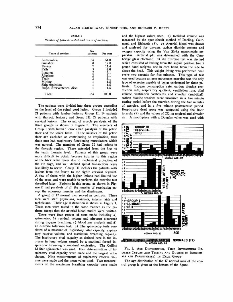
It is to be expected that, with lesions of thespinal cord in the thoracic or cervical segments,the expiratory effort would be more likely to beimpaired than the inspiratory effort presumingthat the abdominal muscles are the powerful ex-piratory muscles. Considering the two compo-nents of the vital capacity as the inspiratory ca-pacity and the expiratory reserve volume, anddefining the expiratory reserve volume as the de-crease in lung volume caused by a forced expira-tion starting from the resting expiratory level,one would expect that with abdominal muscleparalysis the expiratory reserve volume would bedecreased more than inspiratory capacity. Theratio of expiratory reserve volume to vital ca-pacity expressed as percentage is given in TableIII with the probability of there being a significantdifference between paraplegic and control groups.For the paraplegics with lumbar and thoracicspinal cord lesions there was no significant differ-ence between normals and paraplegics. Withcervical lesions, however, the expiratory reservevolume percentage was reduced significantly toapproximately one-half.
Maximum breathing capacity
In Table IV are given the values of maximumbreathing capacity expressed as percentages of thestandard predicted values. These standards arethose of Motley (12) and Baldwin, Cournand, andRichards ( 11 ). The maximum breathing capacity
of the control group of normal individuals was de-termined and a regression formula derived as afunction of age. This formula is:
Maximum breathing capacity/square meter ofbody surface = 92.9 - 0.654 X age + 13.6(liters, 370 C., ambient pressure saturatedwith water vapor).
The percentage of the maximum breathing ca-pacity predicted by this formula is given in TableIV. The probability of a significant differencefrom the predicted, with p < 0.01, is indicated byS (see vital capacity). For Group I (lumbarlesion) the maximum breathing capacity did notdiffer significantly from the normal predicted val-ues of the Baldwin formula or our control group.However, compared with the Motley formula(which has higher values than the Baldwin orour control group) the Group I paraplegics weresignificantly lower. For Groups II and III themaximum breathing capacity values are all sig-nificantly lower than predicted.
TABLE IV
Maximum breathing capacity as percentage of standardpredicted values and predicted values
from control group
Group 1-21Lumbar lesion
Motley standardt 84.4 4 25.3Baldwin standard$ 94.5 i 28.6Control group§ 96.7 4 28.6
Group 11-16Thoracic lesion
Motley standard 61.0 =i 15.6Baldwin standard 70.4 4 17.8Control group 71.1 i 15.6
Group III-24Cervical lesion
Motley standard 48.6 1 15.4Baldwin standard 56.3 d 17.4Control group 56.9 ± 17.4
SNSNS
SSS
SSS
* S, significant difference; NS, no significance.t Motley standard: Maximum breathing capacity per
M.2 equals 97 minus age/2 (12).t Baldwin standard: Maximum breathing capacity per
M.2 equals 86.5 minus 0.522 times age (liters B.T.P.S.per minute (11).
§ Control group: Maximum breathing capacity per M.2equals 90.8 minus 0.68 times age.
776

PULMONARYFUNCTION OF PARAPLEGICS
Residual volume
The residual volume in liters B.T.P.S. per squaremeter of body surface for the paraplegics com-pared with normal controls is shown in Figure 3.A linear regression formula for the normal con-trols was computed and found to be:
Residual volume = 0.64 + 0.0073 x age + 0.16(liters per square meter body surface, 370 C.,ambient pressure saturated with water vapor).
The regression line corresponding to this formulais shown as the center line of Figure 3 with thelimits of standard error of estimate forming thetwo outer lines. On the same figure individualvalues of residual volumes for the three groups ofparaplegics are indicated by points. The majorityof the paraplegics have residual volumes greaterthan normal as is obvious from the location of thepoints on the graph. That they are significantlygreater is shown by computation of the mean ofeach group and the mean of a normal group of thesame age. The means are shown in the small in-set table of Figure 3.
The residual volume/total capacity ratio forpatients and normal controls of the same age is
RESIDUAL x RVOLUME e
liters BTPS/M2 0 OR
22-
2.0-
1.8-
1.6-
1.4-
1.2 -
0.8-
0.6'
TABLE V
Percentage ratio of residual volume/total capacity (R V/TC)for the three groups of paraplegics
P*
Group I (lumbar lesion)-22 32.7 i 8.0 SNormal controls-17 26.3 i 3.5
Group II (thoracic lesion)-16 36.0 : 9.2 SNormal controls-17 25.0 ± 3.5
Group III (cervical lesion)-24 41.3 4 6.1 SNormal controls-17 25.4 + 3.5
* Significant difference.
given in Table V. This ratio for the control groupof 17 normal individuals was found to be:
RV/TV x 100 = 19.50 + 0.201 x age ± 3.50(per cent).
For each of the three groups of patients themean ratio for a group of normals for the sameage was obtained using this formula. This meanratio for each normal group is given in Table V,the mean values for each normal group beingslightly different because of differences in the meanage of each group. The residual volume/total ca-
NORMAL PARAPLEGIC p LESIONIOUP x 0.91 *016 1.27*0.37 S obdmiolD1OUPU O.86.16 1.3020.25 S thorocic*UP m 0.86*0.16 1.22:*0.06 S cervical
%.I I I I_ I I I I ye r
20 25 30 35 40 45 50 5 years
FIG. 3. VARIATION OF RESIDUAL VOLUME* PER SQUAREMETEROF BODYSURFACE WITH AGE FOR NORMAL CONTROLS AND THREE GROUPS OF
PARAPLEGICSThe center line is the linear regression line of the controls and the two
outer lines are the limits of the standard error of estimate. Residual vol-ume is expressed as liters at 370 C., ambient pressure, saturated water vapor.
0 '
""it 0 IN 0 eviffAGE
777

ALLAN HEMINGWAY,ERNEST BORS, AND RICHARD P. HOBBY
TABLE VI
Arterial blood gases and pHfor the three groupsof paraplegics *
I II IIILumbar Thoracic Cervical
02 content x 17.6 17.2 17.5Of ~ 1.5 1.3 1.2
02 capacity x 18.8 18.3 18.6a 1.7 1.7 0.9
Per cent saturation of R 94.4 95.4 95.8hemoglobin 3.0 2.4 1.1
CO2content x 48.3 49.3 48.33.6 4.5 2.5
pH x 7.40 7.42 7.380.07 0.09 0.08
* Oxygen content, oxygen capacity and carbon dioxidecontent are given as ml. of each gas STPD (00 C., 760 mm.pressure, dry) in 100 ml. of whole blood. x is the meanvalue and a the standard deviation.
pacity ratio is significantly greater than normalcontrols of the same age as is shown in Table V.The higher the lesion the greater is this ratio.The total capacity is the sum of vital capacity andresidual volume.
Blood gases
The arterial blood oxygen and carbon dioxide,as well as arterial pH, are shown in Table VI.
Values given are the means and standard devia-tions for each group. These values are normal.
Exercise test
In Table VII are given the data for the fiveminute weight lifting exercise test. The data inrows opposite "rest" are those collected in a fiveminute preexercise resting period.> The post-exercise period immediately following the exerciseperiod lasted five minutes. The values in thetable are the means (x) and the standard devia-tions (a) for the individuals of each group. Withthis "mild" exercise the oxygen consumption rate("702) in ml., 00 C., 760 mm. Hg pressure, dry,per square meter of body surface per minute andthe ventilation rate (VE) in liters, 370 C., ambientpressure, saturated with water vapor per square
meter of body surface per minute, approximatelydoubled. As is characteristic of exercise, therewas a slight rise in respiratory quotient and inventilation coefficient (Vo2/VE).
Respiratory dead space
The respiratory physiological dead space valuesas determined by the Bohr formula (9) and using
TABLE VII
Ventilation data of normal controls and three groups of paraplegics for a five minute weight lifting exercise test *
Normal Lumbar Thoracic Cervical
N Group I Group II Group III15 15i1 14
Number x f x a X O j o
02 consumption rate (OCR) Rest 124 18 121 14 124 14 132 22ml. STPD/min. M2 Ex 251 40 259 32 274 27 211 36
Post Ex 141 19 153 26 155 13 145 32
CO2production rate (CPR) Rest 95 11 92 13 98 11 108 20ml. STPD/min. M2 Ex 210 37 200 25 227 21 180 31
Post Ex 120 16 131 25 130 16 146 30
Respiratory quotient Rest 77 5 76 9 79 7 82 5Ex 84 5 77 8 83 6 85 9Post Ex 85 5 85 11 83 10 83 6
Ventilation rate (VE) Rest 3.12 0.42 3.67 0.17 3.50 0.64 3.81 0.80BTPS/min. M2 Ex 6.17 0.80 6.35 0.73 6.94 0.32 5.99 0.98
Post Ex 3.85 0.35 4.88 0.96 4.46 0.55 4.30 0.75
OCR/VE Rest 39.8 3.5 33.4 3.4 35.3 5.9 34.7 4.2Ex 40.9 5.8 41.0 4.5 40.1 4.5 36.7 6.7Post Ex 37.0 4.9 31.5 4.4 34.3 3.4 33.8 5.0
02 debt 214 13 198 20 148 64 108 60
* Each test was divided into three successive periods including a five minute preexercise (resting) period, the fiveminute exercise period, and a five minute postexercise period.
778

PULMONARYFUNCTION OF PARAPLEGICS
TABLE VIII
Respiratory dead space (VD) (in ml. per square meter of body surface) and the dead space/tidal volume ratio(VD/ VT) expressed as percentage *
Before exercise During exercise After exercise
VD VD/VT VD VD/VT VD VD/VT(ml.) (%) (ml.) (%) (ml.) (%)
Normal group x 87.9 31.6 110.0 23.3 87.7 24.8(N = 14) f 9.2 3.1 25.8 7.5 17.3 5.9
Group I x 93.9 31.8 121.8 24.1 96.1 26.2(N = 15) a 17.2 5.5 26.9 5.9 25.3 5.9
Group II 98.8 29.5 94.1 17.2 97.1 27.220.7 5.2 20.3 4.8 16.2 4.8
Group III R 100.1 28.4 86.4 21.1 94.8 25.9ar 21.5 5.9 16.8 6.5 26.0 7.4
* x is the average for the group and a is the standard deviation. N is the number of individuals in the group.
carbon dioxide analyses of end-tidal (alveolar)air and expired air are given in Table VIII withvalues of the dead space/tidal volume ratio. Forthe normal control group and the patients withlumbar spinal transections the dead space volumeincreased with exercise and the dead space per-centage of the tidal volume decreased, a finding inagreement with previous investigators (9). How-ever, for the patients with thoracic lesions (GroupII) the dead space decreased slightly (but insig-nificantly) with exercise, while for patients withcervical lesions the dead space fell more. If astatistical comparison is made of the dead spacechange with exercise and Group I, or the normalcontrol group, compared with the Group III(cervical lesion), a statistically significant differ-ence between the two groups (p < 0.001) exists.
DISCUSSION
In the management of paraplegics, respiratorydysfunction is not as serious a clinical problem asbowel and bladder regulation, decubitus ulcers andurinary infection. In tetraplegics, respiratory dys-function occurs early in the post-traumatic phasewhen a tracheostomy may be needed and later if anintercurrent upper respiratory infection develops.With an upper respiratory infection, difficulty isexperienced by the patient due to failure of activeexpectoration. Removal of tracheobronchial se-cretions can be aided if necessary by postural drain-age, splinting of the abdomen and liquefaction ofbronchial secreta with expectorants. Usually,
however, for routine activity of the tetraplegic theexisting, but impaired, cough mechanism withtracheobronchial ciliary function is adequate.
It is evident from the data on vital capacity,maximum breathing capacity, exercise ventilationand blood gases that the ventilation of the para-plegics with lumbar lesions is essentially normal.This is readily explainable since the muscularparalysis of these patients is limited to that of thelower extremities and pelvic floor while the re-spiratory musculature is functioning normally.The patients of Group III with lower cervical le-sions have paralysis of abdominal and intercostalmuscles, and the sole remaining effective respira-tory muscle is the diaphragm. In spite of this ex-tensive respiratory muscle paralysis, the entireventilation as performed by this one remainingfunctional muscle is surprisingly effective. Ap-proximately, for this group, the vital capacity istwo-thirds of normal and the maximum breathingcapacity one-half of normal.
The expiratory reserve volume is the decreasein volume of the lung caused by a forced expirationcommencing from the resting expiratory level.For this forced expiration the expiratory musclesare active participants. Since with thoracic spinalcord lesions part of the expiratory muscles areparalyzed, and with cervical lesions all are para-lyzed, it is to be expected that the expiratory re-serve volume would be decreased. It was foundthat with the normal control group and Group I(lumbar lesion) and II (thoracic lesion) the ex-
779

ALLAN HEMINGWAY, ERNEST BORS, AND RICHARD P. HOBBY
piratory reserve volume percentage of the vitalcapacity was approximately the same, i.e., approxi-mately 17 to 19 per cent. With Group III (cervi-cal lesions) the expiratory reserve volume wasapproximately 9 per cent of the vital capacity, areduction from the normal value of approximatelyone-half. The surprising finding is that these pa-tients with no functional expiratory muscles canexhale forcibly this much air from the resting ex-piratory level. The reason for this forcible ex-halation is not readily apparent. Possibly theshoulder girdle muscles participate in exhalationmore than is generally believed. Another ex-planation is that the residual volume of these pa-tients is increased approximately 500 ml., as isshown in Figure 3, which would indicate thatthey are breathing from a more inspiratory chestposition than normal. The term resting expira-tory position of the chest, although it implies min-imal or no diaphragmatic muscle tonus, does notnecessarily mean that such tonus is absent. Thework of Culver and Rahn (13) and the earlierwork on pressure breathing (14) have providedevidence that at the resting expiratory level theremay be partial contraction of the respiratory mus-culature, and it is possible that in the abnormalcondition of respiratory muscle paralysis wherethe diaphragm is the only muscle of respiration,an increased diaphragmatic tone can occur at theresting expiratory level. This would permit anexpiratory volume of air to be exhaled from theresting expiratory level simply by relaxation ofdiaphragmatic tonus. Although this might ex-plain the unanticipated high value of the expira-tory reserve volume, it does not explain why theresidual volume is elevated. The residual volumewas found to be elevated in all of the three groupsof paraplegics and did not seem to be dependenton the extent of respiratory muscle paralysis. Theonly paralysis common to the three groups wasparalysis of the lower limbs and pelvic floor mus-cles. In addition all the patients led a sedentaryor supine type of living while the controls wereactively working. These differences do not af-ford a simple explanation for the increased residualvolume. One explanation which is proposed re-servedly is that the pelvic floor muscles supportthe abdominal viscera. Paralysis of these muscleswithdraws some support from the abdominal vis-
cera causing the diaphragm to descend to increaseresidual volume. If this occurs, it imparts to thepelvic floor more of a supporting role for theviscera than is generally believed to occur.
The values observed for blood gases and ven-tilation studies during exercise were essentiallythe same for the patients as the controls. Theseobservations indicate that the extensive paralysisof respiratory muscles causes no serious inter-ference with blood gas exchange in the lung andthat a mild exercise (equal to approximately twoand a half times the resting oxygen consumptionrate) can be performed without ventilatory diffi-culty. The fact that oxygen consumption rateof the paraplegics being essentially the same asthe controls suggests that widespread denervationof the skeletal muscle does not reduce oxygen de-mand of the tissues. This is an observation oftheoretical interest in the control of metabolismand the needs for homothermy.
In the response to mild exercise a normal indi-vidual increases his ventilation more by deepbreathing than by increased respiratory rate. As-sociated with this increased tidal volume the re-spiratory physiological dead space as measured bythe CO2 method and using the Bohr formula isincreased while the dead space/tidal volume frac-tion is reduced (9). This response with valueswhich were similar to those in the literature werefound for the individuals of the normal controlgroup and those of Group I (lumbar lesions).With Group II (thoracic lesions) the mean re-spiratory dead space for the group fell slightly, thedecrease being of questionable significance. WithGroup III, however, the respiratory dead spacewas reduced considerably more with exercise, themean value decreasing from 100.1 to 86.4 (ml.,370 C., ambient pressure, saturated with watervapor per square meter of body surface). If astatistical comparison of the changes in dead spacebefore and during exercise is made for Group I(or the normal control group) and Group III(cervical lesions) a significant difference (p <0.001) for the dead space change of the two groupsis found. The reason for this reduction in deadspace with exercise for the Group III paraplegicsis not clear. Some individuals of this groupbreathed irregularly and erratically during exer-cise. This group, more than the others, during
780

PULMONARYFUNCTION OF PARAPLEGICS
exercise increased their respiratory rate and theirtidal volume increase was less. This irregularbreathing increased the difficulty of sampling end-tidal air. However, large errors in this samplingprobably did not occur since the following end-tidalmean values of CO, (dry, volumes per cent) be-fore (B) and during (D) exercise were obtained:controls, (B) 5.39, (D) 5.43; Group III, (B)4.75, (D) 4.75. The problem of measuring re-spiratory dead space by the CO2 method is one onwhich some of the most heated controversies ofexercise physiology have been waged and many ofthe problems are still unsettled (9). It is con-cluded from the observations here reported thatwith diaphragmatic breathing of cervical para-plegics, physiological dead space does not increasebut may decrease with exercise. This might bedue to the fact that diaphragmatic breathing movesthe lower parts of the lung adjacent to the dia-phragm while the upper part of the lung contain-ing the large airways, and hence the dead space,remains relatively immobile.
SUMMARY
Pulmonary function studies have been made on agroup of 63 paraplegic patients with complete spi-nal transections and a group of 17 normal controls.The patients were divided into three groups: 1)those with lumbar spinal lesions who had paralysisof the lower limbs and pelvic floor but no paralysisof the muscles of breathing; 2) those with thoracicspinal lesions with paralysis of the lumbar musclesand lower intercostals; and 3) those with lowercervical spinal lesions with paralysis of all musclesof breathing except the diaphragm and the acces-sory muscles of respiration. Measurements weremade of vital capacity and components, residualvolume, maximum breathing capacity, arterialblood gases and pH, and ventilation during exer-cise. It was found that the vital capacity and max-imum breathing capacity of patients with lumbarlesions were normal, while the group with lowercervical lesions had a vital capacity approximatelytwo-thirds of normal and a maximum breathingcapacity of one-half of normal. Paraplegics withthoracic lesions had vital capacities and maximumbreathing capacities between the values for the
cervical and lumbar lesions groups. The restingexpiratory reserve volume expressed as a percent-age of the vital capacity was below the normalpercentage only for the group of paraplegics withcervical lesions. For this group the percentagevalue was one-half normal, which is higher thanwas anticipated for patients with only inspiratorymuscle function. Residual volumes of all groupswere significantly higher than the normal group.Arterial blood gases of all groups were normal andthe ventilatory response to exercise for all groupsof paraplegics did not differ from normal.
ACKNOWLEDGMENT
The authors gratefully acknowledge the cooperationand assistance of Dr. Ben Klaumann, formerly Chief ofthe Pulmonary Function Laboratory, and Dr. DonaldLeik, Chief of Medicine, of the Veterans Hospital. Tech-nical assistance was ably rendered by Dr. Wayne Hull,Lucille Baumgartner, Beverly Lim, Ray Power andWilson Henderson.
REFERENCES
1. Keith, A. The mechanism of respiration in man inFurther Advances in Physiology, Leonard Hill,Ed. London, Arnold, 1909, p. 182.
2. Schmidt, C. F. Medical Physiology, Philip Bard, Ed.St. Louis, Mosby, 1957, page 287.
3. Campbell, E. J. M. An electromyographic study ofthe role of the abdominal muscles in breathing.J. Physiol. 1952, 117, 222.
4. Campbell, E. J. M., and Green, J. H. Variations inintra-abdominal pressure and the activity of ab-dominal muscles during breathing. J. Physiol.1953, 122, 282.
5. Lewis, L. The role of the abdominal musculature inrespiration. Conference on Respiratory Physiologyin Poliomyelitis, National Foundation for InfantileParalysis, Los Angeles, 1955.
6. Dickinson, D. G. An electromyographic study ofsome of the muscles used in respiration. Confer-ence on Respiratory Physiology in Poliomyelitis,National Foundation for Infantile Paralysis, LosAngeles, 1955.
7. Floyd, W. F., and Silver, P. H. S. Electromyographicstudy of patterns of activity of the anterior ab-dominal wall muscles in man. J. Anat. (Lond.)1950, 84, 132.
8. Darling, R. C., Cournand, A., and Richards, D. W.,Jr. Studies on the intrapulmonary mixture ofgases. III. An open circuit method for measuringresidual air. J. clin. Invest. 1940, 19, 609.
781
ALLAN HEMINGWAY,ERNEST BORS, AND RICHARD P. HOBBY
9. Rossier, P. H., and Buhlmann, A. The respiratorydead space. Physiol. Rev. 1955, 35, 860.
10. Scholander, P. F. Analyzer for accurate estimationof respiratory gases in one-half cubic centimetersamples. J. biol. Chem. 1947, 167, 235.
11. Baldwin, E. D., Cournand, A., and Richards, D. W.,Jr. Pulmonary insufficiency: I. Physiologicalclassification, clinical methods of analysis, stand-ard values in normal subjects. Medicine 1948, 27,243.
12. Motley, H. L. The use of pulmonary function testsfor disability appraisal: Including evaluation stand-ards in chronic pulmonary disease. Dis. Chest1953, 24, 378.
13. Culver, G. A., and Rahn, H. Reflex respiratorystimulation by chest compression in the dog.Amer. J. Physiol. 1952, 168, 686.
14. Rahn, H., Otis, A. B., Chadwick, L. E., and Fenn,W. 0. The pressure-volume diagram of thethorax and lung. Amer. J. Physiol. 1946, 146,161.
782