METHODOLOGY Open Access A novel cost effective and high ... · biology and evolution strongly...
10
METHODOLOGY Open Access A novel cost effective and high-throughput isolation and identification method for marine microalgae Martin T Jahn 1,2 , Katrin Schmidt 1 and Thomas Mock 1* Abstract Background: Marine microalgae are of major ecologic and emerging economic importance. Biotechnological screening schemes of microalgae for specific traits and laboratory experiments to advance our knowledge on algal biology and evolution strongly benefit from culture collections reflecting a maximum of the natural inter- and intraspecific diversity. However, standard procedures for strain isolation and identification, namely DNA extraction, purification, amplification, sequencing and taxonomic identification still include considerable constraints increasing the time required to establish new cultures. Results: In this study, we report a cost effective and high-throughput isolation and identification method for marine microalgae. The throughput was increased by applying strain isolation on plates and taxonomic identification by direct PCR (dPCR) of phylogenetic marker genes in combination with a novel sequencing electropherogram based screening method to assess the taxonomic diversity and identity of the isolated cultures. For validation of the effectiveness of this approach, we isolated and identified a range of unialgal cultures from natural phytoplankton communities sampled in the Arctic Ocean. These cultures include the isolate of a novel marine Chlorophyceae strain among several different diatoms. Conclusions: We provide an efficient and effective approach leading from natural phytoplankton communities to isolated and taxonomically identified algal strains in only a few weeks. Validated with sensitive Arctic phytoplankton, this approach overcomes the constraints of standard molecular characterisation and establishment of unialgal cultures. Keywords: Marine microalgae, Direct PCR, Isolation, Cultivation, Taxonomy Background Marine microalgae are unicellular photosynthetic eu- karyotes of major ecological and economic importance worldwide. Ecologically, they are the base of the marine food web and contribute to at least 30% of annual CO 2 fixation worldwide and therefore massively impact glo- bal biogeochemical cycles [1,2]. Economically, diverse marine microalgae are used or have the potential to be used as nutraceuticals, for the production of pharma- ceuticals [3,4], cosmetics [5], for bioremediation [6-8], and biofuels [9]. In recent years, the emerging application of culture- independent omics approaches like metagenomics and metatranscriptomics delivered comprehensive insights into the gene repertoire and activity of marine microalgal com- munities [10-13]. However, results from high-throughput omics approaches ideally need to be scrutinized by experi- ments with isolated strains from the same communities if the scientific endeavour goes beyond purely describing the diversity and abundance of genes and transcripts in rela- tion to environmental conditions. Similarly, in the field of microalgae biotechnology, novel isolation and identifica- tion protocols are essential for identifying specific traits like lipid content [14,15] or any other bioactive compounds [16]. Thus, there is a high demand to develop novel iso- lation and identification protocols. However, laborious standard procedures such as single-cell isolation of * Correspondence: [email protected] 1 School of Environmental Sciences, University of East Anglia, Norwich Research Park, Norwich NR4 7TJ, UK Full list of author information is available at the end of the article PLANT METHODS © 2014 Jahn et al.; licensee BioMed Central Ltd. This is an Open Access article distributed under the terms of the Creative Commons Attribution License (http://creativecommons.org/licenses/by/4.0), which permits unrestricted use, distribution, and reproduction in any medium, provided the original work is properly credited. The Creative Commons Public Domain Dedication waiver (http://creativecommons.org/publicdomain/zero/1.0/) applies to the data made available in this article, unless otherwise stated. Jahn et al. Plant Methods 2014, 10:26 http://www.plantmethods.com/content/10/1/26
Transcript of METHODOLOGY Open Access A novel cost effective and high ... · biology and evolution strongly...

PLANT METHODSJahn et al. Plant Methods 2014, 10:26http://www.plantmethods.com/content/10/1/26
METHODOLOGY Open Access
A novel cost effective and high-throughputisolation and identification method for marinemicroalgaeMartin T Jahn1,2, Katrin Schmidt1 and Thomas Mock1*
Abstract
Background: Marine microalgae are of major ecologic and emerging economic importance. Biotechnologicalscreening schemes of microalgae for specific traits and laboratory experiments to advance our knowledge on algalbiology and evolution strongly benefit from culture collections reflecting a maximum of the natural inter- andintraspecific diversity. However, standard procedures for strain isolation and identification, namely DNA extraction,purification, amplification, sequencing and taxonomic identification still include considerable constraints increasingthe time required to establish new cultures.
Results: In this study, we report a cost effective and high-throughput isolation and identification method for marinemicroalgae. The throughput was increased by applying strain isolation on plates and taxonomic identification bydirect PCR (dPCR) of phylogenetic marker genes in combination with a novel sequencing electropherogram basedscreening method to assess the taxonomic diversity and identity of the isolated cultures. For validation of theeffectiveness of this approach, we isolated and identified a range of unialgal cultures from natural phytoplanktoncommunities sampled in the Arctic Ocean. These cultures include the isolate of a novel marine Chlorophyceaestrain among several different diatoms.
Conclusions: We provide an efficient and effective approach leading from natural phytoplankton communities toisolated and taxonomically identified algal strains in only a few weeks. Validated with sensitive Arctic phytoplankton,this approach overcomes the constraints of standard molecular characterisation and establishment of unialgalcultures.
Keywords: Marine microalgae, Direct PCR, Isolation, Cultivation, Taxonomy
BackgroundMarine microalgae are unicellular photosynthetic eu-karyotes of major ecological and economic importanceworldwide. Ecologically, they are the base of the marinefood web and contribute to at least 30% of annual CO2
fixation worldwide and therefore massively impact glo-bal biogeochemical cycles [1,2]. Economically, diversemarine microalgae are used or have the potential to beused as nutraceuticals, for the production of pharma-ceuticals [3,4], cosmetics [5], for bioremediation [6-8],and biofuels [9].
* Correspondence: [email protected] of Environmental Sciences, University of East Anglia, NorwichResearch Park, Norwich NR4 7TJ, UKFull list of author information is available at the end of the article
© 2014 Jahn et al.; licensee BioMed Central LtCommons Attribution License (http://creativecreproduction in any medium, provided the orDedication waiver (http://creativecommons.orunless otherwise stated.
In recent years, the emerging application of culture-independent omics approaches like metagenomics andmetatranscriptomics delivered comprehensive insights intothe gene repertoire and activity of marine microalgal com-munities [10-13]. However, results from high-throughputomics approaches ideally need to be scrutinized by experi-ments with isolated strains from the same communities ifthe scientific endeavour goes beyond purely describing thediversity and abundance of genes and transcripts in rela-tion to environmental conditions. Similarly, in the field ofmicroalgae biotechnology, novel isolation and identifica-tion protocols are essential for identifying specific traits likelipid content [14,15] or any other bioactive compounds[16]. Thus, there is a high demand to develop novel iso-lation and identification protocols. However, laboriousstandard procedures such as single-cell isolation of
d. This is an Open Access article distributed under the terms of the Creativeommons.org/licenses/by/4.0), which permits unrestricted use, distribution, andiginal work is properly credited. The Creative Commons Public Domaing/publicdomain/zero/1.0/) applies to the data made available in this article,

Jahn et al. Plant Methods 2014, 10:26 Page 2 of 10http://www.plantmethods.com/content/10/1/26
strains, DNA extraction, purification, amplification, se-quencing and taxonomic identification include severaltime, cost and space consuming constraints.To overcome these constraints, we developed a new
cost effective and high-throughput isolation and identifi-cation method for marine microalgae. We combinedhigh throughput isolation by streaking cells from enrich-ment cultures on agar plates with subsequent cultivationin multi-well plates. To assess as to whether a culturewas unialgal or not, we applied direct PCR (dPCR) byonly using boiling MiliQ water to lyse the cells in combin-ation with a novel sequencing electropherogram based as-sessment method. While using the V4 as the most variablesmall subunit (SSU) [17], the underlying idea was that mo-lecular marker sequences of different species possess dif-ferent bases at the same position. This concept is similarto the detection of intraspecific point mutations exploitingsequencing electropherogram tracefiles [18]. The am-biguous base-calls detected as a biased uncalled/calledpeak ratio increase the position specific error pro-bability (Pe) [19], which decreases the per-base QualityValues (QV = -10 log10(Pe)) as a standard quality metric[20]. The per-base quality values were used in ourapproach to evaluate the presence or absence of an uni-algal culture.This new approach is relatively cost effective, time sav-
ing and high throughput to overcome the constraints ofstandard molecular characterisation (e.g. by DGGE orRFLP) and establishment of unialgal cultures withoutthe need of DNA extraction and cloning. To validate theefficiency of this approach, we isolated and identifiedalgal strains from natural phytoplankton communities ofthe Arctic Ocean.
ResultsThe objective of this study was to establish a cost andtime effective method for microalgal isolation and iden-tification. Using the methods described below, we wereable to obtain 24 unialgal cultures consisting of 7 uniqueribotypes based on the V4 18S rDNA region.
Efficiency of growing algae on plates and dPCRUsing the high-throughput isolation technique of streak-ing enriched natural microalgal communities across agarplates, on 59.3% (35 of 59) of the incubated plates algalgrowth was detected. From about three quarters (77.1%;27 of 35) of these plates, it was possible to pick single col-onies. Moreover, all (158 of 158) of the picked coloniestransferred to 12-well plates showed visible growth underthe microscope after 1.5 weeks of cultivation. In a prelim-inary study, primers amplifying the whole 18S rDNA(~1750 bp) region were used for unialgal assessment andtaxonomic identification. However, dPCR amplicon se-quencing from the 5′ end of the whole 18S rDNA region
lacked sufficient variability compared to the V4 sub-regionon the 18S rRNA gene. By combining the dPCRs of thewhole 18S rDNA and of the V4 region of 18S rDNA, thedPCR approach succeeded in 70.25% (85 of 121) of the re-actions. Furthermore, the amplicons obtained by dPCR, asshown in Figure 1, had identical size compared to the con-trol PCR conducted with extracted DNA. Also, no add-itional bands were visible for dPCRs.About 65% of the screened cultures (24 of 37) were
identified as unialgal based on our new electropherogram-based assessment. Figure 2 illustrates the discriminationprinciple between sequences from unialgal cultures andmixed populations.
Taxonomy and geographic originIn total, 6 different taxonomic groups were identifiedbased on V4-18S rDNA sequences. NCBI nucleotideBLAST searches (Table 1) revealed that all groups com-prised microalgae including an array of 4 different classeswith Bacillariophyceae, Fragilariophyceae, Coscinodisco-phyceae and Chlorophyceae (Table 1; Figure 3). Noticeablemorphological features of the novel Chlorophyceae strainare its contractile vacuoles, two isokont flagella, stigmata,pyrenoids and the size of 10 μm (Figure 4). With the ex-ception of this novel Chlorophyceae strain (Figure 3b), di-atoms made up the vast majority of isolated species(Table 1). Amongst diatoms pennate species were twice asoften isolated and identified by BLAST searches as centricspecies, which is in agreement with our microscopic ob-servations (10 of 16 isolates). However, it was found thatthe V4 region failed to resolve differences within the fam-ily Fragilariaceae between the genera Syndrea, Fragilariaand Synedropsis (Table 1, Figure 3a), despite equal se-quence quality and length. A similar situation was foundin two cultures between the best hits Nitzschia thermalisand Amphora sp. (1-80-1-M and 2-80-27-M). However,taxonomic groups clustered with high bootstrap support(Figure 3a).Using our approach, we were most successful in isolat-
ing the pennate diatom Cylindrotheca closterium 9 timesfrom a variety of 5 different sampling locations alongmost of the latitudinal transect (latitude: 65.246- 78.839)of this study (Figure 5). The Fragilariaceae-cluster(Figure 3a) in contrast was only recovered as an isolatefrom samples originating from the northernmost sites(Figure 5). On the west side of the transect, a novelChlorophyceae was isolated from the chlorophyll max-imum in a depth of 10 m. Chaetoceros cf. neogracile andSkeletonema marinoi were collected from location 2-80-51-M and 2-80-8-M, respectively (Figure 5).
DiscussionIn recent years, huge efforts were made to establish cul-ture collections holding thousands of marine algae strains

Figure 1 Comparison of PCR- Products utilizing extracted DNA and direct culture as template. Kit extracted DNA is amplified in threereplicates (Repl.) using the primers TAReuk454FWD1 and TAReukREV3 [36]. Amplification from direct culture (dPCR) in two replicates using sameprimers as described in the methods section. – represents negative control. Whole PCR products (50 μl) are separated and visualised by ethidiumbromide staining on a 1% TAE agarose gel.
Jahn et al. Plant Methods 2014, 10:26 Page 3 of 10http://www.plantmethods.com/content/10/1/26
like in the National Center for Marine Algae and Micro-biota (NCMA). Novel approaches of cryopreservation[21,22] reduced culture maintenance efforts considerably[23]. This study reports an approach that enabled us toestablish a range of unialgal cultures from Arctic Oceansamples (1) under cost effective conditions due to theomission of DNA-extraction and cloning (2) with lowspace requirements due to the use of 12-well format (3)within processing times of three weeks.In accordance with previous studies, the isolated spe-
cies Cylindrotheca closterium [24], Skeletonama marinoi[25] and Chaetoceros cf. neogracile [26] were alreadyidentified in the Arctic Ocean and are available in cul-ture collections. The given morphological features of thenovel Chlorophyceae strain together with the clusteringof its V4 18S rDNA ribotype into Chlamydomonas mayindicate closer affiliation towards this genus. Even thoughseveral different Chlamydomonas species were identifiedin Antarctic saline lakes [27,28], on Arctic glaciers [29], orin sea ice of the brackish Baltic Sea [30], this would be, toour knowledge, the first record of a marine Chlamydomo-nas strain from the deep Chlorophyll maximum layer inthe open ocean. However, further characterisation of this
Figure 2 Representative sequencing electropherogram sections. Compseudonana laboratory culture (b) non-unialgal culture 1-80-15-M with 2 m(2-80-8-M). The color code refers to the per base Quality Values (QV) as the
strain is needed what is beyond the scope of this method-ical paper. It remains to be seen how significant marineChlorophyceae species are in terms of diversity, abun-dance and activity in relation to members of the classPrasinophyta.Every isolation method has biases towards specific
groups to be successfully isolated. Plating, as our methodof choice, was reported to exclude some flagellates andcoccoids and most dinoflagellates [23]. Alternatively, com-bining dPCR with other isolation techniques like single-cell sorting [31,32] may increase the spectrum of isolatedstrains and especially those that won’t grow well on agarplates. However, the costs of single-cell isolation and itsbiases (e.g. selection against filamentous and larger algae)seem to object to our approach.The success rates of our dPCR approach clearly em-
phasise the advantages of using microalgae cultures asthey grow without the need of DNA extraction as de-scribed previously [33-35]. However, a limitation of dPCRmight be the use of the V4 region. Nevertheless, the V4 re-gion used as a molecular marker in this study representsthe most variable SSU region [17]. However, dinoflagellatespossess less variability in this region [36] making it more
pared are the base calling signal noise of (a) unialgal Thalassiosiraorphospecies (c) unialgal classified culture of Skeletonema marinoi–10 log10(Pe), with Pe as the base call error probability.
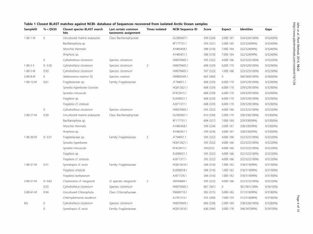
Table 1 Closest BLAST matches against NCBI- database of Sequences recovered from isolated Arctic Ocean samples
SampleID % <QV20 Closest species BLAST searchhits
Last certain commontaxonomic assignment
Times isolated NCBI Sequence ID Score Expect Identities Gaps
1-80-1-M 0 Uncultured marine eukaryote Class: Bacillariophyceae GU385607.1 599 (324) 3.00E-167 324/324(100%) 0/324(0%)
Bacillariophyta sp. KF177731.1 593 (321) 2.00E-165 323/324(99%) 0/324(0%)
Nitzschia thermalis AY485458.1 588 (318) 7.00E-164 322/324(99%) 0/324(0%)
Amphora sp. AY485451.1 588 (318) 7.00E-164 322/324(99%) 0/324(0%)
0 Cylindrotheca closterium Species: closterium HM070405.1 595 (322) 4.00E-166 322/322(100%) 0/322(0%)
1-80-3-S 0- 0.30 Cylindrotheca closterium Species: closterium 3 HM070405.1 608 (329) 6.00E-170 329/329(100%) 0/329(0%)
1-80-5-M 0.93 Cylindrotheca closterium Species: closterium HM070405.1 597 (323) 1.00E-166 323/323(100%) 0/323(0%)
2-80-8-M 0 Skeletonema marinoi (5) Species: marinoi HM805045.1 665 (360) 0 360/360(100%) 0/360(0%)
1-80-15-M 0.61 Fragilariaceae sp. Family: Fragilariaceae JF794051.1 608 (329) 6.00E-170 329/329(100%) 0/329(0%)
Synedra hyperborea Grunow HQ912621.1 608 (329) 6.00E-170 329/329(100%) 0/329(0%)
Synedra minuscula EF423415.1 608 (329) 6.00E-170 329/329(100%) 0/329(0%)
Fragilaria sp. EU090021.1 608 (329) 6.00E-170 329/329(100%) 0/329(0%)
Fragilaria cf. striatula AJ971377.1 608 (329) 6.00E-170 329/329(100%) 0/329(0%)
0 Cylindrotheca closterium Species: closterium HM070405.1 595 (322) 4.00E-166 322/322(100%) 0/322(0%)
2-80-27-M 0.30 Uncultured marine eukaryote Class: Bacillariophyceae GU385607.1 610 (330) 2.00E-170 330/330(100%) 0/330(0%)
Bacillariophyta sp. KF177731.1 604 (327) 7.00E-169 329/330(99%) 0/330(0%)
Nitzschia thermalis AY485458.1 599 (324) 3.00E-167 328/330(99%) 0/330(0%)
Amphora sp. AY485451.1 599 (324) 3.00E-167 328/330(99%) 0/330(0%)
1-80-30-M 0- 0.31 Fragilariaceae sp. Family: Fragilariaceae 3 JF794051.1 595 (322) 4.00E-166 322/322(100%) 0/322(0%)
Synedra hyperborea HQ912621.1 595 (322) 4.00E-166 322/322(100%) 0/322(0%)
Synedra minuscula EF423415.1 595(322) 4.00E-166 322/322(100%) 0/322(0%)
Fragilaria sp. EU090021.1 595 (322) 4.00E-166 322/322(100%) 0/322(0%)
Fragilaria cf. striatula AJ971377.1 595 (322) 4.00E-166 322/322(100%) 0/322(0%)
1-80-37-M 0.31 Synedropsis cf. recta Family: Fragilariaceae HQ912616.1 584 (316) 1.00E-162 318/319(99%) 0/319(0%)
Fragilaria striatula EU090018.1 584 (316) 1.00E-162 318/319(99%) 0/319(0%)
Fragilaria barbararum AJ971376.1 584 (316) 1.00E-162 318/319(99%) 0/319(0%)
2-80-51-M 0- 0.62 Chaetoceros cf. neogracile cf. species: neogracile 2 JN934684.1 595 (322) 4.00E-166 322/322(100%) 0/322(0%)
0.55 Cylindrotheca closterium Species: closterium HM070405.1 667 (361) 0 361/361(100% 0/361(0%)
2-80-61-M 0.94 Uncultured Chlorophyta Class: Chlorophyceae FN690710.1 582 (315) 3.00E-162 317/318(99%) 0/318(0%)
Chlamydomonas raudensis AJ781313.1 555 (300) 7.00E-154 312/318(98%) 0/318(0%)
NA 0 Cylindrotheca closterium Species: closterium HM070405.1 606 (328) 2.00E-169 328/328(100%) 0/328(0%)
0 Synedropsis cf. recta Family: Fragilariaceae HQ912616.1 636 (344) 3.00E-178 346/347(99%) 0/347(0%)
Jahnet
al.PlantMethods
2014,10:26Page
4of
10http://w
ww.plantm
ethods.com/content/10/1/26
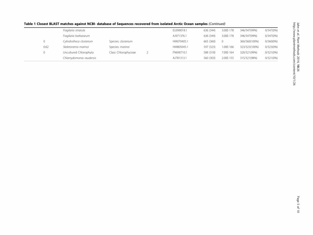
Table 1 Closest BLAST matches against NCBI- database of Sequences recovered from isolated Arctic Ocean samples (Continued)
Fragilaria striatula EU090018.1 636 (344) 3.00E-178 346/347(99%) 0/347(0%)
Fragilaria barbararum AJ971376.1 636 (344) 3.00E-178 346/347(99%) 0/347(0%)
0 Cylindrotheca closterium Species: closterium HM070405.1 665 (360) 0 360/360(100%) 0/360(0%)
0.62 Skeletonema marinoi Species: marinoi HM805045.1 597 (323) 1.00E-166 323/323(100%) 0/323(0%)
0 Uncultured Chlorophyta Class: Chlorophyceae 2 FN690710.1 588 (318) 7.00E-164 320/321(99%) 0/321(0%)
Chlamydomonas raudensis AJ781313.1 560 (303) 2.00E-155 315/321(98%) 0/321(0%)
Jahnet
al.PlantMethods
2014,10:26Page
5of
10http://w
ww.plantm
ethods.com/content/10/1/26

1 80 5 M-Cylindrotheca closterium ID-C6
1 80 15 M-Cylindrotheca closterium ID-G5
1 80 3 S-Cylindrotheca closterium ID-F3
1 80 3 S-Cylindrotheca closterium ID-E6
1 80 3 S-Cylindrotheca closterium ID-C13
1 80 1 M-Cylindrotheca closterium ID-E2
2 80 51 M-Cylindrotheca closterium ID-D15
1 80 37 M-Synedropsis cf. recta Fragilaria sp. ID-C2
1 80 15 M-Synedra sp. Fragilaria sp. ID-F5
1 80 30 M-Synedra sp. Fragilaria sp ID-D8
1 80 30 M-Synedra sp. Fragilaria sp ID-E4
1 80 30 M-Synedra sp. Fragilaria sp ID-F6
1 80 1 M-Bacillariophyceae ID-G4
2 80 27 M-Bacillariophyceae ID-F1
2 80 8 M-Skeletonema marinoi ID-C8
2 80 51 M-Chaetoceros cf. neogracile ID-C12
2 80 51 M-Chaetoceros cf. neogracile ID-D1299
68
62
99
81
96
53
93
100
0.05
Bacillariaceae
Fragilariaceae
Thalassiosirales
Chaetocerotaceae
Urospora penicilliformis (AB049417.1)
Acrosiphonia duriuscula .rav tenuis (AB049418.1)
Blidingia dawsonii (DQ001138.1)
Chlorella emersonii (FR865654.2)
Chlorococcum diplobionticum (U70587.1)
Chlorococcum sp. J7 (AB713407.1)
Chlorococcum ellipsoideum (U70586.1)
.Chlamydomonas sp. SAG 75.94 (AF514399.1)
Chlamydomonas raudensis (JF343798.1)
uncultured Chlorophyta (FN690710.1)
Chlorophyceae strain ID-F7 | Additional Surface Sample
Chlorophyceae strain ID-D16 | 2 80 61 M
Chlorophyceae strain ID-F8 | Additional Surface Sample
Pseudoscourfieldia marina (AF122888.1)
Pycnococcus provasolii (X91264.1)
Dolichomastix tenuilepis (FN562449.1)
Ostreococcus tauri (CAID01000012.1)
Micromonas pusilla (AY954997.1)
Mantoniella squamata (X73999.1)
Mamiellophyceae
Arabidopsis thaliana (AC004708.1)
Selaginella moellendorffii (ADFJ01002076.1)
100
8 6
9 4
100
9 9
9 9
9 9
5 5
8 9
7 1
9 8
9 1
9 5
9 8
2 7
9 6
9 8
9 9
0.05
Thalassiosira pseudonana CCMP1335
Chlorophyceae
Prasinophytes
Land plants
Ulvophyceae
Trebouxiophyceae
a
b
Figure 3 Maximum-likelihood (ML) trees built from the alignments of V4 18S rDNA sequences. Molecular phylogeny of (a) isolated diatomgroups and (b) Chlorophyceae with related clades. Nucleotide sequences obtained in the underlying study indicated by species names in bold.Further sequences were obtained from the SILVA database (www.arb-silva.de) given with accession numbers. The trees with the highest loglikelihood ((a) -1355.5135; (b) -2321.7603) were inferred using the Maximum Likelihood method based on the Kimura 2-parameter model withMEGA6. The fraction of replicate trees in which the associated taxa clustered together is shown next to the branches (1000 bootstraps). Theoutgroups were (a) Arabidopsis thaliana and (b) Mus musculus. All positions with less than 80% site coverage were excluded for tree construction.The scale bar represents number of substitutions per site.
Jahn et al. Plant Methods 2014, 10:26 Page 6 of 10http://www.plantmethods.com/content/10/1/26
difficult to taxonomically characterise isolates without am-biguity. Despite the fact that we had longer reads (average361 bp) available for BLAST searches against NCBI com-pared with Stoeck et al. [36] (average 270 bp), it was still
not sufficient to resolve taxonomies within Fragilariaceaeand between Nitzschia sp. and Amphora sp.. In fact, theV4- region as a molecular marker was found to be too con-served to allow taxonomic resolution in these cases.

Figure 4 Phase contrast micrograph of novel Chlorophyceaeisolate with isokont flagella (FL), pyrenoid (PY), stigma (ST) andcontractile vacuole (CV). The cells, about 10 μm in size, weregrown at 4°C, 24 h day light, 150 μmol photon m-2 s-1. Magnification100×, scale bar = 10 μm.
Jahn et al. Plant Methods 2014, 10:26 Page 7 of 10http://www.plantmethods.com/content/10/1/26
The use of sequencing electropherograms for analyt-ical purposes like the detection of point mutations [18]or multiple clone sequences [37] is frequently reported.In our case, using the novel electropherogram basedanalysis allowed distinction between sequences from a sin-gle strain/species and sequences from multiple strains/species. A crucial step is the formulation of a well-defined
Figure 5 Arctic Ocean Map representing distribution of isolated algaeidentifier (trial-Polarstern cruise number- Cruise stop number-depth [S = Sua BLAST search against GeneBank.
algorithm for an objective trimming of the sequences. Therequirements in this context are twofold. On one hand,sequences from unialgal cultures have to be trimmed atregular drops of quality at the end and the beginning ofthe sequence reads. On the other hand, sequences frommixed communities containing low quality reads shouldonly be trimmed to a distinct lower length limit for a reli-able assessment as described in the methods section. Weexpect that interspecific length polymorphisms of the V4region increase the sensitivity of our culture assessmentdue to the fact that only one base shift would lead to ascrewed sequence.
ConclusionsOur method is suitable for establishing unialgal culturesfrom mixed natural communities within a few weeks ona cost effective and high-throughput basis. Further im-provements could include isolation on low-meting agarfor sensitive species such as flagellates, picking of algalcolonies from plates with robots and cultivation in 96-wellplates under various conditions (e.g. different media, lightand temperature) to increase the likelihood of isolatingrare species or strains.
Materials and methodsStudy sites and sample collectionFor the low cost and high throughput isolation and iden-tification of marine arctic microalgae a total of 27 watersamples was taken along a latitudinal gradient (65.25°N
. Each ▲ denotes a sampling point with hyphen separated uniquerface/M = Chlorophyll max]) and closest certain taxonomy according to

Jahn et al. Plant Methods 2014, 10:26 Page 8 of 10http://www.plantmethods.com/content/10/1/26
to 79.37°N) from the Arctic Ocean during June and July2012. Briefly, 12 L seawater was sampled either at thechlorophyll maximum (23 samples; depths 7-110 m) orat the surface (4 samples; depth 5 m) using a Niskin bot-tle rosette sampler. Additionally, at each sampling depth,temperature, salinity, surface irradiance as well as chloro-phyll and nutrient concentration (NO3, NH, PO4, Silicate)were measured (see Additional file 1). Sea water was pre-filtered through a 100 μm mesh and the flow-throughfraction (<100 μm) was transferred into f/2-medium [38]for enrichment of natural microalgal communities. Whilsttransferred regularly into fresh medium, the samples wereenriched cultured 425 days at 4°C and about 150 μmolphoton m-2 s-1 for ca. 50 generations before unialgal cul-tures were isolated. However, the time for enrichment isvariable depending on the temperature-dependent growthrates of the algal communities.
High throughput microalgae isolationIsolation of microalgae into unialgal cultures was doneby streaking the enriched microalgal communities acrossagar plates as described previously [23]. In short, envir-onmental sample cultures were plated on chilled petridishes containing f/2-medium solidified with 1% (w/v)agar. Subsequently, the agar plates were incubated at4°C, 24 h day cycle, 150 μmol photon m-2 s-1 in a lightthermostat (Rumed, Rubarth Apparate GmbH, Laatzen,Germany) for 1-2 weeks. Clearly separated colonieswere picked from the plates at the end of the stripingand transferred each to 3 ml fresh f/2-medium providedin space efficient 12- well plates. Plates without clearlyseparated colonies where discarded. Inoculated 12-wellplates were incubated for 1.5 weeks at 4°C, 24 h daylight, 150 μmol photon m-2 s-1 to increase cell density.These cultures were screened for a) the presence ofalgae cells (fluorescence emission from chlorophyll a)and b) for visual inspection of having unialgal culturesbased on uniform morphology of at least 200 individualalgal cells using a phase contrast microscope at 400×maginfication (Olympus BX40, Olympus Optical Co.,Ltd., Japan) equipped with Olympus Camedia C-7070wide-zoom digital camera. Cultures that met both cri-teria were kept for further molecular analysis.
Direct polymerase chain reactionFor the direct PCR (dPCR)- amplification of ribosomalDNA, a volume of 500 μl suspended culture from eachof the positive wells according to the visual inspectioncriteria (see above) was transferred to 1.5 ml centrifugetubes and incubated for 5 min at 100°C (Dry Bath Heat-ing System, Starlab, Milton Keynes, United Kingdom) toinactivate protease activity. Then algal cells were harvestedby centrifugation at 16,000 rpm for 10 min at roomtemperature (Eppendorf centrifuge 5418 R, Germany) and
the supernatant was discarded. In order to disrupt thealgal cell integrity the pellet was re-suspended properlywith 100 μl boiling MiliQ-water. The 4°C chilled suspen-sion was either used directly for PCR or stored at -20°Cuntil further use.Primers TAReuk454FWD1 (5′-CCAGCA(G⁄C)C(C⁄T)G
CGGTAATTCC-3′) and TAReukREV3 (5′-ACTTTCGTTCTTGAT(C⁄T)(A⁄G)A-3′) [36] were used to amplifythe V4- region of the 18S rDNA using TC-512 PCR Sys-tem (Techne Co. Staffs, UK). The dPCR was carried outin 50 μL reaction tubes with 10 μl prepared suspensionas template, 2.5 U/μl Taq DNA polymerase (GoTaq®Flexi DNA polymerase, Promega, Madison, WI, USA),1× Taq reaction Buffer, 2 mM MgCl2, 0.2 mM eachdNTP, and 0.4 μM of each primer. The parameters ofthermal cycling of Stoeck et al. (2010) [36] were slightlymodified to 30 s initial denaturation at 98°C, 10 × (98°C,10 s; 53°C, 30 s; 72°C, 30 s), 20 × (98°C, 10 s; 48°C, 30 s;72°C, 30 s) and 10 min final extension at 72°C.
Gel purification and sequencingThe dPCR-products were visualised on 1% (w/v) TAE-agarose gels stained with ethidium bromide. Ampliconbands of the expected size of 421 bp (Fragilariopsis cylin-drus) were cut and gel purified using the NucleoSpin® Geland PCR Clean-up kit (Macherey-Nagel GmbH & Co.KG, Düren, Germany) according to the manufacturer’sinstructions. The DNA yield and purity of the purifieddPCR-products were evaluated using the NanoDropND-1000 spectrophotometer (NanoDrop Technologies,Wilmington, USA). Finally, utilising the TA-Reuk454FWD1forward primer, the amplicons were Sanger-sequencedon a ABI 3730XL sequencer by Eurofins MWG Operon(Ebersberg, Germany).
Nucleotide sequence analysisThe sequencing chromatogram trace (.ab1- format) wasanalysed and trimmed using the ABI Sequence Scannerv1.0 (Applied Biosystems™). Sequence trimming as wellas evaluation of the unialgal status was based on imple-mented per-base Quality Values (QV) as –10 log10(Pe),with Pe as the base call error probability [19]. These QVconsider chromatogram features like peak spacing, un-called/called peak ratio and peak resolution. The se-quences were trimmed: a) at the 5′end after the first25-35 bp when the QV consecutive was >20 in a 20 bpwindow and b) at the 3′end starting after 350 bases, be-fore the first 20 consecutive basecalls contained morethan 1 bases with < QV20. Whilst taking the sequencingmachine basecalling accuracy of 98.5% [39] into account,the trimmed sequences were classified as unialgal, whenthe fraction of <20QV basecalls was smaller than onepercent. For taxonomic identification BLAST sequencesimilarity searches [40] of as unialgal classified cultures
Jahn et al. Plant Methods 2014, 10:26 Page 9 of 10http://www.plantmethods.com/content/10/1/26
against the NCBI database (http://www.ncbi.nlm.nih.gov;release 199) were performed using the megablast algo-rithm. Multiple sequence alignments of the obtained V418S rDNA-sequences were done using ClustalX v1.6[41] and curated using Gblocks v0.91b [42]. A rootedphylogenetic tree was produced by MEGA v6.0 [43]using the maximum likelihood method based on theKimura 2-parameter model [44] excluding positions withless than 80% site coverage. The robustness of the in-ferred tree was estimated using a bootstrap analysis con-sisting of 1000 resampling’s of the data.The nucleotide sequences have been deposited in Gen-
Bank and a representative set of cultures was depositedin the Culture Collection of Algae and Protozoa (CCAP)under accession numbers given in Additional file 2.
Additional files
Additional file 1: Metadata of study sites.
Additional file 2: Culture accession numbers of this study.
Competing interestsThe authors declare that they have no competing interests.
Authors’ contributionsThe experiments were conceived by MTJ, KS and TM and performed by MTJ.The data was analysed by MTJ. KS performed microscopy of theChlorophyceae strain and collected the samples. MTJ and TM co-wrote thepaper. All authors read and approved the final manuscript.
AcknowledgementsTM acknowledges the Natural Environment Research Council (NERC) forfunding (NE/K004530/1). We would like to thank Klaus Valentin from theAlfred-Wegener-Institute for Polar and Marine Research for supporting thefield campaigns. Furthermore, we would like to thank Captain Schwarze andthe Polarstern crews of the ARK27-1 and ANT29-1 expeditions for their vitalhelp during sampling.
Author details1School of Environmental Sciences, University of East Anglia, NorwichResearch Park, Norwich NR4 7TJ, UK. 2Current address: Department of BotanyII, Julius-Maximilians University Würzburg, Julius-von-Sachs-Platz 3, 97082Würzburg, Germany.
Received: 28 May 2014 Accepted: 23 July 2014Published: 7 August 2014
References1. Platt T, Fuentes-Yaco C, Frank KT: Marine ecology: spring algal bloom and
larval fish survival. Nature 2003, 423:398–399.2. Gosselin M, Levasseur M, Wheeler PA, Horner RA, Booth BC: New
measurements of phytoplankton and ice algal production in the ArcticOcean. Deep Sea Res Part II Top Stud Oceanography 1997, 44:1623–1644.
3. Schwartz RE, Hirsch CF, Sesin DF, Flor JE, Chartrain M, Fromtling RE, HarrisGH, Salvatore MJ, Liesch JM, Yudin K: Pharmaceuticals from cultured algae.J Ind Microbiol 1990, 5:113–123.
4. Borowitzka MA: Microalgae as sources of pharmaceuticals and otherbiologically active compounds. J Appl Phycol 1995, 7:3–15.
5. Kim SK, Ravichandran YD, Khan SB, Kim YT: Prospective of thecosmeceuticals derived from marine organisms. Biotechnol Bioproc E 2008,13:511–523.
6. Lee RF, Valkirs AO, Seligman PF: Importance of microalgae in thebiodegradation of tributyltin in estuarine waters. Environ Sci Technol 1989,23:1515–1518.
7. Cardinale BJ: Biodiversity improves water quality through nichepartitioning. Nature 2011, 472:86–89.
8. EI-Sheekh M, Ghareib M, EL-Souod GA: Biodegradation of phenolic andpolycyclic aromatic compounds by some algae and Cyanobacteria.J Bioremediation Biodegradation 2011, 3:133.
9. Waltz E: Biotech’s green gold? Nat Biotech 2009, 27:15–18.10. Toseland A, Daines SJ, Clark JR, Kirkham A, Strauss J, Uhlig C, Lenton TM,
Valentin K, Pearson GA, Moulton V, Mock T: The impact of temperature onmarine phytoplankton resource allocation and metabolism. Nat ClimChange 2013, 3:979–984.
11. Cuvelier ML, Allen AE, Monier A, McCrow JP, Messié M, Tringe SG, Woyke T,Welsh RM, Ishoey T, Lee J-H, Binder BJ, DuPont CL, Latasa M, Guigand C,Buck KR, Hilton J, Thiagarajan M, Caler E, Read B, Lasken RS, Chavez FP,Worden AZ: Targeted metagenomics and ecology of globally importantuncultured eukaryotic phytoplankton. Proc Natl Acad Sci 2010,107:14679–14684.
12. Worden AZ, Lee J-H, Mock T, Rouzé P, Simmons MP, Aerts AL, Allen AE,Cuvelier ML, Derelle E, Everett MV, Foulon E, Grimwood J, Gundlach H,Henrissat B, Napoli C, McDonald SM, Parker MS, Rombauts S, Salamov A,Von Dassow P, Badger JH, Coutinho PM, Demir E, Dubchak I, Gentemann C,Eikrem W, Gready JE, John U, Lanier W, Lindquist EA, et al: Green evolutionand dynamic adaptations revealed by genomes of the marinepicoeukaryotes Micromonas. Science 2009, 324:268–272.
13. Marchetti A, Schruth DM, Durkin CA, Parker MS, Kodner RB, Berthiaume CT,Morales R, Allen AE, Armbrust EV: Comparative metatranscriptomics identifiesmolecular bases for the physiological responses of phytoplankton tovarying iron availability. Proc Natl Acad Sci 2012, 109:E317–E325.
14. Chen W, Zhang C, Song LR, Sommerfeld M, Hu Q: A high throughput Nilered method for quantitative measurement of neutral lipids inmicroalgae. J Microbiol Meth 2009, 77:41–47.
15. Slocombe SP, Zhang QY, Black KD, Day JG, Stanley MS: Comparison ofscreening methods for high-throughput determination of oil yields inmicro-algal biofuel strains. J Appl Phycol 2013, 25:961–972.
16. Plaza M, Santoyo S, Jaime L, García-Blairsy Reina G, Herrero M, Señoráns FJ,Ibáñez E: Screening for bioactive compounds from algae. J Pharm BiomedAnal 2010, 51:450–455.
17. Wuyts J, De Rijk P, Van de Peer Y, Pison G, Rousseeuw P, De Wachter R:Comparative analysis of more than 3000 sequences reveals theexistence of two pseudoknots in area V4 of eukaryotic small subunitribosomal RNA. Nucleic Acids Res 2000, 28:4698–4708.
18. Davies H, Bignell GR, Cox C, Stephens P, Edkins S, Clegg S, Teague J,Woffendin H, Garnett MJ, Bottomley W, Davis N, Dicks E, Ewing R, Floyd Y,Gray K, Hall S, Hawes R, Hughes J, Kosmidou V, Menzies A, Mould C,Parker A, Stevens C, Watt S, Hooper S, Wilson R, Jayatilake H, Gusterson BA,Cooper C, Shipley J, Hargrave D, et al: Mutations of the BRAF gene inhuman cancer. Nature 2002, 417:949–954.
19. Ewing B, Green P: Base-calling of automated sequencer traces usingphred. II. error probabilities. Genome Res 1998, 8:186–194.
20. Applied Biosystems™: User bulletin- KB™ basecaller software v1.4.1.[http://tools.lifetechnologies.com/content/sfs/manuals/cms_079032.pdf]
21. Day JG, Fleck RA, Benson EE: Cryopreservation-recalcitrance in microalgae:novel approaches to identify and avoid cryo-injury. J Appl Phycol 2000,12:369–377.
22. Bui TVL, Ross IL, Jakob G, Hankamer B: Impact of procedural steps andcryopreservation agents in the cryopreservation of Chlorophytemicroalgae. PLoS One 2013, 8:e78668.
23. Andersen RA, Kawachi M: Traditional microalgae isolation techniques. InAlgal culturing techniques. Edited by Andersen RA. Burlington, USA: ElsevierAcademic Press; 2005:83–100.
24. Booth BC, Horner RA: Microalgae on the arctic ocean section, 1994:species abundance and biomass. Deep Sea Res Part II Top StudOceanography 1997, 44:1607–1622.
25. Zhang F, He JF, Xia LH, Cai MH, Lin L, Guang YZ: Applying and comparingtwo chemometric methods in absorption spectral analysis ofphotopigments from Arctic microalgae. J Microbiol Meth 2010, 83:120–126.
26. Vaulot D, Le Gall F, Marie D, Guillou L, Partensky F: The Roscoff CultureCollection (RCC): a collection dedicated to marine picoplankton.Nova Hedwigia 2004, 79:49–70.
27. Lizotte MP, Priscu JC: Photosynthesis- irradiance relationships inphytoplankton from the physically stable water column of a perenniallyice- covered lake (Lake Bonney, Antarctica). J Phycol 1992, 28:179–185.

Jahn et al. Plant Methods 2014, 10:26 Page 10 of 10http://www.plantmethods.com/content/10/1/26
28. Pocock T, Lachance M-A, Pröschold T, Priscu JC, Kim SS, Huner NPA:Identification of a psychrophilic green alga from Lake Bonney Antarctica:Chlamydomonas raudensis Ettl. (UWO 241) Chlorophyceae. J Phycol 2004,40:1138–1148.
29. Säwström C, Mumford P, Marshall W, Hodson A, Laybourn-Parry J: Themicrobial communities and primary productivity of cryoconite holes inan Arctic glacier (Svalbard 79°N). Polar Biol 2002, 25:591–596.
30. Majaneva M, Rintala J-M, Piisilä M, Fewer DP, Blomster J: Comparison ofwintertime eukaryotic community from sea ice and open water in theBaltic Sea, based on sequencing of the 18S rRNA gene. Polar Biol 2012,35:875–889.
31. Doan TTY, Sivaloganathan B, Obbard JP: Screening of marine microalgaefor biodiesel feedstock. Biomass Bioenergy 2011, 35:2534–2544.
32. Montero MF, Aristizábal M, García Reina G: Isolation of high-lipid contentstrains of the marine microalga Tetraselmis suecica for biodieselproduction by flow cytometry and single-cell sorting. J Appl Phycol 2011,23:1053–1057.
33. Wan MX, Rosenberg JN, Faruq J, Betenbaugh MJ, Xia JL: An improvedcolony PCR procedure for genetic screening of Chlorella and relatedmicroalgae. Biotechnol Lett 2011, 33:1615–1619.
34. Radha S, Fathima AA, Iyappan S, Ramya M: Direct colony PCR for rapididentification of varied microalgae from freshwater environment. J ApplPhycol 2013, 25:609–613.
35. Zamora I, Feldman J, Marshall W: PCR-based assay for mating type anddiploidy in Chlamydomonas. Biotechniques 2004, 37:534–536.
36. Stoeck T, Bass D, Nebel M, Christen R, Jones MD, Breiner HW, Richards TA:Multiple marker parallel tag environmental DNA sequencing reveals ahighly complex eukaryotic community in marine anoxic water. Mol Ecol2010, 19(Suppl 1):21–31.
37. Eurofins MWG Operon: Sequencing result guide. 2011, 2014. http://www.eurofinsgenomics.eu.
38. Guillard RRL: Culture of Phytoplankton for feeding marine invertebrates.In Culture of Marine Invertebrate Animals. Edited by Smith WL, Chanley MH.New York, USA: Plenum Press; 1975:29–60.
39. Applied Biosystems™: System profile applied biosystems 3130 and 3130xlgenetic analyzers. [www3.appliedbiosystems.com/cms/groups/mcb_marketing/documents/generaldocuments/cms_041990.pdf]
40. Altschul SF, Gish W, Miller W, Myers EW, Lipman DJ: Basic local alignmentsearch tool. J Mol Biol 1990, 215:403–410.
41. Thompson JD, Gibson TJ, Plewniak F, Jeanmougin F, Higgins DG: TheCLUSTAL X windows interface: flexible strategies for multiple sequencealignment aided by quality analysis tools. Nucleic Acids Res 1997,25:4876–4882.
42. Talavera G, Castresana J: Improvement of phylogenies after removingdivergent and ambiguously aligned blocks from protein sequencealignments. Syst Biol 2007, 56:564–577.
43. Tamura K, Stecher G, Peterson D, Filipski A, Kumar S: MEGA6: MolecularEvolutionary Genetics Analysis version 6.0. Mol Biol Evol 2013, 30:2725–2729.
44. Kimura M: A simple method for estimating evolutionary rates of basesubstitutions through comparative studies of nucleotide sequences.J Mol Evol 1980, 16:111–120.
doi:10.1186/1746-4811-10-26Cite this article as: Jahn et al.: A novel cost effective and high-throughputisolation and identification method for marine microalgae. Plant Methods2014 10:26.
Submit your next manuscript to BioMed Centraland take full advantage of:
• Convenient online submission
• Thorough peer review
• No space constraints or color figure charges
• Immediate publication on acceptance
• Inclusion in PubMed, CAS, Scopus and Google Scholar
• Research which is freely available for redistribution
Submit your manuscript at www.biomedcentral.com/submit