Method to Detect Contaminants in Heparin Using … · mechanism for radical depolymerization of ......
5
Method to Detect Contaminants in Heparin Using Radical Depolymerization and Liquid Chromatography−Mass Spectrometry Guoyun Li, †,‡ Chao Cai, ‡ Lingyun Li, ‡ Li Fu, ‡ Yuqing Chang, ‡ Fuming Zhang, ‡ Toshihiko Toida, § Changhu Xue, † and Robert J. Linhardt* ,‡ † College of Food Science and Technology, Ocean University of China, Qingdao, Shandong 266003, China ‡ Department of Chemistry and Chemical Biology, Department of Biology, Department of Chemical and Biological Engineering, Department of Biomedical Engineering, Center for Biotechnology and Interdisciplinary Studies, Rensselaer Polytechnic Institute, Troy, New York 12180, United States § Graduate School of Pharmaceutical Sciences, Chiba University, Chiba, Japan, 260-8675 * S Supporting Information ABSTRACT: Heparin is a critically important anticoagulant drug that was contaminated with a persulfonated polysacchar- ide in 2008, resulting in a number of severe adverse reactions, some leading to death. Controversy remains as to the precise composition of the 2008 contaminant, and new information suggests that heparin may now be subject to adulteration with a new, difficult to detect, contaminant, N-sulfo oversulfated chondroitin sulfate. This study synthesizes this new potential contaminant and describes the use of radical depolymerization followed by liquid chromatography−mass spectrometry to detect N-sulfo oversulfated chondroitin sulfate and to confirm the structure of the 2008 contaminant as oversulfated chondroitin sulfate and not oversulfated heparan sulfate. H eparin is an important and widely used anticoagulant that is an essential component of modern medicine. 1 This polysaccharide natural product is prepared in ton quantities principally from pig intestinal tissues. 2 In 2008 there was a contamination crisis involving much of the world’s heparin supply coming from China and associated with severe anaphlyactoid reactions leading to patient deaths. 3 The contamination of heparin was believed to be deliberate, and the adulterant causing these reactions was identified as an oversulfated chondroitin sulfate (OSCS) 4 that was prepared by the chemical sulfonation 5 of an inexpensive nutraceutical polysaccharide, chondroitin sulfate (CS). 6 In the aftermath of the 2008 contamination crisis, there was some disagreement as to whether or not OSCS was the sole contaminant in the adulterated heparin and it was speculated that heparan sulfate (HS), a side-stream in heparin manufacturing, might have been chemically sulfonated to prepare oversulfated heparan sulfate (OSHS) and also used as an adulterant. 7 A rigorous discussion of this possibility ensued, 8,9 but a consensus arose that the principal contaminant present was OSCS and that no conclusive evidence supported the presence of OSHS in contaminated lots of heparin. 10,11 Since the contamination crisis there has been extensive efforts aimed at ensuring the safety of the heparin supply. 6 Better control has been placed on the collection and processing of animal tissues used to prepare raw heparin, new analytical methods have been develop to detect OSCS in heparin, 12 the monographs controlling heparin active pharmaceutical ingre- dient (API) have been revised, 13 and new efforts are underway to chemically synthesize or bioengineer heparins to reduce dependence on animal-sourced heparin. 14 The updated United States Pharmacopeia (USP) monograph now contains two assays useful for determining the absolute concentration of OSCS in heparin API, relying on proton nuclear magnetic resonance spectrometry ( 1 H NMR) and ion-exchange high- performance liquid chromatography (HPLC). 13 One additional assay examines the relative amounts of glucosamine (present in heparin and HS) and galactosamine (present in chondroitin sulfates), to test for both OSCS contaminant and chondroitin sulfate impurities, both containing galactosamine. 13 Because of the polyanionic nature of glycosaminoglycans, capillary electro- phoresis (CE) has also applied to analyze both contaminants and impurities in heparin, such as OSCS and dermatan sulfate. 16 Despite these efforts there was a disturbing report that a new contaminant, N-deacetylated/N-sulfonated OSCS (NSOSCS), which is difficult to detect using current analytical methods, was making its way into heparin API in Asian countries. 15 The current USP 1 H NMR assay, focused on the absence of the N-acetyl signal associated with OSCS at 2.08 Received: November 8, 2013 Accepted: December 17, 2013 Published: December 17, 2013 Letter pubs.acs.org/ac © 2013 American Chemical Society 326 dx.doi.org/10.1021/ac403625a | Anal. Chem. 2014, 86, 326−330
Transcript of Method to Detect Contaminants in Heparin Using … · mechanism for radical depolymerization of ......

Method to Detect Contaminants in Heparin Using RadicalDepolymerization and Liquid Chromatography−Mass SpectrometryGuoyun Li,†,‡ Chao Cai,‡ Lingyun Li,‡ Li Fu,‡ Yuqing Chang,‡ Fuming Zhang,‡ Toshihiko Toida,§
Changhu Xue,† and Robert J. Linhardt*,‡
†College of Food Science and Technology, Ocean University of China, Qingdao, Shandong 266003, China‡Department of Chemistry and Chemical Biology, Department of Biology, Department of Chemical and Biological Engineering,Department of Biomedical Engineering, Center for Biotechnology and Interdisciplinary Studies, Rensselaer Polytechnic Institute,Troy, New York 12180, United States§Graduate School of Pharmaceutical Sciences, Chiba University, Chiba, Japan, 260-8675
*S Supporting Information
ABSTRACT: Heparin is a critically important anticoagulantdrug that was contaminated with a persulfonated polysacchar-ide in 2008, resulting in a number of severe adverse reactions,some leading to death. Controversy remains as to the precisecomposition of the 2008 contaminant, and new informationsuggests that heparin may now be subject to adulteration witha new, difficult to detect, contaminant, N-sulfo oversulfatedchondroitin sulfate. This study synthesizes this new potentialcontaminant and describes the use of radical depolymerizationfollowed by liquid chromatography−mass spectrometry todetect N-sulfo oversulfated chondroitin sulfate and to confirmthe structure of the 2008 contaminant as oversulfatedchondroitin sulfate and not oversulfated heparan sulfate.
Heparin is an important and widely used anticoagulant thatis an essential component of modern medicine.1 This
polysaccharide natural product is prepared in ton quantitiesprincipally from pig intestinal tissues.2 In 2008 there was acontamination crisis involving much of the world’s heparinsupply coming from China and associated with severeanaphlyactoid reactions leading to patient deaths.3 Thecontamination of heparin was believed to be deliberate, andthe adulterant causing these reactions was identified as anoversulfated chondroitin sulfate (OSCS)4 that was prepared bythe chemical sulfonation5 of an inexpensive nutraceuticalpolysaccharide, chondroitin sulfate (CS).6 In the aftermath ofthe 2008 contamination crisis, there was some disagreement asto whether or not OSCS was the sole contaminant in theadulterated heparin and it was speculated that heparan sulfate(HS), a side-stream in heparin manufacturing, might have beenchemically sulfonated to prepare oversulfated heparan sulfate(OSHS) and also used as an adulterant.7 A rigorous discussionof this possibility ensued,8,9 but a consensus arose that theprincipal contaminant present was OSCS and that noconclusive evidence supported the presence of OSHS incontaminated lots of heparin.10,11
Since the contamination crisis there has been extensiveefforts aimed at ensuring the safety of the heparin supply.6
Better control has been placed on the collection and processingof animal tissues used to prepare raw heparin, new analyticalmethods have been develop to detect OSCS in heparin,12 the
monographs controlling heparin active pharmaceutical ingre-dient (API) have been revised,13 and new efforts are underwayto chemically synthesize or bioengineer heparins to reducedependence on animal-sourced heparin.14 The updated UnitedStates Pharmacopeia (USP) monograph now contains twoassays useful for determining the absolute concentration ofOSCS in heparin API, relying on proton nuclear magneticresonance spectrometry (1H NMR) and ion-exchange high-performance liquid chromatography (HPLC).13 One additionalassay examines the relative amounts of glucosamine (present inheparin and HS) and galactosamine (present in chondroitinsulfates), to test for both OSCS contaminant and chondroitinsulfate impurities, both containing galactosamine.13 Because ofthe polyanionic nature of glycosaminoglycans, capillary electro-phoresis (CE) has also applied to analyze both contaminantsand impurities in heparin, such as OSCS and dermatansulfate.16 Despite these efforts there was a disturbing report thata new contaminant, N-deacetylated/N-sulfonated OSCS(NSOSCS), which is difficult to detect using current analyticalmethods, was making its way into heparin API in Asiancountries.15 The current USP 1H NMR assay, focused on theabsence of the N-acetyl signal associated with OSCS at 2.08
Received: November 8, 2013Accepted: December 17, 2013Published: December 17, 2013
Letter
pubs.acs.org/ac
© 2013 American Chemical Society 326 dx.doi.org/10.1021/ac403625a | Anal. Chem. 2014, 86, 326−330

ppm, fails to detect the N-deacetylated polysaccharide,NSOSCS, and the current HPLC assay is often not performedat a sufficiently high salt elution to detect the presence of thevery highly sulfated NSOSCS (OSCS has four negativelycharged sulfo groups/disaccharide repeating unit whileNSOSCS has five sulfo groups/disaccharide repeating unit).Moreover, the relative ratio of glucosamine to galactosaminecan be readily manipulated through the addition of smallamounts of the inexpensive monosaccharide, glucosamine. Thesimilar capillary electrophoresis (CE) migration rates ofglycosaminoglycan-based contaminants also make it difficultto distinguish these polysaccharides by CE. Furthermore,enzymatic depolymerization reactions using polysaccharidelyases, pioneered in our laboratory for the selectivedepolymerization of natural glycosaminoglycans for oligosac-charide analysis,17,18 does not work on the unnatural structurespresent in chemically sulfonated glycosaminoglycans. On thebasis of these concerns, our laboratory undertook to devise a
sensitive assay that could be used to detect all chemicallysulfonated glycosaminoglycan-based contaminants.Reactive oxygen species (ROS), generated through the use of
chemical reagents and radiation have been previously used todepolymerize sulfated polysaccharides.19−22 Unlike enzymaticor base catalyzed depolymerization, ROS-based depolymeriza-tion is relatively nonselective fragmenting a wide variety ofpolysaccharides, even ones resistant to enzymatic depolymeri-zation.23 ROS-based depolymerization has been applied toglycosaminoglycans, such as heparin,19 chondroitin/dermatansulfate,20 hyaluronan,21 and marine glycosaminoglycans.22
These studies demonstrate that ROS-based depolymerizationis a highly reproducible route to degrade glycosaminoglycans.Moreover, unlike acid hydrolysis, there is no preferentialcleavage of side chains or sulfo groups and the primarystructure of the molecule is retained after ROS-baseddepolymerization. As a consequence, we selected ROS-baseddepolymerization to investigate the glycosaminoglycans resist-ant to enzymatic depolymerization, including OSCS, OSHS,
Figure 1. Oxidative depolymerization and HILIC-MS of heparin, OSHS, OSCS, and NSOSCS. (a) Structure of polysaccharides and theirpersulfonated derivatives, where R is H or SO3
− with the superscript referring to its ring-position, Y is COCH3, or SO3−, m = 0 or 1, n = 8−60
(depending on chain molecular weight), and their oxidative depolymerization to a mixture of oligosaccharides. (b) HILIC-MS the mixture ofoligosaccharides afforded through oxidative depolymerization affords the following structures with diagnostic [M − 2H]2‑ ions. (c) Proposedmechanism for radical depolymerization of heparin, heparan sulfate, chondroitin sulfate, and their sulfonated derivatives.19,26 Cupric acetate andhydrogen peroxide for hydroxyl radicals that can abstract the hydrogen, resulting in a radical species that can fragment the chain at the glycosidiclinkage. Further radical reactions can result in the recovery of the reducing end sugar breakdown of the nonreducing end sugar. R3 and R4 are H orSO3
− or the anomeric carbon of the next sugar, Y is COCH3, or SO3−.
Analytical Chemistry Letter
dx.doi.org/10.1021/ac403625a | Anal. Chem. 2014, 86, 326−330327

Figure 2. Extracted ion chromatograms of peaks (a) at m/z 357.4674 (±5 ppm) and at (b) m/z 538.9639 (±5 ppm). Samples are heparin (black),contaminated heparin sample (red), oversulfated chondroitin sulfate (green), and oversulfated heparan sulfate (blue).
Figure 3. HILIC-MS analysis of the mixture of oligosaccharides afforded through oxidative depolymerization of heparin, OSHS, OSCS, andNSOSCS. (a) High-resolution mass analysis of depolymerized oligomers. The total ion chromatogram of ROS radical depolymerized OSCS analyzedby HILIC-MS. Peak 2, indicated by the star, was selected as a characteristic marker for the presence of OSCS. The mass spectrum for peak 2((1,2,2,6), where the oligosaccharide compositions are given as (HexA, HexN, Ac, SO3)) is shown (blue). The theoretical mass data for 2 (1,2,2,6) isalso presented (red). (b) HILIC-MS EIC of mass 376.4405 (±5 ppm) of oxidatively depolymerized: heparin (black), contaminated heparin sample(red), OSCS (green), and OSHS (blue). (c) HILIC-MS EIC of mass 538.9639 (±5 ppm) of oxidatively depolymerized: heparin (black),contaminated heparin sample (red), OSCS (green), and NSOSCS (blue). (d) Detection of different amounts of OSCS added to heparin followingoxidative depolymerization by HILIC-MS EIC of mass 538.963.
Analytical Chemistry Letter
dx.doi.org/10.1021/ac403625a | Anal. Chem. 2014, 86, 326−330328

and NSOSCS, using copper ion and hydrogen peroxidefollowed by liquid chromatography−mass spectrometry (LC−MS).The syntheses of oversulfated polysaccharides were adapted
from published methods (see the Supporting Information fordetailed methods and Figure S1). OSCS was prepared throughthe treatment of the tetrabutylammonium salt of CS with sulfurtrioxide−pyridine complex in dimethylformamide.5 OSHS wassynthesized from porcine intestinal HS and relied on the samechemistry but required a subsequent N-sulfonation in aqueoussodium bicarbonate to replace N-sulfo groups lost on O-sulfonation.24 The synthesis of NSOSCS began with de-N-acetylation of CS using hydrazine/hydrazine sulfate, O-sulfonation in dimethylformamide, and aqueous N-sulfona-tion.24 1H NMR characterization of each derivative confirmedtheir structures (Figures S2−S4 in the Supporting Informa-tion).The oxidative depolymerization of OSCS, OSHS, NSOSCS,
and contaminated heparin relied on treatment with hydrogenperoxide and cupric acetate at 45 °C for 3 h, after which thereaction was quenched with sodium bisulfite (Figure 1).19 Thelevel, of polysaccharide degradation, was controlled by alteringthe amount of hydrogen peroxide (Figure S5 in the SupportingInformation), and the conditions for nearly completelydegradation (0.6% H2O2 in water v/v) were applied. Hydro-philic interaction liquid chromatography (HILIC)-MS analysis,which had been recently demonstrated on low molecularweight heparins,25 was utilized for the direct analysis of ROS-treated chemically sulfonated glycosaminoglycans.Direct analysis of oxidatively depolymerized OSCS and
OSHS by HILIC-MS showed m/z 357.4674 and m/z 538.9639peaks corresponding to the [M − 2H]2‑ ion for tetrasulfateddisaccharide and hexasulfated trisaccharide, respectively (Figure2). The OSCS-derived and OSHS-derived hexasulfatedtrisaccharides showed baseline separation on HILIC so theywere used for quantitative analysis (Figures 2 and 3). Usingextracted ion chromatography, as little as 0.1 wt % OSCS couldbe detected in heparin (Figure 3 and S6) with a 97−105%recovery (Table 1). Examination of a formulated sample ofcontaminated heparin showed 19.45 ± 0.37 wt % OSCS and noOSHS present (Figure 3b) consistent with its previous analysisby a number of other methods.11 Next, we analyzed oxidativelydepolymerized NSOSCS by HILIC-MS, and it showed an m/z376.4405 peak corresponding to [M − 2H]2‑ ion for a uniquepentasulfated disaccharide (Figure 3c).Following oxidative depolymerization the absence of a
characteristic HILIC-MS peaks for OSHS tetrasulfateddisaccharide and hexasulfated trisaccharide, corresponding to[M − 2H]2‑ ions of m/z 357.4674 and m/z 538.9639, againconfirm the identity of the contaminant as OSCS in a 2008contaminated heparin sample.A rapid and sensitive method for the analysis of unnatural,
chemically sulfonated polysaccharide contaminants in heparinhas been developed that relies on oxidative depolymerization
followed by LC−MS. This method confirms that OSCS was thesole contaminant in Chinese heparin contaminated in 2008 andthat OSHS was not present. This method is also capable ofdetecting a new potential contaminant, NSOSCS, which cannotbe detected by current USP assays and has been recentlyreported present in heparin API produced in Asia. NSOSCScontaminated heparin samples are not currently available to testour method, so it was only tested on heparin spiked withNSOCS.
■ METHODSChemical Synthesis of Oversulfated Glycosaminogly-
cans. OSCS was synthesized by the chemical O-sulfonation ofthe tetrabutylammonium salt of chondroitin sulfate with sulfurtrioxide-pyridine in anhydrous N, N′-dimethylformamide.OSHS and NSOSCS were synthesized from heparan sulfateand chondroitin sulfate, respectively, by hydrazine/hydrazinesulfate-catalyzed de-N-acetylation and oxidation with iodinefollowed by chemical O-sulfonation of their tetrabutylammo-nium salts with sulfur trioxide-pyridine in anhydrous N, N′-dimethylformamide and subsequent N-sulfonation in aqueoussodium carbonate using sulfur trioxide-trimethylamine complex.
Controlled Oxidative Depolymerization and HILICLC−FTMS Analysis. Glycosaminoglycan samples weredegraded by controlled oxidative depolymerization usinghydrogen peroxide and cupric acetate. Sodium bisulfite wasadded to terminate the reaction by removing excess unreactedhydrogen peroxide, and the reaction mixture was thenlyophilized. The mixture of oligosaccharides, generated fromoxidative depolymerization of heparin, OSHS, OSCS, andNSOSCS, were fractionated by a HILIC-HPLC column usingan aqueous ammonium acetate−acetonitrile gradient connectedonline to the standard ESI source of the LTQ-Orbitrap XLFTMS.
■ ASSOCIATED CONTENT*S Supporting InformationComplete experimental methods, compound characterization,and spectral data. This material is available free of charge via theInternet at http://pubs.acs.org.
■ AUTHOR INFORMATIONCorresponding Author*E-mail: [email protected]. Mailing address: Biotech 4005,Rensselaer Polytechnic Institute, Troy, NY 12180.NotesThe authors declare no competing financial interest.
■ ACKNOWLEDGMENTSThe authors are grateful for the support of the NationalInstitutes of Health in the form of Grants GM38060,HL096972, and ES020903 to R.J.L. and by grants from theChina Scholarship Council and Program for ChangjiangScholars and Innovative Research Team in University (Grant
Table 1. Recovery of OSCS from Contaminated Heparin Sample
contaminated heparin,μg
amount of OSCS in contaminatedheparin, μg
amount of OSCS added,μg
determined amount ofOSCS, μg
OSCSrecovery, %
RSD %n = 3
1 95.0 18.5 5.0 23.4 99.5 2.722 90.0 17.5 10.0 28.9 105 1.763 80.0 15.6 20.0 36.3 102 1.824 70.0 13.6 30.0 42.5 97.3 2.93
Analytical Chemistry Letter
dx.doi.org/10.1021/ac403625a | Anal. Chem. 2014, 86, 326−330329
IRT1188) and National Marine Public Welfare ScientificResearch Project of China (Grant No. 201105029).
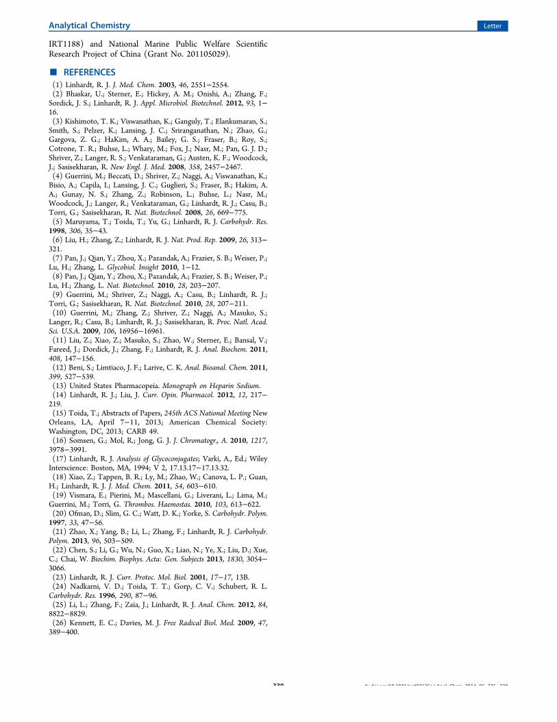
■ REFERENCES(1) Linhardt, R. J. J. Med. Chem. 2003, 46, 2551−2554.(2) Bhaskar, U.; Sterner, E.; Hickey, A. M.; Onishi, A.; Zhang, F.;Sordick, J. S.; Linhardt, R. J. Appl. Microbiol. Biotechnol. 2012, 93, 1−16.(3) Kishimoto, T. K.; Viswanathan, K.; Ganguly, T.; Elankumaran, S.;Smith, S.; Pelzer, K.; Lansing, J. C.; Sriranganathan, N.; Zhao, G.;Gargova, Z. G.; HaKim, A. A.; Bailey, G. S.; Fraser, B.; Roy, S.;Cotrone, T. R.; Buhse, L.; Whary, M.; Fox, J.; Nasr, M.; Pan, G. J. D.;Shriver, Z.; Langer, R. S.; Venkataraman, G.; Austen, K. F.; Woodcock,J.; Sasisekharan, R. New Engl. J. Med. 2008, 358, 2457−2467.(4) Guerrini, M.; Beccati, D.; Shriver, Z.; Naggi, A.; Viswanathan, K.;Bisio, A.; Capila, I.; Lansing, J. C.; Guglieri, S.; Fraser, B.; Hakim, A.A.; Gunay, N. S.; Zhang, Z.; Robinson, L.; Buhse, L.; Nasr, M.;Woodcock, J.; Langer, R.; Venkataraman, G.; Linhardt, R. J.; Casu, B.;Torri, G.; Sasisekharan, R. Nat. Biotechnol. 2008, 26, 669−775.(5) Maruyama, T.; Toida, T.; Yu, G.; Linhardt, R. J. Carbohydr. Res.1998, 306, 35−43.(6) Liu, H.; Zhang, Z.; Linhardt, R. J. Nat. Prod. Rep. 2009, 26, 313−321.(7) Pan, J.; Qian, Y.; Zhou, X.; Pazandak, A.; Frazier, S. B.; Weiser, P.;Lu, H.; Zhang, L. Glycobiol. Insight 2010, 1−12.(8) Pan, J.; Qian, Y.; Zhou, X.; Pazandak, A.; Frazier, S. B.; Weiser, P.;Lu, H.; Zhang, L. Nat. Biotechnol. 2010, 28, 203−207.(9) Guerrini, M.; Shriver, Z.; Naggi, A.; Casu, B.; Linhardt, R. J.;Torri, G.; Sasisekharan, R. Nat. Biotechnol. 2010, 28, 207−211.(10) Guerrini, M.; Zhang, Z.; Shriver, Z.; Naggi, A.; Masuko, S.;Langer, R.; Casu, B.; Linhardt, R. J.; Sasisekharan, R. Proc. Natl. Acad.Sci. U.S.A. 2009, 106, 16956−16961.(11) Liu, Z.; Xiao, Z.; Masuko, S.; Zhao, W.; Sterner, E.; Bansal, V.;Fareed, J.; Dordick, J.; Zhang, F.; Linhardt, R. J. Anal. Biochem. 2011,408, 147−156.(12) Beni, S.; Limtiaco, J. F.; Larive, C. K. Anal. Bioanal. Chem. 2011,399, 527−539.(13) United States Pharmacopeia. Monograph on Heparin Sodium.(14) Linhardt, R. J.; Liu, J. Curr. Opin. Pharmacol. 2012, 12, 217−219.(15) Toida, T.; Abstracts of Papers, 245th ACS National Meeting NewOrleans, LA, April 7−11, 2013; American Chemical Society:Washington, DC, 2013; CARB 49.(16) Somsen, G.; Mol, R.; Jong, G. J. J. Chromatogr., A. 2010, 1217,3978−3991.(17) Linhardt, R. J. Analysis of Glycoconjugates; Varki, A., Ed.; WileyInterscience: Boston, MA, 1994; V 2, 17.13.17−17.13.32.(18) Xiao, Z.; Tappen, B. R.; Ly, M.; Zhao, W.; Canova, L. P.; Guan,H.; Linhardt, R. J. J. Med. Chem. 2011, 54, 603−610.(19) Vismara, E.; Pierini, M.; Mascellani, G.; Liverani, L.; Lima, M.;Guerrini, M.; Torri, G. Thrombos. Haemostas. 2010, 103, 613−622.(20) Ofman, D.; Slim, G. C.; Watt, D. K.; Yorke, S. Carbohydr. Polym.1997, 33, 47−56.(21) Zhao, X.; Yang, B.; Li, L.; Zhang, F.; Linhardt, R. J. Carbohydr.Polym. 2013, 96, 503−509.(22) Chen, S.; Li, G.; Wu, N.; Guo, X.; Liao, N.; Ye, X.; Liu, D.; Xue,C.; Chai, W. Biochim. Biophys. Acta: Gen. Subjects 2013, 1830, 3054−3066.(23) Linhardt, R. J. Curr. Protoc. Mol. Biol. 2001, 17−17, 13B.(24) Nadkarni, V. D.; Toida, T. T.; Gorp, C. V.; Schubert, R. L.Carbohydr. Res. 1996, 290, 87−96.(25) Li, L.; Zhang, F.; Zaia, J.; Linhardt, R. J. Anal. Chem. 2012, 84,8822−8829.(26) Kennett, E. C.; Davies, M. J. Free Radical Biol. Med. 2009, 47,389−400.
Analytical Chemistry Letter
dx.doi.org/10.1021/ac403625a | Anal. Chem. 2014, 86, 326−330330