Metabolism of LY654322, A Growth Hormone...
40
DMD # 37598 1 Metabolism of LY654322, A Growth Hormone Secretagogue, to An Unusual Diimidazopyridine Metabolite Anthony G. Borel, Timothy M. Jones, Robert J. Barbuch, David A. Jackson, Palaniappan Kulanthaivel, Edward Mattiuz, Valentine J. Klimkowski, William J. Wheeler and Gregory A. Rener Lilly Research Laboratories, Eli Lilly and Company, Indianapolis, Indiana DMD Fast Forward. Published on February 23, 2011 as doi:10.1124/dmd.110.037598 Copyright 2011 by the American Society for Pharmacology and Experimental Therapeutics. This article has not been copyedited and formatted. The final version may differ from this version. DMD Fast Forward. Published on February 23, 2011 as DOI: 10.1124/dmd.110.037598 at ASPET Journals on June 20, 2018 dmd.aspetjournals.org Downloaded from
Transcript of Metabolism of LY654322, A Growth Hormone...

DMD # 37598 1
Metabolism of LY654322, A Growth Hormone Secretagogue, to
An Unusual Diimidazopyridine Metabolite
Anthony G. Borel, Timothy M. Jones, Robert J. Barbuch, David A. Jackson, Palaniappan
Kulanthaivel, Edward Mattiuz, Valentine J. Klimkowski, William J. Wheeler and
Gregory A. Rener
Lilly Research Laboratories, Eli Lilly and Company, Indianapolis, Indiana
DMD Fast Forward. Published on February 23, 2011 as doi:10.1124/dmd.110.037598
Copyright 2011 by the American Society for Pharmacology and Experimental Therapeutics.
This article has not been copyedited and formatted. The final version may differ from this version.DMD Fast Forward. Published on February 23, 2011 as DOI: 10.1124/dmd.110.037598
at ASPE
T Journals on June 20, 2018
dmd.aspetjournals.org
Dow
nloaded from

DMD # 37598 2
Running Title: Metabolism of LY654322 to an Unusual
Diimidazopyridine
Anthony G. Borel, PhD
Department of Drug Disposition, Lilly Research Laboratories
Eli Lilly and Company
Lilly Corporate Center
Indianapolis, IN 46285
Tel: 317-277-6736; Fax: 317-655-1185
e-mail: [email protected]
Page and number count
Text pages: 20
Tables: 3
Figures: 8
References: 14
Abstract: 238
Introduction: 238
Results: 1485
Discussion: 813
Abbreviations:
This article has not been copyedited and formatted. The final version may differ from this version.DMD Fast Forward. Published on February 23, 2011 as DOI: 10.1124/dmd.110.037598
at ASPE
T Journals on June 20, 2018
dmd.aspetjournals.org
Dow
nloaded from

DMD # 37598 3
DQFCOSY, double quantum filtered correlated spectroscopy; GH, growth hormone;
HMBC, heteronuclear multiple bond correlation; HSQC, heteronuclear single quantum
coherence; LC, liquid chromatography; MS, mass spectrometry; MS/MS, tandem mass
spectrometry; MS3, 3 stages of tandem mass spectrometry; MS4, 4 stages of tandem mass
spectrometry; MSn, multiple stages of tandem mass spectrometry; THF, tetrahydrofolate;
TOCSY, total correlation spectroscopy
This article has not been copyedited and formatted. The final version may differ from this version.DMD Fast Forward. Published on February 23, 2011 as DOI: 10.1124/dmd.110.037598
at ASPE
T Journals on June 20, 2018
dmd.aspetjournals.org
Dow
nloaded from

DMD # 37598 4
Abstract
LY654322 was rapidly cleared in rats and dogs by renal excretion of parent and
metabolism (oxidative and hydrolytic). Among the metabolites identified in the urine of
rats and dogs was M25, which was structurally unusual. Indeed, the characterization of
M25 and investigation into its disposition relied on the convergence of diverse analytical
methodologies. M25 eluted after parent on reverse phase chromatography with an MH+
at m/z 598 (parent + 35 Da). Given its increased lipophilicity and its mass difference
compared with parent, it was evident that M25 was not a phase 2 conjugate. Subsequent
LC/MS/MS, LC/MS3, LC/MS4, and accurate mass experiments identified the structure of
M25 as having two replicates of the 1-(4-fluorophenyl)-1-methyl-2-oxo-2-pyrrolidinyl
substructure flanking a central aromatic core of composition C7H3N5 that was refractory
to fragmentation. Compared with the UV spectrum of parent (λmax = 213 nm), M25
displayed a bathochromic shift (λmax = 311 nm), which substantiated extensive
conjugation within the central core. Subsequent NMR analysis of M25 isolated from
dog urine coupled with molecular modeling revealed the structure to be consistent with a
diimidazopyridine core with two symmetrically substituted 1-(4-fluorophenyl)-1-methyl-
2-oxo-2-pyrrolidinyl moieties. Using a structural analogue with a similar chromophore to
M25, LC/UV was used to quantitate M25 and determine its urinary disposition. The
formation of M25 appears consistent with hydrolysis of LY654322 to an aminoimidazole,
dimerization of the latter with the loss of NH3, C-formylation, and subsequent ring
closure and aromatization with loss of H2O.
This article has not been copyedited and formatted. The final version may differ from this version.DMD Fast Forward. Published on February 23, 2011 as DOI: 10.1124/dmd.110.037598
at ASPE
T Journals on June 20, 2018
dmd.aspetjournals.org
Dow
nloaded from

DMD # 37598 5
Introduction
Current medical advances have resulted in an increase in human life span and a
commensurate increase in the elderly population. Unfortunately, increased longevity has
been accompanied by an elevated emergence in conditions of the elderly including frailty
– characterized by declining organ function and physical performance. It is thought that
the decrease in pituitary growth hormone (GH) with age might be a contributing factor,
and that maintenance of GH could be beneficial in the treatment of frailty in the elderly
(Lamberts et al., 1997). In order to elicit the biological episodic profile of GH, the
development of synthetic peptide GH secretagogues is considered a viable approach to
the augmentation of GH secretion (Smith, 2005).
LY654322 (Fig. 1) is a GH secretagogue, and in vivo pharmacokinetic and metabolism
studies were conducted as part of late stage discovery studies in rats and dogs. The
objectives of these studies were to characterize the pharmacokinetics of parent drug and
to delineate major clearance pathways. LY654322 was cleared by renal excretion and
hepatic metabolism. Sixteen (16) metabolites were identified altogether, and derived
from pathways that involved C-oxidation, N-oxidation and amide hydrolysis. Among
these metabolites was M25, which was structurally unique and appeared to be the result
of the fusion of two substructures of the parent molecule around an aromatic core. Given
its novelty, the characterization of M25 was considered worthwhile. This report
describes the identification, urinary disposition and proposed formation of M25.
This article has not been copyedited and formatted. The final version may differ from this version.DMD Fast Forward. Published on February 23, 2011 as DOI: 10.1124/dmd.110.037598
at ASPE
T Journals on June 20, 2018
dmd.aspetjournals.org
Dow
nloaded from

DMD # 37598 6
Methods
Chemicals. Compounds LY654322(2-methylalanyl-N-{1-[(1R)-1-(4-fluorophenyl)-1-
methyl-2-oxo-2-pyrrolidin-1-ylethyl]-1H-imidazol-4-yl}-5-phenyl-D-norvalinamide) and
LSN60645 (1,7-dimethyl-1,7-dihydrodiimidazo[4,5-b:4',5'-e]pyridine) were synthesized
at Lilly Research Laboratories.
Animal Studies. All animal experiments were conducted according to protocols
approved by the Eli Lilly Animal Care and Use Committee. Male Fischer 344 rats (200
to 275 g) were obtained from Harlan Sprague Dawley, Indianapolis, IN. Female beagle
dogs (12.5 to 14.5 kg) were obtained from Marshall Farms, North Rose, NY. LY654322
was formulated in 50 mM phosphate buffer (pH 6) and 10% acacia for intravenous and
oral administration, respectively. Animals were dosed following an overnight fast. Food
was restored 2 hours post-dose and free access to water occurred throughout.
Sample Collection. Blood samples were collected in heparinized containers on
ice, and plasma was harvested by centrifugation. Plasma, urine, and feces samples were
stored at -70 °C prior to analysis.
Rat Studies. To determine plasma pharmacokinetics, three rats were administered
LY654322 by tail vein at 3 mg/kg or oral gavage at 30 mg/kg. Blood was sampled from
the jugular vein of rats at 0.05 and 0.25 (intravenous only), and 1, 2, 3, 4, 6, 8, and 10 h
post-dose. To determine urinary metabolites, three rats were housed in metabolism
cages. Urine was collected at 24 and 48 h.
Dog Studies. To determine plasma pharmacokinetics, three dogs were
administered LY654322 by cephalic vein at 1 mg/kg or oral gavage at 3 mg/kg. Blood
This article has not been copyedited and formatted. The final version may differ from this version.DMD Fast Forward. Published on February 23, 2011 as DOI: 10.1124/dmd.110.037598
at ASPE
T Journals on June 20, 2018
dmd.aspetjournals.org
Dow
nloaded from

DMD # 37598 7
was sampled from the jugular vein of dogs at 0.05 and 0.25 (intravenous only), and 1, 2,
3, 4, 6, 8, 10 and 24 h post-dose. To determine urinary metabolites, two dogs were
administered LY654322 at 20 mg/kg by oral capsule, housed in metabolism cages and
urine collected for 24 h.
In Vitro Studies.
Hepatocytes. Cryopreserved hepatocytes (In Vitro Technologies, Baltimore, MD)
were incubated (1 million cells per incubation) in Hepatocyte Maintenance Medium
(Lonza, Walkersville, MD) containing 50 μM LY654322. Conditions were maintained at
37°C in a 5% CO2 atmosphere. Control incubations without hepatocytes or without
substrate were conducted in each study. After 4 h, incubations were sonicated, frozen on
dry ice and stored at –70°C until analysis.
Liver slices. Rat and dog liver slices (2 slices per incubation) of approximately 8
mm in diameter and 200 to 250 μm in thickness were incubated in Waymouth’s 752/1
medium (Cell and Molecular Technologies, Lavallette, NJ) containing 50 μM LY654322.
Conditions were maintained at 37°C in a 95% O2:5% CO2 atmosphere. Control
incubations without slices or without substrate were conducted in each study. After 24 h,
incubations were sonicated, frozen on dry ice and stored at –70°C until analysis.
Equipment and analytical conditions.
Quantitation of LY654322. LC/MS/MS quantitation of LY654322 in rat and dog
plasma was performed on a Sciex API 3000 mass spectrometer (Foster City, CA) by
monitoring for MH+ with the ion transition m/z 563.4 to 192.4. Plasma samples were
extracted by mixing with a 2x volume of acetonitrile, centrifuged and introduced into the
mass spectrometer source with a Metachem Monochrom C18 column (2.1 x 50 mm; 5
This article has not been copyedited and formatted. The final version may differ from this version.DMD Fast Forward. Published on February 23, 2011 as DOI: 10.1124/dmd.110.037598
at ASPE
T Journals on June 20, 2018
dmd.aspetjournals.org
Dow
nloaded from

DMD # 37598 8
μm) using a combination of mobile phase A: 0.1% formic acid in water and mobile phase
B: 0.1% formic acid in methanol, at a flow rate of 350 μL/min. Mobile phase B was
delivered as a gradient from 20% to 75% over 0.8 min and restored to 20% at 0.9 min.
The calibration range was 3.9 to 4000 ng/mL. Samples above the limit of quantification
were diluted and reanalyzed to yield results within the calibration range.
Pharmacokinetic analysis. The pharmacokinetic parameters of LY654322 in
plasma were calculated by a noncompartmental method using WinNonlin. The terminal
half-life (t1/2) was calculated from the first-order elimination rate constant k where t1/2 =
0.693/k. The area under the plasma concentration time curve, determined by the
trapezoidal rule, was extrapolated to infinity using k to determine AUC0-∞. Plasma
clearance and bioavailability were calculated from AUC0-∞, dose and k.
Identification of metabolites.
LC/MS of metabolites. Urine samples were vortex mixed and centrifuged and transferred
to the autosampler for injection. Plasma samples were extracted by mixing with a 2x
volume of acetonitrile, centrifuged and concentrated to approximately the original
volume. Hepatocyte and liver slice samples were extracted by mixing with an equal
volume of acetonitrile, centrifuged and diluted with an equal volume of 0.2% formic acid.
Electrospray LC/MS/MS analysis was performed on a Thermo Finnigan LCQ mass
spectrometer (San Jose, CA), with a spray voltage of 5.0 kV and a capillary temperature
of 225 °C. Product ion spectra were generated at a relative collision energy of 40%. Full
scan spectra were acquired in the positive ion mode. There was also in line UV detection
with a Finnigan Surveyor PDA. Samples were introduced into the mass spectrometer
source via a Supelco Discovery C18 column (2.1 x 150 mm, 5 μm), using a combination
This article has not been copyedited and formatted. The final version may differ from this version.DMD Fast Forward. Published on February 23, 2011 as DOI: 10.1124/dmd.110.037598
at ASPE
T Journals on June 20, 2018
dmd.aspetjournals.org
Dow
nloaded from
DMD # 37598 9
of mobile phase A: 0.2% formic acid in water and mobile phase B: 10% isopropyl
alcohol in acetonitrile, at a flow rate of 200 μL/min. The following profile for mobile
phase B was used: initial, 10%; 2 min, 10%; 22 min, 50%; 32 min, 90%; 36 min, 90%;
36.1 min, 10%; 42 min (stop), 10%. Accurate mass measurements were conducted on a
Micromass QToF-2/MS using positive ion electrospray, a resolution of 8,500 FWHM, a
cone voltage of 40 V, a collision energy of 25 V, and a lock mass of 311.0814
(protonated molecular ion of the sulfadimethoxine standard).
Isolation of M25.
Urine from the individual dogs was pooled (approximately 300 mL) and applied directly
to two Varian Mega Bond ElutTM C18 SPE columns, which were washed with water
(approximately 60 mL each) and eluted with methanol (approximately 50 mL each). The
combined eluate was evaporated to dryness and the residue was reconstituted in 2.5 mL
of water/acetonitrile (85:15). The sample was centrifuged and the supernatant was
fractionated on a Supelco Discovery C18 column (4.6 x 250 mm, 5 μm) using the same
solvent system and gradient described above. Fractions were collected in 15-second
intervals from 5 to 35 minutes at a flow rate of 4.0 mL/min. Pooling of fractions for M25
was based upon purity and concentration as measured by LC/MS. After pooling, the
solvent was evaporated and the dried material was used for NMR analysis.
NMR analysis of M25.
NMR spectra were recorded at 25.0 °C on an Inova 500 MHz NMR spectrometer
equipped with a pulsed-field gradient and a Nalorac MDG 500 3-mm probe (Varian Inc.,
Palo Alto, CA). Compounds were dissolved in approximately 200 μL of CD3OD and
transferred to a 3-mm NMR tube. In the case of M25, approximately 40 μL of ND4OD
This article has not been copyedited and formatted. The final version may differ from this version.DMD Fast Forward. Published on February 23, 2011 as DOI: 10.1124/dmd.110.037598
at ASPE
T Journals on June 20, 2018
dmd.aspetjournals.org
Dow
nloaded from

DMD # 37598 10
was added to sharpen the NMR peaks. Chemical shifts were referenced to the residual
solvent signal at δ 3.3 for 1H and δ 49 for 13C. Two-dimensional NMR experiments
including double quantum filtered correlated spectroscopy (DQFCOSY), total correlation
spectroscopy (TOCSY), heteronuclear single quantum coherence (HSQC), and
heteronuclear multiple bond correlation (HMBC) were performed using Varian standard
pulse sequences.
Molecular modeling of M25.
Molecular modeling was performed using Maestro and MacroModel software
(Schrodinger LLC, 2008). For M25, an extensive conformational search was done using
the Mixed Torsional/Low-Mode Sampling procedure (2000 maximum steps), the
OPLS2005 forcefield, the GBSA continuum aqueous solvation treatment, and complete
energy minimization to convergence. Using the identified M25 global minimum energy
structure and an equivalently minimized structure for LSN60645, NMR shielding tensors
were calculated using the ab initio program Gaussian 03 (Frisch et al., 2004) by Density
Functional Theory (B3LYP functional, the 6-31++G(d,p) basis set, and GIAO method)
and without geometry optimization. From these, the calculated chemical shift difference
for the pyridinyl proton in M25, compared to LSN60645, was estimated by the difference
in the calculated isotropic magnetic shielding tensor for the equivalent proton in each
molecule.
Quantitation of M25.
M25 was quantitated in dog urine by LC/UV using the analogue, LSN60645, as a
surrogate analytical standard, since both compounds contained the same diagnostic
diimidazopyridine chromophore. LC/UV response at 311 nm was selective for M25 and
This article has not been copyedited and formatted. The final version may differ from this version.DMD Fast Forward. Published on February 23, 2011 as DOI: 10.1124/dmd.110.037598
at ASPE
T Journals on June 20, 2018
dmd.aspetjournals.org
Dow
nloaded from

DMD # 37598 11
LSN60645. LC/UV detection was performed on a Waters 996 photodiode array detector
(220 - 500 nm). Chromatography was performed using a Supelco Discovery C18 column
(2.1 x 150 mm, 5 μm) with the same mobile phase conditions as described above in
LC/MS of Metabolites for the quantitation of M25 in urine, and an isocratic delivery of
mobile phase B for the quantitation of LSN60645 in an aqueous solution matrix. The
calibration range was 8 to 1000 ng/mL.
LSN60645 and M25 both contained the same chromophore responsible for absorbance at
λ = 311 nm, and consequently, their extinction coefficients at this wavelength were
assumed to be the same. Since a standard was available for LSN60645, its extinction
coefficient was determined by measuring its absorbance at known concentration in
chloroform at λmax = 311 nm on a Spectra Max Plus spectrophotometer (Molecular
Devices, Sunnyvale, CA) with 1 cm quartz cuvettes. The extinction coefficient for
LSN60645 was calculated to be 12,741 M-1 cm-1.
This article has not been copyedited and formatted. The final version may differ from this version.DMD Fast Forward. Published on February 23, 2011 as DOI: 10.1124/dmd.110.037598
at ASPE
T Journals on June 20, 2018
dmd.aspetjournals.org
Dow
nloaded from

DMD # 37598 12
Results
Pharmacokinetics of LY2456322. The pharmacokinetics of LY654322 were
investigated in rats and dogs. Following intravenous administration, mean values for
plasma clearance and terminal half-life were 75 mL/min/kg and 2 hours, respectively, in
rats and 33 mL/min/kg and 5 hours, respectively, in dogs. Following oral administration,
the bioavailability in rats and dogs was 74% and 33%, respectively.
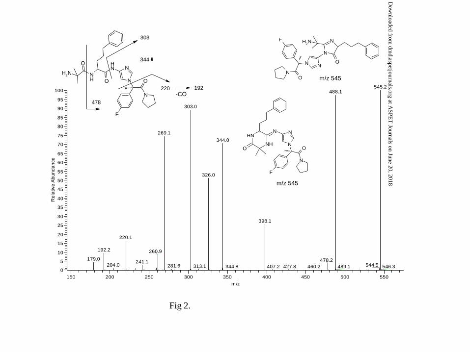
LC/MS/MS of LY654322. The LC/MS/MS spectrum of LY654322 is shown in Fig. 2.
MH+ at m/z 563 undergoes a facile loss of H2O likely through amide – imidic acid
tautomerization at either of the two peptide linkages followed by intramolecular
condensation to the corresponding imine at m/z 545. Subsequent loss of (CH3)2C=NH
from m/z 545 would then afford the ion at m/z 488. Other characteristic fragment ions of
parent were m/z 478, m/z 344, m/z 303, m/z 220 and m/z 192. Accurate mass data and
proposed elemental compositions of these ions are shown in Table 1.
Metabolism of LY654322. Plasma and urine samples from rats and dogs dosed with
LY654322 were profiled by LC/UV and LC/MS/MS. LY654322 was cleared by urinary
excretion of parent and metabolism. Sixteen (16) metabolites were identified between
rats and dogs collectively, with considerable overlap occurring between species.
Metabolism of parent was related to the following structural regions: (A) aliphatic and N-
oxidation, (B) aliphatic and aromatic oxidation and (C) amide hydrolysis (Fig. 1).
Among the downstream metabolites of LY654322 was M25 which was identified only in
the urine of rats and dogs. M25 was not identified as a metabolite in rat or dog liver
This article has not been copyedited and formatted. The final version may differ from this version.DMD Fast Forward. Published on February 23, 2011 as DOI: 10.1124/dmd.110.037598
at ASPE
T Journals on June 20, 2018
dmd.aspetjournals.org
Dow
nloaded from

DMD # 37598 13
slices or human hepatocytes, nor was it detected as an impurity in parent drug in
incubations where liver slices and hepatocytes were absent. Taken together, these results
indicated that M25 was an authentic urinary metabolite of LY654322. M25 was
structurally unusual and subject to detailed investigation.
Characterization of M25. LC/MS analysis of rat and dog urine identified M25 as
eluting after parent on reverse phase chromatography (Fig. 3). Furthermore, its MH+ at
m/z 598 exceeded MH+ of parent by 35 Da (Table 2) and was not the product of direct
phase 2 conjugation as detailed below. Given its increased lipophilicity and its mass
difference compared with parent, it was evident that the metabolic pathway to M25 was
not straightforward. Valuable insight into the structure of M25 was gained through a
series of MSn experiments (Fig. 4). MS/MS of M25 afforded ions m/z 220 and m/z 192,
which were common with parent and indicated that the 1-(4-fluorophenyl)-1-methyl-2-
oxo-2-pyrrolidinyl substructure was retained. The complementary ion to m/z 220 at m/z
379 was fragmented, and the surprising recurrence of ions m/z 220 and m/z 192
suggested that M25 consisted of two replicates of the 1-(4-fluorophenyl)-1-methyl-2-oxo-
2-pyrrolidinyl substructure. The complementary ion to m/z 220 at m/z 160 was
fragmented, and on this occasion, there was only a weak loss of HCN, indicating that m/z
160 was very stable and likely aromatic. The structure of M25 was further probed with
the application of accurate mass MS and MS/MS experiments, which ascribed what was
envisioned to be the central core the formula C7H5N5 (neutral molecule), a composition
requiring eight double bond equivalents (Table 1). In order to rationalize the existing
data, the following structure was proposed: two symmetrically disposed N-[1-(4-
fluorophenyl)-1-methyl-2-oxo-2-pyrrolidinyl] substituents to a fused diimidazopyridine.
This article has not been copyedited and formatted. The final version may differ from this version.DMD Fast Forward. Published on February 23, 2011 as DOI: 10.1124/dmd.110.037598
at ASPE
T Journals on June 20, 2018
dmd.aspetjournals.org
Dow
nloaded from

DMD # 37598 14
Since the diimidazopyridine substructure was critical to the characterization of M25, it
was used to interrogate the Lilly database for a structural precedent. The hit, LSN60645,
a diimidazopyridine analogue, was fortuitous, since it served as a conduit to a series of
informative UV and NMR experiments.
The UV spectrum of parent was characterized by a single λmax at 213 nm (Fig. 5A).
M25, on the other hand displayed, two λmaxima – one at 208 nm, comparable with
parent, and another with a bathochromic shift at 311 nm, consistent with extensive
conjugation (Fig. 5B). The UV spectra of M25 matched that of the standard LSN60645,
supporting the diimidazopyridine substructure of the central core.
NMR was critical in reconciling the connectivity of the proposed heterocycle core to
other substructures of parent in the metabolite. Although the molecular weight of M25
was 35 Da higher than LY654322, the 1H-NMR spectrum of the metabolite was
strikingly less complex than parent (see Fig. 6 for the aromatic region of LY654322 and
M25, and Table 3 for the chemical shift assignments). The aromatic region of the proton
NMR spectrum of M25 consisted of only four resonances, two of which (δ 7.39 br dd; δ
7.27 br t) were assigned to the 4-flurophenyl substituent analogous to the parent
compound (Table 3). Furthermore, the recurrence of the pyrrolidinyl protons and their
respective carbons in M25 compared with parent supported the retention of the 1-(4-
flurophenyl)-1-methyl-2-oxo-2-pyrrolidinyl substructure in M25 (Table 3). The mutually
coupled imidazolyl protons of parent at δ 7.24 and 7.17 were conspicuously absent in
M25 and instead these resonances were replaced by two new resonances at δ 8.23
This article has not been copyedited and formatted. The final version may differ from this version.DMD Fast Forward. Published on February 23, 2011 as DOI: 10.1124/dmd.110.037598
at ASPE
T Journals on June 20, 2018
dmd.aspetjournals.org
Dow
nloaded from
DMD # 37598 15
(singlet, 2 protons) and δ 6.92 (singlet, 1 proton). The lack of any other resonances,
specifically resonances due to the 2-methylalanyl-5-phenyl-D-norvalinyl moiety of the
parent, in M25 (Table 3) and the fact that the intensity of the singlet proton at δ 6.92 is
approximately one-fourth of the equivalent 4-fluorophenyl protons at δ 7.39 or δ 7.27
revealed that M25 possesses an element of symmetry. Since the UV spectrum of M25
was very similar to LSN60645, a diimidazo pyridinyl derivative, the proton NMR
spectrum of M25 was compared with LSN60645 (Fig. 6). The comparison revealed that
both spectra showed a singlet resonance with 2 proton intensity in the range δ 8.2 to 8.3,
which was assigned to the magnetically equivalent 2 imidazolyl protons of a highly
conjugated system. However, the pyridinyl proton (H-19) which resonated at δ 8.06 in
LSN60645, a normal chemical shift associated with its chemical environment, appeared
with a pronounced up-field shift at δ 6.92 (Δδ = 1.14) in M25.
In order to rationalize the NMR of M25, molecular modeling experiments were
performed. A conformational search resulted in a global minimum energy structure (Fig.
7), and other conformations within 2.6 kcal/mole of the global minimum (not shown), all
of which positioned the pyridinyl proton perpendicular to the two fluorophenyl rings.
Oriented as such, this proton should experience significant shielding due to the aromatic
ring current and a pronounced diamagnetic shift. Indeed, calculating the pyridinyl proton
chemical shift difference between M25 (in the illustrated conformation) and LSN60645
by ab initio quantum mechanical methods, gave a value Δδ = 0.82, comparable to the up-
field shift that was experimentally observed. This supports the contention that in M25 the
This article has not been copyedited and formatted. The final version may differ from this version.DMD Fast Forward. Published on February 23, 2011 as DOI: 10.1124/dmd.110.037598
at ASPE
T Journals on June 20, 2018
dmd.aspetjournals.org
Dow
nloaded from

DMD # 37598 16
ring current from the phenyl groups in close proximity to the pyridinyl proton, should
perturb its chemical shift up-field, with respect to the same proton in LSN60645.
Identification of metabolites related to M25 (Table 2).
M2: M2 was identified in the plasma and urine of rats and dogs. The full scan
MS of M2 showed MH+ (C16H20FN4O) at m/z 303, suggesting an aminoimidazole
structure resulting from hydrolysis of the peptide linkage proximal to the imidazole. The
LC/MS/MS spectrum of M2 showed several ions consistent with the proposed structure,
including m/z 220 (C13H15FNO, resulting from loss of 4-aminoimidazole), m/z 204
(C11H11FN3, resulting from loss of N-formylpyrrolidine), m/z 192 (C12H15FN, resulting
from loss of 4-aminoimidazole and CO), and m/z 84 (C3H6N3, protonated 4-
aminoimidazole).
M19: M19 was identified in rat plasma only. The full scan MS of M19 showed
MH+ (C15H23N2O3) at m/z 279, suggestive of the carboxylic acid, complementary to M2,
formed by hydrolysis of the peptide linkage proximal to the imidazole. The supporting
LC/MS/MS product ion spectrum contained m/z 194 (C11H16NO2, resulting from loss of
3,3-dimethylaziridin-2-one) and m/z 103 (C4H11N2O, protonated 2-amino-2-methyl-
propanamide).
M23, M24: M23 and M24 were identified in the urine of dogs, but not rats. The
full scan MS of M23 and M24 showed MH+ (C22H28FN4O7) at m/z 479, suggestive of
glucuronide conjugates of M2. M23 and M24 had comparable product ion spectra. Key
product ions included: m/z 345, the loss of C4H6O5 from the glucuronide; m/z 303, loss
of C6H8O6 from the glucuronide to form the aglycone; m/z 260, loss of the 1-(4-
This article has not been copyedited and formatted. The final version may differ from this version.DMD Fast Forward. Published on February 23, 2011 as DOI: 10.1124/dmd.110.037598
at ASPE
T Journals on June 20, 2018
dmd.aspetjournals.org
Dow
nloaded from

DMD # 37598 17
fluorophenyl)-1-methyl-2-oxo-2-pyrrolidinyl substructure; m/z 242, loss of water from
m/z 260; m/z 220, the 1-(4-fluorophenyl)-1-methyl-2-oxo-2-pyrrolidinyl substructure;
m/z 192, loss of carbon monoxide from m/z 220. These data define the site of
glucuronidation to the aminoimidazole ring.
Urinary Excretion of M25 in Rats and Dogs. Given the identification of M25 in the
urine of both rats and dogs, and the similar UV spectra of M25 and LSN60645 in the
region λ = 308 to 312 nm, UV spectroscopy was exploited as a tool to quantitate M25 in
urine and determine the extent of urinary excretion in animals. In dog urine, the
chromatographic peak for M25 was well-isolated and the UV range was very selective,
allowing M25 to be quantitated and its urinary disposition assessed at 0.3% of the dose in
24 hr. Although M25 was identified in rat urine, it could not be quantitated because of
high chromatographic interference.
This article has not been copyedited and formatted. The final version may differ from this version.DMD Fast Forward. Published on February 23, 2011 as DOI: 10.1124/dmd.110.037598
at ASPE
T Journals on June 20, 2018
dmd.aspetjournals.org
Dow
nloaded from

DMD # 37598 18
Discussion
The preclinical pharmacokinetics and metabolism of LY654322 were determined as part
of late stage discovery characterization. The pharmacokinetics were characterized in rats
and dogs as having high clearance comparable with hepatic blood flow in both species
(Davies and Morris, 1993). The compound was extensively metabolized. One of the
metabolites identified during the course of these studies was M25, excreted in rat and dog
urine, which had LC/MS/MS and UV properties that sparked our interest. The MS4
fragmentation sequence of M25 (m/z 598 → 379 → 160 → 133) coupled with the
bathochromic shift in its λmax compared with parent indicated that M25 resulted from
substantial molecular modification of LY654322. NMR characterization of M25 and
molecular modeling substantiated the dimerization of two imidazo fragments from parent
with endogenous C-atom incorporation to afford a diimidazopyridine analogue with a
pyridinyl plane of symmetry.
The initial step in the metabolism of LY654322 to M25 is proposed to occur through the
hydrolysis of the peptide linker to afford the aminoimidazole M2, which was identified in
the plasma and urine of both rats and dogs. Hydrolysis was catalyzed in the liver based
on the identification of M2 in rat and dog liver slices and human hepatocytes. M19, the
complementary carboxylic acid in the peptide linkage to M2, as well as the downstream
M2 glucuronides, M23 and M24, supported the occurrence of M2.
The organ(s), subcellular compartment(s) and conditions which contribute to the
formation of M25 from M2 are not known. Although M2 was formed in liver slices and
This article has not been copyedited and formatted. The final version may differ from this version.DMD Fast Forward. Published on February 23, 2011 as DOI: 10.1124/dmd.110.037598
at ASPE
T Journals on June 20, 2018
dmd.aspetjournals.org
Dow
nloaded from

DMD # 37598 19
hepatocytes, M25 was not a metabolite in vitro. It is possible that in vitro conditions
were not optimized for the formation of M25 from M2 or the formation of M25 takes
place extra-hepatically.
The subsequent incorporation of the M2 substructure in M25 is envisioned in the
metabolic sequence described in Fig. 8. M2 tautomerizes to the imine which undergoes
nucleophilic attack by another aminoimidazole molecule to form the intermediate 1,
which eliminates NH3 to couple the two aminoimidazole subunits as the N-linked
condensation product 2. Subsequently, the carbon of the central pyrininium heterocycle
is added by formylation through a C-centered conjugate nucleophilic attack from one of
the imidazo subunits to form 3. The latter is positioned for an entropically-favored
intramolecular nucleophilic attack from the C-center of the other imidazo subunit to
afford ring closure to 4, and ensuing aromatization with the loss of H2O to afford M25.
None of the proposed intermediates 1 – 4 were identified as in vivo metabolites in rats or
dogs.
The coupling, formylation and cyclization steps in this metabolic scheme are noteworthy.
Firstly, the chemical condensation of aminoimidazoles described in the literature
(Bredereck et al., 1964a, 1964b), requires high temperatures (>100 °C). The proposed
dimerization of M2 is thought to proceed under physiological conditions, although the
dimer (intermediate 2) was not detected in liver slices or hepatocytes. It is not known
whether this process is enzyme-catalyzed. Secondly, although the C-source in its
cyclization is conjectural (vide infra), experiments were conducted to ensure that single
carbon introduction in the formation of M25 was not an artifact of analysis or processing.
This article has not been copyedited and formatted. The final version may differ from this version.DMD Fast Forward. Published on February 23, 2011 as DOI: 10.1124/dmd.110.037598
at ASPE
T Journals on June 20, 2018
dmd.aspetjournals.org
Dow
nloaded from

DMD # 37598 20
In control experiments, formic acid in the mobile phase was replaced with acetic acid or
ammonium acetate for LC/MS, and in both instances M25 was present to the same extent.
Conversely, urine samples were incubated in 1% formic acid, and no change in M25 was
discerned. Taken together, these experiments supported M25 being an authentic
metabolite derived from the incorporation of carbon from an endogenous source.
This article has not been copyedited and formatted. The final version may differ from this version.DMD Fast Forward. Published on February 23, 2011 as DOI: 10.1124/dmd.110.037598
at ASPE
T Journals on June 20, 2018
dmd.aspetjournals.org
Dow
nloaded from

DMD # 37598 21
The proposed C-formylation in the formation of M25 is based on precedence in the
literature for N- and C-formylations, and invokes N10-formyltetrahydrofolate as a carbon
source. N-formylation in the metabolism of xenobiotics is an uncommon pathway. It has
been described for aromatic (Santti and Hopsu-Havu, 1968; Gothoskar et al., 1979;
TjØrnelund et al., 1991) and aliphatic (Mutlib et al., 2002; Obach et al., 2006) amines.
Alternatively, N-formylation in the biosynthesis of purines is well-recognized, being
mediated by N10-formyltetrahydrofolate as the carbon source (Zhang et al., 2008). C-
formylation, as a biotransformation, has very little precedence in the literature – one
example describes the formation a C-formyl secondary metabolite of phencyclidine
mediated through an enamine (Zhao et al., 1991). The reaction was catalyzed by a
mitochondrial enzyme with either N5- or N10-formyltetrahydrofolate as the carbon source.
The C-formylation of intermediate 2 through a conjugate addition (Fig. 8) appears
analogous to that described for phencyclidine. In this case, however, the reaction is
driven forward by cyclization and aromatization steps to form M25, interestingly
analogous to the N-centered formylation and addition steps in purine biosynthesis (Zhang
et al., 2008).
In summary, LY654322 was metabolized in rats and dogs to M25, which was
characterized by LC/MSn, UV, NMR and molecular modeling, as an unusual
diimidazopyridine. The formation of M25 was rationalized by an initial amide hydrolysis
of LY654322 to the aminoimidazole M2, which underwent dimerization, C-formylation,
cyclization and aromatization.
This article has not been copyedited and formatted. The final version may differ from this version.DMD Fast Forward. Published on February 23, 2011 as DOI: 10.1124/dmd.110.037598
at ASPE
T Journals on June 20, 2018
dmd.aspetjournals.org
Dow
nloaded from

DMD # 37598 22
Authorship Contributions
Participated in research design: Borel
Conducted experiments: Barbuch, Mattiuz, Jackson, Klimkowski, Rener, Kulanthaivel,
Jones
Performed data analysis: Borel, Barbuch, Wheeler, Klimkowski, Rener, Kulanthaivel,
Jones
Wrote or contributed to the writing of the manuscript: Borel, Barbuch, Klimkowski,
Rener, Kulanthaivel
This article has not been copyedited and formatted. The final version may differ from this version.DMD Fast Forward. Published on February 23, 2011 as DOI: 10.1124/dmd.110.037598
at ASPE
T Journals on June 20, 2018
dmd.aspetjournals.org
Dow
nloaded from

DMD # 37598 23
References
Bredereck H, Effenberger F and Rainer G (1964a). Säureamid-Reaktionen, XXXIV. 7-
Substituierte purine und ihre aufspaltung zu 4.5-diamino-pyrimidinen. Justus Liebigs
Annalen der Chemie 673: 82-87.
Bredereck H, Effenberger F and Rainer G (1964b). Säureamid-Reaktionen, XXXV.
Uber den mechanismus der neuen purin-synthese. Justus Liebigs Annalen der Chemie
673: 88-92.
Davies B and Morris T (1993) Physiological parameters in laboratory animals and
humans. Pharm Res 10: 1093–1095.
Frisch MJ, Trucks GW, Schlegel HB, Scuseria GE, Robb MA, Cheeseman JR,
Montgomery Jr. JA, Vreven T, Kudin KN, Burant JC, Millam JM, Iyengar SS, Tomasi J,
Barone V, Mennucci B et al. (2004) Gaussian, Inc., Wallingford CT, Gaussian 03,
Revision C.02.
Gothoskar SV, Benjamin T, Roller PP, and Weisberger EK (1979) N-Formylation of an
aromatic amine as a metabolic pathway. Xenobiotica 9: 533-537.
Lamberts SWJ, van den Beld A, and der Lely A-J (1997). The endocrinology of aging.
Science 278: 419-424.
This article has not been copyedited and formatted. The final version may differ from this version.DMD Fast Forward. Published on February 23, 2011 as DOI: 10.1124/dmd.110.037598
at ASPE
T Journals on June 20, 2018
dmd.aspetjournals.org
Dow
nloaded from

DMD # 37598 24
Mutlib AE, Dickenson P, Chen S-Y, Espina RJ, Daniels JS, and Gan L-S (2002)
Bioactivation of benzylamine to reactive intermediates in rodents: Formation of
glutathione, glutamate, and peptide conjugates. Chem Res Toxicol 15: 1190-1207.
Obach RS, Reed-Hagen AE, Krueger SS, Obach BJ, O’Connell TN, Zandi KS, Miller S,
and Coe JW (2006) Metabolism and disposition of varnicline, a selective α4β2
acetylcholine receptor partial agonist, in vivo and in vitro. Drug Metab Dispos 34: 121-
130.
Santti RSS, and Hopsu-Havu VK (1968) Transformylation of carcinogenic aromatic
amines by kynurenine formamidase: a detoxification mechanism. Biochem Pharmacol
17: 1110-1113.
Smith RG (2005). Endocrine Reviews 26: 346-360.
Schrodinger LLC, New York, NY (2008), Maestro, version 8.5, and MacroModel,
version 9.6.
TjØrnelund J, Hansen SH, and Cornett C (1991) New metabolites of the drug 5-
aminosalicylic acid. II. N-Formyl-5-aminosalicylic acid. Xenobiotica 21: 605-612.
This article has not been copyedited and formatted. The final version may differ from this version.DMD Fast Forward. Published on February 23, 2011 as DOI: 10.1124/dmd.110.037598
at ASPE
T Journals on June 20, 2018
dmd.aspetjournals.org
Dow
nloaded from

DMD # 37598 25
Zhang Y, Morar M, and Ealick SE (2008) Structural biology of the purine biosynthetic
pathway. Cell Mol Life Sci 65: 3699–3724.
Zhao Z, Leung LY, Trevor A, and Neal Castagnoli Jr N (1991) C-Formylation in the
presence of rat brain mitochondria of the 2,3,4,5-tetrahydroyridinium metabolite derived
from the psychotomimetic drug phencyclidine. Chem. Res. Toxicol. 4: 426-429.
This article has not been copyedited and formatted. The final version may differ from this version.DMD Fast Forward. Published on February 23, 2011 as DOI: 10.1124/dmd.110.037598
at ASPE
T Journals on June 20, 2018
dmd.aspetjournals.org
Dow
nloaded from

DMD # 37598 26
Footnotes
Current affiliation: IsotopicSolutions, LLC, Indianapolis, Indiana (W.J.W.)
This article has not been copyedited and formatted. The final version may differ from this version.DMD Fast Forward. Published on February 23, 2011 as DOI: 10.1124/dmd.110.037598
at ASPE
T Journals on June 20, 2018
dmd.aspetjournals.org
Dow
nloaded from

DMD # 37598 27
Legends for Figures
Fig. 1. Structure of LY654322, showing the principal sites of metabolism. (A) Aliphatic
and N-oxidation, (B) aliphatic and aromatic oxidation, and (C) amide hydrolysis.
Fig. 2. LC/MS/MS of LY654322, illustrating fragment ion assignments and proposed
structures for the ions at m/z 545 resulting from loss of H2O from parent.
Fig. 3. LC/MS extracted ion chromatogram of dog urine illustrating metabolites related
to M25.
Fig. 4. LC/MS, LS/MS3 and LC/MS4 of M25 in dog urine. (A) MS/MS of MH+ at m/z
598, (B) MS/MS of the fragment at m/z 379, (C) MS/MS of the fragment at m/z 160.
Fig. 5. LC UV spectra of (A) LY654322, (B) M25 and LSN60645 superimposed.
Fig. 6. 1H NMR spectra of the aromatic region of (A) LY654322, (B) M25 and (C)
LSN60645.
Fig. 7. Molecular model of M25 illustrating a low-energy conformation with the
pyridinyl proton perpendicular to the two 4-flouorophenyl rings and predisposed to a
diamagnetic shift because of aromatic ring current.
This article has not been copyedited and formatted. The final version may differ from this version.DMD Fast Forward. Published on February 23, 2011 as DOI: 10.1124/dmd.110.037598
at ASPE
T Journals on June 20, 2018
dmd.aspetjournals.org
Dow
nloaded from

DMD # 37598 28
Fig. 8. Proposed metabolism of LY654322 to form M25. Intermediates 1 – 4 were not
identified as metabolites in rats or dogs.
This article has not been copyedited and formatted. The final version may differ from this version.DMD Fast Forward. Published on February 23, 2011 as DOI: 10.1124/dmd.110.037598
at ASPE
T Journals on June 20, 2018
dmd.aspetjournals.org
Dow
nloaded from

DMD # 37598 29
Table 1. Accurate mass data for LY654322 and its metabolite M25.
Compound Ion Composition Measured Mass
Calculated Mass
mDa ppm
LY654322 563 C31 H 40N6 O3F 563.3113 563.3146 -3.3 -5.9
LY654322 488 C28 H 31N5 O2F 488.2449 488.2462 -1.3 -2.6
LY654322 398 C21 H 25N5 O2F 398.1989 398.1992 -0.3 -0.8
LY654322 344 C18 H 26N5 O2
344.2104 344.2087 1.7 5.1
LY654322 179 C8 H 11N 4O 179.0923 179.0933 -1 -5.5
M25 598 C33H 34 N7 O2F2 598.2723 598.2742 -1.9 -3.2
M25 379 C20 H20 N 6OF 379.1683 379.1683 0 0
M25 220 C13 H 15NOF 220.1133 220.1138 -0.5 -2.3
M25 192 C12H15 NF 192.1187 192.1189 -0.2 -1.0
M25 160 C7 H6N5 160.0619 160.0623 -0.4 -2.5
LY654322
M25
This article has not been copyedited and formatted. The final version may differ from this version.DMD Fast Forward. Published on February 23, 2011 as DOI: 10.1124/dmd.110.037598
at ASPE
T Journals on June 20, 2018
dmd.aspetjournals.org
Dow
nloaded from
DMD # 37598 30
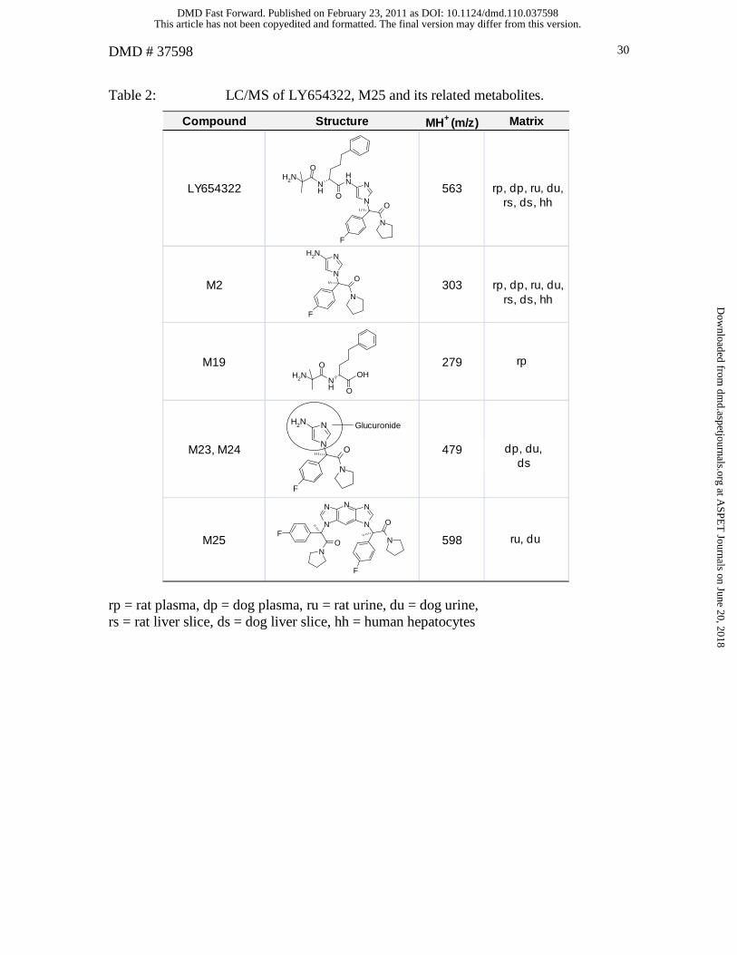
Table 2: LC/MS of LY654322, M25 and its related metabolites.
rp = rat plasma, dp = dog plasma, ru = rat urine, du = dog urine, rs = rat liver slice, ds = dog liver slice, hh = human hepatocytes
Compound Structure MH+ (m/z) Matrix
LY654322 563 rp, dp, ru, du
M2 303 rp, dp, ru, du
M19 279 rp
M23, M24 479 du
M25 598 ru, du
N
N
O
NNH
F
O
NH
ONH2
NO
NNH2
F
N
ONH
ONH2
OH
NO
NNH2
F
N
Glucuronide
N
NO
FN
N
O
F
N NN
rp, dp, ru, du,rs, ds, hh
dp, du,ds
rp, dp, ru, du,rs, ds, hh
rp
ru, du
This article has not been copyedited and formatted. The final version may differ from this version.DMD Fast Forward. Published on February 23, 2011 as DOI: 10.1124/dmd.110.037598
at ASPE
T Journals on June 20, 2018
dmd.aspetjournals.org
Dow
nloaded from

DMD # 37598 31
Table 3. Chemical shift assignmentsa of LY654322, M25 and LSN60645 in CD3OD.
mult = multiplicity, br = broad, m = multiplet, s = singlet, t = triplet, dd = doublet of doublet, obsc = obscured, J = coupling constant in Hz, NA = not applicable,
ND = not detected.
aAssignments were facilitated by the analysis of 1H, DQFCOSY, TOCSY, HSQC and HMBC spectra. bCoupling constants are not entered for M25 when identical with those of LY654322.
Substructure Position LSN606451H δ mult (J in Hz) 13C δ 1H δ mult 13C δ 1H δ mult
1, 1’ 7.40 br dd (9, 5) 130.6 7.39 br dd 130.7 NA
2, 2’ 7.21 br t (8.5) 117.2 7.27 br t 117.2 NA
3 2.21 s 26.3 2.32 s 25.6 NA
4 3.55 t (7) 49.6 3.53 t 49.6 NA
5 1.78 m 24 1.75 m 23.5 NA
6 1.76, 1.63 m 27.5 1.72, 1.64 27.4 NA
7 3.05, 2.67 m 48.7 2.89, 2.74 m 48.6 NA
8 7.24 d (1.5) 134.7 NA NA NA
9 7.17 d (1.5) 109.1 NA NA NA
LY654322 M25
89
N
N
89
N
N
F
1 1'
2'2
O
N
3
4
56
7
This article has not been copyedited and form
atted. The final version m
ay differ from this version.
DM
D Fast Forw
ard. Published on February 23, 2011 as DO
I: 10.1124/dmd.110.037598
at ASPET Journals on June 20, 2018 dmd.aspetjournals.org Downloaded from

DMD # 37598 32
Table 3 (continued). Chemical shift assignmentsa of LY654322, M25 and LSN60645 in CD3OD.
mult = multiplicity, br = broad, m = multiplet, s = singlet, t = triplet, dd = doublet of doublet, obsc = obscured, J = coupling constant in Hz, NA = not applicable,
ND = not detected.
aAssignments were facilitated by the analysis of 1H, DQFCOSY, TOCSY, HSQC and HMBC spectra. bCoupling constants are not entered for M25 when identical with those of LY654322.
Substructure Position LSN606451H δ mult (J in Hz) 13C δ 1H δ mult 13C δ 1H δ mult
10,10’ 1.30 s 28.4, 28.5 NA NA NA
11 4.49 dd (8, 5) 54.4 NA NA NA
12 1.84,1.72 m 33.2 NA NA NA
13 1.66 m 28.8 NA NA NA
14 2.61 m 36.4 NA NA NA
15, 15’ 7.13 obsc 129.5 NA NA NA
16, 16’ 7.22 obsc 129.4 NA NA NA
17 7.12 obsc 126.9 NA NA NA
18, 18’ NA NA 8.23 s ND 8.30 s
19 NA NA 6.92 s 108 8.06 s
LY654322 M25
10
16'
15'
17
16
12
15
13
11
14
10'
NHO
NH210
16'
15'
17
16
12
15
13
11
14
10'
NHO
NH2
18
19
18'
N
N N
N
N
18
19
18'
N
N N
N
N
This article has not been copyedited and form
atted. The final version m
ay differ from this version.
DM
D Fast Forw
ard. Published on February 23, 2011 as DO
I: 10.1124/dmd.110.037598
at ASPET Journals on June 20, 2018 dmd.aspetjournals.org Downloaded from

N
N
O
NNH
F
O
NH
O
NH2
A
B
C
Fig 1.
This article has not been copyedited and formatted. The final version may differ from this version.DMD Fast Forward. Published on February 23, 2011 as DOI: 10.1124/dmd.110.037598
at ASPE
T Journals on June 20, 2018
dmd.aspetjournals.org
Dow
nloaded from
450020_s_h_24a #965-993 RT: 18.75-19.09 AV: 13 NL: 7.31E6
F: + c d Full ms2 [email protected] [ 145.00-575.00]
150 200 250 300 350 400 450 500 550
m/z
0
5
10
15
20
25
30
35
40
45
50
55
60
65
70
75
80
85
90
95
100
Re
lative
Ab
un
da
nce
545.2488.1
303.0
269.1
344.0
326.0
398.1
220.1
192.2 260.9
179.0 478.2241.1544.5204.0 281.6 313.1 489.1407.2 460.2427.8 546.3344.8
N O
NNH
F
ONH
O
NH2
N
478
303
220
344
-CO192
N
O
N
F
N
NH2
N
O
N
m/z 545
NH
NHN
NO
N
F
N
O
m/z 545
Fig 2.
This article has not been copyedited and formatted. The final version may differ from this version.DMD Fast Forward. Published on February 23, 2011 as DOI: 10.1124/dmd.110.037598
at ASPE
T Journals on June 20, 2018
dmd.aspetjournals.org
Dow
nloaded from

RT: 8.34 - 27.37 SM: 5G
10 12 14 16 18 20 22 24 26
Time (min)
10
20
30
40
50
60
70
80
90
100
Rel
ativ
e Ab
unda
nce
M23
M25
LY654322
M2
M24
Fig 3.
This article has not been copyedited and formatted. The final version may differ from this version.DMD Fast Forward. Published on February 23, 2011 as DOI: 10.1124/dmd.110.037598
at ASPE
T Journals on June 20, 2018
dmd.aspetjournals.org
Dow
nloaded from

654322_u_d_0-24 #1272-1285 RT: 21.66-21.78 AV: 5 NL: 1.50E7
F: + c d Full ms2 [email protected] [150.00-610.00]
150 200 250 300 350 400 450 500 550 600
m/z
0
10
20
30
40
50
60
70
80
90
100
Re
lative
Ab
un
da
nce
379.0
380.1
220.1192.2
359.2 499.0280.1219.3 220.9 554.1260.2 539.2400.7 579.3309.0 479.9
N
N
O
F
N
N
O
F
N NN
379
220
-CO
654322_u_d_ms3 #955-995 RT: 22.09-22.89 AV: 10 NL: 1.04E6
F: + c ESI Full ms3 [email protected] [email protected] [100.00-400.00]
100 120 140 160 180 200 220 240 260 280 300 320 340 360 380 400
m/z
0
10
20
30
40
50
60
70
80
90
100
Re
lative
Ab
un
da
nce
160.2
220.1
192.2128.7 161.2 358.0149.2 238.4217.8111.3 280.2 297.2250.0 305.6 382.3334.8190.7
N N
N
O
F
N NN
160
220
654322_u_d_ms4_020125162046 #805 RT: 17.53 AV: 1 NL: 2.74E3
F: + c ESI Full ms4 [email protected] [email protected] [email protected] [50.00-175.00]
50 60 70 80 90 100 110 120 130 140 150 160 170
m/z
0
10
20
30
40
50
60
70
80
90
100
Re
lative
Ab
un
da
nce
133
N N
N NN
133
A
B
C
Fig 4.
This article has not been copyedited and formatted. The final version may differ from this version.DMD Fast Forward. Published on February 23, 2011 as DOI: 10.1124/dmd.110.037598
at ASPE
T Journals on June 20, 2018
dmd.aspetjournals.org
Dow
nloaded from

d:\ghs\654322_u_d_0-24 1/11/2002 6:07:44 PM 0-24 hr post-dose dog urine
study DTAU01, pooled 0-24 hr dog urine, animals 317112, 317122
RT: 0.00 - 36.99
0 5 10 15 20 25 30 35
Time (min)
0
50000
100000
150000
200000
250000
300000
uA
U
0
20
40
60
80
100
Re
lative
Ab
un
da
nce
18.88
19.6718.33 23.13 32.85 35.7130.1425.005.25 8.93 13.533.99
2.03
6.725.23
NL: 1.18E8
m/z=
562.50-563.50 F:
+ c ESI Full ms
[150.00-1000.00]
MS
450020_s_h_24a
NL: 3.08E5
nm=206.5-207.5
PDA
450020_s_h_24a
654322_u_d_0-24 #1114-1119 RT: 18.55-18.63 AV: 6 SB: 15 18.38-18.47 , 18.75-18.88 NL: 4.44E4 microAU
200 220 240 260 280 300 320 340
wavelength (nm)
0
10
20
30
40
50
60
70
80
90
100
Re
lative
Ab
so
rba
nce
213.0
LY654322
A
N
N
O
NNH
F
O
NH
O
NH2
A
200 220 240 260 280 300 320 340 360
wavelength (nm)
100
0
5
10
15
20
25
30
35
40
45
50
55
60
65
70
75
80
85
90
95
Re
lati
ve
Ab
so
rba
nc
e
312.0
208.0
206.0
311.0
N N
N NN
CH3
CH3
N
N
O
F
N
N
O
F
N NN
M25
LSN60645
B
Fig 5.
This article has not been copyedited and formatted. The final version may differ from this version.DMD Fast Forward. Published on February 23, 2011 as DOI: 10.1124/dmd.110.037598
at ASPE
T Journals on June 20, 2018
dmd.aspetjournals.org
Dow
nloaded from

18, 18’
19
19
18, 18’
1
2
1
8
9
2, 16
15, 17
N
N N
NN
CH3
CH3
18’ 18
19
8
15
2
1
17
16
9
18
2
119
18’
2
1
Fig 6.
This article has not been copyedited and formatted. The final version may differ from this version.DMD Fast Forward. Published on February 23, 2011 as DOI: 10.1124/dmd.110.037598
at ASPE
T Journals on June 20, 2018
dmd.aspetjournals.org
Dow
nloaded from
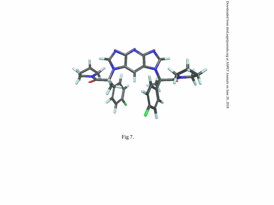
Fig 7.
This article has not been copyedited and formatted. The final version may differ from this version.DMD Fast Forward. Published on February 23, 2011 as DOI: 10.1124/dmd.110.037598
at ASPE
T Journals on June 20, 2018
dmd.aspetjournals.org
Dow
nloaded from

N
N
O
NNH
F
O
NH
O
NH2
N
N
O
F
N
N
O
F
N NN
N
N
O
NNH
2
F
N
N
O
F
N
N
O
F
N NN
NH2
H
N
N
O
F
N
N
O
F
N NN
CHO
H
N
N
O
F
N
N
O
F
N NN
OH
N
N
O
F
N
N
O
F
N NN
H
CH
O
N
N
O
F
NNH
H
THF
NH3
H2O
N10 - THF
LY654322
M21 2
34
M25
*
***
Fig 8.
This article has not been copyedited and formatted. The final version may differ from this version.DMD Fast Forward. Published on February 23, 2011 as DOI: 10.1124/dmd.110.037598
at ASPE
T Journals on June 20, 2018
dmd.aspetjournals.org
Dow
nloaded from