Met receptor signaling is required for sensory nerve development ...
11
Met receptor signaling is required for sensory nerve development and HGF promotes axonal growth and survival of sensory neurons Flavio Maina, 1 Mark C. Hilton, 2 Carola Ponzetto, 3 Alun M. Davies, 2 and Ru ¨ diger Klein 1,4 1 Cell Regulation Programme, European Molecular Biology Laboratory, 69117 Heidelberg, Germany; 2 School of Biological and Medical Sciences, Bute Medical Buildings, University of St. Andrews, St. Andrews, Fife KY16 9TS, Scotland, UK; 3 Department of Biomedical Sciences and Oncology, University of Torino, 10126 Torino, Italy The development of the nervous system is a dynamic process during which factors act in an instructive fashion to direct the differentiation and survival of neurons, and to induce axonal outgrowth, guidance to, and terminal branching within the target tissue. Here we report that mice expressing signaling mutants of the hepatocyte growth factor (HGF) receptor, the Met tyrosine kinase, show a striking reduction of sensory nerves innervating the skin of the limbs and thorax, implicating the HGF/Met system in sensory neuron development. Using in vitro assays, we find that HGF cooperates with nerve growth factor (NGF) to enhance axonal outgrowth from cultured dorsal root ganglion (DRG) neurons. HGF also enhances the neurotrophic activities of NGF in vitro, and Met receptor signaling is required for the survival of a proportion of DRG neurons in vivo. This synergism is specific for NGF but not for the related neurotrophins BDNF and NT3. By using a mild signaling mutant of Met, we have demonstrated previously that Met requires signaling via the adapter molecule Grb2 to induce proliferation of myoblasts. In contrast, the actions of HGF on sensory neurons are mediated by Met effectors distinct from Grb2. Our findings demonstrate a requirement for Met signaling in neurons during development. [Key Words: Hepatocyte growth factor; nerve growth factor; Met; axonal growth; neuronal survival; signal transduction] Received May 23, 1997; revised version accepted October 3, 1997. The development of neurons in the peripheral nervous system (PNS) is a complex, yet highly stereotyped pro- cess that is controlled in part by external cues from other cells. Such cues include (1) factors that regulate the pro- liferation and differentiation of neuronal precursor cells, (2) factors that promote axonal outgrowth and guide axons to their targets, (3) survival factors that match the number of neurons to their targets, and (4) local cues that refine initial connections by regulating terminal branch- ing and synaptogenesis (Goodman and Shatz 1993; Dav- ies 1994; Kennedy and Tessier-Lavigne 1995; Henderson 1996; Lewin and Barde 1996). Although much progress has been made in the identification of molecules with the above activities, in many cases it remains to be de- termined which of these molecules are actually required for PNS development. Moreover, many of these factors have multiple effects not only on neurons but also on neighboring cells, making it difficult to distinguish be- tween indirect target-mediated effects and direct effects on neurons. One such pleiotropic factor is hepatocyte growth fac- tor (HGF) which, besides promoting cell survival and proliferation of a number of cell types, stimulates cell migration and dissociation of epithelial sheets (Gherardi and Stoker 1991; Brinkmann et al. 1995). Through gene targeting experiments in mice, it was shown that HGF and its receptor, the Met tyrosine kinase, are required for the development of placenta, liver, and muscle of the limbs and trunk (Bladt et al. 1995; Schmidt et al. 1995; Uehara et al. 1995; Maina et al. 1996). Met expression has also been observed in various structures of the em- bryonic and postnatal nervous system, including spinal cord motor neurons and migrating neural crest cells (Sonnenberg et al. 1993; Jung et al. 1994; Andermarcher et al. 1996). Recently, HGF was shown to be a chemoat- tractant and survival factor for spinal motor neurons in vitro (Ebens et al. 1996). In addition, hgf-/- mice showed 4 Corresponding author. E-MAIL [email protected]; FAX 49 6221 387516. GENES & DEVELOPMENT 11:3341–3350 © 1997 by Cold Spring Harbor Laboratory Press ISSN 0890-9369/97 $5.00; www.genesdev.org 3341 Cold Spring Harbor Laboratory Press on April 2, 2018 - Published by genesdev.cshlp.org Downloaded from
Transcript of Met receptor signaling is required for sensory nerve development ...
Met receptor signaling is requiredfor sensory nerve development andHGF promotes axonal growth andsurvival of sensory neuronsFlavio Maina,1 Mark C. Hilton,2 Carola Ponzetto,3 Alun M. Davies,2 and Rudiger Klein1,4
1Cell Regulation Programme, European Molecular Biology Laboratory, 69117 Heidelberg, Germany; 2School of Biological andMedical Sciences, Bute Medical Buildings, University of St. Andrews, St. Andrews, Fife KY16 9TS, Scotland, UK;3Department of Biomedical Sciences and Oncology, University of Torino, 10126 Torino, Italy
The development of the nervous system is a dynamic process during which factors act in an instructivefashion to direct the differentiation and survival of neurons, and to induce axonal outgrowth, guidance to, andterminal branching within the target tissue. Here we report that mice expressing signaling mutants of thehepatocyte growth factor (HGF) receptor, the Met tyrosine kinase, show a striking reduction of sensory nervesinnervating the skin of the limbs and thorax, implicating the HGF/Met system in sensory neurondevelopment. Using in vitro assays, we find that HGF cooperates with nerve growth factor (NGF) to enhanceaxonal outgrowth from cultured dorsal root ganglion (DRG) neurons. HGF also enhances the neurotrophicactivities of NGF in vitro, and Met receptor signaling is required for the survival of a proportion of DRGneurons in vivo. This synergism is specific for NGF but not for the related neurotrophins BDNF and NT3. Byusing a mild signaling mutant of Met, we have demonstrated previously that Met requires signaling via theadapter molecule Grb2 to induce proliferation of myoblasts. In contrast, the actions of HGF on sensoryneurons are mediated by Met effectors distinct from Grb2. Our findings demonstrate a requirement for Metsignaling in neurons during development.
[Key Words: Hepatocyte growth factor; nerve growth factor; Met; axonal growth; neuronal survival; signaltransduction]
Received May 23, 1997; revised version accepted October 3, 1997.
The development of neurons in the peripheral nervoussystem (PNS) is a complex, yet highly stereotyped pro-cess that is controlled in part by external cues from othercells. Such cues include (1) factors that regulate the pro-liferation and differentiation of neuronal precursor cells,(2) factors that promote axonal outgrowth and guideaxons to their targets, (3) survival factors that match thenumber of neurons to their targets, and (4) local cues thatrefine initial connections by regulating terminal branch-ing and synaptogenesis (Goodman and Shatz 1993; Dav-ies 1994; Kennedy and Tessier-Lavigne 1995; Henderson1996; Lewin and Barde 1996). Although much progresshas been made in the identification of molecules withthe above activities, in many cases it remains to be de-termined which of these molecules are actually requiredfor PNS development. Moreover, many of these factorshave multiple effects not only on neurons but also on
neighboring cells, making it difficult to distinguish be-tween indirect target-mediated effects and direct effectson neurons.
One such pleiotropic factor is hepatocyte growth fac-tor (HGF) which, besides promoting cell survival andproliferation of a number of cell types, stimulates cellmigration and dissociation of epithelial sheets (Gherardiand Stoker 1991; Brinkmann et al. 1995). Through genetargeting experiments in mice, it was shown that HGFand its receptor, the Met tyrosine kinase, are required forthe development of placenta, liver, and muscle of thelimbs and trunk (Bladt et al. 1995; Schmidt et al. 1995;Uehara et al. 1995; Maina et al. 1996). Met expressionhas also been observed in various structures of the em-bryonic and postnatal nervous system, including spinalcord motor neurons and migrating neural crest cells(Sonnenberg et al. 1993; Jung et al. 1994; Andermarcheret al. 1996). Recently, HGF was shown to be a chemoat-tractant and survival factor for spinal motor neurons invitro (Ebens et al. 1996). In addition, hgf−/− mice showed
4Corresponding author.E-MAIL [email protected]; FAX 49 6221 387516.
GENES & DEVELOPMENT 11:3341–3350 © 1997 by Cold Spring Harbor Laboratory Press ISSN 0890-9369/97 $5.00; www.genesdev.org 3341
Cold Spring Harbor Laboratory Press on April 2, 2018 - Published by genesdev.cshlp.orgDownloaded from
marked defects in limb nerve branching. However, be-cause motor neurons depend on extrinsic factors pro-vided by muscle cells and because the HGF/Met systemis required for muscle development, this study did notprovide conclusive evidence for a requirement of Metsignaling in motor neurons.
To study target-independent functions of HGF, wehave focused on the development of sensory neuronsthat innervate the skin of the limbs and thorax, as incontrast to motor neurons, their projections are not in-fluenced by cues on muscle cells (Lewis et al. 1981). Herewe show that mice carrying a signaling mutant of Met(metD/D) have intercostal nerves that are much reducedin length and elaborate fewer terminal branches, demon-strating a requirement for HGF/Met signaling in sensoryneuron development. Using in vitro assays, we find thatHGF cooperates with nerve growth factor (NGF) in en-hancing the axonal growth rate of dorsal root ganglion(DRG) sensory neurons. In addition, HGF enhances theneurotrophic activities of NGF in vitro, and Met receptorsignaling is required for the survival of a proportion ofDRG neurons in vivo.
Previous work on Met receptor signaling has revealedthat the biological activity of Met depends on the pres-ence of two phosphotyrosine residues in the carboxy-terminal tail, which act as multifunctional docking sitesfor SH2-containing effectors and activate an array oftransductional pathways (Ponzetto et al. 1994). Mutationof both tyrosine residues in the mouse genome (metD/D)is lethal and causes defects identical to the phenotype of
met null mutants (Maina et al. 1996). In contrast, a mildmutation of Met (metGrb2/Grb2), in which the directbinding of Grb2 is uncoupled (Ponzetto et al. 1996), re-vealed an additional function of Met during myogenesis(Maina et al. 1996). Met signaling via Grb2 is required formyoblast proliferation but not for myoblast migration orfor survival of hepatocytes. In this report we have shownthat in contrast to its myogenic function, Met signalingvia Grb2 is not required for sensory neuron development.
Results
Limb motor innervation defects correlatewith reduction of muscle in met mutant embryos
To investigate whether Met signaling is required for thedevelopment of the PNS, we examined wild-type andmutant embryos in which met was replaced with loss-of-function versions of the receptor impaired in signaltransduction (metGrb2/Grb2, metD/D). The most severe ofthese mutants (metD/D), like homozygous Splotch mice(Franz et al. 1993) [in which pax-3, an upstream regulatorof met (Epstein et al. 1996), is mutated], specifically lackmuscles derived from migratory precursors, such as limband shoulder muscles (Maina et al. 1996). Whole-mountimmunostaining with anti-neurofilament antibodiesshowed that, at embryonic day 12.5 (E12.5) in metD/D
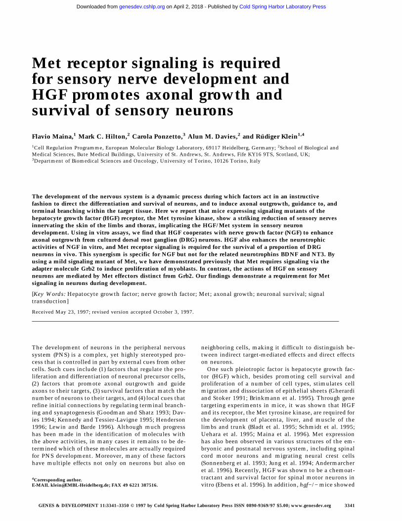
embryos, the brachial plexus in the forelimb is appropri-ately patterned, but all nerves emanating from it areshortened dramatically and lack branching (Fig. 1C,F).
Figure 1. Abnormal limb innervationin mice carrying mutant alleles of theMet receptor gene. (A–C) Whole-mount,anti-neurofilament-stained E12.5 mouseembryos (forelimb and thorax region,dorsal up, anterior left) carrying eitherthe wild-type knock-in allele of met(metWT/WT) (A,D,G), a mild signaling mu-tant (metGrb2/Grb2) (B,E,H), or a severe sig-naling mutant (metD/D) (C,F,I). (D–F)Drawings of limb nerves. Note the severelyreduced length and lack of terminal branch-ing of all limb nerves in the metD/D mutantembryo (F). In metGrb2/Grb2 mutants, thenervus radialis is severely affected, whichcould explain the observed deficiency inforelimb extension in metGrb2/Grb2 new-born mice (Maina et al. 1996). (G–I Fore-limbs of whole-mount, anti-neurofila-ment-stained E12.5 mouse embryos beforeclearing, showing the superficial innerva-tion of the skin. The defects are fully pen-etrant with six out of six embryos affected.Some metD/D mutant embryos also showmisdirection of spinal nerves that inner-vate the hindlimb (data not shown). (N.ax.)nervus axillaris; (N. med.) nervus medi-anus; (N.mu) nervus musculocutaneus;(N.ra.) nervus radialis; (N.th.) nervus tho-racodorsalis; (N.ul.) nervus ulnaris. Scalebar in A–C, 1.1 mm; in (G–I) 0.6 mm.
Maina et al.
3342 GENES & DEVELOPMENT
Cold Spring Harbor Laboratory Press on April 2, 2018 - Published by genesdev.cshlp.orgDownloaded from
For example, all branches of the nervus thoracodorsalis,which contains only motor fibers that innervate themusculus latissimus dorsii (lost in these mutants), arecompletely absent in metD/D embryos. Mutant embryoscarrying the milder metGrb2/Grb2 allele, where the samemuscle is reduced but not absent, showed that branchingof the nervus thoracodorsalis is indistinguishable fromthat of metWT/WT control embryos (Fig. 1A,B,D,E). InmetGrb2/Grb2 mutants, only the most anterior limbnerves (nervus radialis and musculocutaneous), inner-vating an area of the limb where the reduction ofmuscles is more pronounced (Maina et al. 1996), are af-fected (Fig. 1B,E). This innervation defect correlates withthe reduction in muscle mass and could be secondary tothe lack of muscles.
Reduced outgrowth and branching of sensory nervesin the limbs and thorax of met mutant mice
In contrast to motor nerves, the development of sensorynerve fibers in the limb is not dependent on the presenceof limb muscles (Lewis et al. 1981), The superficial net-work of sensory nerves in the skin is reduced drastically
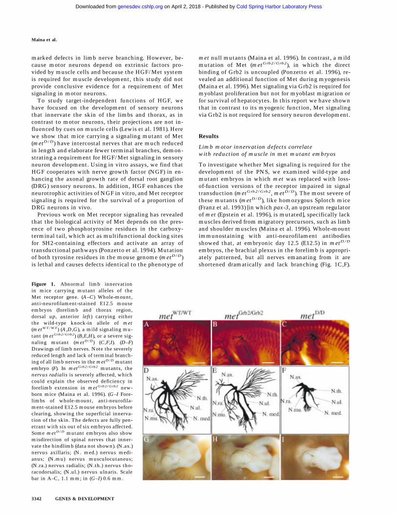
in metD/D mutants, both in the limbs (Fig. 1I) and thetrunk region. Interestingly, the branches of superficialskin nerves appear to be normal in metGrb2/Grb2 mutants(Fig. 1H). To better define the effects of Met signalingindependent of its myogenic role in neurons in the limb,we performed a detailed analysis of the peripheral nervesinnervating the trunk region. As shown in Figure 2, thedevelopment of thoracic spinal nerves is severely af-fected in E12.5 metD/D mutant embryos compared tometWT/WT control embryos. Both the dorsal and ventralarms of the lateral cutaneous branches, which innervatethe skin and muscle of the thorax, are severely com-pressed with much fewer ramifications than controls(Fig. 2A,C,D,F). The defect is most prominent in the su-perficial ventral arms, which sprout significantly fewerside branches, and are reduced in length by an average of>60% compared to metWT/WT embryos or heterozygouslittermates (Fig. 2G–I; data not shown). Similar analysison E13.5 mutant embryos revealed the same defects, sug-gesting that the lack of nerve branching was not causedby a general delay in embryonic development (data notshown). The overall patterning of the spinal nerve net-work, however, appears to be preserved in metD/D mu-
Figure 2. Defects in spinal nerve out-growth and branching in met mutant mice.(A–C) Anti-neurofilament-stained ventralbranches of thoracic spinal nerves (dorsaldown, ventral up). Note the reduction inlength and numbers, and the compressedappearance of side branches in the metD/D
mutant embryos (C). (D–F) Representativedrawings of thoracic DRG and spinalnerves. Only dorsal rami and lateral cuta-neous branches are shown; the distal end ofthe intercostal nerve does not appear to beaffected and is omitted for clarity. (G–I)Quantitative analysis of the defects in spi-nal nerve development. (E) The parametersthat were used for quantification. (G) Thenumber of side branches (indicated in redin D–F) were determined between the ori-gin of the lateral cutaneous branch and thepoint where the main axon bundle dividesinto two or three equally sized terminalprojections. (H) Length of the most proxi-mal part of the peripheral axon bundlemeasured from the exit points of the cen-tral processes of the DRG to the origin ofthe lateral cutaneous branch. (I) Length ofthe most distal arms of the lateral cutane-ous branch. Note that the length of theproximal part of the spinal nerve does notchange in metD/D mutant embryos. In con-trast, there is a strong reduction in lengthof the most distal arms of the lateral cuta-neous branch in metD/D mutant embryoscompared to the controls (statistical analy-
sis was carried out using Student’s t-test; P < 0.0001). Similar results were obtained using an antibody against neuron-specific tubulin(data not shown). No significant differences in the number of ramifications or length of spinal nerves were observed in metGrb2/Grb2
embryos compared to metWT/WT. Four peripheral axon bundles, from three different mutant mice, for each genotype, were analyzed.Scale bar in A–C, 0.3 mm.
Met signaling in sensory neuron development
GENES & DEVELOPMENT 3343
Cold Spring Harbor Laboratory Press on April 2, 2018 - Published by genesdev.cshlp.orgDownloaded from
tant embryos, suggesting a role for Met signaling in ter-minal nerve growth and branching, rather than in initialaxonal guidance. In contrast, metGrb2/Grb2 mutant em-bryos are indistinguishable from their wild-type litter-mates (Fig. 2B,E,G–I), indicating that Grb2 signalingdownstream of Met is not required for peripheral nervegrowth and branching.
HGF cooperates with NGF to enhance axonaloutgrowth from cultured DRG neurons
To investigate whether HGF-induced Met signaling di-rectly affects the growth of axons from wild-type DRGneurons, we studied the effect of HGF alone and in com-bination with other neuritogenic factors, such as NGF,brain-derived neurotrophic factor (BDNF), and neuro-
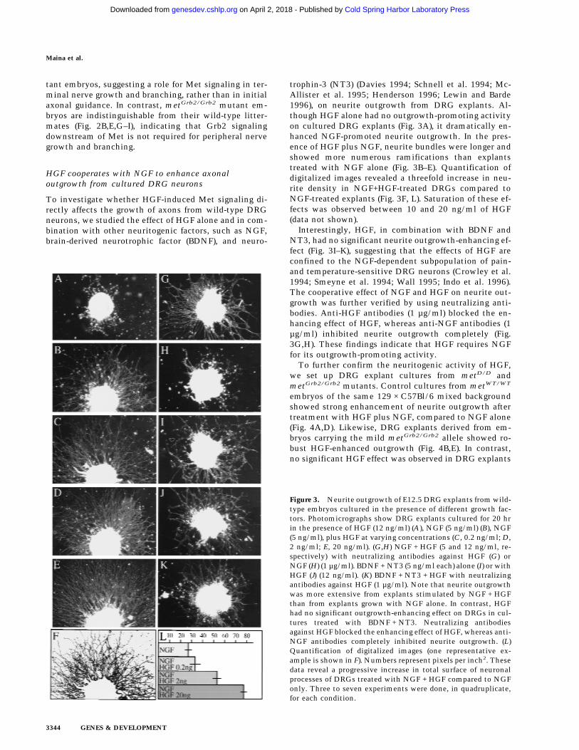
trophin-3 (NT3) (Davies 1994; Schnell et al. 1994; Mc-Allister et al. 1995; Henderson 1996; Lewin and Barde1996), on neurite outgrowth from DRG explants. Al-though HGF alone had no outgrowth-promoting activityon cultured DRG explants (Fig. 3A), it dramatically en-hanced NGF-promoted neurite outgrowth. In the pres-ence of HGF plus NGF, neurite bundles were longer andshowed more numerous ramifications than explantstreated with NGF alone (Fig. 3B–E). Quantification ofdigitalized images revealed a threefold increase in neu-rite density in NGF+HGF-treated DRGs compared toNGF-treated explants (Fig. 3F, L). Saturation of these ef-fects was observed between 10 and 20 ng/ml of HGF(data not shown).
Interestingly, HGF, in combination with BDNF andNT3, had no significant neurite outgrowth-enhancing ef-fect (Fig. 3I–K), suggesting that the effects of HGF areconfined to the NGF-dependent subpopulation of pain-and temperature-sensitive DRG neurons (Crowley et al.1994; Smeyne et al. 1994; Wall 1995; Indo et al. 1996).The cooperative effect of NGF and HGF on neurite out-growth was further verified by using neutralizing anti-bodies. Anti-HGF antibodies (1 µg/ml) blocked the en-hancing effect of HGF, whereas anti-NGF antibodies (1µg/ml) inhibited neurite outgrowth completely (Fig.3G,H). These findings indicate that HGF requires NGFfor its outgrowth-promoting activity.
To further confirm the neuritogenic activity of HGF,we set up DRG explant cultures from metD/D andmetGrb2/Grb2 mutants. Control cultures from metWT/WT
embryos of the same 129 × C57Bl/6 mixed backgroundshowed strong enhancement of neurite outgrowth aftertreatment with HGF plus NGF, compared to NGF alone(Fig. 4A,D). Likewise, DRG explants derived from em-bryos carrying the mild metGrb2/Grb2 allele showed ro-bust HGF-enhanced outgrowth (Fig. 4B,E). In contrast,no significant HGF effect was observed in DRG explants
Figure 3. Neurite outgrowth of E12.5 DRG explants from wild-type embryos cultured in the presence of different growth fac-tors. Photomicrographs show DRG explants cultured for 20 hrin the presence of HGF (12 ng/ml) (A), NGF (5 ng/ml) (B), NGF(5 ng/ml), plus HGF at varying concentrations (C, 0.2 ng/ml; D,2 ng/ml; E, 20 ng/ml). (G,H) NGF + HGF (5 and 12 ng/ml, re-spectively) with neutralizing antibodies against HGF (G) orNGF (H) (1 µg/ml). BDNF + NT3 (5 ng/ml each) alone (I) or withHGF (J) (12 ng/ml). (K) BDNF + NT3 + HGF with neutralizingantibodies against HGF (1 µg/ml). Note that neurite outgrowthwas more extensive from explants stimulated by NGF + HGFthan from explants grown with NGF alone. In contrast, HGFhad no significant outgrowth-enhancing effect on DRGs in cul-tures treated with BDNF + NT3. Neutralizing antibodiesagainst HGF blocked the enhancing effect of HGF, whereas anti-NGF antibodies completely inhibited neurite outgrowth. (L)Quantification of digitalized images (one representative ex-ample is shown in F). Numbers represent pixels per inch2. Thesedata reveal a progressive increase in total surface of neuronalprocesses of DRGs treated with NGF + HGF compared to NGFonly. Three to seven experiments were done, in quadruplicate,for each condition.
Maina et al.
3344 GENES & DEVELOPMENT
Cold Spring Harbor Laboratory Press on April 2, 2018 - Published by genesdev.cshlp.orgDownloaded from
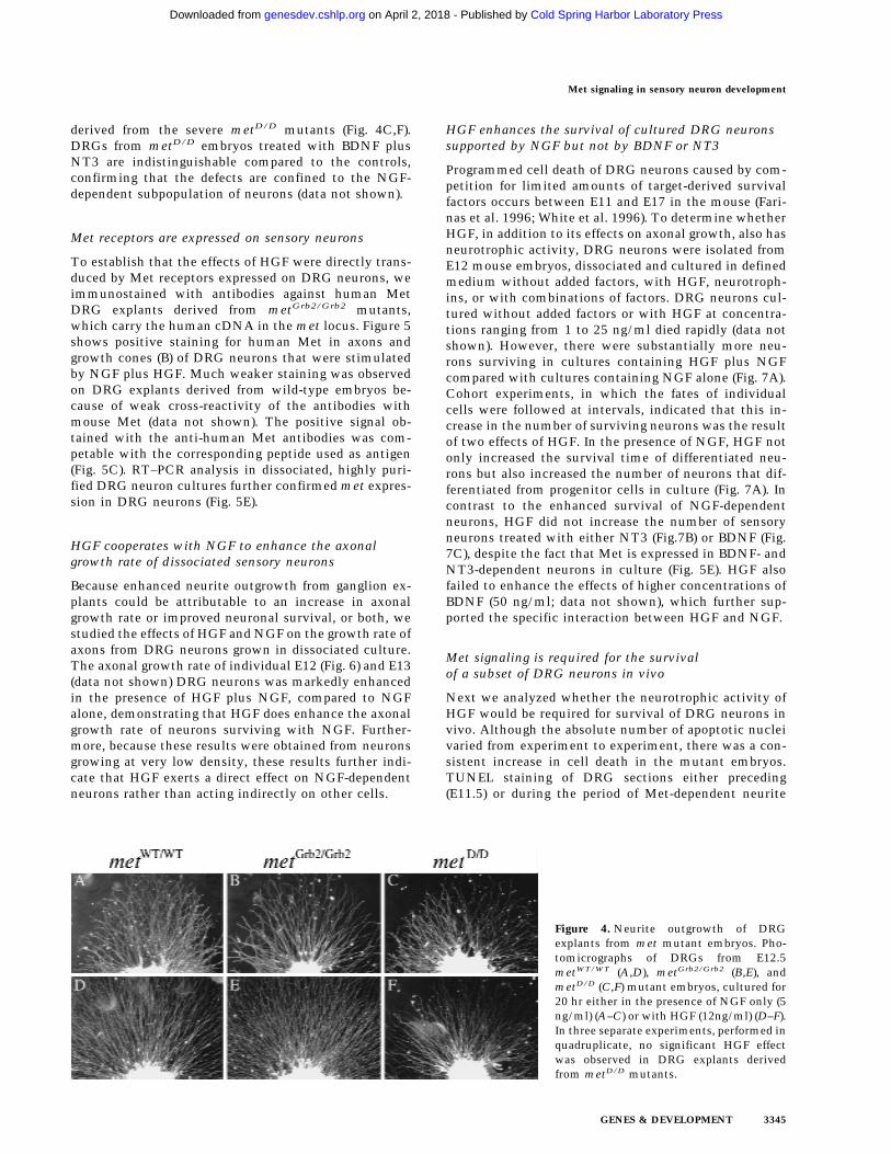
derived from the severe metD/D mutants (Fig. 4C,F).DRGs from metD/D embryos treated with BDNF plusNT3 are indistinguishable compared to the controls,confirming that the defects are confined to the NGF-dependent subpopulation of neurons (data not shown).
Met receptors are expressed on sensory neurons
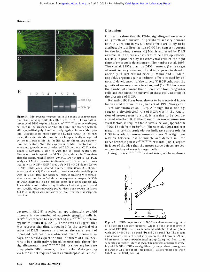
To establish that the effects of HGF were directly trans-duced by Met receptors expressed on DRG neurons, weimmunostained with antibodies against human MetDRG explants derived from metGrb2/Grb2 mutants,which carry the human cDNA in the met locus. Figure 5shows positive staining for human Met in axons andgrowth cones (B) of DRG neurons that were stimulatedby NGF plus HGF. Much weaker staining was observedon DRG explants derived from wild-type embryos be-cause of weak cross-reactivity of the antibodies withmouse Met (data not shown). The positive signal ob-tained with the anti-human Met antibodies was com-petable with the corresponding peptide used as antigen(Fig. 5C). RT–PCR analysis in dissociated, highly puri-fied DRG neuron cultures further confirmed met expres-sion in DRG neurons (Fig. 5E).
HGF cooperates with NGF to enhance the axonalgrowth rate of dissociated sensory neurons
Because enhanced neurite outgrowth from ganglion ex-plants could be attributable to an increase in axonalgrowth rate or improved neuronal survival, or both, westudied the effects of HGF and NGF on the growth rate ofaxons from DRG neurons grown in dissociated culture.The axonal growth rate of individual E12 (Fig. 6) and E13(data not shown) DRG neurons was markedly enhancedin the presence of HGF plus NGF, compared to NGFalone, demonstrating that HGF does enhance the axonalgrowth rate of neurons surviving with NGF. Further-more, because these results were obtained from neuronsgrowing at very low density, these results further indi-cate that HGF exerts a direct effect on NGF-dependentneurons rather than acting indirectly on other cells.
HGF enhances the survival of cultured DRG neuronssupported by NGF but not by BDNF or NT3
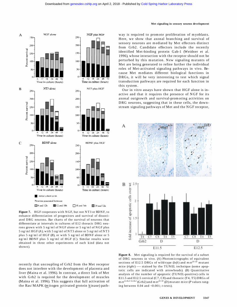
Programmed cell death of DRG neurons caused by com-petition for limited amounts of target-derived survivalfactors occurs between E11 and E17 in the mouse (Fari-nas et al. 1996; White et al. 1996). To determine whetherHGF, in addition to its effects on axonal growth, also hasneurotrophic activity, DRG neurons were isolated fromE12 mouse embryos, dissociated and cultured in definedmedium without added factors, with HGF, neurotroph-ins, or with combinations of factors. DRG neurons cul-tured without added factors or with HGF at concentra-tions ranging from 1 to 25 ng/ml died rapidly (data notshown). However, there were substantially more neu-rons surviving in cultures containing HGF plus NGFcompared with cultures containing NGF alone (Fig. 7A).Cohort experiments, in which the fates of individualcells were followed at intervals, indicated that this in-crease in the number of surviving neurons was the resultof two effects of HGF. In the presence of NGF, HGF notonly increased the survival time of differentiated neu-rons but also increased the number of neurons that dif-ferentiated from progenitor cells in culture (Fig. 7A). Incontrast to the enhanced survival of NGF-dependentneurons, HGF did not increase the number of sensoryneurons treated with either NT3 (Fig.7B) or BDNF (Fig.7C), despite the fact that Met is expressed in BDNF- andNT3-dependent neurons in culture (Fig. 5E). HGF alsofailed to enhance the effects of higher concentrations ofBDNF (50 ng/ml; data not shown), which further sup-ported the specific interaction between HGF and NGF.
Met signaling is required for the survivalof a subset of DRG neurons in vivo
Next we analyzed whether the neurotrophic activity ofHGF would be required for survival of DRG neurons invivo. Although the absolute number of apoptotic nucleivaried from experiment to experiment, there was a con-sistent increase in cell death in the mutant embryos.TUNEL staining of DRG sections either preceding(E11.5) or during the period of Met-dependent neurite
Figure 4. Neurite outgrowth of DRGexplants from met mutant embryos. Pho-tomicrographs of DRGs from E12.5metWT/WT (A,D), metGrb2/Grb2 (B,E), andmetD/D (C,F) mutant embryos, cultured for20 hr either in the presence of NGF only (5ng/ml) (A–C) or with HGF (12ng/ml) (D–F).In three separate experiments, performed inquadruplicate, no significant HGF effectwas observed in DRG explants derivedfrom metD/D mutants.
Met signaling in sensory neuron development
GENES & DEVELOPMENT 3345
Cold Spring Harbor Laboratory Press on April 2, 2018 - Published by genesdev.cshlp.orgDownloaded from
outgrowth (E12.5) revealed an approximately twofoldincrease in the number of apoptotic ganglion cells inmetD/D, compared to age-matched metWT/WT or hetero-zygous mutants (Fig. 8A,B). These results suggest thatMet receptor signaling is required for the survival of asubset of DRG neurons in vivo. As the same levels ofincreased cell death are observed over 2 consecutivedays, one would expect the final numbers of DRG neu-rons to be significantly reduced. Interestingly, the mildersignaling mutant metGrb2/Grb2 did not show any increasein apoptotic DRG neurons, indicating that Met signalingvia Grb2 is not required for its neurotrophic activities.
Discussion
Our results show that HGF/Met signaling enhances axo-nal growth and survival of peripheral sensory neuronsboth in vitro and in vivo. These effects are likely to beattributable to a direct action of HGF on sensory neuronsfor the following reasons: (1) Met is expressed by DRGneurons at the time met mutant mice develop deficits;(2) HGF is produced by mesenchymal cells at the righttime of embryonic development (Sonnenberg et al. 1993;Thery et al. 1995) to act on DRG neurons; (3) the targetof most sensory neurons, the skin, appears to developnormally in met mutant mice (F. Maina and R. Klein,unpubl.), arguing against indirect effects caused by ab-normal development of the target; (4) HGF enhances thegrowth of sensory axons in vitro; and (5) HGF increasesthe number of neurons that differentiate from progenitorcells and enhances the survival of these early neurons inthe presence of NGF.
Recently, HGF has been shown to be a survival factorfor cultured motoneurons (Ebens et al. 1996; Wong et al.1997; Yamamoto et al. 1997). Although these findingssuggest a physiological role of HGF/Met in the regula-tion of motoneuron survival, it remains to be demon-strated whether HGF, like many other motoneuron sur-vival factors, is required for in vivo survival (Oppenheim1996). The defects in hgf−/− (Ebens et al. 1996) and metmutant mice (this study) do not indicate a direct role forHGF in regulating motoneuron numbers. The tight cor-relation between loss of muscle and defects in limbnerve branching in metGrb2/Grb2 mutants (Fig. 1) arguesin favor of the idea that the motor nerve defects are sec-ondary to loss of muscle target cells.
Using the metGrb2/Grb2 mutant mice, we have shown
Figure 5. Met receptor expression in the axons of sensory neu-rons stimulated by NGF plus HGF in vitro. (A,B) Immunofluo-rescence of DRG explants from metGrb2/Grb2 mutant embryos,cultured in the presence of NGF plus HGF and stained with anaffinity-purified polyclonal antibody against human Met pro-tein. Because these mice carry the human cDNA in the metlocus, the chimeric Met protein can be specifically recognizedby the anti-human Met antibodies against the unique carboxy-terminal peptide. Note the expression of Met receptors in theaxons and growth cones of cultured DRG neurons. (C) The Metsignal is completely blocked with the antigenic peptide. (D)Phase-contrast image of the DRG explant, shown in C, to visu-alize the axons. Magnification: 20× (A,C,D); 40× (B). (E) RT–PCRanalysis of Met expression in dissociated DRG neuron culturestreated with NGF + HGF (lanes 1,5), NT3 + HGF (lanes 2,6) orBDNF + HGF (lanes 3,7) and in intact DRGs (lanes 4,8; shorterexposure of lane 8). Dissociated cultures were substantially purewith only 5%–10% non-neuronal cells, indicating Met expres-sion in neurons. Lanes 5–8 show the expected met-specific 550-bp DNA fragment in an ethidium bromide-stained agarose gel.These data were confirmed by Southern blot using an internalmet-specific oligonucleotide probe (data not shown). In lanes1–4 PCR analysis was performed without reverse transcriptionreaction.
Figure 6. HGF cooperates with NGF to enhance axonal growthof dissociated sensory neurons. Graph of the axonal growthrates of E12 DRG neurons incubated with NGF alone (s) orwith NGF + HGF at 5 ng/ml (d) and 25 ng/ml (m). The meansand standard errors of serial measurements of between 79 and89 neurons in each experimental group (compiled from threeseparate experiments) are shown. The neurites of neurons grow-ing with NGF + HGF were significantly longer than those grow-ing with NGF alone at all time points (P values ranging between0.023 and <0.0001; t-tests).
Maina et al.
3346 GENES & DEVELOPMENT
Cold Spring Harbor Laboratory Press on April 2, 2018 - Published by genesdev.cshlp.orgDownloaded from
recently that uncoupling of Grb2 from the Met receptordoes not interfere with the development of placenta andliver (Maina et al. 1996). In contrast, a direct link of Metwith Grb2 is required for the development of muscles(Maina et al. 1996). This suggests that full activation ofthe Ras/MAPK (mitogen activated protein kinase) path-
way is required to promote proliferation of myoblasts.Here, we show that axonal branching and survival ofsensory neurons are mediated by Met effectors distinctfrom Grb2. Candidate effectors include the recentlyidentified Met-binding protein Gab-1 (Weidner et al.1996), whose interaction with the receptor should not beperturbed by this mutation. New signaling mutants ofMet are being generated to refine further the individualroles of Met-activated signaling pathways in vivo. Be-cause Met mediates different biological functions inDRGs, it will be very interesting to test which signaltransduction pathways are required for each function inthis system.
Our in vitro assays have shown that HGF alone is in-active and that it requires the presence of NGF for itsaxonal outgrowth and survival-promoting activities onDRG neurons, suggesting that in these cells, the down-stream signaling pathways of Met and the NGF receptor,
Figure 7. HGF cooperates with NGF, but not NT3 or BDNF, toenhance differentiation of progenitors and survival of dissoci-ated DRG neurons. Bar charts of the survival of neurons thatdifferentiate at intervals in cultures of E12 thoracic DRG neu-rons grown with 5 ng/ml of NGF alone or 5 ng/ml of NGF plus5 ng/ml HGF (A), with 5 ng/ml of NT3 alone or 5 ng/ml of NT3plus 5 ng/ml of HGF (B), or with 5 ng/ml of BDNF alone or 5ng/ml BDNF plus 5 ng/ml of HGF (C). Similar results wereobtained in three other experiments of each kind (data notshown).
Figure 8. Met signaling is required for the survival of a subsetof DRG neurons in vivo. (A) Photomicrographs of equivalentsections of E12.5 DRGs of wild-type (left) and metD/D mutantmice (right) —- stained by the TUNEL technique (some apop-totic cells are indicated with arrowheads). (B) Quantitativeanalysis of the number of apoptotic (TUNEL-positive) cells inE11.5 and E12.5 cervical (C7, C8) and thoracic (T4, T5) DRGs ofmetGrb2/Grb2 (Grb2) and metD/D (D) mutant mice (P values rang-ing between 0.04 and <0.001; t-tests).
Met signaling in sensory neuron development
GENES & DEVELOPMENT 3347
Cold Spring Harbor Laboratory Press on April 2, 2018 - Published by genesdev.cshlp.orgDownloaded from

TrkA, converge. This is different from the situation inmotoneurons, where HGF alone is effective in promot-ing the survival of motor neurons in culture (Ebens et al.1996; Wong et al. 1997; Yamamoto et al. 1997). It re-mains to be determined whether or not HGF can actsynergistically with neurotrophins in this system. Inter-estingly, HGF is unable to enhance the activities of NGFon another population of sensory neurons located in thetrigeminal ganglion, indicating that effects of HGF arenot attributable to generalized increased fitness of sen-sory neurons (A. Forgie and A.M. Davies, unpubl.). Insupport of this observation, we have been unable to finddefects in outgrowth or branching of peripheral trigemi-nal nerves (F. Maina and R. Klein, unpubl.). Our resultsindicate the presence of cell type-specific effector(s) ofTrkA whose signaling is enhanced by activated Met re-ceptors, either by stabilizing signaling activities or byblocking a specific inhibitor of the TrkA pathway. So far,we have no evidence for possible interactions betweenMet and TrkB or TrkC, the receptors for BDNF and NT3,respectively (Klein 1994; Barbacid 1995).
NGF and its receptor TrkA are important for the sur-vival of pain and temperature-sensitive DRG neuronswhose peripheral axons largely terminate in the skin(Crowley et al. 1994; Smeyne et al. 1994; Wall 1995; Indoet al. 1996). The dual requirement observed in vitro ofHGF and NGF explains the lack of superficial nervebranches in the skin of metD/D mutant embryos. NGF iscurrently being developed as a therapeutic drug for thetreatment of peripheral sensory neuropathies, often ob-served in human patients suffering from diabetes or can-cer therapy (Barinaga 1994). Considering the strong en-hancement of NGF-induced sprouting by HGF, combi-nations of the two factors may prove to be even moreeffective (Nishi 1994).
Materials and methods
Whole-mount immunohistochemistry
Generation and initial phenotypic analysis of metWT/WT,metGrb2/Grb2, and metD/D knock-in mutant mice were previ-ously described (Maina et al., 1996). E12.5 and E13.5 embryoswere fixed overnight at 4°C in Dent’s fixative (1:4, DMSO/methanol), bleached in H2O2 (1:2, 30% H2O2/Dent’s fixative) atroom temperature, and incubated overnight with anti-NF160antibody (N-5264; Sigma) or anti-b-Tubulin Isotype III (T-8660;Sigma). After washing with TBS (10 mM Tris-HCl at pH 8.0, 100mM NaCl), embryos were incubated overnight with secondaryantibody (goat anti-mouse IgG-conjugated with horseradish per-oxidase, Fab fragment, A-3682; Sigma). After several washes inTBS, embryos were stained with diaminobenzidine and H2O2.After staining, the embryos were cleared by dehydrating inmethanol and incubated first in 50% BABB (1:2, benzyl alcohol/benzyl benzoate) and finally in 100% BABB.
In situ detection of apoptosis
Paraffin-embedded embryo sections (7 µm) were deparaffinized,incubated in H2O containing 0.3% H2O2 for 30 min, washed,and incubated with proteinase K (2 µg/ml) in PBS for 8 min atroom temperature. Apoptotic cells were detected as described in
the In Situ Cell Death Detection Kit, POD (Boehringer). Sec-tions were counterstained with methyl green.
RT–PCR and Southern blot
RNA was extracted from E12.5 DRGs and primary neuronstreated for 24 hr with NGF plus HGF, NT3 plus HGF or BDNFplus HGF, using the acid phenol–chloroform method (Chomc-zynski and Sacchi 1987), and subjected to reverse transcriptionwith oligo(dT)15 primers and PCR amplification. The met-spe-cific primers were as follows: 58-AGAGACCAGCAGCTTCAG-TTACCG-38 (sense primer corresponding to nucleotides 2189–2213 of mouse c-met cDNA) and 58-CCACTCTATATTTAG-CTCGCTGTTCAG-38 (antisense primer corresponding to nu-cleotides 2704–2730). A met-specific oligonucleotide (58-AG-AGTGCCAGTGGAGACTCTCGCAGCTCTG-38; antisenseprimer corresponding to nucleotides 2635–2684), internal to thePCR product, was labeled with [g-32]ATP using the T4 poly-nucleotide kinase and used as probe for the Southern blot.
Neurite outgrowth assays
DRGs from E12.5 wild-type CD1 or mutant 129 × C57B1/6 em-bryos were dissected and cultured in tissue culture dishescoated with poly-L-ornithine and laminin in Dulbecco’s modi-fied Eagle medium/F12 (DMEM/F12) (Puschel et al. 1996), withthe indicated growth factors. NGF and monoclonal anti-NGFantibodies were purchased from Boehringer Mannheim; BDNFand NT3 were generously provided by Regeneron Pharmaceuti-cals. Polyclonal anti-HGF antibodies were purchased from R&DSystems.
Quantification of outgrowth activity
The density of neurite outgrowth was analyzed by measure-ment and comparison of digitalized micrographs (800-bpi reso-lution) with the NIH-Image 1.6 program (developed at the U.S.National Institutes of Health and available on the Internet athttp:/ /rsb.info.nih.gov/nih-image/). Circular areas were se-lected in four different positions at two distances from the DRG(to obtain additional data on neurite length). The mean inten-sity was measured as the sum of intensities of all pixels in thearea divided by the total number of pixels per area. In addition,for each image analyzed, the mean intensity of areas (2–4) con-taining no neurites was measured and subtracted as back-ground.
Immunofluorescence staining
DRG explants were fixed in methanol, washed with PBS fol-lowed by PBS plus 0.3% Tween, before incubating with block-ing solution for 30 min and with primary antibody for 1 hr. Theantibodies used were anti-mouse–Met (polyclonal sc-162, SantaCruz), anti-human–Met (affinity-purified polyclonal sc-161,Santa Cruz) and affinity-purified fluorescein (FITC)–conjugatedgoat anti-rabbit (Jackson Immunoresearch Laboratories). Forneutralization, the antibodies against Met were incubated for 2hr at room temperature with a 10-fold excess of the correspond-ing peptide antigen.
Neuronal cultures
Thoracic DRG from E12 mouse embryos were trypsinized, dis-sociated, and plated at low density (500–2000 per dish) in serum-
Maina et al.
3348 GENES & DEVELOPMENT
Cold Spring Harbor Laboratory Press on April 2, 2018 - Published by genesdev.cshlp.orgDownloaded from
free medium in 60-mm petri dishes that had been coated withpoly-L-ornithine and laminin (Paul and Davies 1995). To studythe effects of neurotrophins and HGF on the differentiation andsurvival of neurons, the fate of individual cells was followed atintervals in these cultures (Paul and Davies 1995). The locationsof all neurons within a 144-mm2 grid were recorded 6 hr afterplating as were the locations of all neurons that subsequentlydifferentiated from progenitor cells at further time intervals inthe same grid. The fates of all of the neurons in each of thesecohorts (alive or dead) was followed at intervals up to 72 hr inculture. The number of surviving neurons in each cohort isexpressed as a percentage of the size of the initial cohort iden-tified at 6 hr. Neurotrophins and HGF were added to the cul-tures with the identification of the initial cohort at 6 hr. Tostudy the effect of NGF and HGF on axonal growth rate, accu-rate serial drawings of the same neurons were made with adrawing tube from which total neurite length was calculated asdescribed previously (Davies 1989). The length measurementsreported were not influenced by any differences in neuronalsurvival under the different experimental conditions used be-cause only neurons that survived the entire culture period (up to72 hr) were included in the analysis.
Acknowledgments
We thank R. Adams for help with DRG cultures, R. Saffrich forhelp with quantification of DRG neurite outgrowth, Genentechfor providing recombinant HGF, and Regeneron Pharmaceuti-cals for recombinant BDNF and NT3. Part of this work wassupported by a European Commission Biotechnology networkgrant (to R.K. and A.M.D.) and by Telethon-Italy (to C.P.).
The publication costs of this article were defrayed in part bypayment of page charges. This article must therefore be herebymarked ‘‘advertisement’’ in accordance with 18 USC section1734 solely to indicate this fact.
References
Andermarcher, E., M.A. Surani, and E. Gherardi. 1996. Co-ex-pression of the HGF/SF and c-met genes during early mouseembryogenesis precedes reciprocal expression in adjacenttissues during organogenesis. Dev. Genetics 18: 254–266.
Barbacid, M. 1995. Neurotrophic factors and their receptors.Curr. Opin. Cell Biol. 7: 148–155.
Barinaga, M. 1994. Neurotrophic factors enter the clinic. Sci-ence 264: 772–774.
Bladt, F., D. Riethmacher, S. Isenmann, A. Aguzzi, and C. Birch-meier. 1995. Essential role for the c-met receptor in the mi-gration of myogenic precursor cells into the limb bud. Na-ture 376: 768–771.
Brinkmann, V., H. Foroutan, M. Sachs, K.M. Weidner, and W.Birchmeier. 1995. Hepatocyte growth factor/scatter factorinduces a variety of tissue-specific morphogenic programs inepithelial cells. J. Cell Biol. 131: 1573–1586.
Chomczynski, P. and N. Sacchi. 1987. Single step method ofRNA isolation by acid guanidinium thiocyanate-phenol-chloroform extraction. Anal. Biochem. 162: 156–159.
Crowley, C., S.D. Spencer, M.C. Nishimura, K.S. Chen, S. Pitts-Meek, M.P. Armanini, L.H. Ling, S.B. McMahon, D.L.Shelton, A.D. Levinson, and H.S. Phillips. 1994. Mice lack-ing nerve growth factor display perinatal loss of sensory andsympathetic neurons yet develop basal forebrain cholinergicneurons. Cell 76: 1001–1012.
Davies, A.M. 1989. Intrinsic differences in the growth rate ofearly nerve fibres related to target distance. Nature 337: 553–555.
———. 1994. Role of neurotrophins in the developing nervoussystem. J. Neurobiol. 25: 1334–1348.
Ebens, A., K. Brose, E.D. Leonardo, M.G. Hanson, F. Bladt, C.Birchmeier, B.A. Barres, and M. Tessier-Lavigne. 1996. He-patocyte growth factor/Scatter factor is an axonal chemoat-tractant and a neurotrophic factor for spinal motor neurons.Neuron 17: 1157–1172.
Epstein, J.A., D.N. Shapiro, J. Cheng, P.Y.P. Lam, and R.L. Maas.1996. Pax3 modulates expression of the c-Met receptor dur-ing limb muscle development. Proc. Natl. Acad. Sci.93: 4213–4218.
Farinas, I., C.K. Yoshida, C. Backus, and L.F. Reichardt. 1996.Lack of neurotrophin-3 results in death of spinal sensoryneurons and premature differentiation of their precursors.Neuron 17: 1065–1078.
Franz, T., R. Kothary, M.A. Surani, Z. Halata, and M. Grim.1993. The Splotch mutation interferes with muscle develop-ment in the limbs. Anat. Embryol. 187: 153–160.
Gherardi, E. and M. Stoker. 1991. Hepatocyte growth factor-scatter factor: Mitogen, motogen, and met. Cancer Cells3: 227–232.
Goodman, C.S. and C.J. Shatz. 1993. Developmental mecha-nisms that generate precise patterns of neuronal connectiv-ity. Cell/Neuron (Suppl.) 72/10: 77–98.
Henderson, C.E. 1996. Role of neurotrophic factors in neuronaldevelopment. Curr. Opin. Neurobiol. 6: 64–70.
Indo, Y., M. Tsuruta, Y. Hayashida, M.A. Karim, K. Ohta, T.Kawano, H. Mitsubuchi, H. Tonoki, Y. Awaya, and I. Mat-suda. 1996. Mutations in the TRKA/NGF receptor gene inpatients with congenital insensitivity to pain with anhidro-sis. Nature Genet. 13: 485–488.
Jung, W., E. Castren, M. Odenthal, G.F. Vande Woude, T. Ishii,H.-P. Dienes, D. Lindholm, and P. Schirmacher. 1994. Ex-pression and functional interaction of hepatocyte growthfactor-scatter factor and its receptor c-met in mammalianbrain. J. Cell Biol. 126: 485–494.
Kennedy, T.E. and M. Tessier-Lavigne. 1995. Guidance and in-duction of branch formation in developing axons by target-derived diffusible factors. Curr. Opin. Neurobiol. 5: 83–90.
Klein, R. 1994. Role of neurotrophins in mouse neuronal devel-opment. FASEB J. 8: 738–744.
Lewin, G.R. and Y.-A. Barde. 1996. Physiology of the neuro-trophins. Annu. Rev. Neurosci. 19: 289–317.
Lewis, J., A. Chevallier, M. Kieny, and L. Wolpert. 1981. Musclenerve branches do not develop in chick wings devoid ofmuscle. J. Embryol. Exp. Morphol. 64: 211–232.
Maina, F., F. Casagranda, E. Audero, A. Simeone, P. Comoglio,R. Klein, and C. Ponzetto. 1996. Uncoupling of Grb2 fromthe Met receptor in vivo reveals complex roles in muscledevelopment. Cell 87: 531–542.
McAllister, A.K., D.C. Lo, and L.C. Katz. 1995. Neurotrophinsregulate dendritic growth in developing visual cortex. Neu-ron 15: 791–803.
Nishi, R. 1994. Neurotrophic factors: Two are better than one.Science 265: 1052–1053.
Oppenheim, R.W. 1996. Neurotrophic survival molecules formotoneurons: An embarrassment of riches. Neuron 17: 195–197.
Paul, G. and A.M. Davies. 1995. Sensory neurons require extrin-sic signals to switch neurotrophin dependence during theearly stages of target field innervation. Dev. Biol. 171: 590–605.
Ponzetto, C., A. Bardelli, Z. Zhen, F. Maina, P. dalla Zonca, S.Giordano, A. Graziani, G. Panayotou, and P. Comoglio.1994. A multifunctional docking site mediates signaling andtransformation by the hepatocyte growth factor/scatter fac-
Met signaling in sensory neuron development
GENES & DEVELOPMENT 3349
Cold Spring Harbor Laboratory Press on April 2, 2018 - Published by genesdev.cshlp.orgDownloaded from
tor receptor family. Cell 77: 261–271.Ponzetto, C., Z. Zhen, E. Audero, F. Maina, A. Bardelli, M.L.
Basile, S. Giordano, R. Narsimhan, and P.M. Comoglio.1996. Specific uncoupling of GRB2 from the Met receptor:Differential effect on transformation and motility. J. Biol.Chem. 271: 14119–14123.
Puschel, A.W., R.H. Adams, and H. Betz. 1996. The sensoryinnervation of the mouse spinal cord may be patterned bydifferential expression of and differential responsiveness tosemaphorins. Mol. Cell. Neurosci. 7: 419–431.
Schmidt, C., F. Bladt, S. Goedecke, V. Brinkmann, W. Zschle-sche, M. Sharpe, E. Gherardi, and C. Birchmeier. 1995. Scat-ter factor/hepatocyte growth factor is essential for liver de-velopment. Nature 373: 699–702.
Schnell, L., R. Schneider, R. Kolbeck, Y.A. Barde, and M.E.Schwab. 1994. Neurotrophin-3 enhances sprouting of corti-cospinal tract during development and after adult spinal cordlesion. Nature 367: 170–173.
Smeyne, R.J., R. Klein, A. Schnapp, L.K. Long, S. Bryant, A.Lewin, S.A. Lira, and M. Barbacid. 1994. Severe sensory andsympathetic neuropathies in mice carrying a disrupted Trk/NGF receptor gene. Nature 368: 246–249.
Sonnenberg, E., D. Meyer, K.M. Weidner, and C. Birchmeier.1993. Scatter factor/hepatocyte growth factor and its recep-tor, the c-met tyrosine kinase, can mediate a signal exchangebetween mesenchyme and epithelia during mouse develop-ment. J. Cell Biol. 123: 223–235.
Thery, C., M.J. Sharpe, S.J. Batley, C.D. Stern, and E. Gherardi.1995. Expression of HGF/SF, HGF1/MSP, and c-met sug-gests new functions during early chick development. Dev.Genet. 17: 90–101.
Uehara, Y., O. Minowa, C. Mori, K. Shiota, J. Kuno, T. Noda,and N. Kitamura. 1995. Placental defect and embryonic le-thality in mice lacking hepatocyte growth factor/scatter fac-tor. Nature 373: 702–705.
Wall, P.D. 1995. Independent mechanisms converge on pain.Nature Med. 1: 740–741.
Weidner, K.M., S. Di Cesare, M. Sachs, V. Brinkmann, J. Be-hrens, and W. Birchmeier. 1996. Interaction between Gab1and the c-Met receptor tyrosine kinase is responsible for ep-ithelial morphogenesis. Nature 384: 173–176.
White, F.A., I. Silos-Santiago, D.C. Molliver, M. Nishimura, H.Phillips, M. Barbacid, and W.D. Snider. 1996. Synchronousonset of NGF and TrkA survival dependence in developingdorsal root ganglia. J. Neurosci. 16: 4662–4672.
Wong, V., D.J. Glass, R. Arriaga, G.D. Yancopoulos, R.M. Lind-say, and G. Conn. 1997. Hepatocyte growth factor promotesmotor neuron survival and synergizes with ciliary neuro-trophic factor. J. Biol. Chem. 272: 5187–5191.
Yamamoto, Y., J. Livet, R. Vejsada, R.A. Pollock, V. Arce, O.deLapeyriere, A.C. Kato, and C.E. Henderson. 1997. Hepato-cyte growth factor (HGF/SF) is a muscle-derived survivalfactor for a subpopulation of embryonic motoneurons. De-velopment 124: 2903–2913.
Maina et al.
3350 GENES & DEVELOPMENT
Cold Spring Harbor Laboratory Press on April 2, 2018 - Published by genesdev.cshlp.orgDownloaded from

10.1101/gad.11.24.3341Access the most recent version at doi: 11:1997, Genes Dev.
Flavio Maina, Mark C. Hilton, Carola Ponzetto, et al. HGF promotes axonal growth and survival of sensory neuronsMet receptor signaling is required for sensory nerve development and
References
http://genesdev.cshlp.org/content/11/24/3341.full.html#ref-list-1
This article cites 40 articles, 11 of which can be accessed free at:
License
ServiceEmail Alerting
click here.right corner of the article or
Receive free email alerts when new articles cite this article - sign up in the box at the top
Cold Spring Harbor Laboratory Press
Cold Spring Harbor Laboratory Press on April 2, 2018 - Published by genesdev.cshlp.orgDownloaded from