Mesendoderm Extension and Mantle Closure in...
21
Mesendoderm Extension and Mantle Closure in Xenopus laevis Gastrulation: Combined Roles for Integrin a 5 b 1 , Fibronectin, and Tissue Geometry Lance A. Davidson,* Benjamin G. Hoffstrom,* Raymond Keller,² and Douglas W. DeSimone* ,1 *Department of Cell Biology, School of Medicine, University of Virginia Health System, Charlottesville, Virginia 22908; and ²Department of Biology, University of Virginia, Charlottesville, Virginia 22903 We describe mesendoderm morphogenesis during gastrulation in the frog Xenopus laevis and investigate the mechanics of these movements with tissue explants. When a dorsal marginal zone explant is plated onto fibronectin, the mesendoderm moves away from the dorsal axial tissues as an intact sheet. Mesendodermal cells within these explants display monopolar protrusive activity and radially intercalate during explant extension. Live time-lapse confocal sequences of actin dynamics at the margin of these extending explants prompt us to propose that integrin-mediated traction drives these movements. We demonstrate that integrin a 5 b 1 recognition of the synergy site located within the type III 9 repeat of fibronectin is required for mesendoderm extension. Normal mesendoderm morphogenesis occurs with a unique “cup-shaped” geometry of the extending mesendodermal mantle and coincides with a higher rate of tissue extension than that seen in the simpler dorsal marginal zone explant. These higher rates can be reconstituted with “in-the-round” configurations of several explants. We propose several mechanically based hypotheses to explain both the initial fibronectin-dependent extension of the mesendoderm and additional requirement of tissue geometry during the high-velocity closure of the mesendodermal mantle. © 2002 Elsevier Science (USA) Key Words: mesendoderm; integrin; fibronectin; cell migration; radial intercalation. INTRODUCTION During gastrulation in vertebrates, head mesoderm and ventral mesendoderm move great distances and come to lie between the ventral ectoderm and the ventral endoderm. Further elaboration of ventral organs, such as the heart, liver, kidney, and blood, depend on the success of these movements. Failure of mesendoderm morphogenesis re- sults in severe, and frequently lethal, embryonic pheno- types. In the frog Xenopus laevis, these movements also result in the enclosure of the blastocoel and the isolation of the blastocoel from the ventral ectoderm. Fate mapping and lineage analysis have identified where the mesendoderm originates, when it begins to move, and the tissues to which it contributes (Bauer et al., 1994; Keller and Tibbetts, 1989; Nakatsuji, 1975; Nakatsuji and Johnson, 1982; Nieuwkoop and Florschutz, 1950; Vodicka and Gerhart, 1995; Win- klbauer et al., 1996; Winklbauer and Schurfeld, 1999; Lane and Sheets, 2000) (see a summary in Fig. 1A, light orange mesendoderm; after Keller, 1991 and Keller and Tibbets, 1989). However, the mechanism driving vertebrate mesen- doderm morphogenesis and the cell behaviors responsible for these movements are poorly understood. One of the reasons for this is that, in frog, like most vertebrates, the cell behaviors are obscured by either the overlying endoderm or ectoderm, forcing the use of static cell lineage and fate mapping to identify the bulk movements. It is also difficult to distinguish local force generating mechanisms in the mesendoderm from those acting at a distance from other tissues since mesendoderm morphogenesis occurs at the same time as a number of other gastrulation move- ments, including vegetal endoderm “rotation” (Winklbauer and Schurfeld, 1999), dorsal mesoderm convergent exten- sion (Keller and Winklbauer, 1992), and ectoderm epiboly (Keller, 1978, 1980). In Xenopus, however, the local force 1 To whom correspondence should be addressed. Fax: (804) 982- 3912. E-mail: [email protected]. Developmental Biology 242, 109 –129 (2002) doi:10.1006/dbio.2002.0537, available online at http://www.idealibrary.com on 0012-1606/02 $35.00 © 2002 Elsevier Science (USA) All rights reserved. 109
Transcript of Mesendoderm Extension and Mantle Closure in...

Developmental Biology 242, 109–129 (2002)doi:10.1006/dbio.2002.0537, available online at http://www.idealibrary.com on
Mesendoderm Extension and Mantle Closure inXenopus laevis Gastrulation: Combined Roles forIntegrin a5b1, Fibronectin, and Tissue Geometry
Lance A. Davidson,* Benjamin G. Hoffstrom,* Raymond Keller,†and Douglas W. DeSimone* ,1
*Department of Cell Biology, School of Medicine, University of Virginia Health System,Charlottesville, Virginia 22908; and †Department of Biology, University of Virginia,Charlottesville, Virginia 22903
We describe mesendoderm morphogenesis during gastrulation in the frog Xenopus laevis and investigate the mechanics ofthese movements with tissue explants. When a dorsal marginal zone explant is plated onto fibronectin, the mesendodermmoves away from the dorsal axial tissues as an intact sheet. Mesendodermal cells within these explants display monopolarprotrusive activity and radially intercalate during explant extension. Live time-lapse confocal sequences of actin dynamicsat the margin of these extending explants prompt us to propose that integrin-mediated traction drives these movements. Wedemonstrate that integrin a5b1 recognition of the synergy site located within the type III9 repeat of fibronectin is requiredfor mesendoderm extension. Normal mesendoderm morphogenesis occurs with a unique “cup-shaped” geometry of theextending mesendodermal mantle and coincides with a higher rate of tissue extension than that seen in the simpler dorsalmarginal zone explant. These higher rates can be reconstituted with “in-the-round” configurations of several explants. Wepropose several mechanically based hypotheses to explain both the initial fibronectin-dependent extension of themesendoderm and additional requirement of tissue geometry during the high-velocity closure of the mesendodermalmantle. © 2002 Elsevier Science (USA)
Key Words: mesendoderm; integrin; fibronectin; cell migration; radial intercalation.
INTRODUCTION
During gastrulation in vertebrates, head mesoderm andventral mesendoderm move great distances and come to liebetween the ventral ectoderm and the ventral endoderm.Further elaboration of ventral organs, such as the heart,liver, kidney, and blood, depend on the success of thesemovements. Failure of mesendoderm morphogenesis re-sults in severe, and frequently lethal, embryonic pheno-types. In the frog Xenopus laevis, these movements alsoresult in the enclosure of the blastocoel and the isolation ofthe blastocoel from the ventral ectoderm. Fate mapping andlineage analysis have identified where the mesendodermoriginates, when it begins to move, and the tissues to whichit contributes (Bauer et al., 1994; Keller and Tibbetts, 1989;Nakatsuji, 1975; Nakatsuji and Johnson, 1982; Nieuwkoop
1 To whom correspondence should be addressed. Fax: (804) 982-
3912. E-mail: [email protected].0012-1606/02 $35.00© 2002 Elsevier Science (USA)All rights reserved.
and Florschutz, 1950; Vodicka and Gerhart, 1995; Win-klbauer et al., 1996; Winklbauer and Schurfeld, 1999; Laneand Sheets, 2000) (see a summary in Fig. 1A, light orangemesendoderm; after Keller, 1991 and Keller and Tibbets,1989). However, the mechanism driving vertebrate mesen-doderm morphogenesis and the cell behaviors responsiblefor these movements are poorly understood. One of thereasons for this is that, in frog, like most vertebrates, thecell behaviors are obscured by either the overlyingendoderm or ectoderm, forcing the use of static cell lineageand fate mapping to identify the bulk movements. It is alsodifficult to distinguish local force generating mechanismsin the mesendoderm from those acting at a distance fromother tissues since mesendoderm morphogenesis occurs atthe same time as a number of other gastrulation move-ments, including vegetal endoderm “rotation” (Winklbauerand Schurfeld, 1999), dorsal mesoderm convergent exten-sion (Keller and Winklbauer, 1992), and ectoderm epiboly
(Keller, 1978, 1980). In Xenopus, however, the local force109
110 Davidson et al.
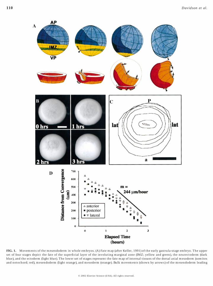
FIG. 1. Movements of the mesendoderm in whole embryos. (A) Fate map (after Keller, 1991) of the early gastrula-stage embryo. The upperset of four stages depict the fate of the superficial layer of the involuting marginal zone (IMZ; yellow and green), the neurectoderm (darkblue), and the ectoderm (light blue). The lower set of stages represent the fate map of internal tissues of the dorsal axial mesoderm (somites
and notochord; red), mesendoderm (light orange), and mesoderm (orange). Bulk movements (shown by arrows) of the mesendoderm leading© 2002 Elsevier Science (USA). All rights reserved.
sitio
111Mesendoderm Extension and Mantle Closure
generating tissues can be isolated and cell behaviors withinthem resolved by using a combination of microsurgery,molecular manipulation, and digital imaging techniques.
In view of the observation that the prospective anteriorleading edge of the mesendoderm comes into contact with
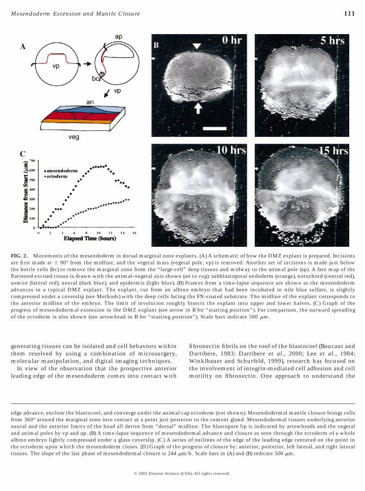
FIG. 2. Movements of the mesendoderm in dorsal marginal zone eare first made at 6 90° from the midline, and the vegetal mass (vethe bottle cells (bc) to remove the marginal zone from the “large-cflattened excised tissue is drawn with the animal–vegetal axis showsomite (lateral red), neural (dark blue), and epidermis (light blue).advances in a typical DMZ explant. The explant, cut from an alcompressed under a coverslip (see Methods) with the deep cells facthe anterior midline of the embryo. The limit of involution rougprogress of mesendodermal extension in the DMZ explant (see arroof the ectoderm is also shown (see arrowhead in B for “starting po
edge advance, enclose the blastocoel, and converge under the animafrom 360° around the marginal zone into contact at a point just posneural and the anterior limits of the head all derive from “dorsal”and animal poles by vp and ap. (B) A time-lapse sequence of mesenalbino embryo lightly compressed under a glass coverslip. (C) A sethe ectoderm upon which the mesendoderm closes. (D) Graph of th
tissues. The slope of the last phase of mesendodermal closure is 244 mm/h© 2002 Elsevier Science (USA
fibronectin fibrils on the roof of the blastocoel (Boucaut andDarribere, 1983; Darribere et al., 2000; Lee et al., 1984;Winklbauer and Schurfeld, 1999), research has focused onthe involvement of integrin-mediated cell adhesion and cellmotility on fibronectin. One approach to understand the
ts. (A) A schematic of how the DMZ explant is prepared. Incisionspole, vp) is removed. Another set of incisions is made just beloweep tissues and midway to the animal pole (ap). A fate map of theto veg): subblastoporal endoderm (orange), notochord (central red),
ames from a time-lapse sequence are shown as the mesendodermembryo that had been incubated in nile blue sulfate, is slightly
he FN-coated substrate. The midline of the explant corresponds toisects the explant into upper and lower halves. (C) Graph of theB for “starting position”). For comparison, the outward spreading
n”). Scale bars indicate 500 mm.
ectoderm (not shown). Mesendodermal mantle closure brings cellsr to the cement gland. Mesendodermal tissues underlying anteriorine. The blastopore lip is indicated by arrowheads and the vegetalrmal advance and closure as seen through the ectoderm of a wholef outlines of the edge of the leading edge centered on the point ingress of closure by: anterior, posterior, left lateral, and right lateral
xplangetalell” dn (an(B) Frbinoing thly bw in
l capteriomidldoderies oe pro
. Scale bars in (A) and (B) indicate 500 mm.
). All rights reserved.

112 Davidson et al.
role of fibronectin (FN) in mesendoderm morphogenesisand gastrulation is to introduce function-blocking antibod-ies or peptide mimics of FN cell-binding sites into theblastocoel of blastula-stage embryos. “Successful gastrula-tion” in these analyses has been equated with closure of theblastopore and differentiation of dorsal axial tissues; how-ever, the phenotypes observed are likely to be more com-plex. In the urodele Pleurodeles watlii, injection of FN- andintegrin-blocking reagents into the blastocoel is reported tostop mesoderm involution and extension (Boucaut et al.,1984a,b; Darribere et al., 1988, 1990). In contrast, injectionof integrin function-blocking GRGDSP peptides into theblastocoel of the anuran Xenopus laevis slows but does notprevent blastopore closure, while dorsal axial tissues arereported to converge and extend (Winklbauer and Keller,1996). Intrablastocoelic injection of a monoclonal antibodythat blocks adhesion to the RGD-containing type III10
repeat of FN also results in the retardation of blastoporeclosure (Ramos et al., 1996) and leads to defects in differ-entiation (Marsden and DeSimone, 2001). These data areconsistent with earlier observations showing that removalof the blastocoel roof (i.e., the substrate for migration) stopsneither blastopore closure nor convergent extension (Kellerand Jansa, 1992).
Cell culture techniques have also been used extensivelywith single cells isolated from prospective mesendoderm orfrom activin-induced animal cap ectoderm to understandthe role of FN in the motility of individual cells. Onceremoved from the embryo and grown on a FN substrate,dissociated cells or tissue fragments from the head meso-derm of both Xenopus and Pleurodeles are sensitive to thesame reagents that produced variable effects in wholeembryos. Function-blocking antibodies and GRGDSP pep-tides inhibit blastomere adhesion and cell migration onFN-coated substrates (Boucaut et al., 1990; Ramos et al.,1996; Winklbauer and Keller, 1996).
Both approaches, studies of single cell motility and theinterpretation of phenotypes of whole embryos after re-agents are injected into the blastocoel, are limited in theirability to decipher the mechanism of mesendoderm mor-phogenesis. One limitation of single-cell studies is that cellmotility within the context of a tissue may differ signifi-cantly from the motility of a single cell in isolation. Cells intissues interact with one another through cell–cell adhe-sion and coordinate motility through other local cell–cellinteractions such as contact inhibition. Similarly, experi-ments that generate whole-embryo phenotypes are limitedby their inability to reveal specific lesions on the cellularlevel; the global effects of some reagents obscure localeffects due to specific cell–cell interactions. Thus, therelationship between results obtained from studies of singlecells and those from whole embryos is unclear.
Several key questions remain concerning the mechanicsof mesendoderm morphogenesis. What are the forces thesecell behaviors generate and how do they move the ventralmesendoderm such large distances so quickly? To what
extent is mesendoderm morphogenesis dependent on cell-© 2002 Elsevier Science (USA
substrate traction and to what extent are the movementsdependent on tissue-deforming forces acting between mes-endodermal cells? How do these movements and the me-chanical “context” of the embryo depend on FN/integrininteractions?
To answer some of these questions, we describe cellbehaviors associated with mesendoderm movements, con-struct predictive models of how morphogenesis proceeds inthe mesendoderm, and test these hypotheses using molecu-lar and microsurgical tools. We analyze movements of themesendodermal mantle in whole embryos at low magnifi-cation, which shows the general kinetics of the cellularmovements involved. We call the movements of the mes-endoderm “mesendoderm extension” since these tissueslengthen during gastrulation to enclose the blastocoel (note:mesendoderm extension should not be confused with con-vergent extension of the mesoderm). To characterize thecell behaviors responsible for these movements, we usemicrosurgery to create an explant where cell behaviors canbe observed in vivo with high-resolution microscopy tech-niques. We describe two distinct models by which cellbehaviors are converted to the forces that drive mesend-oderm extension. After investigating a paradoxically lowrate of tissue extension in the explant with respect to a highrate of extension seen in the whole embryo, we haveidentified a geometrical factor that enables the rapid phaseof mesendodermal mantle closure. Finally we propose sev-eral models that predict how integrin-mediated cell-substrate adhesion and cell–cell adhesion generate andorganize forces driving mesendoderm mantle extension andclosure.
METHODS
Embryo Culture, Microsurgery, and ExplantCulture
Embryos were obtained by standard methods (Kay and Peng,1991) and staged according to Nieuwkoop and Faber (1967). Bothpigmented and albino embryos were used. To enhance contrast inlow-light fluorescence microscopy, embryos were injected with 1.5nl of 25 mg/ml rhodamine dextran amine (RDA; Molecular Probes)at the one-cell stage. To visualize actin in explants, whole embryoswere injected with 0.5 nl per embryo of Alexa 568 (MolecularProbes) conjugated actin protein at the 16-cell stage. Vitellinemembranes were removed mechanically and microsurgery wascarried out with hair-loops and -knives in Danilchik’s For Amy(DFA) consisting of: 53 mM NaCl, 5 mM Na2CO3, 4.5 mM KGluconate, 32 mM Na Gluconate, 1 mM CaCl2, 1 mM MgSO4, 1 gof bovine serum albumin (BSA) per 1 liter, and buffered to pH 8.3with 1 M bicine (Sater et al., 1993). Antibiotic/antimycotic (SigmaA-9909; 0.1 U penicillin, 0.1 mg streptomycin, 0.25 mg amphoteri-cin B per ml) was added to DFA to reduce explant contamination.After microsurgery, each explant was transferred by pipette to adish or chamber, positioned, and gently compressed with a cover-slip fragment held in place with high-vacuum silicone grease (DowChemical).
For marginal zone explants, embryos were selected at stage 102
before the mesendodermal lip contacts the underside of the neural
). All rights reserved.
113Mesendoderm Extension and Mantle Closure
anlagen (Poznanski and Keller, 1997). This event is not readilyvisible from the exterior staging criterion listed in Nieuwkoop andFaber (1967), and selection must be made after the embryo is“opened.” After vitelline removal, two incisions were made 90degrees from the anterior midline. Vegetal endoderm was thenremoved along with tissues that had undergone internal involution(Nieuwkoop and Florschutz, 1950). The explant was then trimmedalong the vegetal side such that several rows of subblastoporalendoderm were left attached. Small wounds in the leading edgethat occur during microsurgery or during transfer to a FN-coateddish can reduce the regularity of movements in the leading edgecells.
For “cap-less” and “donut” explants, embryos were selected atstage 11.5, when the mesendodermal “cup” was approximately 700mm across. An incision through the multilayered epithelium wasmade around the equator of the embryo without injuring theunderlying mesoderm. This “cap” was then lifted gently and teasedoff the mesendodermal mantle, taking care not to injure the leadingedge. To make a “donut-explant,” another incision was made toremove a contiguous “rim” of the mesendodermal mantle “cup”from the remainder of the embryo. This incision ran around theequator and extended into the blastocoel.
Integrin- and FN-Blocking Antibodies
Purified mAbs for 4B12 and 1F7 directed against the RGD andsynergy site containing type III repeats of Xenopus FN, respec-tively, were generated from hybridoma ascites fluid by recombi-nant protein G affinity chromatography (Pharmacia LKB) as previ-ously described and characterized (Ramos and DeSimone, 1996;Ramos et al., 1996). The P8D4 mAb directed against the a5b1
integrin was purified in a similar manner. Details regarding thepreparation and characterization of P8D4 will be described else-where (B.G.H. and D.W.D., manuscript in preparation). Specificfunction-blocking activities of each purified mAb were confirmedby using cells dissociated from early gastrula-stage embryos platedonto FN. Purified mAbs were concentrated in DFA to between 5and 25 mg/ml. mAbs were used at concentrations from 0.1 to 1mg/ml to block FN substrates or to investigate mesendodermalextension in the dorsal marginal zone (DMZ).
Immunocytochemistry and in Situ Protocol
Actin in whole embryos and explants was fixed for immuno-staining according to Kurth et al. (1999). Antibodies to actin(Cedarlane Laboratories, CLT9001) were used at 1:200. Wholeembryos were prepared as “half-mounts” for sectioning accordingto Davidson and Keller (1999). Explants were processed intact onthe original substrate, dehydrated, and cleared in benzyl benzoateand benzyl alcohol (BB:BA; 2:1). Optical sections were collected byusing a confocal laser scan head (PCM2000; Nikon) mounted on aninverted compound microscope (Nikon). Notochord tissues inexplants were immunostained with the tor70 antibody (Kushner,1984) according to Domingo and Keller (1995). A peroxidase-conju-gated goat anti-mouse IgM antibody (Jackson ImmunoResearch)was used to amplify the tor70 signal using the tyramide–fluo-rescein substrate of peroxidase. The fluorescence color reactionwas carried out according to Davidson and Keller (1999).
RNA in situ hybridization was carried out on explants accordingto Davidson and Keller (1999) by using alkaline phosphatase andeither NBT/BCIP (Promega) or magenta-phos (Biosynth) for detec-
tion. Images of in situ hybridizations and tor70 immunostains were© 2002 Elsevier Science (USA
collected by using a digital color camera (Hamamatsu) mounted ona stereoscope (Olympus) equipped for epifluorescence.
Substrate Preparation
FN and recombinant substrates were prepared according toRamos and DeSimone (1996). Briefly, glass coverslips were cleanedwith dilute acid in ethanol then flamed. Human plasma FN (RocheMolecular Biochemicals) or the GST 9.11 fragment of Xenopus FN(Ramos and DeSimone, 1996) were prepared to 20 mg/ml andincubated on glass or plastic petri dishes (Fisher) overnight at 4°C.The substrates were then washed with PBS and blocked with DFA(containing 1 mg/ml BSA) for 1 h prior to receiving explants.
Microscopy and Morphometric Analysis
Time-lapse sequences were collected with digital (Hamamatsu4742, “Orca”) and analog CCD cameras (Dage/MTI and Hama-matsu) mounted on compound upright, compound inverted, andstereo microscopes (Olympus and Zeiss). Image acquisition wascontrolled by software (Metamorph, Universal Imaging Corp. andNIH-Image, Wayne Rasband, version 1.62, http://rsb.info.nih.gov/nih-image). For the low-light fluorescence time-lapse sequences, ashutter (Uniblitz; Vincent Associates) or a filter wheel (SutterInstruments) was also controlled by the software. Morphometricanalysis was carried out on time-lapse sequences by usingNIH-Image (custom macros written by L. Davidson). Time-lapseconfocal sequences of live explants were collected from a laserscanning confocal microscope (Nikon PCM2000) with a 603 oilobjective.
RESULTS
Mesendodermal Movements in Vivo
Static fate maps have tracked the gross tissue movementsof the mesendoderm (Fig. 1A), but the specific cell behav-iors responsible for these movements have not been ob-served directly. In time-lapse sequences of intact wholealbino embryos, the fluid-filled blastocoel is visible as a“shadow” when viewed through the translucent animal capectoderm with incident illumination (Fig. 1B). The leadingedge of the mesendoderm is visible along the perimeter ofthis shadow. Convergent movements of the leading edgecan be seen as the shadow decreases in diameter. Themesendoderm migrates on the blastocoel-facing surface ofthe animal cap ectoderm and converges at a point on thatsurface. Because both the leading edge and the animal capectoderm are visible, rates of mesendoderm movementwith respect to their ectodermal substrate could be mea-sured. The movements of the margin of the mesendodermwere tracked and the rate of movement determined (Figs.1C and 1D; 244 mm/h). In contrast to a previous study
(Nakatsuji, 1975) that indicated that the ventral mesendo-). All rights reserved.
114 Davidson et al.
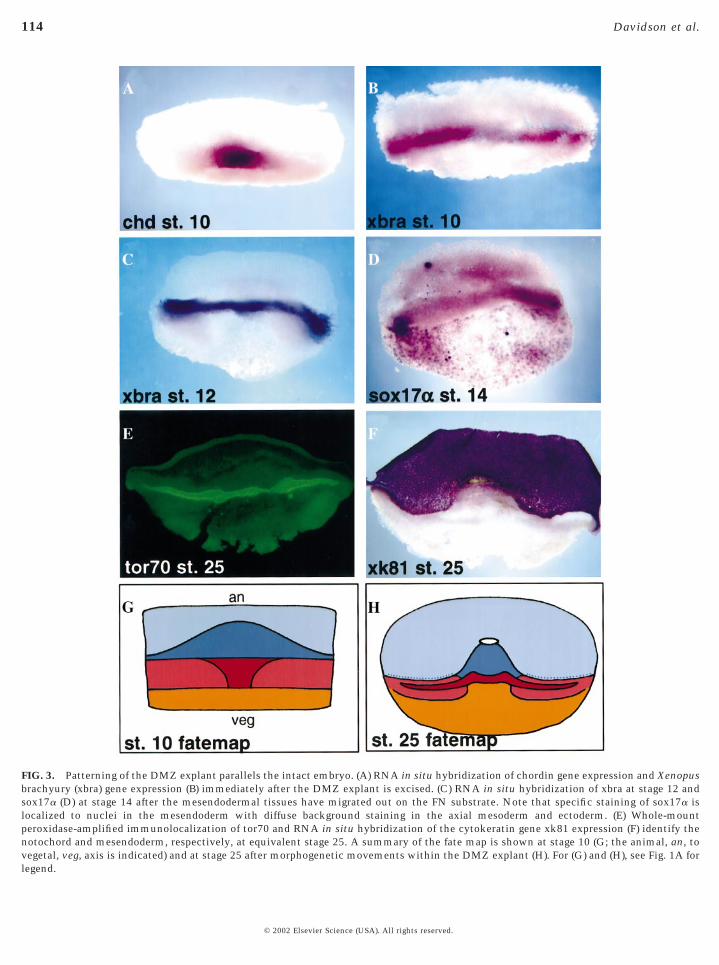
FIG. 3. Patterning of the DMZ explant parallels the intact embryo. (A) RNA in situ hybridization of chordin gene expression and Xenopusbrachyury (xbra) gene expression (B) immediately after the DMZ explant is excised. (C) RNA in situ hybridization of xbra at stage 12 andsox17a (D) at stage 14 after the mesendodermal tissues have migrated out on the FN substrate. Note that specific staining of sox17a islocalized to nuclei in the mesendoderm with diffuse background staining in the axial mesoderm and ectoderm. (E) Whole-mountperoxidase-amplified immunolocalization of tor70 and RNA in situ hybridization of the cytokeratin gene xk81 expression (F) identify thenotochord and mesendoderm, respectively, at equivalent stage 25. A summary of the fate map is shown at stage 10 (G; the animal, an, tovegetal, veg, axis is indicated) and at stage 25 after morphogenetic movements within the DMZ explant (H). For (G) and (H), see Fig. 1A for
legend.© 2002 Elsevier Science (USA). All rights reserved.
FIG
.4.
Tim
e-la
pse
sequ
ence
sof
mes
endo
derm
exte
nsi
on.(
A)R
epre
sen
tati
veti
me-
laps
ese
quen
ceof
exte
ndi
ng
mes
endo
derm
inw
hic
hin
divi
dual
nu
clei
are
trac
ked
(red
cros
ses)
over
90m
in.A
box
draw
nov
era
grou
pof
cell
sdo
esn
otde
form
asth
esh
eet
exte
nds
but
inst
ead
tran
sloc
ates
asth
esh
eet
mov
es.
Scal
eba
ris
250
mm
.(B
)T
rack
s(b
lack
lin
es)
cove
red
byce
lln
ucl
ei(r
eddo
ts)
at5-
min
inte
rval
sov
erth
eco
urs
eof
the
tim
e-la
pse
sequ
ence
in(A
).Sh
ort
trac
ks
indi
cate
cell
sth
atar
rive
onth
esu
bstr
ate
duri
ng
the
cou
rse
ofth
eti
me-
laps
ese
quen
ce.
(C)
Rea
rran
gem
ent
isra
ream
ong
cell
sin
the
mes
endo
derm
.Fr
ames
at20
-min
inte
rval
sfr
oma
tim
e-la
pse
sequ
ence
show
litt
lece
llre
arra
nge
men
t.A
gree
nce
llth
atst
arts
beh
ind
the
lead
ing
edge
at0
min
un
derc
uts
and
tak
esov
era
posi
tion
atth
ele
adin
ged
gew
ith
in40
min
.(D
)Lea
din
ged
gece
lls
exte
nd
broa
dla
mel
lae
inth
edi
rect
ion
ofm
esen
dode
rmm
ovem
ent.
Scal
eba
ris
100
mm
.
115Mesendoderm Extension and Mantle Closure
© 2002 Elsevier Science (USA). All rights reserved.

116 Davidson et al.
FIG. 5. Actin-rich lamellae mark the leading edge as well as cells within the mesendodermal mass of both DMZ explants and whole embryos. (A)An optical confocal section shows that actin localizes strongly to the leading edge of the mesendoderm in whole embryos. Arrowheads show intenseactin localization in the leading edge as well as in cells within the mesendodermal mantle. (B) A sum of 12 confocal sections from an en-face view ofthe leading edge of a DMZ explant show strong actin localization to the leading edge. Occasional smaller lamellae are visible behind the leading edge(arrow). (C) An X-Z section taken at along the dotted line in the confocal stack collected in (B). (D) Confocal time lapse of actin dynamics at the leadingedge (an arrow shows the direction of mesendodermal explant extension). Several cells can be seen in frames from a 24-min-long time-lapse sequence.These cells exhibit complex actin dynamics at the leading edge: intense actin localization at cell–cell boundaries and actin within lamellae at theleading edge as well as in lamellae extended by cells within the mass of the mesendoderm. One cell (marked by “*” at 0 min) overtakes cells alreadyon the leading edge. Another cell (marked by “1”) extends a lamella onto the substrate behind the leading edge. Fluorophore-conjugated actin was
injected into embryos at the 16-cell stage targeting the midline. Single frames were collected every 15 s from the plane of the substrate. One frame is213 mm across.© 2002 Elsevier Science (USA). All rights reserved.
in p
117Mesendoderm Extension and Mantle Closure
derm does not extend, we found that the posterior leadingedge starts to extend later than the anterior leading edge butultimately reaches the same rate of extension as the ante-rior mesendoderm. While the optically translucent celllayers of the albino animal cap ectoderm allow the identi-fication of the leading edge mesendoderm, these tissuesobscure the tracking of individual cells.
Mesendoderm Movements in an Explantof the DMZ
To resolve individual cells and their behaviors, we devel-oped an explant from the marginal zone of early gastrula-
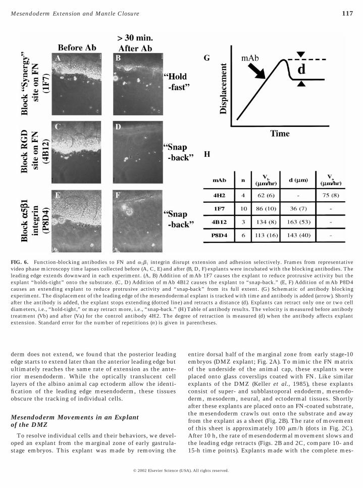
FIG. 6. Function-blocking antibodies to FN and a5b1 integrin divideo phase microscopy time lapses collected before (A, C, E) and aleading edge extends downward in each experiment. (A, B) Additioexplant “holds-tight” onto the substrate. (C, D) Addition of mAbcauses an extending explant to reduce protrusive activity and “experiment. The displacement of the leading edge of the mesendodeafter the antibody is added, the explant stops extending (dotted lindiameters, i.e., “hold-tight,” or may retract more, i.e., “snap-back.”treatment (Vb) and after (Va) for the control antibody 4H2. The dextension. Standard error for the number of repetitions (n) is given
stage embryos. This explant was made by removing the
© 2002 Elsevier Science (USA
entire dorsal half of the marginal zone from early stage-10embryos (DMZ explant; Fig. 2A). To mimic the FN matrixof the underside of the animal cap, these explants wereplaced onto glass coverslips coated with FN. Like similarexplants of the DMZ (Keller et al., 1985), these explantsconsist of super- and subblastoporal endoderm, mesendo-derm, mesoderm, neural, and ectodermal tissues. Shortlyafter these explants are placed onto an FN-coated substrate,the mesendoderm crawls out onto the substrate and awayfrom the explant as a sheet (Fig. 2B). The rate of movementof this sheet is approximately 100 mm/h (dots in Fig. 2C).After 10 h, the rate of mesendodermal movement slows andthe leading edge retracts (Figs. 2B and 2C, compare 10- and
extension and adhesion selectively. Frames from representativeB, D, F) explants were incubated with the blocking antibodies. ThemAb 1F7 causes the explant to reduce protrusive activity but thecauses the explant to “snap-back.” (E, F) Addition of mAb P8D4
-back” from its full extent. (G) Schematic of antibody blockingexplant is tracked with time and antibody is added (arrow). Shortlyd retracts a distance (d). Explants can retract only one or two cellable of antibody results. The velocity is measured before antibody
e of retraction is measured (d) when the antibody affects explantarentheses.
sruptfter (n of4B12snaprmale) an(H) Tegre
15-h time points). Explants made with the complete mes-
). All rights reserved.

0 mm
118 Davidson et al.
endodermal lip, i.e., including the tissue that contacts theunderside of the ectoderm, display similar rates of mesen-doderm migration (data not shown). The rate of mesendo-derm movement is not simply a feature of any tissue placedon FN. While prospective epidermis adheres and spreads onFN, its rate of movement is limited to less than 25 mm/h(triangles in Fig. 2C), and unlike the mesendoderm, exten-
FIG. 7. The “propulsive leading edge” model for mesendoderm eleading edge.” The early mesendoderm consists of a leading edge gmesendodermal mantle (a). The mesendodermal mantle passivelyinitial length (l0). Forces generated from the leading edge pull onmodel, forces for extension are the result of cycles of extension andDMZ explants are placed onto an FN-coated plastic dish. Both arethe midline bisects the explant. (C) The two explants in (B) 18 h latof the upper explant and the ectoderm of the lower explant. (D) A tframes from the time-lapse sequence in (D) show continuing movemThese four cells move at an average 90 mm/h after the collision (distance moved). Scale bar for time-lapse frame in (D) indicates 50
sion continues beyond 10 h.
© 2002 Elsevier Science (USA
Early Morphogenetic Movements of theMesendoderm in the DMZ Explant Are Decoupledfrom the Morphogenesis of the Axial Mesoderm
We confirmed the presence of mesendoderm in our ex-plants with an analysis of gene expression. To visualizegene expression patterns specific to the marginal zone, we
sion and a test of that model. (A) A schematic of the “propulsiveting force (f) through its area of contact with the remainder of thets deformation with an elastic modulus (e) and stretches from anesendodermal mantle deforming it to the new length (l). In this
traction of protrusive lamellae along the free leading edge. (B) Twoted such that the ectoderm is up, the mesendoderm is down, andw that a collision has occurred between the mesendodermal sheet
lapse sequence shows collision of the two explants. (E) Additionalof cells (see four arrows) behind the leading edge after the collision.es indicate where the cells start and tails are proportional to the.
xteneneraresisthe mcon
oriener shoime-ent
cross
carried out a battery of RNA in situ hybridizations (Figs.
). All rights reserved.

119Mesendoderm Extension and Mantle Closure
FIG. 8. High rate of mesendoderm extension proceeds in a cap-less explant. Movements of the mesendoderm in “cap-less” explant. (A)Schematic showing how cap-less explants were prepared. (B) A representative time-lapse sequence of mesendodermal advance and closure
of the cap-less explant. (C) A series of outlines taken every 5 min of the edge of the leading edge centered on the point upon which the© 2002 Elsevier Science (USA). All rights reserved.

120 Davidson et al.
3A–3F). These patterns revealed several interesting featuresof these explants. Figures 3A and 3B show the extent of theDMZ that remains in these explants following microsur-gery. Explants fixed immediately after excision werestained for expression of chordin (Fig. 3A) and xbra (Fig. 3B).The blastoporal lip is clear at the lower boundary of chordinexpression in Fig. 3A. Under the lip can be seen a smallquantity of subblastoporal endoderm. Expression of xbra atthis stage is characteristic of prospective notochord andsomites. The fate map of the DMZ explant shortly afterexcision is summarized in Fig. 3G. After plating the explantonto FN, the mesendodermal tissues migrate out and awayfrom the dorsal axial tissues. Xbra gene expression atequivalent stage 12 (Fig. 3C) shows the location of thedorsal axial tissues in the explant while sox17a geneexpression (Fig. 3D) indicates how far the mesendoderm hasmigrated by equivalent stage 14. Thus, the early morpho-genetic movements of the mesendoderm in explants aremechanically isolated, decoupled from the morphogenesisof more axial mesoderm. Finally, Figs. 3E and 3F illustratethe ultimate patterning of these explants. The differentia-tion marker tor70 shows the extent of notochord formation(Fig. 3E). Epidermal differentiation is indicated by sharplyrestricted gene expression of the cytokeratin marker xk81.Clearly, medial convergence of the axial mesoderm isblocked but differentiation is not (Fig. 3H).What is clearfrom these gene expression patterns is that the mesendo-derm in the DMZ explant extends perpendicular to theaxial mesoderm, mimicking the normal movement inwhole embryos, where head, lateral, and posterior mesendo-derm move away from the dorsal axis (Fig. 1A).
Extension of DMZ Mesendoderm Explant IsCoincident with Radial Intercalation within theSheet and Protrusive Activity at the Leading Edge
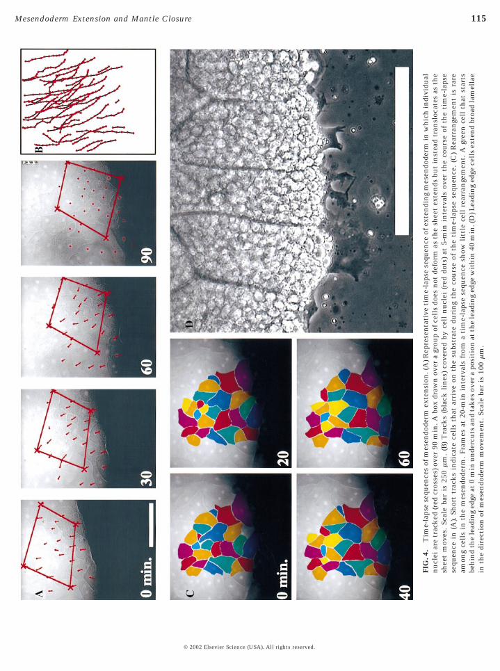
We observed that movement of the mesendoderm in theDMZ explant occurs in the absence of mediolateral inter-calation of mesendoderm cells (Fig. 4A). A box drawnaround a typical group of mesendoderm cells does notdeform as predicted for convergent extension (i.e., lengthenand narrow, cf. Keller and Tibbetts, 1989) but insteadappears to translocate en masse. Tracks of individual cellnuclei within the sheet move in the same direction as theextending sheet (Fig. 4B). Extension of the mesendoderm is
mesendoderm converges. (D) Graph of the progress of closure inmesendoderm, 240 mm/h, is shown as the slope of the trace. Scaleclosure in the cap-less explant. Individual nuclei are tracked overtissue are translocating rather than mediolaterally intercalating (Freducing the number of cells along the margin from 12 to 6. Radmesoderm shown by the expression of the notochord-specific genenot seen on the ventral surface (right panel) where mantle closure o
upper left of both panels.© 2002 Elsevier Science (USA
accompanied by only a small degree of neighbor exchange(Fig. 4C) consistent with previous work (Keller and Tib-betts, 1989).
The tissue architecture of the mesendodermal sheet inthese explants mimics the architecture of the mesendoder-mal mantle in whole embryos. When the explant is placedonto an FN-coated substrate, a single layer of cells contactand adhere to the substrate. Explants typically begin withmultiple layers of cells. As the mesendoderm migrates outfrom the axial mesoderm, it thins from multiple layers ofcells occasionally becoming only one-cell layer thick. Thecells in direct contact with the FN substrate become“shingled” just as cells within the whole embryo (Fig. 5A;Winklbauer et al., 1991). Cells in deeper layers join thosecells already on the FN substrate through the process ofradial intercalation. Radial intercalation occurs at the lead-ing edge and throughout the mesendodermal mantle, evenas the tissue moves forward. Radial intercalation is seen ascells from deeper layers send out protrusions between cellsthat are already contacting the substrate. Within a fewminutes, these deep-originating cells intercalate, attach tothe substrate, and begin moving in the same direction astheir neighboring cells (see Fig. 11D; see time-lapse se-quence shown in Fig. 4 online at http://www.dbtimelapses.org/timelapses.html). Intercalating cells frequently appearon the FN substrate next to cells that had intercalatedearlier, as if taking advantage of openings or weak locationsin the cohesive cell mass (e.g., yellow cells in Fig. 4C).
Time-lapse sequences show that cells occasionally jointhe leading edge (e.g., Fig. 5D) and, like their neighbors,exhibit extensive arrays of cellular protrusions (Fig. 4D).Similar to migrating isolated mesoderm cells in culture(Selchow and Winklbauer, 1997), the leading edge cells inthe DMZ explant extend large dynamic lamellae in thedirection of movement. Xenopus mesendoderm cells areextremely yolk granule laden. Lamellae are yolk-free,readily observed by phase microscopy, and often extend upto 30 mm from the cell body. Nearly all the cells on theleading edge extend lamellae (Figs. 4D and 5D); those thatdo not are either in the process of cell division or in theprocess of being removed or passed by actively protrudingneighbor cells. Even though these cells are mesenchymaland not epithelial in nature, we have never observed cellsdetaching from their neighbors and migrating off the lead-ing edge.
cap-less explant (indicated by arrow in B). The velocity of thein (B) indicates 500 mm. (E) A representative time lapse of mantlein. A bounding box deforms as the mantle closes but cells within) Cell rearrangement occurs as the edge of the mantle converges,ntercalation brings three new cells onto the substrate. (H) Axialdin is present on the dorsal surface (three explants; left panel) buts. The dorsal side of an equivalent stage-14 embryo is shown in the
thebar
60 m). (Gial ichorccur
). All rights reserved.

121Mesendoderm Extension and Mantle Closure
Actin Organization in the DMZ Explant ParallelsActin Cytoskeleton in the Whole Embryo
Tissue movements and patterning in DMZ explants ap-pear to reflect events in whole embryos; however, we do notknow whether the subcellular distribution of specific mo-lecular components of the DMZ explant parallel thoseobserved in the whole embryo. To resolve this question, weinvestigated the localization of actin in both DMZ explantsand whole embryos.
There is striking similarity between actin localization inthe whole embryo (Fig. 5A) and the DMZ explant (Figs. 5Band 5C). Actin is strongly localized to the leading edge inboth the DMZ explant (Fig. 5B) and the mesendodermalmantle in vivo (Fig. 5A). Actin can also be found behind theleading edge in vivo as well as in the DMZ explant (arrow-heads in Fig. 5A and arrow in Fig. 5B), perhaps marking cellsin the process of radial intercalation. Several differencesbetween the DMZ explant and the whole embryo can alsobe observed. While the leading edge in whole embryosdevelops discrete actin-rich structures in the direction ofmesendoderm movement, the long lamellae present in theexplant are not observed in the whole embryo (Fig. 5C vs.Fig. 5A). This absence may reflect the difference between anFN substrate on a glass scaffold and the FN fibrils found onthe deformable scaffold of the animal cap ectoderm. Theabsence of long actin-rich lamellae in the whole embryomay also reflect a fixation artifact due to the difficulty ofsimultaneously preserving actin and lamellae.
Since actin fixation has many known disadvantages, weinjected a fluorescently coupled actin into early cleavage-stage embryos and followed actin dynamics in live DMZexplants (Fig. 5D; see time-lapse sequences of actin dynam-ics online at http://www.dbtimelapses.org/timelapses.html).Confocal time-lapse sequences of actin dynamics in theleading edge support the observations of actin in the fixedsamples. Actin localizes to the dynamic lamellae seen inphase contrast. Actin can also be seen at cell–cell bound-aries but appears more intense just behind advancing cellmargins. Two cells in the sequence illustrate the role ofactin in the process of radial intercalation and the additionof a cell to the leading edge, respectively.
Actin dynamics in the explant differ significantly fromfixed actin seen in isolated cells. Previous workers have
FIG. 9. High rate of mesendoderm extension proceeds in a donutA representative time-lapse sequence of mesendodermal advance acultured on a minimal substrate (GST 9.11) containing the RGD anseries of outlines taken every 5 min of the edge of the leading edge ceof the progress of closure in the donut explant. The velocity of the(B) indicates 200 mm. (E) A representative time lapse of mantle clobounding box deforms as the mantle closes, but (F) cells within tisrearrangement occurs as the edge of the mantle converges, reducintercalation brings five new cells onto the substrate. (H) Axial meis absent from both dorsal surface (three explants; left panel) and th
side of an equivalent stage-14 embryo is shown in the upper left of bot© 2002 Elsevier Science (USA
observed actin localization in fixed isolated mesendoderm(Selchow and Winklbauer, 1997) and activin-induced ani-mal cap cells (Gurdon et al., 1999) and found extensiveactin localized throughout the cell with considerable actinlocalized to both the leading and trailing edges of migratingcells. From our observations of actin in fixed whole em-bryos and within live cells of the DMZ explant, we also seeactin at the leading edge of cells but not at the trailing edge.Cells migrating in isolation (Winklbauer and Selchow,1992) appear to lack the tightly regulated morphology seenin explants where cells develop a polarized, shingled shapeand maintain highly persistent directed protrusive activity.
What Is the Role of Integrin/FN in MesendodermExtension?
Since microfilaments form extensively at the substratelevel of the leading edge and integrins function as a me-chanical link between the cytoskeleton and the extracellu-lar matrix (Sastry and Horwitz, 1993), we decided to inves-tigate the role of integrin-mediated cell-substrate adhesionin driving mesendoderm extension. The forces driving thesemovements may be generated by each cell that makescontact with the FN substrate. In order to test this hypoth-esis, we utilized antibodies capable of blocking specificcell-binding domains of Xenopus FN (Ramos and DeSi-mone, 1996). The first antibody we used, mAb 1F7, blockscell recognition of the “synergy” site located in the type III9
repeat of the FN molecule. This antibody has no effect onthe attachment of single cells to FN but does block spread-ing and migration. Recently, mAb 1F7 has also been shownto block FN fibril formation in the animal cap ectoderm(Marsden and DeSimone, 2001). DMZ explants were placedonto a recombinant fragment of Xenopus FN (GST 9.11)that contains both the synergy and RGD sites, whichrepresent the minimal set of binding domains necessary forcell attachment and migration in this system (Ramos andDeSimone, 1996). There is no difference in the rate ofextension of mesendoderm on either GST 9.11 or humanplasma FN (data not shown). The mesendoderm was thenallowed to migrate out and antibody was added to theculture media (arrow in Fig. 6G). Forward movement of theDMZ explant ceases and the leading edge lamellae retractafter mAb 1F7 is added, demonstrating that the “synergy”
nt. (A) Schematic showing how donut explants were prepared. (B)osure of the donut explant over the course of 1 h. This explant wasnergy site of the central cell-binding domain of Xenopus FN. (C) Aed on the point upon which the mesendoderm converges. (D) Graphng edge, 237 mm/h, is shown by the slope of the trace. Scale bar inin the donut explant. Individual nuclei are tracked over 40 min. Are translocating rather than mediolaterally intercalating. (G) Cell
the number of cells along the margin from eight to five. Radialm shown by the expression of the notochord-specific gene chordinntral surface (right panel) where mantle closure occurs. The dorsal
expland cld synterleadisuresue aing
sodere ve
h panels.
). All rights reserved.
122 Davidson et al.
© 2002 Elsevier Science (USA). All rights reserved.
123Mesendoderm Extension and Mantle Closure
site on FN is required for extension of the mesendodermwithin the DMZ explant (Figs. 6A, 6B, and 6H). When anantibody known to block the RGD-containing type III10
repeat sequence of FN is used (mAb 4B12), mesendodermalmovement not only stops, but the already extended mesen-doderm detaches and retracts (Figs. 6C, 6D, and 6H; acontrol nonfunction blocking antibody to FN, mAb 4H2,has no effect; Fig. 6H). Since recognition of both RGD andsynergy involves the integrin a5b1 (Danen et al., 1995), wealso tested an antibody that blocks the function of the a5b1
integrin directly (mAb P8D4; B.G.H. and D.W.D., manu-script in preparation). Application of mAb P8D4 causesmesendodermal movement to stop and the already ex-tended mesendoderm to retract from its previous position(Figs. 6E, 6F, and 6H for mAb P8D4), confirming theinvolvement of a5b1 integrin. Thus, while adhesion to theFN substrate via the RGD site is a prerequisite for attach-ment and maintenance of the extending mesendoderm, it isthe involvement of integrin a5b1 with the “synergy” regionof FN that enables mesendoderm morphogenesis in theDMZ explant.
Are the Forces That Drive Mesendoderm ExtensionGenerated at the Leading Edge?
The presence of strong protrusive activity coupled withdynamic actin polymerization in the leading edge and thesensitivity of these protrusions to synergy-blocking anti-bodies prompts us to propose the “propulsive leading edge”model for mesendoderm extension (Fig. 7A). This modelposits that the forces driving extension are the direct resultof protrusive activity in the first row of cells along theleading edge. Studies of protrusive activity in single cellsdemonstrate that protrusions can generate considerableforce (Dembo and Wang, 1999; Galbraith and Sheetz, 1999;Oliver et al., 1994; Pelham and Wang, 1999). One predictionof this model is that if one could block protrusive activityspecifically at the leading edge, then one could blockextension of the entire mesendodermal sheet. However,inhibitors of the contractile actomyosin machinery in-volved in the generation of force at the leading edge mayalter force generating mechanisms operating elsewhere inthe tissue or even alter tissue stiffness. Alternatively,according to the model, physically blocking the progress ofthe leading edge should bring movement within the sheetto a halt.
With this testable prediction, we set out to block theprogress of the leading edge in the DMZ explant by placingtwo explants onto FN (Fig. 7B), oriented such that themesendoderm of one explant (upper explant in 7B) wouldextend and collide with the ectoderm of the other explant(lower explant in 7B). The explants collide as expected (Fig.7C), bringing the leading edge to a halt (Fig. 7D). However,cells further back in the mesendodermal mass continue tomove forward at the same rate as an unconfined explant(arrows in Fig. 7E). Thus, tissues behind the leading edge
continue to move forward even when progress of the© 2002 Elsevier Science (USA
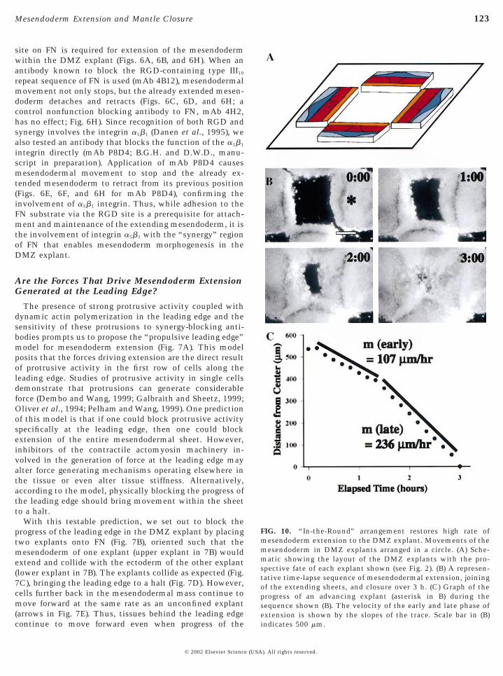
FIG. 10. “In-the-Round” arrangement restores high rate ofmesendoderm extension to the DMZ explant. Movements of themesendoderm in DMZ explants arranged in a circle. (A) Sche-matic showing the layout of the DMZ explants with the pro-spective fate of each explant shown (see Fig. 2). (B) A represen-tative time-lapse sequence of mesendodermal extension, joiningof the extending sheets, and closure over 3 h. (C) Graph of theprogress of an advancing explant (asterisk in B) during thesequence shown (B). The velocity of the early and late phase ofextension is shown by the slopes of the trace. Scale bar in (B)
indicates 500 mm.). All rights reserved.

124 Davidson et al.
leading edge is physically blocked. Thus, protrusive activityof the leading edge is not solely responsible for the move-ment of the entire mesendodermal mantle. Alternativemodels for the forces driving mesendoderm extension in theDMZ explant are proposed in the discussion.
In-The-Round Geometry of the Leading Edge IsBoth Necessary and Sufficient for the IncreasedRate of Movement Observed in Vivo
Comparison of the rate of mesendoderm movement inthe DMZ explant and movement of the leading edge in vivoreveals that the DMZ explant achieves only half the rate ofextension seen in vivo (compare Figs. 1D and 2C). The rateof DMZ extension we observe is consistent with the di-rected migratory rate of mesendodermal tissue “slugs” cutfrom the DMZ (Winklbauer, 1990) and placed onto FN. Thisrate is also consistent with the randomly directed migratoryrate of single cells dissociated from mesendoderm isolatedfrom the DMZ (Ramos et al., 1996). While explants rarelyperform morphogenetic movements as well as the intactembryo, we could identify three characteristics of the DMZexplant that might limit the efficiency of cell migration.The first feature is the organization of the extracellularmatrix presented to the explant. Explants can extend on aminimal substrate consisting of the GST 9.11 fragment ofXenopus laevis FN. In contrast, the mesendodermal mantlein vivo sees a milieu of complex extracellular matrix in theblastocoel, including FN fibrils, bound to the underside ofthe animal cap. Substrate rigidity is another contrastingfactor between the environment of the DMZ explant andthe intact embryo. Cultured cells can change morphologyand migration rate when grown on substrates of differentelastic modulii (Choquet et al., 1997; Lo et al., 2000).Traction exerted by cells in the DMZ explant are met withthe mechanical resistance of molecular FN adsorbed ontoglass. In vivo, the same cells attach to FN fibrils that arebound to a much more pliant substrate of the animal capectoderm. Finally, the linear geometry of the DMZ explantwith both ends “open,” excised from lateral tissues, con-trasts with the contiguous 360° perimeter of the leadingedge in vivo. To identify which of the characteristics of theDMZ explant reduces the rate of extension, we developedseveral additional explants where effects of these featurescould be evaluated.
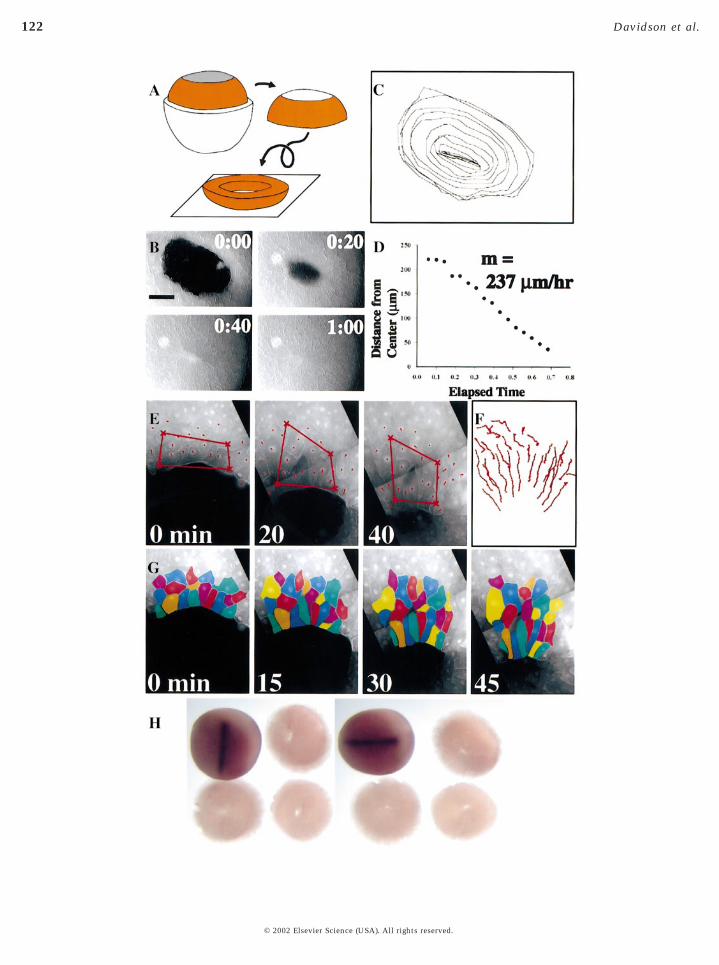
The simplest of these explants revealed that neithersubstrate complexity nor substrate mechanical propertieswere responsible for the increased rate of movement seen inthe intact embryo. The “cap-less” explant is made from astage-11.5 embryo that has had its animal cap ectodermremoved (Fig. 8A). This explant is then plated onto anFN-coated glass coverslip and its movements recorded in atime-lapse sequence (Fig. 8B). Analysis of these movementsshows that the cap-less explant recapitulates the migratoryrate of the intact embryo (Figs. 8C and 8D; n 5 8). Thus, FNfibrils are not required for the increased rate of movement
in the intact embryo.© 2002 Elsevier Science (USA
Mesendoderm closure in the cap-less explant occurs inthe absence of mediolateral intercalation. A bounding boxdrawn around cells in the mesendodermal mantle deforms(Fig. 8E) as the mantle closes. Cells along the leading edgeconverge, however, tracks of individual nuclei translocateen masse (Fig. 8F) just as they do in the DMZ explant.Radial intercalation is also seen as the mantle closes (seeyellow cells in Fig. 8G). In contrast, with the case of theDMZ explant, cells in the closing mantle rearrange as themantle closes. As the perimeter of the closing mantledecreases, cells leave the leading edge and join the secondtier of cells (e.g., six cells leave the leading edge during thetime lapse shown in Fig. 8G).
Dorsal axial tissues do not contribute directly to mantleclosure in the cap-less explant. To show that the anteriorend of the notochord was not driving closure of the mantle,we characterized cap-less explants immediately after clo-sure for the expression of chordin (Fig. 8H). While the dorsalface of the explant clearly shows the location of the noto-chord, the ventral surface shows the complete absence ofthe notochord from the mesendodermal mantle.
Because the cap-less explant retains an intact blastocoelas well as the rest of the embryo and is cultured onfull-length FN, we thought unknown components remain-ing within the blastocoel or nonintegrin cell substrate-binding sites might contribute to the high rate of move-ment observed. We tested this hypothesis by excising theleading edge from the cap-less explant (the “donut” explant;Fig. 9A; n 5 2), plating the explant on the minimal fragmentof FN containing a single RGD and synergy site (GST 9.11)and recording its movements in a time-lapse sequence (Fig.9B). Remarkably, the leading edge in the donut explant(Figs. 9C and 9D) extends with a rate comparable to boththe cap-less explant and the intact embryo. Both the cap-less and the donut explant retain the leading edge in acontiguous 360°.
Mesendoderm closure in the donut explant occurs with-out mediolateral intercalation and in the total absence ofaxial mesoderm. A bounding box drawn around cells in thedonut explant shows limited convergence of the leadingedge of cells (Fig. 9E) as the mantle closes. Like the cap-lessexplant, convergence appears to be driven by geometrysince tracks of individual nuclei translocate en masse (Fig.9F). Radial intercalation is also seen as the donut closes (seeyellow cells in Fig. 9G). Like the cap-less explant, cells inthe closing mantle of the donut explant rearrange as themargin of the mantle closes. As the perimeter of the mantledecreases, cells leave the leading edge and join the secondtier of cells (e.g., three cells leave the leading edge duringthe time lapse shown in Fig. 9G). Lack of chordin expres-sion confirms the complete absence of axial mesoderm inthe donut explant (Fig. 9H). While the cap-less explantretains prospective axial mesoderm with its possible con-tribution to a higher rate of closure, the donut explant doesnot include axial mesoderm but still retains the high rate ofmovement of the mesendoderm observed in the whole
embryo.). All rights reserved.
125Mesendoderm Extension and Mantle Closure
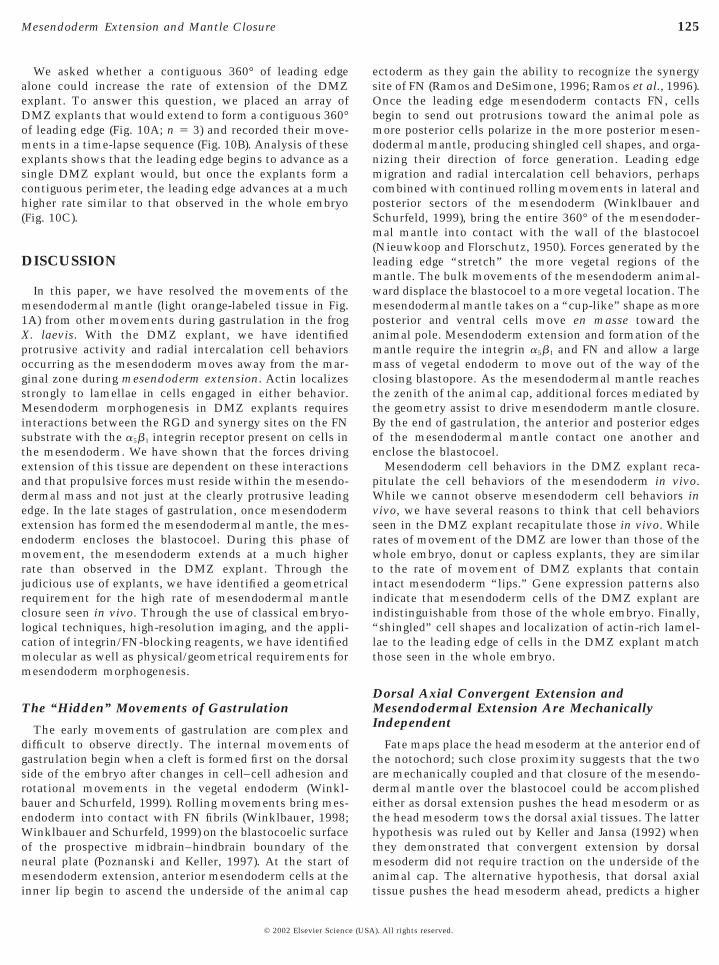
We asked whether a contiguous 360° of leading edgealone could increase the rate of extension of the DMZexplant. To answer this question, we placed an array ofDMZ explants that would extend to form a contiguous 360°of leading edge (Fig. 10A; n 5 3) and recorded their move-ments in a time-lapse sequence (Fig. 10B). Analysis of theseexplants shows that the leading edge begins to advance as asingle DMZ explant would, but once the explants form acontiguous perimeter, the leading edge advances at a muchhigher rate similar to that observed in the whole embryo(Fig. 10C).
DISCUSSION
In this paper, we have resolved the movements of themesendodermal mantle (light orange-labeled tissue in Fig.1A) from other movements during gastrulation in the frogX. laevis. With the DMZ explant, we have identifiedprotrusive activity and radial intercalation cell behaviorsoccurring as the mesendoderm moves away from the mar-ginal zone during mesendoderm extension. Actin localizesstrongly to lamellae in cells engaged in either behavior.Mesendoderm morphogenesis in DMZ explants requiresinteractions between the RGD and synergy sites on the FNsubstrate with the a5b1 integrin receptor present on cells inthe mesendoderm. We have shown that the forces drivingextension of this tissue are dependent on these interactionsand that propulsive forces must reside within the mesendo-dermal mass and not just at the clearly protrusive leadingedge. In the late stages of gastrulation, once mesendodermextension has formed the mesendodermal mantle, the mes-endoderm encloses the blastocoel. During this phase ofmovement, the mesendoderm extends at a much higherrate than observed in the DMZ explant. Through thejudicious use of explants, we have identified a geometricalrequirement for the high rate of mesendodermal mantleclosure seen in vivo. Through the use of classical embryo-logical techniques, high-resolution imaging, and the appli-cation of integrin/FN-blocking reagents, we have identifiedmolecular as well as physical/geometrical requirements formesendoderm morphogenesis.
The “Hidden” Movements of Gastrulation
The early movements of gastrulation are complex anddifficult to observe directly. The internal movements ofgastrulation begin when a cleft is formed first on the dorsalside of the embryo after changes in cell–cell adhesion androtational movements in the vegetal endoderm (Winkl-bauer and Schurfeld, 1999). Rolling movements bring mes-endoderm into contact with FN fibrils (Winklbauer, 1998;Winklbauer and Schurfeld, 1999) on the blastocoelic surfaceof the prospective midbrain–hindbrain boundary of theneural plate (Poznanski and Keller, 1997). At the start ofmesendoderm extension, anterior mesendoderm cells at the
inner lip begin to ascend the underside of the animal cap© 2002 Elsevier Science (USA
ectoderm as they gain the ability to recognize the synergysite of FN (Ramos and DeSimone, 1996; Ramos et al., 1996).Once the leading edge mesendoderm contacts FN, cellsbegin to send out protrusions toward the animal pole asmore posterior cells polarize in the more posterior mesen-dodermal mantle, producing shingled cell shapes, and orga-nizing their direction of force generation. Leading edgemigration and radial intercalation cell behaviors, perhapscombined with continued rolling movements in lateral andposterior sectors of the mesendoderm (Winklbauer andSchurfeld, 1999), bring the entire 360° of the mesendoder-mal mantle into contact with the wall of the blastocoel(Nieuwkoop and Florschutz, 1950). Forces generated by theleading edge “stretch” the more vegetal regions of themantle. The bulk movements of the mesendoderm animal-ward displace the blastocoel to a more vegetal location. Themesendodermal mantle takes on a “cup-like” shape as moreposterior and ventral cells move en masse toward theanimal pole. Mesendoderm extension and formation of themantle require the integrin a5b1 and FN and allow a largemass of vegetal endoderm to move out of the way of theclosing blastopore. As the mesendodermal mantle reachesthe zenith of the animal cap, additional forces mediated bythe geometry assist to drive mesendoderm mantle closure.By the end of gastrulation, the anterior and posterior edgesof the mesendodermal mantle contact one another andenclose the blastocoel.
Mesendoderm cell behaviors in the DMZ explant reca-pitulate the cell behaviors of the mesendoderm in vivo.While we cannot observe mesendoderm cell behaviors invivo, we have several reasons to think that cell behaviorsseen in the DMZ explant recapitulate those in vivo. Whilerates of movement of the DMZ are lower than those of thewhole embryo, donut or capless explants, they are similarto the rate of movement of DMZ explants that containintact mesendoderm “lips.” Gene expression patterns alsoindicate that mesendoderm cells of the DMZ explant areindistinguishable from those of the whole embryo. Finally,“shingled” cell shapes and localization of actin-rich lamel-lae to the leading edge of cells in the DMZ explant matchthose seen in the whole embryo.
Dorsal Axial Convergent Extension andMesendodermal Extension Are MechanicallyIndependent
Fate maps place the head mesoderm at the anterior end ofthe notochord; such close proximity suggests that the twoare mechanically coupled and that closure of the mesendo-dermal mantle over the blastocoel could be accomplishedeither as dorsal extension pushes the head mesoderm or asthe head mesoderm tows the dorsal axial tissues. The latterhypothesis was ruled out by Keller and Jansa (1992) whenthey demonstrated that convergent extension by dorsalmesoderm did not require traction on the underside of theanimal cap. The alternative hypothesis, that dorsal axial
tissue pushes the head mesoderm ahead, predicts a higher). All rights reserved.

126 Davidson et al.
FIG. 11. Models of mesendoderm extension. (A) The mesendoderm extends as a contiguous multicell layered mass. Cells in contact withthe FN-coated substrate extend tractive actin-rich protrusions at their leading edges and develop a highly polarized shingled shape. Cellswithin the mass also extend actin-rich protrusions as they attempt to radially intercalate. The contiguity of the mesendodermal mass ismaintained by cell–cell adhesions (arrow indicates direction of movement). (B) The propulsive leading edge model proposes traction by asingle row of cells at the front of the mass (asterisk) generates traction. (C) The distributed traction model proposes that all cells in contactwith the substrate generate traction (asterisk). (D) The radial intercalation model proposes that cells interdigitate radially (asterisks) andthen wedge between neighboring cells that are already on the substrate. These radial intercalation behaviors generate expansion forces
within the mass that act to advance the leading edge.© 2002 Elsevier Science (USA). All rights reserved.

127Mesendoderm Extension and Mantle Closure
rate of movement on the anterior side of the leading edgedue to the force generated by converging and extendingdorsal axial tissues. It came as a surprise to us that we couldfind no difference in the rate of movement between theanterior- and posterior-leading mesendoderm. Two otherresults, the independence of extension of the DMZ explantwhere extension occurs perpendicular to the dorsal axis,and the rapid closure of the mesendodermal mantle in thedonut explant where the mesendoderm is microsurgicallyseparated from the dorsal axis, provide evidence that theprocesses of convergent extension in the dorsal axial meso-derm and closure of the mesendodermal mantle are me-chanically independent of each other.
However, convergent extension of the dorsal mesodermmay serve a redundant role ensuring mantle closure duringgastrulation whenever mesendodermal extension fails. Ifmesendoderm extension fails, convergent extension in thedorsal mesoderm may still be capable of pushing the ante-rior edge of the mesendoderm over the blastocoel. Once themesendoderm reaches a “permissive” point, cell–cell adhe-sion might be capable of driving mantle closure over theblastocoel. Such a backup role for convergent extension indorsal axial tissues might explain the success of gastrula-tion in zebrafish or mouse mutants even when mesendoder-mal migration is compromised.
A Signaling Role for FN?
It has been proposed that FN has a signaling function inearly embryos in addition to its role as an adhesive sub-strate (Marsden and DeSimone, 2001; Ramos and DeSi-mone, 1996; Ramos et al., 1996; Winklbauer and Schurfeld,1999). From an analysis of isolated cell motility, Ramos etal. (1996) found that cell recognition of the RGD site wasrequired for cell attachment. Once cells were attached, cellrecognition of the synergy site was required before Xenopusmesendodermal cells could spread or migrate. We see analo-gous behavior during extension in our DMZ explants.Blocking either cell recognition of the RGD site or blockingthe a5b1 integrin causes the DMZ explant to lose itsattachment to the substrate. Likewise, blocking cell recog-nition of the synergy site brings DMZ explant extension toa halt. Thus, “synergy” recognition appears to engage themachinery driving cell motility. The question still remain-ing is whether the machinery is engaged via a direct“outside–in” mechanism where attachment to RGD/synergy directly organize the cytoskeleton or whether inte-grin avidity is indirectly induced after stimulation of intra-cellular signal transduction pathways following synergy-site recognition.
Winklbauer and coworkers (Nagel and Winklbauer, 1999;Wacker et al., 1998; Winklbauer and Keller, 1996; Winkl-bauer et al., 1992) proposed that the animal cap ectodermproduces cues localized to the fibrillar array of FN thatguide dissociated mesendodermal cells and small tissuefragments toward the pole of the animal cap. However, we
have determined that cues from the animal cap are not© 2002 Elsevier Science (USA
required for directed movements in the DMZ explant ormantle closure in capless or donut explants. Our study hasshown that, while such cues may exist, they are notrequired to guide larger more intact explants away from thedorsal axial mesoderm and bring about mantle closureunder conditions where a fibrillar matrix is not present.
Biomechanical Models of MesendodermalExtension and Blastocoel Enclosure
We have broken down the gastrulation movements of themesendoderm into two distinct phases: the first phase inwhich integrin-mediated cell interactions with FN areresponsible for extension of the mesendoderm leading edgealong the walls of the animal cap ectoderm, and the secondphase in which extension-mediating forces are joined byforces acting “in-the-round” (Figs. 8–10). Together theseforces contribute to the rapid closure of the mesendodermalmantle.
Our studies suggest at least three possible models toexplain mesendoderm extension in DMZ explants (Fig. 11).First, the “propulsive leading edge” model proposes that theforces driving the extension of this tissue originate solelywithin the leading edge itself (Fig. 11B). Leading edge cellssend out actin-rich protrusions and elongate as extensionproceeds, as if they were pulling out the trailing mass of themesendoderm. This model acting alone seems unlikelybecause we observed that cells behind the leading edgecontinue to advance even when the progress of the leadingedge is physically blocked. The second model (Fig. 11C),“distributed traction,” is based on the observation that cellsat the leading margin as well as within the mass developpolarized actin-rich protrusions in the direction of mesen-doderm extension. This model predicts that propulsiveforces are generated among many contacts scatteredthroughout the mesendodermal mass. Similar processes canoperate during wound healing (Fenteany et al., 2000) whencells further back from the wound margin appear to pushthe margin forward. A third model for mesendoderm exten-sion (Fig. 11D), “radial intercalation,” is based on the“vertical” interdigitation of cells within the extendingexplant. This model proposes that the forces driving exten-sion result as deep cells join the single layer of cells visibleat the level of the substrate (Fig. 4C). Thus, these newlyadded cells might act as “wedges” that push the leadingedge forward from behind. In Xenopus, radial cell interca-lation may play a role in driving numerous morphogeneticmovements such as vegetal endoderm rotation that pre-cedes mesendodermal extension (Winklbauer and Schur-feld, 1999), the earliest extension movements of the dorsalaxial mesoderm (Wilson and Keller, 1991), the epibolicmovements of the animal cap ectoderm (Keller, 1980;Marsden and DeSimone, 2001), and neural tube formation(Davidson and Keller, 1999). However, radial intercalationdoes not appear to drive an increase in surface area of theextending DMZ explant, the cap-less explant, or the donut
explant (bounding boxes in Figs. 4A, 8E, or 9E, respectively).). All rights reserved.
128 Davidson et al.
Thus, distributed traction is the most likely contributor tomesendoderm extension.
Our work identifies additional mechanisms that mightassist the machinery of mesendoderm extension during thefinal phase of gastrulation when the mesendodermalmantle encloses the blastocoel. The first of these proposesthat a “contractile purse string” draws the margin of themesendodermal mantle closed. In a manner analogous toactin-rich purse strings seen in wound healing (Martin andLewis, 1992) and at the margin of the epidermis in Drosoph-ila dorsal closure (Edwards et al., 1997), acto-myosin struc-tures at the leading edge could pull the margins closed.Another proposes that “forced convergence” operates in theunique geometry of the mesendodermal mantle to bringmore cells into the mesendodermal mass than are necessaryto expand the sheet. For instance, when the cell sheet in theDMZ explant extends by radial intercalation, the source ofdeep cells can become exhausted as the sheet becomes asingle cell layer thick and movement of the leading edgemight slow. Alternatively, a continued supply of new cells,brought about as more and more tissues are channeled intoa geometrically confined space at the apex of the blastocoelroof, might enable the high rate of closure (near the point ofconvergence in Fig. 1B). Support for the latter model comesfrom our experiments to recreate the “in-the-round” condi-tions (Figs. 1B, 8, and 9) with multiple colliding DMZexplants (Fig. 10). Additional support for the latter modelcan be seen in patterns of cell rearrangement during closureof the cap-less and donut explants. As the mantle closes,cells detach from the leading edge and take up residence inthe second tier of cells. This rearrangement might tran-siently reduce the mechanical load on the remaining lead-ing edge cells allowing them to close faster. A more precisecellular basis for the higher rate of mesendoderm extensionis the subject of another study (L.A.D. and D.W.D., manu-script in preparation).
Molecular, Cellular, and Physical Mechanismsof Mesendoderm Morphogenesis
Thinking as an engineer might, we can define structure asthe molecular composition and cellular organization oftissues, while the term process can be defined as theregulation of both direction and magnitude of forces drivingmorphogenesis. In this paper, through the use of integrin-and FN-blocking antibodies and microsurgical techniques,we have decoupled morphogenetic machines; i.e., by isolat-ing the morphologic structure from its location in theembryo we have begun to dissect the molecular role ofcell–extracellular matrix adhesion in the process. In doingso, we have resolved tissue movements, cell behaviors, andmolecular dependencies that shed light on the mechanismsthat drive morphogenesis of the ventral mesoderm in ver-
tebrate embryos.© 2002 Elsevier Science (USA
ACKNOWLEDGMENTS
This work has been supported by the American Cancer Society(RPG93-038-06-CSM to D.W.D.) and the National Institutes ofHealth (USPHS/NICHHD R01-HD26402 to D.W.D. and R01-HD25594 to R.E.K.). L.D. is a Postdoctoral Fellow of the AmericanCancer Society. Confocal facilities provided by the W. M. KeckCenter for Cellular Imaging at the University of Virginia.
REFERENCES
Bauer, D. V., Huang, S., and Moody, S. A. (1994). The cleavage stageorigin of Spemann’s Organizer: Analysis of the movements ofblastomere clones before and during gastrulation in Xenopus.Development 120, 1179–1189.
Boucaut, J. C., and Darribere, T. (1983). Fibronectin in early am-phibian embryos. Migrating mesodermal cells contact fibronectinestablished prior to gastrulation. Cell Tissue Res. 234, 135–145.
Boucaut, J. C., Darribere, T., Boulekbache, H., and Thiery, J. P. (1984a).Prevention of gastrulation but not neurulation by antibodies tofibronectin in amphibian embryos. Nature 307, 364–367.
Boucaut, J. C., Darribere, T., Poole, T. J., Aoyama, H., Yamada,K. M., and Thiery, J. P. (1984b). Biologically active syntheticpeptides as probes of embryonic development: A competitivepeptide inhibitor of fibronectin function inhibits gastrulation inamphibian embryos and neural crest cell migration in avianembryos. J. Cell Biol. 99, 1822–1830.
Boucaut, J. C., Johnson, K. E., Darribere, T., Shi, D. L., Riou, J. F.,Bache, H. B., and Delarue, M. (1990). Fibronectin-rich fibrillarextracellular matrix controls cell migration during amphibiangastrulation. Int. J. Dev. Biol. 34, 139–147.
Choquet, D., Felsenfeld, D. P., and Sheetz, M. P. (1997). Extracel-lular matrix rigidity causes strengthening of integrin-cytoskeleton linkages. Cell 88, 39–48.
Danen, E. H., Aota, S., van Kraats, A. A., Yamada, K. M., Ruiter,D. J., and van Muijen, G. N. (1995). Requirement for the synergysite for cell adhesion to fibronectin depends on the activationstate of integrin alpha 5 beta 1. J. Biol. Chem. 270, 21612–21618.
Darribere, T., Guida, K., Larjava, H., Johnson, K. E., Yamada, K. M.,Thiery, J. P., and Boucaut, J. C. (1990). In vivo analyses of integrinbeta 1 subunit function in fibronectin matrix assembly. J. CellBiol. 110, 1813–1823.
Darribere, T., Skalski, M., Cousin, H. L., Gaultier, A., Montmory,C., and Alfandari, D. (2000). Integrins: Regulators of embryogen-esis. Biol. Cell 92, 5–25.
Darribere, T., Yamada, K. M., Johnson, K. E., and Boucaut, J. C.(1988). The 140-kDa fibronectin receptor complex is required formesodermal cell adhesion during gastrulation in the amphibianPleurodeles waltlii. Dev. Biol. 126, 182–194.
Davidson, L. A., and Keller, R. E. (1999). Neural tube closure inXenopus laevis involves medial migration, directed protrusiveactivity, cell intercalation and convergent extension. Develop-ment 126, 4547–4556.
Dembo, M., and Wang, Y. L. (1999). Stresses at the cell-to-substrate in-terface during locomotion of fibroblasts. Biophys. J. 76, 2307–2316.
Domingo, C., and Keller, R. (1995). Induction of notochord cellintercalation behavior and differentiation by progressive signalsin the gastrula of Xenopus laevis. Development. 121, 3311–3321.
Edwards, K. A., Demsky, M., Montague, R. A., Weymouth, N., andKiehart, D. P. (1997). GFP-moesin illuminates actin cytoskeletondynamics in living tissue and demonstrates cell shape changes
during morphogenesis in Drosophila. Dev. Biol. 191, 103–117.). All rights reserved.
129Mesendoderm Extension and Mantle Closure
Fenteany, G., Janmey, P. A., and Stossel, T. P. (2000). Signalingpathways and cell mechanics involved in wound closure byepithelial cell sheets. Curr. Biol. 10, 831–838.
Galbraith, C. G., and Sheetz, M. P. (1999). Keratocytes pull withsimilar forces on their dorsal and ventral surfaces. J. Cell Biol.147, 1313–1324.
Gurdon, J. B., Standley, H., Dyson, S., Butler, K., Langon, T., Ryan,K., Stennard, F., Shimizu, K., and Zorn, A. (1999). Single cells cansense their position in a morphogen gradient. Development 126,5309–5317.
Kay, B. K., and Peng, H. B. (1991). “Xenopus laevis: Practical Usesin Cell and Molecular Biology.” Academic Press, New York.
Keller, R. (1991). Early embryonic development of Xenopus laevis.Methods Cell Biol. 36, 61–113.
Keller, R., and Jansa, S. (1992). Xenopus gastrulation without ablastocoel roof. Dev. Dyn. 195, 162–176.
Keller, R., and Tibbetts, P. (1989). Mediolateral cell intercalation inthe dorsal, axial mesoderm of Xenopus laevis. Dev. Biol. 131,539–549.
Keller, R., and Winklbauer, R. (1992). Cellular basis of amphibiangastrulation. Curr. Top. Dev. Biol. 27, 39–89.
Keller, R. E. (1978). Time-lapse cinemicrographic analysis of super-ficial cell behavior during and prior to gastrulation in Xenopusleavis. J. Morphol. 157, 223–248.
Keller, R. E. (1980). The cellular basis of epiboly: An SEM study ofdeep-cell rearrangement during gastrulation in Xenopus laevis. J.Embryol. Exp. Morphol. 60, 201–234.
Keller, R. E., Danilchik, M., Gimlich, R., and Shih, J. (1985). Thefunction and mechanism of convergent extension during gastru-lation in Xenopus laevis. J. Embryol. Exp. Morphol. 89, 185–209.
Kurth, T., Fesenko, I. V., Schneider, S., Munchberg, F. E., Joos,T. O., Spieker, T. P., and Hausen, P. (1999). Immunocytochemi-cal studies of the interactions of cadherins and catenins in theearly Xenopus embryo. Dev. Dyn. 215, 155–169.
Kushner, P. D. (1984). A library of monoclonal antibodies toTorpedo cholinergic synaptosomes. J. Neurochem. 43, 775–786.
Lane, M. C., and Sheets, M. D. (2000). Designation of the anterior/posterior axis in pregastrula Xenopus laevis. Dev. Biol. 225, 37–58.
Lee, G., Hynes, R., and Kirschner, M. (1984). Temporal and spatialregulation of fibronectin in early Xenopus development. Cell 36,729–740.
Lo, C. M., Wang, H. B., Dembo, M., and Wang, Y. L. (2000). Cellmovement is guided by the rigidity of the substrate. Biophys. J.79, 144–152.
Marsden, M., and DeSimone, D. W. (2001). Regulation of cellpolarity, radial intercalation and epiboly in Xenopus: Novel rolesfor integrin and fibronectin. Development 128, 3635–3647.
Martin, P., and Lewis, J. (1992). Actin cables and epidermal move-ment in embryonic wound healing. Nature 360, 179–183.
Nagel, M., and Winklbauer, R. (1999). Establishment of substratumpolarity in the blastocoel roof of the Xenopus embryo. Develop-ment 126, 1975–1984.
Nakatsuji, N. (1975). Studies on the gastrulation of amphibianembryos: Cell movement during gastrulation in Xenopus laevisembryos. Roux’s Arch. Dev. Biol. 178, 1–14.
Nakatsuji, N., and Johnson, K. E. (1982). Cell locomotion in vitroby Xenopus laevis gastrula mesodermal cells. Cell. Motil. 2,149–161.
Nieuwkoop, P. D., and Faber, J. (1967). “Normal Tables of Xenopus laevis(Daudin).” Elsevier North-Holland Biomedical Press, Amsterdam.
Nieuwkoop, P. D., and Florschutz, P. A. (1950). Quelque caracteresspeciaux de la gastrulation et de la neurulation de l’oeuf de
© 2002 Elsevier Science (USA
Xenopus laevis, Daud. et de quelque autres Anoures. Arch. Biol.61, 113–150.
Oliver, T., Lee, J., and Jacobson, K. (1994). Forces exerted bylocomoting cells. Semin. Cell Biol. 5, 139–147.
Pelham Jr., R. J., and Wang, Y. (1999). High resolution detection ofmechanical forces exerted by locomoting fibroblasts on thesubstrate. Mol. Biol. Cell 10, 935–945.
Poznanski, A., and Keller, R. (1997). The role of planar and earlyvertical signaling in patterning the expression of Hoxb-1 inXenopus. Dev. Biol. 184, 351–366.
Ramos, J. W., and DeSimone, D. W. (1996). Xenopus embryonic celladhesion to fibronectin: position-specific activation of RGD/Synergy site-dependent migratory behavior at gastrulation.J. Cell Biol. 134, 1–14.
Ramos, J. W., Whittaker, C. A., and DeSimone, D. W. (1996).Integrin-dependent adhesive activity is spatially controlled byinductive signals at gastrulation. Development 122, 2873–2883.
Sastry, S. K., and Horwitz, A. F. (1993). Integrin cytoplasmic domains:Mediators of cytoskeletal linkages and extra- and intracellular initi-ated transmembrane signaling. Curr. Opin. Cell Biol. 5, 819–831.
Sater, A. K., Steinhardt, R. A., and Keller, R. (1993). Induction ofneuronal differentiation by planar signals in Xenopus embryos.Dev. Dyn. 197, 268–280.
Selchow, A., and Winklbauer, R. (1997). Structure and cytoskeletalorganization of migratory mesoderm cells from the Xenopusgastrula. Cell Motil. Cytoskeleton 36, 12–29.
Vodicka, M. A., and Gerhart, J. C. (1995). Blastomere derivation anddomains of gene expression in the Spemann Organizer of Xeno-pus laevis. Development 121, 3505–3518.
Wacker, S., Brodbeck, A., Lemaire, P., Niehrs, C., and Winklbauer,R. (1998). Patterns and control of cell motility in the Xenopusgastrula. Development 125, 1931–1942.
Wilson, P., and Keller, R. (1991). Cell rearrangement during gastru-lation of Xenopus: Direct observation of cultured explants.Development 112, 289–300.
Winklbauer, R. (1990). Mesodermal cell migration during Xenopusgastrulation. Dev. Biol. 142, 155–168.
Winklbauer, R. (1998). Conditions for fibronectin fibril formationin the early Xenopus embryo. Dev. Dyn. 212, 335–345.
Winklbauer, R., and Keller, R. E. (1996). Fibronectin, mesodermmigration, and gastrulation in Xenopus. Dev. Biol. 177, 413–426.
Winklbauer, R., Nagel, M., Selchow, A., and Wacker, S. (1996).Mesoderm migration in the Xenopus gastrula. Int. J. Dev. Biol.40, 305–311.
Winklbauer, R., and Schurfeld, M. (1999). Vegetal rotation, a newgastrulation movement involved in the internalization of the me-soderm and endoderm in Xenopus. Development 126, 3703–3713.
Winklbauer, R., and Selchow, A. (1992). Motile behavior andprotrusive activity of migratory mesoderm cells from the Xeno-pus gastrula. Dev. Biol. 150, 335–351.
Winklbauer, R., Selchow, A., Nagel, M., and Angres, B. (1992). Cellinteraction and its role in mesoderm cell migration duringXenopus gastrulation. Dev. Dyn. 195, 290–302.
Winklbauer, R., Selchow, A., Nagel, M., Stoltz, C., and Angres, B.(1991). Mesoderm cell migration in the Xenopus gastrula. In “Gas-trulation: Movements, Patterns, and Molecules” (R. Keller, W.Clark, and F. Griffin, Eds.), pp. 147–168. Plenum Press, New York.
Received for publication October 10, 2001Revised November 14, 2001
Accepted November 14, 2001Published online January 15, 2002
). All rights reserved.