Mericarp micromorphology and anatomy of Salvia hedgeana ... · ActaBot.Croat.70(1),65–80,2011...
17
See discussions, stats, and author profiles for this publication at: https://www.researchgate.net/publication/51022702 Mericarp micromorphology and anatomy of Salvia hedgeana Dönmez, S. Huberi Hedge and S. Rosifolia Sm. (section Salvia Hedge, Lamiaceae) Article in Acta Botanica Croatica · April 2011 DOI: 10.2478/v10184-010-0011-8 · Source: OAI CITATIONS 8 READS 88 4 authors, including: Some of the authors of this publication are also working on these related projects: Systematics of the genus Lathyrus in Central Anatolia, Turkey View project Speciation and molecular phylogeny View project Hatice Colgecen Bülent Ecevit Üniversitesi 38 PUBLICATIONS 93 CITATIONS SEE PROFILE Musa Doğan Middle East Technical University 98 PUBLICATIONS 1,027 CITATIONS SEE PROFILE All content following this page was uploaded by Hatice Colgecen on 16 June 2015. The user has requested enhancement of the downloaded file.
Transcript of Mericarp micromorphology and anatomy of Salvia hedgeana ... · ActaBot.Croat.70(1),65–80,2011...
See discussions, stats, and author profiles for this publication at: https://www.researchgate.net/publication/51022702
Mericarp micromorphology and anatomy of Salvia hedgeana Dönmez, S.
Huberi Hedge and S. Rosifolia Sm. (section Salvia Hedge, Lamiaceae)
Article in Acta Botanica Croatica · April 2011
DOI: 10.2478/v10184-010-0011-8 · Source: OAI
CITATIONS
8READS
88
4 authors, including:
Some of the authors of this publication are also working on these related projects:
Systematics of the genus Lathyrus in Central Anatolia, Turkey View project
Speciation and molecular phylogeny View project
Hatice Colgecen
Bülent Ecevit Üniversitesi
38 PUBLICATIONS 93 CITATIONS
SEE PROFILE
Musa Doğan
Middle East Technical University
98 PUBLICATIONS 1,027 CITATIONS
SEE PROFILE
All content following this page was uploaded by Hatice Colgecen on 16 June 2015.
The user has requested enhancement of the downloaded file.
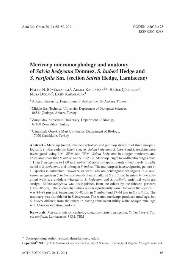
Acta Bot. Croat. 70 (1), 65–80, 2011 CODEN: ABCRA25ISSN 0365–0588
Mericarp micromorphology and anatomy
of Salvia hedgeana Dönmez, S. huberi Hedge and
S. rosifolia Sm. (section Salvia Hedge, Lamiaceae)
HATÝCE N. BÜYÜKKARTAL1, AHMET KAHRAMAN2,*, HATÝCE ÇÖLGEÇEN3,MUSA DOÐAN2, ERSÝN KARABACAK4
1 Ankara University, Department of Biology, 06100 Ankara, Turkey.
2 Middle East Technical University, Department of Biological Sciences,06531 Çankaya, Ankara, Turkey.
3 Zonguldak Karaelmas University, Department of Biology,67100 Zonguldak, Turkey.
4 Çanakkale Onsekiz Mart University, Department of Biology,17020 Çanakkale, Turkey
Abstract – Mericarp (nutlet) micromorphology and pericarp structure of three morpho-logically similar endemic Salvia species; Salvia hedgeana, S. huberi and S. rosifolia wereinvestigated using LM, SEM and TEM. Salvia hedgeana has larger mericarps andabscission scars than S. huberi and S. rosifolia. Mericarp length to width ratio ranges from1.11 in S. hedgeana to 1.60 in S. huberi. Mericarp shape is mainly ovoid, rarely broadlyovoid in S. hedgeana, and oblong in S. huberi. The mericarp surface sculpturing pattern inall species is colliculate. However, exocarp cells are pentangular-hexangular in S. hed-
geana, irregular in S. huberi and rounded and smaller in S. rosifolia. In Salvia huberi anti-clinal walls are undulate whereas in S. hedgeana and S. rosifolia anticlinal walls arestraight. Salvia hedgeana was distinguished from the others by the thickest pericarp(146–185 µm). The sclerenchymatous region significantly varied between the species. Itwas 84–99 µm in S. hedgeana, 56–82 µm in S. huberi and 27–61 µm in S. rosifolia. Themesocarp was also thicker in S. hedgeana. The wetted mericarps produced mucilage, butS. huberi differed from the others in having translucent-milky white opaque mucilagewith fibres or radiating cordons.
Keywords: Mericarp, micromorphology, anatomy, Salvia hedgeana, Salvia huberi, Sal-
via rosifolia, Lamiacaeae, SEM, TEM
ACTA BOT. CROAT. 70 (1), 2011 65
* Corresponding author: e-mail: [email protected]
®2011 by Acta Botanica Croatica, the Faculty of Science, University of Zagreb. All rights reserved.
U:\ACTA BOTANICA\Acta-Botan 1-11\Buyukkartal.vp28. o ujak 2011 16:07:15
Color profile: DisabledComposite 150 lpi at 45 degrees
Introduction
Lamiaceae (the mint family) has a cosmopolitan distribution represented by nearly7200 species belonging to 236 genera, including many well-known plants, herbs, shrubsand trees of horticultural, economic and medicinal significance (HARLEY et al. 2004). It isthe third largest family in Turkey with 45 genera and 574 species, 256 of which are en-demic. The rate of endemism is 44.5% in this family (DAVIS 1965–1985, DAVIS et al. 1988,GÜNER et al. 2000).
Some mericarp (nutlet) characters can be used successfully at many taxonomic levels,depending on the characters chosen and the variation present (GUERIN 2005). Studies onmericarp micromorphology and pericarp anatomy in the family have proved the usefulnessof these characters for species and generic level in the family Lamiaceae (WAGNER 1914;ISLEY 1947; WOJCIECHOWSKA 1958, 1961, 1966, 1972; HEDGE and LAMOND 1968; HUSAIN
et al. 1990; REJDALI 1990; RYDING 1992, 1995, 2001, 2010; MARIN et al. 1994; TURNER andDELPRETE 1996; BUDANTSEV and LOBOVA 1997; ZHOU et al. 1997; DULETI]-LAUŠEVIC andMARIN 1999; MOSQUERO et al. 2002; GUERIN 2005; MOON and HONG 2006; KAYA andDÝRMENCÝ 2008; SALMAKI et al. 2008). ISLEY (1947) distinguished the tribes Ajugaea,Scutellarieae and Stachyeae in USA based on mericarp characters. WOJCIECHOWSKA (1966)investigated mericarp morphology including in some genera of Lamiaceae and pointed outthat there were often important differences at both generic and species level. RYDING (1995)showed that the groups of Prunella and Cleonia plus Lepechinia and Chaunostoma weredistinguishable from other labiates by differences in pericarp structure. BUDANTSEV andLOBOVA (1997) divided the subtribe Nepetinae (as tribe Nepeteae) into three informal genericgroups based on differences in pericarp structure and other characters. WOJCIECHOWSKA
(1966) and MOON and HONG (2006) pointed out that the genus Lycopus had a pericarp anat-omy and mericarp morphology unique in the shape of corky crests and corky ring and thedistribution of glandular trichomes, which were well distinguishable from the other generain the tribe Mentheae. MOON and HONG (2006) also found that mericarp morphological andanatomical characters of Lycopus were useful as diagnostic characters at the specific andinterspecific levels.
Salvia L., the largest genus of the family Lamiaceae, is represented by about 1000 spe-cies. It is distributed extensively in tropical and temperate areas of the Old and New World,with three distinct regions of population: Central and South America (500 spp.), WesternAsia (200 spp.) and Eastern Asia (100 spp.) (WALKER and SYTSMA 2007). Based on themost recent classification (HARLEY et al. 2004), Lamiaceae is divided into seven subfa-milies and the large subfamily Nepetoideae is divided into the three tribes Elsholtzieae,Mentheae and Ocimeae. Mentheae is divided into the three subtribes Salviinae, Menthinaeand Nepetinae. However, the genera Melissa and Heterolamium are unclassified at thisrank. According to their classification, the genus Salvia is classified into the subtribeSalviinae. Salvia species possess only two thecae on each stamen separated by an elongateconnective while most Mentheae have four stamens (WALKER et al. 2004).
Mericarp micromorphology and anatomy are poorly reported in Salvia. In twenty Sal-
via species from Afghanistan, HEDGE (1970) reported that characters of myxocarpy (i.e. theproduction of mucilage when mericarps get in contact with water), thickness of thepericarp and the proportions of its individual layers as well as morphology of mericarps
66 ACTA BOT. CROAT. 70 (1), 2011
BÜYÜKKARTAL H. N., KAHRAMAN A., ÇÖLGEÇEN H., DOÐAN M., KARABACAK E.
U:\ACTA BOTANICA\Acta-Botan 1-11\Buyukkartal.vp28. o ujak 2011 16:07:15
Color profile: DisabledComposite 150 lpi at 45 degrees

could be used in the systematics of the genus. Mericarp micromorphological characters ofdifferent Salvia species was investigated by MARIN et al. (1996) and ORAN (1997). Theyboth pointed out that the variation in thickness of exocarp (=epicarp), mesocarp andendocarp morphology may be useful additional taxonomical markers.
In Turkey, there are 98 Salvia species reported (HEDGE (1982; KAHRAMAN et al. inpress), 52 endemic species (53%), divided into seven sections Salvia Hedge (syn. Eu-
sphace Benth.), Aethiopis Benth., Plethiosphace Benth., Drymosphace Benth., Horminum
Benth. and Hemisphace Benth. (BOISSIER 1879, DOÐAN et al. 2007). Leaf (pinnatisect, tri-sect or simple), calyx (membranous or thick textured, upper lip 2-sulcate or not, concave ornot), corolla (upper lip falcate or not, tube squamulate or not, annulate or not) and stamen(type A, B or C) characteristics are the most important means for distinguishing these sec-tions. The section Salvia has usually pinnatisect leaves, thick-texture calyces, straight up-per corolla lips, annulate corolla tubes and type A stamens with shorter staminal connec-tives than filaments and larger upper theca than the lower theca.
Salvia hedgeana Dönmez, S. huberi Hedge and S. rosifolia Sm. included in the sectionSalvia are endemic to Turkey. They show morphologically close similarities to each other(DÖNMEZ 2001), especially S. huberi and S. rosifolia (HEDGE 1982, personal observation).Salvia hedgeana is also close to S. caespitosa Montbret et Aucher ex Benth. in terms of itshabit and distribution area. Even though micromorphological and anatomical studies canbe used as a powerful tool in delimitation of species, there have been no mericarp micro-morphological and anatomical studies on the species examined. This study aims to exam-ine micromorphological and anatomical characteristics of mericarps of the three Salvia
species which have not been studied to date; using light microscopy, scanning electron mi-croscopy and transmission electron microscopy.
Materials and methods
Different populations of Salvia, Salvia hedgeana Dönmez, S. huberi Hedge and S.
rosifolia Sm. were investigated in localities presented in table 1. The herbarium specimenswere deposited in Department of Biological Sciences, Middle East Technical University,Ankara, Turkey and the Herbarium of Çanakkale Onsekiz Mart University.
Light microscopy (LM)
Mericarps were first examined using a stereomicroscope to ensure that they were ofnormal size and maturity. The ripe mericarps from each population were measured in orderto determine the average mericarp size and abscission scar diameter. The pericarp structureof the mericarps was investigated from softened herbarium specimens. The mature mericarpswere placed in distilled water for 24 hours, 3% glutaraldehyde and then %1 osmiumtetraoxide. The materials were dehydrated through a graduated ethanol series, embedded inEpon 812 (LUFT 1961), sectioned, stained with methylene blue and toluidine blue, and per-manently mounted. These sections were examined for the pericarp features, and thicknessof the pericarp and mesocarp, sclerenchymatous region, endocarp, size of crystals and mu-cilaginous cells were measured and photographed under LM.
MERICARP MICROMORPHOLOGY AND ANATOMY OF SALVIA HEDGEANA
ACTA BOT. CROAT. 70 (1), 2011 67
U:\ACTA BOTANICA\Acta-Botan 1-11\Buyukkartal.vp28. o ujak 2011 16:07:15
Color profile: DisabledComposite 150 lpi at 45 degrees
Scanning Electron Microscopy (SEM)
Mature mericarps were directly placed on aluminium stubs using double-sided adhe-sive tape, sputter coated with gold using a Hummer VII gold-coating apparatus, thenviewed with JEOL-6060 SEM and photographed.
Transmission Electron Microscopy (TEM)
Ultrathin sections of the mericarps were stained with uranyl acetate (STEMPAK andWARD 1964) and lead citrate (SATO 1967), then examined with JEOLL CX-100 TEM andphotographed.
Each quantitative character was analyzed for its mean and median values, range, stan-dard deviation and significance, using the Statistica version 9 software. Significance of dif-ferences was tested using one-way ANOVA and Tukey’s honestly significant difference
68 ACTA BOT. CROAT. 70 (1), 2011
BÜYÜKKARTAL H. N., KAHRAMAN A., ÇÖLGEÇEN H., DOÐAN M., KARABACAK E.
Tab. 1. List of the studied populations of Salvia hedgeana, S. huberi and S. rosifolia.
Species Collection data Collection number
S. hedgeana B7 Sivas: Divriði, Karasar Pass, Uzunkaya to Kayaburun,1450 m, steppe, 25.vii.2008
A. Kahraman 1589Ca,b
B7 Sivas: Divriði, Karasar Pass, near Uzunkaya village,1460 m, steppe, 9.vii.2006
A. Kahraman 1255Aa,c
S. huberi A8 Bayburt: 12 km from Bayburt to Maden, 1615 m,steppe and slopes, 12.vii.2006
E. Karabacak 4979a
A8 Erzurum: 41 km from Erzurum to Tortum, 2121 m,roadside slopes, 14.vii.2006
A. Kahraman 1474a,b,c
A8 Erzurum: Tortum Waterfall, 1010 m, scrubs, 22.vi.2008 E. Karabacak 6137a
A8 Erzurum: Tortum, above Balikli village, 1342 m,stony slopes, 22.vi.2008
E. Karabacak 6144a
A8 Erzurum: 10 km from Tortum to Yusufeli, 1367 m,calcareous slopes and screes, 9.vii.2007
E. Karabacak 5611a
S. rosifolia A8 Bayburt: Gümüþhane to Bayburt, Niþantaþý village,Osluk (Korgan) bridge environs, 1616 m, steppe androcky slopes, 11.vii.2006
E. Karabacak 4963a
A9 Kars: 22 km from Kaðýzman to Kars, 1581 m,roadside slopes, 13.vii.2007
A. Kahraman 1470a,c
A9 Kars: 28 km from Kaðýzman to Kars, 1819 m,roadside slopes, 29.vii.2008
A. Kahraman 1607a,b
A9 Kars: Kaðýzman to Kars, near Kötek village, 1642 m,screes, 8.vii.2007
E. Karabacak 5585a
A9 Erzurum: Oltu, Çamlýdere, 1443 m, slopes, 22.vi.2008 E. Karabacak 6161a
aSpecimens used for morphological studies of mericarps by stereomicroscopy; bSpecimens used for micromor-phological studies of mericarps by SEM; cSpecimens used for anatomical studies of mericarps by LM and TEM.
U:\ACTA BOTANICA\Acta-Botan 1-11\Buyukkartal.vp28. o ujak 2011 16:07:15
Color profile: DisabledComposite 150 lpi at 45 degrees
(HSD) test. The terminology was adjusted for mericarp surface sculpturing (STEARN 1992,SALMAKI et al. 2008) and for pericarp structure (HEDGE 1970, RYDING 2010).
To determine for the absence or presence of myxocarpy, we treated with distilled waterten mericarps from each species examined, following RYDING (1992). Mucilage character-istics regarding colour, consistency and general appearance were followed according toHEDGE (1970).
Results
The significance of differences in basic parameters defines each species (Fig. 1, Tab. 2).Three characters such as mericarp width, pericarp thickness and sclerenchymatous regionthickness allow clear discrimination of the three species. Mericarp length/width ratio helps toseparate Salvia huberi from S. hedgeana and S. rosifolia whereas size of exocarp cells distin-guishes between S. rosifolia and S. hedgeana and S. huberi. Mericarp length, abscission scarsand mesocarp thickness contribute the distinction between S. hedgeana and the remainingtaxa. Nonetheless, some of these characters indicate to some degree overlapping values.
Among the studied qualitative characters of the species, shape of mericarp and exocarpcells and structure of anticlinal walls were found to be the most significant diagnostic char-acters (Tab. 3).
Mericarp micromorphology
Size of mericarps measured from different populations of the species varied from 2.46mm (Salvia huberi) to 4.36 mm (S. hedgeana) in length and 1.83 mm (S. huberi) to 3.44 (S.
hedgeana) in width. Shape of the mericarps was mainly ovoid, rarely broadly ovoid as in S.
hedgeana or oblong as in S. huberi (Fig. 2). They ranged in length to width ratio from 1.11(S. hedgeana) to 1.60 (S. huberi). When mericarps were not fully matured, their colour wasgreenish at first, and then turned to dark brown or blackish. Transverse sections of themericarps were also rounded-trigonous. Abscission scars were almost spherical and theirdiameter ranged between 0.41 mm as in S. huberi and 0.83 mm as in S. hedgeana.
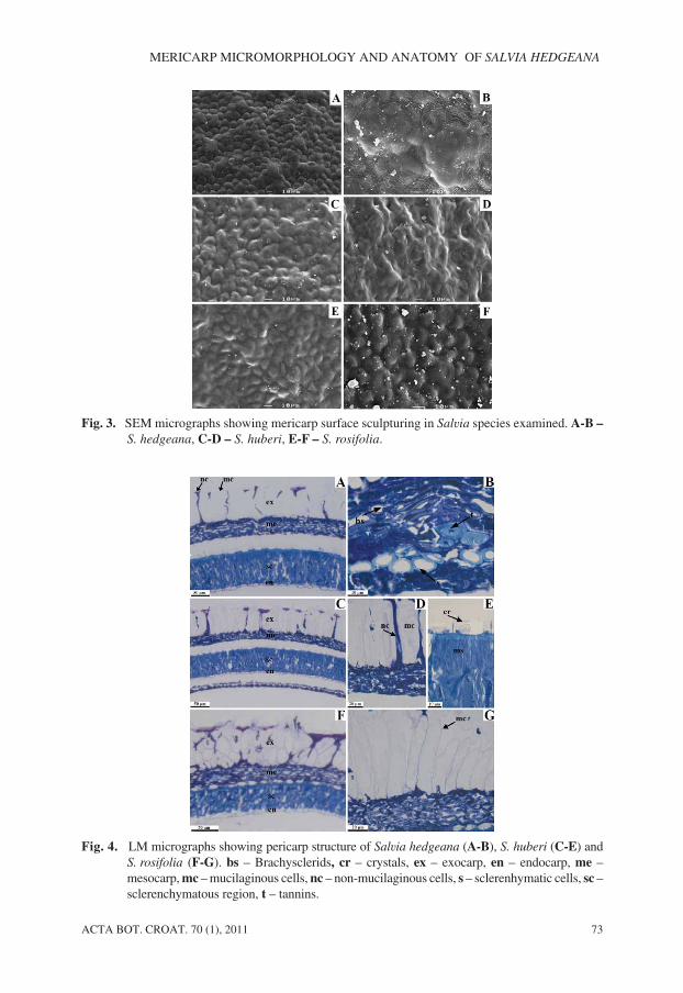
In the species examined, the mericarps were glabrous and the sculpturing pattern was ofthe colliculate type characterized by small hill-like eminences, spaced, covering through-out the mericarp surface. The colliculate sculpturing could be subdivided based on shape ofexocarp cells: pentangular or hexangular in S. hedgeana (Fig. 3A), irregular in S. huberi
(Figs. 3C-D) and rounded in S. rosifolia (Figs. 3E-F). Size of these cells varied from 7.45µm as in S. rosifolia to 27.79 µm as in S. huberi. Striation partially occurred on their surface(Fig. 3B). Anticlinal walls were represented by straight channels in S. hedgeana and S.
rosifolia or undulate channels in S. huberi. Outer periclinal walls were convex, but alsoconsisted of small holes in S. huberi (Figs. 3C-D).
Mericarp anatomy
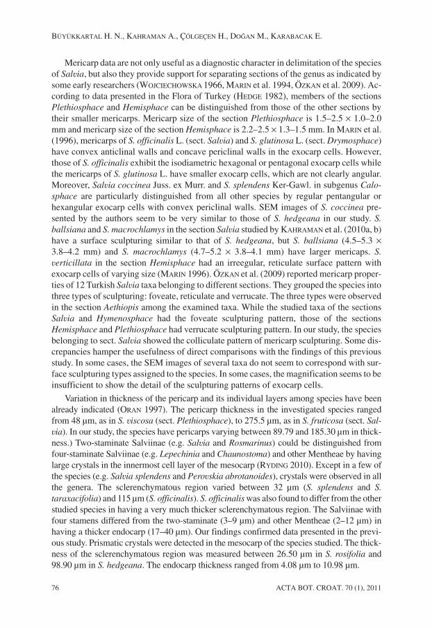
The LM and TEM investigation revealed a pericarp differentiated into four main re-gions: the exocarp (outer epidermis), mesocarp, sclerenchyma region and endocarp (innerepidermis) (Figs. 4A, C, F). The outermost region was the exocarp, consisting of a singlelayer of parenchymatous cells. These cells were differentiated into two types: large oblongor oval mucilaginous cells in groups of one or more and narrow non-mucilaginous cells
ACTA BOT. CROAT. 70 (1), 2011 69
MERICARP MICROMORPHOLOGY AND ANATOMY OF SALVIA HEDGEANA
U:\ACTA BOTANICA\Acta-Botan 1-11\Buyukkartal.vp28. o ujak 2011 16:07:15
Color profile: DisabledComposite 150 lpi at 45 degrees

70 ACTA BOT. CROAT. 70 (1), 2011
BÜYÜKKARTAL H. N., KAHRAMAN A., ÇÖLGEÇEN H., DOÐAN M., KARABACAK E.
Fig. 1. Box plots of the main discriminant quantitative characters in Salvia species examined.
U:\ACTA BOTANICA\Acta-Botan 1-11\Buyukkartal.vp28. o ujak 2011 16:07:15
Color profile: DisabledComposite 150 lpi at 45 degrees

AC
TA
BO
T.C
RO
AT
.70(1),2011
71
ME
RIC
AR
PM
ICR
OM
OR
PHO
LO
GY
AN
DA
NA
TO
MY
OF
SA
LV
IAH
ED
GE
AN
A
Tab. 2. Overview of quantitative characteristics of the species accessions examined. Numbers refer to means ± standard deviations [medians] and (ranges).N = 50, *p < 0.001, NS = not significant.
Character S. hedgeana S. huberi S. rosifolia p Tukey’s HSD test
Mericarp length (mm) 3.66 ± 0.37 [3.70]
(3.12–4.36)2.90 ± 0.27 [2.86]
(2.46–3.54)2.96 ± 0.26 [2.90]
(2.56–3.63)* S. hedgeana-*-S. huberi, S.
rosifolia
Mericarp width (mm) 2.88 ± 0.33 [2.78]
(2.26–3.44)2.17 ± 0.20 [2.13]
(1.83–2.86)2.36 ± 0.23 [2.34]
(1.91–2.87)* S. hedgeana-*-S. huberi, S.
rosifolia; S. huberi-*-S. rosifolia
Mericarp length/width ratio 1.28 ± 0.06 [1.28]
(1.11–1.38)1.34 ± 0.10 [1.32]
(1.20–1.60)1.25 ± 0.05 [1.25]
(1.17–1.36)* S. huberi-*-S. hedgeana, S.
rosifolia
Abscission scar diameter (mm) 0.69 ± 0.08 [0.71]
(0.55–0.83)0.56 ± 0.08 [0.55]
(0.41–0.75)0.60 ± 0.09 [0.60]
(0.46–0.79)* S. hedgeana-*-S. huberi, S.
rosifolia
Size of exocarp cells (µm) 15.88 ± 3.84 [15.06]
(10.06–24.04)17.49 ± 4.00 [17.26]
(11.38–27.79)11.66 ± 2.11 [11.39]
(7.45–16.43)* S. rosifolia-*- S. hedgeana, S.
huberi
Pericarp thickness (µm) 169.39 ± 10.23 [170.00]
(145.53–185.30)129.30 ± 12.01 [130.65]
(100.83–151.90)114.87 ± 13.83 [122.35]
(89.79–133.22)* S. hedgeana-*-S. huberi, S.
rosifolia; S. huberi-*-S. rosifolia
Mesocarp thickness (µm) 64.26 ± 6.31 [62.73]
(55.64–75.91)47.17 ± 8.53 [45.44]
(34.58–61.91)45.06 ± 7.33 [47.08]
(31.18–56.20)* S. hedgeana-*-S. huberi, S.
rosifolia
Crystal length/width ratio 0.50 ± 0.09 [0.48]
(0.33–0.65)0.47 ± 0.10 [0.51]
(0.30–0.60)0.44 ± 0.06 [0.43]
(0.35–0.55)NS
Sclerenchymatous regionthickness (µm)
91.01 ± 3.77 [91.30]
(84.00–98.90)69.42 ± 8.05 [70.25]
(55.50–82.34)46.38 ± 11.27 [47.15]
(26.50–60.90)* S. hedgeana-*-S. huberi, S.
rosifolia; S. huberi-*-S. rosifolia
Endocarp thickness (µm) 7.16 ± 1.93 [6.80]
(4.50–10.98)6.82 ± 1.60 [6.77]
(4.08–10.62)7.43 ± 1.25 [7.18]
(5.13–10.56)NS
U:\ACTA BOTANICA\Acta-Botan 1-11\Buyukkartal.vp
28. oujak 2011 16:07:15
Color profile: Disabled
Composite 150 lpi at 45 degrees
with thin cavities, located between the former cells (Figs. 4A, C, D, F, G). The mucilagi-nous cells had thickened walls and their size was very variable within the same mericarp.Hence, we measured the pericarp thickness without the mucilaginous cells. It ranged from89.79 µm in S. rosifolia to 185.30 µm in S. hedgeana. The mucilage pocket encircled thecentral cytoplasmic column with darkly staining mucilage (Figs. 5C, F).
Below the exocarp, there is the mesocarp region (31.18–75.91 µm thick) composed of adark amorphous mass of several strongly compressed and almost indistinguishable cells.This region contained several groups of sclerenchymatic cells with the luminar cavity en-larged (Figs. 4B, D). Brachysclereids (=stone cells) were characterized by thick walls(Figs. 4B, 5A, C, E). These cells accumulated tannins. The innermost layer of the mesocarpcontained prismatic crystals (Figs. 4A, C, E, F). They were 6.67–13.22 long µm and15.14–32.52 µm wide. They varied in length to width ratio between 0.30 and 0.65.
The mesocarp was followed by the sclerenchymatous region consisting of a layer ofthick-walled macrosclereids, i.e. Malpighian cells (Figs. 4E, 5B, D) in which tannins accu-mulated (Fig. 5B). The sclerenchymatous region thickness varied between 26.50 µm in S.
rosifolia and 98.90 µm thick in S. hedgeana, with small and rounded luminar cavities at ornear the centre (Fig. 5). The thickness of the sclerenchymatous layer was nearly half of thetotal pericarp thickness.
72 ACTA BOT. CROAT. 70 (1), 2011
BÜYÜKKARTAL H. N., KAHRAMAN A., ÇÖLGEÇEN H., DOÐAN M., KARABACAK E.
Tab. 3. Characteristics in Salvia examined.
Character S. hedgeana S. huberi S. rosifolia
Mericarp shape Ovoid to broadly ovoid Ovoid to oblong OvoidMericarp transversesection
Rounded-trigonous Rounded-trigonous Rounded-trigonous
Mericarp colour Mostly blackish Mostly blackish Mostly blackishAbscission scar shape ±spherical ±spherical ±sphericalSurface sculpturing type Colliculate Colliculate ColliculateShape of exocarp cells Pentangular-hexangular Irregular RoundedAnticlinal walls Represented by
straight channelsRepresented byundulate channels
Represented bystraight channels
Periclinal walls Convex Convex with smallholes
Convex
Fig. 2. SEM micrographs showing general appearance of mericarps in Salvia species examined.A – S. hedgeana, B – S. huberi, C – S. rosifolia.
U:\ACTA BOTANICA\Acta-Botan 1-11\Buyukkartal.vp28. o ujak 2011 16:07:16
Color profile: DisabledComposite 150 lpi at 45 degrees
ACTA BOT. CROAT. 70 (1), 2011 73
MERICARP MICROMORPHOLOGY AND ANATOMY OF SALVIA HEDGEANA
Fig. 3. SEM micrographs showing mericarp surface sculpturing in Salvia species examined. A-B –
S. hedgeana, C-D – S. huberi, E-F – S. rosifolia.
Fig. 4. LM micrographs showing pericarp structure of Salvia hedgeana (A-B), S. huberi (C-E) andS. rosifolia (F-G). bs – Brachysclerids, cr – crystals, ex – exocarp, en – endocarp, me –mesocarp, mc – mucilaginous cells, nc – non-mucilaginous cells, s – sclerenhymatic cells, sc –sclerenchymatous region, t – tannins.
U:\ACTA BOTANICA\Acta-Botan 1-11\Buyukkartal.vp28. o ujak 2011 16:07:17
Color profile: DisabledComposite 150 lpi at 45 degrees

The endocarp, consisting of a single layer of transversely arranged cells, was 4.08–10.98 µm thick (Figs. 4A, C, F).
A mucilage reaction (myxocarpy) was seen in the wetted mericarps of the studied spe-cies. In S. huberi and S. rosifolia mucilage was formed after the first half hour of wetting,but in S. hedgeana the time taken was longer than one hour. According to colour, consis-tency and degree of transparency, two mucilage types were observed: transparent andfibreless or having radiating cordons embraced by the mass of mucilage, and translu-cent-milky white opaque with fibres or radiating cordons embraced by the mass of muci-lage. The former type was observed in S. hedgeana (Fig. 6A) and S. rosifolia (Fig. 6D)whereas the latter type was detected in S. huberi (Fig. 6B-C).
Discussion
The size, shape, length to width ratio, abscission scar diameter, size and shape ofexocarp cells and structure of anticlinal walls varied significantly in the investigatedspecies (Tabs. 2, 3). Salvia hedgeana was easily distinguished from S. huberi and S.
rosifolia by large mericarps and abscission scars. Their mericarps were ovoid, but S. huberi
had also oblong mericarps. In addition, the mericarps ranged in their length to width ratiofrom 1.11 in S. hedgeana to 1.60 S. huberi. HEDGE (1982) reported that some species of theTurkish Salvia had brown or black mericarps and rounded, trigonous or rounded-trigonous
74 ACTA BOT. CROAT. 70 (1), 2011
BÜYÜKKARTAL H. N., KAHRAMAN A., ÇÖLGEÇEN H., DOÐAN M., KARABACAK E.
Fig. 5. TEM micrographs showing cells in pericarps of S. hedgeana with brachysclereids and tanninsin the mesocarp (A), S. hedgeana with macrosclereids and tannins in the sclerechymatous re-gion (B), S. huberi with mucilaginous cells in the exocarp and with brachysclereids in themesocarp (C), S. huberi with macrosclereids in the sclerenchymatous region (D), S. rosifolia
with brachysclereids in the mesocarp (E), and S. rosifolia with mucilaginous cells in theexocarp (F). br – brachysclereids, ma – macrosclereids, mu – mucilaginous cells, t – tannins.
U:\ACTA BOTANICA\Acta-Botan 1-11\Buyukkartal.vp28. o ujak 2011 16:07:17
Color profile: DisabledComposite 150 lpi at 45 degrees

mericarps (the most common) in transverse sections. However, the investigated specieshad mericarps similar in colour, transverse sections, as well as, abscission scar shape. Theabscission scar and mericarp shape were invariable in the tribe Saturejeae while these char-acters varied significantly in the Westringieae (HUSAIN et al. 1990).
The studied species showed a similar sculpturing pattern, but exocarp cells werepentangular-hexangular in S. hedgeana, irregular in S. huberi and rounded and smaller in S.
rosifolia. While exocarp cells of S. huberi had undulate anticlinal walls, those of S.
hedgeana and S. rosifolia had straight anticlinal walls. Small holes were also present on theouter periclinal walls of S. huberi. Though mericarp sculpturing was found to be useful forseparating species within the sections in the genus Stachys, it did not seem to be a helpfulcharacter at the infrageneric level (SALMAKI et al. 2008). Mericarp shape, nature of theabscission scar, nature of surface sculpturing, exocarp cell shape and sculpturing, and na-ture of the indumentum were mainly useful for infrageneric delimitation in the generaHemigenia and Microcorys (GUERIN 2005).
According to the mericarp anatomical properties of the species examined, the thickestpericarp was found in S. hedgeana, whereas S. rosifolia had the thinnest pericarp. More-over, the thickness of the sclerenchymatous region significantly varied among the species.DULETI]-LAUŠEVIC and MARIN (1999) determined that the pericarp thickness in the tribeNepetoideae of Lamiaceae was often correlated with dimensions of mericarps. However,they also indicated that the pericarps of Mentha aquatica mericarps were very thick (ap-proximately 95 µm), even though the species had very small mericarps (nearly 0.8 mm �
0.6 mm). In our work, S. hedgeana had the largest mericarps that were composed of thethickest pericarp structure. In the mesocarp and sclerenchmatous region, tannins were rec-ognized. They were thought to protect the plant against dehydration, rotting and damage bypredators such as animals and insects (FAHN 1990).
ACTA BOT. CROAT. 70 (1), 2011 75
MERICARP MICROMORPHOLOGY AND ANATOMY OF SALVIA HEDGEANA
Fig. 6. Stereomicroscopic micrographs showing the mucilaginous reaction (myxocarpy) on mericarpsin Salvia hedgeana (A), S. huberi (B-C), and S. rosifolia (D).
U:\ACTA BOTANICA\Acta-Botan 1-11\Buyukkartal.vp28. o ujak 2011 16:07:18
Color profile: DisabledComposite 150 lpi at 45 degrees
Mericarp data are not only useful as a diagnostic character in delimitation of the speciesof Salvia, but also they provide support for separating sections of the genus as indicated bysome early researchers (WOJCIECHOWSKA 1966, MARIN et al. 1994, ÖZKAN et al. 2009). Ac-cording to data presented in the Flora of Turkey (HEDGE 1982), members of the sectionsPlethiosphace and Hemisphace can be distinguished from those of the other sections bytheir smaller mericarps. Mericarp size of the section Plethiosphace is 1.5–2.5 � 1.0–2.0mm and mericarp size of the section Hemisphace is 2.2–2.5 � 1.3–1.5 mm. In MARIN et al.(1996), mericarps of S. officinalis L. (sect. Salvia) and S. glutinosa L. (sect. Drymosphace)have convex anticlinal walls and concave periclinal walls in the exocarp cells. However,those of S. officinalis exhibit the isodiametric hexagonal or pentagonal exocarp cells whilethe mericarps of S. glutinosa L. have smaller exocarp cells, which are not clearly angular.Moreover, Salvia coccinea Juss. ex Murr. and S. splendens Ker-Gawl. in subgenus Calo-
sphace are particularly distinguished from all other species by regular pentangular orhexangular exocarp cells with convex periclinal walls. SEM images of S. coccinea pre-sented by the authors seem to be very similar to those of S. hedgeana in our study. S.
ballsiana and S. macrochlamys in the section Salvia studied by KAHRAMAN et al. (2010a, b)have a surface sculpturing similar to that of S. hedgeana, but S. ballsiana (4.5–5.3 �
3.8–4.2 mm) and S. macrochlamys (4.7–5.2 � 3.8–4.1 mm) have larger mericaps. S.
verticillata in the section Hemisphace had an irreegular, reticulate surface pattern withexocarp cells of varying size (MARIN 1996). ÖZKAN et al. (2009) reported mericarp proper-ties of 12 Turkish Salvia taxa belonging to different sections. They grouped the species intothree types of sculpturing: foveate, reticulate and verrucate. The three types were observedin the section Aethiopis among the examined taxa. While the studied taxa of the sectionsSalvia and Hymenosphace had the foveate sculpturing pattern, those of the sectionsHemisphace and Plethiosphace had verrucate sculpturing pattern. In our study, the speciesbelonging to sect. Salvia showed the colliculate pattern of mericarp sculpturing. Some dis-crepancies hamper the usefulness of direct comparisons with the findings of this previousstudy. In some cases, the SEM images of several taxa do not seem to correspond with sur-face sculpturing types assigned to the species. In some cases, the magnification seems to beinsufficient to show the detail of the sculpturing patterns of exocarp cells.
Variation in thickness of the pericarp and its individual layers among species have beenalready indicated (ORAN 1997). The pericarp thickness in the investigated species rangedfrom 48 µm, as in S. viscosa (sect. Plethiosphace), to 275.5 µm, as in S. fruticosa (sect. Sal-
via). In our study, the species have pericarps varying between 89.79 and 185.30 µm in thick-ness.) Two-staminate Salviinae (e.g. Salvia and Rosmarinus) could be distinguished fromfour-staminate Salviinae (e.g. Lepechinia and Chaunostoma) and other Mentheae by havinglarge crystals in the innermost cell layer of the mesocarp (RYDING 2010). Except in a few ofthe species (e.g. Salvia splendens and Perovskia abrotanoides), crystals were observed in allthe genera. The sclerenchymatous region varied between 32 µm (S. splendens and S.
taraxacifolia) and 115 µm (S. officinalis). S. officinalis was also found to differ from the otherstudied species in having a very much thicker sclerenchymatous region. The Salviinae withfour stamens differed from the two-staminate (3–9 µm) and other Mentheae (2–12 µm) inhaving a thicker endocarp (17–40 µm). Our findings confirmed data presented in the previ-ous study. Prismatic crystals were detected in the mesocarp of the species studied. The thick-ness of the sclerenchymatous region was measured between 26.50 µm in S. rosifolia and98.90 µm in S. hedgeana. The endocarp thickness ranged from 4.08 µm to 10.98 µm.
76 ACTA BOT. CROAT. 70 (1), 2011
BÜYÜKKARTAL H. N., KAHRAMAN A., ÇÖLGEÇEN H., DOÐAN M., KARABACAK E.
U:\ACTA BOTANICA\Acta-Botan 1-11\Buyukkartal.vp28. o ujak 2011 16:07:18
Color profile: DisabledComposite 150 lpi at 45 degrees
Myxocarpy, the production of mucilage by wetted mericarps, is widespread in thesubfamily Nepetoideae (WAGNER 1914; HEDGE 1970; SWARBRICK 1971; WITZTUM 1978;RYDING 1992, 2001, 2010; DULETI]-LAUŠEVIC and MARIN 1999; HARLEY et al. 2004).Morphologically allied species had similar mucilage properties (HEDGE 1970). In thisstudy, we observed that the species did produce mucilage on their mericarp surfaces, but S.
huberi seemed to differ from the others in colour, consistency and degree of transparency.In a total of over forty Salvia species investigated in Southwest Asia, only three failed toproduce mucilage on the mericarp surface (HEDGE 1970). All the Salvia species knownfrom Afghanistan had mucilage formation, with one exception (HEDGE 1970). These muci-lage-producing species were grouped into four basic types according to their mucilagecharacteristics: transparent, translucent, milky opaque and brownish opaque. The occur-rence of mucilage was found in 14 Salvia species in Jordan which had not been previouslyexamined (ORAN 1997). However, one species, S. napifolia (sect. Hemisphace), onlysometimes produced a very small amount or no visible mucilage. The greatest mucilageproduction was detected in S. viridis (sect. Horminum). There was no mucilage on themericarp surface of all the taxa studied in Lycopus, but a moderately strong mucilaginousreaction was seen in Elsholtzia blanda Benth. in the tribe Elsholtzieae (MOON and HONG
2006).
Mucilage plays a significant role in anchoring the mericarp to the soil. Presence or ab-sence of mucilage seems to be a very homoplastic character in Nepetoideae; most of thespecies-rich genera and many of the genera with few species contained myxocarpic as wellas non-myxocarpic species (RYDING 1992). Therefore, the amount of mucilage may evolvequickly in order to adapt the species to different biological conditions. Even though thepresence of mucilage may provide a considerable selective advantage under certain condi-tions, the production of mucilage may be costly for the plants, and it may be quickly lostwhere it has no function. Plants growing in moist habitats more often had non-mucilagi-nous mericarps (RYDING 1992, 2001). The whole of Lycopus species grow in low wetplaces and have non-mucilaginous mericarps (MOON and HONG 2006). The species of Sal-
via examined grow in dry habitats and have mucilaginous mericarps.
Mericarp micromorphological and anatomical characteristics might be helpful in theidentification of the species studied. Nevertheless, the value of these characteristics can bebetter appreciated by examining other species of Salvia.
Acknowledgements
The authors thank the Scientific and Technical Research Council of Turkey for the fi-nancial assistance (TUBÝTAK-TBAG-104 T 450) and Çanakkale Onsekiz Mart University Re-search Fund (2007/14).
References
BOISSIER, E. P., 1879: Flora Orientalis. Genevae et Basileae.BUDANTSEV, A. V., LOBOVA, T. A., 1997: Fruit morphology, anatomy and taxonomy of tribe
Nepeteae (Labiatae). Edinburgh Journal of Botany 54, 183–216
ACTA BOT. CROAT. 70 (1), 2011 77
MERICARP MICROMORPHOLOGY AND ANATOMY OF SALVIA HEDGEANA
U:\ACTA BOTANICA\Acta-Botan 1-11\Buyukkartal.vp28. o ujak 2011 16:07:18
Color profile: DisabledComposite 150 lpi at 45 degrees
DAVIS, P. H., 1965–1985: Flora of Turkey and the east Aegean islands, 1–9. Edinburgh Uni-versity Press, Edinburgh.
DAVIS, P. H., MILL, R. R., TAN, K., 1988: Flora of Turkey and the east Aegean islands, 10(First Supplement). Edinburg University Press, Edinburg.
DOÐAN, M., AKAYDIN, G., CELEP, F., BAGHERPOUR, S., KAHRAMAN, A., KARABACAK, E.,2007: Infrageneric delimitation of Salvia L. (Labiatae) in Turkey. Proceedings 7 Inter-national Symposium on plant life of southwest Asia, Eskiºehir, 1–12.
DÖNMEZ, A., 2001: A new Turkish species of Salvia L. (Lamiaceae). Botanical Journal ofthe Linnean Society 137, 413–416.
DULETI]-LAUŠEVIC, S., MARIN, P. D., 1999: Pericarp structure and myxocarpy in selectedgenera of Nepetoideae (Lamiaceae). Nordic Journal of Botany 19, 435–446.
FAHN, A., 1990. Plant anatomy. Pergamon, Oxford.
GUERIN, G. R., 2005: Nutlet morphology in Hemigenia R. Br. Microcorys R. Br. (Lamia-ceae). Plant Systematics and Evolution 254, 49–68.
GÜNER, A., ÖZHATAY, N., EKÝM, T., BAªER, K. H. C., 2000: Flora of Turkey and the eastAegean islands 11 (Second Supplement). Edinburgh University Press, Edinburgh.
HARLEY, R. M., ATKINS, S., BUDANTSEV, A. L., CANTINO, P. D., CONN, B. J., GRAYER, R.,HARLEY, M. M., DE KOK, R., KRESTOVSKAJA, T., MORALES, R., PATON A. J., RYDING, O.,UPSON, T., 2004: Labiatae. In: KADEREIT, J. W. (ed.), The families and genera of vascu-lar plants 7, Lamiales, 167–275. Springer-Verlag, Berlin.
HEDGE, I. C., LAMOND, J., 1968: Studies in the flora of Afghanistan: 7, Labiatae. Notesfrom the Royal Botanic Garden 28, 97–123.
HEDGE, I. C., 1970: Observations on the mucilage of Salvia fruits. Notes from the RoyalBotanic Garden Edinburgh 30, 79–95.
HEDGE, I. C., 1982: Flora of Turkey and the east Aegean islands. In: DAVIS, P.H. (ed.), Salvia
L., 7, 400–461. Edinburgh University Press, Edinburgh.
HUSAIN, S. Z., MARIN, P. D., ŠILI], C, QAISER, M., PETKOVI], B., 1990: A micromorpholo-gical study of some representative genera in the tribe Satureae (Lamiaceae). BotanicalJournal of the Linnean Society 103, 59–80.
ISLEY, D., 1947: Investigations in seed classification by family characteristics. Iowa Agri-cultural Experimental University Journal of Research 12, 247–260.
KAHRAMAN, A., DOGAN, M., CELEP, F., KOYUNCU, M., AKAYDIN, G., 2010a: Morphology,anatomy, palynology and nutlet micromorphology of the rediscovered Turkish endemicSalvia ballsiana (Lamiaceae) and their taxonomic implications. Nordic Journal of Bot-any 28, 91–99.
KAHRAMAN, A., CELEP, F., DOÐAN, M., 2010b: Morphology, anatomy, palynology andnutlet micromorphology of Salvia macrochlamys (Labiatae) in Turkey. Biologia 65,219–227.
KAHRAMAN, A., DOGAN, M., CELEP, F., in press: A new endangered species of Salvia
(Lamiaceae) from Turkey. Nordic Journal of Botany.
KAYA, A., DIRMENCI, T., 2008: Nutlet surface micromorphology of the genus Nepeta L.,(Lamiaceae) in Turkey. Turkish Journal of Botany 32, 103–112.
78 ACTA BOT. CROAT. 70 (1), 2011
BÜYÜKKARTAL H. N., KAHRAMAN A., ÇÖLGEÇEN H., DOÐAN M., KARABACAK E.
U:\ACTA BOTANICA\Acta-Botan 1-11\Buyukkartal.vp28. o ujak 2011 16:07:19
Color profile: DisabledComposite 150 lpi at 45 degrees
LUFT, J. H., 1961: Improvements in epoxy resin embedding methods. Journal of Biophysi-cal and Biochemical Cytology 9, 409.
MARIN, P. D., PETKOVI], B. P., DULETI], S., 1994: Nutlet sculpturing of selected Teucrium
species (Lamiaceae): A character of taxonomic significance. Plant Systematics andEvolution 192, 199–214.
MARIN, P. D., DULETI], S., PETKOVI], B., 1996: Nutlet ornamentation in selected Salvia L.species (Lamiaceae). Flora Mediterranea 6, 203–211.
MOON, H. K., HONG, S. P., 2006: Nutlet morphology and anatomy of the genus Lycopus(Lamiaceae. Mentheae). Journal of Plant Research 119, 633–644.
MOSQUERO, M. A. M., JUAN, R., PASTOR, J. E., 2002: Morphological and anatomical stud-ies on nutlets of Nepeta L. (Lamiaceae) from South-West Spain. Acta Botanica Malaci-tana 27, 15–26.
ORAN, S. A., 1997: Nutlet anatomy of the genus Salvia L. in Jordan. Flora Mediterranea 7,27–40.
ÖZKAN, M., AKTAÞ, K., ÖZDEMÝR, C., GUERIN, G., 2009: Nutlet morphology and its taxo-nomic utility in Salvia (Lamiaceae: Mentheae) from Turkey. Acta Botanica Croatica68, 105–115.
REJDALI, M., 1990: Seed morphology and taxonomy of the North African species of Side-
ritis L. (Lamiaceae). Botanical Journal of the Linnean Society 103, 317–324.RYDING, O., 1992: The distribution and evolution of myxocarpy in Lamiaceae. In: HARLEY,
R. M. and REYNOLDS, T. (eds.), Advances in Labiate science, 85–97. The Royal Botani-cal Garden, Kew.
RYDING, O., 1995: Pericarp structure and phylogeny of Lamiaceae-Verbenaceae complex.Plant Systematics and Evolution 198, 101–141.
RYDING, O., 2001: Myxocarpy in the Nepetoideae (Lamiaceae) with notes on myxodia-spory in general. Systematics and Geography of Plants 71, 503–514.
RYDING, O., 2010: Pericarp structure and phylogeny of tribe Mentheae (Lamiaceae). PlantSystematics and Evolution 285, 165–175.
SALMAKI, Y., ZARRE, S., JAMZAD, Z., 2008: Nutlet micromorphology and its systematic im-plication in Stachys L. (Lamiaceae) in Iran. Feddes Repertorium 119, 607–621.
SATO, J. E., 1967: A modified method for lead staining of thin sections. Journa of ElectronMicroscopy 16, 133.
STEARN, W. T., 1992: Botanical Latin, London.STEMPAK, J. G., WARD, W. P., 1964: An improved staining method for electron microscopy.
Journal Cell Biology 22, 697.SWARBRICK, J. T., 1971: External mucilage production by seeds of British plants. Botanical
Journal of the Linnean Society 64, 157–162.TURNER, B. L., DELPRETE, P. G., 1996: Nutlet sculpturing in Scutellaria sect. Resinosa
(Lamiaceae) and its taxonomic utility. Plant Systematics and Evolution 199, 109–120.WAGNER, S., 1914: Contribution à l’étude anatomique du fruit des labiées. Université de
Paris, Lons Le-Saunier.WALKER, J. B., SYTSMA, K. J., TREUTLEIN, J., WINK, M., 2004: Salvia (Lamiaceae) is not
monophletic: Implications for the systematics, radiation, and ecological specializationsof Salvia and tribe Mentheae. American Journal of Botany 91, 1115–1125.
ACTA BOT. CROAT. 70 (1), 2011 79
MERICARP MICROMORPHOLOGY AND ANATOMY OF SALVIA HEDGEANA
U:\ACTA BOTANICA\Acta-Botan 1-11\Buyukkartal.vp28. o ujak 2011 16:07:19
Color profile: DisabledComposite 150 lpi at 45 degrees
WALKER, J. B., SYTSMA, K. J., 2007: Staminal evolution in the genus Salvia (Lamiaceae):Molecular phylogenetic evidence for multiple origins of the staminal lever. Annals ofBotany 100, 375–391.
WITZTUM, A., 1978: Mucilaginous plate cells in the nutlet epidermis of Coleus blumei
Benth. (Labiatae). Botanical Gazette 139, 430–435.WOJCIECHOWSKA, B., 1958: Taxonomy, morphology and anatomy of seeds in the genus
Salvia L. (In Polish). Monographie Botanicae 6, 1–56.WOJCIECHOWSKA, B., 1961: Fruits in the middle European species of some genera of Sta-
chyoideae (fam. Labiatae). Monographie Botanicae 12, 89–120.WOJCIECHOWSKA, B., 1966: Morphology and anatomy of fruits and seeds in the family La-
biatae with particular respect to medicinal species. Monographie Botanicae 21, 119–243.WOJCIECHOWSKA, B., 1972: Morphology and anatomy of fruits of Scutellaria, Chaiturus,
Galeobdolon and Sideritis of the family Labiatae. Monographie Botanicae 37, 137–168.ZHOU, S. L., PAN, K. Y., HONG, D. Y., 1997: Pollen and nutlet morphology in Mosla
(Labiatae) and their systematic value. Israel Journal of Plant Sciences 45, 343–350.
80 ACTA BOT. CROAT. 70 (1), 2011
BÜYÜKKARTAL H. N., KAHRAMAN A., ÇÖLGEÇEN H., DOÐAN M., KARABACAK E.
U:\ACTA BOTANICA\Acta-Botan 1-11\Buyukkartal.vp28. o ujak 2011 16:07:19
Color profile: DisabledComposite 150 lpi at 45 degrees
View publication statsView publication stats