Merged files title - UCL Discovery
154
Title Page The skeletal muscle channelopathies: phenotype, genotype and pathogenesis Emma Louise Matthews UCL Submission for PhD examination
Transcript of Merged files title - UCL Discovery

Title Page
The skeletal muscle channelopathies: phenotype, genotype and pathogenesis
Emma Louise Matthews
UCL
Submission for PhD examination

Declaration
I, Emma Louise Matthews confirm that the work presented in this thesis is my own.
Where information has been derived from other sources, I confirm that this has been
indicated in the thesis.

Abstract
The skeletal muscle channelopathies are a group of inherited disorders due to the
dysfunction of voltage gated channels in the sarcolemma resulting in abnormal
membrane excitability. Simplistically they are broadly divided into those that result from
an “over excited” membrane (the non-dystrophic myotonias) and those due to an
inexcitable one (the periodic paralyses). Skeletal muscle channelopathies were described
clinically long before they were genotyped or hypotheses regarding pathogenesis fully
evolved. This thesis explores all three, the phenotype, the genotype and recent insights
into the pathogenesis.
Detailed clinical and neurophysiologic examination of a large group of patients identified
new aspects of the phenotype including neonatal presentations with important
implications for early life care. Morphological findings are also expanded with the
presence of inflammatory infiltrates, not previously described in the channelopathies.
Extensive DNA sequencing of causative genes was undertaken in a carefully genotyped
cohort. In hypokalaemic periodic paralysis an exclusive relationship between mutations
and the channel voltage sensor emerged which relates closely to recent
electrophysiological evidence of a “gating pore” disease mechanism. A small but
significant minority of cases remain however where no mutation is found. The
implication of other potential genetic mechanisms or even undescribed genes in these
cases is discussed.
Current drug therapies are also examined in three separate cohorts and evidence suggests
acetazolamide, a commonly prescribed treatment, may only be effective in 50-60% of
those with hypokalaemic periodic paralysis. A tentative relationship between efficacy and
genotype also emerges.
Patch clamp studies show significant loss of function of the main alpha pore of the
sodium channel in periodic paralysis but the implications of this in light of the “gating
pore” hypothesis are discussed. Tentative explorations are made as to the viability of
performing future studies in myoctes as opposed to the traditional HEK cell model with
early experiments illustrating limitations.

Acknowledgements
Firstly I must thank the patients and families I met during this thesis. The “skeletal
muscle channelopathies” are a small community but one filled with individuals who
meet their problems with unending humour and who gave me their time and
experience so generously.
The entire Neurogenetics Laboratory staff also receives a big thank you. There is not
a single member of the department who didn’t help me in some way during my thesis.
All the staff at the MRC Centre for Neuromuscular diseases are equally deserving of
thanks. It must be said that both departments not only helped me in practical terms but
extended a very warm welcome and injected frequent humour into the often long and
lonely hours.
Some individuals however cannot go without special mention. Anne Grayson – I will
never forget the day you walked with me to Gower Street in the rain. Mary Davis –
those hours spent behind the door in your office were very special and I miss you.
Mary Sweeney, I have said it before and will say it again, there would have been no
thesis without you but it is your friendship that I treasure most.
My supervisors, Stephanie Schorge, who taught me that the best way to survive
electrophysiology is to always, have wine in the fridge! Prof Hanna –who taught and
inspired me enormously but especially for that first opportunity and continued belief I
will always be grateful.
Finally, to Mr Matthews whose selflessness was infinite, and this thesis can only be
dedicated to my very precious family.

Abbreviations
ACZ: acetazolamide
ATP: adenosine triphosphate
ATS: Andersen Tawil syndrome
CA: carbonic anhydrase
CMAP: compound muscle action potential
CK: creatine kinase
DM1: myotonic dystrophy type 1
DM2: myotonic dystrophy type 2 (or PROMM proximal myotonic myopathy)
DMEM: Dulbecco's Modified Eagle Medium
ECG: electrocardiogram
EMG: electromyography
FBS: Foetal bovine serum
GOSH: Great Ormond Street Hospital
HEK: human embryonic kidney
HyperPP: hyperkalaemic periodic paralysis
HypoPP: hypokalaemic periodic paralysis
IRK: inward rectifying potassium channel
MC: myotonia congenita
NCG: National Commissioning Group
NDM: non-dystrophic myotonia
NHNN: National Hospital for Neurology and Neurosurgery
PAM: potassium aggravated myotonia
PCR: polymerase chain reaction
PMC: paramyotonia congenita
RyR: ryanodine receptor
RyR1: isoform of ryandine receptor primarily expressed in skeletal muscle
SCM: sodium channel myotonia
TBE: Tris Borate Ethylenediaminetetraacetic acid
TTX: tetrodotoxin

WT: wild type

Contents
Page no.
Introduction
1.0 The Role of voltage-gated channels in muscle cells 2
1.1 Structure of Nav1.4 and Cav1.1 4
1.2 Elements determining the ion selectivity of Nav1.4 and Cav1.1 5
1.3 Channel activation and inactivation 7
1.4 Clinical features of diseases associated with voltage gated ion 9
channel dysfunction in skeletal muscle
1.5 Clinical Neurophysiology 17
1.6 Genetics of the skeletal muscle channelopathies 21
1.7 Pathogenesis of the skeletal muscle channelopathies 33
1.8 Treatment of the skeletal muscle channelopathies 38
1.9 Mechanisms of action of acetazolamide 43
1.10 Mechanisms of muscle degeneration in the skeletal muscle 46
channelopathies
1.11 Morbidity in the skeletal muscle channelopathies 48
1.12 Summary of Aims 49
Methods
2.0 Examining the phenotype of the skeletal muscle channelopathies 50
2.1 Gene Sequencing 51
2.2 Analysis of genetic data 57
2.3 ZNF9 analysis 57
2.4 Mutagenesis experiments 59
2.5 Cell culture methods 63
2.6 Transfection of human embryonic kidney cells 65
2.7 Patch clamp experiments 66
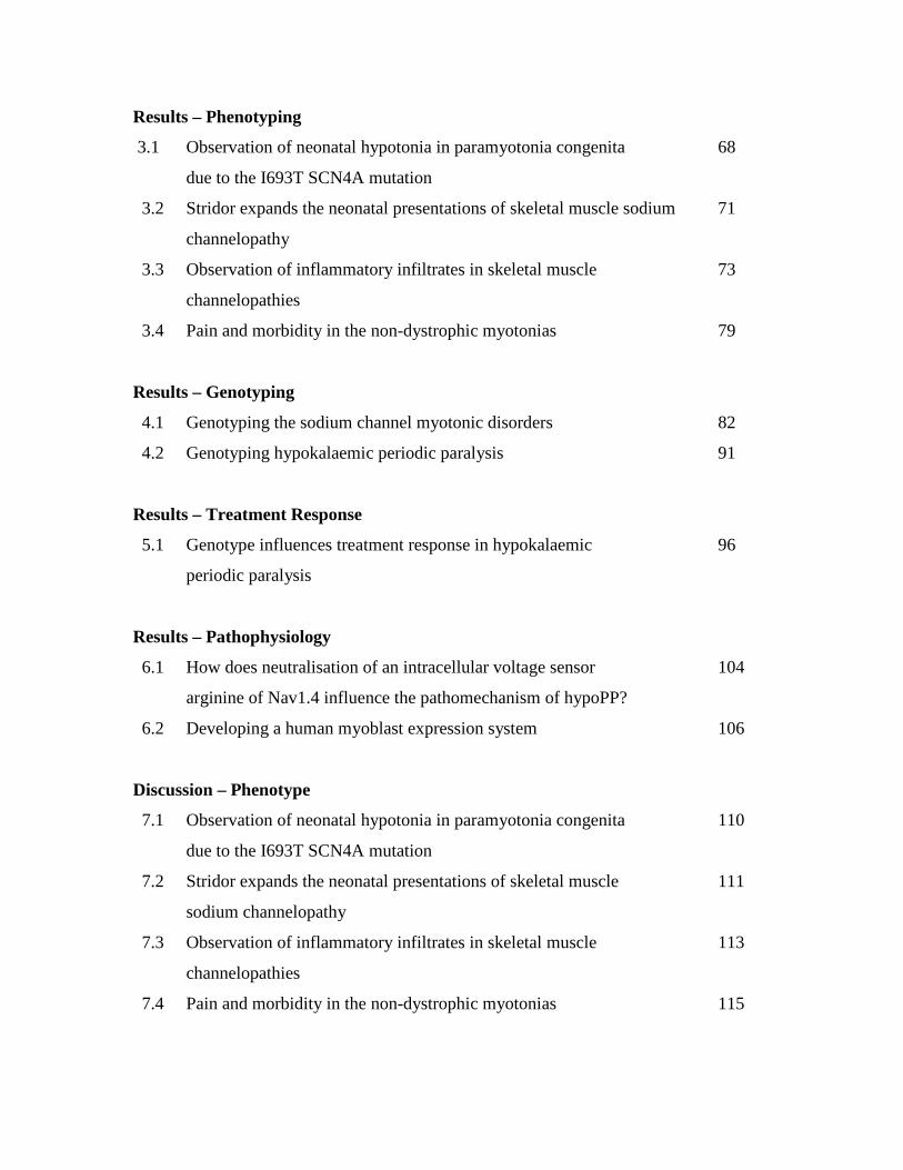
Results – Phenotyping
3.1 Observation of neonatal hypotonia in paramyotonia congenita 68
due to the I693T SCN4A mutation
3.2 Stridor expands the neonatal presentations of skeletal muscle sodium 71
channelopathy
3.3 Observation of inflammatory infiltrates in skeletal muscle 73
channelopathies
3.4 Pain and morbidity in the non-dystrophic myotonias 79
Results – Genotyping
4.1 Genotyping the sodium channel myotonic disorders 82
4.2 Genotyping hypokalaemic periodic paralysis 91
Results – Treatment Response
5.1 Genotype influences treatment response in hypokalaemic 96
periodic paralysis
Results – Pathophysiology
6.1 How does neutralisation of an intracellular voltage sensor 104
arginine of Nav1.4 influence the pathomechanism of hypoPP?
6.2 Developing a human myoblast expression system 106
Discussion – Phenotype
7.1 Observation of neonatal hypotonia in paramyotonia congenita 110
due to the I693T SCN4A mutation
7.2 Stridor expands the neonatal presentations of skeletal muscle 111
sodium channelopathy
7.3 Observation of inflammatory infiltrates in skeletal muscle 113
channelopathies
7.4 Pain and morbidity in the non-dystrophic myotonias 115
Discussion – Genotype
8.1 Genotyping the sodium channel myotonic disorders 117
8.2 Genotyping hypokalaemic periodic paralysis 120
Discussion – Treatment
9.1 Genotype influences treatment response in hypokalaemic 124
periodic paralysis
Discussion – Pathophysiology
10.1 Functional Consequences of the R675G SCN4A mutation 127
10.2 Establishing a myoblast system 128
Reference List 130
Appendix 1: Publication List 146

1
Introduction
The skeletal muscle channelopathies are a rare group of episodic neuromuscular disorders
including the periodic paralyses and the non-dystrophic myotonias. Mutations in different
genes are responsible for each sub-group but they are all genes that code for voltage gated
skeletal muscle ion channels. These ion channels regulate muscle membrane excitability,
and consequently these diseases share a common pathology of altered sarcolemmal
excitability. The resultant symptoms include episodes of muscle stiffness as well as
episodes of weakness. The stiffness, or myotonia is associated with hyper-excitability of
the membrane. In contrast, weakness, or paralysis, arises when muscle membranes
become inexcitable.
Given these two major types of symptoms, these channelopathies can be classified in two
broad groups: The first are the non-dystrophic myotonias, in which myotonia is the
predominant symptom, this group encompasses myotonia congenita, paramyotonia
congenita and the sodium channel myotonias. The second group is characterised
predominantly by paralysis rather than myotonia, and includes the primary periodic
paralyses hyperkalemic periodic paralysis, hypokalaemic periodic paralysis and
Andersen-Tawil Syndrome.
Skeletal Muscle
Channelopathy
Disease Causing Gene Mode of Inheritance
Myotonia Congenita CLCN-1 Autosomal Dominant or
Recessive
Paramyotonia Congenita SCN4A Autosomal Dominant
Sodium Channel Myotonia SCN4A Autosomal Dominant
Hyperkalaemic Periodic
Paralysis
SCN4A Autosomal Dominant
Hypokalaemic Periodic
Paralysis
SCN4A (10% of cases)
CACNA1S (70% of
cases)
Autosomal Dominant
Andersen-Tawil Syndrome KCNJ2 Autosomal Dominant
Table 1: Skeletal muscle channelopathies associated with voltage-gated ion channels
and their causative genes

2
This thesis focuses primarily on the muscle channelopathies due to mutations of the
SCN4A and CACNA1S genes. For practical reasons I will consider these in two
categories:
1. SCN4A only: Paramyotonia congenita, sodium channel myotonia and
hyperkalaemic periodic paralysis
2. SCN4A or CACNA1S: Hypokalaemic periodic paralysis
1.0 The role of voltage-gated channels in muscle cells
Skeletal muscle is an electrically excitable tissue. Changes in the muscle membrane
potential due to the movement of charged ions in and out of the cell stimulate a cascade
of reactions that result in co-ordinated muscle contraction and subsequent relaxation.
Nav1.4, the voltage gated sarcolemmal sodium channel encoded by SCN4A supports the
rising phase of action potentials in skeletal muscle (Fig 1). In cardiac muscles, Nav1.5
(SCN5A) predominates, while in neurons several different isoforms of sodium channel
are used, but not Nav1.4. Thus mutations or drugs targeting Nav1.4 can be expected to
have relatively specific effects on skeletal muscle excitability.
CACNA1S encodes the skeletal muscle voltage-dependent calcium channel, Cav1.1. Like
Nav1.4, expression of Cav1.1 is predominantly restricted to skeletal muscle, with cardiac
muscle expressing Cav1.2 (CACNA1C), and neurons utilising a diverse group of voltage
gated calcium channels but not Cav1.1. Upon depolarisation by Nav1.4, Cav1.1 channels
are activated. These channels, although designated voltage-gated calcium channels, are
physically coupled to ryanodine receptors (RyR1) in the sarcoplasmic reticulum, and the
activation of Cav1.1 leads to opening of RyR1 and release of calcium from intracellular
stores (Fig 2). It is thought that the activation of RyR1 via physical coupling, not via
calcium influx, is the main function of Cav1.1 in healthy muscle cells(Catterall., 1995;
Brini., 2004).

3
The other two genes associated with similar muscle disorders are CLCN1, which encodes
the voltage-gated chloride channel ClC-1, and KCNJ2, which encodes an inward
rectifying potassium channel, Kir2.1. ClC-1 is restricted to skeletal muscle cells where it
is thought to help set the muscle resting potential and facilitates repolarisation when
potassium accumulates in T-tubules(Cannon., 2006). As is consistent with its restricted
expression in skeletal muscles, mutations in CLCN1 lead to a purely muscular disorder,
myotonia congenita.
KCNJ2, in contrast, is expressed in a variety of cell types, including cardiac myocytes,
and mutations in this gene lead to Andersen-Tawil syndrome which has symptoms
associated with this relatively wide distribution, including long QT, facial dysmorphia,
and a muscle phenotype of periodic paralysis. In skeletal muscle cells, it has recently
been proposed that mutations in KCNJ2 that disrupt the expression of Kir2.1 lead to an
elevation of the resting membrane potential of myoblasts, indicating that in healthy cells
this channel may be required to maintain resting membrane potential(Sacconi et al.,
2009).
Fig 1: Schematic representation of a muscle action potential
Depolarisation Repolarisation
Na+ channels K+ channels
open close
K+ channels open
Na+ channels close
Nerve stimulus
Threshold for activation
-90mv
Membrane potential
Na+ ions enter the
cell
K+ ions leave the cell
Resting membrane potential
0mv
Cl- ions leave the cell
Ca2+ ions enter the
cell
Depolarisation Repolarisation
Na+ channels K+ channels
open close
K+ channels open
Na+ channels close
Nerve stimulus
Threshold for activation
-90mv
Membrane potential
Na+ ions enter the
cell
K+ ions leave the cell
Resting membrane potential
0mv
Cl- ions leave the cell
Ca2+ ions enter the
cell

4
Fig 2: Schematic representation of the skeletal muscle voltage gated ion channels
1.1 Structure of Nav1.4 and Cav1.1
Nav1.4 and Cav1.1 channels both consist of a core α subunit and one or more accessory
subunits. In both cases, the α-subunits form the ion conducting pore of the channel and
thus far only mutations in the α-subunits have been associated with the skeletal muscle
channelopathies. As such only the α-sub-units are considered in detail here. The α sub-
units of Nav1.4 and Cav1.1 have similar structures consistent with a shared evolutionary
history linked to a single ancestral channel(Cannon., 2007). Both subunits are comprised
of four domains, each of which contains six transmembrane segments. The four domains
fold together to form a single ion-conducting pore (Fig 3).
RYR
SR
Ca
Ca
ClK
Na
Ca
Surface membrane
T Tubule
Cav1.1 RYR
SR
Ca
Ca
ClK
Na
Ca
Surface membrane
T Tubule
Cav1.1

5
Fig 3: Schematic representation of the A:Cav 1.1 and B: Nav 1.4 channels. C:
representation of the alignment of the four domains to form a single ion selective
pore.
1.2 Elements determining the ion selectivity of Nav1.4 and Cav1.1
Although Nav1.4 and Cav1.1 are structurally related, they are permeable to different ions.
In both channels the S5 and S6 segments from each domain assemble together to create a
central channel pore through which ions enter the cell(Varadi et al., 1999; Catterall.,
1995). However, Nav1.4 is highly selective for sodium, while Cav1.1 is most permeable
to calcium ions. The loops between segments S5 and S6 are known as the P loops and
site-directed mutagenesis experiments have revealed that a small number of amino acid
residues in these P loops determine the ion selectivity for each respective channel. For
calcium channels four glutamic acid residues (EEEE) have been identified as the key
amino acids in determining selectivity for calcium(Tang et al., 1993; Mikala et al., 1993).
I
IIIIV
II
+ +
+ +
+
+ +
+ +
+
++
+ +
+ +
+ +
+ +
+
A C
+ +
+ +
+ +
+ +
+ +
+ +
+ +
+ +
+ +
+ +
+ +
+
B
I
IIIIV
II
+ +
+ +
+
+ +
+ +
+
++
+ +
+ +
+ +
+ +
+
A C
+ +
+ +
+ +
+ +
+ +
+ +
+ +
+ +
+ +
+ +
+ +
+
B

6
The amino acids occupying the comparable positions in the voltage gated sodium
channels (aspartate, glutamate, lysine, alanine, DEKA) have similarly been implicated in
setting the selectivity of sodium channels(Heinemann et al., 1992; Favre et al., 1996)(See
Figs 4 and 5).
Fig 4: Schematic representation of Cav1.1 highlighting amino acid residues
responsible for channel ion selectivity.
Fig 5: Schematic representation of Nav1.4 highlighting amino acid residues
responsible for channel ion selectivity and the IFM motif/inactivation gate.
+ +
+ +
+ +
+ +
+ +
+ +
++
+ +
+ +
+ +
+ +
+
P loop
S5 and S6 pore forming
segments
AKED
IF M
+ +
+ +
+ +
+ +
+ +
+ +
++
+ +
+ +
+ +
+ +
+
P loop
S5 and S6 pore forming
segments
AAKKEED
IF M
+ +
+ +
+
+ +
+ +
+
++
+ +
+ +
+ +
+ +
+
P loop
S5 and S6 pore forming
segments
EEEE
Voltage sensor
+ +
+ +
+
+ +
+ +
+
++
+ +
+ +
+ +
+ +
+
P loop
S5 and S6 pore forming
segments
EEEE+ +
+ +
+
+ +
+ +
+
++
+ +
+ +
+ +
+ +
+
P loop
S5 and S6 pore forming
segments
EEEEEEE
Voltage sensor

7
1.3 Channel activation and inactivation
Both Nav1.4 and Cav1.1 are activated by depolarisation of the muscle membrane. In the
case of Nav1.4 the activation is rapid, and the main role of the channel is to allow sodium
ions to enter the cell, further depolarizing it, and allowing more voltage-gated channels to
open. In the case of Cav1.1, it appears the structural change upon activation is sufficient
to mediate the main effect, a physical activation of the ryanodine receptors, however
Cav1.1 proteins still contain a channel pore that is opened in response to depolarization,
and which allows calcium to enter the muscle cell.
In common with many voltage gated channels, both Nav1.4 and Cav1.1 use positively
charged segments within the pore-forming α subunits to detect changes in membrane
voltage(Catterall., 2010). The S4 segments of both channels contain positively charged
residues (arginines or lysines) at every third position surrounded by hydrophobic
residues. These segments with their abundance of electrical charge behave as the voltage
sensors(Yang et al., 1996a) by moving towards the cytoplasmic side of the membrane in
response to membrane depolarisation. It is this outward movement that produces a
conformational change at the intracellular surface of the channels(Catterall., 1995), and
opens the central pore of the channel to allow influx of ions (Fig 6).
Fig 6: Outward movement of the S4 segment in response to depolarization produces
a conformational change in the channel which opens the central pore.
6
5
4 32
1
4
6
5
3
2
1
4
Pore closed Pore open
66
5
4 3322
11
4
66
55
33
22
11
44
Pore closed Pore open

8
Although significant advances in understanding the function of the S4 segments as the
voltage sensors have been made the exact role of each segment is not fully
understood(Catterall., 2010). It is likely that individual segments may have slightly
different roles e.g. in the sodium channel evidence suggests the S4 segments of domains
III and IV play a more significant role in fast inactivation(Cha et al., 1999).
In basic terms the channels can be in one of three states: closed, open or inactive. When
the sarcolemma is at its resting potential the channels are closed and allow no movement
of sodium or calcium across the membrane. In response to depolarisation the S4 segments
(voltage sensors) move outwards and the pore undergoes a responding conformational
change such that the channel moves into the open state and allows the influx of ions
through the central pore. With sustained depolarisation, the channels inactivate stopping
the flow of ions. As the sarcolemma repolarises the channels return to the closed state and
recover from inactivation (see Fig 7).
Fig 7: States of activation and inactivation of Nav1.4 in response to changes in
membrane potential.
Open Inactive Closed Closed
Activation Fast Inactivation Recovery from inactivation
Membrane depolarisation Membrane repolarisation
Potassium channels open
Open Inactive Closed Closed
Activation Fast Inactivation Recovery from inactivation
Open Inactive Closed Closed
Activation Fast Inactivation Recovery from inactivation
Membrane depolarisation
Potassium channels open

9
The sodium channels can undergo two forms of inactivation. After a brief depolarisation
of the muscle membrane the channel undergoes fast inactivation which occurs in
milliseconds and is mediated by the cytoplasmic linker between domains III and IV that
acts as a hinged “inactivation gate” swinging across and blocking the cytoplasmic side of
the pore to inactivate the channel. The specific inactivation particle consists of three
residues in the DIII-IV linker (see Fig 5) known as the “IFM motif” (isoleucine,
phenylalanine, methionine) that are thought to bind to an acceptor site on the cytoplasmic
mouth of the pore blocking the passage of sodium ions(Vassilev et al., 1988; Patton et al.,
1992; McPhee et al., 1994). After longer depolarisations the channel inactivates over a
period of seconds to minutes by a process of slow inactivation. Fast and slow inactivation
are thought to be structurally independent processes, but the mechanism for slow
inactivation is less well understood and no specific structural part of the channel has been
identified as a slow inactivation gate(Vedantham et al., 1998).
While ion selectivity, activation and inactivation are thought to be processes which are
intrinsic to α subunits the full physiological function of these channels, as is common to
most ion channels, is dependent on the assembly of several proteins, including multiple
accessory subunits(Catterall., 1995). However since this project is focused on the clinical
manifestation of diseases which are so far only associated with mutations in the α
subunits, the accessories will not be considered here.
1.4 Clinical features of diseases associated with voltage gated ion channel
dysfunction in skeletal muscle
The non-dystrophic myotonias
Paramyotonia Congenita
Eulenberg first described and named Paramyotonia Congenita (PMC) in 1886 after
studying six generations of a German family affected with the disease(Eulenberg A.,
1886). PMC is due to mutations of the SCN4A gene and is inherited in an autosomal
dominant manner.

10
Symptoms usually present in the first decade. Affected individuals experience episodes of
muscle stiffness (myotonia) and weakness or paralysis that are markedly worsened or
precipitated by cold environments and with periods of exercise. The muscles of the face
and hands tend to be most severely affected. The myotonia commonly lasts seconds to
minutes but the paralysis can last hours.
Sodium Channel Myotonia
Before the recognition of SCN4A as the disease causing gene for paramyotonia congenita
there were many reports of other myotonic phenotypes that did not fit the typical
presentation for PMC. These phenotypes were united by features such as a delayed onset
of myotonia following rest after exertion, little or no cold exacerbation but significant
exacerbation following potassium ingestion. They were purely myotonic phenotypes with
no associated episodes of muscle paralysis. Individually they were described as
acetazolamide responsive myotonia congenita(Trudell et al., 1987; Ptacek et al., 1994b),
myotonia fluctuans(Ricker et al., 1990; Lennox et al., 1992; Ricker et al., 1994) and
myotonia permanens(Lerche et al., 1993; McClatchey et al., 1992b). Together due to
their shared features they are termed the potassium aggravated myotonias (PAM).
Other phenotypes were reported that had overlapping features of PMC and PAM in that
they were purely myotonic and often exacerbated by potassium ingestion but also by
exposure to cold(Heine et al., 1993; Koch et al., 1995; Wu et al., 2001).
The distinguishing feature between all these collective phenotypes and PMC has been
that, unlike PMC, they are purely myotonic disorders with a lack of any associated
episodes of muscle paralysis. As all of these phenotypes have been shown to be allelic to
PMC and due to mutations in SCN4A some authors collectively use the term sodium
channel myotonia (SCM)(Fournier et al., 2004) to encompass all of these purely
myotonic phenotypes.
Predominantly it is the face and limb muscles affected by these two groups of myotonic
disorders (PMC and SCM) but respiratory compromise which can be severe has been
reported as has dysphagia(Lerche et al., 1993);(Colding-Jorgensen et al., 2006).
The sodium channel myotonias can be generally relatively easily separated from
paramyotonia congenita by their absence of episodic weakness but they can have

11
considerable clinical overlap with the dominant form of myotonia congenita (MC) due to
mutations in the CLCN-1 gene. Table 2 outlines the pertinent clinical features for each
sub group of non-dystrophic myotonia. The major features of overlap between SCM and
dominant MC are the presence of the “warm-up” phenomenon used to describe an
improvement in myotonia with repetitive activity. This is often described as the
distinguishing feature of myotonia congenita but it can be present in the sodium channel
myotonias. If so however, it often fluctuates with paramyotonia, (myotonia that worsens
with repetition as is seen in PMC), whereas in myotonia congenita only the warm up
phenomenon will be seen. The distribution of muscle involvement can also be very
similar for SCM and dominant MC and often episodic weakness is also absent in
dominant myotonia congenita. The recessive form of myotonia congenita is not easily
confused with sodium channel myotonia but the clinical features are outlined in Table 2
for comparative purposes.
Development of a progressive proximal myopathy is described in PMC and SCM
although whether the onset or severity of this correlates to age or episodes of myotonia
and/or paralysis is unknown(Schoser et al., 2007).

12
*Myotonia typically develops after a short period of exercise e.g. 10 minutes or on
resting after a period of exercise.
Table 2: Pertinent clinical features of the non-dystrophic myotonias
Recessive MC Dominant MC Paramyotonia
Congenita
Sodium Channel
Myotonia
Inheritance Recessive Dominant Dominant Dominant
Causative gene CLCN-1 CLCN-1 SCN4A SCN4A
Myotonia
distribution
Lower limbs
more than
upper limbs
Upper limbs
more than
lower limbs
Facial muscles
may be
involved
Upper limbs and
face
more than lower
limbs
Upper limbs,
face ,
extraocular,
more than lower
limbs
Myotonia cold
sensitivity
None or
minimal
None or
minimal
Yes – often
dramatic
Variable –
ranging from
none to severe
Warm up
phenomenon
Present Present Absent May be present
Paradoxical
myotonia
Absent Absent Present May be present
Delayed onset
myotonia
after exercise*
Absent Absent Absent May be present
Characteristic of
myotonia
fluctuans
Episodic
muscle
weakness
Common,
develops on
initiation of
movement but
transient and
improves
rapidly
Uncommon Common often
exacerbated by
cold and/or
exercise and
frequently
prolonged for
several hours
Not reported
Eyelid
myotonia
Infrequent Infrequent Common Common

13
The Periodic Paralyses
Hyperkalaemic Periodic Paralysis
The periodic paralyses are thought to be the most common of the skeletal muscle
channelopathies although the exact incidence is unknown(Venance et al., 2006).
Hyperkalaemic periodic paralysis is due to mutations in SCN4A and is inherited in an
autosomal dominant manner. Onset of symptoms is usually within the first decade. The
prominent symptom is episodic muscle paralysis frequently occurring after exercise or
following the ingestion of potassium rich foods e.g. bananas and tomatoes. Attacks
usually last from a few minutes to hours and myotonia can occur. ECG abnormalities
secondary to raised serum potassium levels may be seen during an attack of paralysis.
Paramyotonia congenita, sodium channel myotonia and hyperkalaemic periodic paralysis
are all allelic disorders due to mutations of the SCN4A gene. There are often overlapping
clinical features and it has been proposed that these disorders represent different ends of a
spectrum rather than being individual diseases(Cannon., 2000).
Hypokalaemic Periodic Paralysis
Hypokalaemic periodic paralysis (hypoPP) is also inherited in an autosomal dominant
manner. It is characterised by attacks of flaccid skeletal muscle paralysis in association
with reduced serum potassium levels. Onset is commonly in the first or second decade
although presentation in the third decade has been described(Miller et al., 2004). Attacks
of muscle paralysis are precipitated by factors that reduce serum potassium levels such as
a large carbohydrate meal. Attacks occur most commonly following strenuous exercise or
during the night or early morning. They typically last in the region of hours to days
although some patients will report it is weeks to months before full muscle strength
returns.
It is predominantly the limb muscles which are affected in hypoPP but occasionally
severe respiratory muscle involvement is described(Kil et al., 2009b; rzel-Hezode et al.,
2009b). Cardiac muscle is not inherently affected but potassium levels outside the normal
range can cause ECG changes(Kim et al., 2005; Kil et al., 2009a; Hecht et al., 1997)
(flattened ST segments, U waves, prolonged QT interval) that may be pro-arrhythmic and

14
require cardiac monitoring while potassium levels are restored. One case of severe sinus
bradycardia requiring temporary pacemaker has been reported during an episode of
hypokalaemia(Maffe et al., 2009) and another case report has described sudden death
amongst relatives of a genetically confirmed individual with hypoPP(Hecht et al., 1997).
Whether such severe outcomes are directly or exclusively attributable to hypokalaemia is
not fully established.
Many patients with hyperPP and hypoPP will function independently between attacks of
paralysis but in a significant number a fixed myopathy develops that can be
debilitating(Fouad et al., 1997; Miller et al., 2004; Biemond et al., 1934). It is of note that
three independent series all reported abnormal morphological findings in 100% of
hypoPP patients who had been biopsied(Fouad et al., 1997; Sternberg et al., 2001; Miller
et al., 2004). In a minority these were non-specific myopathic changes with vacuolar
myopathy and tubular aggregates accounting for the majority. The reports of permanent
muscle weakness were more varied from 25% to 72% of genotyped individuals. The
mechanism of this myopathy is not understood. It is postulated that it is independent of
paralytic attack frequency or severity(Buruma et al., 1978; Links et al., 1990) although
there is evidence that increasing age is associated with the myopathy(Links et al., 1990).
Only 10% of cases of hypokalaemic periodic paralysis are due to mutations of the
SCN4A gene with the majority being caused by mutations in the CACNA1S gene(Fouad
et al., 1997; Miller et al., 2004; Sternberg et al., 2001). Approximately 20% remain
genetically undefined. There is no clear clinical distinction between hypoPP arising from
the different genetic sources.
Andersen-Tawil Syndrome
Andersen-Tawil syndrome is the only muscle channelopathy in which the causative gene,
KCNJ2(Plaster et al., 2001), is expressed in tissue other than skeletal muscle. As such the
triad of periodic paralysis, dysmorphic features and cardiac conduction defects comprise
the characteristic phenotype(Andersen et al., 1971; Tawil et al., 1994). As with nearly all
the channelopathies it is an autosomal dominant disorder. The type of periodic paralysis
can be either hyper or hypokalaemic although most commonly it is hypoPP(Davies et al.,
2005). The dysmorphic features include mandibular micrognathia, short stature,

15
clinodactyly, syndactyly, hypertelorism and low set ears(Andersen et al., 1971; Tawil et
al., 1994; Davies et al., 2005; Haruna et al., 2007) but can be very subtle and easily
missed. Likewise there are often no cardiac symptoms and unless an ECG is performed
for unrelated circumstances the cardiac conduction defects are often undetected. For these
reasons hypokalaemic periodic paralysis may be the only obvious presentation and some
individuals may be clinically misdiagnosed. This can have significant consequences as
the cardiac conduction defects described include abnormal U waves, prolonged QUc
interval, prolonged QTc interval, bigeminy and bidirectional VT. Rarely sudden death
can occur(Tawil et al., 1994; Davies et al., 2005; Zhang et al., 2005; Haruna et al., 2007).
There is very little data as to the natural history of any of the skeletal muscle
channelopathies, particularly with regards to the proximal myopathy. Additionally, there
are few phenotype-genotype correlations, something which is exacerbated by significant
variability in the severity of the phenotype amongst individuals with the same mutation.
There are even examples of this occurring amongst members of a single kindred. The
skeletal muscle channelopathies are generally considered to be more severe in males than
females. A significant worsening of symptoms is also commonly reported during
pregnancy. Both of these factors imply there may be a hormonal influence on the
phenotype. There is some suggestive in vitro evidence to support this(Fialho et al., 2008)
but it is yet to be fully established or understood.
An allied disease where hormones are clearly shown to be of significance is thyrotoxic
periodic paralysis. This is most common in Asian males and represents a form of
hypokalaemic periodic paralysis in which attacks only occur in relation to deranged
thyroid function. It is not to be missed as treatment of the thyrotoxicosis and return to a
euthyroid state will abolish the attacks of muscle paralysis. Equally although the thyroid
function may be abnormal, the patient may not be obviously thyrotoxic and diagnosis
requires a degree of suspicion.

16
The first aim of this thesis is to study the phenotypes among a large cohort of
genotyped channelopathy patients to explore any additional recurring features and
assess phenotype-genotype correlations.
Hyperkalaemic PP Hypokalaemic PP Andersen-Tawil
Syndrome
Causative
gene
SCN4A CACNA1S
SCN4A
KCNJ2
Inheritance Autosomal dominant Autosomal dominant Autosomal dominant
Episodic
skeletal
muscle
paralysis
Yes Yes Yes
Duration of
paralysis
Commonly minutes to
hours
Commonly hours to
days
Variable, minutes to
days
Ictal
potassium
levels
High Low Low, high or normal
Precipitators
of paralysis
Potassium rich foods
Rest after exercise
Large carbohydrate
load
Rest after exercise
Dependant on ictal
potassium
Typical time
of attacks
Any time of day During night or early
morning
Any – dependant on
ictal potassium
Cardiac
conduction
defects
Only those attributable
to severe
hyperkalaemia; resolve
with restoration of
normal potassium
values
Only those
attributable to severe
hypokalaemia; resolve
with restoration of
normal potassium
values
Common especially
abnormal u waves,
prolonged QUc
interval and
ventricular
arrhythmias,
irrespective of
potassium levels
Dysmorphic
features
No No Common especially
short stature,
mandibular
hypoplasia,
clinodactyly and low
set ears
Table 3: Summary of clinical features of the primary periodic paralyses
17
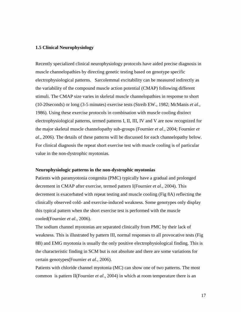
1.5 Clinical Neurophysiology
Recently specialized clinical neurophysiology protocols have aided precise diagnosis in
muscle channelopathies by directing genetic testing based on genotype specific
electrophysiological patterns. Sarcolemmal excitability can be measured indirectly as
the variability of the compound muscle action potential (CMAP) following different
stimuli. The CMAP size varies in skeletal muscle channelopathies in response to short
(10-20seconds) or long (3-5 minutes) exercise tests (Streib EW., 1982; McManis et al.,
1986). Using these exercise protocols in combination with muscle cooling distinct
electrophysiological patterns, termed patterns I, II, III, IV and V are now recognized for
the major skeletal muscle channelopathy sub-groups (Fournier et al., 2004; Fournier et
al., 2006). The details of these patterns will be discussed for each channelopathy below.
For clinical diagnosis the repeat short exercise test with muscle cooling is of particular
value in the non-dystrophic myotonias.
Neurophysiologic patterns in the non-dystrophic myotonias
Patients with paramyotonia congenita (PMC) typically have a gradual and prolonged
decrement in CMAP after exercise, termed pattern I(Fournier et al., 2004). This
decrement is exacerbated with repeat testing and muscle cooling (Fig 8A) reflecting the
clinically observed cold- and exercise-induced weakness. Some genotypes only display
this typical pattern when the short exercise test is performed with the muscle
cooled(Fournier et al., 2006).
The sodium channel myotonias are separated clinically from PMC by their lack of
weakness. This is illustrated by pattern III, normal responses to all provocative tests (Fig
8B) and EMG myotonia is usually the only positive electrophysiological finding. This is
the characteristic finding in SCM but is not absolute and there are some variations for
certain genotypes(Fournier et al., 2006).
Patients with chloride channel myotonia (MC) can show one of two patterns. The most
common is pattern II(Fournier et al., 2004) in which at room temperature there is an

18
immediate CMAP decrement after the short exercise test which recovers quickly and
diminishes with repetition, reflecting the transient weakness observed clinically (Fig 8C).
This pattern is most frequently seen in recessive MC but can be observed in any muscle
ion channel disorder in which there is a loss of sarcolemmal chloride conductance. It is
therefore also seen in dominant MC and in both DM1 and DM2. There is now clear
evidence that the myotonia in DM1 and DM2 is secondary to reduced chloride
conductance(Charlet et al., 2002). In recessive MC cooling has little further effect (Fig
8C). However, in dominant MC the CMAP decrement may be worsened or only seen
with cooling(Fournier et al., 2006) making it essential to perform the short exercise test at
both room temperature and with the muscle cooled (Fig 8D). However, some patients
with dominant MC show a normal response (pattern III)(Fournier et al., 2006) to all
provocative tests (Fig 8B), even with muscle cooling which is indistinguishable
electrophysiologically from sodium channel myotonia. Clinical history and examination
usually helps to distinguish between the two and guide genetic analysis but both of these
can have considerable overlap between the two groups as discussed earlier (see Table 2).
Table 4 outlines the most common electrophysiological pattern observed in the repeat
short exercise test with muscle cooling for each of the NDMs. Variability exists and
where muscle cooling has already proven useful in improving diagnosis, repetitive nerve
stimulation may have a future role to play in distinguishing the sub-types of NDM. There
is some evidence that a reduction in CMAP may be provoked by repetitive nerve
stimulation in certain cases of recessive MC where exercise testing even with the muscle
cooled has failed to produce any such decrement(Michel et al., 2007). In this way
repetitive nerve stimulation may become an additional future tool to guide the genetic
analysis towards recessive MC in cases that may otherwise be thought to be dominant
MC or SCM.
Neurophysiologic patterns in the primary periodic paralyses
For hyperkalaemic periodic paralysis and hypokalaemic periodic paralysis due to calcium
channel mutations electrophysiological patterns IV and V were determined (Table 4). In
summary, pattern IV describes an increase in CMAP that is exaggerated by repetition in

19
the short exercise test, and an immediate increase in CMAP followed by a later reduction
in CMAP in the long exercise test. Pattern V illustrates the lack of change observed in
CMAP during the short exercise test and the prolonged decrement in CMAP during the
long exercise test of patients with HypoPP due to calcium channel mutation. It is
important to note that while these patterns are undoubtedly useful in each group they
were performed in a relatively small number of individuals all carrying the same
causative gene mutation (6 T704M and 13 R528H). Only two hypoPP patients with
sodium channel mutations were studied each with a different mutation and each showing
a different electrophysiological pattern. No large scale studies have been performed in
those with Andersen-Tawil syndrome. As a result the electrophysiological patterns are
perhaps less clearly defined for the periodic paralyses than the myotonic disorders.
Fig 8: Common electrophysiological patterns seen in the repetitive short exercise
test at room temp and with the muscle cooled in the NDMs (produced by Dr Tan)
AUTOSOMAL DOMINANT MYOTONIA CONGENITA
0
20
40
60
80
100
120
2 20 40 60 72 90 110 130 142 160 180 200
TIME (S)
CM
AP
AM
PL
ITU
DE
, %
BA
SE
LIN
E
Room Temperature After Cooling
AUTOSOMAL RECESSIVE MYOTONIA CONGENITA
0
20
40
60
80
100
120
140
2 20 40 60 72 90 110 130 142 160 180 200
TIME (S)
CM
AP
AM
PL
ITU
DE
, %
BA
SE
LIN
E
Room Temperature After Cooling
PARAMYOTONIA CONGENITA
0
20
40
60
80
100
120
140
2 10 20 30 40 50 60 72 80 90 100 110 120 130 142 150 160 170 180 190 200
TIME (S)
CM
AP
AM
PL
ITU
DE
, %
BA
SE
LIN
E
Room Temperature After Cooling
POTASSIUM AGGRAVATED MYOTONIA
0
20
40
60
80
100
120
140
2 10 20 30 40 50 60 72 80 90 100 110 120 130 142 150 160 170 180 190 200
TIME (S)
CM
AP
AM
PL
ITU
DE
, %
BA
SE
LIN
E
Room Temperature After Cooling
A
CD
B
AUTOSOMAL DOMINANT MYOTONIA CONGENITA
0
20
40
60
80
100
120
2 20 40 60 72 90 110 130 142 160 180 200
TIME (S)
CM
AP
AM
PL
ITU
DE
, %
BA
SE
LIN
E
Room Temperature After Cooling
AUTOSOMAL DOMINANT MYOTONIA CONGENITA
0
20
40
60
80
100
120
2 20 40 60 72 90 110 130 142 160 180 200
TIME (S)
CM
AP
AM
PL
ITU
DE
, %
BA
SE
LIN
E
Room Temperature After Cooling
AUTOSOMAL RECESSIVE MYOTONIA CONGENITA
0
20
40
60
80
100
120
140
2 20 40 60 72 90 110 130 142 160 180 200
TIME (S)
CM
AP
AM
PL
ITU
DE
, %
BA
SE
LIN
E
Room Temperature After Cooling
AUTOSOMAL RECESSIVE MYOTONIA CONGENITA
0
20
40
60
80
100
120
140
2 20 40 60 72 90 110 130 142 160 180 200
TIME (S)
CM
AP
AM
PL
ITU
DE
, %
BA
SE
LIN
E
Room Temperature After Cooling
PARAMYOTONIA CONGENITA
0
20
40
60
80
100
120
140
2 10 20 30 40 50 60 72 80 90 100 110 120 130 142 150 160 170 180 190 200
TIME (S)
CM
AP
AM
PL
ITU
DE
, %
BA
SE
LIN
E
Room Temperature After Cooling
PARAMYOTONIA CONGENITA
0
20
40
60
80
100
120
140
2 10 20 30 40 50 60 72 80 90 100 110 120 130 142 150 160 170 180 190 200
TIME (S)
CM
AP
AM
PL
ITU
DE
, %
BA
SE
LIN
E
Room Temperature After Cooling
POTASSIUM AGGRAVATED MYOTONIA
0
20
40
60
80
100
120
140
2 10 20 30 40 50 60 72 80 90 100 110 120 130 142 150 160 170 180 190 200
TIME (S)
CM
AP
AM
PL
ITU
DE
, %
BA
SE
LIN
E
Room Temperature After Cooling
POTASSIUM AGGRAVATED MYOTONIA
0
20
40
60
80
100
120
140
2 10 20 30 40 50 60 72 80 90 100 110 120 130 142 150 160 170 180 190 200
TIME (S)
CM
AP
AM
PL
ITU
DE
, %
BA
SE
LIN
E
Room Temperature After Cooling
A
CD
B

20
Paramyotonia
congenita
Sodium
channel
myotonia
Dominant MC Recessive MC HyperPP HypoPP
Myotonic
potentials
Yes Yes Yes Yes Yes – less
frequent/florid than
NDMs
No
Repeat SET
at room
temp
Gradual and persistent
reduction in CMAP Enhanced by repetition
No significant
change of the
CMAP from
baseline*
Little or no
decrement in CMAP Early decrement
in CMAP with
rapid recovery Improves with
repetition
Increase in CMAP
exaggerated by
repetition
No change
in CMAP
Repeat SET
with muscle
cooling
Gradual and persistent
reduction in CMAP
enhanced further by
cooling (see Fig )
No significant
change of the
CMAP from
baseline * (see Fig )
Early decrement
with rapid recovery
and reduction with
repetition may be
seen (see Fig )
Cooling has little
further effect (see Fig )
ND ND
Long
exercise test
Prolonged decline in
CMAP
No change in
CMAP
No change in
CMAP or slight
early decrement
only
No change in
CMAP or slight
early decrement
only
Early increase in
CMAP followed by
prolonged decline in
CMAP
Prolonged
decrease in
CMAP
Fournier
pattern
I III II/III II IV V
*Note: this same pattern may be observed in dominant MC SET: short exercise test, CMAP: compound muscle action potential
Table 4: Electrophysiological patterns seen in the non-dystrophic myotonias and the primary periodic paralyses

21
1.6 Genetics of the skeletal muscle channelopathies
The Skeletal Muscle Sodium Channelopathies
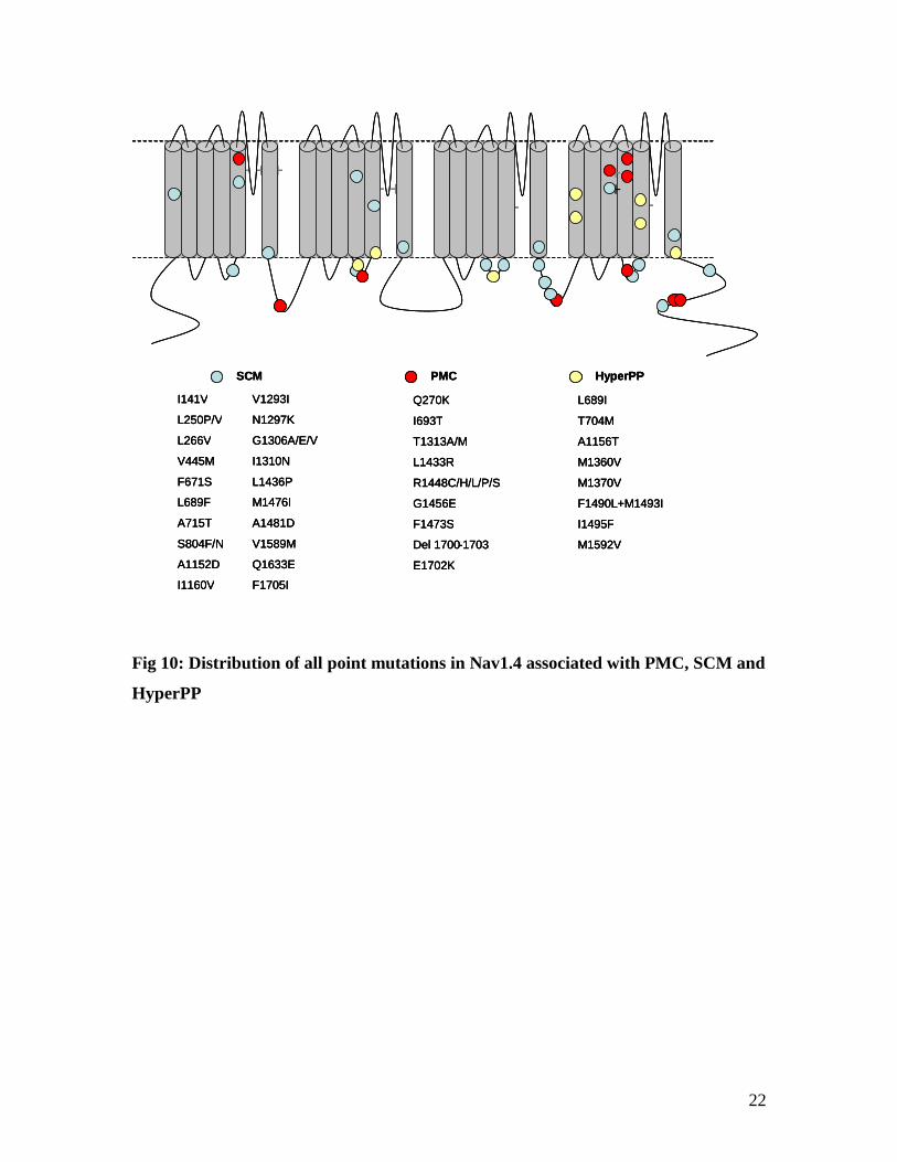
The SCN4A gene has 24 exons that code for the α-subunit of the voltage gated skeletal
muscle sodium channel, Nav1.4. All of the disorders due to mutations in this gene are
inherited in an autosomal dominant manner. Every mutation, with the exception of one
small deletion, associated with the sodium skeletal muscle channelopathies has been a
point mutation. Mutations have been reported throughout the gene although exons 13, 22
and 24 are recognised as “hotspots”. Table 5 outlines all reported SCN4A mutations
including the described phenotypes and functional consequences. Briefly, the most
common mutations associated with PMC are T1313M and substitutions at the R1448
position in exons 22 and 24. The most frequently occurring SCM mutations are G1306A
and V1589M in exons 22 and 24.The mutations most commonly seen in hyperkalaemic
periodic paralysis are the T704M and M1592V mutations in exons 13 and 24. Although
these exons represent hotspots (Fig 9), mutations have been reported throughout the gene
and there is no clustering or discernible pattern to their position in the protein (Fig 10).
Fig 9: Common Nav1.4 mutations associated with PMC, SCM and HyperPP.
+
+
+
+
+
+
+
+
+
+
+
+
+
+
+
+
COOH
NH
I II III IV
T704M
M1592V
T1313M
R1448
V1589M
G1306Most common hyperPP mutations
Most common PMC mutations
Most common SCM mutations
+
+
+
+
+
+
+
+
+
+
+
+
+
+
+
+
COOH
NH
I II III IV
T704M
M1592V
T1313M
R1448
V1589M
G1306
+
+
+
+
+
+
+
+
+
+
+
+
+
+
+
+
COOH
NH
I II III IV
T704M
M1592V
T1313M
R1448
V1589M
G1306Most common hyperPP mutations
Most common PMC mutations
Most common SCM mutations
Most common hyperPP mutations
Most common PMC mutations
Most common SCM mutations
22
Fig 10: Distribution of all point mutations in Nav1.4 associated with PMC, SCM and
HyperPP
+ + + + + + + + + +
+ + +
+ + +
+ + + + +
I141V V1293I
L250P/V N1297K
L266V G1306A/E/V
V445M I1310N
F671S L1436P
L689F M1476I
A715T A1481D
S804F/N V1589M
A1152D Q1633E
I1160V F1705I
SCM
Q270K
I693T
T1313A/M
L1433R
R1448C/H/L/P/S
G1456E
F1473S
Del 1700 - 1703
E1702K
PMC
L689I
T704M
A1156T
M1360V
M1370V
F1490L+M1493I
I1495F
M1592V
HyperPP
+ + + + + + + + + +
+ + +
+ + +
+ + + + +
+ +
I141V V1293I
L250P/V N1297K
L266V G1306A/E/V
V445M I1310N
F671S L1436P
L689F M1476I
A715T A1481D
S804F/N V1589M
A1152D Q1633E
I1160V F1705I
SCM
I141V V1293I
L250P/V N1297K
L266V G1306A/E/V
V445M I1310N
F671S L1436P
L689F M1476I
A715T A1481D
S804F/N V1589M
A1152D Q1633E
I1160V F1705I
SCM
Q270K
I693T
T1313A/M
L1433R
R1448C/H/L/P/S
G1456E
F1473S
Del 1700 - 1703
E1702K
PMC
Q270K
I693T
T1313A/M
L1433R
R1448C/H/L/P/S
G1456E
F1473S
Del 1700 - 1703
E1702K
PMC
L689I
T704M
A1156T
M1360V
M1370V
F1490L+M1493I
I1495F
M1592V
HyperPP
L689I
T704M
A1156T
M1360V
M1370V
F1490L+M1493I
I1495F
M1592V
HyperPP

23
Mutation Exon Protein
Position
Phenotype Clinical Features Major Pathomechanism Reference
I141V 4 DI/S1 SCM Myotonia following rest
after exertion
Cold exacerbated myotonia
No weakness
Enhanced activation
Enhanced slow
inactivation
(Petitprez et al.,
2008)
L250P/V 6 DI/S5 SCM Myotonia with warm up
phenomenon
No response to cooling
Unknown (Trip et al.,
2008)
L266V 6 DI/S5 SCM Cold exacerbated myotonia
No weakness
Impaired fast inactivation
Accelerated recovery
from fast inactivation
(Wu et al.,
2001)
Q270K 6 DI/S5 PMC Cold exacerbated myotonia
Cold exacerbated paralysis
Unknown (Fournier et al.,
2006)
V445M 9 DI/S6 SCM – painful congenital
myotonia
Painful myotonia
No weakness
Enhanced activation
Impaired fast inactivation
Enhanced slow
inactivation
(Rosenfeld et
al., 1997;
Takahashi et
al., 1999)
E452K 9 DI-II PMC Myotonia
Episodic weakness
No cold exacerbation
clinically
Unknown (Dupre et al.,
2009)
F671S 12 DII/S4 SCM Severe myotonia
Generalised hypertophy and
joint retraction
No weakness reported
Unknown (Dupre et al.,
2009)
L689I 13 DII/S4-5 HyperPP Episodic muscle weakness
especially following rest
after exercise
Positive K challenge
Pseudomyotonic discharges
on EMG, no myotonic
discharges
Enhanced activation
Impaired slow
inactivation
(Bendahhou et
al., 2002)
L689F 13 DII/S4-5 SCM Myotonia with warm up
No response to cooling
Unknown (Trip et al.,
2008)

24
I693T 13 DII/S4-5 PMC Cold and exercise induced
weakness
Myotonia on EMG
Enhanced activation
Impaired slow
inactivation
(Plassart et al.,
1996; Plassart-
Schiess et al.,
1998; Hayward
et al., 1999)
I693T
Different kindreds
Neonatal hypotonia
Cold and exercise induced
myotonia and weakness
(Matthews et
al., 2008a)
T704M 13 DII/S5 HyperPP Episodic muscle weakness
exacerbated by rest after
exercise
Positive K challenge
Enhanced activation
Impaired slow
inactivation
(Ptacek et al.,
1991;
Bendahhou et
al., 1999a)
A715T 13 DII/S5 SCM Cold exacerbated myotonia
Myotonia worse after rest
Warm up phenomenon
No weakness
Unknown (Fournier et al.,
2006)
S804F 14 DII/S6 SCM Cold exacerbated myotonia
Severe myotonia after
anaesthesia
Impaired fast inactivation (McClatchey et
al., 1992a;
Green et al.,
1998)
S804F different
kindred
SCM – myotonia fluctuans Fluctuating myotonia
Delayed onset myotonia
following rest after exercise
No cold exacerbated
myotonia
No weakness
(Ricker et al.,
1994)
S804N 14 DII/S6 SCM Cold exacerbated myotonia
Myotonia worse after rest
Warm-up phenomenon
No weakness
Unknown (Fournier et al.,
2006)
A1152D 19 DIII/S4-5 SCM* Cold exacerbated myotonia
No weakness
Impaired fast inactivation (Bouhours et
al., 2005)
A1156T 19 DIII/S4-5 HyperPP Episodic muscle weakness
following rest after exertion
associated with raised
Impaired fast inactivation
Slow inactivation
indistinct from WT
(McClatchey et
al., 1992a;
Hayward et al.,

25
serum K levels
Muscle stiffness (hands and
face)
EMG myotonia
1999)
I1160V 19 DIII/S4-5 SCM – acetazolamide
responsive myotonia
Fluctuating severe painful
myotonia
Potassium aggravated
myotonia
Paradoxical myotonia and
warm up phenomenon
No weakness
Marked response to
acetazolamide
Enhanced recovery from
fast inactivation
Slower deactivation
(Trudell et al.,
1987; Ptacek et
al., 1994b;
Richmond et
al., 1997)
V1293I 21 DIII/S6 SCM* Cold exacerbated myotonia
Occasional myotonia after
heavy exercise
No weakness
Enhanced activation
Accelerated recovery
from fast inactivation
(Koch et al.,
1995; Green et
al., 1998)
N1297K 21 DIII-DIV SCM-severe neonatal non-
dystrophic myotonia
Neonatal onset
Cold induced severe
myotonia and weakness
Hypoxia from resp muscle
myotonia
Psychomotor retardation
Fatal outcome
Unknown (Gay et al.,
2008)
G1306A 22 DIII-DIV SCM – myotonia fluctuans Myotonia of fluctuating
severity and frequency
Myotonia exacerbated by
potassium, rest after
exertion and anaesthesia
No cold exacerbation
No weakness
Impaired fast inactivation (Lerche et al.,
1993; Ricker et
al., 1994;
Mitrovic et al.,
1995)
G1306E 22 DIII-DIV SCM – myotonia
permanens
Severe permanent myotonia
Respiratory muscle
myotonia causing episodic
hypoxia and acidosis
without treatment
No weakness
Impaired fast inactivation
Enhanced activation
(Lerche et al.,
1993; Mitrovic
et al., 1995)

26
G1306E
Different kindred
SCM Exercise, cold and
potassium exacerbated
myotonia
No significant respiratory
myotonia
No weakness
(Colding-
Jorgensen et al.,
2006)
G1306V 22 DIII-DIV SCM Cold exacerbated myotonia
or chronic myotonia
(different kindreds)
No weakness
Impaired fast inactivation (Van den Bergh
P et al., 991;
McClatchey et
al., 1992b;
Mitrovic et al.,
1995)
G1306V
Different kindred
SCM Exercise exacerbated
myotonia
Impaired fast inactivation (Lerche et al.,
1993)
I1310N 22 DIII-DIV SCM Cold exacerbated myotonia
Myotonia worse after rest
Warm-up phenomenon
No weakness
Unknown (Fournier et al.,
2006)
T1313A 22 DIII-IV PMC Cold and exercise induced
myotonia
Occasional cold and
exercise induced paralysis
Impaired fast inactivation
(Bouhours et
al., 2004)
T1313M 22 DIII-IV PMC Cold and exercise
exacerbated myotonia and
weakness
Impaired fast inactivation
Enhanced recovery from
inactivation
(McClatchey et
al., 1992b;
Ptacek et al.,
1993; Hayward
et al., 1996;
Yang et al.,
1994)
M1360V 23 DIV/S1 HyperPP Episodic weakness
especially early morning
and following rest after
exercise
Clinical weakness observed
following K challenge and
cooling forearm muscles
EMG myotonia
Impaired inactivation
(Wagner et al.,
1997)

27
M1370V 23 DIV/S1 HyperPP Episodic limb weakness
Muscle stiffness face and
hands when exercising in
cold
Positive K challenge
EMG myotonia
Unknown (Okuda et al.,
2001)
L1433R 24 DIV/S3 PMC Cold and exercise
exacerbated myotonia
Paralysis induced by
exercise when muscles
cooled
Impaired fast inactivation
Enhanced recovery from
inactivation
(Ptacek et al.,
1993; Yang et
al., 1996b;
Yang et al.,
1994)
L1436P 24 DI/S3 SCM Cold and exercise
exacerbated myotonia
Unknown (Matthews et
al., 2008b)
R1448C 24 DIV/S4 PMC Cold exacerbated myotonia
and paralysis
Impaired fast inactivation
Impaired deactivation
Enhanced recovery from
inactivation
(Ptacek et al.,
1992; Yang et
al., 1994;
Featherstone et
al., 1998)
R1448H 24 DIV/S4 PMC Cold exacerbated myotonia
and paralysis
Potassium aggravated
myotonia following
potassium challenge
No potassium aggravated
weakness
Impaired fast inactivation
Enhanced recovery from
inactivation
(Ptacek et al.,
1992; Yang et
al., 1994)
R1448L 24 DIV/S4 PMC Cold and exercise
exacerbated myotonia
Unknown (Matthews et
al., 2008b)
R1448P 24 DIV/S4 PMC Cold exacerbated myotonia
and weakness
Impaired fast inactivation
Impaired deactivation
(Featherstone et
al., 1998; Wang
et al., 1995)
R1448S 24 DIV/S4 PMC Myotonia
Exercise exacerbated
weakness
Impaired fast inactivation
Impaired deactivation
(Bendahhou et
al., 1999b)
G1456E 24 DIV/S4 PMC Cold and exercise
exacerbated myotonia
Cold induced weakness
Unknown (Sasaki et al.,
1999)

28
F1473S 24 DIV/S4-S5 PMC Cold and exercise
exacerbated myotonia
Cold induced weakness
Impaired fast inactivation (Mitrovic et al.,
1996;
Fleischhauer et
al., 1998)
M1476I 24 DIV/S4-S5 SCM** Variable myotonia,
majority of cases
asymptomatic (myotonia on
EMG) or mild symptoms
Cold exacerbated and in
some painful myotonia
Rare paralytic episodes in
1/44 patients
Unknown (Rossignol et
al., 2007)
M1476I SCM Mild to asymptomatic
myotonia (detected on
EMG)
Warm up present
No weakness clinically and
no CMAP decrement with
SET
Unknown (Dupre et al.,
2009)
A1481D 24 DIV/S4-S5 SCM Cold aggravated myotonia
Myotonia profoundly worse
after anaesthetic
Occasional exercise
exacerbated myotonia
No weakness
Unknown (Schoser et al.,
2007)
F1490L_M1493I 24 DIV/S5 HyperPP Episodic muscle weakness
especially on waking
Myotonia muscles of hands
and face
EMG myotonia
Enhanced slow
inactivation
(Bendahhou et
al., 2000)
I1495F 24 DIV/S5 HyperPP Episodic muscle weakness
following rest after exertion
Positive K challenge
Enhanced slow
inactivation
(Bendahhou et
al., 1999a)
V1589M 24 DIV/S6 SCM Cold and potassium
aggravated myotonia
Myotonia also exacerbated
by anaesthetic
Enhanced recovery from
inactivation
(Heine et al.,
1993; Mitrovic
et al., 1994)
29
No weakness
V1589M
Different kindred
PMC Cold and exercise
exacerbated myotonia
Subjective cold and
exercise exacerbated
weakness
EMG reduced CMAP after
repeat exercise but no
CMAP decrement with
muscle cooling
(Ferriby et al.,
2006)
M1592V 24 DIV/S6 HyperPP Impaired slow
inactivation
Q1633E 24 C-terminus SCM Cyanotic attacks in infancy
Myotonia exacerbated by
cold and potassium
No paralytic attacks
Impaired fast inactivation (Kubota et al.,
2009)
T1700_E1703 del 24 C-terminus PMC Paradoxical myotonia of
hand muscles
Significant reduction in
CMAP with muscle cooling
Unknown (Michel et al.,
2007)
E1702 K 24 C-terminus PMC Very brief symptoms,10-
15secs several times a day
or night
Unknown (Miller et al.,
2004)
F1705I 24 C-terminus SCM Cold induced myotonia
Warm up phenomenon
described
No weakness
Impaired fast inactivation (Wu et al.,
2005)
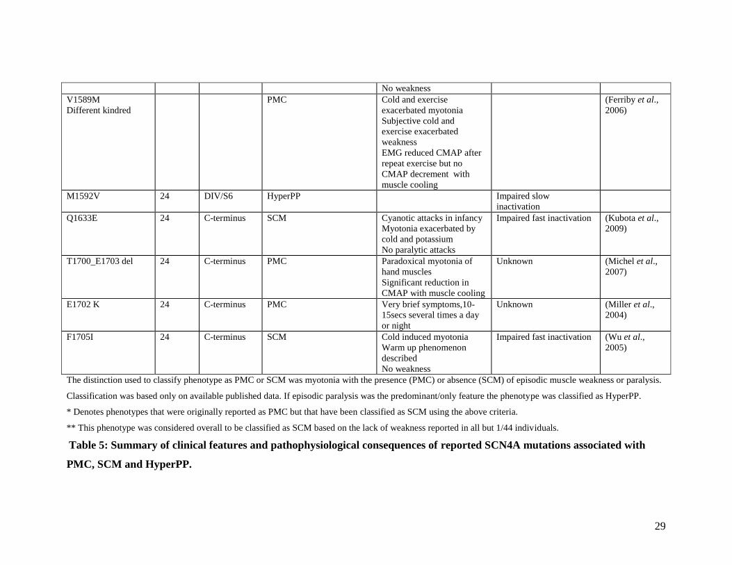
The distinction used to classify phenotype as PMC or SCM was myotonia with the presence (PMC) or absence (SCM) of episodic muscle weakness or paralysis.
Classification was based only on available published data. If episodic paralysis was the predominant/only feature the phenotype was classified as HyperPP.
* Denotes phenotypes that were originally reported as PMC but that have been classified as SCM using the above criteria.
** This phenotype was considered overall to be classified as SCM based on the lack of weakness reported in all but 1/44 individuals.
Table 5: Summary of clinical features and pathophysiological consequences of reported SCN4A mutations associated with
PMC, SCM and HyperPP.

30
Hypokalaemic Periodic Paralysis
Only approximately 10% of cases of hypokalaemic periodic paralysis are due to
mutations in the SCN4A gene(Sternberg et al., 2001). This less frequent genotype is
sometimes referred to as HypoPP 2. The majority of cases, estimated at between 60-70%,
are due to mutations in the CACNA1S gene that codes for the voltage gated skeletal
muscle calcium channel Cav1.1 (the dihydropyridine receptor). Approximately 20% of
cases are genetically undefined(Fouad et al., 1997; Miller et al., 2004; Sternberg et al.,
2001). All mutations that have been reported in either gene associated with hypokalaemic
periodic paralysis have been substitutions of positively charged arginine residues in the
voltage sensors (S4 segments) of the channels(Ptacek et al., 1994a; Jurkat-Rott et al.,
1994; Bulman et al., 1999; Jurkat-Rott et al., 2000; Bendahhou et al., 2001; Sternberg et
al., 2001; Davies et al., 2001; Kim et al., 2004; Wang et al., 2005). In total substitutions
of only 5 arginine residues have been reported in association with hypokalaemic periodic
paralysis. Substitution of a sixth arginine residue has been reported as causing a
potassium sensitive normokalaemic periodic paralysis (Fig 11).

31
Fig 11: Voltage sensor mutations of A: Cav1.1 and B: Nav1.4 associated with
HypoPP. *These mutations are associated with a different phenotype described as
potassium sensitive normokalaemic periodic paralysis.
+ +
+ +
+
++
+ +
+
++
++
+ +
++
+ +
+
A R528H/G R1239H/G
++
+ +
+ +
+ +
+ +
+ +
+ +
+ +
+ +
+ +
++
+
R669H
R672H/G
R675G/Q/W* R1132Q
B
+ +
+ +
+
++
+ +
+
++
++
+ +
++
+ +
+
A R528H/G R1239H/G
++
+ +
+ +
+ +
+ +
+ +
+ +
+ +
+ +
+ +
++
+
R669H
R672H/G
R675G/Q/W* R1132Q
B

32
Mutation Gene Exon Protein
position
Reported
Phenotype
Major
Pathomechanism
Reference
R528G/H CACNA1S 11 DII/S4 Hypokalaemic
periodic paralysis
Slower kinetics of
activation
Reduced current
density
(Lapie et al.,
1996; Morrill
et al., 1999)
R1239G/H CACNA1S 30 DIV/S4 Hypokalaemic
periodic paralysis
Slower kinetics of
activation
Reduced current
density
(Morrill et al.,
1999)
R669H SCN4A 12 DII/S4 Hypokalaemic
periodic paralysis
Enhanced slow
inactivation
Enhanced fast
inactivation
Reduced current
density
(Struyk et al.,
2000;
Kuzmenkin et
al., 2002)
R672G/H SCN4A 12 DII/S4 Hypokalaemic
periodic paralysis
Enhanced slow
inactivation
(R672G only)
Enhanced fast
inactivation
Reduced current
density
(Kuzmenkin et
al., 2002)
R675G/Q/W SCN4A 13 DII/S4 Potassium
sensitive
normokalaemic
periodic paralysis
Unknown (Vicart et al.,
2005)
R1132Q SCN4A 18 DIII/S4 Hypokalaemic
periodic paralysis
Enhanced fast and
slow inactivation
(Carle et al.,
2006)
Table 6: Summary of pathophysiological consequences of voltage sensor mutations
associated with HypoPP

33
The second aim of this thesis is to address whether a carefully phenotyped cohort of
channelopathy patients can be 100% genotyped by exomic sequencing of known
disease causing genes.
1.7 Pathogenesis of the skeletal muscle channelopathies
The skeletal muscle sodium channelopathies
The mutations in the SCN4A gene associated with PMC, SCM and HyperPP are scattered
throughout the gene affecting all domains of the Nav1.4 channel (see Fig 10). The
functional consequence of all these mutations is to produce a “gain of function” effect of
channel activity. This is achieved either by enhanced activation or impaired inactivation
of the sodium channel (see table 5).
Normally when the sodium channel is activated from its resting state by depolarization,
the channel opens rapidly, allowing an influx of sodium ions through the sodium
selective central pore into the cell. This rapid influx of positively charged sodium ions
depolarises the cell, and produces an action potential that triggers muscle contraction.
Sodium channels usually inactivate rapidly, stopping the influx of sodium ions, and, in
conjunction with potassium and chloride channels, promoting a return to resting
potentials (see Fig 1).
Normally a single action potential in a neuron leads to a single contraction in its target
muscle fibre. Myotonia results when a sequential train of action potentials is evoked in
the muscle in response to a single action potential nerve stimulus. Mutations that cause
myotonia do so by delaying inactivation of the sodium channel (see Table 5 and Fig 12)
which results in a hyper-excitable muscle cell. When inactivation is delayed the sodium
channel is open for longer and the resulting influx of sodium ions consequently persists
for longer. The persistent inward depolarising sodium current produces a comparable
increase in the outward repolarising potassium current which leads to a greater
accumulation of potassium ions in the t-tubules. This accumulation of positive charge

34
makes the sarcolemma susceptible to further spontaneous depolarisations with
accompanying muscle contraction.
In contrast to the hyper-excitable sarcolemma associated with myotonia the episodes of
flaccid muscle paralysis experienced in the sodium skeletal muscle channelopathies
reflect an unexcitable sarcolemma. This occurs due to incomplete inactivation of the
sodium channel (see Table 5 and Fig12). Once the sodium channel has opened there is
large persistent inward sodium current due to the inability of the channel to close. This
persistent current leads to sustained depolarisation of the sarcolemma and an inability to
support any further action potentials.
The key difference between these two situations is that in the unexcitable state, the
sodium channels fail to shut, preventing the cell from repolarising, while in the hyper
excitable state the channels do inactivate completely, only more slowly than normal.
Fig 12: Diagrammatic representation of impaired inactivation of Nav1.4 in PMC
and HyperPP.
Sodium current
-90mv
0mvMe
mb
rane
Po
ten
tia
l
WT sodium
current
Delayed
inactivation
Incomplete
inactivation
Sodium current
-90mv
0mvMe
mb
rane
Po
ten
tia
l
WT sodium
current
Delayed
inactivation
Incomplete
inactivation
WT sodium
current
Delayed
inactivation
Incomplete
inactivation
35
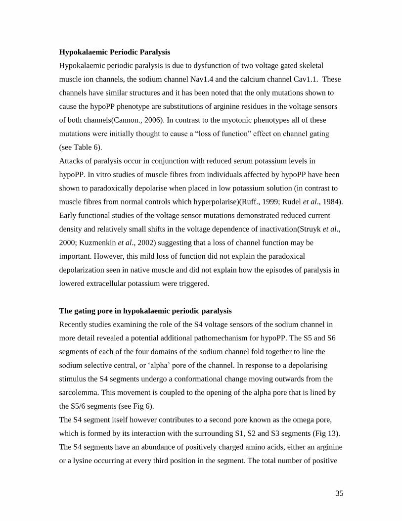
Hypokalaemic Periodic Paralysis
Hypokalaemic periodic paralysis is due to dysfunction of two voltage gated skeletal
muscle ion channels, the sodium channel Nav1.4 and the calcium channel Cav1.1. These
channels have similar structures and it has been noted that the only mutations shown to
cause the hypoPP phenotype are substitutions of arginine residues in the voltage sensors
of both channels(Cannon., 2006). In contrast to the myotonic phenotypes all of these
mutations were initially thought to cause a “loss of function” effect on channel gating
(see Table 6).
Attacks of paralysis occur in conjunction with reduced serum potassium levels in
hypoPP. In vitro studies of muscle fibres from individuals affected by hypoPP have been
shown to paradoxically depolarise when placed in low potassium solution (in contrast to
muscle fibres from normal controls which hyperpolarise)(Ruff., 1999; Rudel et al., 1984).
Early functional studies of the voltage sensor mutations demonstrated reduced current
density and relatively small shifts in the voltage dependence of inactivation(Struyk et al.,
2000; Kuzmenkin et al., 2002) suggesting that a loss of channel function may be
important. However, this mild loss of function did not explain the paradoxical
depolarization seen in native muscle and did not explain how the episodes of paralysis in
lowered extracellular potassium were triggered.
The gating pore in hypokalaemic periodic paralysis
Recently studies examining the role of the S4 voltage sensors of the sodium channel in
more detail revealed a potential additional pathomechanism for hypoPP. The S5 and S6
segments of each of the four domains of the sodium channel fold together to line the
sodium selective central, or ‘alpha’ pore of the channel. In response to a depolarising
stimulus the S4 segments undergo a conformational change moving outwards from the
sarcolemma. This movement is coupled to the opening of the alpha pore that is lined by
the S5/6 segments (see Fig 6).
The S4 segment itself however contributes to a second pore known as the omega pore,
which is formed by its interaction with the surrounding S1, S2 and S3 segments (Fig 13).
The S4 segments have an abundance of positively charged amino acids, either an arginine
or a lysine occurring at every third position in the segment. The total number of positive

36
charges varies for each S4 segment. The omega pore is not uniform in diameter and the
narrowest portion is occupied by different positively charged residues depending on
whether the membrane is resting (S4 down) or depolarised (S4 up).
Fig 13: The movement of the S4 segment through the omega pore
Under normal conditions the charged residues in the S4 segments form salt bridges with
other segments which effectively block any free ions from flowing through the omega
pore. Recent work has shown that neutralisation of either of the two outer arginine
residues in the DII/S4 segment of SCN4A has led to a loss of integrity of the omega pore
and allows monovalent cations to “leak” through the omega pore and into the cell when
the cell is hyperpolarised.
It is known that the amino acid that is substituted for the arginine residue affects the size
of this leak. If the arginine is replaced by a histidine, a proton leak through the omega
pore is favoured. Any other amino acid substitution at this site allows less selectivity and
a greater variety of monovalent cations can pass including sodium(Sokolov et al., 2007;
Struyk et al., 2007).
While the omega pore leak has been hypothesised as an additional pathomechansim in
hypoPP the precise consequences of this “gain of channel function” on muscle cell
homeostasis and sarcolemmal excitability are not yet fully understood. It is suggested that
movement of protons and other ions including sodium via the gating pore may disrupt pH
homeostasis and lead to intracellular sodium accumulation via activation of several ion
transporters including the sodium-hydrogen anti-port exchanger (see Fig 14)(Struyk et
al., 2008b; Jurkat-Rott et al., 2009). However, the cause of the paradoxical membrane
depolarisation in low potassium solution and the stimulus for the low serum potassium
Hyperpolarised Depolarised Hyperpolarised Depolarised

37
itself that occurs in HypoPP remains less clear although possible mechanisms are
discussed.
Fig 14: An aberrant current permeable to protons could stimulate the NHE and
NBC transporters with a resultant increase in intra-cellular sodium ions.
Stimulation of the monocarboxylate transporter would result in increased lactate
efflux from the cell which has been proposed as contributory to vacuolar formation
in HypoPP.
A further aim of my thesis was to examine the functional consequences of Nav1.4
hypoPP mutations in light of the recently described gating pore current.
Na/K ATPase H +
H + Na 2+ 2K + ATP
ADP + Pi
HCO3 - + H + CO 2 + H 2 O CA
MCT
NHE
NBC
Na 2+
CA
Aberrant
omega pore
current
MCT: monocarboxylate transporter NHE: Sodium hydrogen anti - port exchanger NBC: sodium dependant bicarbonate transporter CA :carbonic anhydrase
3Na +
H +
H +
Lactate
Na + 2K + ATP ADP + Pi
HCO3 - + H + CO 2 + H 2 O CA
MCT
NHE
NBC
Na +
CA
Aberrant
omega pore
current
MCT: monocarboxylate transporter NHE: Sodium hydrogen anti - port exchanger NBC: sodium dependant bicarbonate transporter CA :carbonic anhydrase
3Na

38
Role of inward rectifying potassium channels in hypokalaemic periodic paralysis
Muscle fibre resting membrane potential (VREST) is controlled by the membrane
permeability to K+ ions. It has been suggested that inhibition of the outward component
of the inward rectifying potassium channels could account for the abnormal membrane
response to low serum potassium and also for the lowered serum potassium itself due to
intracellular accumulation of potassium(Hofmann et al., 1970; Ruff., 1999). Furthermore,
barium which blocks inward rectifying potassium channel current(Standen et al., 1978)
produces reduced twitch force in the skeletal muscles of mammals in vitro in low
potassium solution.(Gallant., 1983) Reduced ATP dependant potassium channel (a
subgroup of IRK channel) current has also been identified in vitro from muscle biopsies
of hypoPP patients.(Tricarico et al., 1999) The aberrant inward depolarizing gating pore
current has recently been shown to contribute to the probability of the membrane
paradoxically depolarising in the presence of low potassium solution and also to the
reduced outward current component of the IRK channels.(Jurkat-Rott et al., 2009; Struyk
et al., 2008a) However, it is not clear why the potassium conductance should be reduced
by dysfunction of the voltage sensors of Cav1.1 or Nav1.4. One possible explanation is
that a proton selective gating pore causes an acidic intracellular environment and IRK
channels are known to be inhibited at this pH(Struyk et al., 2008a).
1.8 Treatment of the skeletal muscle channelopathies
Some patients with the skeletal muscle channelopathies feel their symptoms are not
restrictive enough to take medication and will manage their illness by avoiding
precipitating factors as far as possible. When drug therapy is considered a number of
therapeutic agents exists.
Myotonic disorders: early treatments
Where treatment is considered numerous membrane stabilising agents have been
employed in the myotonic disorders. These are currently mainly use dependant blockers
of sodium channels e.g. anti-convulsants, local anaesthetics or anti-arrhythmics. As these

39
target persistent sodium currents they are ideal to inhibit the abnormal sodium channels
with impaired inactivation seen in PMC, SCM and hyperPP but have little or no effect on
the functioning of wild type sodium channels.
In early studies, procainamide, quinine and glucocorticosteroids were employed. A small
randomized double blind trial compared the efficacy of each of these treatments in
relation to placebo in 20 individuals with myotonic disorder (16 myotonic dystrophy,
4 myotonia congenita). The diagnosis was made on a clinical basis without genetic
confirmation. The trial lasted 12 weeks and all participants received each of the four
treatments for a 3 week period with no washout period. An end-point of at least
a 50% reduction in the duration of hand grip myotonia, measured by EMG and timed
clinically, was employed. Using this endpoint, 6/20 participants taking quinine, 15/20
taking procainamide, 15/19 taking prednisone (one patient did not receive prednisone)
and 0/20 taking placebo showed improvement(Leyburn et al., 1959). This study, although
imperfect, illustrated a low efficacy of quinine. Despite the suggested benefits of
procainamide and prednisone the side effect profile of both these drugs restricts their use
and they are no longer recommended as therapeutic agents in the non-dystrophic
myotonias.
The carbonic anhydrase inhibitor acetazolamide is commonly used in the periodic
paralyses and has been reported to be beneficial in the non-dystrophic myotonias.(Trudell
et al., 1987; Ferriby et al., 2006) In a small series of nine patients with myotonia, seven
diagnosed clinically with myotonia congenita and two with paramyotonia congenita, all
cases reported a subjective and objective (timed measurements of myotonia)
improvement in myotonia with acetazolamide. However, one individual with
paramyotonia congenita developed quadriparesis 12 h after the ingestion of
acetazolamide.(Griggs et al., 1978) Larger studies of acetazolamide use in the non-
dystrophic myotonias have not been performed, and while there is evidence of some
benefit, it is not generally considered as a first line agent for the treatment of myotonia.
The anti-convulsant phenytoin has been shown to improve the righting time of myotonic
mice turned onto their backs.(Aichele et al., 1985) Ricker et al.(Ricker et al., 1978)
reported subjective improvement in muscle stiffness and an improved timed walk in one
patient with myotonia congenita and a dose dependant improvement in isometric force in
40
another. The lidocaine derivative tocainide gave encouraging results initially(Rudel et al.,
1980; Streib., 1987) but was eventually withdrawn from the market due to the risk of
potentially fatal agranulocytosis.(Volosin et al., 1985) Synthesis of tocainide analogues
has been attempted in vitro and may be of value for future study as anti-myotonic agents
if the efficacy and side effect profiles are favourable.(Catalano et al., 2008)
Myotonic disorders: Class I anti-arrhythmics
More recently class I anti-arryhthmics have offered potential for treatment. Flecainide, a
class Ic anti-arrhythmic, has been shown to be effective in vitro(Aoike et al., 2006)
although its use in clinical practice as an anti-myotonic agent is rarely
reported.(Rosenfeld et al., 1997) An improvement in clinical symptoms and cold induced
EMG findings with propafenone, another class Ic anti-arrhythmic has been reported in a
single case of paramyotonia congenita.(Alfonsi et al., 2007)
The class Ib anti-arrhythmic mexiletine is generally considered to be the first-line
treatment of choice by myologists but a randomized controlled trial is required. It is
usually well tolerated with only minor side effects reported. Importantly it has
proarrhythmic potential and therefore pre- and post-treatment ECGs are essential to
ensure satisfactory QT interval. More extensive cardiac evaluation prior to
commencement is important if there is an abnormal baseline ECG or a history of cardiac
disease. Single case reports have shown that mexiletine is effective in treating myotonia
in both sodium and chloride channel disorders.(Ceccarelli et al., 1992; Jackson et al.,
1994) However, a recent Cochrane review highlighted the lack of adequate randomized
double blind placebo controlled trials to prove efficacy.(Trip et al., 2006)
The ability to conduct such trials is partly hampered by the difficulty in quantitating
myotonia(Torres et al., 1983; Hammaren et al., 2005; Logigian et al., 2005; Moxley, III
et al., 2007; Hogrel., 2009) and in recruiting adequate numbers of patients to achieve
statistical power. A recent study employed trunk sway analysis to measure the warm up
phenomenon in recessive myotonia congenita and proposed that with further evaluation
this may offer an alternative potential end-point for therapeutic trials.(Horlings et al.,
2009) Sodium MRI has also recently been proposed as a possible outcome measure in
41
patients with sodium channel diseases. An increase in intramuscular sodium content was
demonstrated to accompany muscle weakness following exercise of cooled muscles in
paramyotonia congenita. In a small group of patients this increase was significantly
reduced following treatment with mexiletine.(Weber et al., 2006) It is possible this
technique could be used to monitor response to treatment in both a clinical and research
setting.
In vitro studies continue to identify pharmacological agents that preferentially block
sodium channels in the open state, thereby targeting persistent sodium currents.(Wang et
al., 2008b; Wang et al., 2008a) These studies may identify future therapies. No safe drugs
are currently available which specifically act on the ClC-1 channel. A number of
experimental approaches may have future implications for the treatment of myotonia
congenita including oligonucleotides to promote exon skipping, trans-splicing and
restoration of normal protein trafficking.(Cleland et al., 2008)
Hypokalaemic periodic paralysis: potassium supplements and potassium sparing
diuretics
In the 1930s it was recognised that lowered potassium levels were associated with attacks
of paralysis(Biemond et al., 1934) prompting treatment of these episodes with potassium
supplements and dietary modification to limit carbohydrate intake. It was realised
however that although often effective in aborting or reducing the duration and severity of
paralytic attacks, potassium supplementation had no effect on their frequency or on the
development of a permanent muscle weakness.
In the late 1950s Conn performed extensive experiments on 2 patients with hypokalaemic
periodic paralysis and reported that the attacks of paralysis were not only associated with
hypokalaemia but also hypernatraemia. He proposed that it was even possible the
hypernatraemia was the primary stimulus for the paralytic attacks with hypokalaemia
being secondary(Conn et al., 1957). While he did not suggest hyperaldosteronism was the
cause per se he did identify raised aldosterone levels during attacks in his patients.
However others did not corroborate these findings(Poskanzer et al., 1961). Conn
suggested a low sodium diet was beneficial in reducing both paralytic attacks and

42
myopathy. He also proposed carbonic anhydrase inhibitors may be effective due to their
ability to increase sodium excretion in the urine. In practical terms the restrictive sodium
diet proposed by Conn was difficult for patients to tolerate and others did not find sodium
restriction to be as beneficial(Poskanzer et al., 1961).
Based on his research however spironolactone, a known inhibitor of aldosterone, was
trialled as therapy for hypokalaemic periodic paralysis. In the initial patient studied,
subjective and objective observations of not only reduced paralytic attacks but also
improved inter-attack muscle strength were noted. This treatment with spironolactone did
produce a slight increase in sodium excretion with normal potassium serum levels being
maintained but the authors argued the effects on sodium and potassium balance were too
minimal to adequately explain the full mechanism of action of spironolactone in
producing such significant symptom reduction in hypokalaemic periodic
paralysis(Poskanzer et al., 1961). Spironolactone and other potassium sparing diuretics
are still used today as therapy for hypoPP although often not as first line agents(Kim et
al., 2001; Bendahhou et al., 2001; Kim et al., 2005; Kim et al., 2007). Gynaecomastia
can limit the tolerability of spironolactone for male patients.
Carbonic anhydrase inhibitors
Acetazolamide was first used in 1962 to lower the elevated potassium levels associated
with paralytic attacks in hyperkalaemic periodic paralysis(McArdle., 1962). A few years
later, despite seeming paradoxical in terms of potassium balance, it was also noted to be
an effective prophylactic agent in hypokalaemic periodic paralysis(Resnick et al., 1968).
An observational study further suggested acetazolamide may improve inter-attack muscle
strength in some patients(Griggs et al., 1970). Although no randomised controlled trials
have been performed using acetazolamide a plethora of case reports and series support its
beneficial effects (see Table 13 in Results). Another carbonic anhydrase inhibitor,
dichlorphenamide is the only therapy for hypoPP to have undergone a randomised double
blind placebo controlled cross over trial. The results of this trial showed a significant
efficacy of dichlorphenamide in reducing attack frequency although the inclusion criteria
were based on clinical diagnosis of hypoPP and not genetic confirmation(Tawil et al.,

43
2000; Sansone et al., 2008). Others have reported a benefit of dichlorphenamide on inter-
attack weakness(Dalakas et al., 1983). There are no trials comparing the use of
dichlorphenamide to acetazolamide in periodic paralysis.
Acetazolamide rapidly became the treatment of choice for hypokalaemic periodic
paralysis, and almost half a century later remains the frontline treatment. However with
genetic advances and the discovery of two causative genes it was noted that some patients
did not respond to acetazolamide treatment or indeed found it to be
detrimental(Bendahhou et al., 2001; Sternberg et al., 2001). It was initially suggested that
it was only the minority of patients with hypoPP 2 due to mutations in SCN4A who
reacted adversely to acetazolamide therapy but other reports of hypoPP 2 patients who
benefited from acetazolamide refuted this(Bulman et al., 1999; Kim et al., 2004; Venance
et al., 2004). Despite its popularity as a therapeutic agent for hypokalaemic periodic
paralysis the disease specific mechanism of action of acetazolamide is not understood.
Importantly despite some suggestive case reports it is not established in either the
myotonic disorders or hypokalaemic periodic paralysis if currently available treatments
have any influence on the development of permanent myopathy.
In my thesis I attempt to quantify the efficacy of acetazolamide in hypokalaemic
periodic paralysis and whether genotype has any influence on treatment response.
1.9 Mechanisms of action of acetazolamide
Carbonic anhydrase inhibition
Carbonic anhydrase is an enzyme that catalyses the reversible reaction converting carbon
dioxide and water into protons and bicarbonate.
Acetazolamide is a sulphonamide that inhibits carbonic anhydrase (CA) and it is
generally considered that its main therapeutic mechanism of action in hypoPP is
CO2 + H20 H2CO3 H+ + HCO3

44
somehow linked to this capacity. Inhibition of CA in the renal tubules by acetazolamide
leads to increased urinary loss of bicarbonate, sodium and some potassium with a
resultant metabolic acidosis (Fig 15). It remains unclear how this alteration of pH could
reduce paralytic attacks or prevent the development of myopathy. One study did examine
the effect of pH on the R669H SCN4A mutation and pH on the R672H/G SCN4A
mutations expressed in a HEK cell system. The deleterious effects of the histidine
substitutions could be ameliorated by a more acidic pH whereas the glycine substitution
was insensitive to alterations in pH. The authors proposed this may predict that those
hypoPP patients with a glycine substitution would not benefit from acetazolamide
(Kuzmenkin et al., 2002).
Fig 15: Acetazolamide(ACZ) inhibits the carbonic anhydrase(CA) present in the
tubular lumen preventing the conversion of H2CO3 to CO2 and H2O. H2CO3
dissociates to H+ and HCO3. Bicarbonate is lost in the tubular lumen producing a
metabolic acidosis. Within the proximal tubular cell itself ACZ blocks the CA
preventing the conversion of CO2 and H20 to H+ and HCO3. The reduction in
protons reduces the re-absorption of sodium ions which are excreted in the urine.
H+
Na+
HC03 +
H2C03
C02 + H2O
C02+H20
H2C03
H+ +HC03
Na+
CAACZ x
x CAACZ
Lumen Proximal tubular cell
H+
Na+
HC03 +
H2C03
C02 + H2O
C02+H20
H2C03
H+ +HC03
Na+
CAACZ x
x CAACZ
Lumen Proximal tubular cell

45
Carbonic anhydrase isoenzymes
At least 14 isoenzymes of CA exist in humans. Different isoenzymes are preferentially
expressed in the cytosol, cell membranes or mitochondria of different tissues. Each
isoform demonstrates a different degree of catalytic activity and affinity for
sulfonamides.(Clare et al., 2006; Supuran et al., 2000)
The subcellular localisation of several isoforms has been studied in animal models and
shown to be present at variable levels in the sarcolemma and sarcoplasmic reticulum of
skeletal muscle(Wetzel et al., 1998; Wetzel et al., 2007; Scheibe et al., 2008). The
presence of different isoforms of CA in skeletal muscle raises questions about how the
contribution of each isoform may influence CA treatment response and how this might be
influenced by the proposed proton permeable gating pore. Intracellular isoforms of CA
can only be reached by the lipophilic membrane permeable sulphonamides. Hydrophilic
sulphonamides such as acetazolamide or the alternative carbonic anhydrase inhibitor
dichlorphenamide, which is also used in hypoPP, cannot easily cross the sarcolemma and
would not be expected to have any significant direct effect on the intracellular isoforms.
This suggests any benefit derived from acetazolamide relies on inhibition of extracellular
carbonic anhydrase or carbonic anhydrase on the extracellular surface of the membrane.
Acetazolamide and activation of sarcolemmal calcium activated potassium channels
[KCa2+]
Tricarico et al used the potassium depleted rat as an animal model of hypoPP to explore
the hypothesis that the mechanism of action of acetazolamide and other carbonic
anhydrase inhibitors was not exclusively related to their inhibition of CA(Tricarico et al.,
2004). A dose dependant increase of calcium activated potassium channel [KCa2+
]
activity and restoration of the serum potassium levels to within the normal range was
observed in the muscle fibres of potassium depleted rats in whom treatment with
acetazolamide prevented insulin induced attacks of paralysis(Tricarico et al., 2000;
Tricarico et al., 2004). The ability of acetazolamide to enhance the sarcolemmal
conductance of potassium seems particularly relevant when considered in light of the
studies discussed that implicate inhibition of IRK channel conductance in the
pathomechanism of hypoPP.

46
Effects of acetazolamide on inter-attack weakness
The reported clinical benefit of acetazolamide in preventing or improving inter-attack
weakness has also been explored using the K depleted rat model. Vacuoles are a common
morphological finding in primary and secondary hypokalaemic periodic paralysis. They
are considered to represent localised swelling and vacuolation of the t-tubules secondary
to increased osmolarity caused by the local accumulation of ions or metabolites
(including lactate). The rat model demonstrated a vacuolar myopathy and an increased
efflux of lactate from muscle fibres in vitro. Muscle biopsies from rats treated with
acetazolamide demonstrated significantly reduced vacuoles and lactate efflux(Tricarico et
al., 2008).
These observations are particularly interesting when considered in light of the proposed
proton leak described in hypoPP. Accumulation of intracellular protons could produce an
increased efflux of lactate by stimulating the proton linked monocarboxylate transporter
(Fig 14). Potentially this pathway may partly explain the reports of acetazolamide
ameliorating inter-attack muscle weakness.
1.10 Mechanisms of muscle degeneration in the skeletal muscle channelopathies
Patients with periodic paralysis have been frequently reported to exhibit vacuoles and/or
tubular aggregates on muscle biopsy(Venance et al., 2006) and as discussed there is some
evidence suggesting lactate efflux may contribute to this. However, the myopathological
findings in NDMs are not defined well and often reported to be non-specific(Miller et
al., 2004). Furthermore, with the characteristic clinical history and examination findings
coupled with the recent advances in electrophysiological techniques a diagnosis of NDM
is usually apparent and it is now rare that a muscle biopsy will be performed in such
patients other than as a research procedure.
It is clear muscle damage can occur in the NDMs but its pathomechansim and frequency
are unknown. It has been postulated that the abnormally prolonged intramuscular influx
of sodium that is known to occur via the mutant sodium channels may be responsible for
muscle degeneration(Bradley et al., 1990) but the specifics of such a mechanism have not

47
been shown. However, it is of note that there is evidence for increased intracellular
sodium contributing to cell necrosis in the mouse model of Duchenne muscular
dystrophy(Hirn et al., 2008). One recent study has employed ultrasound to assess
permanent muscle changes in the NDMs. Using ultrasound measurements of eight
muscles, (four upper limb and four lower limb) in a group of 63 genetically confirmed
NDM patients an increase in the mean echo intensity compared with controls from all
muscles examined except the rectus femoris was observed. The ultrasound changes were
considered to indicate structural muscle damage such as fatty infiltration or fibrosis. This
change was most marked in the forearm flexors where the increased echogenicity
correlated negatively with muscle power. There was no positive correlation between echo
intensity and age for individual muscles except the rectus femoris although the sum of the
scores did show a significant positive correlation(Trip et al., 2009b).
Recently, a mouse model of hyperkalaemic periodic paralysis has been engineered by
introducing the murine equivalent of the SCN4A mis-sense mutation M1592V(Hayward
et al., 2008). This mutation causes both myotonia and paralysis in humans(Rojas et al.,
1991; Kelly et al., 1997) and was demonstrated to produce these same symptoms in the
mouse verifying its use as a model of the human disease. At a few months of age the mice
heterozygous for this mutation already displayed subtle myopathic changes. In those that
were homozygous significant muscle abnormalities were seen including an increase in
fibre size variability, frequent internal nuclei and large scattered vacuoles. These early
changes were present at a few months of age before any spontaneous episodes of
paralysis had been observed. Furthermore, they were shown to increase with age in the
heterozygotes while muscle force generation declined(Hayward et al., 2008). This
supports the clinical observations that myopathy increases with age in humans(Links et
al., 1990; Plassart et al., 1994) and may be independent of paralytic attacks in the
periodic paralyses(Buruma et al., 1978; Links et al., 1990).
Although this animal model is of a hyperkalaemic periodic paralysis genotype the same
symptoms of myotonia and muscle weakness occur in the allelic disorders PMC and
SCM. The pathomechanism is also a gain of function of the sodium channel in both. It is
likely that future insights into muscle degeneration gained from the study of this model
will also have implications for our understanding of PMC and SCM. The possibility that

48
myopathy develops independently of symptom frequency or severity may influence
future approaches to therapy which is currently aimed at relieving symptoms. As such,
many patients with minimal or manageable symptoms decline pharmacological treatment.
1.11 Morbidity in the skeletal muscle channelopathies
Very little is known about the impact of the muscle channelopathies on quality of life and
these disorders have often been regarded as benign. A single study has recently examined
this in a group of 62 genetically confirmed NDM patients and found painful myotonia
and fatigue to be the best predictors of poor general health perception and physical
functioning(Trip et al., 2009a). In this study painful myotonia was reported in 28% of
those with myotonia congenita and 57% with sodium channelopathy. In addition, there
are numerous case reports where pain which is often severe, is described in the
NDMs(Vicart et al., 2004; Rosenfeld et al., 1997; Ptacek et al., 1994b; Colding-
Jorgensen et al., 2006; Fialho et al., 2007; Walsh et al., 2007; Wang et al., 2008c). This
suggests that pain is a frequent symptom that may have been previously under-recognised
and possibly undertreated in the NDMs.

49
1.12 Summary of Aims
The specific aims of this thesis were:
1. To examine the phenotype of the skeletal muscle channelopathies in patients referred
to the NCG service for channelopathies at the NHNN to determine
a. if there were any clinically useful phenotype-genotype correlations that
could be made and
b. if there were more extensive aspects to the phenotypes than is currently
recognised.
2. To genetically characterize patients to assess if new mutations in known genes existed
and if these genes could account for 100% of a carefully phenotyped cohort or if new
genes may be implicated.
3. To ascertain the efficacy of acetazolamide therapy in hypokalaemic periodic paralysis
and if genotype has any influence on treatment response.
4. To functionally characterize mutations in hypokalaemic periodic paralysis in light of
the recently described gating pore seen in some of the Nav1.4 mutations.

50
Methods
2.0 Examining the phenotype of the skeletal muscle channelopathies
Information on the phenotype of patients with skeletal muscle channelopathies was
examined from several sources. Where possible, patients were seen and examined
personally through the specialist neuromuscular clinic at the MRC Centre for
Neuromuscular Diseases (under the government funded NCG scheme for rare diseases)
when they attended for clinical care or when they participated in a natural history trial of
non-dystrophic myotonias. The natural history trial was a collaborative effort between the
MRC Centre for Neuromuscular Diseases and five other American and Canadian centres.
All participants consented to the study which had ethical approval from local ethics
committee and included DNA analysis to allow correlation of genotype with any
emergent phenotypic features. Data collected included history of illness and symptoms,
relevant past medical history, family history and drug therapy. Patients were examined
for the presence of myotonia, and muscle strength was recorded by Elizabeth Dewar
(senior physiotherapist) using standardised tests. All patients seen at the centre also
underwent electrophysiological testing with Dr Veronica Tan including standard NCS,
EMG and specialised short and long exercise tests. All patients enrolled in the natural
history trial also completed quality of life questionnaires.
The majority of patients were personally examined by me, e.g. all of the participants in
the natural history trial, the English adults detailed in the neonatal phenotypes and all
others who attended the clinic who have been described in this thesis. When it was not
possible to personally examine individuals (e.g. the family in Australia, the individuals
with inflammatory biopsies) information was obtained from a clinical and genetic
database of all those referred to the diagnostic NCG service. In selected cases the
referring clinician was contacted to request supplemental information and in some cases,
a muscle biopsy and DNA samples. Muscle biopsies were reviewed with Dr Janice
Holton (consultant pathologist). DNA samples were analysed as detailed in the following
sections. Data gathered on patients seen during the thesis was added to the clinical and
51
genetic database held at the MRC Centre for Neuromuscular Diseases. A record was kept
of all unusual phenotypes and endeavours made to obtain complete details as above.
2.1 Gene Sequencing
Direct automated sequencing of all 24 exons of SCN4A, 44 exons of CACNA1S and one
exon of KCNJ2 was carried out for all patients with available DNA and no pre-existing
genetic diagnosis. Selected samples were additionally analysed for the presence of the
quadruplet nucleotide repeat expansion in intron 1 of the ZNF9 gene associated with
myotonic dystrophy type 2 (DM2). A small number of samples had direct automated
sequencing performed of all exons of CLCN 1 associated with myotonia congenita and
the RYR 1 gene. These two genes were analysed by the diagnostic labs at the NHNN and
GOSH.
Primer Design
KCNJ2 primers had been previously designed and optimised by the diagnostic genetic
laboratory at the NHNN. Aliquots of these primers were kindly given to me by the lab.
Primers for SCN4A and CACNA1S were designed and PCR conditions optimised as
follows.
Gene sequences were identified using the Ensemble database:
http://www.ensembl.org/index.html. Using these sequences forward and reverse primers
were designed for each exon using the Primer 3 web-based programme:
http://frodo.wi.mit.edu/primer3/. Optimal primers were designed to begin within
approximately 100 base pairs from the beginning or end of each exon and to produce a
fragment size of approximately 450 base pairs. Primers were then blasted against the
human genome to ensure specificity using another website: http://genome.ucsc.edu/cgi-
bin/hgPcr?org=Human&db=hg18&hgsid=90948004. Any non-specific primers were
redesigned. Finally all primers were analysed for the presence of single nucleotide
polymorphisms (SNPs) using:http://ngrl.man.ac.uk/SNPCheck/SNPCheck.html. Any
primers containing SNPs were also re-designed. Once optimised, primers were tagged
with M13 universal primer sequences before ordering from SIGMA.

52
Exon Forward Primer Reverse Primer
M13 TGTAAAACGACGGCCAGT CAGGAAACAGCTATGACC
Ex1 GCACATCTCCCAGTCCTGAT TGGATGGCAGACAGACAGAG
Ex2_3 CTCTGTCTGTCTGCCATCCA CCACACAGAGGTGCAAACAC
Ex4 GCCACCTGAGTGGCATATTT GGGCTGCCTCATGTGATTAT
Ex5 AGGAGCTTTGGGGGTGTCT ATCTGCCCTCTGGTCACG
Ex6 CAAGCCTTGAAGATGGAGTAGG GAGGCTACCCTAGGGACTGG
EX7_8 GTGTCCATGTGGGTGACTTG CCAGCTGGAAGAGGTTCTCC
Ex9 ATGTATGGAAAGGGGCACTG ACCCTGGGTCCTCTATCTCC
Ex10 CTTTGGTCCCTACCCTGTCA TCCTCCTGAATCCAGTCCAG
Ex11 GCTTGGAAGGTGGAACAAAA CATACAGCCAGACAGCTCCA
Ex12 CTCTGTGACAGGGCCTCATG TCCTCACCCCACCCCCATCC
Ex13 CTGCCTTGGGTGGTGGTC TTGGGGAGATAGAAAACAGTCA
Ex14a TGCCCAACATGTGGTAGTTT GCGGCTCCTTCTTCTCATC
Ex14b CCCAAGGACATCATGCTCA GCTCCAGGTCACAGGAGAAT
Ex15 AGGGCTGGATGAATGAATGA GCTCCAAGCTAGGTCTGCAC
Ex16 TGTTGGATCCAGCTGTGAAG CCCTTCCTGTGTGTGGAGAC
Ex17 ATGGCAATTTGCTGTTGTTG TGACAGCCTCTGGATGTAGC
Ex18 GTGCCAGGCTCCTACAGGT CTCTGGTGGTGGTTGGAGTA
Ex19 AGGAAGCCCTCCCTAATTGA AGTGACACTGGGGTTGGGTA
Ex20 GTTGTACCCAACCCCAGTGT CTGAGGGCAGGACCCATC
Ex21 CGTTCTGTCCCCGAGACTT GGGTTTGTGCAATGGAGAGT
Ex22 GCTGCCTTAAAGGTGAGGT GGGTGGAAGGCAGGAAAC
Ex23 CTGCCTGTCCATGTCCTGT GTCTTCCCGAGTGCCTCA
Ex24a CTCTGAGACTTGAGCAGAGCAC TCTCGAACAGGCAGATGATG
Ex24b TTGCCTACGTCAAGAAGGAGTC AAGTCTGAGAGGCGGCTGTA
Ex24c TCTCCTTCCTCATCGTGGTC ACCTCCTCGTGCTTCCTCTT
Ex24d AAGCAGACCATGGAGGAGAA AGGCACAGTCCCAGATTCAA
Table 7: SCN4A Primer sequences

53
Exon Forward Primer Reverse Primer
M13 TGTAAAACGACGGCCAGT CAGGAAACAGCTATGACC
1 GGCTGGGTGGAATGACAG TGTGATCACCCCGAATCC
2 GGCATCAGAGTCACAGTCCA GACCAATGAGTGCGTGCTTA
3 TTCCATCAGCAAGTGCAAAC TGATCCACTCTCCACCAACA
4 TATTTGCCGTCTCTCCCCTA TAGGAAGGGGACCCAGAACT
5 AGTTCTGGGTCCCCTTCCTA CCAGGAGAAACCCATTCTCA
6 TTCACTGTCACCGTCTGCTC GTGGTCACGCAAGTCAGAGA
7 CAGGGTTGGGTGGAGTCTTA AATTGAAGGGCTCCTGTGTG
8 GGTGAGCAGGAAGTGAGAGG ACCATTTTGAGCCATTTTGC
9 TGGGCTCTTTAGCTCTTGGA GTCCCTCAGAGCAGGAACTG
10 GCCTGGAGTTTGTCTGAAGG GGGTATGTTGCTTTGCCACT
11 GGGAGTCAGGAGAAGGGAAG AACCTGCACAGATCCCAGAC
12 CCCTCCCTCTGTGTCACATC CCCACCTTGATCTTGAAGGA
13 AGTAACAGGCGTGAGCCACT GGGCCTGGCTACCTAGAAAC
14 TGAGGGCCTTCCACTATGTC CCAGAAGGCCAACTTGTCTC
15 GCCCTCTCACCACAGAGAAG AGGAGTCCCTGGAGATGGAT
16 CCAAGCAGTGGGTATGTGAA ATGGAGGGGTACAGGTAGGG
17 CAACATGCAGCCACAAAGTT AGATGAGAGCCGCATCAATC
18 GGAGCAGGAGGTGTATTCCA CAGCATAAAGCAGGCAGTCA
19 CTTCACCCTCTCTGCCACTC TGCCAGTCTCCACCTCTTTT
20 TGTTCTCCAAGCTCCTCCAT CTGTGGGTGGCTAGAAGCTC
21 GGAAGGTGGGAGTGTTTTCA AGACTTGCTGCCTCCTGATG
22 AGGAATGGGAGGGGTTACAG GGCTCCTTGTGCTTGAGAGT
23 GAGCTGTGGGAATGAACGAT AGCCTAAAGGCTGAGCTTCC
24 AGGAGGGACACTCACAGAGG AGGAGGGACACTCACAGAGG
25 CTTCCCAGCCTGACTCTCTG CACCCTTAGGCCTCTCTTCC
26 TTCACAGTCCTCAGCCACAG TCCATGTAGCACCTCAGCAC
27 CCCCTCCCCTTCTGTGTTAT GAGCAAGTTGGGAGCAGAAG

54
28 AAGAGCAAAGGGGAGAGGAG AGTTACCTCTCTGCCCAGCA
29 CCCACAGAGCTTCAGTCCTC AAGCCGCTATATCCATGCAC
30 AGTTCCTCCATGCCACAAAC GAGCGCCAGTCAGTGTCTTA
31 GACCCTGGAAACTGCACGTA ATCTGACACTCCAGCCATCC
32 CCCCCACACATTGACTTACC CAGGTCACACACCCATGAAG
33 AGGCAGGAGCAGATAGGTGA TGGCAGCCTCTCTGGAGTAT
34 GTGTGATGTGGGTGGTTCAG AAATGAAGGGGGAAGGAGAA
35 TCCCATATCTGAGGGAATGC CCATCAGGTCCTCACCAGTT
36 TCAGCAATGTCACCCAACTC TGGGGTCCTCCCTCTACTTC
37 GCCTGTGGGGAGAAAGTACA CTGGTCCGTTCTCAGATTCC
38 CGGACCAGATCCTCCATAAA CTTCTGGGCTTCCTTTTTCC
39 TCTCCTGGGCTGGAGAGATA TGGTAGGGAAATGTGGGAAA
40 CCATCCTCCTTTGGCCTTAT GCTGACATTGTCCTCCCAGT
41 TGAGGGAAAAGGATGTTTGG TTAGCATGAATGGGCTTTCC
42 AAGCGAGCCTCTGTTGACTC CCACCAATGAGCAAAATTCC
43 CCTGGCCTGATACTTGGAGT CTGTTGGCCCTACCCTCTCT
44 AGAGGGTAGGGCCAACAGAG TTTTGAGGTGGTTCCTGACC
Table 8: CACNA1S Primer Sequences
PCR Reagents and Reactions
PCR reactions to amplify each exon of SCN4A and CACNA1S were performed using a
number of different reagents and conditions but the optimal methods most commonly
used were:
1. A 25μL reaction contained 200ng genomic DNA, 12.5μL of Amplitaq 360 MasterMix
(Applied Biosysytems), 10pmol of each primer (forward and reverse) and 9.5μL of
ddnH20.
2. A 25μL reaction contained 200ng genomic DNA, 5μL of 10 x PCR buffer without
MgCl2 (Applied Biosystems), 4μL of 25mM MgCl2, 5μL of 2mM dNTPs, 15pmol of

55
each primer (forward and reverse), and 2.5 units of AmpliTaq Gold polymerase (Applied
Biosystems).
For each method all constituents except the DNA were pipetted into a 1.5ml eppendorf
and vortexed for 5 secs. 24μL were then pipetted into each well of a 96 well PCR plate.
1μL of DNA was added to each well at this stage. A control well substituting sterilized
H20 for DNA was included for each exon (i.e. each set of primers). The 96 well plate
was sealed with a transparent self-adhesive seal and centrifuged for 1min at low speed to
collect samples at the bottom of the wells.
9700 PCR machines were used. Variable PCR cycling conditions were also trialed but the
optimum conditions consisted of an initial denaturing step of 95°C for 10 minutes
followed by 30 cycles of 95°C for 30 seconds, 55°C(for CACNA1S primers)/60°C(for
SCN4A primers) for 30 seconds, 72°C for 30 seconds, and a final extension step of 72°C
for 7 minutes.
Agarose Gels
PCR products were run on 2% agarose gels. Gels were made using 2g of agarose powder
per 100ml of 1 x TBE (Tris Borate EDTA) which was microwaved on full power for 2-3
mins to dissolve. Ethidium bromide (5μL) was added to the gel. The gel was stood to
cool for a further 3-5 mins before being poured into a tray with gel combs and end plates
and allowed to set for 1 hour. 5μL of PCR product from each well of the 9700 PCR plate
was mixed with 3μL of orange G (as a loading dye) and transferred to the gel chambers.
A DNA size marker was placed in the first well of each row of samples. Additional 1 x
TBE was poured over the gel as a buffer and the lid placed on the tray. Electrodes were
connected to the power supply and 65Volts run through the tray. Gels were left to run for
45mins or until the loading dye had run ¾ of the length of the tray. The gel was then
carefully removed from the tray and placed under UV light in a light box to identify the
presence of DNA fragments of expected size (using the DNA size marker for
comparison) bound to ethidium bromide and hence success or failure of the PCR
reaction.

56
PCR Clean Up
If the PCR was successful the remainder of the PCR product was cleaned to remove all
constituents except the DNA fragments. The PCR plate was first centrifuged for 1min to
collect samples at the bottoms of wells. 30μL of sterilized water was added to each well
and the plate was resealed and re-spun on the centrifuge for a further 1min. The diluted
product was then transferred to a 96 well PCR clean up plate and placed on a vacuum for
5-10mins or until dry. The dry DNA was re-constituted with 40μL of sterilized water and
placed on a rotator for 30mins before being transferred to a clean 96 well PCR plate.
Sequencing Reagents and Reactions
Samples were sequenced (bidirectionally) using the ABI Big Dye Terminator Sequencing
Kit version 1.1, M13 universal primers, and an ABI Model 3730xl Automated DNA
Sequencer.
Each 10μL sequencing reaction contained the following reagents: 1μL of Big Dye, 2μL
of 5 x sequencing buffer, 10pmol of M13 universal primer (forward or reverse), 3.5μL of
the clean PCR product and 2.5μL of sterilized ddnH20. All constituents with the
exception of PCR product were pipetted into a 1.5ml eppendorf in volumes adequate for
100 reactions and vortexed for 5secs. 6.5μL was then pipetted into each well of a 96 well
9800 PCR plate and 3.5μL of PCR product added to the sequencing reagents. The plate
was sealed and centrifuged for 1 min to collect samples at the bottom of the wells.
A 9800 PCR machine was used and the following cycling conditions: an initial step of
96°C for one minute, followed by 25 cycles of 96°C for 10secs, 50°C for 5secs, 60°C for
1min 15secs, and then held at 4°C.
After sequencing the samples were cleaned in order to remove all reagents. The clean up
plates were centrifuged for 3mins at 2150rpm and the flow though discarded. The
samples were centrifuged for 1 min at low speed to ensure they collected in the bottom of
the wells. 10μL of sterilized ddnH20 was added to each well, the plate resealed and
centrifuged for a further 1 min. All samples were then transferred to the clean up plate
which was placed on top of a clean PCR plate. Plates were spun together for 3 mins at
2150rpm so that the cleaned sequencing product was filtered into the clean PCR plate.

57
The samples were sequenced bidirectionally using an ABI Model 3730xl automated DNA
sequencer. All primers were initially tested and all conditions optimized on control DNA
samples that were known to be of good quality before proceeding to research samples.
2.2 Analysis of genetic data
Data was analyzed using the version 2.5 SeqScape Analysis software (ABI). A reference
data group and project template was created for each gene using the seqscape manager
software. The reference data group is the sequence against which the analysis programme
compares the sample sequence. In essence this involves importing and accurately
numbering the coding sequence flanked by a small portion of intron for each exon.
If a sequence variant was identified that predicted an amino acid change it was compared
to the reference gene sequence in Ensemble for any known single nucleotide
polymorphisms. A second database, dbSNP: http://www.ncbi.nlm.nih.gov/projects/SNP/,
was additionally searched. If neither database identified the sequence variant as a
polymorphism it was entered into a SIFT (sorting intolerant from tolerant) prediction
programme: http://sift.jcvi.org/www/SIFT_BLink_submit.html to prioritise which
changes were most likely to be deleterious and therefore of a pathological nature. A more
laborious but similar approach was employed by also checking isoform sequences of
other animal species to determine the conservation of the amino acid change. If these
combined efforts indicated the change was likely to be pathological control samples were
analysed (minimum of 166 chromosomes) for the presence of the amino acid substitution.
2.3 ZNF9 anlaysis
One of the aims of this work was to assess whether known disease genes accounted for
all cases examined with EMG proven myotonia. A minority of samples were identified in
whom testing of the CLCN1 gene (performed by the diagnostic lab) and SCN4A revealed
no pathogenic changes. These samples were analysed for the presence of the quadruplet
nucleotide repeat expansion in intron 1 of the ZNF9 gene associated with myotonic
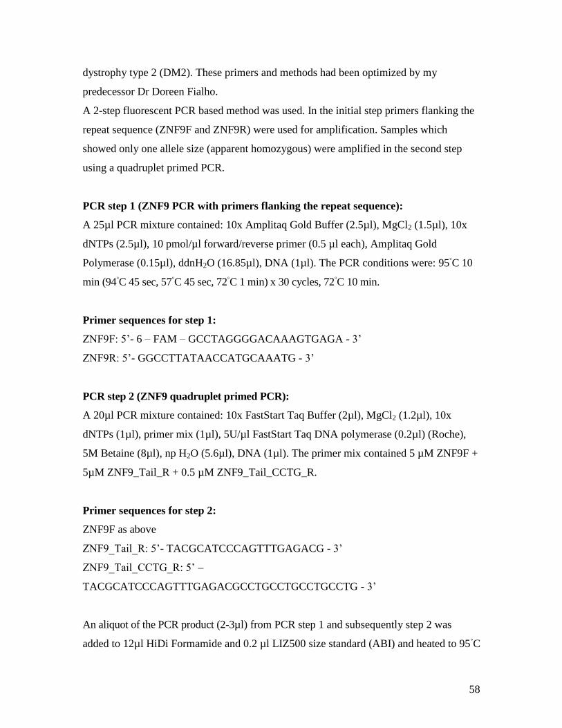
58
dystrophy type 2 (DM2). These primers and methods had been optimized by my
predecessor Dr Doreen Fialho.
A 2-step fluorescent PCR based method was used. In the initial step primers flanking the
repeat sequence (ZNF9F and ZNF9R) were used for amplification. Samples which
showed only one allele size (apparent homozygous) were amplified in the second step
using a quadruplet primed PCR.
PCR step 1 (ZNF9 PCR with primers flanking the repeat sequence):
A 25µl PCR mixture contained: 10x Amplitaq Gold Buffer (2.5µl), MgCl2 (1.5µl), 10x
dNTPs (2.5µl), 10 pmol/µl forward/reverse primer (0.5 µl each), Amplitaq Gold
Polymerase (0.15µl), ddnH2O (16.85µl), DNA (1µl). The PCR conditions were: 95C 10
min (94C 45 sec, 57
C 45 sec, 72
C 1 min) x 30 cycles, 72
C 10 min.
Primer sequences for step 1:
ZNF9F: 5’- 6 – FAM – GCCTAGGGGACAAAGTGAGA - 3’
ZNF9R: 5’- GGCCTTATAACCATGCAAATG - 3’
PCR step 2 (ZNF9 quadruplet primed PCR):
A 20µl PCR mixture contained: 10x FastStart Taq Buffer (2µl), MgCl2 (1.2µl), 10x
dNTPs (1µl), primer mix (1µl), 5U/µl FastStart Taq DNA polymerase (0.2µl) (Roche),
5M Betaine (8µl), np H2O (5.6µl), DNA (1µl). The primer mix contained 5 µM ZNF9F +
5µM ZNF9_Tail_R + 0.5 µM ZNF9_Tail_CCTG_R.
Primer sequences for step 2:
ZNF9F as above
ZNF9_Tail_R: 5’- TACGCATCCCAGTTTGAGACG - 3’
ZNF9_Tail_CCTG_R: 5’ –
TACGCATCCCAGTTTGAGACGCCTGCCTGCCTGCCTG - 3’
An aliquot of the PCR product (2-3µl) from PCR step 1 and subsequently step 2 was
added to 12µl HiDi Formamide and 0.2 µl LIZ500 size standard (ABI) and heated to 95C
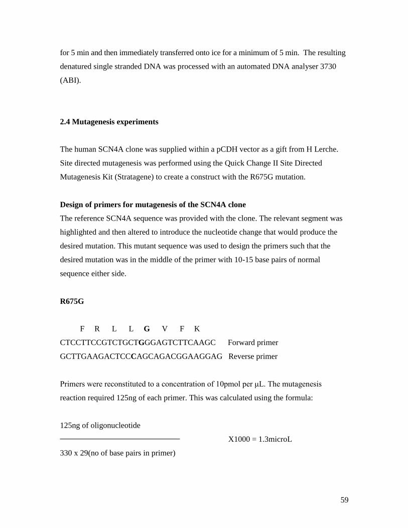
59
for 5 min and then immediately transferred onto ice for a minimum of 5 min. The resulting
denatured single stranded DNA was processed with an automated DNA analyser 3730
(ABI).
2.4 Mutagenesis experiments
The human SCN4A clone was supplied within a pCDH vector as a gift from H Lerche.
Site directed mutagenesis was performed using the Quick Change II Site Directed
Mutagenesis Kit (Stratagene) to create a construct with the R675G mutation.
Design of primers for mutagenesis of the SCN4A clone
The reference SCN4A sequence was provided with the clone. The relevant segment was
highlighted and then altered to introduce the nucleotide change that would produce the
desired mutation. This mutant sequence was used to design the primers such that the
desired mutation was in the middle of the primer with 10-15 base pairs of normal
sequence either side.
R675G
F R L L G V F K
CTCCTTCCGTCTGCTGGGAGTCTTCAAGC Forward primer
GCTTGAAGACTCCCAGCAGACGGAAGGAG Reverse primer
Primers were reconstituted to a concentration of 10pmol per μL. The mutagenesis
reaction required 125ng of each primer. This was calculated using the formula:
125ng of oligonucleotide
X1000 = 1.3microL
330 x 29(no of base pairs in primer)
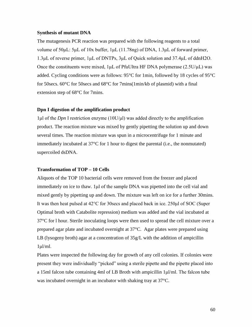
60
Synthesis of mutant DNA
The mutagenesis PCR reaction was prepared with the following reagents to a total
volume of 50μL: 5μL of 10x buffer, 1μL (11.78ng) of DNA, 1.3μL of forward primer,
1.3μL of reverse primer, 1μL of DNTPs, 3μL of Quick solution and 37.4μL of ddnH2O.
Once the constituents were mixed, 1μL of PfuUltra HF DNA polymerase (2.5U/μL) was
added. Cycling conditions were as follows: 95°C for 1min, followed by 18 cycles of 95°C
for 50secs. 60°C for 50secs and 68°C for 7mins(1min/kb of plasmid) with a final
extension step of 68°C for 7mins.
Dpn I digestion of the amplification product
1μl of the Dpn I restriction enzyme (10U/μl) was added directly to the amplification
product. The reaction mixture was mixed by gently pipetting the solution up and down
several times. The reaction mixture was spun in a microcentrifuge for 1 minute and
immediately incubated at 37°C for 1 hour to digest the parental (i.e., the nonmutated)
supercoiled dsDNA.
Transformation of TOP – 10 Cells
Aliquots of the TOP 10 bacterial cells were removed from the freezer and placed
immediately on ice to thaw. 1μl of the sample DNA was pipetted into the cell vial and
mixed gently by pipetting up and down. The mixture was left on ice for a further 30mins.
It was then heat pulsed at 42°C for 30secs and placed back in ice. 250μl of SOC (Super
Optimal broth with Catabolite repression) medium was added and the vial incubated at
37°C for l hour. Sterile inoculating loops were then used to spread the cell mixture over a
prepared agar plate and incubated overnight at 37°C. Agar plates were prepared using
LB (lysogeny broth) agar at a concentration of 35g/L with the addition of ampicillin
1μl/ml.
Plates were inspected the following day for growth of any cell colonies. If colonies were
present they were individually “picked” using a sterile pipette and the pipette placed into
a 15ml falcon tube containing 4ml of LB Broth with ampicillin 1μl/ml. The falcon tube
was incubated overnight in an incubator with shaking tray at 37°C.

61
Plasmid DNA purification
Bacteria that had multiplied overnight and were suspended in the falcon tubes were
transferred to a 1.5 mL tube and spun in a microcentifuge for 5mins. This produced a
pellet of bacterial cells and the remaining supernatant was discarded. Plasmid DNA
purification was performed using a Qiaprep mini-prep kit (Qiagen) and according to the
kit manual.
The bacterial cells were resuspended by adding 250μl of Buffer P1 (resuspension buffer)
and mixed by gently pipetting up and down. An additional 250μl of Buffer P2 (lysis
buffer) was then added and the solution gently mixed by inverting the eppendorf. The
lysis reaction was left for 5mins then neutralized by adding 350μl of Buffer P3
(neutralization buffer) and the tube immediately mixed by inverting 5 times. The solution
was then spun in a microcentrifuge for 10mins at 13 000rpm.
While the lysed bacteria were spinning new tubes were labeled and binding columns
placed inside. The binding columns were washed by adding 500μl of Buffer PB and
centrifuged for 60secs. The flow through was discarded.
The original tubes were removed from the centrifuge and a white pellet could be
observed. The supernatant was transferred to the pre-prepared binding columns and the
columns spun for 60secs. The flow through was discarded. 750μl of Buffer PE (wash
buffer) was added to the binding column which was spun again for 30-60secs. The flow
through was discarded and the tube spun for a further 60secs. Any additional flow
through was discarded. Columns were placed in clean labeled tubes. The DNA was eluted
by adding 50μl of Buffer EB. Columns were left to stand for 1min and then spun for
1min. The flow through comprised of purified DNA which was frozen.
Confirming successful mutagenesis of the SCN4A clone
Purified DNA was digested using the following reagents: 1.5μl DNA, 1.5μl 10 x buffer,
0.15μl 100x BSA, 0.3μl Enzyme ACC 65I, 12μl of H20 and incubated at 37°C for
60mins. A 1% agarose gel was prepared as previously detailed and digestion samples run
on the gel with a lkb DNA ladder. The presence of 2 fragments of 4.5kb and 7.0kb
indicated successful DNA mutagenesis. To further confirm, DNA was also directly
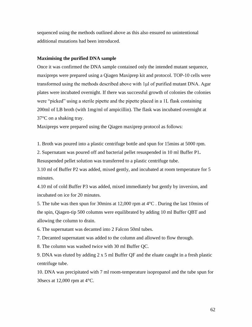
62
sequenced using the methods outlined above as this also ensured no unintentional
additional mutations had been introduced.
Maximising the purified DNA sample
Once it was confirmed the DNA sample contained only the intended mutant sequence,
maxipreps were prepared using a Qiagen Maxiprep kit and protocol. TOP-10 cells were
transformed using the methods described above with 1μl of purified mutant DNA. Agar
plates were incubated overnight. If there was successful growth of colonies the colonies
were “picked” using a sterile pipette and the pipette placed in a 1L flask containing
200ml of LB broth (with 1mg/ml of ampicillin). The flask was incubated overnight at
37°C on a shaking tray.
Maxipreps were prepared using the Qiagen maxiprep protocol as follows:
1. Broth was poured into a plastic centrifuge bottle and spun for 15mins at 5000 rpm.
2. Supernatant was poured off and bacterial pellet resuspended in 10 ml Buffer P1.
Resuspended pellet solution was transferred to a plastic centrifuge tube.
3.10 ml of Buffer P2 was added, mixed gently, and incubated at room temperature for 5
minutes.
4.10 ml of cold Buffer P3 was added, mixed immediately but gently by inversion, and
incubated on ice for 20 minutes.
5. The tube was then spun for 30mins at 12,000 rpm at 4°C . During the last 10mins of
the spin, Qiagen-tip 500 columns were equilibrated by adding 10 ml Buffer QBT and
allowing the column to drain.
6. The supernatant was decanted into 2 Falcon 50ml tubes.
7. Decanted supernatant was added to the column and allowed to flow through.
8. The column was washed twice with 30 ml Buffer QC.
9. DNA was eluted by adding 2 x 5 ml Buffer QF and the eluate caught in a fresh plastic
centrifuge tube.
10. DNA was precipitated with 7 ml room-temperature isopropanol and the tube spun for
30secs at 12,000 rpm at 4°C.

63
11. The supernatant was poured off, 5ml 70% ethanol added and the tube spun for 5mins
at 12, 000 rpm at 4°C.
12. Supernatant was poured off; the tube respun for 5mins at 12, 000 rpm at 4°C.
13. DNA was allowed to air-dry for 5mins; resuspended in 500l dH20 and, transferred to
an eppendorf.
Confirmation of the correct DNA sequence was again performed by restriction digest and
direct DNA sequencing.
2.5 Cell culture methods
All cell work was performed under sterile technique in sterile tissue culture cabinets
within a dedicated tissue culture facility.
Thawing human embryonic kidney (HEK) cells
Stocks of HEK cells were stored in a liquid nitrogen facility. Prior to removing cells from
storage flasks for cell culture were prepared in the fume hood. Growth medium
constituted DMEM, 10% FBS and 1% glutamax (Invitrogen). 50ml aliquots were stored
in the fridge and removed to warm to room temperature. 5ml were transferred using bulb
pipettes to a pre-labeled sterile cell culture flask. A further 5mls were pipetted to a 10ml
falcon tube. At this stage cell vials were removed from the liquid nitrogen tank and
immediately transferred to the water tank (37°C) in the cell culture laboratory until
thawed. The cells in solution were transferred to the falcon tube and mixed gently with
the fresh medium. The falcon tube was spun down at 12 000rpm for 5mins to create a cell
pellet. The supernatant was discarded and 5mls of fresh warm growth medium gently
mixed with the cells using a bulb pipette. This solution was transferred to the prepared
culture flask and the solution gently washed repeatedly over the flask base 20 times in an
attempt to create an even distribution of cells and avoid “clumps”. The flask was placed
in the incubator at 37°C.

64
Splitting HEK cells
HEK cells were split when approximately 80% confluent. This was guided by direct
observation of the flask under a microscope on a daily basis but as a general guide cells
were split three times a week. A new flask was prepared by labeling and loading with
5mls of fresh growth medium warmed to room temperature. Growth medium was
removed from the flask containing the HEK cell culture with a bulb pipette and
discarded. 1ml of trypsin was added to the flask and the flask rolled gently to coat the
whole cell layer. Effectiveness of the trypsin was guided by sight as the cell layer could
be seen to detach from the flask base but as a general guide this took approximately 3-
5mins. The reaction was terminated at this point by the addition of 5mls of fresh growth
medium. The cells now in trypsin/growth medium solution were transferred to a 10ml
falcon tube and the tube spun at 12 000rpm for 5mins. A cell pellet was now visible and
the medium was discarded. 5mls of fresh media was added to the pellet and mixed gently
by pipetting up and down. Approximately 1ml of this solution (volume transferred
depended on confluency of cells and size of the pellet) was transferred to the prepared
flask containing fresh growth medium and washed twenty times over the bottom of the
flask to achieve an even distribution of cells. The new flask was returned to the incubator
at 37°C.
Myoblast cultures
Aliquots of a healthy culture of normal myoblasts were given to me by Dr Carl Adkin at
the Institute of Child Health. These had been cultured from healthy muscle donated by a
teenager undergoing scoliosis surgery. Thawing, culture and splitting of cells was
performed along similar methods to the HEK cells but a different growth medium was
used comprising of: 500ml Skeletal Muscle Cell Growth Medium(Promocell C-23060),
25ml supplement mix(Promocell), 50ml FBS and 7.5ml L-glutamine. Myoblasts were
also split at approx. 50% confluence to prevent fusion and formation of myotubes.

65
Reverse transcription PCR
RNA was extracted from robust myoblast cell culture using an RNAeasy kit (Quiagen).
Reverse transcription PCR was performed using an ABI high capacity cDNA RT kit.
Each 10l reaction contained: 2l 10xbuffer, 0.8l dNTPs, 2l primer, 1l RT and 4.2l
of H20.
2.6 Transfection of human embryonic kidney cells
HEK cells were split in the manner described above 24hours prior to cell transfection.
When the cell pellet was reconstituted with 5mls of fresh growth media one drop of the
solution was added to six 5ml petri dishes which each contained three 1cm circular glass
coverslips that had been pre-coated with poly-D-lysine, and 2-3mls of growth media.
Petri dishes were incubated overnight at 37°C to allow for early growth of the cells.
Human Embryonic Kidney cells (HEK cells) were transfected using the calcium
phosphate precipitation method. The purified mutant SCN4A DNA, WT SCN4A DNA,
SCN1B DNA samples and GFP aliquots were analysed with a nanodrop to assess the
DNA concentration in each sample. This was to allow calculation for the transfection
reaction such that each mixture contained a 1:1:1 ratio of SCN1A, SCN1B and GFP
(green fluorescent protein). As such 12.7l of calcium chloride, 4l of GFP, 4l of WT
SCN4A DNA, 1.5l of SCN1B DNA and 77.8l of H2O were pipetted into a sterile
eppendorf to a total volume of 100l. 100l of BES
(bis-hydroxyethylaminoethansulfonate) was added to the mixture and gently pipetted up
and down. Constituents for the mutant DNA transfection were mixed in a second sterile
eppendorf: 12.7l of calcium chloride, 4l of GFP, 20l of mutant SCN4A DNA, 1.5l
of SCN1B DNA and 61.8l of H2O to a total volume of 100l and mixed gently with
100l of BES. Each reaction was left to stand at room temperature for 10mins. The
mixture was then distributed evenly between three of the prepared petri dishes containing
glass cover slips and the dish gently swirled before being placed back in the incubator for
8-10hours. After this time the media was changed to replenish the dish with fresh growth

66
media. Transfected cells were incubated for 48hrs before being used in patch clamp
experiments.
Coating cover slips with poly-D-lysine
50ml of poly-D-lysine was poured into a sterile autoclaved glass bottle and made up to
100ml with autoclaved water. This was then divided into 10ml aliquots in falcon tubes for
freezing. 200 cover slips were placed in a 50ml falcon tube with one 10ml aliquot of
poly-D-lysine. This was placed on a rotator for 1hour to evenly coat all the cover slips.
After one hour the poly-D-lysine was poured off and the cover slips rinsed several times
with sterile water. The cover slips were then autoclaved to sterilise prior to use in patch
clamp experiments.
2.7 Patch clamp experiments
Patch clamp experiments were performed on HEK cells 48-72 hours post-transfection.
Standard whole-cell patch clamp recordings were performed at room temperature. The
external bath solution used to bathe the coverslips and cells was prepared as a 1L solution
containing:
mM 1L
145 NaCl 8.47g
4 KCl 2ml(2M)
1.8 CaCl2 1.8ml(1M)
1 MgCl2 1ml(1M)
10 HEPES 2.383g
The external solution was pH tested and brought to a pH of 7.35 with NaOH(1M), usually
requiring approximately 3-5ml.
67
Several pipette solutions were trialed but the internal pipette solution used for all cells in
the final analysis contained CsCl (150mM); Hepes (10mM) and EGTA (10mM) at a pH
of 7.3. Pipettes were fabricated using capillary glass and a pipette puller using pipettes
with resistances of between 2.5-5MΩ. Cells with series resistance above 10MΩ were
discarded. To control variability, recordings from WT SCN4A cells were routinely
intercalated with recordings from mutant SCN4A cells. Leak subtracted currents were
recorded using –P/4 protocol with an Axopatch 200B amplifier (Molecular Devices,
Sunnyvale, CA, USA). Data was sampled at 20kHz and filtered at 5kHz, and analysed
using LabView software (v. 8.0, National Instruments, Austin, TX, USA) with programs
written by Prof D Kullman.
68
Results - Phenotyping
Phenotype can be markedly diverse in the skeletal muscle channelopathies even among
individuals with the same genotype. An example of the phenotypic variability that can be
observed is provided by the SCN4A G1306E mutation associated with sodium channel
myotonia. The original phenotype reported with this mutation was so severe the
individual suffered permanent myotonia that included the respiratory muscles and led to
hypoxia and acidosis requiring ventilatory support(Lerche et al., 1993). In contrast, a
more recent report observed that although affected individuals had relatively severe
myotonia they did not exhibit respiratory involvement and were able to carry out daily
activities including work without treatment.(Colding-Jorgensen et al., 2006) As a
consequence of this wide variability there are few clinically useful genotype-phenotype
correlations. The channelopathy database was updated and reviewed regularly to identify
potentially informative phenotypes and any possible genotype-phenotype correlations
that emerged were explored further.
3.1 Observation of neonatal hypotonia in paramyotonia congenita due to the I693T
SCN4A mutation
A female patient, BWL, referred to the clinical service for skeletal muscle
channelopathies with PMC and seen by me described her mother reporting that she had
been “paralysed” and floppy for the first four days after birth with an inability to
breastfeed. The lady’s mother was deceased so further details could not be sought.
Attempts to obtain the birth records proved unsuccessful as they had been destroyed.
This lady was known to have genetically confirmed paramyotonia congenita with the
point mutation I693T having been identified in exon 13 of the SCN4A gene by the
diagnostic clinical service. Due to the unusual birth history and a rather severe phenotype
causing biopsy proven evidence of myopathy at the age of 15 with subsequent use of a
wheelchair from her late twenties this lady was presented as an “interesting case” at an
international meeting of the Non-Dystrophic Myotonias. This led to collaboration with

69
Bertrand Fontaine’s group who attended the meeting and also reported neonatal
hypotonia occurring in families they had seen with the same SCN4A mutation (I693T).
Details of the four French children from two unrelated families were sent to me by
Damien Sternberg.
Phenotype of neonates carrying the I693T SCN4A mutation
Four unrelated families in total (2 in the UK, 2 in France) were identified who all carried
the I693T mutation and reported cases of neonatal hypotonia with variable difficulties in
feeding and respiratory compromise (see Table 9). Symptoms were self-limiting within
10 days in each case. All affected individuals later went on to develop the typical
symptoms of cold and exercise exacerbated myotonia seen in PMC. The mutation was
inherited from the mother in two cases, the father in one and was apparently sporadic in
the other although parental DNA was not available to confirm this.
The proband identified in one of the UK families became pregnant shortly after the
recognition of neonatal hypotonia with this mutation. Advice was given to her and to the
obstetric and paediatric teams involved in her care regarding the possible development of
neonatal hypotonia, feeding or respiratory difficulties along with reassurances that these
symptoms were most likely to be self-limiting and would require only supportive care.
She went on to deliver a healthy baby boy who did display hypotonia and feeding
difficulties requiring a short term nasogastric tube. Symptoms were self limiting and he
was later able to breast feed and to date has had normal development. Subsequent genetic
testing confirmed he had inherited the I693T mutation from his mother.

70
Sex Year
of
birth
APGAR
at birth
Onset of
Hypotonia
Impairment
of sucking-
swallowing
Desaturation Kalemia
(mmol/l)
Motor
Development
Family 1
(English)
Female
1-1
1950 NA Birth Day1-
bottle
fed,unable
to breast
feed
No NA Walking
delayed
(19/12)
Family 2
(English)
Female
2-1
1975 NA Birth No
(hypotonia
limited to
limbs)
No NA Walking
delayed
(22/12)
Family 3
(French)
Female
3-1
1987 10 Day1,
some
hours after
birth
Day1-3
some hours
after birth,
nasogastric
tube
Day1, some
hours after
birth
(cyanosis) ;
no reported
treatment
5.1 then
normal
Normal
Male
3-2
1997 10 Day1,
some
hours after
birth when
bathed
Day1-3
onset in
bath
No Normal NA
Family 4
(French)
Male
4-1
2004 NA Day1 Day1-3
no
treatment
reported
No NA Normal
Male
4-2
2007 10 Day1,some
hours after
birth
Day1-6,
some hours
after birth
nasogastric
tube
Day2-6,
nasal
oxygen
required
Normal NA
NA-Not Available
Table 9: Features of hypotonia and motor development in six neonates carrying the
Nav1.4 I693T mutation.

71
3.2 Stridor expands the neonatal presentations of skeletal muscle sodium
channelopathy
Following on from the observation of this neonatal presentation of paramyotonia
congenita a patient seen by me in clinic and enrolled in the natural history trial with
paramyotonia congenita described her second son as having severe stridor with some
respiratory and feeding difficulties at birth. She queried whether his stridor was related to
PMC. Initially this was uncertain as although the defective Nav1.4 channel would be
present in all skeletal muscles including the larynx, neonatal stridor is common for
numerous other reasons and it was not possible to draw conclusions from a single case.
His medical records were reviewed with permission. Discussion once again with Bertrand
Fontaine and Damien Sternberg’s group in France identified two additional French cases
however which suggested the association was not co-incidental and the stridor may be a
direct result of the myotonic disorder.
Features of neonatal stridor
The lady’s four year old son was referred to paediatric services following the diagnosis of
paramyotonia congenita (PMC) in her. The boy was the mother’s second child and was
born by ventouse delivery at 39 weeks following an uncomplicated pregnancy. APGAR
scores were normal and post natal examination was unremarkable. Within 24 hours of
delivery he was transferred to the neonatal intensive care due to inspiratory stridor and
poor feeding. Over the following week he was unable to take sufficient oral feeds and
required supplemental nasogastric feeding. Stridor persisted and he required intermittent
oxygen therapy for desaturations that occurred while attempting to bottle feed or when
crying. Laryngoscopy showed findings consistent with laryngomalacia.
The infant continued to have persistent inspiratory stridor for the first six months of life.
Feeds were prolonged but he gained weight appropriately and had no further apnoeic
episodes. Motor milestones were mildly delayed, sitting independently at nine months
and walking at 19 months. From the age of one year exotropia was noted. This is

72
currently under investigation by an opthalmologist and possibly reflects myotonia of the
extraocular muscles.
At the age of 23 months he fell in the garden on a cold day and complained of leg
weakness, refusing to stand or walk. The weakness recovered spontaneously within five
hours. A second similar episode occurred at age 27 months after playing in the garden in
winter. In addition to these two episodes of muscle weakness his mother noted muscle
stiffness occurring on an almost daily basis from age two. Both muscle stiffness and
weakness were exacerbated by cold weather and exertion.
At the age of four he continues to have episodes of inspiratory stridor exacerbated by
viral illness, cold weather, and prolonged laughter or crying. A humidifier is helpful in
aborting such episodes.
He has recently been treated with the sodium channel blocker mexiletine which has had
some beneficial effect on his degree of myotonia and exotropia.
The child’s mother, grandfather and great uncle all reported similar episodes of muscle
stiffness and weakness exacerbated by cold and exercise. There was no prior family
history of stridor. All affected family members including the child described had DNA
sent to the diagnostic genetic lab at the NHNN under the NCG scheme for
channelopathies by the specialist responsible for their clinical care and were shown to
have the SCN4A mutation T1313M associated with PMC(McClatchey et al., 1992b).
In addition to this case two further adult cases of PMC in France in whom neonatal
stridor was documented in their medical records were identified by Damien Sternberg
although no further details were available. The first carried an E1702K familial mutation,
and the other an A444D de novo mutation.
Neonatal myotonia was recorded in Eulenburg’s first description of PMC but distinct
neonatal phenotypes of sodium channelopathy have not previously been recognised. A
case of fatal infantile myotonia with severe respiratory compromise was recently reported
in a child carrying a new SCN4A mutation N1297K(Gay et al., 2008). Since describing
the case of stridor another series has reported more severe laryngospasm in three infants
with sodium channel mutations, one of whom also died(Lion-Francois et al., 2010).

73
This emerging evidence of neonatal presentations is of paramount importance when
counselling expectant families with a history of sodium channel disorder and is important
to bear in mind in children presenting with these features even if family history is lacking
as de novo mutations occur. In the milder phenotypes I have described symptoms
generally only required supportive care and invasive investigations are not required. The
recently published more severe cases generally responded to mexiletine therapy.
Ultimately the delivery of neonates at risk of inheriting sodium channel disorders should
be considered relatively high risk and appropriate obstetric, anaesthetic and paediatric
services must be available. The recognition of these phenotypes will limit unnecessary
investigation and prompt early appropriate therapy.
3.3 Observation of inflammatory infiltrates in skeletal muscle channelopathies
Advances in specialised electrophysiological tests and genetic analysis although not
infallible have significantly improved the ability to diagnose skeletal muscle
channelopathies. Many of the reported histopathological findings in the channelopathies
predate the availability of genetic testing and commonly include a vacuolar myopathy or
tubular aggregates in the periodic paralyses although it must be noted that these occur in
all of the sub groups. Biopsy findings in the non-dystrophic myotonias are non-specific
myopathic changes. Morphological analysis is ultimately not diagnostic but prior to the
availability of less invasive investigations it was used to support a diagnosis of
channelopathy and much of the data available is from typical cases without severe or
unusual symptoms. It is rare in the UK that a diagnostic biopsy is now performed in the
channelopathies.
Regular searches of the database of patients referred to the channelopathy service
identified three cases of genetically confirmed skeletal muscle channelopathies who all
underwent muscle biopsies due to reports of either proximal weakness or severe muscle
pain in conjunction with a raised creatine kinase (CK). In each case the biopsies were
reported as showing inflammatory infiltrates which are not a recognised or described
finding in channelopathies. The referring clinicians were contacted in each case to request

74
further clinical details and any available biopsy slides. All three kindly responded with
clinical details and in two cases biopsy material was sent for review by Dr Janice Holton.
In the remaining case biopsy material had unfortunately been destroyed but detailed
biopsy reports were obtained.
Histopathology
Each patient had undergone a muscle biopsy in another centre and tissue sections stained
for haematoxylin and eosin (H&E), Gomori’s trichrome method, lipid and glycogen in
addition to enzyme histochemical preparations for succinic dehydrogenase, cytochrome
oxidase and nicotinamide adenine dinucleotide dehydrogenase tetrazolium reductase
were available for review. Immunohistochemical preparations submitted for review were:
Case 1: CD3 and MHC class I; Case 2: immunohistochemical staining for CD3, CD20,
spectrin, dystrophin, merosin, dysferlin, sarcoglycans, emerin and desmin; and Case 3:
MHC Class I, CD3, CD68 and CD20.
Clinical features of channelopathy patients with inflammatory biopsies
Case One
An 80 year old man complained of episodes of muscle weakness after periods of
prolonged rest from the age of four. Initially only the lower limbs were affected but by
his late teens the upper limbs were also involved. The episodes usually lasted hours and
occasionally days. A clinical diagnosis of familial periodic paralysis was made when he
was aged 21 following reports of similar episodes of weakness in his mother and sister.
From his early 50s mild proximal muscle weakness was noted on examination. At the age
of 56, following a positive potassium challenge his diagnosis was revised to
hyperkalaemic periodic paralysis and he was treated with salbutamol. Creatine Kinase
(CK) at this time was noted to be 436U/L. At the age of 65 he began complaining of
difficulty rising from a chair and climbing stairs. Examination confirmed limb weakness
greater proximally than distally. Clear difficulty rising from a chair without using his

75
hands for leverage was observed and a degree of thigh atrophy was noted. EMG showed
myopathic changes and minimal myotonic discharges. His CK was 2729U/L. Muscle
biopsy showed internal nuclei, fibre atrophy, fibre hypertrophy with occasional split
fibres, some regenerating fibres and an increase in endomysial connective tissue. In
addition there was a dense inflammatory cell infiltrate focused around small capillaries
and necrotic fibres which was demonstrated by immunohistochemistry to consist mainly
of T-lymphocytes with restricted MHC 1 expression. Diagnoses of vasculitis and
polymyositis were considered from the biopsy findings but the overall clinical picture
was felt most consistent with a diagnosis of polymyositis and he was started on
prednisone 60mg once daily. A few hours after the initial dose of steroids he suffered a
severe attack of paralysis lasting 48 hours. Following recovery from this he was restarted
on prednisone 10mg once daily with gradual improvement of his muscle weakness.
Ten years later he re-presented with further complaints of leg weakness and difficulties in
mobilisation. At this time he was on 5mg of prednisone although it is not clear if this had
been continuous since commencement 10 years earlier. Azathioprine 50mg bd was added
to his therapy but he continued to deteriorate and was admitted to hospital where
proximal lower limb power was documented as ranging from 3/5 to 4/5. The azathioprine
dose was further increased to 100mg bd and he was discharged from hospital. When he
was reviewed in clinic 2 months later he reported no real change in his mobility
difficulties although only “mild” proximal leg weakness was recorded. Genetic analysis
by the diagnostic channelopathy service confirmed the presence of the common T704M
point mutation in the SCN4A gene associated with hyperkalaemic periodic
paralysis(Ptacek et al., 1991).
Case Two
A 42 year old lady with the F167L mutation in the CLCN1 gene associated with
myotonia congenita(George, Jr. et al., 1994) complained of daily muscle cramps affecting
all muscle groups but predominantly the limbs from early childhood. This was confirmed
to be myotonia on EMG. Family history revealed similar symptoms in her mother and
two maternal aunts. The patient complained of severe cramps with increasing age and
began to experience episodes of muscle weakness after exertion although in between

76
episodes reported normal muscle power. Examination was generally unremarkable but a
CK was raised at 1109U/L.
Muscle biopsy was performed and demonstrated increased variation in fibre size,
evidence of fibre regeneration and 1 necrotic fibre. Two small foci of inflammation
composed of CD3 immunoreactive T lymphocytes were noted in perimysium and
endomysium (Fig 16). Invasion of intact myofibres by T cells was not evident and MHC
Class I immunohistochemistry was not available for review. There was no evidence of a
vacuolar myopathy and tubular aggregates were not present. She was started on
prednisone with a resultant reduction in CK to 3-400U/L. All attempts to reduce
prednisone were unsuccessful and the patient was unable to tolerate other
immunosuppressants.
Case Three
A 38 year old man complained of episodes of muscles stiffness when exposed to the cold
affecting predominantly his tongue, hands and feet from early childhood. This was
confirmed by electrophysiological testing to be myotonia. There was no family history of
note. Myotonia occurred daily but symptoms remained fairly static until his late teens
when he additionally complained of episodes of acute muscle swelling predominantly
affecting his forearms and thighs. Initially these were non-painful, and CK and muscle
power were normal. Later however he complained of myalgia accompanying the episodes
of swelling which could occur 2-3 times a month and last up to a week. An ultrasound
scan confirmed hypoechoic regions within the muscles but normal subcutaneous tissue.
An intermittent peripheral eosinophilia was noted in conjunction with the episodes of
myalgia. A muscle biopsy showed no evidence of regeneration, necrosis, vacuolation of
fibres or tubular aggregates. Inflammatory infiltrates composed of a mixture of T
lymphocytes and macrophages were present in the perimysium with a single endomysial
cluster (Fig 16B and 16C). Invasion of intact muscle fibres by T cells was not apparent.
Immunohistochemical staining for MHC Class I demonstrated an increase in
sarcolemmal and sarcoplasmic expression (Fig 16D). The biopsy findings were
interpreted as being compatible with polymyositis although the clinical history was not
typical.

77
He had an excellent response to prednisone, 20mg od reduced to 5mg od which reduced
the frequency and severity of the muscle swelling and myalgia although he unfortunately
developed osteopenia. Attempts to introduce other steroid sparing immunosupressive
agents were unsuccessful as he did not respond to methotrexate, cyclosporine, imatinib or
hydroxyurea and did not tolerate azathioprine. Genetic analysis by the diagnostic
channelopathy service confirmed the presence of the L1436P mutation in the SCN4A
gene associated with sodium channel myotonia(Matthews et al., 2008b).
Fig 16: In case 2 a prominent focus of perimysial inflammation was found (A). Similarly
the inflammation in case 3 was predominantly perimysial (B) with focal extension into
the endomysium (arrow in C). The inflammatory cells included abundant T lymphocytes
(C) and increased expression of MHC Class I at the sarcolemma and within the
sarcoplasm of fibres was demonstrated (D). A: case 2; B – D: case 3. A & B:
haematoxylin and eosin, C: CD3 immunohistochemistry; D: MHC Class I
immunohistochemistry. Bar in A represents 50µm in A – D.
A B
C D
A B
C D

78
Case Predominant
clinical features
Examination
findings
Investigations Biopsy Findings Response to
steroid therapy
1 Episodic
muscle
paralysis
Progressive
proximal limb
weakness
Proximal limb
weakness,
MRC grade 3/5
to 4/5
CK 2729U/L
EMG:
polyphasic units + short
duration units,
fibrillations,
fasciculations and
myotonic discharges
Internal nuclei, fibre atrophy, fibre
hypertrophy with occasional split fibres,
some regenerating fibres and an increase in
endomysial connective tissue.
Dense inflammatory cell infiltrate focused
around small capillaries and necrotic fibres
composed mainly of T-lymphocytes.
MHC I upregulation on occasional muscle
fibres.
High dose
prednisone
(60mg) led to
severe paralytic
attack lasting
48hrs
10mg better
tolerated with
improved
muscle strength
2 Muscle cramps
and severe
myalgia
Myotonia
Normal muscle
power
CK 1109U/L
EMG: massive myotonic
discharges at every
insertion point that
precluded analysis of
MUAP configuration,
voluntary recruitment or
interference pattern.
Increased variation in fibre size, evidence
of fibre regeneration and 1 necrotic fibre.
Two foci of inflammation composed of
CD3 immunoreactive T lymphocytes noted
in perimysium and endomysium.
Immunohistochemistry unavailable for
review.
Reduction in
CK to 3-400.
3 Muscle cramps
and episodic
limb oedema
with myalgia
Myotonia
Normal muscle
power
CK normal
EMG: myotonic
discharges
Inflammatory infiltrates composed of a
mixture of T lymphocytes and macrophages
were present in the perimysium with a
single endomysial cluster (Fig 16).
Immunohistochemical staining for MHC
Class I demonstrated an increase in
sarcolemmal and sarcoplasmic expression
Excellent
response with
significant
reduction in
symptoms of
oedema and
myalgia
Table 10: Summary of clinical, biochemical, neurophysiologic and histological findings in each case

79
Channelopathies are rare disorders and for the reasons outlined biopsy samples are
scarce. It is impossible to be certain that the observation of inflammatory infiltrates in
these three cases is not merely co-incidental. To try to ascertain the likelihood of this
however I consulted Dr Gareth Ambler, statistician at UCL who reviewed the data. The
prevalence rates of both skeletal muscle channelopathies and idiopathic inflammatory
myopathy (IIM) have each been estimated to be 1 in 100,000(Emery., 1991; Fontaine.,
1994; Hilton-Jones., 2001). A null hypothesis was taken that the development of both
diseases in these three cases was coincidental. Considered statistically, if the likelihood of
developing a skeletal muscle channelopathy and an IIM is independent then the chance of
having both is one in 10-10
. The probability of observing UK patients with both diseases
(if this association were by chance alone) can be calculated using the Poisson distribution
since p is small and the UK population is large; Poisson lambda = 10-10
(independent
probability of two events in an individual) x 6.5 x 10⁷
(UK population) = 0.0065, and the
probability of observing three or more patients with both diseases (assuming
independence) is 4.5 x 10⁻⁸
. This small p value indicates that the occurrence of both
diseases in the same individual is highly unlikely to be independent.
Whether such inflammatory changes are present in a greater number of channelopathy
patients will be difficult to establish but it is reasonable to suggest that a muscle biopsy
be considered in those in whom a CK>1000IU/L is observed in conjunction with
significant myalgia or weakness. Steroid use has to be interpreted cautiously as in one
case high dose steroids caused significant exacerbation of the periodic paralysis but lower
doses may be beneficial.
3.4 Pain and morbidity in the non-dystrophic myotonias
There is little in the literature that gives consideration to the morbidity of the skeletal
muscle channelopathies. Multiple case reports and one recent series(Trip et al., 2009a) do
suggest pain may be an overlooked feature in the non-dystrophic myotonias. It was a
personal observation that many patients who attended clinic or participated in the natural
history trial of non dystrophic myotonia often volunteered pain as a troubling symptom.

80
To look at this more subjectively all participants enrolled in the natural history study
were asked about their symptoms. In the first year of enrollment of UK based participants
the majority (16/19) reported stiffness as their most prominent symptom which would be
as predicted for these myotonic disorders. All 19 had EMG evidence of myotonia and
in18/19 pathogenic mutations associated with myotonic disorders have been identified.
However 3/19 (16%) reported pain was their most prominent symptom (see Fig 17). Of
these 3, 1 carried a sodium channel mutation and 2 chloride channel mutations.
Fig 17: NDM patient reports of their most prominent clinical symptom
Participants were then asked about specific symptoms in detail including whether they
experienced pain in association with muscle stiffness or even in the absence of muscle
stiffness. If they answered yes they were asked to rate the severity of their maximum
level of pain and their average level of pain on a pain scale of 0 – 9, 9 being the most
severe pain imaginable and 0 being no pain. Of 19 patients 16/19 (84%) reported some
pain, 7 of these associated only with stiffness, 7 both with and without stiffness and 2
without any stiffness exacerbation. Mean maximum and average severities are detailed in
fig 18. There was no discernible difference in severity of pain whether it occurred with
stiffness i.e. painful myotonia or in the absence of stiffness.
0
2
4
6
8
10
12
14
16
Pain Weakness Stiffness Fatigue
No. of patients
0
2
4
6
8
10
12
14
16
Pain Weakness Stiffness Fatigue
No. of patients
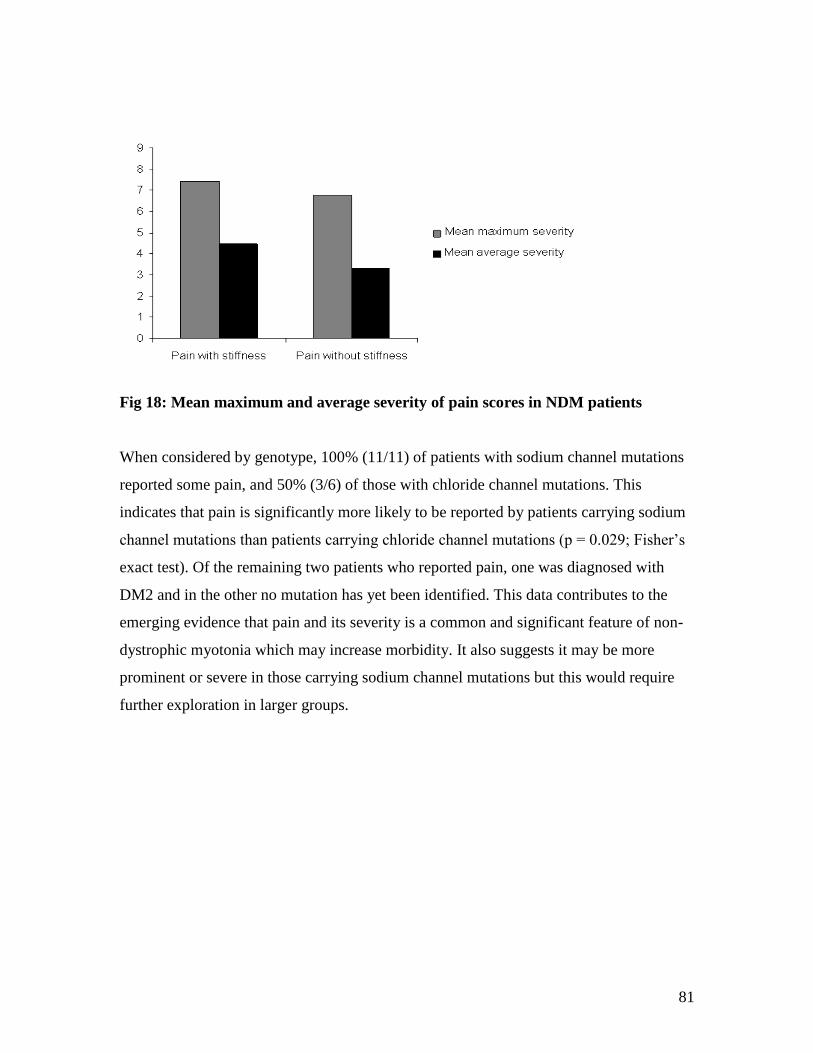
81
Fig 18: Mean maximum and average severity of pain scores in NDM patients
When considered by genotype, 100% (11/11) of patients with sodium channel mutations
reported some pain, and 50% (3/6) of those with chloride channel mutations. This
indicates that pain is significantly more likely to be reported by patients carrying sodium
channel mutations than patients carrying chloride channel mutations (p = 0.029; Fisher’s
exact test). Of the remaining two patients who reported pain, one was diagnosed with
DM2 and in the other no mutation has yet been identified. This data contributes to the
emerging evidence that pain and its severity is a common and significant feature of non-
dystrophic myotonia which may increase morbidity. It also suggests it may be more
prominent or severe in those carrying sodium channel mutations but this would require
further exploration in larger groups.

82
Results – Genotyping
4.1 Genotyping the sodium channel myotonic disorders
Patient identification
Using the channelopathy database, patients were identified with a clinical history
consistent with myotonic sodium channel disease, either paramyotonia congenita, sodium
channel myotonia or hyperkalaemic periodic paralysis. All patients are screened for the
most common SCN4A mutations in exons 13, 22 and 24 by the diagnostic genetic lab as
part of the NCG channelopathy service. I identified 17 patients in whom no such
mutations were found for further study and performed direct DNA sequencing of all 24
exons of SCN4A (analysis of exons 13, 22 and 24 was repeated) and quantitative PCR for
the trinucleotide repeat of ZNF9 known to cause myotonic dystrophy type 2 (DM2). All
those with a phenotype of PMC or SCM had EMG proven myotonia. Cases of
hyperkalaemic periodic paralysis included some with atypical features. If no abnormality
was detected in SCN4A or ZNF9, the CLCN1 gene was kindly sequenced by geneticists
Dr Robyn Labrum or Dr Richa Sud in the diagnostic lab. Selected patients had additional
gene sequencing performed by other centres.
Sodium channel myotonia – new mutations
Of the 17 patients identified, I considered 13 to have PMC or SCM and 4 to have features
of hyerPP. Amino acid substitutions were identified in 9/13 myotonic patients including 3
previously unreported, V445L, L128P and M135L. Controls were run for all of these
novel changes including analysis of conservation across species and use of analysis
websites to predict the likelihood of these changes being pathogenic. V445L and L128P
were highly conserved, absent in controls and predicted to be deleterious to channel
function. M135L was not highly conserved, was present in one control sample and
predicted to be a tolerable amino acid substitution. It was not however listed on any SNP
database as a known polymorphism. Both the patient and the positive control were

83
believed to be of Jewish origin. Taken together these findings suggest that this
substitution may be a polymorphism that is more prevalent in certain ethnic groups.
Fig 19: Electropherograms, conservation across species and position in Nav1.4
channel of new SCN4A mutations
Human IKV L IHA Gorilla IKV L IHT Dog IKV L IHT Mouse IKV L IHT Chick IKV L IHS Zebrafish IK I L IHS
V445L L128P M135L
Human LFS M FIM Gorilla LFS M FIM Dog LFS M FIM Mouse LFS M FIM Chick LFS M FIM Zebrafish LFS L VIM
Human ILA V VAM Gorilla ILA V VAM Dog ILA V VAM Mouse ILA V VAM Chick ILA V VAM Zebrafish ILA V VAM
L128P M135L
V445L
Human IKV L IHA Gorilla IKV L IHT Dog IKV L IHT Mouse IKV L IHT Chick IKV L IHS Zebrafish IK I L IHS
V445L L128P M135L
Human LFS M FIM Gorilla LFS M FIM Dog LFS M FIM Mouse LFS M FIM Chick LFS M FIM Zebrafish LFS L VIM
Human ILA V VAM Gorilla ILA V VAM Dog ILA V VAM Mouse ILA V VAM Chick ILA V VAM Zebrafish ILA V VAM
L128P M135L
V445L

84
Genotyped SCN4A CLCN1 DM 2 Other Genes Phenotype EMG Myotonia
1MZ Yes V1293I Sodium channel myotonia Yes
2LG Yes V445L* Sodium channel myotonia Yes
3LF No Negative Negative Negative DM1 negative Sodium channel myotonia Yes
4EC No Negative Awaited –
more DNA
requested
Negative DM1 negative Infantile myotonia Yes
5BC Yes Negative CRYAB gene c.343delT
(p.Ser115ProfsX14) +
c.343delT
(p.Ser115ProfsX14).
Infantile onset severe limb and
respiratory muscle stiffness
First EMG ?myotonia
Second EMG
Pseudomyotonia
6CM No Negative Negative Negative DM1 negative Sodium channel myotonia Yes
7MH Yes Negative Mutation
detected
Negative Sodium channel myotonia Yes
8BB Yes Q270K Paramyotonia Congenita Yes
9EL Yes E1702K Paramyotonia Congenita Yes
10CO Yes L128P* Sodium Channel Myotonia Yes
11DL Yes V1293I Sodium Channel Myotonia Yes
12JSC No M135L** Paramyotonia Congenita Yes
13RT No Negative Negative Failed Paramyotonia Congenita Yes
14SR No Negative Negative ND RYR1 silent changes only
Brother congenital
myasthenic syndrome
Atypical hyperkalaemic periodic
paralysis
No
15JV Yes Negative Negative Positive Hyperkalaemic Periodic Paralysis Myotonia – not clear if
clinical or EMG
16LD No Negative Negative Negative Congenital myasthenia
genes awaited
Atypical periodic
paralysis/myasthenia
No
17EP No Negative ND ND KCNA1 Negative Hyperkalaemic periodic paralysis No
*denotes new gene mutations not previously reported. **denotes a new single amino acid substitution, not reported as a mutation or
polymorphism but probably benign
Table 11: Summary of genetic results for 17 patients considered clinically to have PMC or hyperPP

85
Sodium channel myotonia – non-genotyped cases
The remaining 4 myotonic cases in whom no mutation was identified are considered in
detail and are referred to by their order in table 11.
Patient No. 3 (LF)
This lady is from a large Sicilian family living in Australia. She complained of muscle
stiffness and paralysis from infancy. She reported the first time she was taken for a
swimming lesson at age 3 she nearly drowned as the cool water caused her muscles to
become stiff. When she was rescued from the bottom of the pool she was found in a
flexed position which required rewarming over about an hour before her muscles could
be relaxed and began contracting normally again. If she exerts herself she can be in bed
with muscle weakness and muscle discomfort for days. Recently she ran to catch a plane
(the first time she had run in 20 years) and ended up paralysed in her airplane seat for the
flight back from Florida to Australia. Cold temperature was a powerful exacerbator of her
myotonic symptoms especially the peripheries, the eyelids and extraocular muscles. Her
neurologist in Australia had ice cooled her eyes for 1 minute after which she could not
open them very easily, had diplopia for several minutes and a mild facial droop. She
spent most of her time “paralysed” in the hotel room after a recent trip to Canada, where
she got cold on the first day. Cold also produced a tension-like headache and cervical
muscle “tightness”. She avoids some foods like olives and some fruits which she thinks
can precipitate attacks. She had no dysmorphic or systemic features suggestive of
myotonic dystrophy.
The long exercise (5minutes) and cooling repetitive stimulation testing were only mildly
positive. In contrast muscle cooling down to 20oC with needle EMG was very suggestive
of the transition of changes that have previously been reported in PMC and she had
associated clinical symptoms which took several hours to resolve.
She responded well to low dose mexiletine reporting after 3 days that it was the first time
in the last 20 years she had not woken in the morning with muscle cramp and pain.

86
In terms of family history it was known her son and daughter had similar symptoms.
They were from a large Sicilian family and her Australian neurologist was making
attempts to identify affected members of preceding generations.
Patient No. 4 (EC)
A male infant referred to Great Ormond Street Hospital at the age of 15 months with
hypotonia. No specific cause had been identified and there was no family history of note.
EMG had shown unexpected but clear myotonic discharges. Myotonic dystrophy had
been excluded by the referring paediatric team.
Patient No. 6 (CM)
A forty-nine year old lady who was first symptomatic during her 3rd pregnancy at the age
of 42 with muscle stiffness that was worse with stress and after exercise. EMG confirmed
myotonia and profound CMAP drop after short exercise test. Her parents were
unaffected, her sister reported vague symptoms but had normal neurophysiology, and 3
sons were asymptomatic.
Patient No. 13 (RT)
A forty-three year old Italian lady who experienced her first symptoms at the age of
twenty. There was evidence of clinical and EMG myotonia, affecting mainly her limbs
but also face, neck and abdominal muscles, triggered by cold. She reported episodes of
muscle paralysis triggered by exercise and cold lasting hours. There was proximal and
distal limb muscle wasting with moderate to severe proximal and mild to moderate distal
muscle weakness. Repetitive stimulation caused a 33% CMAP reduction on
electrophysiological testing. There was no family history of note.
Hyperkalaemic periodic paralysis – non-genotyped cases
Of the remaining four/seventeen patients (see genotyping section 4.1 and table 11)
categorized as having a phenotype consistent with possible hyperkalaemic periodic
paralysis SCN4A mutations were not identified in any. In one a pathological expansion of
the ZNF9 gene associated with DM2 was present. The remaining three had some atypical
87
clinical features and additional genes were or are being sequenced by other laboratories.
Their clinical features are considered in detail.
Patient No 14 (SR)
A 24 year old man previously under the care of paediatric colleagues until the age of 18.
He had been investigated extensively for myopathy but no specific cause was found and
he was given a diagnosis of unspecified congenital myopathy. He described brief
episodes of lower limb weakness that occurred mainly in the cold including one episode
where he fell down some stairs due to his legs giving way. There was some myalgia but
no particular history to suggest myotonia. Symptom severity did seem to be fluctuant and
from the ages of approx 12-14 his limb weakness had been so severe he had required a
wheelchair but this had improved and at the time of his review in clinic he was
independently ambulant and attending college.
On EMG no myotonic discharges were demonstrated. After muscle cooling a reduction in
CMAP amplitude with increase in CMAP area was observed. At the time the significance
of this was unclear and it was thought to possibly be pathological. Later studies by Dr
Veronica Tan (consultant neurophysiologist at the NHNN) of large numbers of patients
with skeletal muscle channelopathies and healthy volunteers showed this change to
actually be a normal variant. This man had no prior family history of myopathy. He was
one of four children and his younger brother was affected with a very similar clinical
course to his own.
During the write up of this thesis I was advised a congenital myasthenia mutation has
been identified in this patient’s brother by another lab but I do not have access to the
formal report. Confirmation of the same mutation in this patient is apparently pending but
is very likely to be the explanation for his symptoms.
Patient No 16 (LD)
A 51 year old male born 7 weeks prematurely. Delayed walking (not until approx 2
years) was observed and he was never able to run or keep up with peers physically due to
a feeling of weakness in his arms and legs. From the age of twenty he avoided stairs due
to limb weakness. There was very slow progression in symptoms until age thirty four

88
following a flu like illness when he complained of worsening muscle weakness. Since
this episode he has continued to complain of episodes of worsening limb weakness 2-3
times a year. Clinical examination at age 35 showed ptosis, external opthalmoplegia and
proximal limb weakness with significantly depressed reflexes. At age 38 he presented
with an episode of bulbar weakness requiring a nasogastric tube and ultimately a
percutaneous endoscopic gastrostomy(PEG) tube for feeding. However there was
spontaneous resolution after 6 months with eventual removal of the PEG. He continues to
have episodes of limb weakness, with some fatigue and dysphagia in addition to
permanent proximal myopathy.
Electrophysiological tests showed myopathic potentials and repetitive nerve stimulation
showed some decrement in CMAP. Stimulation single fibre EMG showed abnormal jitter
and blocking. A McMannis test for periodic paralysis was positive. Muscle biopsy
showed non-specific myopathic changes only.
There is no family history of similar illness or symptoms. He is one of four children (2
brothers and one sister).
Patient No 17 (EP)
A forty eight year old woman who complained of episodes of paralysis from early
childhood (at least age five). In addition she suffered from exercise intolerance, pain and
stiffness especially in the cold, which rendered her unable to walk. She had been given a
previous diagnosis of fibromyalgia and investigations at another centre had included
testing voltage gated potassium channel antibodies which were positive. Two EMGs
performed at our centre on separate occasions did not show any evidence of
neuromyotonia or myotonia. A Mcmannis test was positive on the first occasion and
negative on the second.
This lady extensively researched her family history and provided the following details (as
quoted from her notes):
Her son – now 11 yrs
“At a few hours old, possibly on the 2nd
day, was taken away as he was not
breathing properly.”

89
“Many floppy episodes, but also has Congenital Adrenal Hyperplasia (CAH),
therefore cannot distinguish what may have been PP or CAH.”
“Complains of pains in legs after sport and on waking day after exercise, can’t
walk properly. Sometimes, pains in legs apparent w/o exercise (like football,
swimming etc), although walking, stairclimbing will have been done.”
“Complains of pains in legs and feet on walking distance, though none at outset.”
“On holiday Cyprus, Dec 03, aged 6, was swimming in a freezing swimming
pool. He swam for a short while and then just seized up and sank to the bottom
and lay there unmoving. I had to dive in and drag him out.”
Her daughter – now 16 yrs:
“As a toddler, had many floppy episodes following play. Eventually went into
hospital for week, but attacks did not occur. No diagnosis made. Tested for
narcolepsy, revealed unusual brainwave pattern, but not connected to episodes.”
“Complains of pains in limbs after sport, especially in the arms.”
“Had disabling pain in legs as 8 yr old ?osteomyelitis, kept in hospital.”
“Age 15 had ?viral illness – couldn’t walk, pain in back too severe to move. MRI
scan done, revealed Parr’s (sic) Process in formation of spine, but not responsible
for inability to walk.”
“Age 13 -16 suffered intermittently with chronic fatigue type disorder, floppy
aching limbs, nausea, headaches, GI disturbances. Missed a lot of school.
Coincided with a passion for eating tomatoes.”
Her mother – died in 1997, aged 79, cause of death: myocardial infarction
“My mother had persistent weakness, mostly I recall of her arms. She also was
very fatigued, and needed to sleep during the day. I had little contact with her
after the age of 18.”
Her brother – now 63 yrs:
“Left home as I was born, joined the Royal Navy, and settled in Scotland where
he still lives. I do not know him very well.”
90
“He is my half brother – we share a mother.”
“Admits he has suffered from a disabling pain and weakness condition for over
30 years. Does not like to talk about it at present, but admits he has seen many
doctors with no positive outcome. He is also very intolerant of illness, but spends
his life having sporadic episodes of intense pain with weakness, which then ‘clear
up’. He suffers daily now.”
Her maternal grandmother – died aged 36 yrs in 1930
“Was described as ‘very weak”
Overall the majority of cases studied with a PMC or SCM phenotype could be genotyped
by analysing known disease causing genes. A small minority however could not. Cases 3
and 6 (Table 11) particularly case 3 have phenotypes very suggestive of a sodium channel
myotonic disorder with EMG confirmation of myotonia but no mutation identified. Other
genetic mechanisms e.g. large scale re-arrangements have not been excluded by the direct
sequencing I performed and remain a plausible explanation. Such a large family however
with several living generations of both affected and unaffected individuals offers an ideal
opportunity to consider the possibility of a new disease causing gene.
The four cases classed as a phenotype of hyperkalaemic periodic paralysis did have some
atypical features and the majority of typical cases were again accounted for by mutations
in SCN4A (and hence not selected for further study or described here). In one case a
diagnosis of DM2 was made which was somewhat unexpected from the predominant
complaint of episodes of severe muscle weakness although the description of myotonia
would be consistent with this diagnosis. The remaining three cases were selected as other
atypical phenotypes are beginning to be described including congenital myasthenia with
periodic paralysis(Tsujino et al., 2003) and more recently an atypical periodic paralysis
phenotype with a ryanodine receptor mutation(Zhou et al., 2010). The more of these
atypical cases described and genotyped the clearer a picture of their phenotype will
hopefully emerge, providing future guidance both in terms of diagnosis and
understanding pathogenesis.
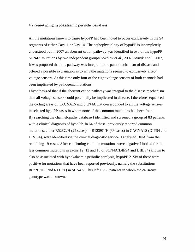
91
4.2 Genotyping hypokalaemic periodic paralysis
All the mutations known to cause hypoPP had been noted to occur exclusively in the S4
segments of either Cav1.1 or Nav1.4. The pathophysiology of hypoPP is incompletely
understood but in 2007 an aberrant cation pathway was identified in two of the hypoPP
SCN4A mutations by two independent groups(Sokolov et al., 2007; Struyk et al., 2007).
It was proposed that this pathway was integral to the pathomechanism of disease and
offered a possible explanation as to why the mutations seemed to exclusively affect
voltage sensors. At this time only four of the eight voltage sensors of both channels had
been implicated by pathogenic mutations.
I hypothesized that if the aberrant cation pathway was integral to the disease mechanism
then all voltage sensors could potentially be implicated in disease. I therefore sequenced
the coding areas of CACNA1S and SCN4A that corresponded to all the voltage sensors
in selected hypoPP cases in whom none of the common mutations had been found.
By searching the channelopathy database I identified and screened a group of 83 patients
with a clinical diagnosis of hypoPP. In 64 of these, previously reported common
mutations, either R528G/H (25 cases) or R1239G/H (39 cases) in CACNA1S (DII/S4 and
DIV/S4), were identified via the clinical diagnostic service. I analysed DNA from the
remaining 19 cases. After confirming common mutations were negative I looked for the
less common mutations in exons 12, 13 and 18 of SCN4A(DII/S4 and DIII/S4) known to
also be associated with hypokalaemic periodic paralysis, hypoPP 2. Six of these were
positive for mutations that have been reported previously, namely the substitutions
R672C/H/S and R1132Q in SCN4A. This left 13/83 patients in whom the causative
genotype was unknown.

92
Fig 20: Illustration of diminishing number of patients with a phenotype of hypoPP
in whom no genetic diagnosis could be made with escalation of genetic analysis
Hypokalaemic periodic paralysis – new mutations
In 4/13 cases three novel mutations were found, all of which neutralized arginine residues
in S4 segments. These mutations were absent from 240 control chromosomes. One
mutation was in the S4 segment of domain III (DIII/S4) of CACNA1S: c.2700G>T;
p.R900S. The other two mutations neutralized arginine residues in S4 segments of
SCN4A; the first in DI c.664C>T;p.R222W and the second in DIII
c.3404G>A;p.R1135H. R222W was found in two apparently unrelated kindreds. All four
individuals with the new mutations had a typical HypoPP phenotype. Age at onset of
attacks of muscle paralysis was in the second decade, with attacks usually occurring at
night or in the early morning, and associated with low serum potassium levels or with
provocative factors that would induce low serum potassium. The frequency and severity
of attacks were not different from those in individuals with previously reported
CACNA1S DIV/S4
CACNA1S DII/S4
SCN4A DII/S4 and DIII/S4
CACNA1S and SCN4A remaining S4 segments
Unknown
KCNJ2
CACNA1S DIV/S4
CACNA1S DII/S4
SCN4A DII/S4 and DIII/S4
CACNA1S and SCN4A remaining S4 segments
Unknown
KCNJ2
CACNA1S DIV/S4
CACNA1S DII/S4
SCN4A DII/S4 and DIII/S4
CACNA1S and SCN4A remaining S4 segments
Unknown
KCNJ2
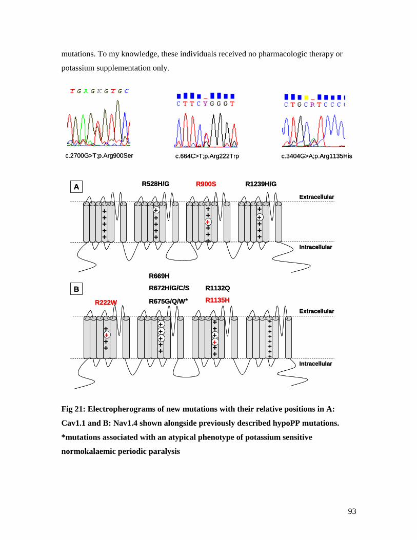
93
mutations. To my knowledge, these individuals received no pharmacologic therapy or
potassium supplementation only.
Fig 21: Electropherograms of new mutations with their relative positions in A:
Cav1.1 and B: Nav1.4 shown alongside previously described hypoPP mutations.
*mutations associated with an atypical phenotype of potassium sensitive
normokalaemic periodic paralysis
c.2700G>T;p.Arg900Ser c.664C>T;p.Arg222Trp c.3404G>A;p.Arg1135Hisc.2700G>T;p.Arg900Ser c.664C>T;p.Arg222Trp c.3404G>A;p.Arg1135His
+ +
+ +
+
+ +
+ +
+ +
+ +
+
+ +
+ +
A R528H/G R1239H/G
++
Extracellular
Intracellular
+ +
+
+ +
+ +
+ +
+ +
+ +
+ +
+ +
R669H
R672H/G/C/S
R675G/Q/W*
B
++
+
Extracellular
Intracellular
+
+
++
R900S
R222W
R1132Q
R1135H
+ +
+ +
+
+ +
+ +
+ +
+ +
+
+ +
+ +
A R528H/G R1239H/G
+++
Extracellular
Intracellular
+ +
+
+ +
+ +
+ +
+ +
+ +
+ +
+ +
R669H
R672H/G/C/S
R675G/Q/W*
B
++++
++
Extracellular
Intracellular
++
++
++++
R900S
R222W
R1132Q
R1135H

94
I then analysed DNA from the remaining 9/13 cases for the entire coding region of
CACNA1S and the coding regions of SCN4A that correspond to the S1-3 segments of
Nav1.4. The incomplete sequencing of SCN4A was due to limited available DNA. I took
a directed approach to the sequencing hypothesizing that if the voltage sensors
themselves were not implicated it was possible that the S1-3 segments would be as
theoretically this may produce a similar cation leak. I found no further mutations with this
analysis.
Finally I analysed the entire coding region of KCNJ2 associated with ATS. A novel
mutation was identified in 1/9 cases. Control chromosomes were sequenced but none
carried the mutation which was highly conserved and predicted to be a deleterious amino
acid substitution. Eight cases remained in whom no mutation was identified.
Gene Mutation No of Cases
CACNA1S R528G/H 25
CACNA1S R1239G/H 39
CACNA1S R900S* 1
SCN4A R672C/H/S 4
SCN4A R1132Q 2
SCN4A R222W* 2
SCN4A R1135H* 1
KCNJ2 V223E* 1
Unidentified 8
*denotes new gene mutations, not previously reported.
Table 12: Summary of genetic results for 83 patients with a phenotype of hypoPP
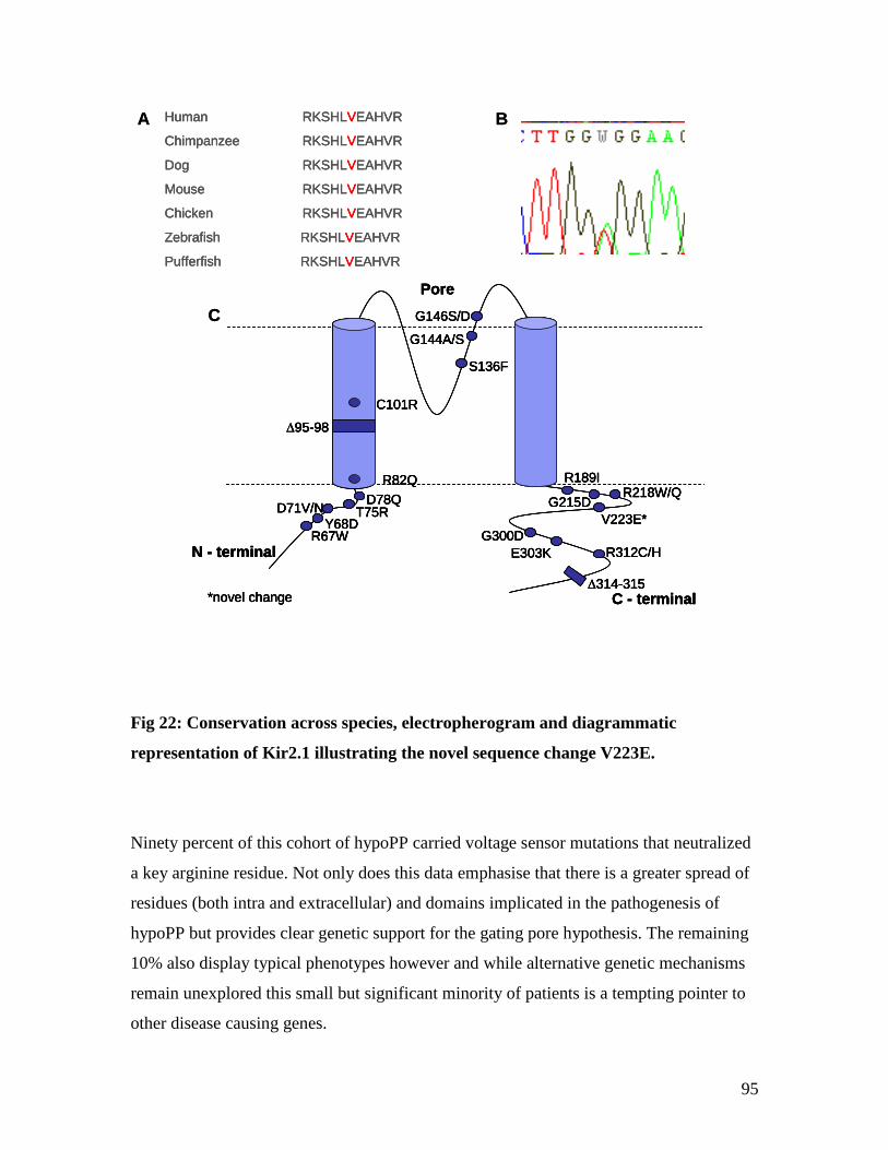
95
Fig 22: Conservation across species, electropherogram and diagrammatic
representation of Kir2.1 illustrating the novel sequence change V223E.
Ninety percent of this cohort of hypoPP carried voltage sensor mutations that neutralized
a key arginine residue. Not only does this data emphasise that there is a greater spread of
residues (both intra and extracellular) and domains implicated in the pathogenesis of
hypoPP but provides clear genetic support for the gating pore hypothesis. The remaining
10% also display typical phenotypes however and while alternative genetic mechanisms
remain unexplored this small but significant minority of patients is a tempting pointer to
other disease causing genes.
C - terminal
R67WY68D
D71V/N
G144A/S
∆95-98
R189I
D78QT75R
R82Q
C101R
G146S/D
S136F
G215D
N - terminal
Pore
*novel change
R218W/Q
E303K
∆314-315
G300D
V223E*
R312C/H
Human RKSHLVEAHVR
Chimpanzee RKSHLVEAHVR
Dog RKSHLVEAHVR
Mouse RKSHLVEAHVR
Chicken RKSHLVEAHVR
Zebrafish RKSHLVEAHVR
Pufferfish RKSHLVEAHVR
A B
C
C - terminal
R67WY68D
D71V/N
G144A/S
∆95-98
R189I
D78QT75R
R82Q
C101R
G146S/D
S136F
G215D
N - terminal
Pore
*novel change
R218W/Q
E303K
∆314-315
G300D
V223E*
R312C/H
Human RKSHLVEAHVR
Chimpanzee RKSHLVEAHVR
Dog RKSHLVEAHVR
Mouse RKSHLVEAHVR
Chicken RKSHLVEAHVR
Zebrafish RKSHLVEAHVR
Pufferfish RKSHLVEAHVR
C - terminal
R67WY68D
D71V/N
G144A/S
∆95-98
R189I
D78QT75R
R82Q
C101R
G146S/D
S136F
G215D
N - terminal
Pore
*novel change
R218W/Q
E303K
∆314-315
G300D
V223E*
R312C/H
C - terminal
R67WY68D
D71V/N
G144A/S
∆95-98
R189I
D78QT75R
R82Q
C101R
G146S/D
S136F
G215D
N - terminal
Pore
*novel change
R218W/Q
E303K
∆314-315
G300D
V223E*
R312C/H
Human RKSHLVEAHVR
Chimpanzee RKSHLVEAHVR
Dog RKSHLVEAHVR
Mouse RKSHLVEAHVR
Chicken RKSHLVEAHVR
Zebrafish RKSHLVEAHVR
Pufferfish RKSHLVEAHVR
A B
C

96
Results – Treatment Response
5.1 Genotype influences treatment response in hypokalaemic periodic paralysis
The mechanism of action of acetazolamide in hypoPP is not understood although it is
generally used as the first line therapeutic agent. Despite this there has never been a
randomized controlled trial to determine its efficacy, partly due to logistical difficulties
given the relative rareness of the disease. There have been several reports of patients with
SCN4A mutations (hypoPP2) responding deleteriously to acetazolamide therapy.
However other reports have disputed this. With the additional discovery of the aberrant
cation leak (including protons) with certain Nav1.4 mutations it is feasible that disturbed
pH homeostasis may offer a mechanism to explain why acetazolamide, a carbonic
anhydrase inhibitor could be beneficial. However histidine substitutions were shown to
produce a proton selective gating pore whereas glycine substitutions allowed a larger and
less selective cation leak, suggesting any disruption in pH that occurs and therefore any
amelioration offered by acetazolamide, may not be equal for all mutations. Whether a
similar hypothesis applies to mutations of Cav1.1 is unclear as no functional work has yet
been carried out to determine the presence or absence of a gating pore.
Literature and patient review
As my aim was to determine if genotype influenced the response to acetazolamide only
cases of genetically confirmed hypokalaemic periodic paralysis were selected (cohort 1)
from the published literature. Given the relative rarity of hypoPP, I conducted a literature
search on Pubmed (www.ncbi.nlm.nih.gov) using the key terms “hypokalaemic periodic
paralysis”, “periodic paralysis”, “acetazolamide” and “carbonic anhydrase inhibitors”.
Only articles published in English in or after 1994 (when the genetic basis of hypoPP was
identified) were considered. Articles meeting these criteria were reviewed and any
reported treatment and treatment response were recorded (Table 13). The data in table 13
was then used to summarise the response to acetazolamide by genotype (Table 14). Other

97
treatments were reported but only the response to acetazolamide was summarised as this
was the most frequently used and the numbers treated with alternatives were very small.
In order to create a comparison group to test the validity of the findings and to attempt to
avoid introducing any bias, case records of patients with genetically confirmed hypoPP
attending the neuromuscular clinic at NHNN under the care of Professor Hanna were
reviewed (cohort 2) by a visiting fellow Dr Simona Portaro. Patients were reviewed at
clinic on a six monthly or once yearly basis. Documented frequency, severity and
duration of paralytic attacks before and after therapy with acetazolamide was analysed to
ascertain response as either beneficial, detrimental or no change. I used this data to
summarise the response to acetazolamide by genotype in the same way as for cohort 1
(table 15).
Patients with SCN4A substitutions at the R675 position who had received acetazolamide
therapy were identified in each cohort. The published phenotype of these mutations
however has been described as potassium sensitive, normokalaemic periodic paralysis
and not hypoPP. I met patients from a single kindred in cohort 2 carrying the R675G
SCN4A mutation many times and they did also demonstrate potassium sensitivity and
normal serum potassium levels during an attack of paralysis. As such they did not display
a typical hypoPP phenotype and after consideration all patients carrying SCN4A R675
substitutions were excluded from analysis as the aim was to determine the efficacy of
acetazolamide in hypoPP as accurately as possible within the limitations of a
retrospective analysis.
Response to acetazolamide by genotype
In cohort 1, 54% (21/39) of patients with a CACNA1S mutation treated with
acetazolamide reported a beneficial response, compared with only 18% (3/17) of those
with an SCN4A mutation. In the combined cohort this was a benefit for only 43% (24/56)
of patients. However, if only patients with the most common HypoPP mutations (R528H,
R1239H) were considered, the benefit improved to 59% (20/34). In cohort 2 the findings
were similar; with 57% (8/14) of patients with CACNA1S mutations which also

98
represented the cohort as a whole, reporting a beneficial response. The positive response
rate increased to 62% (8/13) when the R528H and R1239H group were considered alone.
Closer evaluation of the relationship between precise genotype and treatment response
showed that in both cohorts when one of the arginine residues located at the extracellular
side of the sarcolemma (see Fig 21) was substituted for a glycine residue (R528G,
R1239G, R672G), there was never a beneficial response to acetazolamide, and there was
often a deleterious effect reported.
Fig 23: Histogram depicting the response rate to acetazolamide by genotype for each
cohort
1. All patients in cohort
2. CACNA1S mutations only
3. SCN4A mutations only
4. Common CACNA1S mutations only
5. Extracellular glycine substitutions
0.00%
10.00%
20.00%
30.00%
40.00%
50.00%
60.00%
70.00%
80.00%
90.00%
100.00%
1 2 3 4 5
Cohort 1
Cohort 2
1. All patients in cohort
2. CACNA1S mutations only
3. SCN4A mutations only
4. Common CACNA1S mutations only
5. Extracellular glycine substitutions
0.00%
10.00%
20.00%
30.00%
40.00%
50.00%
60.00%
70.00%
80.00%
90.00%
100.00%
1 2 3 4 5
Cohort 1
Cohort 2

99
This data is retrospective and imperfect, but it does provide some indication of response
rates to acetazolamide and tentatively suggests a correlation with genotype i.e. R to H
substitutions respond the best and R to G substitutions respond poorly. It is likely the
poor response rate in the SCN4A group also reflects this as the amino acid substitutions
are much more varied compared to the CACNA1S group where they are predominantly R
to H. Overall however only approximately 50% of hypoPP patients are adequately treated
by the current first line therapeutic agent, acetazolamide. This represents a significant
unmet need in terms of treatment. Both randomized controlled trials and development of
alternative therapies are required.
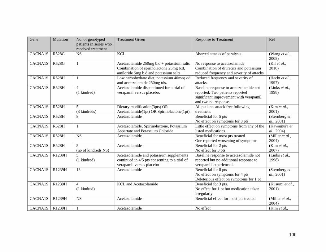
100
Gene Mutation No. of genotyped
patients in series who
received treatment
Treatment Given Response to Treatment Ref
CACNA1S R528G NS KCL Aborted attacks of paralysis (Wang et al.,
2005)
CACNA1S R528G 1 Acetazolamide 250mg b.d + potassium salts
Combination of spirinolactone 25mg b.d,
amiloride 5mg b.d and potassium salts
No response to acetazolamide
Combination of diuretics and potassium
reduced frequency and severity of attacks
(Kil et al.,
2010)
CACNA1S R528H 1 Low carbohydrate diet, potassium 40meq od
and acetazolamide 250mg tds.
Reduced frequency and severity of
attacks.
(Hecht et al.,
1997)
CACNA1S R528H 4
(1 kindred)
Acetazolamide discontinued for a trial of
verapamil versus placebo.
Baseline response to acetazolamide not
reported. Two patients reported
significant improvement with verapamil,
and two no response.
(Links et al.,
1998)
CACNA1S R528H 5
(3 kindreds)
Dietary modification(3pts) OR
Acetazolamide(1pt) OR Spirinolactone(1pt)
All patients attack free following
treatment
(Kim et al.,
2001)
CACNA1S R528H 8 Acetazolamide Beneficial for 5 pts
No effect on symptoms for 3 pts
(Sternberg et
al., 2001)
CACNA1S R528H 1 Acetazolamide, Spirinolactone, Potassium
Aspartate and Potassium Chloride
Little effect on symptoms from any of the
listed medications.
(Kawamura et
al., 2004)
CACNA1S R528H NS Acetazolamide Beneficial for most pts treated.
One reported worsening of symptoms
(Miller et al.,
2004)
CACNA1S R528H 5
(no of kindreds NS)
Acetazolamide Beneficial for 2 pts
No effect for 3 pts
(Kim et al.,
2007)
CACNA1S R1239H 5
(1 kindred)
Acetazolamide and potassium supplements
continued in 4/5 pts consenting to a trial of
verapamil versus placebo
Baseline response to acetazolamide not
reported but no additional response to
verapamil experienced.
(Links et al.,
1998)
CACNA1S R1239H 13 Acetazolamide Beneficial for 8 pts
No effect on symptoms for 4 pts
Deleterious effect on symptoms for 1 pt
(Sternberg et
al., 2001)
CACNA1S R1239H 4
(1 kindred)
KCL and Acetazolamide Beneficial for 3 pts.
No effect for 1 pt but medication taken
irregularly
(Kusumi et al.,
2001)
CACNA1S R1239H NS Acetazolamide Beneficial effect for most pts treated (Miller et al.,
2004)
CACNA1S R1239H 1 Acetazolamide No effect (Kim et al.,

101
2007)
CACNA1S R1239G 1 Spirinolactone 25mg bd Improved symptoms
Note: 2 clinically affected cousins
reported no effect with ACZ treatment
but an improvement with spirinolactone.
Genetic analysis was not performed on
these 2 pts.
(Kim et al.,
2005)
CACNA1S R1239G 3
(1 kindred)
Acetazolamide OR a
combination of spirinolactone, amiloride
and potassium supplements
No effect of ACZ for 2 pts
Deleterious effect of ACZ for 1 pt but
then treated successfully with a
combination of diuretics and potassium
supplements
(Kim et al.,
2007)
CACNA1S R897S 1 Potassium supplements 2-3mmol/kg/day
and acetazolamide 5-10mg/kg/day
Treated from age 2-6years. Frequency
and severity of attacks reduced but
duration of attacks increased.
(Chabrier et al.,
2008)
SCN4A “Sodium
channel
mutations”
NO
4 Acetazolamide Deleterious effect (Miller et al.,
2004)
SCN4A R669H 2 Pt 1:
Dietary regime, potassium supplements and
a carbonic anhydrase inhibitor (not
specified)
Reduced frequency of attacks (Bulman et al.,
1999)
Pt 2:
acetazolamide 250mg bd
Attack free for 2 months then one severe
attack responding to potassium infusion.
No long-term response documented
SCN4A R669H 1 Acetazolamide No response (Kim et al.,
2007)
SCN4A R669H 1 Potassium supplements No attacks during 12 month follow up
period
(Maffe et al.,
2009)
SCN4A R672G 3
(1 kindred)
Acetazolamide Deleterious effect (Sternberg et
al., 2001)
SCN4A R672G 1 Acetazolamide OR
Combination of spirinolactone, amiloride
and potassium supplements
Deleterious effect of ACZ but then
treated successfully with a combination
of diuretics and potassium supplements
(Kim et al.,
2007)
SCN4A R672C NS Either acetazolamide 500mg a day OR
dichlorphenamide 100mg a day
Attack free for at least 6 months on
average
(Kim et al.,
2004)

102
SCN4A R672C 1 Acetazolamide No response
SCN4A R672S 3
(1 kindred)
acetazolamide
triamterene
Severe quadriparesis within 12 - 60 hours
of acetazolamide.
Reduction in frequency and severity of
attacks with Triamterene
(Bendahhou et
al., 2001)
SCN4A R672S 1 Acetazolamide 250mg bd and spirinolactone
100mg per day
Reduced frequency of attacks (Davies et al.,
2001)
SCN4A R672S 1 Acetazolamide 250mg per day and
potassium taken at onset of attack or before
strenuous exercise
Reduced frequency of attacks (Venance et al.,
2004)
SCN4A R675G 5
(1 kindred)
Acetazolamide 250mg per day 1 pt no response to 10 years continuous
treatment
4 pts reported a reduced duration of
attacks with intermittent ACZ treatment
(Vicart et al.,
2004)
SCN4A R675Q 2
(2 kindreds)
Acetazolamide 500mg per day Pt 1: no benefit
Pt 2: attack free since taking ACZ (length
of treatment not documented)
(Vicart et al.,
2004)
SCN4A R675W 1 Acetazolamide 250mg per day Reduced frequency and severity of
attacks
(Vicart et al.,
2004)
SCN4A R1132Q 1 Acetazolamide
Potassium peri-attack
No response to acetazolamide
Potassium ingestion reduced duration of
attack
(Carle et al.,
2006)
NS: Not Specified
Table 13: Details of response to therapy for genetically confirmed cases of hypokalaemic periodic paralysis reported in the
literature.
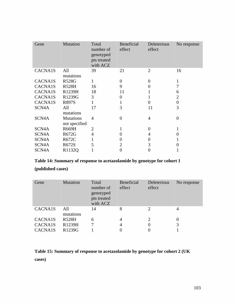
103
Table 14: Summary of response to acetazolamide by genotype for cohort 1
(published cases)
Table 15: Summary of response to acetazolamide by genotype for cohort 2 (UK
cases)
Gene Mutation Total
number of
genotyped
pts treated
with ACZ
Beneficial
effect
Deleterious
effect
No response
CACNA1S All
mutations
39 21 2 16
CACNA1S R528G 1 0 0 1
CACNA1S R528H 16 9 0 7
CACNA1S R1239H 18 11 1 6
CACNA1S R1239G 3 0 1 2
CACNA1S R897S 1 1 0 0
SCN4A All
mutations
17 3 11 3
SCN4A Mutations
not specified
4 0 4 0
SCN4A R669H 2 1 0 1
SCN4A R672G 4 0 4 0
SCN4A R672C 1 0 0 1
SCN4A R672S 5 2 3 0
SCN4A R1132Q 1 0 0 1
Gene Mutation Total
number of
genotyped
pts treated
with ACZ
Beneficial
effect
Deleterious
effect
No response
CACNA1S All
mutations
14 8 2 4
CACNA1S R528H 6 4 2 0
CACNA1S R1239H 7 4 0 3
CACNA1S R1239G 1 0 0 1

104
Results - Pathophysiology
6.1 How does neutralisation of an intracellular voltage sensor arginine of Nav1.4
influence the pathomechanism of hypoPP?
It is known that the common mutations in CACNA1S and SCN4A associated with
hypoPP produce “loss of function” effects on the alpha pore of Cav1.1 and Nav1.4
although this has failed to explain the mechanism of paradoxical membrane
depolarisation of the muscle membrane in the presence of hypokalaemia. The gating pore
hypothesis has offered some new insights into the pathophysiology of hypoPP. It is
noteworthy however that not all the voltage sensors have equal numbers of positive
charge and it is likely that each voltage sensor has additional specificities. As the voltage
sensor moves across the membrane in response to changes in membrane potential it has
been observed that mutations of variably placed arginines determine whether the aberrant
gating pore occurs at depolarised or hyperpolarised potentials.
Mutations of one arginine, R675G/Q/W in DII of Nav1.4 produce a unique phenotype of
potassium sensitive normokalaemic periodic paralysis. Mutations of this arginine, which
is placed more toward the intracellular side of the membrane, have recently been shown
to create a cation leak at depolarised potentials(Sokolov et al., 2008). What is unknown is
whether mutations of these inwardly placed arginine residues also produce a loss of
function effect on the alpha pore.
To investigate the function of the alpha pore, I carried out whole-cell patch clamp
recording in cultured mammalian HEK293 cells. Patch clamp recordings were made from
HEK cells co-transfected with human wild type SCN4A or a mutant construct containing
the R675G mutation and cDNA encoding human SCN1B, the sodium channel beta
subunit, which although not contributing directly to the main pore of the Nav1.4 channel,
has been shown to be important for regulating sodium channel function(Isom., 2001).

105
Effects of R675G on the Nav1.4 alpha pore
Fig 24: Comparison of activation, inactivation, peak current and current density
observed for the R675G mutation and WT Nav1.4
A significant reduction (6 fold) in peak current and current density was observed for the
mutant in comparison to the wild type (Figure 24). There was no difference in the voltage
dependence of activation. There was a suggestion of enhanced inactivation for the mutant
but this was not significant, partly because the small currents produced by SCN4A-
R675G were too close to the noise threshold for accurate measurements of inactivating
currents.
WT Nav1.4R675G
-0.2
0
0.2
0.4
0.6
0.8
1
-70 -50 -30 -10
Voltage (mV)
No
rma
lise
d c
urr
en
t
-0.2
0
0.2
0.4
0.6
0.8
1
-110 -90 -70 -50 -30
Voltage (mV)
No
rma
lise
d c
urr
en
t
10ms
1nA
0
1000
2000
3000
4000
WT R675G
Cu
rre
nt
de
nsity (
pA
)
-4000
-3500
-3000
-2500
-2000
-1500
-1000
-500
0-100 -80 -60 -40 -20 0
Voltage (mV)
Cu
rre
nt
(pA
)
WT Nav1.4R675GWT Nav1.4R675G
-0.2
0
0.2
0.4
0.6
0.8
1
-70 -50 -30 -10
-0.2
0
0.2
0.4
0.6
0.8
1
-70 -50 -30 -10
Voltage (mV)
No
rma
lise
d c
urr
en
t
-0.2
0
0.2
0.4
0.6
0.8
1
-110 -90 -70 -50 -30
-0.2
0
0.2
0.4
0.6
0.8
1
-110 -90 -70 -50 -30
Voltage (mV)
No
rma
lise
d c
urr
en
t
10ms
1nA
10ms
1nA
0
1000
2000
3000
4000
WT R675G
Cu
rre
nt
de
nsity (
pA
)0
1000
2000
3000
4000
WT R675G
Cu
rre
nt
de
nsity (
pA
)
-4000
-3500
-3000
-2500
-2000
-1500
-1000
-500
0-100 -80 -60 -40 -20 0
Voltage (mV)
-4000
-3500
-3000
-2500
-2000
-1500
-1000
-500
0-100 -80 -60 -40 -20 0
Voltage (mV)
Cu
rre
nt
(pA
)
106
The large reduction in peak transient currents is consistent with the effects on the alpha
pore observed for extracellular voltage sensor mutations (R669H, R672G/H/S). All of
these mutations give reduced current density accompanied by small or non-significant
effects on inactivation. The 6-fold reduction in current density for R675G was greater
than that reported for the other mutants (2 to 4 fold reduction).The small differences in
the kinetics of inactivation introduced by these mutants have been discussed in the
literature, and there is a consensus that these kinetic changes cannot explain the
significant reduction in current density that is observed(Struyk et al., 2000; Kuzmenkin et
al., 2002). Although alternative hypotheses have been postulated, such as altered protein
folding or trafficking, no work has been carried out to date to address this specific
question.
In channels containing mutations affecting external arginines, the gating pore current has
been estimated to be between 1% and 0.02% of the current through the main alpha
pore(Cannon., 2010). Given the significantly reduced current density seen in R675G of
approximately 500pA, any leak current of this size would not have been detectable in
these recordings, as the noise inherent to whole cell currents is greater than 5 pA rms (1%
of 500 pA).For this reason although my data show a robust loss of peak current through
the alpha pore, I was unable to assess whether the R675G mutation also introduced a leak
current.
6.2 Developing a human myoblast expression system
All published work examining gating pore currents from SCN4A mutants has been in
xenopus oocytes where very large currents can be obtained. I carried out recordings in
HEK cells, which allow accurate clamping of peak alpha pore currents, but the total
current amplitudes were not sufficient for investigation of leak currents. While both of
these model systems have advantages, they may not replicate the physiology of muscle
cells sufficiently to recapitulate the mechanism of muscle channelopathies. For example,
mutations in SCN4A may be modified by endogenous accessory subunits present only in
muscle cells, or muscle cells may have specialized trafficking or expression machinery
for regulating highly-expressed channels like SCN4A. In addition the pathology of
107
SCN4A mutations may involve buffering (possibly via carbonic anyhydrases inhibited by
acetazolamide). Neither HEK cells or xenopus oocytes would test these sorts of muscle-
specific mechanisms. Therefore, I investigated whether it was possible to culture human
myoblasts and record currents in patch clamp configuration.
Preliminary recordings were made using a myoblast culture from a healthy control
teenager that was kindly donated by Dr Carl Adkin working at the Dubowitz
Neuromuscular Centre. It was established that robust sodium currents can be recorded in
these cells. Recordings made from isolated myoblasts plated on coverslips for only 48-72
hours allowed identification of modest sodium currents (Fig 25). When the voltage
dependence of activation and inactivation of this current was compared with current
obtained from HEK cells transfected with wild type Nav1.4 there were no detectable
significant differences (see figure 25).
In addition to determine whether SCN4A was expressed endogenously in these cells I
extracted RNA from an aliquot of the same myoblast culture and performed reverse
transcription PCR. The cDNA obtained from this procedure underwent further PCR using
primer pairs specific for SCN4A cDNA. An SCN4A clone was used as a positive control
and genomic DNA as a negative control. Fragments of anticipated size for SCN4A cDNA
were amplified for the SCN4A clone and cDNA obtained from the myoblasts. No product
was obtained from the genomic DNA confirming the SCN4A gene was expressed in the
cell culture which is consistent with Nav1.4 channels having contributed to the sodium
current recorded

108
Fig 25: Current density and voltage dependence of activation and inactivation of
sodium currents obtained after 48-72hours of human myoblasts in culture
compared with HEK cells transfected with WT Nav1.4.
Culture time influenced the density of sodium current and also the voltage dependence of
activation. There is a much larger sodium current (average 6nA) and a left shift in
activation of sodium current recorded from more mature myoblasts (day 10) which is
likely to reflect a contribution to the overall current from both Nav1.4 and Nav1.5 (which
has a left shift of activation of approx 10-15mv compared to Nav1.4). These preliminary
experiments have indicated low sodium extracellular solutions will be required to reduce
sodium currents and to provide better clamp of the sodium current seen in mature
myoblasts. In addition, given the likely contribution of the Nav1.5 channels, it may be
necessary to apply tetrodotoxin to quantify the relative contributions of Nav1.4 (TTX
sensitive) and Nav1.5 (TTX insensitive) to the overall sodium current.
Myoblasts day 2-3
WT SCN4A - HEK
Voltage (mV)
Norm
alis
ed C
urr
ent
0
0.2
0.4
0.6
0.8
1
-60 -40 -20 0
0
0.2
0.4
0.6
0.8
1
-110 -90 -70 -50 -30
Voltage (mV)
Norm
alis
ed C
urr
ent
-600
-500
-400
-300
-200
-100
0-100 -80 -60 -40 -20 0 20
Voltage (mV)
Curr
ent (p
A)
Myoblasts day 2-3
WT SCN4A - HEK
Myoblasts day 2-3
WT SCN4A - HEK
Voltage (mV)
Norm
alis
ed C
urr
ent
0
0.2
0.4
0.6
0.8
1
-60 -40 -20 0
0
0.2
0.4
0.6
0.8
1
-110 -90 -70 -50 -30
Voltage (mV)
Norm
alis
ed C
urr
ent
Voltage (mV)
Norm
alis
ed C
urr
ent
0
0.2
0.4
0.6
0.8
1
-60 -40 -20 0
0
0.2
0.4
0.6
0.8
1
-110 -90 -70 -50 -30
Voltage (mV)Voltage (mV)
Norm
alis
ed C
urr
ent
0
0.2
0.4
0.6
0.8
1
-60 -40 -20 0
0
0.2
0.4
0.6
0.8
1
-110 -90 -70 -50 -30
0
0.2
0.4
0.6
0.8
1
-110 -90 -70 -50 -30
Voltage (mV)
Norm
alis
ed C
urr
ent
-600
-500
-400
-300
-200
-100
0-100 -80 -60 -40 -20 0 20
Voltage (mV)
Curr
ent (p
A)
-600
-500
-400
-300
-200
-100
0-100 -80 -60 -40 -20 0 20
Voltage (mV)
Curr
ent (p
A)
A
B C
A
B C

109
Although myoblasts potentially offer a system for recapitulating much more of the
muscle cell physiology, there are many technical considerations to be made and
optimized before a myoblast system may be used to study the gating pore in voltage
sensor mutations of Nav1.4. A major hurdle will be the need for obtaining muscle
biopsies from willing patients who carry these mutations. However the early recordings
made suggest endogenous Nav1.4 is expressed, and consequently characterization of
sodium currents in these cells may be a viable albeit technically challenging future
project.

110
Discussion - Phenotype
Aim
To examine the phenotype of the skeletal muscle channelopathies in patients referred to
the NCG service for channelopathies at the NHNN to determine
c. if there were any clinically useful phenotype-genotype correlations that
could be made and
d. if there were more extensive aspects to the phenotypes than is currently
recognised.
Meeting patients through both the specialist muscle clinic and the natural history trial of
non-dystrophic myotonia combined with analysis of the existing channelopathy database
was an enormously valuable and rewarding experience. Using this approach I was able to
identify several unexplored aspects of the phenotype that had clinically relevant
correlations.
7.1 Observation of neonatal hypotonia in paramyotonia congenita due to the I693T
SCN4A mutation
Neonatal paramyotonia congenita often presents with myotonic phenomenon when an
infant cries or is washed, especially with cool water(Eulenberg A., 1886). Despite the
recognition that myotonic symptoms may present at birth, this was not generally
considered to be particularly detrimental to the neonate or to require any specific
consideration in counseling expectant mothers. Recently a rare lethal neonatal
paramyotonia case with extreme sensitivity to cold was reported and attributed to an
SCN4A mutation (N1297K)(Gay et al., 2008). However, neonatal hypotonia had not
been recognised as a clinical manifestation of muscle sodium channel dysfunction. The
observations I gathered in the six individuals studied from four unrelated French and

111
English families indicate that neonatal hypotonia with additional symptoms of poor
feeding and respiratory distress can be a manifestation of muscle sodium channel
dysfunction common to individuals with the I693T sodium channel mutation.
Pathological muscle hypotonia is an important neonatal presentation and can be a sign of
either a peripheral or central abnormality. Careful diagnostic assessment and often
multiple investigations are required(Laugel et al., 2007). Recognition of a sodium
channel mutation as a cause of neonatal hypotonia may prevent unnecessary and invasive
investigations. For example, a muscle biopsy is very unlikely to be contributory. EMG
assessment has been shown to be beneficial in adults if performed according to specific
protocols(Fournier et al., 2006; Fournier et al., 2004) but these are not easily applicable
in neonates.
Treatment in all the cases studied was supportive as symptoms were self-limiting but it is
important to recognize that symptoms may be cold-triggered and temperature dependent,
indicating, a constant warm ambient temperature is essential in such cases.
All cases studied carried the I693T mutation. The detrimental effects of the I693T
mutation on sodium channel gating are similar to other PMC mutations i.e. the overall
effect is one of impaired inactivation(Plassart-Schiess et al., 1998). These detrimental
effects were exacerbated in vitro by cold temperature but this is not an exclusive finding.
Whether the association of neonatal hypotonia is entirely specific to the I693T mutation
is not yet clear. Paramyotonia congenita is rare and it may be that neonatal hypotonia has
occurred in infants carrying other mutations but the direct association with PMC not been
made or reported.
7.2 Stridor expands the neonatal presentations of skeletal muscle sodium
channelopathy
Initially it was hard to determine from a single case if the occurrence of stridor in the
child with PMC was related to PMC itself. However discussion with Dr Damien
Sternberg in France and his observations of stridor in two other cases was very suggestive
and hypothetically it seemed likely that stridor could reflect laryngeal myotonia.

112
Hyperkalaemic periodic paralysis (HyperPP) is a dominantly inherited disorder which is
allelic to PMC and is characterised by episodes of muscle weakness and myotonia,
although weakness is the predominant symptom(Venance et al., 2006). In both these
disorders gene mutation results in a “gain of function” sodium channel defect. It has been
proposed that the two disorders reflect ends of a spectrum of the same disease(Cannon.,
2006).
Stridor has been described in patients with myotonic dystrophy, in both neonates and
adults.(Heuschkel et al., 2003; Ahmadian et al., 2002) In addition there are several
reports of stridor in American Quarter horses affected by the equine form of
hyperkalaemic periodic paralysis (HyperPP). In one series 63/68 affected horses suffered
stridor in association with exertion, muscle weakness or excitement(Carr et al., 1996).
There is also one case report of an adult with PMC in whom provocative testing with
muscle cooling induced severe laryngeal myotonia with stridor(Ferriby et al., 2006).
Overall it is likely the stridor reflects myotonia of the laryngeal muscles. In addition, the
child described experienced feeding and respiratory difficulties which were also noted in
the cases of neonatal hypotonia suggesting these are common neonatal features of PMC.
Dysphagia and respiratory compromise are rarely reported in adult cases(Lerche et al.,
1993).
Following on from the observations here, another report has recently been published of
severe neonatal laryngospasm occurring in three neonates carrying de novo SCN4A
mutations (G1306E and A799S). These infants had life threatening episodes of stridor
with apnoea and feeding difficulties. Symptoms responded to sodium channel blockers
although in one infant the diagnosis was made posthumously(Lion-Francois et al., 2010).
Taken together these observations of hypotonia and stridor with resultant feeding and
respiratory difficulty indicate that offspring of parents with sodium channel mutations are
at risk of neonatal complications. This is an important development in understanding the
phenotypes of skeletal muscle sodium channelopathies and should be reflected
accordingly in the advice given to patients considering having children and the relevant
clinicians involved in their delivery and early post-natal care.

113
7.3 Observation of inflammatory infiltrates in skeletal muscle channelopathies
The natural history of the skeletal muscle channelopathies is not extensively documented
but development of a proximal myopathy is established particularly in the periodic
paralyses(Becker PE., 1977; Nagamitsu et al., 2000; Miller et al., 2004; Links et al.,
1990; Bradley et al., 1990; Plassart et al., 1996; Vicart et al., 2004). Whether the
myopathy is related to the severity and frequency of paralytic attacks is unclear(Buruma
et al., 1978; Links et al., 1990) but there is some evidence that its development may be
associated with increasing age(Links et al., 1990; Plassart et al., 1994). In addition it is
common for moderate to severe myalgia to be reported(Rosenfeld et al., 1997; Ptacek et
al., 1994b; Colding-Jorgensen et al., 2006; Vicart et al., 2004; Fialho et al., 2007) and
moderately raised creatine kinase(1-2000U/L)(Links et al., 1990; Colding-Jorgensen et
al., 2006; Plassart et al., 1996; Fialho et al., 2007) to be noted in the skeletal muscle
channelopathies although there is no evidence to link these to the presence or absence of
myopathy.
A characteristic history in conjunction with specialised neurophysiologic
techniques(Fournier et al., 2004) allows for a diagnosis in the majority of cases of
skeletal muscle channelopathy and it is rare that a diagnostic muscle biopsy is now
performed in the UK.
In the three cases I studied a clinical diagnosis of skeletal muscle channelopathy had been
made prior to the muscle biopsy and was later genetically confirmed. Biopsy was
performed due to additional clinical features that were deemed unusual or severe for a
muscle channelopathy. In case one the proximal myopathy could have been attributed to
the myopathy that is reported in periodic paralysis but the very high CK (2729U/L) was
atypical for this. The severe myalgia and moderate elevation of CK (1109U/L) reported
in case two are also described features of the channelopathies and it is possible that at
least some of the myalgia is due to myotonia. However as the myalgia became severe
enough to be the patient’s primary complaint a biopsy was performed. The initial
complaint of muscle swelling in case three is not a typical feature of the muscle

114
channelopathies and with the later development of significant myalgia a biopsy was felt
to be warranted. Collectively the clinical features of weakness, myalgia and significantly
elevated CK in these three cases suggest that these symptoms are secondary, at least in
part, to the observed inflammatory process.
The biopsy findings in these cases did not fulfil all morphological criteria for IIM,
particularly the lack of invasion of intact fibres. However upregulation of MHC class I
expression (case I and III) is supportive and it is not unreasonable that a diagnosis of IIM
had been made in these cases, especially when each was considered in isolation. There
are numerous examples of other genetic muscle disorders in which inflammatory
infiltrates have been observed and even diagnosed as IIM(Tidball et al., 2005). It is
impossible to exclude the possibility that the observed inflammatory process is a co-
incidental finding and two independent pathologies (channelopathy and IIM) are present
in each of the three individuals, but statistically this seems unlikely.
Whether this inflammatory response plays a potential pathogenic role in the development
of the myopathy is not known. Morphology data is reported for the skeletal muscle
channelopathies, especially the periodic paralyses, but in many cases pre-dates the
availability of genetic testing. A vacuolar myopathy and or tubular aggregates support the
diagnosis of periodic paralysis. With improved electrophysiological and genetic
diagnostic methods muscle biopsy is now less common. Without a larger number of
biopsy samples from similar cases it is not possible to know how accurately
inflammatory muscle infiltrates may correlate to clinical presentation.
Although there was some improvement in symptoms and a reduction in CK with
prednisone in two cases, in one case high dose prednisone induced a severe paralytic
attack, although low dose steroids were better tolerated. Worsening of the paralytic
symptoms of periodic paralysis with glucocorticoids has been reported(rzel-Hezode et al.,
2009a). Other immunosuppressive agents had no clear additional benefit in any of the
three cases.
Based on these observations it is reasonable to propose that a diagnostic muscle biopsy
should be considered in cases of skeletal muscle channelopathy presenting with the
symptoms of severe myalgia and/or sub acute weakness and an accompanying elevated
CK (>1000U/L) to ascertain the presence of any inflammatory infiltrates. The therapeutic

115
benefit of immunosuppressants is not established by this small number of cases but they
do tentatively indicate steroids could have a role. The optimal dose however is
undetermined and higher doses may be detrimental.
The statistical evidence indicates that the co-existence of an inflammatory process and
skeletal muscle channelopathy is very unlikely to be co-incidental. The possible
implications this may have for understanding of the mechanisms of muscle damage in the
skeletal muscle channelopathies will require future exploration.
7.4 Pain and morbidity in the non-dystrophic myotonias
There are almost no studies reporting on the morbidity or quality of life in any of the
skeletal muscle channelopathies. It was a personal observation that many of the patients I
met however did report pain as a prominent feature of their illness. Data gathered from
the patients I studied in the UK arm of the natural history study in the NDMs has
illustrated pain is a common and prominent aspect of the clinical presentation. It also
seems to be significantly more common in sodium channelopathies compared with
chloride channelopathies. These findings support those recently published in one of the
very few studies that has attempted to analyse morbidity in the channelopathies(Trip et
al., 2009a). This is very suggestive that pain has been an under-recognised and
potentially undertreated feature of the muscle channelopathies. These illnesses have often
been considered to be benign but this data together with the increasing evidence for
development of myopathy in the literature(Links et al., 1990; Buruma et al., 1978; Miller
et al., 2004; Nagamitsu et al., 2000) would indicate quality of life deserves more
consideration. Pain was not exclusively related to symptoms of muscle stiffness
(myotonia) so it is not entirely clear if treating the myotonia would abolish all pain.
However this early data does suggest the presence of pain should be a considered factor
when assessing the benefits or initiation of drug therapy for the NDMs. Analysis of the
combined cohort of patients across all centres participating in the natural history study
will be important to further address this issue.

116
Conclusions: Phenotype
1. Neonatal phenotypes of the skeletal muscle sodium channel myotonic disorders
exist. Severity is variable. Symptoms include hypotonia with feeding and
respiratory difficulties (including stridor) that is transitory and requires supportive
care only. Others support the recognition of neonatal phenotypes and have
additionally reported severe presentations of myotonia, feeding and respiratory
difficulties (including stridor) that can be fatal.
2. Inflammatory changes occur in the muscle biopsies of some channelopathy
patients that statistically seem unlikely to be co-incidental although the exact
significance and any influence this may have on the development of myopathy
requires further exploration.
3. Pain is an under-recognised feature of the skeletal muscle channelopathies and
morbidity associated with these diseases deserves more consideration.

117
Discussion - Genotype
Aim
To genetically characterize patients to assess if new mutations in known genes existed
and if these genes could account for 100% of a carefully phenotyped cohort or if new
genes may be implicated.
8.1 Genotyping the sodium channel myotonic disorders
Paramyotonia congenita and sodium channel myotonia
Of the 17 patients identified with a phenotype consistent with a sodium channel myotonic
disorder, I considered 13 to have PMC or SCM. Of this group 9/13 was genotyped.
Previously reported SCN4A mutations (V1293I, Q270K and E1702K) were identified in
four(Koch et al., 1995; Fournier et al., 2006; Miller et al., 2004) and another three had
novel amino acid substitutions (L128P, V445L and M135L) although it is probable only
L128P and V445L are pathogenic and possible that M135L is a rare polymorphism.
Mutations in SCN4A responsible for myotonic phenotypes appear to affect amino acids
distributed randomly across the protein with no discernible pattern. The novel changes
found here illustrate it is likely that new mutations will continue to be reported
throughout SCN4A. Although exons 13, 22 and 24 represent mutation hotspots screening
of the entire coding region of SCN4A is still required to make a genetic diagnosis in a
significant number of patients.
Of the remaining two patients who were genotyped one carried a CLCN1 mutation. This
illustrates the difficulty that remains, despite more advanced electrophysiological tests, in
differentiating some cases of sodium channel myotonia from chloride channel myotonia
clinically. One group genotyped 93% of their cohort of patients with NDM by employing
in tandem analysis of SCN4A and CLCN1(Trip et al., 2008). This highlights a real need
for both genes to be analysed if the first is negative.
118
The final patient was an atypical case under the care of paediatric colleagues, consultant
paediatrician Dr Elizabeth Wraige. The patient was an infant who had been well until
approximately four months of age. They were then admitted to intensive care in
respiratory distress that required long-term tracheostomy. The child was noted to be
“stiff” and a first EMG suggested difficulty in differentiating between myotonic
potentials and pseudomyotonic potentials. A trial of mexiletine was initially felt to be
helpful but later withdrawn as any benefits seemed to plateau. As a result SCN4A
analysis was undertaken by me but no pathogenic changes were found. A second EMG
later confirmed pseudomyotonic potentials. An initial biopsy at 5 months was non-
specific but a second biopsy at 6 months of age showed changes of myofibrillar
myopathy. Many parallels were noted between this child and a group of Indian-Canadian
children who had been described years earlier as having autosomal recessive fatal
hypertonic muscular dystrophy although no genetic diagnosis had been reached(Lacson et
al., 1994).
As muscle biopsy suggested a myofibrillar myopathy analysis of associated genes was
undertaken by the NCG genetic laboratory at Newcastle. A homozygous mutation was
identified in the CRYAB gene(Forrest et al., 2011). Attempts had been made to contact
the authors of the original hypertonic muscular dystrophy paper but were unsuccessful.
Shortly after the identification of the CRYAB mutation in this infant the same
homozygous CRYAB mutation was identified in the Canadian cohort(Del Bigio et al.,
2011).
From the work done in this thesis and reports in the literature neonatal channelopathy
phenotypes of varying severity are emerging. This includes fatal cases characterised by
myotonia, respiratory and feeding difficulties(Gay et al., 2008). Although the infant
above ultimately displayed evidence of a myofibrillar myopathy the initial presentation
was potentially in keeping with the described sodium channel neonatal phenotypes. It
suggests SCN4A analysis should still be considered in similar cases as the number of
reported neonatal cases and extent of the neonatal phenotype may continue to expand.
The 4/13 patients with a myotonic phenotype in whom no mutation was identified all had
EMG confirmed myotonia. Of these four, one was an infant being investigated for
119
hypotonia and the EMG myotonia was an unexpected finding. Brief bursts of myotonia
are seen in other myopathies(Milone et al., 2012) including congenital myopathies and
the myotonia in this case may ultimately be accounted for in this way, although no
definite diagnosis has yet been reached.
The other three cases however, had phenotypes that can be considered strongly consistent
with non-dystrophic myotonia. Only direct automated sequencing of the coding regions
of SCN4A and CLCN1 was undertaken. It is possible gene deletions or duplications may
occur and would not have been picked up by this method. Recently a pathogenic intron
change has been described in SCN4A in a family with PMC(Kubota et al., 2011). This
particular change would have been detected as it was close to the intron/exon border and
the primers I designed overlapped these regions. However other deeper intron changes
have not been excluded nor have changes in the promoter region of the gene. A final
possibility is that other myotonia genes exist. One of the non-genotyped cases came from
a large Sicilian family and a future project could examine linkage in this family to assess
the possibility.
Hyperkalaemic periodic paralysis
In the group of 17 patients with a sodium channel myotonic phenotype I identified four
patients with a phenotype consistent with hyperkalaemic periodic paralysis. In 1/4 I
identified a pathological expansion of the ZNF9 gene consistent with a diagnosis of
DM2. This patient was described as having episodes of muscle paralysis since childhood,
triggered by cold, antihistamines, and corticosteroids. She also complained of paralysis of
the respiratory muscles and bladder, with fasciculations and myotonia noted during
episodes. Her mother had the same symptoms, her father was unaffected. Her brother was
noted to have muscle hypertrophy and an elevated CK.
The other three cases had some atypical features for hyperkalaemic periodic paralysis and
it is perhaps not surprising that they do not carry any mutations in SCN4A. During my
thesis I was asked to sequence another patient with atypical hyperkalaemic periodic
paralysis by Prof Francesco Muntoni (consultant paediatrician) in whom his team had
identified a ryanodine receptor gene mutation. This patient had no pathogenic SCN4A
mutations and functional expression work carried out by Prof Muntoni’s team confirmed

120
the pathogenicity of the RYR1 gene mutation(Zhou et al., 2010). This case illustrates
however that atypical cases of hyperkalaemic periodic paralysis and potentially other
muscle channelopathies may reflect new disease causing genes. Additionally the
identification of an abnormal expansion of the ZNF9 gene in the fourth case of
hyperkalaemic periodic paralysis I studied was unexpected and emphasises the need to
reflect on these atypical cases. As more are identified, common phenotypic features may
help to group some together and aid in the future identification of additional disease
causing genes.
8.2 Genotyping hypokalaemic periodic paralysis
The new mutations found in CACNA1S and SCN4A in patients with a phenotype of
hypokalaemic periodic paralysis significantly expands the spectrum of S4 segment
arginine mutations which cause HypoPP and adds new genetic evidence to support the
hypothesis that loss of positive charge in S4 voltage sensors is important in the molecular
pathogenesis of this muscle channelopathy. Overall, the 74/83 HypoPP cases genetically
characterized by voltage sensor charge loss argue that arginine mutations in the voltage
sensors of both channels are the overwhelmingly most important cause of HypoPP. These
results, furthermore, suggest that screening for arginine mutations in S4 segments should
yield a genetic diagnosis in approximately 90% of cases of HypoPP.
It remains unclear how S4 mutations lead to the HypoPP phenotype. Previous functional
studies of the effect of the CACNA1S/SCN4A S4 mutations on the gating properties of
the main pore all pointed to a loss of function defect(Lapie et al., 1996; Morrill et al.,
1999; Struyk et al., 2000; Kuzmenkin et al., 2002; Carle et al., 2006). However, such a
loss of function mechanism does not readily explain the prolonged depolarization of the
sarcolemmal membrane associated with attacks of paralysis or the episodic occurrence of
hypokalaemia(Cannon., 2006). The individual contribution of each S4 segment to
channel gating is not yet fully understood. Current evidence suggests that the S4
segments of DIII and IV of Nav1.4 play a more significant role in fast inactivation(Cha et
al., 1999). It is therefore of interest to note that replacement of the outermost arginine in

121
DIV/S4 of SCN4A does not produce a HypoPP phenotype but rather one of
paramyotonia congenita(Ptacek et al., 1992). In contrast, replacement of the third
outermost arginine in DII/S4 of SCN4A produces a potassium-sensitive periodic
paralysis(Vicart et al., 2004). Only one non-arginine substitution has been described in an
S4 segment of either channel, G1456E[SCN4A] also in domain IV, which resulted in a
phenotype of paramyotonia congenita(Sasaki et al., 1999).
Two recent studies have suggested that a gating pore current may be important in the
pathogenesis of HypoPP(Struyk et al., 2007; Sokolov et al., 2007), and this data is fully
consistent with this hypothesis.
In response to depolarizing voltages, S4 segments undergo a conformational change that
moves these segments outwards through a gating pore (omega pore) which leads to the
opening of the central pore of the channel (alpha pore). The outermost arginines in S4
occupy and occlude the narrowest part of the gating pore at the resting membrane
potential while internal arginine residues occlude it at depolarized potentials. Recently, it
was shown that in addition to disrupting the gating of the main pore
(alpha pore), mutations that neutralize the two outermost arginines (R669/R672) in
DII/S4 of Nav1.4 also generate a monovalent cation leak through the gating pore at
hyperpolarized potentials(Sokolov et al., 2007). A histidine substitution at R672 produces
a proton specific pore leak whereas other amino acid substitutions at this position cause a
nonselective cation leak(Struyk et al., 2007). The leak current is thought to be mainly
mediated by protons and could contribute to the pathophysiology of HypoPP, possibly by
an accumulation of intracellular protons and disruption of the intracellular homeostasis of
pH. Consistent with the role of arginines in occluding the gating pore, a 10-fold larger
leak current occurs with a glycine substitution compared with the histidine substitution at
R663 in the rat isoform of Nav1.4 (comparable to R669 in the human isoform)(Struyk et
al., 2007).
The finding of additional mutations that affect S4 arginines adds to the possibility these
arginines play a central role in HypoPP. Furthermore, it expands the number of channel
domains and the range of arginines affected, notably including arginines buried more
deeply in the channel. In the Shaker potassium channel, replacing more intracellular
arginine residues with histidine residues has been shown to produce proton leak currents

122
at depolarized potentials in contrast to substitutions of outer arginines that generate leak
currents at hyperpolarized potentials(Starace et al., 2001). If the mutation of intracellular
arginines associated with hypoPP leads to similar changes in SCN4A, then patients with
mutations affecting these residues may have leak currents at depolarized potentials rather
than at rest. Functional characterization of these new mutations will be important for
determining the pathophysiological effect of these more intracellular arginine residues.
In summary, the results obtained by sequencing all S4 segments of SCN4A and
CACNA1S has shown that new mutations do exist, additional S4 segments are implicated
in disease pathogenesis, and enabled the identification of a mutation in 90% of patients,
adding significant genetic evidence to support the gating pore hypothesis.
Sequencing the entire coding region of CACNA1S and the remaining sequence of
SCN4A coding for all S1-3 segments in the remaining nine cases did not add any new
information. I did hypothesise that neutralisation of key negative charges in the S1-3
segments that interact with the positive charges in the voltage sensors to maintain the
integrity of the omega pore could also produce an aberrant leak current that may be
responsible for some cases of hypoPP. No such mutations were found but only a small
number of genetically unexplained samples remained (nine cases) and it is still possible
such mutations will be found if larger numbers of similar cases are sequenced.
Analysis of the coding region of KCNJ2 identified a novel change in 1/9 remaining cases.
Mutations of KCNJ2 are associated with Andersen-Tawil Syndrome. This is the only
skeletal muscle channelopathy that also has systemic features and is characterised by the
triad of periodic paralysis, dysmorphic features and cardiac conduction defects although
two out of three is considered acceptable for diagnosis(Venance et al., 2006). The
dysmorphic features can be very subtle and the cardiac conduction defects may be
asymptomatic so it is not surprising that occasionally, as in this case, patients are
diagnosed clinically as hypokalaemic periodic paralysis only. This has important
implications however as hypoPP is not associated with conduction defects whereas ATS
is. These conduction defects are often significant and in some cases fatal so it is essential
this diagnosis is not missed. The conduction defects can be asymptomatic but the resting
ECG may still show abnormalities(Venance et al., 2006). Overall this data suggests that

123
ATS should be considered in all otherwise unexplained cases of hypoPP, prompting
performance of a resting ECG and consideration of genetic testing.
Eight cases of hypoPP remain in whom no genetic diagnosis was made. The same genetic
mechanisms that could explain the unidentified myotonic cases, i.e. gene re-
arrangements, intron or promoter region changes are also relevant to these hypoPP cases.
The possibility of new genes however is tantalising. A shared observation between all the
known hypoPP genes (CACNA1S, SCN4A, KCNJ2) is that they all impair inward
rectifying potassium channel current. Mutations of KCNJ18 have recently been identified
as the cause of some cases of thyrotoxic periodic paralysis and more recently sporadic
cases of hypoPP. Future projects could analyse the remaining 8 cases identified here for
this and other potassium channel gene mutations.
Conclusions: Genotype
1. The number of new mutations in known causative genes associated with sodium
channel myotonic disorders, hypoPP and ATS continues to expand.
2. Most typical cases of each phenotype are accounted for by known genes but a small
minority are still genetically unexplained. This may represent a. unexamined genetic
mechanisms or b. new genes.
3. More atypical phenotypes tend not to be accounted for by known genes. These cases
deserve further consideration.
4. The large proportion of hypoPP cases accounted for by voltage sensor mutations
supports the recently proposed “gating pore hypothesis” disease mechanism.

124
Discussion – Treatment
Aim
To assess if genotype has any influence on response to treatment with acetazolamide in
hypokalaemic periodic paralysis.
9.1 Genotype influences treatment response in hypokalaemic periodic paralysis
There are no consensus guidelines for the treatment of hypoPP. Current pharmacological
agents commonly used include potassium supplements, potassium sparing diuretics and
carbonic anhydrase inhibitors (acetazolamide and dichlorphenamide). Dichlorphenamide
is the only therapy for hypoPP to have undergone a randomised double blind placebo
controlled cross over trial. This trial showed a significant efficacy of dichlorphenamide in
reducing attack frequency but the inclusion criteria were based on clinical diagnosis of
hypoPP and not genetic confirmation(Tawil et al., 2000). Aside from this there is very
little trial evidence to support the use of any treatment in hypoPP and no randomised
controlled trial evidence supporting the most common choice; acetazolamide (Sansone et
al., 2008).
The data gathered in this thesis from a literature review of cases and retrospective
analysis of our own patients suggest that at least half the patients treated with
acetazolamide do not get a satisfactory response. A tentative correlation between
detrimental response and genotype does emerge and those patients with substitutions to
glycine of arginine residues situated towards the extracellular side of the voltage sensors
of either Cav1.1 or Nav1.4 may be predicted not to respond to acetazolamide. The
highest response rates were seen in the common CACNA1S arginine(R) to Histidine(H)
substitutions. These observations are particularly noteworthy in light of experimental data
indicating that the deleterious effects of the R672G substitution on channel gating are
insensitive to reductions in pH in vitro. This is in contrast to the deleterious effects of the
R669H mutation which were ameliorated by an acidic pH. These in vitro observations

125
predict that such patients would not respond to acetazolamide which produces a
metabolic acidosis(Kuzmenkin et al., 2002). The deleterious effects studied relate to the
ion selective alpha pore. More recent studies have identified an anomalous proton
selective gating pore due to R669H and R672H SCN4A mutations and a less selective
cation conducting pore in the R672G SCN4A mutation(Struyk et al., 2007; Sokolov et
al., 2007). Similar gating pores have yet to be identified in the CACNA1S mutations but
are proposed as a likely pathomechanism. This may additionally suggest that the R to H
substitutions are likely to have a greater effect on pH concentration gradients and as a
result more likelihood of a beneficial response to acetazolamide therapy compared to the
R to G substitutions.
Overall these findings suggest genotype is an important factor in determining treatment
response to acetazolamide in hypoPP and illustrate an additional benefit of achieving a
genetic diagnosis where possible. This form of retrospective analysis is imperfect
however and no correction could be made for dose or duration of treatment or influence
of co-existing treatments. Additionally only the effect of acetazolamide on episodic
muscle weakness was studied. Any beneficial effect of acetazolamide on preventing
myopathy was not considered. Without randomised controlled trials the exact efficacy of
acetazolamide is unknown. In addition its mechanism of action is unclear although it has
become first line therapy and suggested to have benefit for the majority of patients for
almost half a century(Venance et al., 2006). This data suggest however that the response
rate may be more modest than currently generally considered. This highlights a need for
randomised controlled trials but also suggests development of new treatments is
warranted.

126
Conclusions: Treatment response HypoPP
1. Retrospective analysis suggests acetazolamide is efficacious for only approximately
50% of patients with hypoPP.
2. Those with R to H amino acid substitutions have the most favourable response.
3. Those with R to G amino acid substitutions have the poorest response.
4. Genotypic differences in response may reflect the specific effects of amino acid
substitution on the aberrant channel gating pore current produced.
127
Discussion – Pathophysiology
Aim
To functionally characterize voltage sensor mutations of SCN4A in light of the recently
described gating pore seen in some of the Nav1.4 mutations.
10.1 Functional consequences of the R675G SCN4A mutation
The identification of an aberrant current via the gating pore in some SCN4A mutations
associated with hypoPP has led to significant steps forward in understanding the
pathophysiology of this disorder. Much remains unexplained however. Although all S4
segments act generically as voltage sensors each undoubtedly has its own specificities
and it is likely this even applies to each individual key positively charged residue. The
phenotype of the Nav1.4 mutant I studied, (R675G) is not entirely in keeping with
hypoPP(Vicart et al., 2004). It has many shared features, mainly long attacks of paralysis
lasting hours to days to even weeks. Potassium levels are normal or reduced but oddly
attacks can be precipitated by increased potassium levels. It has been shown however to
introduce a gating pore current as was demonstrated for substitutions of the adjacent
R669 and R672 residues(Sokolov et al., 2008). The difference with R675G is that the
current occurred at depolarising potentials. Clearly R675G shares some phenotypic and
pathological features of other hypoPP causing mutations but has other exclusive aspects.
Expression of the SCN4A R675G mutation in HEK cells shows a significant (6 fold)
reduction in current density through the alpha pore compared with wild type. This is
similar to the reduced current density seen with substitutions of the adjacent arginines
R672 and R669.
These functional experiments confirm that an arginine placed nearer the intracellular
surface of the S4 segment has a similar effect on the gating of the main pore to one on the
extracellular surface in that they all produce a loss of function defect. This is most severe

128
however for R675G in that a 6 fold reduction in current density is seen as opposed to the
2-4 fold reduction described for R669 and R672 substitutions(Struyk et al., 2000;
Kuzmenkin et al., 2002).
The reason for the reduction in current density however is not clear. It cannot be
explained by an impairment of channel opening as the voltage dependence of activation
is indistinguishable from the wild type. Similarly channel inactivation did not
significantly differ between WT and mutant. Other possibilities are that the mutation
interferes with protein production or trafficking such that a reduced number of sodium
channels are expressed at the sarcolemma. Alternatively it may be that the same number
of channels is physically present in the membrane but that a significant proportion is non-
functional.
These hypotheses warrant further exploration by single channel recordings and by
immunofluorescent staining using a Nav1.4 antibody to give an estimate of channel
expression at the sarcolemma.
10.2 Establishing a myoblast system
Preliminary studies suggested it would not be possible to study gating pore currents in an
HEK cell model. Although a system for doing so is established in oocytes, these are far
removed from human skeletal muscle. Early attempts were made to establish if human
muscle cell cultures could provide a viable and more physiologically relevant alternative.
Robust sodium currents were obtained but several limitations were identified even in this
early work including very large currents that are difficult to stabilise and contribution to
the sodium current from Nav1.5 channels. Ultimately human tissue is also a relatively
rare commodity that requires careful handling, storage and culture. However despite the
challenges, a myoblast system has the attraction of being a more physiologically relevant
tissue in which to study disease pathomechanisms, making the establishment of such a
system a worthy future project.
129
Conclusions: Pathophysiology
1. A more intracellularly placed arginine (R675G) in the voltage sensor of domain II,
Nav1.4, has similar although more severe detrimental effects on the alpha pore to
extracellularly placed basic residues studied in this domain.
2. Human muscle cell cultures have potential to be a viable physiologically relevant
model in which to study gating pore currents but there are many technical challenges to
be considered.

130
Reference List
Ahmadian JL, Heller SL, Nishida T, Altman KW. Myotonic dystrophy type 1 (DM1)
presenting with laryngeal stridor and vocal fold paresis. Muscle Nerve 2002; 25: 616-
618.
Aichele R, Paik H, Heller AH. Efficacy of phenytoin, procainamide, and tocainide in
murine genetic myotonia. Exp Neurol 1985; 87: 377-381.
Alfonsi E, Merlo IM, Tonini M, Ravaglia S, Brugnoni R, Gozzini A et al. Efficacy of
propafenone in paramyotonia congenita. Neurology 2007; 68: 1080-1081.
Andersen ED, Krasilnikoff PA, Overvad H. Intermittent muscular weakness,
extrasystoles, and multiple developmental anomalies. A new syndrome? Acta Paediatr
Scand 1971; 60: 559-564.
Aoike F, Takahashi MP, Sakoda S. Class Ic antiarrhythmics block human skeletal muscle
Na channel during myotonia-like stimulation. Eur J Pharmacol 2006; 532: 24-31.
Becker PE. Myotonia congenita and syndromes associated with myotonia. Topics in
Human Genetics. Stuttgart, Germany: Georg Thieme; 1977.
Bendahhou S, Cummins TR, Griggs RC, Fu YH, Ptacek LJ. Sodium channel inactivation
defects are associated with acetazolamide-exacerbated hypokalemic periodic paralysis.
Ann Neurol 2001; 50: 417-420.
Bendahhou S, Cummins TR, Hahn AF, Langlois S, Waxman SG, Ptacek LJ. A double
mutation in families with periodic paralysis defines new aspects of sodium channel slow
inactivation. J Clin Invest 2000; 106: 431-438.
Bendahhou S, Cummins TR, Kula RW, Fu YH, Ptacek LJ. Impairment of slow
inactivation as a common mechanism for periodic paralysis in DIIS4-S5. Neurology
2002; 58: 1266-1272.
Bendahhou S, Cummins TR, Tawil R, Waxman SG, Ptacek LJ. Activation and
inactivation of the voltage-gated sodium channel: role of segment S5 revealed by a novel
hyperkalaemic periodic paralysis mutation. J Neurosci 1999a; 19: 4762-4771.
Bendahhou S, Cummins TR, Kwiecinski H, Waxman SG, Ptacek LJ. Characterization of
a new sodium channel mutation at arginine 1448 associated with moderate paramyotonia
congenita in humans. J Physiol (Lond) 1999b; 518: 337-344.
Biemond A, Daniels A.P. Familial Periodic Paralysis And Its Transition Into Spinal
Muscular Atrophy. Brain 1934; 57: 91-108.

131
Bouhours M, Luce S, Sternberg D, Willer JC, Fontaine B, Tabti N. A1152D mutation of
the Na+ channel causes paramyotonia congenita and emphasizes the role of DIII/S4-S5
linker in fast inactivation. J Physiol 2005; 565: 415-427.
Bouhours M, Sternberg D, Davoine CS, Ferrer X, Willer JC, Fontaine B et al. Functional
characterization and cold sensitivity of T1313A, a new mutation of the skeletal muscle
sodium channel causing paramyotonia congenita in humans. J Physiol 2004; 554: 635-
647.
Bradley WG, Taylor R, Rice DR, Hausmanowa-Petruzewicz I, Adelman LS, Jenkison M
et al. Progressive myopathy in hyperkalemic periodic paralysis. Arch Neurol 1990; 47:
1013-1017.
Brini M. Ryanodine receptor defects in muscle genetic diseases. Biochem Biophys Res
Commun 2004; 322: 1245-1255.
Bulman DE, Scoggan KA, van O, Nicolle MW, Hahn AF, Tollar LL et al. A novel
sodium channel mutation in a family with hypokalemic periodic paralysis. Neurology
1999; 53: 1932-1936.
Buruma OJ, Bots GT. Myopathy in familial hypokalaemic periodic paralysis independent
of paralytic attacks. Acta Neurol Scand 1978; 57: 171-179.
Cannon SC. Physiologic principles underlying ion channelopathies. Neurotherapeutics
2007; 4: 174-183.
Cannon SC. Spectrum of sodium channel disturbances in the nondystrophic myotonias
and periodic paralyses. Kidney Int 2000; 57: 772-779.
Cannon SC. Voltage-sensor mutations in channelopathies of skeletal muscle. J Physiol
2010; 588: 1887-1895.
Cannon SC. Pathomechanisms in channelopathies of skeletal muscle and brain. Annu
Rev Neurosci 2006; 29: 387-415.
Carle T, Lhuillier L, Luce S, Sternberg D, Devuyst O, Fontaine B et al. Gating defects of
a novel Na+ channel mutant causing hypokalemic periodic paralysis. Biochem Biophys
Res Commun 2006; 348: 653-661.
Carr EA, Spier SJ, Kortz GD, Hoffman EP. Laryngeal and pharyngeal dysfunction in
horses homozygous for hyperkalemic periodic paralysis. J Am Vet Med Assoc 1996;
209: 798-803.
Catalano A, Carocci A, Corbo F, Franchini C, Muraglia M, Scilimati A et al. Constrained
analogues of tocainide as potent skeletal muscle sodium channel blockers towards the
development of antimyotonic agents. Eur J Med Chem 2008; 43: 2535-2540.

132
Catterall WA. Structure and function of voltage-gated ion channels. Annu Rev Biochem
1995; 64: 493-531.
Catterall WA. Ion channel voltage sensors: structure, function, and pathophysiology.
Neuron 2010; 67: 915-928.
Ceccarelli M, Rossi B, Siciliano G, Calevro L, Tarantino E. Clinical and
electrophysiological reports in a case of early onset myotonia congenita (Thomsen's
disease) successfully treated with mexiletine. Acta Paediatr 1992; 81: 453-455.
Cha A, Ruben PC, George AL, Jr., Fujimoto E, Bezanilla F. Voltage sensors in domains
III and IV, but not I and II, are immobilized by Na+ channel fast inactivation. Neuron
1999; 22: 73-87.
Chabrier S, Monnier N, Lunardi J. Early onset of hypokalaemic periodic paralysis caused
by a novel mutation of the CACNA1S gene. J Med Genet 2008; 45: 686-688.
Charlet B, Savkur RS, Singh G, Philips AV, Grice EA, Cooper TA. Loss of the muscle-
specific chloride channel in type 1 myotonic dystrophy due to misregulated alternative
splicing. Mol Cell 2002; 10: 45-53.
Clare BW, Supuran CT. A perspective on quantitative structure-activity relationships and
carbonic anhydrase inhibitors. Expert Opin Drug Metab Toxicol 2006; 2: 113-137.
Cleland JC, Griggs RC. Treatment of neuromuscular channelopathies: current concepts
and future prospects. Neurotherapeutics 2008; 5: 607-612.
Colding-Jorgensen E, Duno M, Vissing J. Autosomal dominant monosymptomatic
myotonia permanens. Neurology 2006; 67: 153-155.
Conn JW, Fajans SS, Louis LH, Streeten DH, Johnson RD. Intermittent aldosteronism in
periodic paralysis; dependence of attacks on retention of sodium, and failure to induce
attacks by restriction of dietary sodium. Lancet 1957; 272: 802-805.
Dalakas MC, Engel WK. Treatment of "permanent" muscle weakness in familial
Hypokalemic Periodic Paralysis. Muscle Nerve 1983; 6: 182-186.
Davies NP, Eunson LH, Samuel M, Hanna MG. Sodium channel gene mutations in
hypokalemic periodic paralysis: an uncommon cause in the UK. Neurology 2001; 57:
1323-1325.
Davies NP, Imbrici P, Fialho D, Herd C, Bilsland LG, Weber A et al. Andersen-Tawil
syndrome: new potassium channel mutations and possible phenotypic variation.
Neurology 2005; 65: 1083-1089.
Del Bigio MR, Chudley AE, Sarnat HB, Campbell C, Goobie S, Chodirker BN et al.
Infantile muscular dystrophy in Canadian aboriginals is an alphaB-crystallinopathy. Ann
Neurol 2011; 69: 866-871.

133
Dupre N, Chrestian N, Bouchard JP, Rossignol E, Brunet D, Sternberg D et al. Clinical,
electrophysiologic, and genetic study of non-dystrophic myotonia in French-Canadians.
Neuromuscul Disord 2009; 19: 330-334.
Emery AE. Population frequencies of inherited neuromuscular diseases--a world survey.
Neuromuscul Disord 1991; 1: 19-29.
Eulenberg A. Ueber eine familiäre, durch 6 generationen verfolbare form congenitaler
paramyotonie. Neurologisches Centralblatt 1886; 12: 265-272.
Favre I, Moczydlowski E, Schild L. On the structural basis for ionic selectivity among
Na+, K+, and Ca2+ in the voltage-gated sodium channel. Biophys J 1996; 71: 3110-3125.
Featherstone DE, Fujimoto E, Ruben PC. A defect in skeletal muscle sodium channel
deactivation exacerbates hyperexcitability in human paramyotonia congenita. J Physiol
1998; 506 ( Pt 3): 627-638.
Ferriby D, Stojkovic T, Sternberg D, Hurtevent JF, Hurtevent JP, Vermersch P. A new
case of autosomal dominant myotonia associated with the V1589M missense mutation in
the muscle sodium channel gene and its phenotypic classification. Neuromuscul Disord
2006; 16: 321-324.
Fialho D, Kullmann DM, Hanna MG, Schorge S. Non-genomic effects of sex hormones
on CLC-1 may contribute to gender differences in myotonia congenita. Neuromuscul
Disord 2008; 18: 869-872.
Fialho D, Schorge S, Pucovska U, Davies NP, Labrum R, Haworth A et al. Chloride
channel myotonia: exon 8 hot-spot for dominant-negative interactions. Brain 2007; 130:
3265-3274.
Fleischhauer R, Mitrovic N, Deymeer F, Lehmann-Horn F, Lerche H. Effects of
temperature and mexiletine on the F1473S Na+ channel mutation causing paramyotonia
congenita. Pflugers Arch 1998; 436: 757-765.
Fontaine B. Primary periodic paralysis and muscle sodium channel. Adv Nephrol Necker
Hosp 1994; 23: 191-197.
Forrest KM, Al-Sarraj S, Sewry C, Buk S, Tan SV, Pitt M et al. Infantile onset
myofibrillar myopathy due to recessive CRYAB mutations. Neuromuscul Disord 2011;
21: 37-40.
Fouad G, Dalakas M, Servidei S, Mendell JR, Van den BP, Angelini C et al. Genotype-
phenotype correlations of DHP receptor alpha 1-subunit gene mutations causing
hypokalemic periodic paralysis. Neuromuscul Disord 1997; 7: 33-38.
Fournier E, Arzel M, Sternberg D, Vicart S, Laforet P, Eymard B et al.
Electromyography guides toward subgroups of mutations in muscle channelopathies. Ann
Neurol 2004; 56: 650-661.

134
Fournier E, Viala K, Gervais H, Sternberg D, rzel-Hezode M, Laforet P et al. Cold
extends electromyography distinction between ion channel mutations causing myotonia.
Ann Neurol 2006; 60: 356-365.
Gallant EM. Barium-treated mammalian skeletal muscle: similarities to hypokalaemic
periodic paralysis. J Physiol 1983; 335: 577-590.
Gay S, Dupuis D, Faivre L, Masurel-Paulet A, Labenne M, Colombani M et al. Severe
neonatal non-dystrophic myotonia secondary to a novel mutation of the voltage-gated
sodium channel (SCN4A) gene. Am J Med Genet A 2008; 146: 380-383.
George AL, Jr., Sloan-Brown K, Fenichel GM, Mitchell GA, Spiegel R, Pascuzzi RM.
Nonsense and missense mutations of the muscle chloride channel gene in patients with
myotonia congenita. Hum Mol Genet 1994; 3: 2071-2072.
Green DS, George AL, Jr., Cannon SC. Human sodium channel gating defects caused by
missense mutations in S6 segments associated with myotonia: S804F and V1293I. J
Physiol 1998; 510 ( Pt 3): 685-694.
Griggs RC, Engel WK, Resnick JS. Acetazolamide treatment of hypokalemic periodic
paralysis. Prevention of attacks and improvement of persistent weakness. Ann Intern Med
1970; 73: 39-48.
Griggs RC, Moxley RT, III, Riggs JE, Engel WK. Effects of acetazolamide on myotonia.
Ann Neurol 1978; 3: 531-537.
Hammaren E, Kjellby-Wendt G, Lindberg C. Quantification of mobility impairment and
self-assessment of stiffness in patients with myotonia congenita by the physiotherapist.
Neuromuscul Disord 2005; 15: 610-617.
Haruna Y, Kobori A, Makiyama T, Yoshida H, Akao M, Doi T et al. Genotype-
phenotype correlations of KCNJ2 mutations in Japanese patients with Andersen-Tawil
syndrome. Hum Mutat 2007; 28: 208.
Hayward LJ, Brown RH, Jr., Cannon SC. Inactivation defects caused by myotonia-
associated mutations in the sodium channel III-IV linker. J Gen Physiol 1996; 107: 559-
576.
Hayward LJ, Kim JS, Lee MY, Zhou H, Kim JW, Misra K et al. Targeted mutation of
mouse skeletal muscle sodium channel produces myotonia and potassium-sensitive
weakness. J Clin Invest 2008; 118: 1437-1449.
Hayward LJ, Sandoval GM, Cannon SC. Defective slow inactivation of sodium channels
contributes to familial periodic paralysis. Neurology 1999; 52: 1447-1453.
Hecht ML, Valtysson B, Hogan K. Spinal anesthesia for a patient with a calcium channel
mutation causing hypokalemic periodic paralysis. Anesth Analg 1997; 84: 461-464.

135
Heine R, Pika U, Lehmann-Horn F. A novel SCN4A mutation causing myotonia
aggravated by cold and potassium. Hum Mol Genet 1993; 2: 1349-1353.
Heinemann SH, Terlau H, Stuhmer W, Imoto K, Numa S. Calcium channel
characteristics conferred on the sodium channel by single mutations. Nature 1992; 356:
441-443.
Heuschkel RB, Fletcher K, Hill A, Buonomo C, Bousvaros A, Nurko S. Isolated neonatal
swallowing dysfunction: a case series and review of the literature. Dig Dis Sci 2003; 48:
30-35.
Hilton-Jones D. Inflammatory muscle diseases. Curr Opin Neurol 2001; 14: 591-596.
Hirn C, Shapovalov G, Petermann O, Roulet E, Ruegg UT. Nav1.4 deregulation in
dystrophic skeletal muscle leads to Na+ overload and enhanced cell death. J Gen Physiol
2008; 132: 199-208.
Hofmann WW, Smith RA. Hypokalemic periodic paralysis studies in vitro. Brain 1970;
93: 445-474.
Hogrel JY. Quantitative myotonia assessment using force relaxation curve modelling.
Physiol Meas 2009; 30: 719-727.
Horlings CG, Drost G, Bloem BR, Trip J, Pieterse AJ, van Engelen BG et al. Trunk sway
analysis to quantify the warm-up phenomenon in myotonia congenita patients. J Neurol
Neurosurg Psychiatry 2009; 80: 207-212.
Isom LL. Sodium channel beta subunits: anything but auxiliary. Neuroscientist 2001; 7:
42-54.
Jackson CE, Barohn RJ, Ptacek LJ. Paramyotonia congenita: abnormal short exercise
test, and improvement after mexiletine therapy. Muscle Nerve 1994; 17: 763-768.
Jurkat-Rott K, Lehmann-Horn F, Elbaz A, Heine R, Gregg RG, Hogan K et al. A calcium
channel mutation causing hypokalemic periodic paralysis. Hum Mol Genet 1994; 3:
1415-1419.
Jurkat-Rott K, Mitrovic N, Hang C, Kouzmekine A, Iaizzo P, Herzog J et al. Voltage-
sensor sodium channel mutations cause hypokalemic periodic paralysis type 2 by
enhanced inactivation and reduced current. Proc Natl Acad Sci U S A 2000; 97: 9549-
9554.
Jurkat-Rott K, Weber MA, Fauler M, Guo XH, Holzherr BD, Paczulla A et al. K+-
dependent paradoxical membrane depolarization and Na+ overload, major and reversible
contributors to weakness by ion channel leaks. Proc Natl Acad Sci U S A 2009; 106:
4036-4041.

136
Kawamura S, Ikeda Y, Tomita K, Watanabe N, Seki K. A family of hypokalemic
periodic paralysis with CACNA1S gene mutation showing incomplete penetrance in
women. Intern Med 2004; 43: 218-222.
Kelly P, Yang WS, Costigan D, Farrell MA, Murphy S, Hardiman O. Paramyotonia
congenita and hyperkalemic periodic paralysis associated with a Met 1592 Val
substitution in the skeletal muscle sodium channel alpha subunit--a large kindred with a
novel phenotype. Neuromuscul Disord 1997; 7: 105-111.
Kil TH, Kim JB. Severe respiratory phenotype caused by a de novo Arg528Gly mutation
in the CACNA1S gene in a patient with hypokalemic periodic paralysis. Eur J Paediatr
Neurol 2009b.
Kim JB, Kim MH, Lee SJ, Kim DJ, Lee BC. The genotype and clinical phenotype of
Korean patients with familial hypokalemic periodic paralysis. J Korean Med Sci 2007;
22: 946-951.
Kim JB, Lee KY, Hur JK. A Korean family of hypokalemic periodic paralysis with
mutation in a voltage-gated calcium channel (R1239G). J Korean Med Sci 2005; 20: 162-
165.
Kim MK, Lee SH, Park MS, Kim BC, Cho KH, Lee MC et al. Mutation screening in
Korean hypokalemic periodic paralysis patients: a novel SCN4A Arg672Cys mutation.
Neuromuscul Disord 2004; 14: 727-731.
Kim SH, Kim UK, Chae JJ, Kim DJ, Oh HY, Kim BJ et al. Identification of mutations
including de novo mutations in Korean patients with hypokalaemic periodic paralysis.
Nephrol Dial Transplant 2001; 16: 939-944.
Koch MC, Baumbach K, George AL, Ricker K. Paramyotonia congenita without
paralysis on exposure to cold: a novel mutation in the SCN4A gene (Val1293Ile).
Neuroreport 1995; 6: 2001-2004.
Kubota T, Kinoshita M, Sasaki R, Aoike F, Takahashi MP, Sakoda S et al. New mutation
of the Na channel in the severe form of potassium-aggravated myotonia. Muscle Nerve
2009; 39: 666-673.
Kubota T, Roca X, Kimura T, Kokunai Y, Nishino I, Sakoda S et al. A mutation in a rare
type of intron in a sodium-channel gene results in aberrant splicing and causes myotonia.
Hum Mutat 2011; 32: 773-782.
Kusumi M, Kumada H, Adachi Y, Nakashima K. Muscle weakness in a Japanese family
of Arg1239His mutation hypokalemic periodic paralysis. Psychiatry Clin Neurosci 2001;
55: 539-541.
Kuzmenkin A, Muncan V, Jurkat-Rott K, Hang C, Lerche H, Lehmann-Horn F et al.
Enhanced inactivation and pH sensitivity of Na(+) channel mutations causing
hypokalaemic periodic paralysis type II. Brain 2002; 125: 835-843.

137
Lacson AG, Seshia SS, Sarnat HB, Anderson J, DeGroot WR, Chudley A et al.
Autosomal recessive, fatal infantile hypertonic muscular dystrophy among Canadian
Natives. Can J Neurol Sci 1994; 21: 203-212.
Lapie P, Goudet C, Nargeot J, Fontaine B, Lory P. Electrophysiological properties of the
hypokalaemic periodic paralysis mutation (R528H) of the skeletal muscle alpha 1s
subunit as expressed in mouse L cells. FEBS Lett 1996; 382: 244-248.
Laugel V, Cossee M, Matis J, de Saint-Martin A, Echaniz-Laguna A, Mandel JL et al.
Diagnostic approach to neonatal hypotonia: retrospective study on 144 neonates. Eur J
Pediatr. 2008 May;167(5):517-23.
Lennox G, Purves A, Marsden D. Myotonia fluctuans. Arch Neurol 1992; 49: 1010-1011.
Lerche H, Heine R, Pika U, George AL, Jr., Mitrovic N, Browatzki M et al. Human
sodium channel myotonia: slowed channel inactivation due to substitutions for a glycine
within the III-IV linker. J Physiol 1993; 470: 13-22.
Leyburn P, Walton JN. The treatment of myotonia: a controlled clinical trial. Brain 1959;
82: 81-91.
Links TP, Arnoldus EP, Wintzen AR, van der HJ, Gerritsen JJ, Brandenburg HC. The
calcium channel blocker verapamil in hypokalemic periodic paralysis. Muscle Nerve
1998; 21: 1564-1565.
Links TP, Zwarts MJ, Wilmink JT, Molenaar WM, Oosterhuis HJ. Permanent muscle
weakness in familial hypokalaemic periodic paralysis. Clinical, radiological and
pathological aspects. Brain 1990; 113 ( Pt 6): 1873-1889.
Lion-Francois L, Mignot C, Vicart S, Manel V, Sternberg D, Landrieu P et al. Severe
neonatal episodic laryngospasm due to de novo SCN4A mutations: a new treatable
disorder. Neurology 2010; 75: 641-645.
Logigian EL, Blood CL, Dilek N, Martens WB, Moxley RT, Wiegner AW et al.
Quantitative analysis of the "warm-up" phenomenon in myotonic dystrophy type 1.
Muscle Nerve 2005; 32: 35-42.
Maffe S, Signorotti F, Perucca A, Bielli M, Hladnik U, Ragazzoni E et al. Atypical
arrhythmic complications in familial hypokalemic periodic paralysis. J Cardiovasc Med
(Hagerstown ) 2009; 10: 68-71.
Matthews E, Guet A, Mayer M, Vicart S, Pemble S, Sternberg D et al. Neonatal
hypotonia can be a sodium channelopathy: recognition of a new phenotype. Neurology
2008a; 71: 1740-1742.
Matthews E, Tan SV, Fialho D, Sweeney MG, Sud R, Haworth A et al. What causes
paramyotonia in the United Kingdom? Common and new SCN4A mutations revealed.
Neurology 2008b; 70: 50-53.

138
McArdle B. ADYNAMIA EPISODICA HERED1TARIA AND ITS TREATMENT.
Brain 1962; 85: 121-148.
McClatchey AI, Kenna-Yasek D, Cros D, Worthen HG, Kuncl RW, DeSilva SM et al.
Novel mutations in families with unusual and variable disorders of the skeletal muscle
sodium channel. Nat Genet 1992a; 2: 148-152.
McClatchey AI, Van den BP, Pericak-Vance MA, Raskind W, Verellen C, Kenna-Yasek
D et al. Temperature-sensitive mutations in the III-IV cytoplasmic loop region of the
skeletal muscle sodium channel gene in paramyotonia congenita. Cell 1992b; 68: 769-
774.
McManis PG, Lambert EH, Daube JR. The exercise test in periodic paralysis. Muscle
Nerve 1986; 9: 704-710.
McPhee JC, Ragsdale DS, Scheuer T, Catterall WA. A mutation in segment IVS6
disrupts fast inactivation of sodium channels. Proc Natl Acad Sci U S A 1994; 91: 12346-
12350.
Michel P, Sternberg D, Jeannet PY, Dunand M, Thonney F, Kress W et al. Comparative
efficacy of repetitive nerve stimulation, exercise, and cold in differentiating myotonic
disorders. Muscle Nerve 2007; 36: 643-650.
Mikala G, Bahinski A, Yatani A, Tang S, Schwartz A. Differential contribution by
conserved glutamate residues to an ion-selectivity site in the L-type Ca2+ channel pore.
FEBS Lett 1993; 335: 265-269.
Miller TM, as da Silva MR, Miller HA, Kwiecinski H, Mendell JR, Tawil R et al.
Correlating phenotype and genotype in the periodic paralyses. Neurology 2004; 63: 1647-
1655.
Milone M, McEvoy KM, Sorenson EJ, Daube JR. Myotonia associated with caveolin-3
mutation. Muscle Nerve 2012; 45: 897-900.
Mitrovic N, George AL, Jr., Heine R, Wagner S, Pika U, Hartlaub U et al. K(+)-
aggravated myotonia: destabilization of the inactivated state of the human muscle Na+
channel by the V1589M mutation. J Physiol 1994; 478 Pt 3: 395-402.
Mitrovic N, George AL, Jr., Lerche H, Wagner S, Fahlke C, Lehmann-Horn F. Different
effects on gating of three myotonia-causing mutations in the inactivation gate of the
human muscle sodium channel. J Physiol 1995; 487 ( Pt 1): 107-114.
Mitrovic N, Lerche H, Heine R, Fleischhauer R, Pika-Hartlaub U, Hartlaub U et al. Role
in fast inactivation of conserved amino acids in the IV/S4-S5 loop of the human muscle
Na+ channel. Neurosci Lett 1996; 214: 9-12.

139
Morrill JA, Cannon SC. Effects of mutations causing hypokalaemic periodic paralysis on
the skeletal muscle L-type Ca2+ channel expressed in Xenopus laevis oocytes. J Physiol
1999; 520 Pt 2: 321-336.
Moxley RT, III, Logigian EL, Martens WB, Annis CL, Pandya S, Moxley RT et al.
Computerized hand grip myometry reliably measures myotonia and muscle strength in
myotonic dystrophy (DM1). Muscle Nerve 2007; 36: 320-328.
Nagamitsu S, Matsuura T, Khajavi M, Armstrong R, Gooch C, Harati Y et al. A
"dystrophic" variant of autosomal recessive myotonia congenita caused by novel
mutations in the CLCN1 gene. Neurology 2000; 55: 1697-1703.
Okuda S, Kanda F, Nishimoto K, Sasaki R, Chihara K. Hyperkalemic periodic paralysis
and paramyotonia congenita--a novel sodium channel mutation. J Neurol 2001; 248:
1003-1004.
Patton DE, West JW, Catterall WA, Goldin AL. Amino acid residues required for fast
Na(+)-channel inactivation: charge neutralizations and deletions in the III-IV linker. Proc
Natl Acad Sci U S A 1992; 89: 10905-10909.
Petitprez S, Tiab L, Chen L, Kappeler L, Rosler KM, Schorderet D et al. A novel
dominant mutation of the Nav1.4 alpha-subunit domain I leading to sodium channel
myotonia. Neurology 2008; 71: 1669-1675.
Plassart E, Eymard B, Maurs L, Hauw JJ, Lyon-Caen O, Fardeau M et al. Paramyotonia
congenita: genotype to phenotype correlations in two families and report of a new
mutation in the sodium channel gene. J Neurol Sci 1996; 142: 126-133.
Plassart E, Reboul J, Rime CS, Recan D, Millasseau P, Eymard B et al. Mutations in the
muscle sodium channel gene (SCN4A) in 13 French families with hyperkalemic periodic
paralysis and paramyotonia congenita: phenotype to genotype correlations and
demonstration of the predominance of two mutations. Eur J Hum Genet 1994; 2: 110-
124.
Plassart-Schiess E, Lhuillier L, George AL, Jr., Fontaine B, Tabti N. Functional
expression of the Ile693Thr Na+ channel mutation associated with paramyotonia
congenita in a human cell line. J Physiol 1998; 507 ( Pt 3): 721-727.
Plaster NM, Tawil R, Tristani-Firouzi M, Canun S, Bendahhou S, Tsunoda A et al.
Mutations in Kir2.1 cause the developmental and episodic electrical phenotypes of
Andersen's syndrome. Cell 2001; 105: 511-519.
Poskanzer DC, Kerr DN. Periodic paralysis with response to spironolactone. Lancet
1961; 2: 511-513.
Ptacek LJ, George AL, Jr., Barchi RL, Griggs RC, Riggs JE, Robertson M et al.
Mutations in an S4 segment of the adult skeletal muscle sodium channel cause
paramyotonia congenita. Neuron 1992; 8: 891-897.

140
Ptacek LJ, George AL, Jr., Griggs RC, Tawil R, Kallen RG, Barchi RL et al.
Identification of a mutation in the gene causing hyperkalemic periodic paralysis. Cell
1991; 67: 1021-1027.
Ptacek LJ, Gouw L, Kwiecinski H, McManis P, Mendell JR, Barohn RJ et al. Sodium
channel mutations in paramyotonia congenita and hyperkalemic periodic paralysis. Ann
Neurol 1993; 33: 300-307.
Ptacek LJ, Tawil R, Griggs RC, Engel AG, Layzer RB, Kwiecinski H et al.
Dihydropyridine receptor mutations cause hypokalemic periodic paralysis. Cell 1994a;
77: 863-868.
Ptacek LJ, Tawil R, Griggs RC, Meola G, McManis P, Barohn RJ et al. Sodium channel
mutations in acetazolamide-responsive myotonia congenita, paramyotonia congenita, and
hyperkalemic periodic paralysis. Neurology 1994b; 44: 1500-1503.
Resnick JS, Engel WK, Griggs RC, Stam AC. Acetazolamide prophylaxis in
hypokalemic periodic paralysis. N Engl J Med 1968; 278: 582-586.
Richmond JE, VanDeCarr D, Featherstone DE, George AL, Jr., Ruben PC. Defective fast
inactivation recovery and deactivation account for sodium channel myotonia in the
I1160V mutant. Biophys J 1997; 73: 1896-1903.
Ricker K, Haass A, Hertel G, Mertens HG. Transient muscular weakness in severe
recessive myotonia congenita. Improvement of isometric muscle force by drugs relieving
myotomic stiffness. J Neurol 1978; 218: 253-262.
Ricker K, Lehmann-Horn F, Moxley RT, III. Myotonia fluctuans. Arch Neurol 1990; 47:
268-272.
Ricker K, Moxley RT, III, Heine R, Lehmann-Horn F. Myotonia fluctuans. A third type
of muscle sodium channel disease. Arch Neurol 1994; 51: 1095-1102.
Rojas CV, Wang JZ, Schwartz LS, Hoffman EP, Powell BR, Brown RH, Jr. A Met-to-
Val mutation in the skeletal muscle Na+ channel alpha-subunit in hyperkalaemic periodic
paralysis. Nature 1991; 354: 387-389.
Rosenfeld J, Sloan-Brown K, George AL, Jr. A novel muscle sodium channel mutation
causes painful congenital myotonia. Ann Neurol 1997; 42: 811-814.
Rossignol E, Mathieu J, Thiffault I, Tetreault M, Dicaire MJ, Chrestian N et al. A novel
founder SCN4A mutation causes painful cold-induced myotonia in French-Canadians.
Neurology 2007; 69: 1937-1941.
Rudel R, Dengler R, Ricker K, Haass A, Emser W. Improved therapy of myotonia with
the lidocaine derivative tocainide. J Neurol 1980; 222: 275-278.

141
Rudel R, Lehmann-Horn F, Ricker K, Kuther G. Hypokalemic periodic paralysis: in vitro
investigation of muscle fiber membrane parameters. Muscle Nerve 1984; 7: 110-120.
Ruff RL. Insulin acts in hypokalemic periodic paralysis by reducing inward rectifier K+
current. Neurology 1999; 53: 1556-1563.
Arzel-Hezode M, McGoey S, Sternberg D, Vicart S, Eymard B, Fontaine B.
Glucocorticoids may trigger attacks in several types of periodic paralysis. Neuromuscul
Disord 2009a; 19: 217-219.
Arzel-Hezode M, Sternberg D, Tabti N, Vicart S, Goizet C, Eymard B et al.
Homozygosity for dominant mutations increases severity of muscle channelopathies.
Muscle Nerve 2010 Apr;41(4):470-7.
Sacconi S, Simkin D, Arrighi N, Chapon F, Larroque MM, Vicart S et al. Mechanisms
underlying Andersen's syndrome pathology in skeletal muscle are revealed in human
myotubes. Am J Physiol Cell Physiol 2009; 297: C876-C885.
Sansone V, Meola G, Links TP, Panzeri M, Rose MR. Treatment for periodic paralysis.
Cochrane Database Syst Rev 2008; CD005045.
Sasaki R, Takano H, Kamakura K, Kaida K, Hirata A, Saito M et al. A novel mutation in
the gene for the adult skeletal muscle sodium channel alpha-subunit (SCN4A) that causes
paramyotonia congenita of von Eulenburg. Arch Neurol 1999; 56: 692-696.
Scheibe RJ, Mundhenk K, Becker T, Hallerdei J, Waheed A, Shah GN et al. Carbonic
anhydrases IV and IX: subcellular localization and functional role in mouse skeletal
muscle. Am J Physiol Cell Physiol 2008; 294: C402-C412.
Schoser BG, Schroder JM, Grimm T, Sternberg D, Kress W. A large German kindred
with cold-aggravated myotonia and a heterozygous A1481D mutation in the SCN4A
gene. Muscle Nerve 2007; 35: 599-606.
Sokolov S, Scheuer T, Catterall WA. Gating pore current in an inherited ion
channelopathy. Nature 2007; 446: 76-78.
Sokolov S, Scheuer T, Catterall WA. Depolarization-activated gating pore current
conducted by mutant sodium channels in potassium-sensitive normokalemic periodic
paralysis. Proc Natl Acad Sci U S A 2008; 105: 19980-19985.
Standen NB, Stanfield PR. A potential- and time-dependent blockade of inward
rectification in frog skeletal muscle fibres by barium and strontium ions. J Physiol 1978;
280: 169-191.
Starace DM, Bezanilla F. Histidine scanning mutagenesis of basic residues of the S4
segment of the shaker k+ channel. J Gen Physiol 2001; 117: 469-490.

142
Sternberg D, Maisonobe T, Jurkat-Rott K, Nicole S, Launay E, Chauveau D et al.
Hypokalaemic periodic paralysis type 2 caused by mutations at codon 672 in the muscle
sodium channel gene SCN4A. Brain 2001; 124: 1091-1099.
Streib EW SSYT. Transient Paresis in Myotonic Syndromes: a simplified
electrophysiologic approach. Muscle & Nerve 1982; 5: 719-723.
Streib EW. Paramyotonia congenita: successful treatment with tocainide. Clinical and
electrophysiologic findings in seven patients. Muscle Nerve 1987; 10: 155-162.
Struyk AF, Cannon SC. A Na+ channel mutation linked to hypokalemic periodic
paralysis exposes a proton-selective gating pore. J Gen Physiol 2007; 130: 11-20.
Struyk AF, Cannon SC. Paradoxical depolarization of BA2+- treated muscle exposed to
low extracellular K+: insights into resting potential abnormalities in hypokalemic
paralysis. Muscle Nerve 2008a; 37: 326-337.
Struyk AF, Markin VS, Francis D, Cannon SC. Gating pore currents in DIIS4 mutations
of NaV1.4 associated with periodic paralysis: saturation of ion flux and implications for
disease pathogenesis. J Gen Physiol 2008b; 132: 447-464.
Struyk AF, Scoggan KA, Bulman DE, Cannon SC. The human skeletal muscle Na
channel mutation R669H associated with hypokalemic periodic paralysis enhances slow
inactivation. J Neurosci 2000; 20: 8610-8617.
Supuran CT, Scozzafava A. Carbonic anhydrase inhibitors and their therapeutic potential.
Expert Opinion on Therapeutic Patents 2000; 10: 575-600.
Takahashi MP, Cannon SC. Enhanced slow inactivation by V445M: a sodium channel
mutation associated with myotonia. Biophys J 1999; 76: 861-868.
Tang S, Mikala G, Bahinski A, Yatani A, Varadi G, Schwartz A. Molecular localization
of ion selectivity sites within the pore of a human L-type cardiac calcium channel. J Biol
Chem 1993; 268: 13026-13029.
Tawil R, McDermott MP, Brown R, Jr., Shapiro BC, Ptacek LJ, McManis PG et al.
Randomized trials of dichlorphenamide in the periodic paralyses. Working Group on
Periodic Paralysis. Ann Neurol 2000; 47: 46-53.
Tawil R, Ptacek LJ, Pavlakis SG, DeVivo DC, Penn AS, Ozdemir C et al. Andersen's
syndrome: potassium-sensitive periodic paralysis, ventricular ectopy, and dysmorphic
features. Ann Neurol 1994; 35: 326-330.
Tidball JG, Wehling-Henricks M. Damage and inflammation in muscular dystrophy:
potential implications and relationships with autoimmune myositis. Curr Opin Rheumatol
2005; 17: 707-713.

143
Torres C, Moxley RT, Griggs RC. Quantitative testing of handgrip strength, myotonia,
and fatigue in myotonic dystrophy. J Neurol Sci 1983; 60: 157-168.
Tricarico D, Barbieri M, Camerino DC. Acetazolamide opens the muscular KCa2+
channel: a novel mechanism of action that may explain the therapeutic effect of the drug
in hypokalemic periodic paralysis. Ann Neurol 2000; 48: 304-312.
Tricarico D, Barbieri M, Mele A, Carbonara G, Camerino DC. Carbonic anhydrase
inhibitors are specific openers of skeletal muscle BK channel of K+-deficient rats.
FASEB J 2004; 18: 760-761.
Tricarico D, Lovaglio S, Mele A, Rotondo G, Mancinelli E, Meola G et al.
Acetazolamide prevents vacuolar myopathy in skeletal muscle of K(+) -depleted rats. Br
J Pharmacol 2008; 154: 183-190.
Tricarico D, Servidei S, Tonali P, Jurkat-Rott K, Camerino DC. Impairment of skeletal
muscle adenosine triphosphate-sensitive K+ channels in patients with hypokalemic
periodic paralysis. J Clin Invest 1999; 103: 675-682.
Trip J, de VJ, Drost G, Ginjaar HB, van Engelen BG, Faber CG. Health status in non-
dystrophic myotonias: close relation with pain and fatigue. J Neurol 2009a; 256: 939-947.
Trip J, Drost G, van Engelen BG, Faber CG. Drug treatment for myotonia. Cochrane
Database Syst Rev 2006; CD004762.
Trip J, Drost G, Verbove DJ, van der Kooi AJ, Kuks JB, Notermans NC et al. In tandem
analysis of CLCN1 and SCN4A greatly enhances mutation detection in families with
non-dystrophic myotonia. Eur J Hum Genet 2008; 16: 921-929.
Trip J, Pillen S, Faber CG, van Engelen BG, Zwarts MJ, Drost G. Muscle ultrasound
measurements and functional muscle parameters in non-dystrophic myotonias suggest
structural muscle changes. Neuromuscul Disord 2009b. Jul;19(7):462-7
Trudell RG, Kaiser KK, Griggs RC. Acetazolamide-responsive myotonia congenita.
Neurology 1987; 37: 488-491.
Tsujino A, Maertens C, Ohno K, Shen XM, Fukuda T, Harper CM et al. Myasthenic
syndrome caused by mutation of the SCN4A sodium channel. Proc Natl Acad Sci U S A
2003; 100: 7377-7382.
Van den Bergh P, Van den Wyngaert F, J M Brucher. K+ sensitivity in pure
paramyotonia congenita. Neurology 1991;41(Suppl 1); 419-20.
Varadi G, Strobeck M, Koch S, Caglioti L, Zucchi C, Palyi G. Molecular elements of ion
permeation and selectivity within calcium channels. Crit Rev Biochem Mol Biol 1999;
34: 181-214.

144
Vassilev PM, Scheuer T, Catterall WA. Identification of an intracellular peptide segment
involved in sodium channel inactivation. Science 1988; 241: 1658-1661.
Vedantham V, Cannon SC. Slow inactivation does not affect movement of the fast
inactivation gate in voltage-gated Na+ channels. J Gen Physiol 1998; 111: 83-93.
Venance SL, Cannon SC, Fialho D, Fontaine B, Hanna MG, Ptacek LJ et al. The primary
periodic paralyses: diagnosis, pathogenesis and treatment. Brain 2006; 129: 8-17.
Venance SL, Jurkat-Rott K, Lehmann-Horn F, Tawil R. SCN4A-associated hypokalemic
periodic paralysis merits a trial of acetazolamide. Neurology 2004; 63: 1977.
Vicart S, Sternberg D, Fontaine B, Meola G. Human skeletal muscle sodium
channelopathies. Neurol Sci 2005; 26: 194-202.
Vicart S, Sternberg D, Fournier E, Ochsner F, Laforet P, Kuntzer T et al. New mutations
of SCN4A cause a potassium-sensitive normokalemic periodic paralysis. Neurology
2004; 63: 2120-2127.
Volosin K, Greenberg RM, Greenspon AJ. Tocainide associated agranulocytosis. Am
Heart J 1985; 109: 1392-1393.
Wagner S, Lerche H, Mitrovic N, Heine R, George AL, Lehmann-Horn F. A novel
sodium channel mutation causing a hyperkalemic paralytic and paramyotonic syndrome
with variable clinical expressivity. Neurology 1997; 49: 1018-1025.
Walsh R, Wang Y, Statland J, Bundy B, Barohn RJ, CINCH study group. The
nondystrophic myotonias: genotype-phenotype correlation and longitudinal study.
Clinical phenotype characterization [abstract]. Neurology 2007; 68(Suppl 1):A297.
Wang GK, Calderon J, Wang SY. State- and use-dependent block of muscle Nav1.4 and
neuronal Nav1.7 voltage-gated Na+ channel isoforms by ranolazine. Mol Pharmacol
2008a; 73: 940-948.
Wang GK, Mitchell J, Wang SY. Block of persistent late Na+ currents by antidepressant
sertraline and paroxetine. J Membr Biol 2008b; 222: 79-90.
Wang J, Dubowitz V, Lehmann-Horn F, Ricker K, Ptacek L, Hoffman EP. In vivo
sodium channel structure/function studies: consecutive Arg1448 changes to Cys, His, and
Pro at the extracellular surface of IVS4. Soc Gen Physiol Ser 1995; 50: 77-88.
Wang Q, Liu M, Xu C, Tang Z, Liao Y, Du R et al. Novel CACNA1S mutation causes
autosomal dominant hypokalemic periodic paralysis in a Chinese family. J Mol Med
2005; 83: 203-208.
Wang Y, Statland J, Walsh RJ, Brundy B, Barohn RJ, Herbelin L et al. Clinical
phenotype characterization of nondystrophic myotonias[abstract]. J Clin Neuromusc Dis
2008; 9(1): 367.

145
Weber MA, Nielles-Vallespin S, Huttner HB, Wohrle JC, Jurkat-Rott K, Lehmann-Horn
F et al. Evaluation of patients with paramyotonia at 23Na MR imaging during cold-
induced weakness. Radiology 2006; 240: 489-500.
Wetzel P, Gros G. Inhibition and kinetic properties of membrane-bound carbonic
anhydrases in rabbit skeletal muscles. Arch Biochem Biophys 1998; 356: 151-158.
Wetzel P, Scheibe RJ, Hellmann B, Hallerdei J, Shah GN, Waheed A et al. Carbonic
anhydrase XIV in skeletal muscle: subcellular localization and function from wild-type
and knockout mice. Am J Physiol Cell Physiol 2007; 293: C358-C366.
Wu FF, Gordon E, Hoffman EP, Cannon SC. A C-terminal skeletal muscle sodium
channel mutation associated with myotonia disrupts fast inactivation. J Physiol 2005;
565: 371-380.
Wu FF, Takahashi MP, Pegoraro E, Angelini C, Colleselli P, Cannon SC et al. A new
mutation in a family with cold-aggravated myotonia disrupts Na(+) channel inactivation.
Neurology 2001; 56: 878-884.
Yang N, George AL, Jr., Horn R. Molecular basis of charge movement in voltage-gated
sodium channels. Neuron 1996a; 16: 113-122.
Yang N, Ji S, Zhou M, Ptacek LJ, Barchi RL, Horn R et al. Sodium channel mutations in
paramyotonia congenita exhibit similar biophysical phenotypes in vitro. Proc Natl Acad
Sci U S A 1994; 91: 12785-12789.
Zhang L, Benson DW, Tristani-Firouzi M, Ptacek LJ, Tawil R, Schwartz PJ et al.
Electrocardiographic features in Andersen-Tawil syndrome patients with KCNJ2
mutations: characteristic T-U-wave patterns predict the KCNJ2 genotype. Circulation
2005; 111: 2720-2726.
Zhou H, Lillis S, Loy RE, Ghassemi F, Rose MR, Norwood F et al. Multi-minicore
disease and atypical periodic paralysis associated with novel mutations in the skeletal
muscle ryanodine receptor (RYR1) gene. Neuromuscul Disord 2010; 20: 166-173.