
Meningiomas - Cedars-Sinai · Meningiomas Ray M Chu, MD Brain Tumor center Cedars Sinai Medical...
48
Meningiomas Ray M Chu, MD Brain Tumor center Cedars Sinai Medical Center Department of Neurosurgery
Transcript of Meningiomas - Cedars-Sinai · Meningiomas Ray M Chu, MD Brain Tumor center Cedars Sinai Medical...

Meningiomas
Ray M Chu MD Brain Tumor center
Cedars Sinai Medical Center
Department of Neurosurgery
Overview
bull Definition
bull Epidemiology
bull Types of brain tumors
bull Diagnosis
bull Treatment
Definitions
bull Meninges- coverings of the brain (pia arachnoid dura)
bull Meningiomas arise from arachnoidal cells
bull Along venous sinuses
Location
bull Parasagittalfalcine (25)
bull Convexity (surface of the brain) (19)
bull Sphenoid ridge (17)
bull Suprasellar (9)
bull Posterior fossa (8)
bull Olfactory groove (8)
bull Middle fossaMeckels cave (4)
bull Tentorial (3)
bull Peri-torcular (3)
bull Uncommon lateral ventricle optic nerve foramen magnum spinal
Central Brain Tumor Registry of the US (CBTRUS) 2006-2010
Epidemiology
bull Incidence approx 8100000
ndash Some asymptomatic
bull Peak incidence 45
bull Meningiomas are 2-3 times as common in women as men
ndash Estrogen progesterone receptors
Possible Causes of Brain Tumors and Risk Factors - Environmental
bull 24 RH F with progressive bilateral vision loss for one year
bull R eye can count fingers only
bull L eye 2040 corrected
bull L temporal visual field loss
Clinical Features of Brain Tumors
bull Symptoms of brain tumors are usually associated with increased ICP
bull Monro-Kellie Hypothesis
ndash Skull is a closed system comprising 3 elements
bull 80 brain tissue
bull 10 CSF
bull 10 intravascular blood
ndash If one or more of these components increase in size intracranial pressure will rise
ndash A growing tumor in a closed system will elevate ICP
Clinical Features of Brain Tumors
bull Symptoms
ndash Headache
bull Generalized
bull Worse in the am
bull Aggravated by stooping bending and coughing
ndash Vomiting
bull with acute rise in ICP
bull usually in the morning
Clinical Features of Brain Tumors
bull Signs of focal damage from tumor
ndash Seizures bull Occurs in 30 of
patients with brain tumors
bull Consequence of paroxysmal uncontrolled discharge of neurons
Clinical Features
Frontal Lobe
Contralateral weakness
Expressive dysphasia
Personality changes
Parietal Lobe
Disturbed
sensation
Visual field defect
lower quadrantanopia
Gerstmannrsquos syndrome (Dominant hemisphere)
Rightleft confusion finger agnosia acalculia agraphia
(Non dominant) Dress apraxia
geographic agnosia Construction
apraxia anosognosia
Occipital Lobe
Visual field defect
homonymous hemianopia
Temporal Lobe
Receptive dysphasia
Visual field defect
upper quadrantanopia
WHO Meningioma Grading System
bull Grade I benign ndash Slow growing cells ndash Cells are well differentiated
(resemble normal cells) ndash Least malignant ndash Good prognosis usually
associated with long-term survival
ndash Approximately 80
bull Grade II atypical ndash Relatively slow growing
cells ndash Cells are moderately
differentiated ndash Approximately 15
bull Grade III anaplastic or malignant ndash Actively reproducing
abnormal cells ndash Cells are poorly
differentiated (lack the structure and function of normal cells and grow uncontrollably)
ndash Abnormal cells which reproduce rapidly
ndash Form new blood vessels to maintain rapid growth
ndash Associated with poor prognosis
ndash lt 5
Pathological Classification ndash Tumors of Meninges
bull Meningiomas ndash Most are benign
ndash Slow growing
ndash Arise from arachnoid granulations not dura
ndash Most lie around venous sinuses
ndash Malignant meningiomas bull Rapid recurrence
bull Histologic Features ndash ldquowhorlrdquo pattern
Radiology
bull EvaluationImaging
ndash CT
bullLess costly faster
bullLess detail
ndash MRI
bullgadolinium (contrast) enhances tumor
bullBetter detail
bullMore difficult for patients with claustrophobia problems sitting still
Meningiomas - Imaging
bull Well circumscribed dural tail
bull Striking enhancement with contrast
bull Often causes hyperostosis of adjacent bone (bony thickening)
Treatments
bull Close observation
bull Surgery
ndash Craniotomy
bull Radiation
ndash Fractionated radiation
ndash Radiosurgery
bull Chemotherapy
ndash Only for adjuvant treatment
Observation
bull Small asymptomatic tumors
bull Little mass effect
bull No cerebral edema
bull Caution for certain areas of the brain where a small amount of growth would greatly increase the risks of future treatment
ndash Proximity to the optic oculomotor facial vestibulocochlear vagus nerves carotid artery brain stem spinal cord
Surgical resection
bull Can be curative
bull Can relieve pressure symptoms
bull Precise pathologic diagnosis
bull Weigh risks vs benefits
ndash Location of tumor
ndash Medical health of the patient
Endovascular therapy
bull May require pre-operative angiogram +- embolization
ndash Mapping the vascular supply and drainage from the tumor
ndash Embolization of the vascular feeders
Extent of Resection
bull 5 recurrence 10 years- complete resection including the dural root
bull 10-15- resection of the tumor + coagulation of the dural root
bull 30- resection of the tumor without the dural root
bull 40+- subtotal resection
bull (Simpson grade)
Radiation therapy
ndash Stereotactic radiosurgery (SRS) bull Focused radiation one day treatment
bull X-knife ndash linear accelerator ndash TruBeam Trilogy Novalis Cyberknife
bull Gamma Knife ndash cobalt 60
bull Usually for tumors lt 3 cm
ndash Stereotactic radiotherapy (SRT) bull Fractionated five days
ndash Fractionated radiation bull Six weeks
bull Lesion involving the optic nerve
Chemotherapy
bull Few choices
bull Mainly adjuvant therapy
bull SOM 230- only somewhat effective
bull Avastin (bevacizumab)- not very effective question of causing hemorrhage
bull Sutent (sunitinib)- can be effective small risk of hemorrhage
Prognostic Factors
bull Prognosis is based on ndash Type of tumor
ndash Tumor grade
ndash Location
ndash Spread (if any)
ndash Age of the patient
ndash How long the patient had symptoms before it was diagnosed
ndash How much the tumor has affected the patientrsquos ability to function
bull Favorable prognostic factors
ndash Young age
ndash High Karnofsky performance status (standard way of measuring the ability of cancer patients to perform ordinary tasksADLs)
ndash Lower pathologic grade
Meningiomas
bull Usually benign
bull Can be observed is small asymptomatic
bull Surgical resection is a priority and can be curative
bull Depending on size location health of patient radiosurgery may be indicated
bull Little choices for chemotherapy
Pathological Classification - Tumors of cranial nerves
bull Vestibular Schwannomas (AKA acoustic neuroma)
ndash Benign
ndash Arise from the superior vestibular division of 8th CN
ndash Incidence uarr with
neurofibromatosis (NFT) and with bilateral AN being pathognomonic of neurofibromatosis Type 2
Vestibular Schwannomas - Symptoms
ndash Early Triad of Sx ndash pressure on the 8th CN complex in IAC
bull hearing loss (insidious and progressive)
bull Tinnitis (high pitch)
bull Dysequilibriumvertigo
ndash Later sx- compression of CN and brain stem
bull CN V and VII (gt2 cm in
size)
ndash Otalgia
ndash Facial numbness and weakness
bull CN IX X XII
ndash Hoarseness
ndash Dysphagia
bull Brain stem compression
ndash Cerebellar signs
ndash HA and NV
ndash Diplopia
Vestibular Schwannomas - Evaluation
bull Audiometry
ndash Baseline studies helpful for later comparison
bull MRI
ndash Round or oval enhancing tumor centered on IAC
ndash Tumor lies in cerebello-pontine angle
ndash Hydrocephalus
bull Large tumors may compress the 4th ventricle
Vestibular Schwannomas - Treatment
bull Conservative treatment
ndash Follow symptoms
bull Audiometry for deterioration
ndash Serial imaging for tumor progression
bull Radiation therapy
ndash Alone or in conjunction with surgery
ndash SRS
ndash EBRT
Vestibular Schwannomas - Treatment
bull Surgery
ndash Treatment of choice
ndash Treatment aim
bull Tumor removal with minimal risk
bull Preservation of CN function
bull Retention of useful hearing unless already lost
Vestibular Schwannomas - Treatment
ndash Approaches
bull Suboccipital
ndash Preferred route
ndash Best for preservation of hearing
bull Middle fossa
ndash Reserved for small laterally located tumor
bull Translabyrinthine
ndash Tumor with intracanalicular component
ndash When hearing is non-functional
Vestibular Schwannomas ndash Post-op Care
bull CN and brainstem dysfunction
ndash CN VII
bull Impaired eye closure
ndash Natural tears PRN
ndash CN VIII
bull Vestibular dysfunction
ndash NV
Antiemetic
ndash Balance difficulties
Safety
ndash CN IX X and XII
bull Swallowing difficulty
bull CSF fistula ndash May develop through the
bull skin incision
bull ear (ruptured TM)
bull eustachian tube through the nose (rhinorrea) or back of throat
ndash Risk for meningitis
ndash 25-35 resolves spontaneously
ndash Treatment ndash Elevate HOB
ndash Lumbar drain
ndash Surgical CSF shunting
Pituitary Adenoma ndash Surgery
bull Transphenoidal (procedure of choice)
ndash Post op complications bull Hormonal imbalance
ndash darr ADH ndash Diabetes
Insipidus
Tx DDAVP SQ
ndash darr Cortisol
ndash darr TSH
bull Infection
bull CSF leak
bull Nasal septal perforation
bull Damage to structures in cavernous sinus
bull Ray M Chu MD
bull Neurosurgical Institute
bull 310-423-7900
bull raychucshsorg

Clinical Features of Brain Tumors
bull Signs of focal damage from tumor
ndash Seizures bull Occurs in 30 of
patients with brain tumors
bull Consequence of paroxysmal uncontrolled discharge of neurons
Clinical Features
Frontal Lobe
Contralateral weakness
Expressive dysphasia
Personality changes
Parietal Lobe
Disturbed
sensation
Visual field defect
lower quadrantanopia
Gerstmannrsquos syndrome (Dominant hemisphere)
Rightleft confusion finger agnosia acalculia agraphia
(Non dominant) Dress apraxia
geographic agnosia Construction
apraxia anosognosia
Occipital Lobe
Visual field defect
homonymous hemianopia
Temporal Lobe
Receptive dysphasia
Visual field defect
upper quadrantanopia
WHO Meningioma Grading System
bull Grade I benign ndash Slow growing cells ndash Cells are well differentiated
(resemble normal cells) ndash Least malignant ndash Good prognosis usually
associated with long-term survival
ndash Approximately 80
bull Grade II atypical ndash Relatively slow growing
cells ndash Cells are moderately
differentiated ndash Approximately 15
bull Grade III anaplastic or malignant ndash Actively reproducing
abnormal cells ndash Cells are poorly
differentiated (lack the structure and function of normal cells and grow uncontrollably)
ndash Abnormal cells which reproduce rapidly
ndash Form new blood vessels to maintain rapid growth
ndash Associated with poor prognosis
ndash lt 5
Pathological Classification ndash Tumors of Meninges
bull Meningiomas ndash Most are benign
ndash Slow growing
ndash Arise from arachnoid granulations not dura
ndash Most lie around venous sinuses
ndash Malignant meningiomas bull Rapid recurrence
bull Histologic Features ndash ldquowhorlrdquo pattern
Radiology
bull EvaluationImaging
ndash CT
bullLess costly faster
bullLess detail
ndash MRI
bullgadolinium (contrast) enhances tumor
bullBetter detail
bullMore difficult for patients with claustrophobia problems sitting still
Meningiomas - Imaging
bull Well circumscribed dural tail
bull Striking enhancement with contrast
bull Often causes hyperostosis of adjacent bone (bony thickening)
Treatments
bull Close observation
bull Surgery
ndash Craniotomy
bull Radiation
ndash Fractionated radiation
ndash Radiosurgery
bull Chemotherapy
ndash Only for adjuvant treatment
Observation
bull Small asymptomatic tumors
bull Little mass effect
bull No cerebral edema
bull Caution for certain areas of the brain where a small amount of growth would greatly increase the risks of future treatment
ndash Proximity to the optic oculomotor facial vestibulocochlear vagus nerves carotid artery brain stem spinal cord
Surgical resection
bull Can be curative
bull Can relieve pressure symptoms
bull Precise pathologic diagnosis
bull Weigh risks vs benefits
ndash Location of tumor
ndash Medical health of the patient
Endovascular therapy
bull May require pre-operative angiogram +- embolization
ndash Mapping the vascular supply and drainage from the tumor
ndash Embolization of the vascular feeders
Extent of Resection
bull 5 recurrence 10 years- complete resection including the dural root
bull 10-15- resection of the tumor + coagulation of the dural root
bull 30- resection of the tumor without the dural root
bull 40+- subtotal resection
bull (Simpson grade)
Radiation therapy
ndash Stereotactic radiosurgery (SRS) bull Focused radiation one day treatment
bull X-knife ndash linear accelerator ndash TruBeam Trilogy Novalis Cyberknife
bull Gamma Knife ndash cobalt 60
bull Usually for tumors lt 3 cm
ndash Stereotactic radiotherapy (SRT) bull Fractionated five days
ndash Fractionated radiation bull Six weeks
bull Lesion involving the optic nerve
Chemotherapy
bull Few choices
bull Mainly adjuvant therapy
bull SOM 230- only somewhat effective
bull Avastin (bevacizumab)- not very effective question of causing hemorrhage
bull Sutent (sunitinib)- can be effective small risk of hemorrhage
Prognostic Factors
bull Prognosis is based on ndash Type of tumor
ndash Tumor grade
ndash Location
ndash Spread (if any)
ndash Age of the patient
ndash How long the patient had symptoms before it was diagnosed
ndash How much the tumor has affected the patientrsquos ability to function
bull Favorable prognostic factors
ndash Young age
ndash High Karnofsky performance status (standard way of measuring the ability of cancer patients to perform ordinary tasksADLs)
ndash Lower pathologic grade
Meningiomas
bull Usually benign
bull Can be observed is small asymptomatic
bull Surgical resection is a priority and can be curative
bull Depending on size location health of patient radiosurgery may be indicated
bull Little choices for chemotherapy
Pathological Classification - Tumors of cranial nerves
bull Vestibular Schwannomas (AKA acoustic neuroma)
ndash Benign
ndash Arise from the superior vestibular division of 8th CN
ndash Incidence uarr with
neurofibromatosis (NFT) and with bilateral AN being pathognomonic of neurofibromatosis Type 2
Vestibular Schwannomas - Symptoms
ndash Early Triad of Sx ndash pressure on the 8th CN complex in IAC
bull hearing loss (insidious and progressive)
bull Tinnitis (high pitch)
bull Dysequilibriumvertigo
ndash Later sx- compression of CN and brain stem
bull CN V and VII (gt2 cm in
size)
ndash Otalgia
ndash Facial numbness and weakness
bull CN IX X XII
ndash Hoarseness
ndash Dysphagia
bull Brain stem compression
ndash Cerebellar signs
ndash HA and NV
ndash Diplopia
Vestibular Schwannomas - Evaluation
bull Audiometry
ndash Baseline studies helpful for later comparison
bull MRI
ndash Round or oval enhancing tumor centered on IAC
ndash Tumor lies in cerebello-pontine angle
ndash Hydrocephalus
bull Large tumors may compress the 4th ventricle
Vestibular Schwannomas - Treatment
bull Conservative treatment
ndash Follow symptoms
bull Audiometry for deterioration
ndash Serial imaging for tumor progression
bull Radiation therapy
ndash Alone or in conjunction with surgery
ndash SRS
ndash EBRT
Vestibular Schwannomas - Treatment
bull Surgery
ndash Treatment of choice
ndash Treatment aim
bull Tumor removal with minimal risk
bull Preservation of CN function
bull Retention of useful hearing unless already lost
Vestibular Schwannomas - Treatment
ndash Approaches
bull Suboccipital
ndash Preferred route
ndash Best for preservation of hearing
bull Middle fossa
ndash Reserved for small laterally located tumor
bull Translabyrinthine
ndash Tumor with intracanalicular component
ndash When hearing is non-functional
Vestibular Schwannomas ndash Post-op Care
bull CN and brainstem dysfunction
ndash CN VII
bull Impaired eye closure
ndash Natural tears PRN
ndash CN VIII
bull Vestibular dysfunction
ndash NV
Antiemetic
ndash Balance difficulties
Safety
ndash CN IX X and XII
bull Swallowing difficulty
bull CSF fistula ndash May develop through the
bull skin incision
bull ear (ruptured TM)
bull eustachian tube through the nose (rhinorrea) or back of throat
ndash Risk for meningitis
ndash 25-35 resolves spontaneously
ndash Treatment ndash Elevate HOB
ndash Lumbar drain
ndash Surgical CSF shunting
Pituitary Adenoma ndash Surgery
bull Transphenoidal (procedure of choice)
ndash Post op complications bull Hormonal imbalance
ndash darr ADH ndash Diabetes
Insipidus
Tx DDAVP SQ
ndash darr Cortisol
ndash darr TSH
bull Infection
bull CSF leak
bull Nasal septal perforation
bull Damage to structures in cavernous sinus
bull Ray M Chu MD
bull Neurosurgical Institute
bull 310-423-7900
bull raychucshsorg
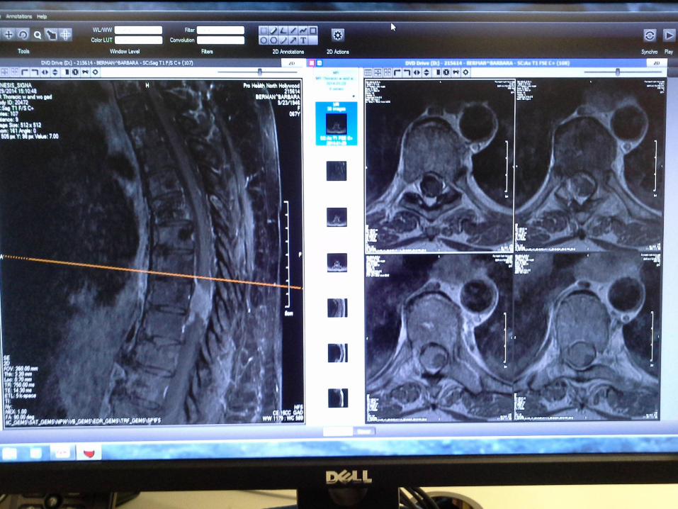

Vestibular Schwannomas - Treatment
ndash Approaches
bull Suboccipital
ndash Preferred route
ndash Best for preservation of hearing
bull Middle fossa
ndash Reserved for small laterally located tumor
bull Translabyrinthine
ndash Tumor with intracanalicular component
ndash When hearing is non-functional
Vestibular Schwannomas ndash Post-op Care
bull CN and brainstem dysfunction
ndash CN VII
bull Impaired eye closure
ndash Natural tears PRN
ndash CN VIII
bull Vestibular dysfunction
ndash NV
Antiemetic
ndash Balance difficulties
Safety
ndash CN IX X and XII
bull Swallowing difficulty
bull CSF fistula ndash May develop through the
bull skin incision
bull ear (ruptured TM)
bull eustachian tube through the nose (rhinorrea) or back of throat
ndash Risk for meningitis
ndash 25-35 resolves spontaneously
ndash Treatment ndash Elevate HOB
ndash Lumbar drain
ndash Surgical CSF shunting
Pituitary Adenoma ndash Surgery
bull Transphenoidal (procedure of choice)
ndash Post op complications bull Hormonal imbalance
ndash darr ADH ndash Diabetes
Insipidus
Tx DDAVP SQ
ndash darr Cortisol
ndash darr TSH
bull Infection
bull CSF leak
bull Nasal septal perforation
bull Damage to structures in cavernous sinus
bull Ray M Chu MD
bull Neurosurgical Institute
bull 310-423-7900
bull raychucshsorg

bull Ray M Chu MD
bull Neurosurgical Institute
bull 310-423-7900
bull raychucshsorg


