memory systems Where do you know what you know? The ...
13
Semantic memory (also called conceptual knowl- edge) is the aspect of human memory that cor- responds to general knowledge of objects, word meanings, facts and people, without connection to any particular time or place 1 . Knowing that you ate Szechuan scallops at the Peking Restaurant in Cambridge last Thursday evening is episodic, not semantic, memory. Knowing that Szechuan refers to a province of China, that food from this region tends to be spicy and that scallops are sea-creatures that live in brittle bivalve shells are all forms of conceptual knowl- edge. Memory for episodic events is not only specific to times and places, it is also largely specific to an individual. Conceptual knowledge, on the other hand, is mostly shared across individuals in a given culture, although its precise scope depends on the individual’s experience. Only a few decades ago, one respectable position held that semantic memory might arise from ‘uni- versal connectivity’ in the brain, and hence have no corresponding stable neural architecture 2 . Because we now know that brain lesions can produce selec- tive disruption of semantic memory, this hypothesis is probably no longer tenable; however, localizing the stable architecture of conceptual knowledge has proved difficult. Indeed, one researcher concluded that “The search for the neuroanatomical locus of semantic memory has simultaneously led us nowhere and everywhere...” (REF. 3) It is perhaps time for an assessment of what we do and do not know about the representation of conceptual knowledge in the human brain. A semantic hub? This Review focuses on one specific issue. Essentially all current theoretical positions about semantic memory share the view that much of the content of our semantic memory relates to perception and action, and is repre- sented in brain regions that overlap with, or possibly even correspond to, the regions that are responsible for perceiving and acting 4–6 . This view about the neural representation of how objects look, sound, move and so on therefore entails commitment to the idea that conceptual knowledge is a widely distributed neural network. Our knowledge of the scallop, for example, includes attributes such as its visual features, which will be represented in or near the brain regions that analyse visual form and colour; its manner of moving on the seabed, represented in or near the brain regions that respond to the perception of this kind of movement; its texture and taste, involving tactile and gustatory regions; the actions its edibility affords, such as cutting with a knife and fork or chewing, supported by frontal and parietal areas; its name or other descriptions that we could apply to it, represented in perisylvian language regions; and so on. The debate centres on the following question: are these distributed brain regions, along with the connections between them, the entire neural basis of semantic memory? Many theories, forming a class that we refer to as the distributed-only view (FIG.1a) offer a positive answer to this question. An alternative position, for which we argue here, is that the sensory-, motor- and language- specific aspects of conceptual knowledge are necessary but not sufficient: this is the distributed-plus-hub view *MRC Cognition and Brain Sciences Unit, 15 Chaucer Road, Cambridge, CB2 7EF, UK. ‡ Neurology Unit, Cambridge University, Cambridge, UK. § Department of Psychology, 1202 West Johnson Street, University of Wisconsin- Madison, Madison, Wisconsin, USA. Correspondence to: K.P. and T.T.R. e-mails: karalyn.patterson@mrc-cbu. cam.ac.uk; [email protected] doi:10.1038/nrn2277 Where do you know what you know? The representation of semantic knowledge in the human brain Karalyn Patterson*, Peter J. Nestor ‡ and Timothy T. Rogers § Abstract | Mr M, a patient with semantic dementia — a neurodegenerative disease that is characterized by the gradual deterioration of semantic memory — was being driven through the countryside to visit a friend and was able to remind his wife where to turn along the not- recently-travelled route. Then, pointing at the sheep in the field, he asked her “What are those things?” Prior to the onset of symptoms in his late 40s, this man had normal semantic memory. What has gone wrong in his brain to produce this dramatic and selective erosion of conceptual knowledge? MEMORY SYSTEMS REVIEWS 976 | DECEMBER 2007 | VOLUME 8 www.nature.com/reviews/neuro © 2007 Nature Publishing Group
Transcript of memory systems Where do you know what you know? The ...

Semantic memory (also called conceptual knowl-edge) is the aspect of human memory that cor-responds to general knowledge of objects, word meanings, facts and people, without connection to any particular time or place1. Knowing that you ate Szechuan scallops at the Peking Restaurant in Cambridge last Thursday evening is episodic, not semantic, memory. Knowing that Szechuan refers to a province of China, that food from this region tends to be spicy and that scallops are sea-creatures that live in brittle bivalve shells are all forms of conceptual knowl-edge. Memory for episodic events is not only specific to times and places, it is also largely specific to an individual. Conceptual knowledge, on the other hand, is mostly shared across individuals in a given culture, although its precise scope depends on the individual’s experience.
Only a few decades ago, one respectable position held that semantic memory might arise from ‘uni-versal connectivity’ in the brain, and hence have no corresponding stable neural architecture2. Because we now know that brain lesions can produce selec-tive disruption of semantic memory, this hypothesis is probably no longer tenable; however, localizing the stable architecture of conceptual knowledge has proved difficult. Indeed, one researcher concluded that “The search for the neuroanatomical locus of semantic memory has simultaneously led us nowhere and everywhere...” (Ref. 3) It is perhaps time for an assessment of what we do and do not know about the representation of conceptual knowledge in the human brain.
A semantic hub?This Review focuses on one specific issue. Essentially all current theoretical positions about semantic memory share the view that much of the content of our semantic memory relates to perception and action, and is repre-sented in brain regions that overlap with, or possibly even correspond to, the regions that are responsible for perceiving and acting4–6. This view about the neural representation of how objects look, sound, move and so on therefore entails commitment to the idea that conceptual knowledge is a widely distributed neural network. Our knowledge of the scallop, for example, includes attributes such as its visual features, which will be represented in or near the brain regions that analyse visual form and colour; its manner of moving on the seabed, represented in or near the brain regions that respond to the perception of this kind of movement; its texture and taste, involving tactile and gustatory regions; the actions its edibility affords, such as cutting with a knife and fork or chewing, supported by frontal and parietal areas; its name or other descriptions that we could apply to it, represented in perisylvian language regions; and so on. The debate centres on the following question: are these distributed brain regions, along with the connections between them, the entire neural basis of semantic memory?
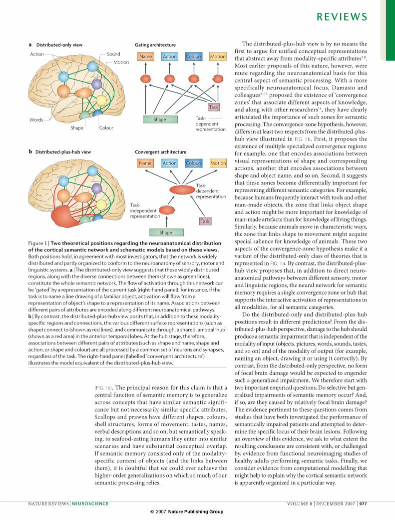
Many theories, forming a class that we refer to as the distributed-only view (fIG.1a) offer a positive answer to this question. An alternative position, for which we argue here, is that the sensory-, motor- and language-specific aspects of conceptual knowledge are necessary but not sufficient: this is the distributed-plus-hub view
*MRC Cognition and Brain Sciences Unit, 15 Chaucer Road, Cambridge, CB2 7EF, UK. ‡Neurology Unit, Cambridge University, Cambridge, UK. §Department of Psychology, 1202 West Johnson Street, University of Wisconsin-Madison, Madison, Wisconsin, USA.Correspondence to: K.P. and T.T.R. e-mails: [email protected]; [email protected]:10.1038/nrn2277
Where do you know what you know? The representation of semantic knowledge in the human brainKaralyn Patterson*, Peter J. Nestor‡ and Timothy T. Rogers§
Abstract | Mr M, a patient with semantic dementia — a neurodegenerative disease that is characterized by the gradual deterioration of semantic memory — was being driven through the countryside to visit a friend and was able to remind his wife where to turn along the not-recently-travelled route. Then, pointing at the sheep in the field, he asked her “What are those things?” Prior to the onset of symptoms in his late 40s, this man had normal semantic memory. What has gone wrong in his brain to produce this dramatic and selective erosion of conceptual knowledge?
m e m o ry s y s t e m s
R E V I E W S
976 | dECEMBER 2007 | vOluME 8 www.nature.com/reviews/neuro
© 2007 Nature Publishing Group
Nature Reviews | Neuroscience
Distributed-only view
Distributed-plus-hub view Convergent architecture
Action Colours MotionName
Action Colours MotionName
Gating architecture
Shape
Shape
Task
Task
Task- dependent representation
Task- independent representation
Action
Words
Sound
Colour Shape
Motion
a
b
Task- dependent representation
(fIG.1b). The principal reason for this claim is that a central function of semantic memory is to generalize across concepts that have similar semantic signifi-cance but not necessarily similar specific attributes. Scallops and prawns have different shapes, colours, shell structures, forms of movement, tastes, names, verbal descriptions and so on, but semantically speak-ing, to seafood-eating humans they enter into similar scenarios and have substantial conceptual overlap. If semantic memory consisted only of the modality- specific content of objects (and the links between them), it is doubtful that we could ever achieve the higher-order generalizations on which so much of our semantic processing relies.
The distributed-plus-hub view is by no means the first to argue for unified conceptual representations that abstract away from modality-specific attributes7,8. Most earlier proposals of this nature, however, were mute regarding the neuroanatomical basis for this central aspect of semantic processing. With a more specifically neuroanatomical focus, damasio and colleagues9–13 proposed the existence of ‘convergence zones’ that associate different aspects of knowledge, and along with other researchers14, they have clearly articulated the importance of such zones for semantic processing. The convergence-zone hypothesis, however, differs in at least two respects from the distributed-plus-hub view illustrated in fIG. 1b. First, it proposes the existence of multiple specialized convergence regions: for example, one that encodes associations between visual representations of shape and corresponding actions, another that encodes associations between shape and object name, and so on. Second, it suggests that these zones become differentially important for representing different semantic categories. For example, because humans frequently interact with tools and other man-made objects, the zone that links object shape and action might be more important for knowledge of man-made artefacts than for knowledge of living things. Similarly, because animals move in characteristic ways, the zone that links shape to movement might acquire special salience for knowledge of animals. These two aspects of the convergence-zone hypothesis make it a variant of the distributed-only class of theories that is represented in fIG. 1a. By contrast, the distributed-plus-hub view proposes that, in addition to direct neuro-anatomical pathways between different sensory, motor and linguistic regions, the neural network for semantic memory requires a single convergence zone or hub that supports the interactive activation of representations in all modalities, for all semantic categories.
do the distributed-only and distributed-plus-hub positions result in different predictions? From the dis-tributed-plus-hub perspective, damage to the hub should produce a semantic impairment that is independent of the modality of input (objects, pictures, words, sounds, tastes, and so on) and of the modality of output (for example, naming an object, drawing it or using it correctly). By contrast, from the distributed-only perspective, no form of focal brain damage would be expected to engender such a generalized impairment. We therefore start with two important empirical questions. do selective but gen-eralized impairments of semantic memory occur? And, if so, are they caused by relatively focal brain damage? The evidence pertinent to these questions comes from studies that have both investigated the performance of semantically impaired patients and attempted to deter-mine the specific locus of their brain lesions. Following an overview of this evidence, we ask to what extent the resulting conclusions are consistent with, or challenged by, evidence from functional neuroimaging studies of healthy adults performing semantic tasks. Finally, we consider evidence from computational modelling that might help to explain why the cortical semantic network is apparently organized in a particular way.
Figure 1 | Two theoretical positions regarding the neuroanatomical distribution of the cortical semantic network and schematic models based on these views. Both positions hold, in agreement with most investigators, that the network is widely distributed and partly organized to conform to the neuroanatomy of sensory, motor and linguistic systems. a | The distributed-only view suggests that these widely distributed regions, along with the diverse connections between them (shown as green lines), constitute the whole semantic network. The flow of activation through this network can be ‘gated’ by a representation of the current task (right-hand panel): for instance, if the task is to name a line drawing of a familiar object, activation will flow from a representation of object’s shape to a representation of its name. Associations between different pairs of attributes are encoded along different neuroanatomical pathways. b | By contrast, the distributed-plus-hub view posits that, in addition to these modality-specific regions and connections, the various different surface representations (such as shape) connect to (shown as red lines), and communicate through, a shared, amodal ‘hub’ (shown as a red area) in the anterior temporal lobes. At the hub stage, therefore, associations between different pairs of attributes (such as shape and name, shape and action, or shape and colour) are all processed by a common set of neurons and synapses, regardless of the task. The right-hand panel (labelled ‘convergent architecture’) illustrates the model equivalent of the distributed-plus-hub view.
R E V I E W S
nATuRE REvIEWS | neuroscience vOluME 8 | dECEMBER 2007 | 977
© 2007 Nature Publishing Group

Expressive vocabularyThe set of words that an individual knows and can retrieve for referring to objects and other concepts in speech or writing.
Receptive vocabularyThe set of words that an individual can comprehend when hearing or reading them.
Acquired disorders of semantic memoryImpaired conceptual knowledge is associated with four principal neurological aetiologies: stroke; viral infec-tion, most commonly herpes simplex virus encephalitis (HSvE); and two forms of neurodegenerative disease: Alzheimer’s disease (Ad) and the semantic variant of fronto-temporal dementia, typically called semantic dementia (Sd). All four conditions give useful clues about the nature and organization of conceptual knowledge, but from different angles.
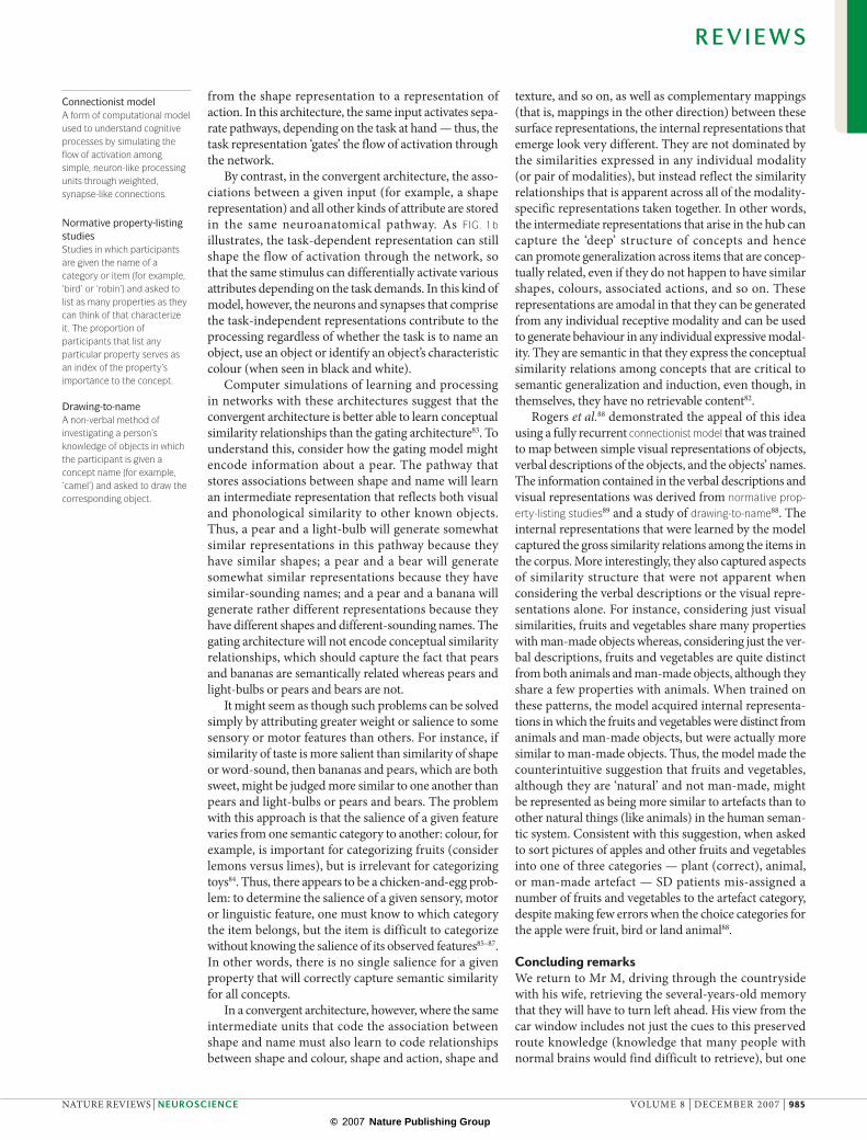
Semantic dementia. Sd is one of the neurodegenerative conditions that belongs to the Fronto-Temporal dementia spectrum15. Recent post-mortem analyses indicate that the typical neuropathology in Sd is abnormal neuronal inclusions of the protein ubiquitin16, a form of pathol-ogy that is best known in motor neuron disease. Sd is marked behaviourally by a progressive deterioration of expressive and receptive vocabulary and of knowledge about the properties of everyday objects, in the context of otherwise well-preserved cognition and memory for recent events (BOX 1). It was first reported in the modern era by Warrington17, Schwartz et al.18 and Snowden et al.19. As indicated in BOX 1, research on Sd provides clear and positive answers to the two questions posed above. That is, Sd represents a selective impairment to seman-tic abilities that affects all modalities of reception and expression, for all kinds of concepts, more or less equally (fIG. 2), and it is the consequence of relatively focal brain lesions. degeneration of the anterior temporal lobes
(ATl) is invariably evident by the time that Sd patients become symptomatic. Bilateral degeneration is the norm, although it is usually asymmetrical: in approximately two-thirds of cases the atrophy is greater on the left side than on the right. The typical pattern seen on structural MRI scans is one of well-defined atrophy of both the left and the right ATl, which is maximal at the temporal pole and on the adjacent rostral-inferior surface.
Alzheimer’s disease. The most prominent cognitive deficit in typical Ad is an impairment of episodic (autobiographical) memory: in particular, the ability to learn new information is progressively abolished. The original hypothesis, which attributed this phenomenon specifically to degeneration of the hippocampus20, is now viewed not as wrong but as incomplete: functional brain imaging, even in patients with mild or early Ad, reveals hypometabolism not only in the bilateral medial temporal lobes, but also in the thalamus, the posterior cingulate gyrus and other parts of the limbic system, which apparently constitute a network that is crucial for the formation of new memories21. Semantic memory is frequently affected in Ad22,23, but typically at a later stage and to a more modest extent than episodic memory. There are a few reported cases in which loss of concep-tual knowledge was an early and significant symptom of Ad. Where detailed neuroanatomical information was available in these cases, however, the pathology sometimes had a distribution that was atypical for Ad and was in fact more similar to that seen in Sd24. It is the contrast between the prototypical distributions of degeneration in these two diseases that is most germane to the current Review, because the relatively widespread degeneration that is observed in Ad apparently leads to a less consistent and usually less profound disruption of semantic memory than is observed in Sd, where the pathology is relatively circumscribed within anterior temporal regions.
Herpes simplex virus encephalitis. The most prominent cognitive consequences of typical HSvE are amnesia for earlier episodes in one’s life and profound difficulty in learning new information. The impact on episodic mem-ory can be attributed to the fact that this viral infection has a predilection for medial temporal lobe structures, including the hippocampus. Some patients with HSvE have no significant disruption to their conceptual knowl-edge, but have other cognitive deficits, most notably in the executive and control abilities that are associated with the frontal lobe. When semantic memory is affected, the deficit is often mild relative to the profound semantic def-icits that are observed in Sd. As in Sd, however, semantic impairment in HSvE is associated with bilateral ATl lesions25,26. Interestingly, the semantic deficit in HSvE is often category-specific, with relatively well-preserved knowledge of man-made things but impaired knowledge of living things. There is considerable debate in the lit-erature as to whether these two domains of conceptual knowledge are represented separately in the brain27, or whether this dissociation can be explained by some fun-damental difference in the nature of the attributes that are
Box 1 | the syndrome of semantic dementia
One of the most prominent symptoms of semantic dementia (SD) is anomia91: failure to name objects, concepts and people, whether in response to stimulus presentation or in spontaneous speech. Patients with other causes of anomia, and occasionally even healthy people, sometimes fail to name something because, although they know what it is, they cannot find the word for it at that moment. Anomia in SD does not result from this kind of word-finding difficulty but instead reflects degraded knowledge of the object or concept. When we asked one of our patients to name a picture of a zebra, she replied: “It’s a horse, ain’t it?” Then, pointing to the stripes, she added, “But what are these funny things for?”
For SD patients, the degree of success in naming (and, indeed, in any measure of conceptual knowledge) is largely independent of the modalities of input and output. Naming is most commonly assessed by presenting pictures and asking for a verbal naming response; however, SD patients are just as unsuccessful, indeed usually more so, if the stimulus is a description of the item to be named (for example, “What do we call the African animal with black and white stripes?”), or is the item’s characteristic sound (for example, a telephone ringing or a dog barking), or if the patient is asked to write the name rather than say it.
The degree of success or failure in any semantic test in SD patients is determined by four principal factors72,92: the severity or stage of progression of the disease; the familiarity of the object (the deficit is always more pronounced for less familiar things, words and concepts); the object’s typicality (there are fewer successes for things that are atypical of their kind); and the specificity of information required by the task (for example, a patient might be able to identify an apple as something to eat without being able to identify it as an apple).
Most cognitive functions apart from semantic memory are reasonably well-preserved in patients with SD, at least until late in the disease. Thus — provided that the tests do not refer to or require access to semantic knowledge — SD patients have reasonably normal capabilities in general intelligence, problem solving, visuo-spatial function, short-term memory, episodic memory, simple calculation skills, and so on.
R E V I E W S
978 | dECEMBER 2007 | vOluME 8 www.nature.com/reviews/neuro
© 2007 Nature Publishing Group

Nature Reviews | Neuroscience
ModelTask one stimulus: Which is coloured correctly?
Task two stimulus: Which is the real animal?
Delayed copy
d Delayed-copy drawingc Colour and object recognition
Target-typical Target-unusual
Severe SDMild SDControls0.00
0.50
0.25
0.75
1.00
Severe SDMild SDControls0.00
0.50
0.25
0.75
1.00
Controls Mild SD Moderate SD Severe SD0
1
2
3
a Picture categorization b Picture naming
Acc
urac
y (d
′)
Item Sept 1991 Sept 1992Mar 1992 Mar 1993
Bird
Bird
Bird
Bird
Bird
Bird
Bird
Bird
Bird
Bird
Bird
Bird
Cat
Cat
Cat
Vehicle
Part of animal
Dog
Horse
Dog
Chicken
Chicken Chicken
Duck
Duck
Duck
Duck
Swan
Swan
Eagle
Ostrich
Peacock
Penguin
Rooster
+
+
+ +
+
+
+
Animal
Animal
Animal
Animal
Prop
ortio
n co
rrec
tPr
opor
tion
corr
ect
GeneralBasicSpecific
Task one performance
Task two performance
Figure 2 | examples of impaired performance on semantic tasks in patients with semantic dementia. This figure illustrates the cross-modal nature of the impairment and the preservation of general relative to specific information in semantic dementia (SD). a | Accuracy at discriminating targets from distractors in a picture-categorization task for four groups of participants: healthy controls and patients with mild, moderate and severe SD72. Participants viewed a category label followed by a colour photograph and were asked whether the picture matched the label. Labels were either general (for example, ‘animal’), basic-level (for example, ‘dog’) or specific (for example, ‘Labrador’). b | Picture-naming responses for one SD patient who was assessed longitudinally68. + denotes a correct response. c | Examples of stimuli and performance (for controls, mild and severe SD patients) on two recognition tasks. In the first task, participants judged which of two items was coloured correctly. Patients with milder SD performed well when the targets had a category-typical colour (for example, the green celery) but poorly when the items had an unusual colour (for example, the orange pumpkin). The judgements of patients with more severe SD were no better than chance (50%) in either condition80. In the second task, participants judged which of two drawings depicted a real animal. Here, both the patients with mild SD and the patients with severe SD achieved normal levels of success for targets with relatively prototypical features (for example, the monkey which, like most animals, has small ears). For stimulus pairs in which the correct choice had unusual features (for example, the elephant, which has very large ears), the patients with mild SD were impaired and the patients with severe SD scored at chance levels70. d | Delayed-copy drawings produced by SD patients71. The patients were shown a model picture which was then removed and, after a 10-second delay, they were asked to reproduce this picture from memory. Properties that are common to most animals, such as eyes and a tail, were preserved in the delayed drawings. Unusual properties that distinguish one animal from others — for example, the hump on the camel and the flippers on the seal — were frequently omitted. Some common properties were also incorrectly added to animals that lack them (for example, the four legs on the delayed drawing of the duck and the tail on the delayed drawing of the frog). Real animal pictures in part c reproduced, with permission, from Ref. 97 (1980) American Psychological Association.
R E V I E W S
nATuRE REvIEWS | neuroscience vOluME 8 | dECEMBER 2007 | 979
© 2007 Nature Publishing Group

AphasiaImpaired language abilities resulting from brain disease or injury.
Cross-modalThe term that is applied to representations or processes that operate across different kinds of sensory, motor and linguistic representations. for instance, representations and processes that receive input from and/or direct output to both visual and auditory representations would be considered cross-modal.
AnomiaThe failure to name objects, concepts and people, whether in response to stimulus presentation or in spontaneous speech.
Volumetric MRIA method that uses finely cut brain slices (usually less than 2 mm thick) to measure the volume of brain structures.
Voxel-by-voxel analysisA method of whole-brain image analysis in which the brain scans of different individuals are fitted to a standard template (to minimize inter-individual differences in brain shape) so that brain regions can be compared systematically across subjects.
Lesion-overlap studyA method that seeks to define a common area of brain damage relevant to a given behavioural deficit by overlaying the scan-defined lesions of multiple subjects with the behavioural deficit in question.
PET activation paradigmAn experimental paradigm that uses PeT to measure changes in cerebral perfusion in response to a stimulus.
characteristic of man-made and living things (for exam-ple, living things might be more heavily weighted towards sensory features and man-made artefacts might be more heavily weighted toward functional attributes)28,29. Once again, the contrast between HSvE and Sd in terms of both the severity of the semantic deficit (which is often either absent or mild in patients with HSvE, as opposed to progressive and ultimately profound in Sd) and its pattern (which is frequently category-specific in HSvE, but very rarely so in Sd) seems most telling in the current context. That is, because both diseases implicate the bilat-eral ATl in semantic processing, it must be the specific nature and/or distribution of the brain abnormalities in Sd that produces the pervasive disruption — across all categories and all modalities — of conceptual knowledge that defines this condition.
Stroke. The most prominent impairment in stroke patients with extensive lesions in the left hemisphere is aphasia, but poor performance on non-verbal semantic tests as well as in verbal comprehension can result from left-hemisphere stroke, especially in a condition called transcortical sensory aphasia (TSA). At least in this cross-modal regard, TSA might resemble Sd; however, there are important differences between the two patients groups. With reference to criteria designed to distinguish impairments of representation from problems in access or retrieval30, some recent work suggests that the seman-tic deficit in TSA may be better described as an impaired ability to retrieve, select and manipulate semantic infor-mation in a task-appropriate fashion31, rather than the degradation of semantic representations themselves which is characteristic of Sd. For example, the anomia that occurs in TSA readily benefits from cueing (for a patient struggling to name a picture of a violin, the cue might be, “It begins with a ‘v’”)32, whereas anomia in Sd is largely unaided by cueing32,33. There is essentially no overlap between the brain regions that are damaged in these two patient populations. Owing to the anatomy of the vascular system, stroke rarely, if ever, produces focal lesions in the ATl, and semantic deficits in TSA typically result from damage to either frontal or parietal regions (or both) that is restricted to the left hemisphere34. In accordance with this view regarding the nature of the semantic impairment in TSA, there is substantial evi-dence from functional imaging of normal individuals that activation of prefrontal cortical areas is associated with selection or control processes35,36.
Summary of semantic disorders. The range of aetiologies associated with impaired performance on semantic tasks reveals important similarities and differences. The cogni-tive and neuroanatomical abnormalities in Sd, especially when contrasted with those from the other aetiologies reviewed above, provide trenchant evidence for the distributed-plus-hub view (fIG. 1b). The strength of this evidence hinges, however, on claims about the relatively focal nature of the pathology in Sd. The remaining issue to be considered in this section is whether the selective conceptual impairment in Sd can be securely attributed to circumscribed lesions of the ATl.
The suggestion that the cognitive syndrome in Sd might result in part from structural and/or functional abnormalities elsewhere in the semantic network5 is challenged by recent findings with (18F)fluoro-2-deoxy-d-glucose positron emission tomography (FdG-PET). Three studies37–39 have confirmed bilateral anterior temporal dysfunction in Sd, but no other area of hypometabolism was consistently observed: one reported more extensive hypometabolism along the length of the inferior left temporal lobe37, the second observed additional hypometabolism in the left insula and orbito-frontal areas38 and the third revealed no sig-nificant reduction in metabolism in any region except the rostral temporal lobes39. In agreement with these studies of resting metabolism, volumetric structural imaging40 indicates relative preservation of the posterior temporal lobe in Sd38,39,41–43. In the dorsal-ventral plane, the supe-rior temporal gyrus is atrophic but also preserved rela-tive to the inferior temporal gyrus40. In other words, both structural and metabolic imaging studies of patients with Sd suggest that the abnormalities are most pronounced in the anterior and inferior parts of the temporal lobes.
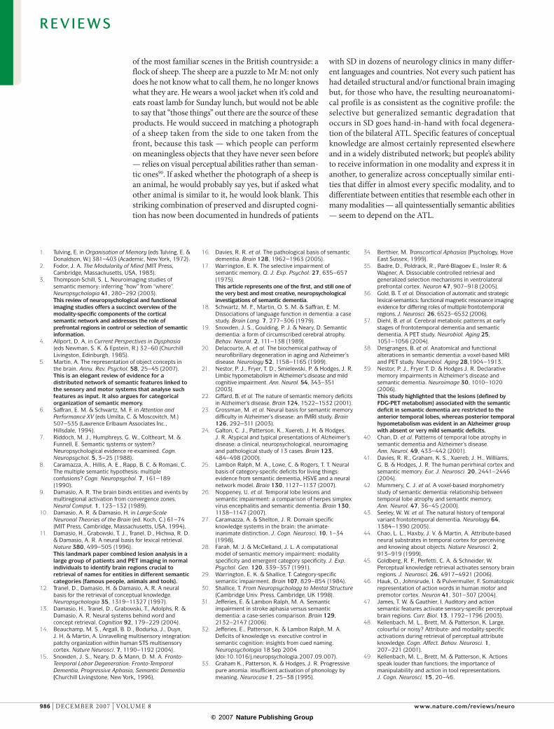
Of course, it is possible that the pathology is more widely distributed in Sd, but that such further abnor-malities are so subtle or variable across patients that they do not meet the standard for statistical significance in voxel-by-voxel analyses. A recent comparison of brain hypometabolism in patients with Ad versus patients with Sd suggests that this is not the case. nestor and colleagues39 demonstrated that hypometabolism is much more widespread in Ad than in Sd and, in particular, that it affects left frontal, occipito-temporal and tem-poro-parietal regions in Ad — areas that have been implicated in the distributed cortical semantic network and which appear relatively normal in Sd. Yet semantic impairment was much milder in Ad patients than in Sd patients, all of whom showed hypometabolism in the ATl (fIG. 3).
Other lesion studies also support the view that the ATl is important for semantic representation and/or processing. Perhaps the best known is a lesion-overlap study that tested picture naming in a large group of anomic patients with unilateral, stable, focal brain lesions11. The stimuli included pictures of famous peo-ple — who must, of course, be named at a specific level (for example, ‘Princess diana’ rather than ‘princess’ or ‘woman’) — and pictures of animals and tools, which are usually named at the basic level (for example, ‘elephant’ or ‘hammer’), which does not distinguish individual category members from one another. Impaired naming of famous faces was associated with the ‘tightest’ lesion overlap, centred on the left temporal pole, whereas impaired animal naming correlated with lesions in the anterior, inferior left temporal lobe. Poor tool-naming revealed the lowest degree of lesion overlap and was associated with damage in the posterior, lateral left tem-poral lobe as well as in the temporo–occipito–parietal junction. lesion–symptom correlation thus implicated the left ATl in naming for two of the three categories tested. When healthy participants named the same items in a PeT activation paradigm, all three stimulus
R E V I E W S
980 | dECEMBER 2007 | vOluME 8 www.nature.com/reviews/neuro
© 2007 Nature Publishing Group

Nature Reviews | Neuroscience
PPTp PPTw Naming WPMCategory fluency0
10
20
30
40
50
60
70
SD AD Control
0.0
1.0
0.75
0.5
0.25
Semantic dementia Alzheimer’s disease
Scor
e
Prop
ortio
n co
rrec
t
a
b
Magnetoencephalography(MeG). A method of measuring physiological activity across the cortex by detecting pertubations in the magnetic field that is generated by the electrical activity of neuronal populations.
types yielded significant blood-flow increases (relative to a control condition) in the left temporal pole, with further activation of the right temporal pole during face-naming and similar but discernibly different acti-vation patterns in the posterior left temporal regions for animal- versus tool-naming11.
Taken together, the neuropsychological literature suggests that the selective but amodal semantic impair-ment in patients with Sd is attributable to relatively focal pathology in the ATl and not to widespread damage in the cortical semantic network. Such data strongly support the distributed-plus-hub view shown in fIG. 1b, and further suggest that the ‘hub’ part of the semantic network is located in the bilateral ATl (BOX 2).
evidence from functional neuroimagingThis conclusion from neuropsychological evidence might seem surprising to researchers who are famil-iar with functional brain-imaging studies of semantic abilities in healthy individuals. Although there have been more than 40 published reports of ATl activation by a wide variety of semantic tasks (fIG. 4), these reports constitute a relatively small proportion of the functional imaging studies of semantic memory in the literature of the past decade. According to both primary sources44–49
and review articles3,5,50,51, imaging studies more often implicate some combination of frontal, posterior tempo-ral, temporo-parietal and parietal regions in the cortical semantic network. In this section we consider whether these results are genuinely at odds with the neuro-psychological data discussed above, and suggest two ways of reconciling the apparently conflicting sources of evidence.
The anterior temporal lobe is ‘shy’ to functional MRI. In standard acquisition protocols for functional MRI (fMRI), which has largely replaced H2
15O-PET for func-tional neuroimaging purposes, the signal-to-noise ratio diminishes substantially near the temporal poles, owing to their proximity to air-filled sinuses (the so-called ‘susceptibility artefact’). One study compared the func-tional activation revealed by PET and by fMRI (using a standard whole-brain acquisition sequence) in a group of people performing a semantic categorization task52. Although both methods showed substantial semantic-related activity in ventral posterior temporal lobes, only PET revealed that this activation extended all the way rostrally to the temporal pole. Other PET evidence has demonstrated significant ATl activation during the semantic tasks of category fluency53, object naming54, category verification55 and word recognition56. Perhaps most saliently — because what could more clearly exem-plify the essence of semantic processing? — the tasks of comprehending connected speech versus various forms of distorted (and hence meaningless) speech, and of reading coherent text versus a meaningless visual control condition, seem to engage the left temporal pole when measured by both PET57,58 and fMRI59.
Other functional imaging methods similarly impli-cate the ATl in semantic processing. For instance, an elegant experiment was performed with anatomically constrained magnetoencephalography (MEG) in which subjects made semantic judgements about spoken or written words60. As expected, the brain’s initial responses to words were detected in the appropriate sensory areas for each modality; however, from approximately 400 ms after stimulus onset, the MEG responses for both modalities converged on the ATl.
Specificity of semantic processing. In the PET component of the object-naming study cited above11, the strong-est ATl activation was observed when participants were required to recognize and name famous people. A subsequent study from the same group61 proposed that the temporal pole might have a special role in the recognition and identification of unique concepts
Figure 3 | Differences between semantic dementia and Alzheimer’s disease in measures of brain function and semantic memory. a | The areas of reduced metabolism (shown as graded grey areas), are widespread in patients with Alzheimer’s disease (AD) and include some regions that are implicated in the cortical semantic network (see fIG. 1). In the AD cases shown, however, there was little evidence of any abnormality in anterior temporal regions, which show substantial and focal hypometabolism in patients with semantic dementia (SD). b | The performance of AD and SD patients, as well as of a group of age-matched healthy controls, on a range of semantic tasks. The SD patients were significantly more impaired than the AD patients on all tasks, even though the brain abnormalities in the AD patients were more widespread. In category-fluency tasks, participants are given 1 minute to list as many examples of a semantic category as they can. PPTp and PPTw indicate the picture and word versions, respectively, of the Pyramids and Palm Trees test of semantic association98. Naming indicates performance on a simple picture-naming task and WPM indicates performance on a 10-alternative, forced-choice word-to-picture matching task. Parts a and b modified, with permission, from Ref. 39 (2006) Academic Press.
R E V I E W S
nATuRE REvIEWS | neuroscience vOluME 8 | dECEMBER 2007 | 981
© 2007 Nature Publishing Group

Voxel-based morphometryA voxel-by-voxel analysis of structural-image data, most commonly the grey-matter segments extracted from T1-weighted MRI.
Conjunction analysisA statistical method used in functional brain imaging research for identifying brain regions that are significantly activated in two or more separate experimental conditions.
— individual people and famous buildings being two prominent examples. A number of functional imaging studies, using both PET and fMRI, have since supported this idea, showing ATl activation related to the recogni-tion of familiar versus unfamiliar buildings62, faces62,63, names64,65 and even voices66.
Semantic dementia patients are profoundly impaired at recognizing famous individuals from photographs, names and verbal descriptions67; this impairment, however, appears to be one notable manifestation of a more general sensitivity to the specificity with which an item must be recognized or categorized. Across a wide array of semantic tasks (fIG. 2), the patients might perform well as long as accurate performance requires only a relatively coarse or general categorization of the stimulus. Thus, severely impaired Sd patients can sometimes call a picture an ‘animal’, without being able to name it ‘chicken’ or even ‘bird’68; they can accurately sort pictures or words into categories such as ‘animal’ versus ‘man-made object’, but not ‘car’ versus ‘boat’69; they will correctly judge — when offered two pictures of a donkey, one with and one without a hump on its back — that ‘the real one’ is the humpless exemplar, but will make the same humpless (and therefore more ‘ani-mal prototypical’, but incorrect) choice for the camel, to which humps are specific70; they will copy-draw a line drawing of a real humped camel a mere 10–15 seconds after studying it, but omit the hump71; and so on. This pattern does not arise simply because tasks that require precise classification are more difficult. For example, Rogers and Patterson72 showed that, whereas healthy controls are faster and more accurate at classifying items at the basic level (for example, ‘dog’) relative to a more general level (for example, ‘animal’), patients with Sd show the reverse pattern: they have greater impairment on the more precise basic-level classifica-tion. Such findings suggest that semantic tasks that require the distinctive classification of a stimulus place particularly strong demands on the ATl regions that are affected in Sd.
Functional imaging studies may likewise indicate that the ATl is most strongly recruited not just by rec-ognition of unique items (such as the Eiffel Tower or Princess diana), but by any form of specific semantic processing. One of several PET studies that support this view used a task in which normal participants were asked to verify, by answering yes or no, whether the stimulus shown in a colour photograph belonged to a particular category55. For example, in trials designed to receive ‘yes’ responses, participants judged on dif-ferent occasions whether the photo of a robin depicted an animal (superordinate level), a bird (basic level) or a robin (subordinate or most specific level). Relative to a control condition, all three semantic-judgement con-ditions activated the bilateral posterior fusiform gyrus and the occipito-temporal cortex, but the subordinate condition, when contrasted with both superordinate and basic levels, activated the ATl bilaterally. The ATl activation peaks aligned closely with the areas that showed the strongest grey-matter reduction in a voxel-based morphometry analysis of atrophy in Sd patients42. The ATl activation peaks for the subordi-nate condition also matched those from another PET study that contrasted the naming of unique items with the (basic-level) naming of common objects73. Similar results have been reported using fMRI74,75. A conjunc-tion analysis of results from four different PET studies of semantic processing, two with words as stimuli and two with pictures, highlighted activation of the inferior ATl; the authors concluded that this region supports a polymodal or amodal network of semantic representa-tions that is recruited when more specific conceptual information is required56 (BOX 3).
This brief Review suggests that activation in ATl regions during semantic tasks is not as scarce as an initial survey of the literature might suggest, especially if one takes into account the low signal-to-noise ratio that is achieved by standard fMRI methods in these regions and the fact that the amount of signal generated in ATl regions is related to the specificity of semantic process-
Box 2 | Why the anterior temporal lobe?
This Review summarizes evidence for the hypotheses that, first, semantic generalization requires a single amodal hub and, second, that the neuroanatomical site of this hub is the anterior temporal lobe (ATL). Assuming, for the moment, that these hypotheses are valid, why is the ATL a neuroanatomically sensible place for the semantic hub? One reason that is sometimes offered is the fact, known from non-human primate physiology93, that many primary sensory and motor areas, along with their related association cortices, connect to the ATL. This is true, but not a unique feature of the ATL: the ‘tri-state’ junction of the temporal, parietal and occipital lobes, including the angular gyrus, has the same feature of being well connected to input from multiple modalities, and indeed some researchers have ascribed a similar cross-modal mapping function to this area14,94. It is worth noting that cross-modal is not the same as a-modal: the region around the angular gyrus might serve to combine information from several modalities but still not have the genuinely amodal function of a semantic hub.
Two neuroanatomical facts about the ATL region might make it an appropriate candidate for extracting amodal conceptual information. First, it is neuroanatomically proximal to the amygdala and to other limbic structures, as well as to the orbito-frontal cortex — regions that are known to be important for the processing of emotion and reward95. Given that affective response to some extent pervades everything that we perceive, do and know, ATL regions might be well suited to computing associations between affect and more value-neutral sensory, motor and linguistic aspects of conceptual knowledge. Second, the ATL regions that seem so crucial to semantic memory are immediately adjacent to the anterior parts of the medial temporal lobe memory system — a system that is critical for rapid learning of new episodic information. As episodes must contribute to the gradual acquisition of new conceptual knowledge, it makes sense for episodic and semantic systems to be situated in close proximity.
R E V I E W S
982 | dECEMBER 2007 | vOluME 8 www.nature.com/reviews/neuro
© 2007 Nature Publishing Group

Unique recognition Basic-level naming Other basic-level tasks
6463
10754 54
10556 565223104
66
62
656465
62
55 55
110
112
104104
108109
103102
106113
11153
10653
Auditory
Generation task
Visual
Both
Other
Nature Reviews | Neuroscience
6465
62556366
64
65
62
55 54107
54
56 23
52
104
105
56
Speech comprehension Reading/word recognition Other semantic tasks
58
106
99
57
100
57
58
100
58
10657
100
99
57
58
100
104
109
103108
102104
106
112 113111
53
53110
106
Non
-ling
uist
ic
Ling
uist
ic
Both
Figure 4 | ATL activation in functional imaging studies of semantic processing. This figure shows the most anteriorly located temporal-lobe activation peaks (plotted in Talairach space) reported in 28 different studies of semantic-task performance in healthy individuals. More than 40 of such studies using a variety of imaging methods, have been published; this set includes those studies that used either positron emission tomography or functional MRI and reported coordinates of anterior temporal lobe (ATL) peaks in either Montreal Neurological Institute (MNI) or Talairach standardized spaces. Peaks originally reported in MNI coordinates were transformed to Talairach space using the transformation described by Brett and colleagues114. The studies varied in the modality of stimulus presentation, the nature of the particular task that was being performed, whether or not the stimuli were linguistic, and the specificity of the categorization that was required for successful task performance. Despite these variations in methodology, the different studies showed activation in remarkably similar regions of the ATL. The numbers indicate the studies from which the data were taken, as numbered in the reference list (see RefS 52–58,61–66,99–113). The shape and colour of the plotted areas indicate the nature of the stimulus that was used in the experiments, as shown in the legend. Where multiple points are plotted for the same study, these represent the most anteriorly located activation peaks in the temporal lobe from different contrast conditions in the same study. For instance, for a study that used both words and pictures as stimuli, and that reported separate anterior-temporal peaks corresponding to the two stimulus sets, both peaks are shown.
R E V I E W S
nATuRE REvIEWS | neuroscience vOluME 8 | dECEMBER 2007 | 983
© 2007 Nature Publishing Group

ing demanded by the task. Of course, a review of the functional imaging literature performed with a different ‘filter’ might reach a different conclusion, especially if it excluded PET studies and included only fMRI studies that lacked techniques designed to maximize the signal in the ‘hard-to-get’ rostral temporal lobes. Indeed, much of the evidence from such imaging studies supports the view that semantic processing involves mainly frontal or pos-terior-temporal loci, and of course our own position also implicates these regions in the larger semantic network.
The current Review is selective in that our primary aim was to determine whether the scarcity of reported ATl activation in the literature to date is sufficient to refute the conclusion drawn from lesion studies, which unambiguously point to ATl regions as being crucial for semantic processing. To paraphrase a recently-departed united States Secretary of defense, we do not believe that the seeming absence of evidence constitutes, in this case, evidence of absence.
Why does the semantic network need a hub?The above considerations support a view of human semantic cognition that raises at least two questions. The first question is why a single region of the brain should contribute to learned associations among widely distrib-uted sensory, motor and linguistic representations. For example, visual representations of objects are apparently coded in the posterior inferior/middle temporal cortex, and knowledge about action is probably supported by frontal and parietal regions76,77. Surely then, the knowl-edge that allows one to brush one’s hair with a hairbrush and one’s teeth with a toothbrush could be coded in direct associations between object-recognition regions in
the posterior-temporal cortex and motor plans in frontal and parietal areas, perhaps via the dorsal visual process-ing stream? Yet the ATl focus of damage in Sd, which is far removed from these sites and does not lie between them, seems to disrupt knowledge about characteristic object use78,79. Furthermore, damage in the same region impairs knowledge of the colours that are character-istic of certain object shapes (for example, grey for an elephant and orange for a carrot)80,81, even though shape and colour are two manifestly visual forms of informa-tion that are thought to be supported by neighbouring regions in the posterior ventral occipito-temporal cortex. Again, why are there not just direct connections between these two modality-specific regions? That is, why the need for a single hub?
The second question relates to the fact that was noted earlier regarding scallops and prawns: conceptual simi-larities between items are not necessarily apparent from their perceptual features. For instance, in one of the sim-ple tasks used in studies with healthy and semantically impaired individuals, called ‘category fluency’, a person might be asked to name as many different fruits as possi-ble in one minute. A typical sequence of responses might be “Apple, orange, banana, pear, grapes, lemon…” and so on. These six objects are very different from one another in colour, shape, texture, how they grow, how they are eaten, et cetera. Yet normal individuals can (whereas Sd patients definitely cannot) perform this task with ease, because knowledge of conceptual similarity allows these objects to be grouped as fruits, even though they have few sensory/motor properties in common. The second question is therefore: how does the semantic system acquire representations that capture such conceptual similarity relationships?
Evidence from computational modelling. The two ques-tions posed above might seem unrelated, but evidence from neural-network models suggests that they are not. Computer simulations with such models have shown that networks that adopt a convergent architecture — in which all forms of information about concepts are, at some point, processed through the same population of neurons and synapses — exhibit functional properties that explain how the semantic system is able to learn conceptual similarity relationships82. An example of such a convergent architecture is illustrated in the right-hand panel of fIG. 1b. Contrasting with this is what might be called a ‘gating’ architecture, illustrated in the right-hand panel of fIG. 1a.
In the gating architecture, the associations between different kinds of attributes are encoded in different neuroanatomical pathways: one pathway stores the asso-ciation between an item’s shape and its name, another stores the association between an item’s shape and the usual action associated with the item, and so on. A representation of the current task determines the path-way through which activation will flow. If the task is to name a line drawing of an object, then activation will flow from the shape representation to the name repre-sentation. If the task is to demonstrate how an object is used, then activation will flow along a different path,
Box 3 | sensitivity to specificity in the anterior temporal lobe
Both neuropsychological and functional imaging data suggest that anterior temporal lobe (ATL) regions are especially taxed by tasks that require very specific recognition or classification of a stimulus. What accounts for this sensitivity to specificity?
One possibility is that a specificity gradient exists in the temporal lobes, such that the greater the specificity with which an item must be recognized or categorized, the more rostral the activation in the temporal lobes will be96. The data in fIG. 4, however, challenge this hypothesis: the ATL regions that are engaged by recognition of unique entities (top left of fIG. 4) are no more anterior than those that are activated by tasks that require discrimination of (non-specific) basic-level categories (top middle and top right of fIG. 4) or other semantic tasks (bottom section of fIG. 4).
The distributed-plus-hub view offers a different explanation. It suggests that ATL regions encode the similarity relations among various concepts, so that semantically related items (for example, various different birds) are coded with similar patterns across ATL neurons. According to this model, naming a particular bird as a ‘robin’ requires the ATL hub to instantiate the robin representation almost exactly, as the name does not apply to other kinds of birds, many of which nevertheless have representations that are very similar to the robin. To name the same item ‘bird’, however, the robin pattern need not be instantiated exactly. Because the name applies to all birds and all birds share similar representations, it is only necessary for the hub to find a representation that is sufficiently ‘bird-like’ to activate the name. Thus, small distortions of the ‘robin’ representation — perhaps resulting from ATL atrophy — will prevent the network from retrieving the robin’s specific name (and other properties that differentiate it from other birds) without disrupting the retrieval of properties that are common to birds. A similar explanation extends to the interpretation of the functional imaging results, if one assumes that a stronger metabolic response in the ATL occurs in tasks that require the differentiation of highly overlapping representations.
R E V I E W S
984 | dECEMBER 2007 | vOluME 8 www.nature.com/reviews/neuro
© 2007 Nature Publishing Group
Connectionist modelA form of computational model used to understand cognitive processes by simulating the flow of activation among simple, neuron-like processing units through weighted, synapse-like connections.
Normative property-listing studiesStudies in which participants are given the name of a category or item (for example, ‘bird’ or ‘robin’) and asked to list as many properties as they can think of that characterize it. The proportion of participants that list any particular property serves as an index of the property’s importance to the concept.
Drawing-to-nameA non-verbal method of investigating a person’s knowledge of objects in which the participant is given a concept name (for example, ‘camel’) and asked to draw the corresponding object.
from the shape representation to a representation of action. In this architecture, the same input activates sepa-rate pathways, depending on the task at hand — thus, the task representation ‘gates’ the flow of activation through the network.
By contrast, in the convergent architecture, the asso-ciations between a given input (for example, a shape representation) and all other kinds of attribute are stored in the same neuroanatomical pathway. As fIG. 1b illustrates, the task-dependent representation can still shape the flow of activation through the network, so that the same stimulus can differentially activate various attributes depending on the task demands. In this kind of model, however, the neurons and synapses that comprise the task-independent representations contribute to the processing regardless of whether the task is to name an object, use an object or identify an object’s characteristic colour (when seen in black and white).
Computer simulations of learning and processing in networks with these architectures suggest that the convergent architecture is better able to learn conceptual similarity relationships than the gating architecture83. To understand this, consider how the gating model might encode information about a pear. The pathway that stores associations between shape and name will learn an intermediate representation that reflects both visual and phonological similarity to other known objects. Thus, a pear and a light-bulb will generate somewhat similar representations in this pathway because they have similar shapes; a pear and a bear will generate somewhat similar representations because they have similar-sounding names; and a pear and a banana will generate rather different representations because they have different shapes and different-sounding names. The gating architecture will not encode conceptual similarity relationships, which should capture the fact that pears and bananas are semantically related whereas pears and light-bulbs or pears and bears are not.
It might seem as though such problems can be solved simply by attributing greater weight or salience to some sensory or motor features than others. For instance, if similarity of taste is more salient than similarity of shape or word-sound, then bananas and pears, which are both sweet, might be judged more similar to one another than pears and light-bulbs or pears and bears. The problem with this approach is that the salience of a given feature varies from one semantic category to another: colour, for example, is important for categorizing fruits (consider lemons versus limes), but is irrelevant for categorizing toys84. Thus, there appears to be a chicken-and-egg prob-lem: to determine the salience of a given sensory, motor or linguistic feature, one must know to which category the item belongs, but the item is difficult to categorize without knowing the salience of its observed features85–87. In other words, there is no single salience for a given property that will correctly capture semantic similarity for all concepts.
In a convergent architecture, however, where the same intermediate units that code the association between shape and name must also learn to code relationships between shape and colour, shape and action, shape and
texture, and so on, as well as complementary mappings (that is, mappings in the other direction) between these surface representations, the internal representations that emerge look very different. They are not dominated by the similarities expressed in any individual modality (or pair of modalities), but instead reflect the similarity relationships that is apparent across all of the modality-specific representations taken together. In other words, the intermediate representations that arise in the hub can capture the ‘deep’ structure of concepts and hence can promote generalization across items that are concep-tually related, even if they do not happen to have similar shapes, colours, associated actions, and so on. These representations are amodal in that they can be generated from any individual receptive modality and can be used to generate behaviour in any individual expressive modal-ity. They are semantic in that they express the conceptual similarity relations among concepts that are critical to semantic generalization and induction, even though, in themselves, they have no retrievable content82.
Rogers et al.88 demonstrated the appeal of this idea using a fully recurrent connectionist model that was trained to map between simple visual representations of objects, verbal descriptions of the objects, and the objects’ names. The information contained in the verbal descriptions and visual representations was derived from normative prop-erty-listing studies89 and a study of drawing-to-name88. The internal representations that were learned by the model captured the gross similarity relations among the items in the corpus. More interestingly, they also captured aspects of similarity structure that were not apparent when considering the verbal descriptions or the visual repre-sentations alone. For instance, considering just visual similarities, fruits and vegetables share many properties with man-made objects whereas, considering just the ver-bal descriptions, fruits and vegetables are quite distinct from both animals and man-made objects, although they share a few properties with animals. When trained on these patterns, the model acquired internal representa-tions in which the fruits and vegetables were distinct from animals and man-made objects, but were actually more similar to man-made objects. Thus, the model made the counterintuitive suggestion that fruits and vegetables, although they are ‘natural’ and not man-made, might be represented as being more similar to artefacts than to other natural things (like animals) in the human seman-tic system. Consistent with this suggestion, when asked to sort pictures of apples and other fruits and vegetables into one of three categories — plant (correct), animal, or man-made artefact — Sd patients mis-assigned a number of fruits and vegetables to the artefact category, despite making few errors when the choice categories for the apple were fruit, bird or land animal88.
Concluding remarksWe return to Mr M, driving through the countryside with his wife, retrieving the several-years-old memory that they will have to turn left ahead. His view from the car window includes not just the cues to this preserved route knowledge (knowledge that many people with normal brains would find difficult to retrieve), but one
R E V I E W S
nATuRE REvIEWS | neuroscience vOluME 8 | dECEMBER 2007 | 985
© 2007 Nature Publishing Group
of the most familiar scenes in the British countryside: a flock of sheep. The sheep are a puzzle to Mr M: not only does he not know what to call them, he no longer knows what they are. He wears a wool jacket when it’s cold and eats roast lamb for Sunday lunch, but would not be able to say that “those things” out there are the source of these products. He would succeed in matching a photograph of a sheep taken from the side to one taken from the front, because this task — which people can perform on meaningless objects that they have never seen before — relies on visual perceptual abilities rather than seman-tic ones90. If asked whether the photograph of a sheep is an animal, he would probably say yes, but if asked what other animal is similar to it, he would look blank. This striking combination of preserved and disrupted cogni-tion has now been documented in hundreds of patients
with Sd in dozens of neurology clinics in many differ-ent languages and countries. not every such patient has had detailed structural and/or functional brain imaging but, for those who have, the resulting neuroanatomi-cal profile is as consistent as the cognitive profile: the selective but generalized semantic degradation that occurs in Sd goes hand-in-hand with focal degenera-tion of the bilateral ATl. Specific features of conceptual knowledge are almost certainly represented elsewhere and in a widely distributed network; but people’s ability to receive information in one modality and express it in another, to generalize across conceptually similar enti-ties that differ in almost every specific modality, and to differentiate between entities that resemble each other in many modalities — all quintessentially semantic abilities — seem to depend on the ATl.
1. Tulving, E. in Organisation of Memory (eds Tulving, E. & Donaldson, W.) 381–403 (Academic, New York, 1972).
2. Fodor, J. A. The Modularity of Mind (MIT Press, Cambridge, Massachusetts, USA, 1983).
3. Thompson-Schill, S. L. Neuroimaging studies of semantic memory: inferring “how” from “where”. Neuropsychologia 41, 280–292 (2003). This review of neuropsychological and functional imaging studies offers a succinct overview of the modality-specific components of the cortical semantic network and addresses the role of prefrontal regions in control or selection of semantic information.
4. Allport, D. A. in Current Perspectives in Dysphasia (eds Newman, S. K. & Epstein, R.) 32–60 (Churchill Livingston, Edinburgh, 1985).
5. Martin, A. The representation of object concepts in the brain. Annu. Rev. Psychol. 58, 25–45 (2007).This is an elegant review of evidence for a distributed network of semantic features linked to the sensory and motor systems that analyse such features as input. It also argues for categorical organization of semantic memory.
6. Saffran, E. M. & Schwartz, M. F. in Attention and Performance XV (eds Umilta, C. & Moscovitch, M.) 507–535 (Lawrence Erlbaum Associates Inc., Hillsdale, 1994).
7. Riddoch, M. J., Humphreys, G. W., Coltheart, M. & Funnell, E. Semantic systems or system? Neuropsychological evidence re-examined. Cogn. Neuropsychol. 5, 3–25 (1988).
8. Caramazza, A., Hillis, A. E., Rapp, B. C. & Romani, C. The multiple semantic hypothesis: multiple confusions? Cogn. Neuropsychol. 7, 161–189 (1990).
9. Damasio, A. R. The brain binds entities and events by multiregional activation from convergence zones. Neural Comput. 1, 123–132 (1989).
10. Damasio, A. R. & Damasio, H. in Large-Scale Neuronal Theories of the Brain (ed. Koch, C.) 61–74 (MIT Press, Cambridge, Massachusetts, USA, 1994).
11. Damasio, H., Grabowski, T. J., Tranel, D., Hichwa, R. D. & Damasio, A. R. A neural basis for lexical retrieval. Nature 380, 499–505 (1996).This landmark paper combined lesion analysis in a large group of patients and PET imaging in normal individuals to identify brain regions crucial to retrieval of names for entities in different semantic categories (famous people, animals and tools).
12. Tranel, D., Damasio, H. & Damasio, A. R. A neural basis for the retrieval of conceptual knowledge. Neuropsychologia 35, 1319–11327 (1997).
13. Damasio, H., Tranel, D., Grabowski, T., Adolphs, R. & Damasio, A. R. Neural systems behind word and concept retrieval. Cognition 92, 179–229 (2004).
14. Beauchamp, M. S., Argall, B. D., Bodurka, J., Duyn, J. H. & Martin, A. Unravelling multisensory integration: patchy organization within human STS multisensory cortex. Nature Neurosci. 7, 1190–1192 (2004).
15. Snowden, J. S., Neary, D. & Mann, D. M. A. Fronto-Temporal Lobar Degeneration: Fronto-Temporal Dementia, Progressive Aphasia, Semantic Dementia (Churchill Livingstone, New York, 1996).
16. Davies, R. R. et al. The pathological basis of semantic dementia. Brain 128, 1962–1963 (2005).
17. Warrington, E. K. The selective impairment of semantic memory. Q. J. Exp. Psychol. 27, 635–657 (1975).This article represents one of the first, and still one of the very best and most creative, neuropsychological investigations of semantic dementia.
18. Schwartz, M. F., Martin, O. S. M. & Saffran, E. M. Dissociations of language function in dementia: a case study. Brain Lang. 7, 277–306 (1979).
19. Snowden, J. S., Goulding, P. J. & Neary, D. Semantic dementia: a form of circumscribed cerebral atrophy. Behav. Neurol. 2, 111–138 (1989).
20. Delacourte, A. et al. The biochemical pathway of neurofibrillary degeneration in aging and Alzheimer’s disease. Neurology 52, 1158–1165 (1999).
21. Nestor, P. J., Fryer, T. D., Smielewski, P. & Hodges, J. R. Limbic hypometabolism in Alzheimer’s disease and mild cognitive impairment. Ann. Neurol. 54, 343–351 (2003).
22. Giffard, B. et al. The nature of semantic memory deficits in Alzheimer’s disease. Brain 124, 1522–1532 (2001).
23. Grossman, M. et al. Neural basis for semantic memory difficulty in Alzheimer’s disease: an fMRI study. Brain 126, 292–311 (2003).
24. Galton, C. J., Patterson, K., Xuereb, J. H. & Hodges, J. R. Atypical and typical presentations of Alzheimer’s disease: a clinical, neuropsychological, neuroimaging and pathological study of 13 cases. Brain 123, 484–498 (2000).
25. Lambon Ralph, M. A., Lowe, C. & Rogers, T. T. Neural basis of category-specific deficits for living things: evidence from semantic dementia, HSVE and a neural network model. Brain 130, 1127–1137 (2007).
26. Noppeney, U. et al. Temporal lobe lesions and semantic impairment: a comparison of herpes simplex virus encephalitis and semantic dementia. Brain 130, 1138–1147 (2007).
27. Caramazza, A. & Shelton, J. R. Domain specific knowledge systems in the brain: the animate-inanimate distinction. J. Cogn. Neurosci. 10, 1–34 (1998).
28. Farah, M. J. & McClelland, J. L. A computational model of semantic memory impairment: modality specificity and emergent category specificity. J. Exp. Psychol. Gen. 120, 339–357 (1991).
29. Warrington, E. K. & Shallice, T. Category-specific semantic impairment. Brain 107, 829–854 (1984).
30. Shallice, T. From Neuropsychology to Mental Structure (Cambridge Univ. Press, Cambridge, UK 1998).
31. Jefferies, E. & Lambon Ralph, M. A. Semantic impairment in stroke aphasia versus semantic dementia: a case-series comparison. Brain 129, 2132–2147 (2006).
32. Jefferies, E., Patterson, K. & Lambon Ralph, M. A. Deficits of knowledge vs. executive control in semantic cognition: insights from cued naming. Neuropsychologia 18 Sep 2004 (doi:10.1016/j.neuropsychologia.2007.09.007).
33. Graham K., Patterson, K. & Hodges, J. R. Progressive pure anomia: insufficient activation of phonology by meaning. Neurocase 1, 25–38 (1995).
34. Berthier, M. Transcortical Aphasias (Psychology, Hove East Sussex, 1999).
35. Badre, D., Poldrack, R., Paré-Blagoev E., Insler R. & Wagner, A. Dissociable controlled retrieval and generalized selection mechanisms in ventrolateral prefrontal cortex. Neuron 47, 907–918 (2005).
36. Gold, B. T. et al. Dissociation of automatic and strategic lexical-semantics: functional magnetic resonance imaging evidence for differing roles of multiple frontotemporal regions. J. Neurosci. 26, 6523–6532 (2006).
37. Diehl, B. et al. Cerebral metabolic patterns at early stages of frontotemporal dementia and semantic dementia. A PET study. Neurobiol. Aging 25, 1051–1056 (2004).
38. Desgranges, B. et al. Anatomical and functional alterations in semantic dementia: a voxel-based MRI and PET study. Neurobiol. Aging 28,1904–1913.
39. Nestor, P. J., Fryer T. D. & Hodges J. R. Declarative memory impairments in Alzheimer’s disease and semantic dementia. Neuroimage 30, 1010–1020 (2006).This study highlighted that the lesions (defined by FDG-PET metabolism) associated with the semantic deficit in semantic dementia are restricted to the anterior temporal lobes, whereas posterior temporal hypometabolism was evident in an Alzheimer group with absent or very mild semantic deficits.
40. Chan, D. et al. Patterns of temporal lobe atrophy in semantic dementia and Alzheimer’s disease. Ann. Neurol. 49, 433–442 (2001).
41. Davies, R. R., Graham, K. S., Xuereb, J. H., Williams, G. B. & Hodges, J. R. The human perirhinal cortex and semantic memory. Eur. J. Neurosci. 20, 2441–2446 (2004).
42. Mummery, C. J. et al. A voxel-based morphometry study of semantic dementia: relationship between temporal lobe atrophy and semantic memory. Ann. Neurol. 47, 36–45 (2000).
43. Seeley, W. W. et al. The natural history of temporal variant frontotemporal dementia. Neurology 64, 1384–1390 (2005).
44. Chao, L. L., Haxby, J. V. & Martin, A. Attribute-based neural substrates in temporal cortex for perceiving and knowing about objects. Nature Neurosci. 2, 913–919 (1999).
45. Goldberg, R. F., Perfetti, C. A. & Schneider, W. Perceptual knowledge retrieval activates sensory brain regions. J. Neurosci. 26, 4917–4921 (2006).
46. Hauk, O., Johnsrude, I. & Pulvermuller, F. Somatotopic representation of action words in human motor and premotor cortex. Neuron 41, 301–307 (2004).
47. James, T. W. & Gauthier, I. Auditory and action semantic features activate sensory-specific perceptual brain regions. Curr. Biol. 13, 1792–1796 (2003).
48. Kellenbach, M. L., Brett, M. & Patterson, K. Large, colourful or noisy? Attribute- and modality-specific activations during retrieval of perceptual attribute knowledge. Cogn. Affect. Behav. Neurosci. 1, 207–221 (2001).
49. Kellenbach, M. L., Brett, M. & Patterson, K. Actions speak louder than functions: the importance of manipulability and action in tool representations. J. Cogn. Neurosci. 15, 20–46.
R E V I E W S
986 | dECEMBER 2007 | vOluME 8 www.nature.com/reviews/neuro
© 2007 Nature Publishing Group

50. Joseph, J. E. Functional neuroimaging studies of category specificity in object recognition: a critical review and meta-analysis. Cogn. Affect. Behav. Neurosci. 1, 119–136.
51. Moore, C. J. & Price, C. J. Three distinct ventral occipitotemporal regions for reading and object naming. Neuroimage 10, 181–192 (1999).
52. Devlin, J. T. et al. Susceptibility-induced loss of signal: comparing PET and fMRI on a semantic task. Neuroimage 11, 589–600 (2000).These experiments highlighted an important methodological issue for fMRI. Using a semantic categorization paradigm and a comparison of imaging methods, the authors established that the significant anterior temporal lobe activation evident with H2
150-PET was largely absent with fMRI — a consequence of MRI susceptibility artefact.
53. Mummery, C. J., Patterson, K., Hodges, J. R. & Wise, R. J. S. Generating ‘tiger’ as an animal name or a word beginning with T: differences in brain activation. Proc. Biol. Sci. 263, 989–995 (1996).
54. Price, C. J., Devlin, J. T., Moore, C. J., Morton, C. & Laird, A. R. Meta-analyses of object naming: effect of baseline. Hum. Brain Mapp. 25, 70–82 (2005).
55. Rogers, T. T. et al. Anterior temporal cortex and semantic memory: reconciling findings from neuropsychology and functional imaging. Cogn. Affect. Behav. Neurosci. 6, 201–213 (2006).
56. Bright, P., Moss, H. & Tyler, L. K. Unitary vs multiple semantics: PET studies of word and picture processing. Brain Lang. 89, 417–432 (2004).This publication is significant because its conjunction analysis of four PET studies, using different kinds of semantic tasks and stimuli, revealed common activation in the anterior temporal lobe, and because it highlights the impact of specificity of semantic processing.
57. Crinion, J. T., Lambon-Ralph, M. A., Warburton, E. A., Howard, D. & Wise, R. J. S. Temporal lobe regions engaged in normal speech comprehension. Brain 126, 1193–1201 (2003).
58. Scott, S. K., Blank, C. C., Rosen, S. & Wise, R. J. S. Identification of a pathway for intelligible speech in the left temporal lobe. Brain 123, 2400–2406 (2000).
59. Lindenberg, R. & Scheef, L. Supramodal language comprehension: role of the left temporal lobe for listening and reading. Neuropsychologia 45, 2407–2415 (2007).
60. Marinkovic K. et al. Spatiotemporal dynamics of modality-specific and supramodal word processing. Neuron 28, 487–497 (2003).This paper demonstrated the potential of MEG to map the early temporal sequence of regional activation in a semantic paradigm. Auditory and visual stimuli each initially engaged their respective sensory cortices but ultimately the activation from each converged on the anterior temporal lobes.
61. Grabowski, T. J. et al. A role for left temporal pole in the retrieval of words for unique entities. Hum. Brain Mapp. 13, 199–212 (2001).
62. Gorno-Tempini, M. L. & Price, C. J. Identification of famous faces and buildings: a functional neuroimaging study of semantically unique items. Brain 124, 2087–2097 (2001).
63. Nakamura, K. et al. Functional delineation of the human occipito-temporal areas related to face and scene processing: a PET study. Brain 123, 1903–1912 (2000).
64. Gorno-Tempini, M. L. et al. The neural systems sustaining face and proper-name processing. Brain 121, 2103–2118 (1998).
65. Tsukiura, T., Mochizuki-Kawai, H. & Fujii, T. Dissociable roles of the bilateral anterior temporal lobe in face-name associations: an event-related fMRI study. Neuroimage 30, 617–626 (2006).
66. Nakamura, K., et al. Neural substrates for recognition for familiar voices: a PET study. Neuropsychologia 39, 1047–1054 (2001).
67. Snowden, J. S., Thompson, J. C. & Neary D. Knowledge of famous faces and names in semantic dementia. Brain 4, 860–872 (2004).
68. Hodges, J. R., Graham, N. & Patterson, K. Charting the progression in semantic dementia: implications for the organisation of semantic memory. Memory 3, 463–495 (1995).
69. Hodges, J. R., Patterson, K. & Tyler, L. K. Loss of semantic memory: implications for the modularity of mind. Cogn. Neuropsychol. 11, 505–542 (1994).
70. Rogers, T. T., Lambon Ralph, M. A., Hodges, J. R. & Patterson, K. Natural selection: the impact of semantic impairment on lexical and object decision. Cogn. Neuropsychol. 21, 331–352 (2004).
71. Patterson, K. & Erzinçlioglu, S. in Drawing and Non-Verbal Intelligence (eds Lange-Kuettner, C. & Vinter, A.) (Cambridge Univ. Press, Cambridge, in the press).
72. Rogers, T. T. & Patterson, K. Object categorization: reversals and explanations of the basic-level advantage. J. Exp. Psychol. Gen., 136, 451–469 (2007).
73. Gorno-Tempini, M. L., Cipolotti, L. & Price, C. J. Category differences in brain activation studies: where do they come from? Proc. Biol. Sci. 267, 1253–1258 (2000).
74. Gauthier, I., Anderson, A. W., Tarr, M. J., Skudlarski, P. & Gore, J. C. Levels of categorization in visual recognition studied using functional magnetic resonance imaging. Curr. Biol. 7, 645–651 (1997).
75. Tyler, L. K. et al. Processing objects at different levels of specificity. J. Cogn. Neurosci. 16, 351–362 (2004).
76. Buxbaum, L. J. & Saffran, E. M. Knowledge of object manipulation and object function: dissociations in apraxic and nonapraxic subjects. Brain Lang. 82, 179–199 (2002).
77. Chao, L. L. & Martin, A. Representation of manipulable man-made objects in the dorsal stream. Neuroimage 12, 478–484 (2000).
78. Hodges, J. R., Bozeat, S., Lambon Ralph, M. A., Patterson, K. & Spatt, J. The role of conceptual knowledge in object use: evidence from semantic dementia. Brain 123, 1913–1925 (2000).
79. Bozeat, S., Lambon Ralph, M. A., Patterson, K. & Hodges, J. R. When objects lose their meaning: what happens to their use? Cogn. Affect. Behav. Neurosci. 2, 236–251 (2002).
80. Adlam, A. L. et al. Semantic dementia and fluent primary progressive aphasia: two sides of the same coin? Brain 129, 3066–80 (2006).
81. Rogers, T. T., Patterson, K. & Graham, K. S. Colour knowledge in semantic dementia: it’s not all black and white. Neuropsychologia (in the press).
82. Rogers, T. T. & McClelland, J. L. Semantic Cognition: A Parallel Distributed Processing Approach (MIT press, Cambridge, Massachusetts, USA, 2004).The general theory of human semantic abilities described in this book explains how we come to know which properties are important for representing which concepts, and why this ability may critically depend upon neuroanatomical convergence within the cortical semantic network.
83. Plaut, D. C. Graded modality-specific specialization in semantics: a computational account of optic aphasia. Cogn. Neuropsychol. 19, 603–639 (2002).
84. Macario, J. F. Young children’s use of color in classification: foods and canonically colored objects. Cogn. Dev. 6, 17–46 (1991).
85. Gelman, R. & Williams, E. M. in Handbook of Child Psychology, Vol. II: Cognition, Perception and Development (eds Kuhn, D. & Siegler, R.) 575–530 (Wiley, New York, 1998).
86. Keil, F. C. Concepts, Kinds and Cognitive Development (MIT Press, Cambridge, Massachusetts, USA, 1989).
87. Murphy, G. L. & Medin, D. L. The role of theories in conceptual coherence. Psychol. Rev. 92, 289–316 (1985).
88. Rogers, T. T. et al. Structure and deterioration of semantic memory: a neuropsychological and computational investigation. Psychol. Rev. 111, 205–235 (2004).This paper describes a model implementation of the distributed-plus-hub theory and uses the model to explain many different aspects of the semantic impairment observed in semantic dementia.
89. Garrard, P., Lambon Ralph, M. A., Hodges, J. R. & Patterson, K. Prototypicality, distinctiveness and intercorrelation: analyses of the semantic attributes of living and nonliving items. Cogn. Neuropsychol. 18, 125–174 (2001).
90. Patterson, K. The reign of typicality in semantic memory. philos. Trans. R. Soc. Lond. B Biol. Sci. 362, 813–821 (2007).
91. Hodges, J. R., Patterson, K., Oxbury, S. & Funnell, E. Semantic dementia: progressive fluent aphasia with temporal lobe atrophy. Brain 115, 1783–1806 (1992).
92. Patterson, K. et al. ‘Pre-semantic’ cognition in semantic dementia: six deficits in search of an explanation. J. Cogn. Neurosci. 18, 169–183 (2006).
This paper demonstrates that the amodal semantic deficit in semantic dementia produces the same pattern of impaired performance across a range of tasks differing in modality of input and output.
93. Gloor, P. The Temporal Lobe and Limbic System (OUP, New York, 1997).
94. Hickok, G. & Poeppel, D. The cortical organization of speech processing. Nature Rev. Neurosci. 8, 393–402 (2007).
95. Adolphs, R. & Spezio, M. Role of the amygdala in processing social stimuli. Prog. Brain Res. 156, 363–378 (2006).
96. Martin, A. & Chao, L. L. Semantic memory and the brain: structure and processes. Curr. Opin. Neurobiol. 11, 194–201 (2001).
97. Snodgrass, J. G. & Vanderwart, M. A standardized set of 260 pictures: norms for name agreement, image agreement, familiarity and visual complexity. J. Exp. Psychol. 6, 174–215 (1980).
98. Howard, D. & Patterson, K. Pyramids and Palm Trees: A Test of Semantic Access from Pictures and Words. (Thames Valley test Co., Bury St Edmunds, UK, 1992)
99. Binder, J. R. et al. Human temporal lobe activation by speech and nonspeech sounds. Cereb. Cortex 10, 512–528 (2000).
100. Davis, M. H. & Johnsrude, I. S. Hierarchical processing in spoken language comprehension. J. Neurosci. 23, 3423–3431 (2003).
101. Scott, S. K., Rosen, S., Lang, H. & Wise, R. J. Neural correlates of intelligibility in speech investigated with noise vocoded-speech—-a positron emission tomography study. J. Acoust. Soc. Am. 120, 1075–1083 (2006).
102. Ferstl, E. C., Rinck, M. & von Cramon, D. Y. Emotional and temporal aspects of situation model processing during text comprehension: an event-related fMRI study. J. Cogn. Neurosci. 17, 724–739 (2005).
103. Noppeney, U., Price, C. J., Duncan, J. S. & Koepp, M. J. Reading skills after left anterior temporal lobe resection: an fMRI study. Brain 128, 1377–1385 (2005).
104. Devlin, J. T. et al. Is there an anatomical basis for category-specificity? Semantic memory studies in PET and fMRI. Neuropsychologia 40, 54–75 (2002).
105. Noppeney, U. & Price, C. J. Retrieval of visual, auditory, and abstract semantics. Neuroimage 15, 917–926 (2002).
106. Papathanassiou, D. et al. A common language network for comprehension and production: a contribution to the definition of language epicentres with PET. Neuroimage 11, 347–357 (2000).
107. Tranel, D., Grabowski, T. J., Lyon, J. & Damasio, H. Naming the same entities from visual or from auditory stimulation engages similar regions of left inferotemporal cortices. J. Cogn. Neurosci. 17, 1293–1305 (2005).
108. Binder, J. R., Westbury, C. F., McKiernan, K. A., Possing, E. T. & Medler, D. A. Distinct brain systems for processing concrete and abstract concepts. J. Cogn. Neurosci. 17, 905–917(2005).
109. Mummery, C. J., Shallice, T. & Price, C. J. Dual-process model in semantic priming: a functional imaging perspective. Neuroimage 9, 516–525 (1999).
110. Small, D. M., Jones-Gotman, M., Zatorre, R. J., Petrides, M. & Evans A. C. A role for the right anterior temporal lobe in taste quality recognition. J. Neurosci. 17, 5136–5142 (1997).
111. Simons, J. S., Koutstaal, W., Prince, S., Wagner, A. & Schacter, D. Neural mechanisms of visual object priming: evidence for perceptual and semantic distinctions in fusiform cortex. Neuroimage 19, 613–626 (2003).
112. Vuilleumier, P., Henson, R. N., Driver, J. & Dolan, R. J. Multiple levels of visual object constancy revealed by event-related fMRI of repetition priming. Nature Neurosci. 5, 491–499 (2002).
113. Phillips, J. A., Humphreys, G. W., Noppeney, U. & Price, C. J. The neural substrates of action retrieval: an examination of semantic and visual routes to action. Vis. Cogn. 9, 662–684 (2002).
114. Brett, M. The MNI brain and the Talairach atlas. MRC Cognition and Brain Sciences Unit [online] http://imaging.mrc-cbu.cam.ac.uk/imaging/MniTalairach (2002).
AcknowledgementsWe gratefully acknowledge help from E. Copper-Pye in prepa-ration of the manuscript.
R E V I E W S
nATuRE REvIEWS | neuroscience vOluME 8 | dECEMBER 2007 | 987
© 2007 Nature Publishing Group

online CorrespondenCe Nature Reviews Neuroscience publishes items of correspondence online. Such contributions are published at the discretion of the Editors and can be subject to peer review. Correspondence should be no longer than 500 words with up to 15 references and should represent a scholarly attempt to comment on a specific Review or Perspective article that has been published in the journal. To view correspondence, please go to our homepage at: http://www.nature.com/reviews/nrn/index.html and follow the link from the Perspective article by Stuart A. Lipton or from the Perspective article by Kriegstein et al.
The following items of correspondence have recently been published:
Receptor abuse-dependent antagonism for neuroprotectionHari Manev
ReplyStuart A. Lipton
This correspondence relates to the article:
Pathologically activated therapeutics for neuroprotectionStuart A. LiptonNature Reviews Neuroscience 8, 803–808 (2007)
Cause and effect in cortical foldingDavid C. Van Essen
ReplyArnold R. Kriegstein, Verónica Martínez Cerdeño and Stephen C. Noctor
This correspondence relates to the article:
Patterns of neural stem and progenitor cell division may underlie evolutionary cortical expansionArnold R. Kriegstein, Stephen C. Noctor and Verónica Martínez CerdeñoNature Rev. Neurosci. 7, 883–890 (2006)
R E V I E W S
988 | dECEMBER 2007 | vOluME 8 www.nature.com/reviews/neuro
© 2007 Nature Publishing Group