Memoirs of the Scientific Sections of the Romanian Academy
19
Memoirs of the Scientific Sections of the Romanian Academy Tome XXXVIII, 2015 CHEMISTRY STUDY ON POLY(VINYL ALCOHOL) COPOLYMERS BIODEGRADATION RODICA LIPŞA 1 , NITA TUDORACHI 1 , ANCA GRIGORAŞ 1 , CORNELIA VASILE 1 and PETRONELA GRĂDINARIU 2 1 “Petru Poni” Institute of Macromolecular Chemistry, Gr. Ghica Voda Alley, 41A, 700487, Iaşi, Romania 2 Institute of Biological Research Iaşi Corresponding author: [email protected]; [email protected] In the study the biodegradability of some poly(vinyl alcohol)-g-aspartic acid copolymers was investigated, using Trichotecium roseum fungus. The biodegraded samples were examined after established days of inoculation (3–21 days). The modification of structure and surface morphology, caused by copolymers biodegradation was determined by atenuated total reflectance fourier transform infrared spectroscopy (ATR-FTIR) and scanning electron microscopy (SEM). By static light scatering analysis (SLS) was concluded that the weight average molecular weight (Mw) of poly(vinyl alcohol)- g-aspartic acid copolymers was major modified by biodegradation. The thermal characteristics of initial and biodegraded samples were studied by differential scanning calorimetry (DSC) and thermo- gravimetry (TG-DTG) analyses. Keywords: Poly(vinyl alcohol)-g-aspartic acid copolymers, Trichotecium roseum fungus, bio- degradation, clear-zone diameter. 1. INTRODUCTION The accumulation of plastics in the environment becomes a matter of great concern leading to long-term environment, impact, economic and waste management problems. Over the last ten years, there was a shift away from investigation on the degradability of traditional plastics, with more and more emphasis placed on the development and use of novel biodegradable polymers [23]. Biodegradation is a natural process by which organic chemicals in the environment are converted to simpler compounds, mineralized and redistributed through elemental cycles such as: the carbon, nitrogen and sulphur cycles. Biodegradation can only occur within the biosphere, as microorganisms play a central role in the biodegradation process [6]. Biodegradable polymers are subject of interest for many years, because they protect the environment by reducing non-biodegradable synthetic plastic waste, minimize the pollution and impact to the environment after being used. Polymers with hydrolysable backbones, reduced crystallinity and additives have been found to be susceptible to biodegradation. Heterochain polymers, particularly those
Transcript of Memoirs of the Scientific Sections of the Romanian Academy

Memoirs of the Scientific Sections of the Romanian Academy Tome XXXVIII, 2015
CHEMISTRY
STUDY ON POLY(VINYL ALCOHOL) COPOLYMERS BIODEGRADATION
RODICA LIPŞA1, NITA TUDORACHI1, ANCA GRIGORAŞ1, CORNELIA VASILE1 and PETRONELA GRĂDINARIU2
1“Petru Poni” Institute of Macromolecular Chemistry, Gr. Ghica Voda Alley, 41A, 700487, Iaşi, Romania
2Institute of Biological Research Iaşi Corresponding author: [email protected]; [email protected]
In the study the biodegradability of some poly(vinyl alcohol)-g-aspartic acid copolymers was investigated, using Trichotecium roseum fungus. The biodegraded samples were examined after established days of inoculation (3–21 days). The modification of structure and surface morphology, caused by copolymers biodegradation was determined by atenuated total reflectance fourier transform infrared spectroscopy (ATR-FTIR) and scanning electron microscopy (SEM). By static light scatering analysis (SLS) was concluded that the weight average molecular weight (Mw) of poly(vinyl alcohol)-g-aspartic acid copolymers was major modified by biodegradation. The thermal characteristics of initial and biodegraded samples were studied by differential scanning calorimetry (DSC) and thermo-gravimetry (TG-DTG) analyses.
Keywords: Poly(vinyl alcohol)-g-aspartic acid copolymers, Trichotecium roseum fungus, bio-degradation, clear-zone diameter.
1. INTRODUCTION
The accumulation of plastics in the environment becomes a matter of great concern leading to long-term environment, impact, economic and waste management problems. Over the last ten years, there was a shift away from investigation on the degradability of traditional plastics, with more and more emphasis placed on the development and use of novel biodegradable polymers [23]. Biodegradation is a natural process by which organic chemicals in the environment are converted to simpler compounds, mineralized and redistributed through elemental cycles such as: the carbon, nitrogen and sulphur cycles. Biodegradation can only occur within the biosphere, as microorganisms play a central role in the biodegradation process [6].
Biodegradable polymers are subject of interest for many years, because they protect the environment by reducing non-biodegradable synthetic plastic waste, minimize the pollution and impact to the environment after being used. Polymers with hydrolysable backbones, reduced crystallinity and additives have been found to be susceptible to biodegradation. Heterochain polymers, particularly those

Rodica Lipşa et al. 2 8
containing oxygen and/or nitrogen atoms in the main chain are the most susceptible to biodegradation. One approach to produce biodegradable plastics is to produce materials based on conventional plastics with enhanced degradability, without compromising the material properties. Biodegradable polymers with additional functional groups on the polymer chains were obtained by polymerization or copolymerization with equivalent functionalized monomers [2, 22]. Numerous biodegradable polymers have been developed in the last two decades. In terms of application, biodegradable polymers are classified in three groups: medical, ecological and dual application, while in terms of origin they are divided in two groups: natural and synthetic [8]. They are much studied and applied also in: packaging, pharmaceutics, agriculture, cosmetics [1].
Poly(vinyl alcohol) (PVA) is a hydrophilic biocompatible, biodegradable, non-toxic and non-carcinogenic polymer, with excellent properties, such as: low permeability and high absorption capacity, wide industrial and agricultural applications. PVA is a well-accepted pharmaceutically safe polymer for both humans and environment and has been used in various pharmaceutical, medical, cosmetic, food, and agricultural products [12]. In the drug delivery area, PVA is widely used as stabilizer during the preparation of micro/nanoparticles as in the emulsion solvent extraction/evaporation process [15, 17]. PVA is considered one of the very few vinyl polymers soluble in water, susceptible to biodegradation in aqueous media by specific microorganisms, implying oxidation of the carbon backbone, followed by random endocleavage of the polymer chains [7]. The initial step is associated with specific oxidation of methine carbon bearing the hydroxyl group, as mediated by oxidase and dehydrogenase type enzymes, to give β-hydroxy-ketone as well as 1,3-diketone moieties. The latter groups are able to facilitate the carbon-carbon bond cleavage as promoted by specific β-diketone hydrolase, leading to the formation of carboxylic and methyl ketone end groups. The bio-degradation of PVA was much studied as it dissolves in wastewater, causing environmental problems. Factors that can affect PVA biodegradability are: the degree of polymerization (DP), the degree of hydrolysis (DS), tacticity of the main chain, ethylene content and 1,2 glycol content [10]. It was found that DP and DS did not have a significant influence on PVA biodegradation. It was reported that 55 species of microorganisms (bacteria, fungi, yeast and mould) would participate in PVA degradation. PVA can be degraded by bacteria as: Pseudomonas, Brevi-bacterium, Bacillus megaterium, Alcaligenes, as carbon and energy source and by fungi as: Fusarium, Aspergillus, Phanerochaete chrysosporium. Pseudomonas is the main bacterium that produces and secretes an enzyme that degrades PVA [10]. In addition, several PVA-degrading enzymes have been identified and characterized [24].
The biodegradation of polymeric materials based on PVA, starch, glycerol and urea, in the presence of some bacteria and fungi localized in sediments, obtained from activated sludges of municipal sewage plant and from landfill took place with important decrease of the physico-mechanical characteristics of the samples, as well as significant mass losses [21]. Trichotecium roseum fungus can

3 Study on poly(vinyl alcohol) copolymers biodegradation
9
be isolated from the leaves of tomato plants (Fig. 1); it is initially white and becomes pale pink on potato dextrose and oatmeal agar [25].
Fig. 1. Trichotecium roseum conidispori.
Our previous researches studied PVA esterification with L-lactic (L-LA) or L-aspartic acid (Asp), to decrease the hydrophilicity, density of intra and inter-molecular hydrogen bonds, improve melt processability and utilization of PVA [19, 20]. Also, the biodegradability of poly(vinyl alcohol)-graft-lactic acid copolymers, with Trichotecium roseum fungus, in controlled laboratory conditions was previously investigated [11]. The choice of this fungus was done with the aim to initiate biodegradation of PVA and copolymers, for possible utilization in packaging or agriculture.
This study investigates the biodegradation of poly(vinyl alcohol)-graft–aspartic acid (PVA-g-Asp) copolymers previously synthesized [19], by Trichotecium roseum fungus, in controlled laboratory conditions.
2. EXPERIMENTAL
2.1. MATERIALS
PVA-g-Asp copolymers, utilized in this biodegradation study were synthesized in molar ratios PVA/Asp 10/0.5 and 10/1. PVA in the copolymers synthesis had hydrolysis degree 98%, number average molecular weight Mn = 18 000, saponifi-cation index 140 ± 30 mg KOH/g, K value 65 ± 5 and 2% ash and was supplied from Romacryl SA (Romania). Other materials were: peptone, malt extract, agar (supplied from Fluka Switzerland) and poly(ethylene glycol) (PEG) with Mn = 6430, analytical reagents, from Sigma Aldrich.
T. roseum is a parasite microorganism, plant residues encountered it as an agent of rotting fruit and has world-wide distribution [14]. This fungus is mostly saprophytic or weakly parasitic. It is present on the surface of cereal grains, wheat, barley, corn and produces rot bread, thus mentioning that the agricultural production performance cannot be achieved without preventing it. The fungus was found as a laboratory contaminant and previously recorded on felled trunks and fallen branches of: Acer, Corylus, Fagus, Prunus, Quercus and Ulmus. It was also isolated from paddy field soil and pink rot infected apples. T. roseum, both “in vivo” and “in vitro” produces cytotoxic metabolic toxins that inhibits alcoholic

Rodica Lipşa et al. 4 10
fermentation, contributing to this pathogen circulation and metabolic adaptation to different environments of life. The extract of the fungus Trichotecium sp., from the Mycosynthetix library of filamentous fungi displayed promising cytotoxicity, with less than 20 wt.% survival of human tumor cells, when treated with 20 µg/mL of crude extract [16].
2.2. CHARACTERIZATION METHODS
Atenuated total reflectance Fourier transform infrared spectra were recorded with a Vertex 70 model (Bruker-Germany) spectrophotometer, in the 600–4000 cm-1 range, with 4 cm-1 resolution and sample scan time 32. The spectrophotometer is equipped with MIRAcle TM ATR accessory, designed for single or multi-reflection attenuated total reflectance (ATR). The ATR crystal plate is made of diamond (1.8 mm diameter) and the solid materials can be put into intimate physical contact with the sampling area through high-pressure clamping, yielding high-quality and reproducible spectra.
Scanning electron microscopy (SEM) analysis was conducted, using a SEM/ ESEM-EDAX-Quanta 200, equipped with a large field detector. The acceleration voltage was 20 kV, under low vacuum mode (60–100 Pa).
SLS measurements were achieved in “batch mode”, using a DAWN-DSP photometer (Wyatt Tech.), equipped with a HeNe laser (633 nm, 5 mW). This detector was calibrated with toluene and normalized with aqueous solution of poly (ethylene glycol) (Mp = 6430 g mol-1), filtered through a crucible with 0.2 µm pores. The light scattering intensities, recorded at 18 angles between 14÷152º were processed by ASTRA software 4.90.07, according to Zimm procedure. The values of the refractive index increments dn/dc were determined by differential refractometry, using an Optilab-rEX refractometer, from Wyatt Technology (Santa Barbara, CA, USA). Measurements were realized in off-line mode at 25ºC and 633 nm.
Differential scanning calorimetry analysis was carried out by means of a power compensated calorimeter Pyris Diamond (Perkin Elmer). The samples, with mass ranging from 7 to 7.5 mg were kept in the cell at 120ºC, for 5 minutes to remove the water traces. Then, they were quenched at 10ºC/min and a nitrogen flow of 20 mL/min passed. The device was calibrated for temperature and energy, using pure indium as standard. The Tg values were determined from the second heating DSC scan. The samples were heated in an open Al2O3 crucible and Al2O3 as reference material was used.
Thermogravimetric analysis of nonbiodegraded and biodegraded samples was performed by means of a STA 449 F1 Jupiter thermobalance (Netzsch-Germany). The samples, with a mass ranging from 7 to 10 mg were heated from 30ºC to 600ºC, with a heating rate of 10ºC/min. Nitrogen gas (99.99% purity), as carrier with flow rate of 50 mL/min was used. The samples were heated in open Al2O3 crucible and Al2O3, as reference material was used. The data collection was carried out with ProteusR software.
5 Study on poly(vinyl alcohol) copolymers biodegradation
11
3. RESULTS AND DISCUSSION
3.1. BIODEGRADATION TEST
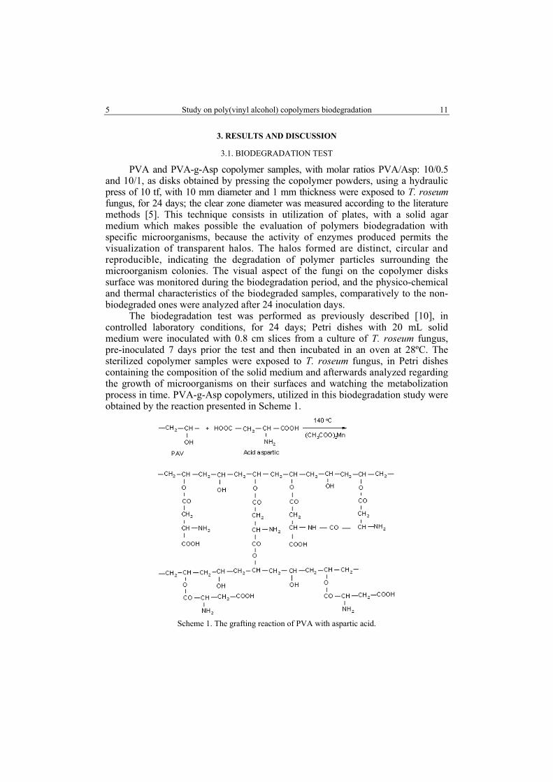
PVA and PVA-g-Asp copolymer samples, with molar ratios PVA/Asp: 10/0.5 and 10/1, as disks obtained by pressing the copolymer powders, using a hydraulic press of 10 tf, with 10 mm diameter and 1 mm thickness were exposed to T. roseum fungus, for 24 days; the clear zone diameter was measured according to the literature methods [5]. This technique consists in utilization of plates, with a solid agar medium which makes possible the evaluation of polymers biodegradation with specific microorganisms, because the activity of enzymes produced permits the visualization of transparent halos. The halos formed are distinct, circular and reproducible, indicating the degradation of polymer particles surrounding the microorganism colonies. The visual aspect of the fungi on the copolymer disks surface was monitored during the biodegradation period, and the physico-chemical and thermal characteristics of the biodegraded samples, comparatively to the non-biodegraded ones were analyzed after 24 inoculation days.
The biodegradation test was performed as previously described [10], in controlled laboratory conditions, for 24 days; Petri dishes with 20 mL solid medium were inoculated with 0.8 cm slices from a culture of T. roseum fungus, pre-inoculated 7 days prior the test and then incubated in an oven at 28ºC. The sterilized copolymer samples were exposed to T. roseum fungus, in Petri dishes containing the composition of the solid medium and afterwards analyzed regarding the growth of microorganisms on their surfaces and watching the metabolization process in time. PVA-g-Asp copolymers, utilized in this biodegradation study were obtained by the reaction presented in Scheme 1.
Scheme 1. The grafting reaction of PVA with aspartic acid.

Rodica Lipşa et al. 6 12
In Table 1 are given the initial molar ratios of the components: PVA/Asp and PVA/LA used in the copolymers synthesis and utilized in the biodegradation studies and some of their characteristics [19, 20].
Table 1 Copolymers and PVA in the biodegradation studies
Copolymer sample
and PVA
Molar ratio PVA/LA PVA/Asp
Average molecular
weight, Mw
Tg, ºC
Tm, ºC
∆Cp, J/g ºC
∆H, J/g
Xc, %
PVA-g-LA 1/1 73156 54 172 * 14 * PVA-g-LA 2.2/1 42739 52 195 * 35.07 * PVA-g-Asp 10/0.5 93440 76.2 220.2 0.169 38.54 24.71 PVA-g-Asp 10/1 206360 80.9 218.0 0.174 52.87 33.89
PVA --- 18000 75.1 220.0 0.118 18.69 11.98 * not determined; Tg = glass transition temperature; Tm = melting temperature; ∆Cp = heat capacity; ∆H = melting enthalpy; Xc = crystallinity degree
Images of PVA and PVA-g-Asp copolymers, before inoculation with T. roseum fungus are shown in Figure 2.
a)
b) c)
Fig. 2. Images of nonbiodegraded samples: a) -PVA; b) -PVA-g-Asp (10/0.5); c) -PVA-g-Asp (10/1).
In Table 2 is given the clear zone diameter of PVA and copolymers, recorded after 6 days of inoculation, determined according the method proposed in the literature [13]. Images showed that the biodegraded copolymer samples were readily colonized by Trichotecium roseum fungus. Fungal hyphae could be seen on PVA-g-Asp and PVA-g-LA samples [10], even after 6 inoculation days.
Table 2 The clear zone diameter
Sample Molar ratios PVA/LA PVA/Asp
Clear zone diameter after
6 inoculation days, mm PVA 1/0 10
PVA-g-LA 1/1 35 PVA-g-LA 2.2/1 20 PVA-g-Asp 10/0.5 20 PVA-g-Asp 10/1 32
7 Study on poly(vinyl alcohol) copolymers biodegradation
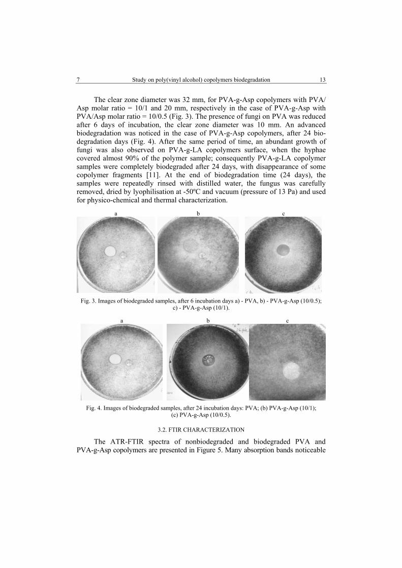
13
The clear zone diameter was 32 mm, for PVA-g-Asp copolymers with PVA/ Asp molar ratio = 10/1 and 20 mm, respectively in the case of PVA-g-Asp with PVA/Asp molar ratio = 10/0.5 (Fig. 3). The presence of fungi on PVA was reduced after 6 days of incubation, the clear zone diameter was 10 mm. An advanced biodegradation was noticed in the case of PVA-g-Asp copolymers, after 24 bio-degradation days (Fig. 4). After the same period of time, an abundant growth of fungi was also observed on PVA-g-LA copolymers surface, when the hyphae covered almost 90% of the polymer sample; consequently PVA-g-LA copolymer samples were completely biodegraded after 24 days, with disappearance of some copolymer fragments [11]. At the end of biodegradation time (24 days), the samples were repeatedly rinsed with distilled water, the fungus was carefully removed, dried by lyophilisation at -50ºC and vacuum (pressure of 13 Pa) and used for physico-chemical and thermal characterization.
a
b c
Fig. 3. Images of biodegraded samples, after 6 incubation days a) - PVA, b) - PVA-g-Asp (10/0.5);
c) - PVA-g-Asp (10/1).
a
b
c
Fig. 4. Images of biodegraded samples, after 24 incubation days: PVA; (b) PVA-g-Asp (10/1); (c) PVA-g-Asp (10/0.5).
3.2. FTIR CHARACTERIZATION
The ATR-FTIR spectra of nonbiodegraded and biodegraded PVA and PVA-g-Asp copolymers are presented in Figure 5. Many absorption bands noticeable

Rodica Lipşa et al. 8 14
in nonbiodegraded PVA spectrum are also observed in the spectrum of bio-degraded PVA, but slightly shifted: at 3276 cm-1 (in non-biodegraded PVA) and 3266 cm-1 (in biodegraded PVA) are recorded υOH stretching vibrations of the hydroxyl groups and intermolecular hydrogen linkages in polyols; at 2911 cm-1 and 1416 cm-1 (nonbiodegraded PVA) and 2911 cm-1, 1420 cm-1 (biodegraded PVA) are recorded the stretching vibrations υCH2 and δCH3 characteristic to methylene groups in the polymer. The bands at 1087 cm-1 (nonbiodegraded PVA) and 1079 cm-1
(biodegraded PVA) are characteristic to υC-OH stretching vibrations in the secondary alcohols. It can be noticed that in PVA biodegraded spectrum some absorption bands disappeared (at 1707, 1566 and 1136 cm-1), characteristic to υCO stretching vibrations of the residual acetyl groups COOCH3 and υC-OH in secondary alcohols. By fungal attack, the residual acetyl groups (υ CO from COOCH3) existent in PVA and CH-OH linkages were destroyed. This result is in accordance with the literature data [13]. The enzyme released by microorganisms can accelerate the polymer degradation.
Fig. 5. FTIR-ATR spectra of PVA and PVA-g-Asp copolymers
nonbiodegraded and biodegraded.

9 Study on poly(vinyl alcohol) copolymers biodegradation
15
In the ATR-FTIR spectra of biodegraded PVA-g-Asp copolymers (Fig. 5) can be noticed that many absorption bands existent in the initial copolymer structure are also found in the biodegraded samples (slightly shifted), suggesting that by biodegradation essential structural modifications in the copolymers structure were not produced. However, in the biodegraded PVA-g-Asp copolymer samples disappeared the absorption bands existent at 1691 and 1592 cm-1 in the initial copolymers, demonstrating that COOH (υC = O) and COO-(υC = O) linkages were destroyed. Also, in the biodegraded samples appeared new absorption bands at: 1656, 1237 and 1135 cm-1, attributed to υCO vibrations of CONHR groups I amide band group and stretching vibrations sym υCO and asym υCO from COOR groups (existent in Trichotecium roseum fungus) and stretching vibrations υC-OH of secondary alcohols (from PVA). Also, in the biodegraded PVA-g-LA samples in the ATR-FTIR spectra could be noticed that the ester groups were destroyed by hydrolytic scission, caused by this fungus [11].
3.3. SCANNING ELECTRON MICROSCOPY
SEM micrographs of PVA and PVA-g-Asp copolymer samples (non-bio-degraded and biodegraded) are presented in Figure 6.
Firstly, on the biodegraded PVA surface can be observed the appearance of tiny holes (Fig. 6 b). In some areas, after 24 biodegradation days, could be noticed that fungal hyphae penetrated into the structure of PVA-g-Asp copolymers (Fig. 6 d, f). The tracks (Fig. 6 d) and fungal hyphae (Fig. 6 f) on their surface suggested the secretion of fungal extra-cellular substances that probably determined the scission of the ester groups and short macromomolecular chains.
Concerning the biodegraded PVA-g-LA copolymers, the SEM micrographs recorded after 24 biodegradation days showed several large holes and cracks that penetrated deep into the samples, with the disappearance of some polymer fragments. The extent of colonization increased with time and after 24 days the PVA-g-LA samples were completely biodegraded. The presence of the ester groups in the copolymers determined a high biodegradation degree, comparatively to PVA, in the presence of this fungus. In the case of the biodegraded PVA-g-LA copolymers the biodegradation degree was also influenced by the molar ratio PVA/LA. The PVA-g-LA copolymer, with molar ratio 1/1 presented an advanced biodegradation and mass losses caused by degradation of the fragments with lower molecular weights. For all the studied samples it could be observed an abundant growth of fungi at the end of the biodegradation time (24 days), when their surface was covered almost 70%–90% with this fungus.

Rodica Lipşa et al. 10 16
Fig. 6. SEM micrographs: a) - PVA; c) - PVA-g-Asp (10/0.5); e) - PVA-g-Asp (10/1)
(nonbiodegraded); b) - PVA; d) - PVA-g-Asp (10/0.5); f) - PVA-g-Asp (10/1) (biodegraded).
3.4. STATIC LIGHT SCATERING ANALYSIS
The light scattering methods allow the exploration of different associations of biomacromolecules, but require suitable solvents for the molecular dissolution of the components [8]. In order to determine the characteristic solution properties (weight average molecular weight Mw, radius of gyration Rg and the second virial coefficient A2), it was necessary to know the specific refractive index increments

11 Study on poly(vinyl alcohol) copolymers biodegradation
17
dn/dc of the sample-solvent pair. Refractometric and SLS measurements were made on unfractionated samples (both for nonbiodegraded and biodegraded samples). The stock and diluted solutions (in range of 10-3–10-4 g/mL for the copolymers; and 10-4–10-5 g/mL for PVA were prepared in deionized water. The solvent was filtered through 0.02 µm Anotop filters (Whatman). Before dilution stage, every stock sample was filtered through membrane filter with pore size of 0.8 µm. Then, all the solutions were transferred into cylindrical quartz scintillation vials (Quartz SUPRASIL, Hellma GmbH & Co. KG, Germany), immediately sealed with plastic caps and kept 2 days for degassing. Thereby, polymer solutions were prepared for “batch mode” laser light scattering measurements. Based on Zimm plots and Berry formalism, characteristic solution properties were determined: weight-average molecular weight Mw, radius of gyration Rg and second virial coefficient A2. The Astra 4.90.07 software allowed modeling of the scattering angle by first-order polynomial functions. Laser light scattering of the macromolecular compounds was described by Zimm equation:
...
2sin
31621 2
2
0
2
2 +++=θ
λπ
θ w
g
w MR
cAMR
Kc (1)
with
( )4
0
222 /4λ
πN
ndcdnK = (2)
where K is the optical constant for vertically polarized incident light, c con-centration (g mL−1) of the scattering species in solution, Rθ Rayleigh ratio (directly proportional to the ratio of the scattered light intensity at angle θ to the incident light intensity), N is the Avogadro number, n solution refractive index, and λ0 is the wavelength of the incident light. In the following calculations, we assumed that the concentration is sufficiently low to neglect the terms containing the higher virial coefficients than A2. The Rg value can be found from the initial slope of a plot Kc/Rθ versus sin2(θ/2) and 1/ wM from the intersection with the ordinate. The second virial coefficient A2 is an important parameter that quantitatively characterizes the thermodynamic interaction between the solution molecules at a specific temperature. The polymer-solvent interactions dominate the polymer-polymer interactions when A2>0. In the case of A2 = 0, both interactions are energetically equivalent and the solvent is called theta solvent. For A2<0, polymer-polymer contact is preferred to polymer-solvent and the solvent is a poor one for the given polymer [3].
The Zimm plots were computed using Berry formalism (eq. 3)
θRKc / = f (sin2 (θ / 2)+ a c) (3)
and the scattering angle dependence was modeled by first-order polynomial functions. For the sake of clarity, the noisy signals of some detectors were rejected.
Rodica Lipşa et al. 12 18
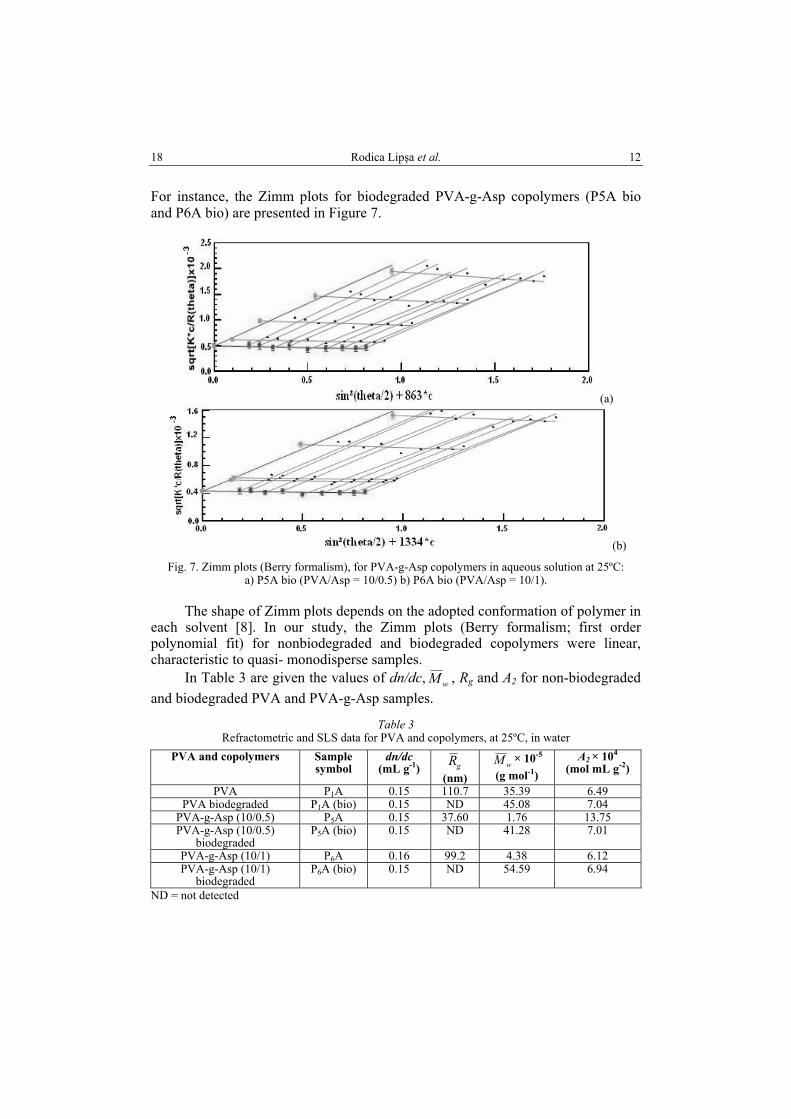
For instance, the Zimm plots for biodegraded PVA-g-Asp copolymers (P5A bio and P6A bio) are presented in Figure 7.
(a)
(b) Fig. 7. Zimm plots (Berry formalism), for PVA-g-Asp copolymers in aqueous solution at 25ºC:
a) P5A bio (PVA/Asp = 10/0.5) b) P6A bio (PVA/Asp = 10/1).
The shape of Zimm plots depends on the adopted conformation of polymer in each solvent [8]. In our study, the Zimm plots (Berry formalism; first order polynomial fit) for nonbiodegraded and biodegraded copolymers were linear, characteristic to quasi- monodisperse samples.
In Table 3 are given the values of dn/dc, wM , Rg and A2 for non-biodegraded and biodegraded PVA and PVA-g-Asp samples.
Table 3 Refractometric and SLS data for PVA and copolymers, at 25ºC, in water
PVA and copolymers Sample symbol
dn/dc (mL g-1) gR
(nm)wM × 10-5
(g mol-1) A2 × 104
(mol mL g-2)
PVA P1A 0.15 110.7 35.39 6.49 PVA biodegraded P1A (bio) 0.15 ND 45.08 7.04
PVA-g-Asp (10/0.5) P5A 0.15 37.60 1.76 13.75 PVA-g-Asp (10/0.5)
biodegraded P5A (bio) 0.15 ND 41.28 7.01
PVA-g-Asp (10/1) P6A 0.16 99.2 4.38 6.12 PVA-g-Asp (10/1)
biodegraded P6A (bio) 0.15 ND 54.59 6.94
ND = not detected

13 Study on poly(vinyl alcohol) copolymers biodegradation
19
The results presented in Table 3 show that the biodegraded samples have a more raised average weight molecular weight wM comparatively with the nonbio-degraded ones, which also slightly increased in the case of PVA-g-LA copolymers [11]. These results suggest that by biodegradation in the presence of Trichotecium roseum fungus, the low molecular weight macromolecular chains were degraded and released in the medium. These results are consistent with the data presented in the literature [4]. Additionally, in the case of biodegraded samples it is possible that the intermolecular interactions dominate in detriment to the intramolecular ones. In the copolymer-solvent systems, a competition between these interactions exists, which was caused by the solvent nature, copolymer structure (hydrophilic-hydrophobic balance) and mixing conditions (temperature, pH, ionic strength) [18]. The positive values of A2 for all nonbiodegraded and biodegraded samples demonstrate the presence of favorable polymer-solvent interactions.
3.5. DIFFERENTIAL SCANNING CALORIMETRY ANALYSIS
In Figure 8 and Table 4 are presented the DSC curves of nonbiodegraded and biodegraded samples and their thermal characteristics. By PVA biodegradation the targeted areas with crystalline structure (corresponding to the melting process at T>160ºC) increased (Fig. 8), as a result of advanced wraps by hydrogen bonding and van der Waals forces. The PVA-g-Asp (except the copolymer with PVA/Asp = 10/1 molar ratio) copolymers have lower Tg values in comparison with PVA, as a result of crystallinity and polarity decrease and free volume increase. Also, the melting temperature (Tm), melting enthalpy (∆H) and crystallinity degree (Xc) values of biodegraded PVA and PVA-g-Asp copolymer samples increased after 24 inoculation days with Trichotecium roseum fungus, showing the disappearance of imperfect crystallites and amorphous domains [11].
Fig. 8. DSC curves of PVA and PVA-g-Asp copolymers,
nonbiodegraded and biodegraded, 24 days.

Rodica Lipşa et al. 14 20
Table 4 Thermal characteristics of PVA and PVA-g-Asp copolymers, by DSC
Sample Molar ratio PVA/Asp
Tg oC
Tm oC
∆Cp J/g oC
∆Hm J/g
P1A – 76 219 0.225 90.01 P1A bio – – 229 – 91.33
P5A 10/0.5 61.35 221 0.155 84.83 P5A bio 10/0.5 – 227 – 91.57
P6A 10/1 93.24 160 0.616 6.45 P6A bio 10/1 – 192 – 31.80
Tg = glass transition temperature Tm = melting temperature ∆Cp = heat capacity
DSC analysis showed that biodegraded PVA and PVA-g-Asp copolymer samples have no glass transition temperature (Tg) (Table 4), so the heat capacity value ∆Cp could not be calculated on 10–250ºC interval; also the melting temperature (Tm) and the melting enthalpy (∆Hm) of biodegraded PVA and copolymers have higher values than the nonbiodegraded samples. The melting domain of bio-degraded PVA becomes narrower (53ºC) comparatively to nonbiodegraded PVA (65ºC), demonstrating that by biodegradation the amorphous-crystalline interface was affected and the crystallinity degree increased from 57.7 wt.% (nonbio-degraded PVA) to 58.6 wt.% (biodegraded PVA). The same behavior can be noticed in the case of PVA-g-Asp copolymers: PVA-g-Asp biodegraded samples have no Tg, consequently ∆Cp could not be calculated on 10–240ºC interval. The Tm and ∆Hm values of PVA-g-Asp biodegraded copolymers are more raised comparatively to the nonbiodegraded samples, suggesting that biodegradation mainly occurs in the amorphous phase, most of this fraction being removed. DSC results suggest that the short macromolecular chains at the crystalline-amorphous interface are biodegraded and the small crystallites are eliminated.
3.6. THERMOGRAVIMETRIC ANALYSES
The thermal behavior of PVA and PVA-g-Asp nonbiodegraded and biodegraded copolymers, in the presence of Trichotecium roseum fungus was also studied by thermogravimetry, in nitrogen atmosphere, with heating rate of 10ºC/min, on the 30–600ºC temperature interval. TG-DTG curves are shown in Figure 9. The thermal behavior of nonbiodegraded and biodegraded PVA with Trichotecium roseum was presented in [11]. The degradation of PVA-g-Asp copolymers was assessed quali-tatively by determination of onset (Tonset), peak (Tpeak) and endset (Tendset) temperatures, as well as by temperatures corresponding to 10 wt.% (T10) and 20 wt.% (T20) mass loss, and ash at 700ºC, from TG/DTG curves. PVA-g-Asp copolymers non-biodegraded and biodegraded present 3 and 4 thermal decomposition stages, respectively that occur with different rates. In the first stage of degradation at around 100ºC, the mass losses are reduced (until maximum 10.13 wt.%),

15 Study on poly(vinyl alcohol) copolymers biodegradation
21
caused by the elimination of physical absorbed water molecules. In the second stage of thermal decomposition, that occurs between 198–384ºC interval for non-biodegraded samples the mass losses have values between 8.77–60.66 wt% in the case of nonbiodegraded samples; for the biodegraded samples the second stage of thermal decomposition is at 243–388ºC and the mass losses are 10.53–67.55 wt.%. In the third thermal decomposition stage, between 296÷490ºC (for nonbiodegraded samples), the mass losses range between 23.59–51.32 wt.%; the biodegraded samples present a third thermal decomposition stage at 314–487ºC, with weight losses of 14.90–66.42 wt.%. The copolymer with PVA/Asp molar ratio 10/1 non-biodegraded and biodegraded has a 4th thermal decomposition stage (408–484ºC), with weight losses ranging between 13.56–18.18 wt.%. Concerning the thermal stability it can be noticed that the biodegraded PVA-g-Asp copolymers present a raised thermal stability in comparison with the nonbiodegraded ones, as it can be observed from T10 as well as T20 values (Table 5). Also, from Table 5 can be noticed that the thermal stability of the copolymers is influenced by PVA/Asp molar ratio: it is higher in the case of the copolymer with PVA/Asp = 10/0.5 molar ratio for T10 as well as for T20. The thermal stability of PVA-g-Asp nonbio-degraded and biodegraded is more raised in comparison with nonbiodegraded and biodegraded PVA. Similar results were obtained in the case of PVA and PVA-g-LA copolymers, suggesting that by biodegradation, the amorphous domains of PVA and copolymers were destroyed, consequently the crystallinity degree increased and the remaining macromolecular chains with higher molecular weight could be more difficult thermally degraded.
Table 5 Thermogravimetric results for PVA-g-Asp nonbiodegraded and biodegraded copolymers
Sample Degradation stage
Tonset ºC
Tpeak ºC
Tendset ºC
W %
T10 ºC
T20 ºC
P5A I II III
residue
207 302 420
227 356 429
247 384 490
7,37 60,66 23,59 8,38
290
325
P5A bio I II III
residue
173 318 415
– 358 434
305 388 487
10,13 67,55 14,90 7,42
294
327
P6A I II III IV
residue
37 198 296 408
81 228 354 436
130 241 388 484
9,22 8,77
51,32 18,18 12,51
183
272
P6A bio I II III IV
residue
58 243 314 409
91 –
348 437
144 279 381 481
4,29 10,53 66,42 13,56 5,20
275
307

Rodica Lipşa et al. 16 22
The heating rate = 10ºC/min; N2, Al2O3, 30–600ºC; Tonset, the temperature at which the thermal decomposition begins; T peak , the temperature at which the degradation rate is maximum; Tendset the temperature at the end of the thermal degradation process; T10 , T20 the temperature corresponding to 10 wt.% and 20 wt.% weight loss; W = weight loss.
Fig. 9 a. TG-DTG curves of PVA-g-Asp (10/0.5),
nonbiodegraded and biodegraded.
Fig. 9 b. TG/DTG curves of PVA-g-Asp (10/1),
nonbiodegraded and biodegraded.

17 Study on poly(vinyl alcohol) copolymers biodegradation
23
4. CONCLUSIONS
The study was conducted in order to find the ability of T. roseum fungus to biodegrade PVA-g-Asp copolymers, previously synthesized by solution polycondensation procedure; the physico-chemical and thermal characteristics of biodegraded PVA-g-Asp samples were studied in comparison with biodegraded PVA ones. The clear zone technique showed that the fungal attack was much more reduced on PVA, comparatively to the studied copolymers. The FTIR-ATR spectra revealed that PVA structure was not much modified after 24 biodegradation days, with this fungus; the FTIR-ATR spectra of biodegraded PVA-g-Asp copolymers showed the scission of the ester and amide groups by biodegradation; hyphae of the fungus are present in their structure. SEM analysis proved to be a useful tool to examine the colonization and biodegradation of PVA copolymers by Trichotecium roseum. SEM micrographs showed an abundant growth of fungi on the copolymers surface, at the end of biodegradation time (24 days); the samples surface was covered with hyphae at a rate of 70–90%, at the end of the biodegradation period. The SLS analysis highlighted that the average molecular weights, Mw of PVA-g-Asp copolymers were much more modified after biodegradation, in comparison with PVA-g-LA copolymers and the low molecular weight fractions were firstly bio-degraded. Also, the thermal stability of biodegraded PVA and PVA-g-Asp samples was more raised comparatively to the nonbiodegraded ones; the same results were previously obtained in the case of PVA-g-LA copolymers [10]; TG-DTG results concluded that by biodegradation the low molecular weight chains and amorphous domains existent in PVA and copolymer samples were destroyed. DSC results demonstrated that the amorphous/crystalline ratios were modified by biodegradation and the small quasi-amorphous crystallites destroyed. Accordingly, the studied PVA, PVA-g-Asp and PVA-g-LA copolymers are biodegradable in the presence of Trichotecium roseum fungus, after maximum 24 days of incubation. Also, it can be concluded that the the graft copolymers biodegradation is much advanced in comparison with PVA as base polymer. By varying the copolymer composition the biodegradation rate would be controlled. The present study suggests the possible utilization of these copolymers in packaging, agriculture, pharmacy, as they are environmentally friendly.
Acknowledgements: The authors are grateful for the financial support from the Romanian National Authority for Scientific Research CNCS-UEFISCDI, project: Bionanomed: Antimicrobial bionanocomposites for medical applications by Grant PCCA/II, no 164/2012 and project: Interdisciplinary research on multifunctional hybrid particles for bio-requirements “INTERBIORES” PN-II-PT-PCCA-2011-3.2-0428, Grant no. 211/2012.
R E F E R E N C E S
1. AMASS W., AMASS A., TIGHE B., A review of biodegradable polymers: uses, current developments in the synthesis and characterization of biodegradable polyesters, blends of biodegradable polymers and recent advances in biodegradation studies, Polymer International, 1998, 47(2), 89–144.

Rodica Lipşa et al. 18 24
2. ARTHAM T., DOBLE M., Biodegradation of physicochemically treated polycarbonate by fungi, Biomacromolecules 2009, 11, 20–28.
3. BISKUP-CZECHOWSKA R., WOJTASZ-PAIAK A., SIKORSKI J., HENKE A., ULANSKI P., ROSIAK J. M., Aqueous Solutions of Hydrochloric Acid as Simple Solvents of Chitosan for Viscosity-and Light –Scattering –Based Molecular Weight Determination, Polish Chitin Society, Monograph XII, 2007, 87–94.
4. BOHLMAN G. M, Handbook of Biodegradable Polymers, Rapra Technology Ltd, Shropshire, U.K., 2005, 183–218.
5. CANGEMI J. M., dos SANTOS A. M., NETO S. C., CHIERICE G. O., Biodegradation of polyurethane derived from castor oil, Polimeros: Ciencia e Tecnologia, 2008, 18 (3), 201–206.
6. CHANDRA R., RUSTGI R., Biodegradable Polymers, Prog. Polym. Sci.,1998, 23, 1273–1335. 7. CHIELLINI E., CORTI A., DEL SARTO G., D’ ANTONE S., Oxo-biodegradable polymers.
Effect of hydrolysis degree on biodegradation behavior of poly(vinyl alcohol), Polymer Degradation and Stability, 2006, 91, 3397–3406.
8. GRIGORAS A. G., CONSTANTIN M., GRIGORAS V. C., DUNCA S. I., OCHIUZ L., Studies on the physico-chemical and antibacterial properties of grafted pullulans solutions, Reactive & Functional Polymers, 2013, 73, 1249–1254.
9. IKADA Y., TSUJI H., Biodegradable polyesters for medical and ecological applications, Macromol. Rapid Commun. 21, 2000, 117–132.
10. LEJA K., LEWANDOWICZ G., Polymer biodegradation and biodegradable polymers - A Review, Polish J. of Environ. Stud, 2010, 19 (2), 255–266.
11. LIPSA R., TUDORACHI N., GRIGORAS V. C., VASILE C., Degradation of poly(vinyl alcohol)-graft-lactic acid copolymers by the fungus Trichotecium Roseum, Journal of Applied Polymer Science, 2015, 132 (14) (in press).
12. LUADTHONG C., TACHAPRUTINUN A., WANICHWECHARUNGRUANG S. P., Synthesis and characterization of micro/nanoparticles of poly(vinyl alcohol-co-vinylcinnamate) derivatives, European Polymer Journal, 2008, 44 (5), 1285–1295.
13. MAARTEN VAN DER ZEE, Handbook of Biodegradable Polymers: Synthesis, Characterization and Applications, Wiley-VCH Verlag GmbH & Co. KGaA, Weiheim, Germany 2011.
14. SHAMSII S. H., SULTANA R., Trichotecium roseum link a new record of Hyphomycetous fungus for Bangladesh, J. Plant Taxon, 2008, 15(1), 77–80.
15. SI-SHEN F., LI M., BING-HUNG C., DANIEL P., Polymeric nanospheres fabricated with natural emulsifiers for clinical administration of an anticancer drug paclitaxel (Taxol), Mater Sci. Eng. C, 2002, 20, 85–92.
16. SY.-CORDERO A. A., GRAF T. N., ADCOCK A. F., KROLL D. J., STEVEN Q-SH, SWANSON M., WANI M. C., PEARCE C. J., OBERLIES N. H., Two cyclodepsipeptides, two sesquiterpenoids and other cytotoxic metabolites from the filamentous fungus Trichotecium, J. Nat. Prod., 2011, 74 (10), 2137–2142.
17. THAM C. Y., HAMID Z. A. A., ISMAIL H., AHMAD Z., Poly(vinyl alcohol) in fabrication of PLA micro- and nanoparticles using solvent evaporation technique, Advanced Materials Research, 2014, 1024, 296–299.
18. TOKIWA Y., CALABIA B. P., UGUWU CH. U., AIBA S., Biodegradability of plastics, Int. J. Mol. Sci., 2009, 10 (9), 3722–3742.
19. TUDORACHI N., LIPSA R., Synthesis and characterization of poly(vinyl alcohol-co-aspartic acid) copolymers, Polimery, 2010, 55 (7–8), 562–567.
20. TUDORACHI N., LIPSA R., Poly(vinyl alcohol)-g-lactic acid copolymers and films with silver nanoparticles, Journal of Applied Polymer Science, 2011, 122, 1109–1120.
21. TUDORACHI N., CASCAVAL C. N., RUSU M., PRUTEANU M., Testing of polyvinyl alcohol and starch mixtures as biodegradable polymeric materials, Polymer Testing, 2000, 19, 785–799.
22. VROMAN I., TIGHZERT L., Biodegradable polymers, Materials, 2009, 2, 307–344.

19 Study on poly(vinyl alcohol) copolymers biodegradation
25
23. WEBB H. K., RUSSELL J. A., CRAWFORD J. IVANOVA E. P., Plastic degradation and its environmental implications with special reference to poly(ethylene terephthalate), Polymers, 2013, 5, 1–18.
24. YAMATSU A., MATSUNAMI T., ATOMI H., IMANKA T., Isolation and characterization of a novel poly(vinyl alcohol) degrading bacterium Spingoyxis sp. PVA 3, Appl. Microbiol. Biotechnol., 2006, 72, 804–811.
25. YUN Y. H., SON S. Y., CHOI CH. W., HONG J. K., KIM Y. S. H., KIM S. H., The occurrence of pink mold rot fungus Trichotecium roseum on tomatoes in Korea, African Journal of Microbiology Research, 2013, 7(13), 1128–1135.
Received April 2, 2015