Membranes - アジレント・テクノロジー株式会社 3 Duralose-UV Membranes Duralose-UV...
26
Membranes INSTRUCTION MANUAL Catalog #420100–#420105 (Duralon-UV™ Membranes), #420106–#420108 (Nitrocellulose Membranes), #420111–#420115 (Duralose-UV™ Membranes), and #420130 (PVDF Membranes) Revision #042001b For In Vitro Use Only *420100-12_042001b/*
Transcript of Membranes - アジレント・テクノロジー株式会社 3 Duralose-UV Membranes Duralose-UV...
Membranes
INSTRUCTION MANUALCatalog #420100–#420105 (Duralon-UV™ Membranes),
#420106–#420108 (Nitrocellulose Membranes),
#420111–#420115 (Duralose-UV™ Membranes),
and #420130 (PVDF Membranes)
Revision #042001b
For In Vitro Use Only
*420100-12_042001b/*
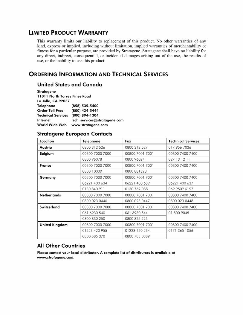
LIMITED PRODUCT WARRANTY
This warranty limits our liability to replacement of this product. No other warranties of anykind, express or implied, including without limitation, implied warranties of merchantability orfitness for a particular purpose, are provided by Stratagene. Stratagene shall have no liability forany direct, indirect, consequential, or incidental damages arising out of the use, the results ofuse, or the inability to use this product.
ORDERING INFORMATION AND TECHNICAL SERVICES
United States and CanadaStratagene11011 North Torrey Pines RoadLa Jolla, CA 92037Telephone (858) 535-5400Order Toll Free (800) 424-5444Technical Services (800) 894-1304Internet [email protected] Wide Web www.stratagene.com
Stratagene European ContactsLocation Telephone Fax Technical Services
Austria 0800 312 526 0800 312 527 017 956 7036
00800 7000 7000 00800 7001 7001 00800 7400 7400Belgium
0800 96078 0800 96024 027 13 12 11
France 00800 7000 7000 00800 7001 7001 00800 7400 7400
0800 100391 0800 881323
Germany 00800 7000 7000 00800 7001 7001 00800 7400 7400
06221 400 634 06221 400 639 06221 400 637
0130 840 911 0130 762 088 069 9509 6197
Netherlands 00800 7000 7000 00800 7001 7001 00800 7400 7400
0800 023 0446 0800 023 0447 0800 023 0448
Switzerland 00800 7000 7000 00800 7001 7001 00800 7400 7400
061 6930 540 061 6930 544 01 800 9045
0800 830 250 0800 825 225
United Kingdom 00800 7000 7000 00800 7001 7001 00800 7400 7400
01223 420 955 01223 420 234 0171 365 1056
0800 585 370 0800 783 0889
All Other CountriesPlease contact your local distributor. A complete list of distributors is available atwww.stratagene.com.
Membranes
CONTENTS
Materials Provided.............................................................................................................................. 1Introduction......................................................................................................................................... 1Stratagene’s Membranes for Nucleic Acid Transfer and Hybridization....................................... 2
Nitrocellulose Membranes..................................................................................................... 2
Duralose-UV Membranes...................................................................................................... 3
Duralon-UV Membranes ....................................................................................................... 3
Stratagene’s Nucleic Acid Hybridization Solution........................................................................... 3QuikHyb® Hybridization Solution......................................................................................... 3
Stratagene’s Membranes for Protein Transfer ................................................................................ 4PVDF Membranes ................................................................................................................. 4
Southern Blotting ................................................................................................................................ 4Hybridization of Long DNA Probes Using Stratagene’s Nucleic Acid Transfer and
Hybridization Membranes ............................................................................................. 4
Hybridization of Oligonucleotide Probes Using Stratagene’s Nucleic Acid Transfer
and Hybridization Membranes ...................................................................................... 8
Stripping of Probes for Reuse Using Stratagene’s Nucleic Acid Transfer and
Hybridization Membranes ........................................................................................... 11
Northern Blotting .............................................................................................................................. 12Transfer of RNA onto the Membrane.................................................................................. 12
Prehybridization of Northern Blots ..................................................................................... 13
Hybridization of Northern Blots.......................................................................................... 13
Post-Hybridization Washes of Northern Blots .................................................................... 14
Screening............................................................................................................................................ 14Plaque Lifts.......................................................................................................................... 14
Colony Lifts......................................................................................................................... 17
Western Blotting ............................................................................................................................... 19Transfer of Proteins from SDS–Polyacrylamide Gels to PVDF Membranes...................... 19
Storage of Western Blots..................................................................................................... 21
Preparation of Media and Reagents ................................................................................................ 22References .......................................................................................................................................... 22Endnotes............................................................................................................................................. 22

Membranes 1
Membranes
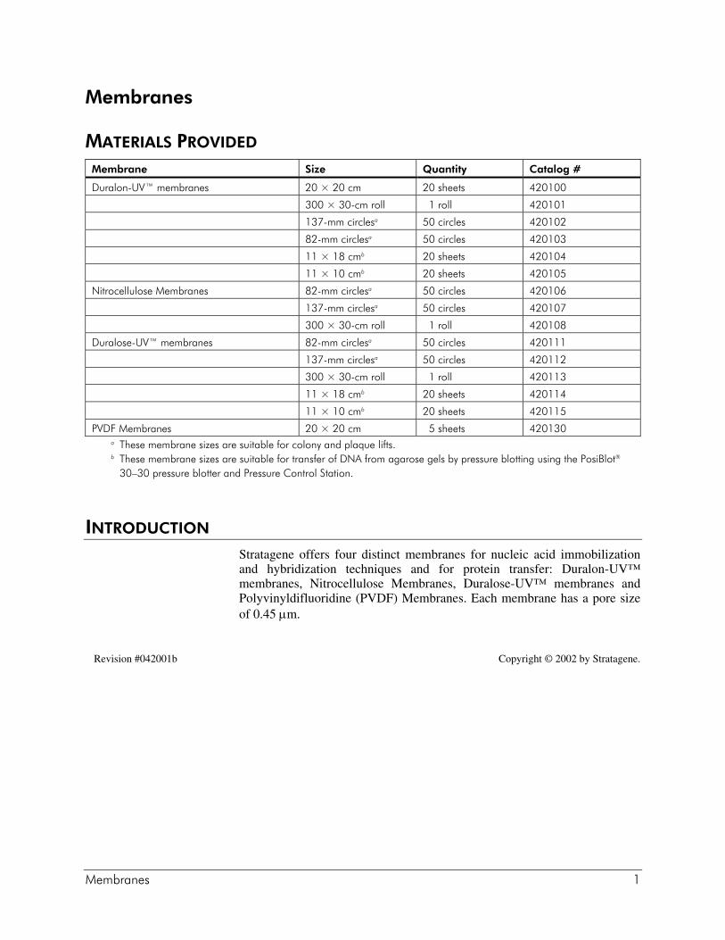
MATERIALS PROVIDED
Membrane Size Quantity Catalog #
Duralon-UV™ membranes 20 × 20 cm 20 sheets 420100
300 × 30-cm roll 1 roll 420101
137-mm circlesa 50 circles 420102
82-mm circlesa 50 circles 420103
11 × 18 cmb 20 sheets 420104
11 × 10 cmb 20 sheets 420105
Nitrocellulose Membranes 82-mm circlesa 50 circles 420106
137-mm circlesa 50 circles 420107
300 × 30-cm roll 1 roll 420108
Duralose-UV™ membranes 82-mm circlesa 50 circles 420111
137-mm circlesa 50 circles 420112
300 × 30-cm roll 1 roll 420113
11 × 18 cmb 20 sheets 420114
11 × 10 cmb 20 sheets 420115
PVDF Membranes 20 × 20 cm 5 sheets 420130a These membrane sizes are suitable for colony and plaque lifts.b These membrane sizes are suitable for transfer of DNA from agarose gels by pressure blotting using the PosiBlot®
30–30 pressure blotter and Pressure Control Station.
INTRODUCTION
Stratagene offers four distinct membranes for nucleic acid immobilizationand hybridization techniques and for protein transfer: Duralon-UV™membranes, Nitrocellulose Membranes, Duralose-UV™ membranes andPolyvinyldifluoridine (PVDF) Membranes. Each membrane has a pore sizeof 0.45 µm.
Revision #042001b Copyright © 2002 by Stratagene.
2 Membranes
STRATAGENE’S MEMBRANES FOR NUCLEIC ACID TRANSFER ANDHYBRIDIZATION
Nucleic acids can be bound to Stratagene’s Duralon-UV membranes,Nitrocellulose Membranes and Duralose-UV membranes by the traditional2-hour vacuum oven baking method or by rapid ultraviolet (UV)crosslinking. Being noncharged, these membranes have increased bindingcapacity, decreased background and can be crosslinked instead of baked.The Stratalinker® UV crosslinker covalently crosslinks nucleic acids tomembranes in ~30 seconds. Each lot of these membranes is manufacturedand tested to exacting specifications and is chosen for maximum signal-to-noise ratio when used in conjunction with the Stratalinker UV crosslinker.These membranes are Triton®-free to ensure excellent plaque and colonyrecovery.
Nitrocellulose MembranesNitrocellulose has traditionally been used for all standard blottingapplications. Stratagene's Nitrocellulose Membranes exhibit the uniformwettability, low background, and 80 µg of RNA or DNA/cm2 bindingcapacity of pure nitrocellulose transfer membranes. These membranes mustbe prewet first with distilled water (dH2O) and then with transfer buffer forthorough transfer. If a membrane does not wet completely, boil themembrane briefly. Nitrocellulose can be sterilized by steam autoclaving.When baking Nitrocellulose Membranes, use a vacuum oven. Ultravioletcrosslink only damp membranes, as nitrocellulose is highly flammable.Reprobing of nitrocellulose can be difficult due to its inherent fragility(fragility increases after baking).
Cleanly cut the nitrocellulose with a razor or similarly sharp instrument toavoid nicks and tears that will eventually enlarge. Avoid handlingNitrocellulose Membranes with bare hands, as the proteins and oilsassociated with fingerprints interfere with the proper wetting of themembrane and may increase the background after hybridization. Alkalinetransfers cannot be performed using Nitrocellulose Membranes. It is best tochoose nitrocellulose for experiments requiring low background from asingle probing. Either side of Stratagene's Nitrocellulose Membranes can beused. For best retention of nucleic acid during blotting, use 20× SSC buffer(see Preparation of Media and Reagents) as a transfer medium.
Nitrocellulose Membranes should be stored at a constant temperature and ina dark, cool place devoid of solvent vapors to prevent these membranesfrom drying out and becoming brittle. Nitrocellulose has wetting agents thatcould dry out if stored at room temperature in dry climates. A 4°C coldroom is a convenient way to maintain constant temperature and humidity.
Note Nitrocellulose Membranes cannot be used with the Illuminator™nonradioactive detection system or with alkaline transfers.
Membranes 3
Duralose-UV MembranesDuralose-UV membranes are reinforced nitrocellulose membranes that havethe tensile strength and reliability of nylon membranes, while retaining thelower background generally observed with nitrocellulose membranes. Eachlot of Duralose-UV membranes binds 80–100 µg of DNA or RNA/cm2 andis 500× stronger than ordinary nitrocellulose membranes. These supportednitrocellulose membranes allow several reprobings and do not require theextensive blocking necessary with nylon membranes for visualization ofcolorimetric reactions. As with conventional nitrocellulose, alkaline transfermethods cannot be used with Duralose-UV membranes, and gloves shouldbe worn when handling these membranes. For best retention of nucleic acidsduring blotting use 20× SSC buffer as a transfer medium.
Note Duralose-UV membranes cannot be used with the Illuminatornonradioactive detection system or with alkaline transfers.
Duralon-UV MembranesDuralon-UV membranes are noncharged nylon membranes that exhibitexceptional strength, thermal stability and increased sensitivity incomparison with conventional nitrocellulose membranes. Each lot ofDuralon-UV membranes will bind at least 500 µg of DNA or RNA/cm2
(compared to 80 µg/cm2 characteristic of nitrocellulose membranes),including fragments as small as 10 bp, and can be reprobed several times.These easy-to-handle membranes should not tear, crack, shrink or curlwhether wet or dry and may be stored at room temperature. Completely cutthrough the polymer supporting threads before separating a measured piecefrom the remainder of the Duralon-UV membrane sheet.
Baking or UV crosslinking is required for fixing the nucleic acids onto thesenoncharged membranes. Duralon-UV membranes can be used in alkalinetransfer systems. The high binding capacity may increase the backgroundafter probing. Duralon-UV membranes are an ideal choice for hybridizationof low-copy-number sequences, unusually small fragments or when multipleprobings are necessary.
STRATAGENE’S NUCLEIC ACID HYBRIDIZATION SOLUTION
QuikHyb® Hybridization SolutionQuikHyb® hybridization solution is a rapid hybridization solution developedat Stratagene. This unique hybridization solution reduces the time requiredfor nucleic acid hybridization down to 1–2 hours. QuikHyb hybridizationsolution can be used for Northern, Southern and slot blots and is compatiblewith any conventional membrane used for nucleic acid transfer.
4 Membranes
STRATAGENE’S MEMBRANES FOR PROTEIN TRANSFER
PVDF MembranesPVDF membranes are a practical alternative to traditional nitrocellulosemembranes in protein transfer and Western blotting applications. Thesemembranes exhibit stronger signals, higher signal-to-noise ratios, greatermechanical strength and a higher protein binding capacity of 125 µg/cm2. Inaddition, these membranes are chemically stable, allowing use of a widerange of solvents for rapid destaining, and are compatible for use inconventional protein staining and immunoblotting protocols.
SOUTHERN BLOTTING
Hybridization of Long DNA Probes Using Stratagene’s Nucleic AcidTransfer and Hybridization Membranes
Transfer of the Target DNA onto the Membrane
1. Electrophorese the DNA samples in an agarose gel, stain the gel withethidium bromide (EtBr) and photograph under UV light with a rulerfor later reference.
2. Depurinate the DNA by soaking the gel in 0.25 N HCl for5–15 minutes, depending on the thickness of the gel (e.g., 5 minutes fora 3-mm-thick gel and 15 minutes for a 4- to 6-mm-thick gel). Thisfragments the DNA and facilitates transfer of DNA through the gel andonto the membrane and is especially important for the transfer of largeDNA fragments.
3. Rinse the gel in dH2O to complete the depurination process anddenature the DNA by soaking the gel in a solution of 1.5 M NaCl and0.5 M NaOH for 15–30 minutes.
4. Neutralize by soaking the gel in a solution of 1.5 M NaCl and 0.5 MTris-HCl (pH 8.0) for 15–30 minutes.
5. Briefly soak the gel in transfer buffer [e.g., either 10–20× SSPE (seePreparation of Media and Reagents), 10–20× SSC buffer or 25 mMsodium phosphate].
Note For Nitrocellulose Membranes or Duralose-UV membranes,use 20× SSC buffer as the transfer buffer to obtain maximumDNA retention.
6. Prewet the membrane in dH2O and then in transfer buffer. If a portionof the membrane resists wetting, pour boiling dH2O over the membraneand rock gently.

Membranes 5
7. Transfer DNA to the membrane by traditional capillary blotting1 or bypressure blotting using Stratagene's PosiBlot 30–30 pressure blotter andPressure Control Station.
8. After the transfer is complete, mark the location of the gel wells on themembrane and then, after re-staining the gel in EtBr, check the gelunder UV light to determine if any DNA has failed to transfer.
9. Either bake the damp membrane under vacuum for 2 hours at 80°C orUV crosslink the transferred DNA to the membrane using theStratalinker UV crosslinker, which takes ~30 seconds.
10. Place the membrane in fresh transfer buffer to gently remove anyagarose gel residue and then on blotting paper to carefully blot off anyexcess transfer buffer.
For nylon membranes, the blot may be rinsed briefly in dH2O to removeexcess salt and any fragments of agarose debris.
Prehybridization of the Membrane for Use with Long DNAProbes
Note Stratagene recommends using QuikHyb hybridization solution forperforming the prehybridization step for long DNA probes. Whenusing QuikHyb hybridization solution, follow the protocolsoutlined in Stratagene's QuikHyb® Hybridization SolutionInstruction Manual.
The prehybridization and hybridization solutions are identical for long DNAprobes. A prehybridization step greatly reduces nonspecific probehybridization (background or noise) and is particularly important for high-binding-capacity membranes such as Duralon-UV membranes. The reagentsshould be of the highest quality and the formamide should be properlydeionized to ensure low backgrounds.
6 Membranes
Conventional prehybridization and hybridization solutions2 or either of thefollowing prehybridization and hybridization solutions may be used.
Prehybridization and Hybridization Solution A2× 1,4-piperazine-diethanesulfonic acid (PIPES) buffer§
50% deionized formamide0.5% (w/v) sodium dodecyl sulfate (SDS)ll
100 µg/ml of sonicated or sheared, denatured salmon sperm DNA#
Prehybridization and Hybridization Solution B6× SSC buffer or 6× SSPE buffer5× Denhardt's reagent§
0.5% (w/v) SDSll
100 µg/ml of sonicated or sheared, denatured salmon sperm DNA#
50% deionized formamide (optional)
Note Prehybridize the Duralose-UV membranes or theNitrocellulose Membranes in 0.05 ml of prehybridizationsolution/cm2. Prehybridize the Duralon-UV membranes in0.25 ml of prehybridization solution/cm2.
1. Combine all the reagents except the salmon sperm DNA and warm theprehybridization solution to ~50°C. Denature the salmon sperm DNAby boiling for 5 minutes and add the salmon sperm DNA to the warmedprehybridization solution.
2. Seal the crosslinked membrane with the prehybridization solution in aheat-sealable hybridization bag, minimizing trapped air bubbles.Massage the hybridization bag to completely wet the membrane.Alternatively, the membranes can be incubated in hybridization tubesor bottles instead of in heat-sealable hybridization bags if using ahybridization oven.
3. Incubate the hybridization bag at 42°C (if using a prehybridizationsolution that contains deionized formamide) or at 68°C (if using aprehybridization solution that does not contain deionized formamide)for at least 1 hour with constant agitation. The prehybridizationtemperature should always equal the hybridization temperature (seeHybridization of Radiolabeled DNA Probes).
§ See Preparation of Media and Reagentsll For Duralon-UV membranes, increase the SDS in the prehybridization and hybridization
solution A to 1% (w/v).# The salmon sperm DNA serves as a blocking agent for possible nonspecific probe binding
sites and will function only as such when it is sonicated or sheared and thendenatured.
Membranes 7
Hybridization of Radiolabeled DNA ProbesDuring the prehybridization period, a DNA fragment probe may begenerated by nick translation or by random primed synthesis. Freshlylabeled probes result in the lowest background. The DNA to be labeledshould be free of contaminating DNA, RNA and proteins. Afterradiolabeling of the probe, unincorporated radiolabel should be removed toprovide the highest hybridization signal-to-background ratio. Excessradionucleotides are efficiently removed from labeled probes usingStratagene's NucTrap® probe purification columns and Push Column BetaShield Device.
A reasonable specific activity for the probe is ≥108 dpm/µg for nicktranslation protocols and ≥109 dpm/µg for random oligonucleotide labelingprocedures. For most applications, 1 × 10⁶–5 × 10⁶ cpm/ml of labeled probemay be required.
1. Double-stranded DNA (dsDNA) must be denatured to serve effectivelyas a probe; therefore, boil the probe for 5 minutes with the salmonsperm DNA and then add the probe directly to the prehybridizedmembrane without removing the prehybridization solution.Alternatively, the prehybridization solution can be poured off and thedenatured probe and salmon sperm DNA can be added to fresh,warmed hybridization solution and then added to the blot.
2. Massage the hybridization bag to distribute the probe evenly.
3. Hybridize with constant agitation for 12–18 hours at the followingtemperatures:
Hybridization temperaturesProbe-to-target sequencehomologies For hybridization solutions with 50%
deionized formamideFor aqueous hybridization solutionswithout 50% deionized formamide
95–100% 42°C 68°C
90–95% 37°C 68°C
85–90% 32°C 68°C
If the homology is <85%, refer to Reference 1.
8 Membranes
Post-Hybridization Washes of Long DNA Probes
1. Pour off the hybridization solution into a suitable liquid radioactivewaste container. Wash the blot once for 10 minutes at roomtemperature in a small volume of a 1× SSC buffer and 0.1% (w/v) SDSwash solution to remove any probe not hybridized to the blotted DNAand to remove the hybridization solution.
2. Wash the membrane as indicated below:
High-Stringency Washes
For 95–100% homology between the probe and the target, wash threetimes for 15 minutes each in a 0.1× SSC buffer and 0.1% (w/v) SDSwash solution at 55–65°C.
Medium-Stringency Washes
For 90–95% homology, wash three times for 15 minutes each in a0.1× SSC buffer and 0.1% (w/v) SDS wash solution at 40–50°C.
Low-Stringency Washes
For 85–90% homology, wash three times for 15 minutes each in a0.1× SSC buffer and 0.1% (w/v) SDS wash solution at roomtemperature to 37°C.
3. When the washes are complete, briefly blot the membrane on blottingpaper and either seal the damp (not wet) membrane in a heat-sealablehybridization bag or in plastic wrap. Expose the membrane toautoradiographic film at –80°C in an X-ray cassette with anintensifying screen. Exposure time is determined principally by theamount of target DNA on the blot, the specific activity of the probe andthe stringency of the post-hybridization washes.
Note that exposure of the hybridized and washed membrane while dampallows for more stringent washes to be performed later, if necessary, andallows for the probe to be stripped from the blot for hybridization withanother probe.
Hybridization of Oligonucleotide Probes Using Stratagene’s NucleicAcid Transfer and Hybridization Membranes
Transfer of the DNA onto the MembraneTreat the DNA within the gel and blot the DNA from the gel onto thechosen membrane as indicated previously in Transfer of the Target DNAonto the Membrane.
Membranes 9
Prehybridization of Southern Blots for OligonucleotideProbes
Note Stratagene recommends using QuikHyb hybridization solution forperforming the prehybridization step for oligonucleotide probes.When using QuikHyb hybridization solution, follow the protocolsoutlined in Stratagene's QuikHyb® Hybridization SolutionInstruction Manual.
Prehybridize the membrane in the following prehybridization solution at0.05–0.1 ml/cm2 for 1–2 hours at 42°C with constant agitation in a heat-sealable hybridization bag:
6× SSC buffer5× Denhardt's reagent20 mM NaH2PO4
500 µg/ml of sheared or sonicated, denatured salmon sperm DNA(see Prehybridization of the Membrane for Use with Long DNAProbes)
Hybridization of Oligonucleotide Probes to Southern BlotsDuring the prehybridization period, an oligonucleotide probe may beproduced using T4 polynucleotide kinase (PNK) and high-specific-activity[γ-32P]ATP (>3000 Ci/mmol). Stratagene offers a KinAce-It™ kinasing kit.Unincorporated radiolabel must be removed from the probe prior tohybridization (see Hybridization of Radiolabeled DNA Probes). It is notnecessary to denature the oligonucleotide probes. An acceptable specificactivity for an oligonucleotide probe is ≥109 cpm/µg. The oligonucleotideprobe concentration should be at least 0.5 × 10⁶ cpm/ml.
1. Pour off the prehybridization solution and add the probe to thehybridization bag with the minimum volume of the followinghybridization solution:
6× SSC buffer20 mM NaH2PO4
0.4% (w/v) SDSll
500 µg/ml of sonicated or sheared, denatured salmon sperm DNA(see Prehybridization of the Membrane for Use with Long DNAProbes)
2. Massage the hybridization bag to evenly distribute the probe. Incubate12–18 hours at 42°C with constant agitation.
ll The SDS concentration can be increased to 1% (w/v) for Duralon-UV membranes.
10 Membranes
Post-Hybridization Washes of Oligonucleotide ProbesCalculate the approximate melting temperature (Tm) of the oligonucleotideprobe ≤18 bases in length by using the following formula2:
Note The first method below overestimates the Tm of hybrids involvinglonger nucleotides. The second formula works only for Na+
concentrations of ≤1 M.
Oligonucleotides Shorter than 18 BasesTm = 2°C(A + T) + 4°C(G + C)
Oligonucleotides 14 Bases and Longer (up to 60–70Nucleotides)
Tm = 81.5 + 16.6(log10[Na+]) + 0.41(%G + C) – (600/N)
where N is the chain length.
1. Pour the hybridization solution into a suitable liquid radioactive wastecontainer.
2. Wash the membrane once at room temperature for 10 minutes with a6× SSC buffer and 0.1% (w/v) SDS wash solution to remove anyunbound probe and the hybridization solution.
3. Wash the membrane twice for 15 minutes each at a temperature 10°Cbelow the calculated Tm.
4. Wash the membrane several times for 15 minutes each at a temperature5°C below the calculated Tm, if necessary, in a 6× SSC buffer and 0.1%(w/v) SDS wash solution.
5. Blot any excess solution off the membrane with blotting paper andexpose the damp membrane for 1–24 hours at –80°C as described in thePost-Hybridization Washes of Long DNA Probes.
Note that only the damp membranes can be rewashed or stripped of theoriginal probe and rehybridized to a new probe. Once a membrane has driedfollowing hybridization, the probe is irreversibly bound to the membrane.
Membranes 11
Stripping of Probes for Reuse Using Stratagene’s Nucleic Acid Transferand Hybridization Membranes
The probes can be stripped off the Southern blots only if the membraneshave been kept damp.
Duralon-UV MembranesFor Duralon-UV membranes, use one of the three methods listed below toremove the residual probe.
1. Wash the membrane for 30 minutes at 65°C in the following washsolution:
50% formamide6× SSPE buffer or 6× SSC buffer
2. Pour a boiling solution of 0.1× SSC buffer and 0.1% (w/v) SDS or aboiling solution of 0.1× SSPE buffer and 0.1% (w/v) SDS over theSouthern blot and wash the membrane for 15 minutes (after removingthe membrane from heat). Repeat several times if necessary.
3. Wash the membrane for 15 minutes at 65°C in dH2O.
Duralose-UV MembranesFor Duralose-UV membranes, use one of the four methods listed below toremove the residual probe.
1. Boil the membrane in dH2O for 5 minutes.
2. Wash the membrane for 1 hour at 90–95°C in the following washsolution:
0.5× Denhardt's reagent25 mM Tris-HCl (pH 7.5)0.1% (w/v) SDS
3. Wash the membrane for 15 minutes at 65°C in dH2O.
4. Pour a boiling solution of 0.1× SSC buffer and 0.1% (w/v) SDS or aboiling solution of 0.1× SSPE buffer and 0.1% (w/v) SDS over theSouthern blot and wash the membrane for 15 minutes (after removingthe membrane from heat). Repeat several times if necessary.
12 Membranes
Nitrocellulose MembranesFor Nitrocellulose Membranes, pour a boiling solution of 0.1× SSPE bufferand 0.1% (w/v) SDS or a boiling solution of 0.1× SSC buffer and 0.1%(w/v) SDS over the Southern blot and wash the membrane for 15 minutes(after removing the membrane from heat). Repeat several times if necessary.
Check the Southern blot with a Geiger counter for residual radioactivity.Blot off any excess buffer, seal the damp membrane in plastic wrap or in aheat-sealable hybridization bag and expose the hybridization bag toautoradiographic film.
To reprobe the stripped Southern blot, the investigator must againprehybridize the Southern blot as described in the following sections:Prehybridization of the Membrane for Use with Long DNA Probes andPrehybridization of Southern Blots for Oligonucleotide Probes.
NORTHERN BLOTTING
Transfer of RNA onto the MembraneTo allow a more complete transfer for Northern blots, it is important toeither run a vertical gel or a thin horizontal gel.
Pretreatment of Formaldehyde Gels Prior to NorthernTransfer
1. Following electrophoresis, stain the formaldehyde gel in EtBr (5 µg/ml)and then destain the formaldehyde gel in deionized water. Photographthe formaldehyde gel under UV light with a ruler for later reference.
2. If the Northern blot will be probed for transcripts ≤~2 kb, rinse theformaldehyde gel in two changes of transfer buffer (10–20× SSCbuffer) to remove the formaldehyde.
3. If the Northern blot will be probed for transcripts >~2 kb or if theformaldehyde gel is >1% agarose, perform the following pretreatments:
a. Soak the formaldehyde gel in a 0.05 N NaOH and 0.15 M NaClsolution for 20–30 minutes.
b. Neutralize the formaldehyde gel in a 0.1 M Tris-HCl (pH 7.5) and0.15 M NaCl solution for 30 minutes.
If using a vertical gel apparatus (1.5- to 3-mm-thick gels), the soakingand neutralizing steps may be performed for 15 minutes each.
4. Prewet the membrane first in dH2O and then in transfer buffer (if usingNitrocellulose Membranes or Duralose-UV membranes, blot in20× SSC buffer). Never submerge a dry membrane in SSC buffer, sincethis may cause uneven wetting and blemishes in the membrane.
Membranes 13
5. Using 10× SSC buffer, transfer the RNA to a solid support bytraditional capillary method or by pressure blotting using the PosiBlot30–30 pressure blotter and Pressure Control Station. When blottingonto Nitrocellulose Membranes or Duralose-UV membranes, use20× SSC buffer as the transfer buffer.
6. After transfer, mark the location of the wells on the membrane. Placethe membrane on blotting paper to remove any excess buffer. Eitherbake the damp membrane under vacuum for 2 hours at 80°C or UVcrosslink the transferred RNA to the membrane using the StratalinkerUV crosslinker.
For nylon membranes, the blot may be rinsed briefly in dH2O to removeexcess salt and any fragments of agarose debris.
Prehybridization of Northern Blots
Note Stratagene recommends using QuikHyb hybridization solution forperforming the prehybridization step. When using QuikHybhybridization solution, follow the protocols outlined inStratagene's QuikHyb® Hybridization Solution InstructionManual.
Using 0.1–0.5 ml/cm2, prehybridize the membrane in the following solutionfor ~1 hour at 42°C with constant agitation in a heat-sealable hybridizationbag:
50% deionized formamide 10% dextran sulfate 1% (w/v) SDS 1 M NaCl100 µg/ml of denatured, sonicated salmon sperm DNA
Hybridization of Northern BlotsThe hybridization solution is the same as the prehybridization solution listedin the previous section. The probe concentration should be5 × 105–5 × 10⁶ cpm/ml. Incubate the hybridization bag overnight at 42°C.
14 Membranes
Post-Hybridization Washes of Northern Blots
1. Pour the hybridization mixture into a suitable liquid radioactive wastecontainer.
2. Wash the membrane one time at room temperature for 15 minutes witha 0.1× SSC buffer and 0.1% (w/v) SDS wash solution to remove anyunbound probe and the hybridization solution.
3. Wash the membrane according to the suggestions in step 2 of Post-Hybridization Washes of Long DNA Probes for high-, medium- andlow-stringency washes.
4. Blot any excess solution off the membrane with blotting paper andexpose the damp membrane with an intensifying screen overnight at–80°C.
SCREENING
Plaque Lifts
Plating the Phage
1. The agar plates used to plate the phage should be ~2 days old for bestresults. If only freshly poured plates are available, dry the plates out ina 37°C incubator for several hours with the lids askew.
2. Determine the titer of the phage stock before plating for plaque lifts.Avoid confluent plates of plaques (>50,000 pfu/150-mm plate).Ideally, individual plaques should be discernible.
Include a lift from a blank plate (plaque free, only top agar and platingcells) for each hybridization bag.
3. Add 48–50°C top agar to the phage preabsorbed to fresh plating cells,roll the tube back and forth between the palms of the hands gently butquickly to mix and pour the top agar onto an agar plate on a levelsurface. Avoid vortexing or other methods that introduce air bubblesinto the solution. Let the plates set for 20 minutes.
4. Incubate the inverted plates for 8–16 hours at 37°C. The size of theplaques obtained is phage and/or host dependent.
Membranes 15
Lifting the PlaquesUp to four plaque lifts per plate are possible. Always handle the membraneswith gloved hands.
1. After incubation, shake off the condensation from the plate lids ifnecessary and chill the plates inverted at 4°C for 2 hours. This will helpprevent the top agar from sticking to the membranes.
2. Prior to performing plaque lifts, label the membrane of choice with apen. Label each membrane with the plate number and a designation ofthe order of the lift from each plate. For example, if three membranesare to be lifted from plate one, label the membranes 1A, 1B, and 1C,respectively. To ensure future orientation, mark the first membrane tobe lifted with a series of asymmetrically placed dots ~5 mm from themembrane edge. For example, if the circular membrane was a clockface, one dot would be marked between one and three o'clock, two dotswould be marked between five and seven o'clock and three dots wouldbe marked between nine and twelve o'clock.
3. Place the first membrane onto the surface of the plate without trappingair bubbles. Gently curve the membrane in half and touch its center tothe center (line of diameter) of the plate and allow the wetting action topull the remainder of the membrane onto the plate. Once the membraneis thoroughly wet, punch holes through the pen-marked dots and intothe agar with a 16- or 18-gauge syringe needle.
4. Hold the plate up to a light and, on the underside of the plate, use a labmarker to note the position of the needle holes.
5. After 2 minutes, carefully remove the first membrane with forceps andplace the membrane into the denaturing solution (see Treating thePlaque-Lift Membranes).
6. Lay the next membrane carefully onto the top agar and let themembrane wet. Again hold the plate up to a light (membrane sideforward) and use the lab maker dots as a guide to repeat the needleholes through the membrane. Remove the membrane after 5 minutesand immerse the membrane in denaturing solution.
Repeat for one or two more membranes as necessary. If the top agarsticks, try lifting the membrane from the plate in a different area or trychilling the plates at 4°C before proceeding with duplicate lifts.
16 Membranes
Treating the Plaque-Lift Membranes
1. Denature each membrane for 2 minutes at room temperature bysubmerging the membranes in a denaturing solution of 1.5 M NaCl and0.5 M NaOH. Gently rub off any clinging agar residue from themembranes with gloved fingertips. Residual agar will contribute to thebackground. Separate the membranes so that the entire surface isexposed to the denaturing solution.
2. Neutralize the membranes for 5 minutes by submerging the membranesin a neutralizing solution of 1.5 M NaCl and 0.5 M Tris-HCl (pH 8.0)at room temperature. Do not allow the membranes to stick together.
3. Rinse the membranes briefly in a solution of 0.2 M Tris-HCl (pH 7.5)and 2× SSC buffer at room temperature.
4. Blot the membranes on blotting paper and bake the membranesbetween sheets of blotting paper for 2 hours at 80°C under vacuum orUV crosslink the damp membranes in the Stratalinker UV crosslinker(see Transfer of the Target DNA onto the Membrane).
5. Cover the master plates with Parafilm® laboratory film and store theinverted master plates at 4°C for subsequent phage isolation.
Prehybridization of Plaque-Lift MembranesFor long DNA probes, use hybridization solution A from Prehybridizationof the Membrane for Use with Long DNA Probes. For oligonucleotideprobes, use the prehybridization solution from Prehybridization of SouthernBlots for Oligonucleotide Probes.
Prehybridize with 2–3 ml of solution/137-mm membrane and0.8–1.2 ml/87-mm membrane. Membranes that will be hybridized with thesame probe (up to 20) may be placed in the same heat-sealable hybridizationbag. To ensure that all the membranes are thoroughly wet, individually weteach membrane in a tray of prehybridization solution before placing themembrane in the hybridization bag. Squeeze out any air bubbles betweenthe membranes before sealing the hybridization bag. Prehybridize themembranes for 2 hours at the appropriate hybridization temperature (seeHybridization of Radiolabeled DNA Probes or Hybridization ofOligonucleotide Probes to Southern Blots).
Hybridization of Plaque-Lift MembranesRefer to Hybridization of Radiolabeled DNA Probes or Hybridization ofOligonucleotide Probes to Southern Blots for the solutions and probes. Forlong-stranded probes, use ~0.2 × 10⁶ cpm/filter and, for oligonucleotideprobes, use 1 × 10⁶–1 × 10⁷ cpm/filter.
Post-Hybridization Washes of Plaque-Lift MembranesRefer to the Post-Hybridization Washes of Long DNA Probes and Post-Hybridization Washes of Oligonucleotide Probes for the post-hybridizationprocedure.
Membranes 17
Colony LiftsNote that colony screening should be performed on duplicate sets ofmembranes. This protocol is used to make multiple replica plates oftransformations. The original or master membrane can be saved to pickcolonies which have been identified by the screening of replica membranes.
Plating of Transformed Bacterial Cells
1. Spread the transformants in liquid culture directly onto NitrocelluloseMembranes already positioned on LB agar plates (see Preparation ofMedia and Reagents) containing an appropriate antibiotic.Approximately 200 µl of liquid culture/137-mm membrane and~100 µl of liquid culture/82-mm membrane is adequate. Leave a borderat the edge of the membrane, which is free of bacteria.
2. Incubate the plates at 37°C until the colonies are 0.5–1 mm in diameter.The plates should not be confluent with colonies; ideally, individualcolonies should be discernible. To minimize the background, thecolonies must be ≤1 mm in diameter. These membranes will serve asthe master membranes.
Replicating the Membranes
1. Remove the master membranes and place the membrane colony side upon blotting paper.
2. Carefully place a second Nitrocellulose Membrane that has been preweton an LB agar plate containing an appropriate antibiotic onto themaster membrane. Mark the membrane with a needle as described instep 2 of Lifting the Plaques, after applying gentle pressure with aweighed glass plate or replicating tool.
3. Carefully peel the membranes apart to avoid smearing the colonies andplace the replica membrane colony side up on an LB agar platecontaining an appropriate antibiotic. Repeat this procedure with asecond prewetted membrane.
4. Store the master membrane inverted at 4°C on an LB agar platecontaining an appropriate antibiotic with the colony side up. It may benecessary to incubate the master membrane on a fresh LB agar platecontaining an appropriate antibiotic for 3–4 hours to regenerate thecolonies.
18 Membranes
Growing the Replicated ColoniesIncubate the replicated membranes on an LB agar plate containing anappropriate antibiotic at 37°C until the colonies are 1 mm in diameter.
For low-copy-number plasmids or for plasmids containing inserts >10 kb,grow the replica membranes on LB agar plates containing chloramphenicol(0.15 µg/ml) (see Preparation of Media and Reagents) overnight. This maydouble the colony size. Chloramphenicol will amplify the plasmids withinthe cells. This step is unnecessary with the pBluescript® phagemid, since thepBluescript phagemid has a high copy number.
Treating the Replica Membranes
1. Prepare three trays each containing several sheets of blotting paper.Saturate the trays with the following solutions. Avoid excess fluid.
Tray 1A solution of 0.5 M NaOHTray 2A solution of 1 M Tris-HCl (pH 7.5)Tray 3A solution of 0.5 M Tris-HCl (pH 7.5) and 1.5 M NaCl
2. Place the replica membranes colony side up on the wet blotting paperin Tray 1 for 5 minutes and then blot the membranes on dry blottingpaper colony side up for 5 minutes.
3. Place the replica membranes onto the blotting paper in Tray 2 for2 minutes and then blot the membranes dry for 2 minutes. Place thereplica membranes into Tray 3 for 10–15 minutes and then blot themembranes dry for 2 minutes.
4. Either UV crosslink the DNA for ~30 seconds or bake the membranesbetween pieces of blotting paper at 80°C for 1.5–2 hours to fix theDNA to the membrane.
5. Prehybridize and then hybridize the membrane as outlined inHybridization of Long DNA Probes Using Stratagene's Nucleic AcidTransfer and Hybridization Membranes for long DNA fragment probesand Hybridization of Oligonucleotide Probes Using Stratagene'sNucleic Acid Transfer and Hybridization Membranes foroligonucleotide probes.
Post-Hybridization Washes of Colony LiftsRefer to Post-Hybridization Washes of Long DNA Probes and Post-Hybridization Washes of Oligonucleotide Probes for the post-hybridizationwash procedure.
Membranes 19
WESTERN BLOTTING
Transfer of Proteins from SDS–Polyacrylamide Gels to PVDF MembranesTransfer of proteins from SDS–polyacrylamide gels to a solid support suchas the PVDF Membranes can be achieved using one of the followingmethods: tank electroblotting or semidry electroblotting (see TankElectroblotting and Semidry Electroblotting).
Tank Electroblotting
Note Wear gloves at all times when handling the SDS–polyacrylamidegel, Whatman® 3MM paper and PVDF Membranes. Oils andsecretions from the skin may prevent the transfer of proteins fromthe gel to the membrane.
1. Following electrophoresis, place the SDS–polyacrylamide gel intransfer buffer (see Preparation of Media and Reagents) and allow thegel to equilibrate for 15–20 minutes prior to blotting.
2. Cut two pieces of Whatman 3MM paper and one piece of the PVDFMembrane to the exact size of the SDS–polyacrylamide gel. If theWhatman 3MM paper and the membrane are larger than the gel, theoverhanging edges of the Whatman 3MM paper and the membrane maytouch, possibly preventing the transfer of the protein from the gel.
3. Soak the two pieces of Whatman 3MM paper in a shallow traycontaining a small amount of transfer buffer.
4. Prewet the PVDF Membrane in 5–10 ml of 100% (v/v) methanol for5 seconds and then soak the membrane in 500 ml of dH2O for5 minutes to remove the methanol.
5. Equilibrate the PVDF Membrane with 500 ml of transfer buffer for10–15 minutes prior to use in blotting.
Note The PVDF Membrane must remain wet at all times. If thePVDF Membrane dries out, rewet the membrane in methanoland dH2O as described in step 4 above and proceed with theprotein transfer.
6. Assemble the transfer cassette as follows:
a. Lay the bottom half of a plastic cassette flat on the laboratorybench.
b. Place one piece of moistened Whatman 3MM paper from step3 above onto the plastic cassette. Using a glass pipet as a roller,squeeze out any air bubbles.
20 Membranes
c. Remove the glass plates holding the SDS–polyacrylamide gelfrom the electrophoresis tank. Carefully position the gel exactly ontop of the wet Whatman 3MM paper. Remove any trapped airbubbles using a gloved hand.
d. Place the wet PVDF Membrane from step 4 above onto the gel.Again make sure that the PVDF Membrane is exactly aligned andthat no air bubbles are trapped between the membrane and the gel.
e. Overlay the second sheet of moistened Whatman 3MM paper fromstep 3 above exactly on top of the PVDF Membrane. Remove anytrapped air bubbles.
f. Place a porous pad on top of the second sheet of Whatman 3MMpaper in order to complete the cassette assembly and to ensureuniform membrane contact with the gel.
7. Insert the assembled cassette into the transfer apparatus. In order fortransfer from the gel to the membrane to take place, it is essential thatthe membrane side of the cassette assembly faces the anode (+)electrode when the cassette is inserted into the transfer apparatus.
8. Fill the transfer apparatus with 4–5 liters of transfer buffer and connectthe cooling coil to a suitable circulating water bath.
9. Transfer the proteins from the SDS–polyacrylamide gel to the PVDFMembrane at 70 V for 1–2 hours.
10. After transfer is complete, disconnect the cooling coil and disassemblethe cassette assembly.
11. Transfer the PVDF Membrane to a tray containing Amido black orCoomassie® brilliant blue. Stain the membrane for 10 minutes andfollow with a rapid destaining in a 50% (v/v) methanol and 10% (v/v)acetic acid solution for 10 minutes. Following destaining, wash themembrane in dH2O.
Membranes 21
Semidry ElectroblottingFollowing electrophoresis, place the SDS–polyacrylamide gel in transferbuffer and allow the gel to equilibrate for 15–20 minutes prior to blotting.For the semidry electroblotter to be used, consult the respectivemanufacturer’s instructions for details on the assembly procedure andrecommended buffers.
Notes When assembling the transfer cassette, be sure to remove alltrapped air bubbles in order to avoid bare spots on the PVDFMembrane.
If transferring proteins from thin SDS–polyacrylamide gels, placethe moistened PVDF Membrane directly onto the gel while the gelis still on the electrophoretic glass plate. Invert the glass plate andcarefully place the PVDF Membrane and the gel onto themoistened Whatman 3MM paper. Remove the glass plate andcontinue assembly.
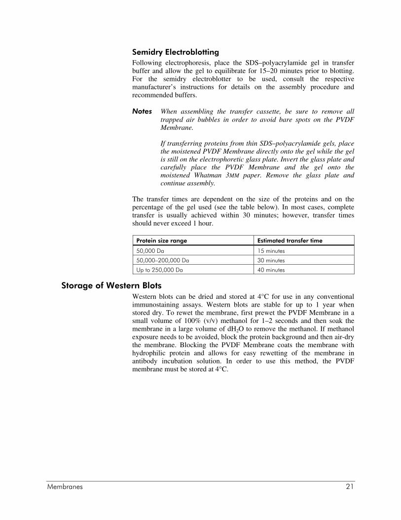
The transfer times are dependent on the size of the proteins and on thepercentage of the gel used (see the table below). In most cases, completetransfer is usually achieved within 30 minutes; however, transfer timesshould never exceed 1 hour.
Protein size range Estimated transfer time
50,000 Da 15 minutes
50,000–200,000 Da 30 minutes
Up to 250,000 Da 40 minutes
Storage of Western BlotsWestern blots can be dried and stored at 4°C for use in any conventionalimmunostaining assays. Western blots are stable for up to 1 year whenstored dry. To rewet the membrane, first prewet the PVDF Membrane in asmall volume of 100% (v/v) methanol for 1–2 seconds and then soak themembrane in a large volume of dH2O to remove the methanol. If methanolexposure needs to be avoided, block the protein background and then air-drythe membrane. Blocking the PVDF Membrane coats the membrane withhydrophilic protein and allows for easy rewetting of the membrane inantibody incubation solution. In order to use this method, the PVDFmembrane must be stored at 4°C.
22 Membranes
PREPARATION OF MEDIA AND REAGENTS
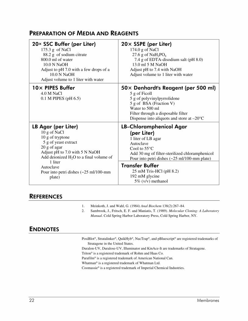
20× SSC Buffer (per Liter)175.3 g of NaCl 88.2 g of sodium citrate800.0 ml of water 10.0 N NaOHAdjust to pH 7.0 with a few drops of a
10.0 N NaOHAdjust volume to 1 liter with water
20× SSPE (per Liter)174.0 g of NaCl 27.6 g of NaH2PO4
7.4 g of EDTA-disodium salt (pH 8.0) 13.0 ml 5 M NaOHAdjust pH to 7.4 with NaOHAdjust volume to 1 liter with water
10× PIPES Buffer4.0 M NaCl0.1 M PIPES (pH 6.5)
50× Denhardt's Reagent (per 500 ml)5 g of Ficoll5 g of polyvinylpyrrolidone5 g of BSA (Fraction V)Water to 500 mlFilter through a disposable filterDispense into aliquots and store at –20°C
LB–Chloramphenicol Agar(per Liter)1 liter of LB agarAutoclaveCool to 55°CAdd 30 mg of filter-sterilized chloramphenicolPour into petri dishes (~25 ml/100-mm plate)
LB Agar (per Liter)10 g of NaCl10 g of tryptone 5 g of yeast extract20 g of agarAdjust pH to 7.0 with 5 N NaOHAdd deionized H2O to a final volume of
1 literAutoclavePour into petri dishes (~25 ml/100-mm
plate)
Transfer Buffer 25 mM Tris-HCl (pH 8.2)192 mM glycine 5% (v/v) methanol
REFERENCES
1. Meinkoth, J. and Wahl, G. (1984) Anal Biochem 138(2):267–84.2. Sambrook, J., Fritsch, E. F. and Maniatis, T. (1989). Molecular Cloning: A Laboratory
Manual. Cold Spring Harbor Laboratory Press, Cold Spring Harbor, NY.
ENDNOTES
PosiBlot®, Stratalinker®, QuikHyb®, NucTrap®, and pBluescript® are registered trademarks ofStratagene in the United States.
Duralon-UV, Duralose-UV, Illuminator and KinAce-It are trademarks of Stratagene.Triton® is a registered trademark of Rohm and Haas Co.Parafilm® is a registered trademark of American National Can.Whatman® is a registered trademark of Whatman Ltd.Coomassie® is a registered trademark of Imperial Chemical Industries.