
Melatonin promotes the BMP9-induced osteogenic ...
13
RESEARCH Open Access Melatonin promotes the BMP9-induced osteogenic differentiation of mesenchymal stem cells by activating the AMPK/β- catenin signalling pathway Tianyuan Jiang 1 , Chao Xia 1 , Xiaoting Chen 1 , Yan Hu 1 , Yan Wang 1 , Jin Wu 2 , Shuyan Chen 1* and Yanhong Gao 1* Abstract Background: Mesenchymal stem cells (MSCs) play a crucial role in maintaining the dynamic balance of bone metabolism. Melatonin may have a regulatory effect on bone metabolism by regulating the lineage commitment and differentiation signalling pathways of MSCs. Among the BMP families, the osteogenesis of BMP9 is considered to be one of the strongest in MSCs. Here, we explored whether melatonin and BMP9 act synergistically on MSC osteogenic differentiation. Methods: The C3H10T1/2 osteogenic differentiation function induced by melatonin synergizes with BMP9, as detected by the expression of osteogenic markers at different periods. The result was further confirmed by foetal limb explant culture and in vivo stem cell implantation experiments. The effects of the AMPK/β-catenin pathway on the osteogenic differentiation of C3H10T1/2 cells were evaluated by Western blotting. Results: Melatonin combined with BMP9 significantly enhanced the expression of osteogenic markers at different periods in C3H10T1/2 cells, effectively enhancing BMP9-induced bone formation in cultured foetal explants and ectopic bone formation in vivo in stem cell transplantation experiments. Melatonin increases the expression of BMP9 in C3H10T1/2 cells and induces Smad1/5/8 translocation from the cytoplasm to the nucleus. In addition, melatonin and BMP9 synergistically promote AMPK and β-catenin phosphorylation, which can be largely eliminated by AMPK siRNA pretreatment. Conclusions: Melatonin and BMP9 in C3H10T1/2 cells synergistically promote osteogenic differentiation at least in part by activating the AMPK/β-catenin signalling pathway. Keywords: Melatonin, BMP9, Osteogenic differentiation, Mesenchymal stem cells Background Mesenchymal stem cells (MSCs) are pluripotent stem cells derived from mesoderm that can differentiate into osteoblasts, adipocytes and chondrocytes with strong self-renewal and multi-directional differentiation [1, 2]. Bone is one of the few organs that retains the potential for regeneration and can be continuously remodelled throughout life [3]. The osteogenic differentiation of MSCs plays an important role in bone regeneration and remodelling. This process is regulated by a variety of hormones, transcription factors and cellular signalling pathways, including bone morphogenetic protein (BMP), Wnt, insulin-like growth factor (IGF), epidermal growth factor and growth hormone, and is involved in multiple pathway interactions [3–6]. Bone morphogenetic proteins (BMPs) are important signalling pathways for cell proliferation and differenti- ation during embryonic development and play a key role in regulating the osteogenic differentiation of mesenchy- mal stem cells [7]. BMPs belong to the transforming growth factor (TGF) superfamily and have at least 15 © The Author(s). 2019 Open Access This article is distributed under the terms of the Creative Commons Attribution 4.0 International License (http://creativecommons.org/licenses/by/4.0/), which permits unrestricted use, distribution, and reproduction in any medium, provided you give appropriate credit to the original author(s) and the source, provide a link to the Creative Commons license, and indicate if changes were made. The Creative Commons Public Domain Dedication waiver (http://creativecommons.org/publicdomain/zero/1.0/) applies to the data made available in this article, unless otherwise stated. * Correspondence: [email protected]; [email protected]; [email protected] 1 Department of Geriatrics, Xinhua Hospital, Shanghai Jiaotong University School of Medicine, Shanghai 200092, China Full list of author information is available at the end of the article Jiang et al. Stem Cell Research & Therapy (2019) 10:408 https://doi.org/10.1186/s13287-019-1511-7
Transcript of Melatonin promotes the BMP9-induced osteogenic ...
RESEARCH Open Access
Melatonin promotes the BMP9-inducedosteogenic differentiation of mesenchymalstem cells by activating the AMPK/β-catenin signalling pathwayTianyuan Jiang1, Chao Xia1, Xiaoting Chen1, Yan Hu1, Yan Wang1, Jin Wu2, Shuyan Chen1* and Yanhong Gao1*
Abstract
Background: Mesenchymal stem cells (MSCs) play a crucial role in maintaining the dynamic balance of bonemetabolism. Melatonin may have a regulatory effect on bone metabolism by regulating the lineage commitmentand differentiation signalling pathways of MSCs. Among the BMP families, the osteogenesis of BMP9 is consideredto be one of the strongest in MSCs. Here, we explored whether melatonin and BMP9 act synergistically on MSCosteogenic differentiation.
Methods: The C3H10T1/2 osteogenic differentiation function induced by melatonin synergizes with BMP9, asdetected by the expression of osteogenic markers at different periods. The result was further confirmed by foetallimb explant culture and in vivo stem cell implantation experiments. The effects of the AMPK/β-catenin pathway onthe osteogenic differentiation of C3H10T1/2 cells were evaluated by Western blotting.
Results: Melatonin combined with BMP9 significantly enhanced the expression of osteogenic markers at differentperiods in C3H10T1/2 cells, effectively enhancing BMP9-induced bone formation in cultured foetal explants andectopic bone formation in vivo in stem cell transplantation experiments. Melatonin increases the expression ofBMP9 in C3H10T1/2 cells and induces Smad1/5/8 translocation from the cytoplasm to the nucleus. In addition,melatonin and BMP9 synergistically promote AMPK and β-catenin phosphorylation, which can be largely eliminatedby AMPK siRNA pretreatment.
Conclusions: Melatonin and BMP9 in C3H10T1/2 cells synergistically promote osteogenic differentiation at least inpart by activating the AMPK/β-catenin signalling pathway.
Keywords: Melatonin, BMP9, Osteogenic differentiation, Mesenchymal stem cells
BackgroundMesenchymal stem cells (MSCs) are pluripotent stemcells derived from mesoderm that can differentiate intoosteoblasts, adipocytes and chondrocytes with strongself-renewal and multi-directional differentiation [1, 2].Bone is one of the few organs that retains the potentialfor regeneration and can be continuously remodelledthroughout life [3]. The osteogenic differentiation of
MSCs plays an important role in bone regeneration andremodelling. This process is regulated by a variety ofhormones, transcription factors and cellular signallingpathways, including bone morphogenetic protein (BMP),Wnt, insulin-like growth factor (IGF), epidermal growthfactor and growth hormone, and is involved in multiplepathway interactions [3–6].Bone morphogenetic proteins (BMPs) are important
signalling pathways for cell proliferation and differenti-ation during embryonic development and play a key rolein regulating the osteogenic differentiation of mesenchy-mal stem cells [7]. BMPs belong to the transforminggrowth factor (TGF) superfamily and have at least 15
© The Author(s). 2019 Open Access This article is distributed under the terms of the Creative Commons Attribution 4.0International License (http://creativecommons.org/licenses/by/4.0/), which permits unrestricted use, distribution, andreproduction in any medium, provided you give appropriate credit to the original author(s) and the source, provide a link tothe Creative Commons license, and indicate if changes were made. The Creative Commons Public Domain Dedication waiver(http://creativecommons.org/publicdomain/zero/1.0/) applies to the data made available in this article, unless otherwise stated.
* Correspondence: [email protected];[email protected]; [email protected] of Geriatrics, Xinhua Hospital, Shanghai Jiaotong UniversitySchool of Medicine, Shanghai 200092, ChinaFull list of author information is available at the end of the article
Jiang et al. Stem Cell Research & Therapy (2019) 10:408 https://doi.org/10.1186/s13287-019-1511-7
different subtypes present in humans. BMP2, BMP4,BMP7 and BMP9, all play an important role in promot-ing osteogenic differentiation and bone formation.Among these proteins, BMP2 and BMP7 have been eval-uated in clinical trials for the treatment of tibial fracturesand spinal fusion [8, 9]. BMP9 is considered to be one ofthe most effective factors for inducing mesenchymalstem cell osteogenesis in various BMPs. BMP9 phos-phorylates the transcription factor smad1, smad5 orsmad8. These phosphorylated r-smad factors bind tosmad4 and localize to the nucleus to promote the ex-pression of osteogenic genes [10, 11]. In addition, somefactors or signals, such as Wnt/β-catenin, ATRA andEGF, interact with BMP9 to enhance the BMP9-inducedosteogenic differentiation of MSCs [3, 4, 12].Melatonin (N-acetyl-5-methoxytryptamine) is a hor-
mone secreted by the pineal gland that affects circadianrhythm, regulates the sleep-wake cycle, inhibits tumourgrowth and regulates immunity [13]. In most mammals,including humans, melatonin receptors have two sub-types, MT1 and MT2, which belong to the G protein-coupled receptor family [14, 15]. Melatonin exerts itsphysiological regulation mainly through receptors dis-tributed in the hypothalamus (PT) and suprachiasmatic(SCN), and peripheral tissues, such as the nervous sys-tem, retina, immunity system, reproductive system andendocrine system tissues, also show a distribution ofthese receptors. Studies have shown that melatonin mayhave a regulatory effect on bone metabolism: promotingthe osteogenic differentiation of BMSCs while inhibitingadipogenic differentiation [16, 17]; inhibiting osteoclastformation and activation, thereby inhibiting bone resorp-tion [18]; and activating the antioxidant defence systemto maintain the self-renewal and differentiation ofBMSCs after long-term passage [19]. Additionally, theexpression of melatonin receptors was also found inMSCs. It is currently believed that the osteogenic effectsof melatonin are mainly mediated through the MT2 re-ceptor, and the mechanisms involved include TGF-β[20], the Wnt/β-catenin pathway [15] and the MAPKsignalling pathway [21]. It has been found that in pituit-ary AtT20 cells and rat granulosa cells, melatonin pro-motes the expression of BMP4 or BMP6 andphosphorylation of Smad1/5/8 downstream of BMPs[22, 23]. The melatonin-induced expression of osteo-genic markers, such as BMP4 or BMP2, was also foundin mouse pre-myoblast cell lines, C2C12 cells and hu-man dental pulp stem cells [24, 25]. However, whetherthis effect of melatonin on BMPs is also present inMSCs and how the interaction between melatonin andBMPs occurs during the osteogenic differentiation ofMSCs is still unclear, so further research is needed.The specific purpose of this study was to investigate the
effects of melatonin on the BMP9-induced osteogenic
differentiation of MSCs and to reveal the underlying mo-lecular mechanisms of this effect. Our results suggest thatmelatonin combined with BMP9 promotes osteogenic dif-ferentiation by activating the AMPK/β-catenin signallingpathway. Therefore, melatonin may provide another po-tentially effective option for enhancing BMP9-inducedosteogenesis in bone marrow mesenchymal stem cells.This effect has important clinical significance for promot-ing the application of MSCs in the field of bone tissue en-gineering and research, treatment and prevention of thepathogenesis of bone metabolism diseases, such as osteo-porosis, and fracture healing.
MethodsCell culture and chemicalsC3H10T1/2 cells were purchased from Shanghai Insti-tutes for Biological Sciences (Shanghai, China), and thecells were cultured in Dulbecco’s modified Eagle’smedium (DMEM) with 10% foetal bovine serum (FBS),100 U/ml penicillin and 100 mg/ml streptomycin. Theculture environment was 37 °C in 5% CO2. Melatoninwas diluted to 100 mM as storage concentrations inDMSO. DMSO was used as a solvent control. Unlessotherwise indicated, all chemicals were purchased fromSigma-Aldrich (St. Louis, MO, USA).
Alkaline phosphatase (ALP) activity and stainingThe total cellular protein concentration of the samplewas determined using a BCA protein assay (Beyotime,China). The activity of ALP was quantitatively deter-mined by modified Great EscAPe SEAP Chemilumines-cence Assay (BD Clontech, USA) as previouslydescribed. According to the definition of enzyme activ-ity, the alkaline phosphatase activity in the sample wascalculated. Qualitative detection of ALP activity was per-formed using a BCIP/NBT alkaline phosphatase stainingassay kit (Beyotime, China).
Alizarin red S stainingAlizarin red S staining was carried out as describedabove [8–10]. After the corresponding treatment,C3H10T1/2 cells were cultured in the presence of 50 μg/ml ascorbic acid, 100 nM dexamethasone and 10mM β-glycerophosphate. The medium was changed every 3days. After 21 days of culture, the medium was removed,the cells were washed twice with PBS and fixed at roomtemperature for 15 min using 4% paraformaldehyde, andthe cells were washed twice more with PBS. The fixedcells were incubated with 2% Alizarin red S solution(Sigma-Aldrich) for 10 min at room temperature andthen washed thoroughly in PBS. Calcium mineral depos-ition staining was observed by light microscopy (LeicaDMI 3000B, Germany).
Jiang et al. Stem Cell Research & Therapy (2019) 10:408 Page 2 of 13
Immunohistochemical (IHC) stainingAfter 14 days of the corresponding treatment,C3H10T1/2 cells were fixed with 4% paraformaldehydeand washed with PBS. The cells were permeabilized with0.1% Triton-X (Sigma, USA) and blocked with 10% BSA(Beyotime, China) to reduce non-specific staining. Sub-sequently, the cells were incubated with anti-osteocalcin(OCN) antibody (sc30045, Santa Cruz Biotechnology)and anti-osteopontin (OPN) antibody (ab91655, Abcam)at 4 °C overnight. The cells were washed three timeswith DPBS and incubated with a biotin-labelled second-ary antibody (Santa Cruz Biotechnology, USA) for 20min at 37 °C. The cells were washed three times withDPBS, and a streptavidin-HRP conjugate was added tothe cells to incubate for 20 min at 37 °C. The presence ofthe expected protein was observed by diaminobenzidine(DAB) staining and examined under a microscope. Con-trol IgG staining was used as a negative control.
Western blot analysisThe cells were lysed by ice-cold RIPA lysis buffer(Biyuntian, China) containing protease inhibitors, andthe protein concentration was quantified using a BCAprotein assay kit (Biyuntian). Each group of proteins wasdenatured by boiling, and then sodium dodecyl sulfate-polyacrylamide gel electrophoresis (SDS-PAGE) wasused to separate the proteins. Subsequently, the proteinswere transferred to a polyvinylidene fluoride (PVDF)membrane (Millipore, USA) by electrophoresis. Then,the membranes were blocked in 5% skim milk for 1 hand incubated overnight at 4 °C in diluted primary anti-body. Finally, images of the target strip were developedusing a Western Chemiluminescent HRP Substrate Kit(Millipore, USA). Image Lab software (Bio-Rad, USA)was used for semi-quantitative analysis. Primary anti-bodies directed against AMPK (D5A2 for monoclonalantibody, Cell Signaling Technology), p-AMPK (40H9for monoclonal antibody, Cell Signaling Technology), β-catenin (polyclonal antibodies, Cell Signaling Technol-ogy), p-β-catenin (polyclonal antibodies, Cell SignalingTechnology), p-Smad-1/5/8(D5B10 for monoclonal anti-body, Cell Signaling Technology), Smad-1/5/8 (N-18 formonoclonal antibody, Santa Cruz Biotechnology) andGADPH (6C5 for monoclonal antibody, Beyotime) wereused.
Transient transfection with small interfering RNAsFor RNA interference against AMPKα1/2 and CTNNB1,siRNA duplexes were synthesized, corresponding toAMPKα1/2 (target sequence: 5′-AAGAGAAGCAGAAGCACGACG-3′), CTNNB1 (sense: 5′-AUCACAGAUGUUGAAACAUTT-3′ and antisense: 5′-AUGUUUCAACAUCUGUGAUGG-3′) and control (5′-AAGCCGGTATGCCGGTTAAGT-3′). The cells were transfected
with 25 nM siRNA using Lipofectamine 2000 transfec-tion reagent (Invitrogen, USA) according to the manu-facturer’s instructions and subsequently treated at 2 daysafter transfection.
Immunofluorescence stainingCells cultured in 24-well plates were fixed with 4% para-formaldehyde for 20 min at room temperature and thenpermeabilized using 0.5% Triton X-100 for 20 min. Thefixed cells were then blocked in 5% BSA for 2 h and in-cubated overnight at 4 °C in a suitably diluted primaryantibody against p-Smad1/5/8 (1:500, Cell Signaling).After washing with PBS, the cells were incubated withAlexa Fluor 647-labelled goat anti-rabbit IgG (H+L)antibody (1:500 dilution; Beyotime, China) at roomtemperature in the dark for 1 h. The cells were then in-cubated with DAPI for 10 min and sealed with sealing li-quid containing an anti-fluorescence quencher, andimages were captured on a Leica DMI 3000B invertedfluorescence microscope.
Foetal limb explant cultureFoetal limbs were obtained by dissecting mouse embryos(E18.5) under sterile conditions and culturing in DMEMcontaining 0.5% BSA, 50 μg/ml ascorbic acid, 1 mMβ-glycerophosphate and 100 mg/ml penicillin and strepto-mycin (Sigma) at 37 °C in 5% CO2. After 24 h of in vitroculture, the limbs were treated accordingly. The mediumwas changed every 3 days. After 10 days of culture, cal-cein (100 mM, Sigma, USA) was added to the medium.After 12 days, the skin and muscle were removed, andnew bone formation was assessed using fluorescence mi-croscopy and histology. Each group includes at least fivelimb explants.
C3H10T1/2 implantation and micro-computedtomographic (μCT) analysisMale athymic nude mice (4 weeks old) were obtainedfrom Shanghai Laboratory Animal Research Center(Shanghai, China). C3H10T1/2 cells were treated withAdBMP9, AdGFP and melatonin alone or in combin-ation, and the infection efficiency was confirmed byfluorescence microscopy at 24 h later. After 7 days oftreatment, the cells were harvested for subcutaneous in-jection (5 × 10⁶ cells per injection) into the flanks ofathymic nude (nu/nu) mice (5 animals per group). Then,intrathecal injection was performed with melatonin (5mg/kg/day) or PBS, and after 5 weeks, the animals wereeuthanized, and the specimen was removed and fixed.Scanning and histological evaluation of ectopic bonewere performed using a high-resolution μCT system(SkyScan, Belgium) and analysis of the data using μCTanalyser software (SkyScan, Belgium). All experimentswere performed in accordance with the guidelines for
Jiang et al. Stem Cell Research & Therapy (2019) 10:408 Page 3 of 13
animal experiments of the Xinhua Hospital EthicsCommittee.
Histological evaluationThe removed tissues were fixed with 4% paraformalde-hyde, then decalcified and dehydrated with paraffin em-bedded. Serial sections of paraffin-embedded sampleswere stained with haematoxylin and eosin (H&E) or Mas-son’s trichrome stain as previously described [26, 27].
RNA extraction and real-time reverse transcriptionpolymerase chain reaction (real-time RT-PCR) analysisThe cells were seeded in 6-well plates, and treated withAdBMP9, AdGFP and melatonin alone or in combinationfor 2 days to extract total cellular RNA and perform reversetranscription. The qPCR reaction was carried out using aSYBR Green Premix Ex TaqTM kit (Takara, Japan). The re-action conditions were as follows: 95 °C for 5 s; 60 °C for 30s; 40 cycles. GAPDH was used as the internal referencegene, and the relative quantification was performed by theΔΔCt method (Comparative Delta-delta Ct).
Isolation of mouse bone marrow mesenchymal stem cells(BMSCs)Primary MSCs were collected from the bone marrow ofC57 mice using the method of Soleimani et al. [28]. The 8-week-old mouse was sacrificed by cervical dislocation, andthen the femur and tibia were separated. The bone marrowwas washed out with Dulbecco’s modified Eagle’s mediumlow glucose (DMEM LG, SH30021.01, Hyclone) with a 1-ml syringe until the bones turned white. The bone marrowextract was filtered through a 70-μm cell strainer and pel-leted at 1000 rpm for 5min. The cell pellet was resus-pended in 15mL DMEM LG containing 10% FBS and 100mg/ml Pen-Strep and inoculated into a T-75 cm2 flask. Thesolution was changed every 3 days. After the cell fusion de-gree reaches 80–90%, the cells are passaged, and the fourthgeneration can be used for experiments.
Statistical analysisAll data are expressed as the means ± standard error (SE).The experiments were performed at least three times toensure reproducibility. Statistical differences between thetwo groups were determined using one-way analysis ofvariance (ANOVA) or Student’s t test. A P value < 0.05 (*)was considered statistically significant.
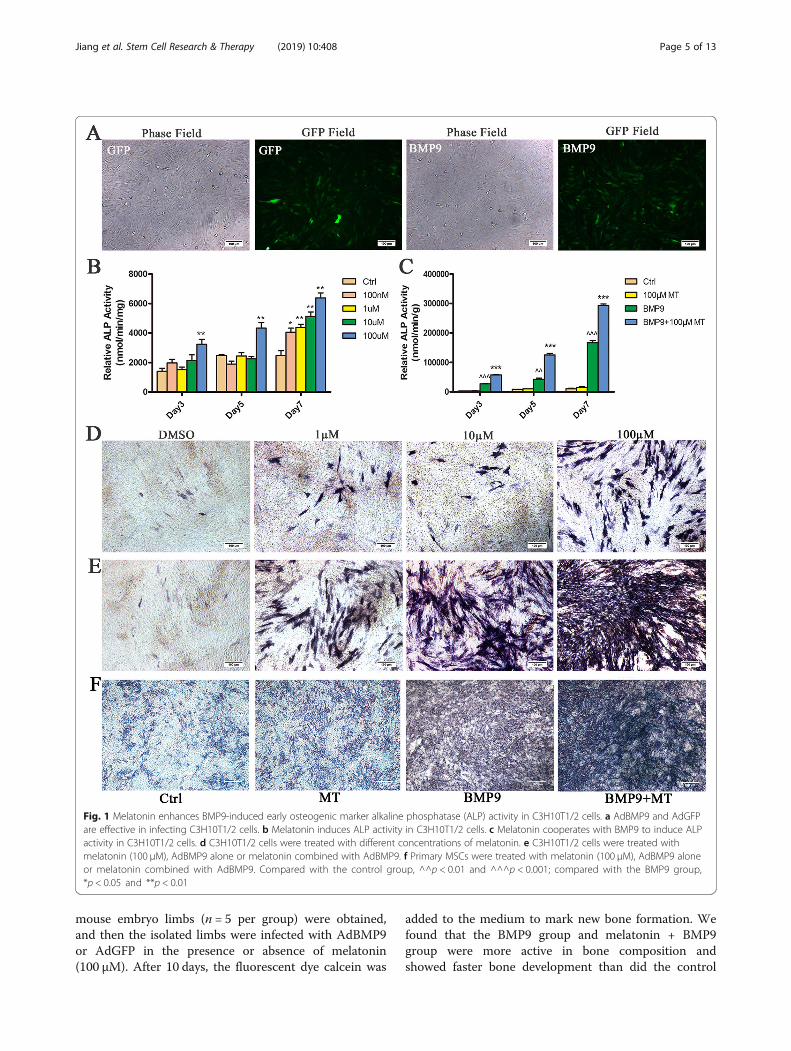
ResultsMelatonin synergizes with BMP9 to induce the ALPactivity of C3H10T1/2 cellsTo explore the effects of melatonin and BMP9 in synergis-tically inducing the osteogenic differentiation of MSC/C3H10T1/2 s, we used a recombinant adenovirus express-ing human BMP9 as described above and demonstrated
that this recombinant adenovirus is capable of efficientlytransducing C3H10T1/2 cells (Fig. 1a). We treatedC3H10T1/2 cells with 0 nM (control), 100 nM, 1 μM,10 μM and 100 μM melatonin for 3, 5 and 7 days to deter-mine the effect of melatonin on the osteogenic differenti-ation of C3H10T1/2 cells. The ALP activity of C3H10T1/2 cells increased with increasing melatonin dose and100 μM melatonin was able to induce ALP activity to thegreatest extent (Fig. 1b, d), which was selected for subse-quent experiments. Next, we used melatonin (100 μM)and BMP9 alone or in combination to stimulateC3H10T1/2 cells. The ALP activity assay showed thatBMP9 induced ALP activity earlier and stronger thanmelatonin stimulation. The combination of both mela-tonin and BMP9 can further enhance ALP activity (Fig. 1c,e). In addition, we performed the same experiment in pri-mary MSC cells, and the results were similar to those inC3H10T1/2 cells, and the combination of melatonin andBMP9 further enhanced ALP activity (Fig. 1f). In sum-mary, the data obtained indicate that melatonin can syner-gize with BMP9 to induce ALP activity in C3H10T1/2cells.
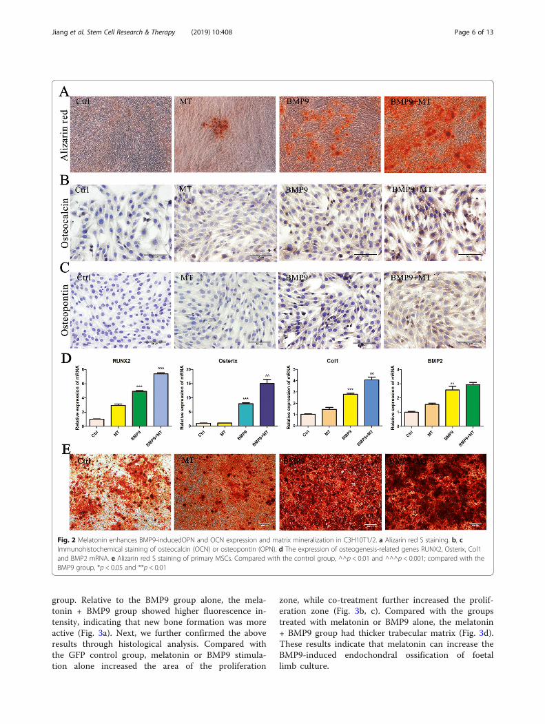
The combination of melatonin and BMP9 enhances thelate osteogenic marker expression and matrixmineralization of C3H10T1/2 cellsTo further confirm these results, we analysed the effect ofmelatonin on BMP9-induced late osteogenic markers. Theresults of Alizarin red S staining showed that the formationof calcium mineral deposits in C3H10T1/2 cells stimulatedby melatonin and BMP9 was significantly increased com-pared with BMP9 infection alone, and the matrixmineralization was obvious (Fig. 2a). Immunohistochemicalstaining showed that melatonin significantly enhanced theexpression of the BMP9-induced late osteogenic markersosteocalcin (Fig. 2b) and osteopontin (Fig. 2c). In addition,we examined the expression of osteogenesis-related genesRUNX2, Osterix, Col1 and BMP2 mRNA. The resultsshowed that melatonin combined with BMP9 significantlyincreased the expression levels of RUNX2, Osterix andCol1 mRNA, while the increase of BMP2 mRNA expres-sion levels was not statistically significant (Fig. 2d). Inaddition, we also performed Alizarin red S staining in pri-mary MSC cells, and the results were similar to those inC3H10T1/2 cells, the combination of melatonin and BMP9further enhanced the formation of calcium mineral depositsin MSCs (Fig. 2e). Based on these results, we conclude thatmelatonin signalling can synergize with BMP9-inducedosteogenic signalling in C3H10T1/2 cells.
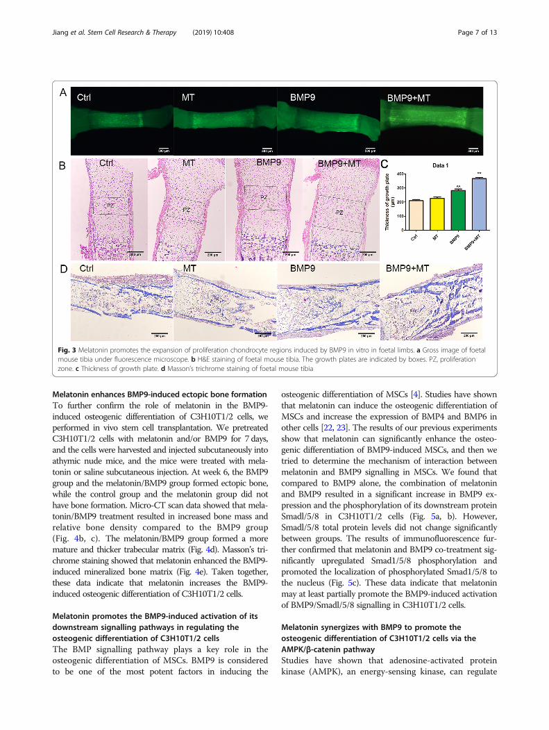
Melatonin promotes BMP9-induced osteogenesis ofembryonic limbs in vitroNext, we examined the effects of melatonin and BMP9on bone development through foetal limb culture. E18.5
Jiang et al. Stem Cell Research & Therapy (2019) 10:408 Page 4 of 13
mouse embryo limbs (n = 5 per group) were obtained,and then the isolated limbs were infected with AdBMP9or AdGFP in the presence or absence of melatonin(100 μM). After 10 days, the fluorescent dye calcein was
added to the medium to mark new bone formation. Wefound that the BMP9 group and melatonin + BMP9group were more active in bone composition andshowed faster bone development than did the control
Fig. 1 Melatonin enhances BMP9-induced early osteogenic marker alkaline phosphatase (ALP) activity in C3H10T1/2 cells. a AdBMP9 and AdGFPare effective in infecting C3H10T1/2 cells. b Melatonin induces ALP activity in C3H10T1/2 cells. c Melatonin cooperates with BMP9 to induce ALPactivity in C3H10T1/2 cells. d C3H10T1/2 cells were treated with different concentrations of melatonin. e C3H10T1/2 cells were treated withmelatonin (100 μM), AdBMP9 alone or melatonin combined with AdBMP9. f Primary MSCs were treated with melatonin (100 μM), AdBMP9 aloneor melatonin combined with AdBMP9. Compared with the control group, ^^p < 0.01 and ^^^p < 0.001; compared with the BMP9 group,*p < 0.05 and **p < 0.01
Jiang et al. Stem Cell Research & Therapy (2019) 10:408 Page 5 of 13
group. Relative to the BMP9 group alone, the mela-tonin + BMP9 group showed higher fluorescence in-tensity, indicating that new bone formation was moreactive (Fig. 3a). Next, we further confirmed the aboveresults through histological analysis. Compared withthe GFP control group, melatonin or BMP9 stimula-tion alone increased the area of the proliferation
zone, while co-treatment further increased the prolif-eration zone (Fig. 3b, c). Compared with the groupstreated with melatonin or BMP9 alone, the melatonin+ BMP9 group had thicker trabecular matrix (Fig. 3d).These results indicate that melatonin can increase theBMP9-induced endochondral ossification of foetallimb culture.
Fig. 2 Melatonin enhances BMP9-inducedOPN and OCN expression and matrix mineralization in C3H10T1/2. a Alizarin red S staining. b, cImmunohistochemical staining of osteocalcin (OCN) or osteopontin (OPN). d The expression of osteogenesis-related genes RUNX2, Osterix, Col1and BMP2 mRNA. e Alizarin red S staining of primary MSCs. Compared with the control group, ^^p < 0.01 and ^^^p < 0.001; compared with theBMP9 group, *p < 0.05 and **p < 0.01
Jiang et al. Stem Cell Research & Therapy (2019) 10:408 Page 6 of 13
Melatonin enhances BMP9-induced ectopic bone formationTo further confirm the role of melatonin in the BMP9-induced osteogenic differentiation of C3H10T1/2 cells, weperformed in vivo stem cell transplantation. We pretreatedC3H10T1/2 cells with melatonin and/or BMP9 for 7 days,and the cells were harvested and injected subcutaneously intoathymic nude mice, and the mice were treated with mela-tonin or saline subcutaneous injection. At week 6, the BMP9group and the melatonin/BMP9 group formed ectopic bone,while the control group and the melatonin group did nothave bone formation. Micro-CT scan data showed that mela-tonin/BMP9 treatment resulted in increased bone mass andrelative bone density compared to the BMP9 group(Fig. 4b, c). The melatonin/BMP9 group formed a moremature and thicker trabecular matrix (Fig. 4d). Masson’s tri-chrome staining showed that melatonin enhanced the BMP9-induced mineralized bone matrix (Fig. 4e). Taken together,these data indicate that melatonin increases the BMP9-induced osteogenic differentiation of C3H10T1/2 cells.
Melatonin promotes the BMP9-induced activation of itsdownstream signalling pathways in regulating theosteogenic differentiation of C3H10T1/2 cellsThe BMP signalling pathway plays a key role in theosteogenic differentiation of MSCs. BMP9 is consideredto be one of the most potent factors in inducing the
osteogenic differentiation of MSCs [4]. Studies have shownthat melatonin can induce the osteogenic differentiation ofMSCs and increase the expression of BMP4 and BMP6 inother cells [22, 23]. The results of our previous experimentsshow that melatonin can significantly enhance the osteo-genic differentiation of BMP9-induced MSCs, and then wetried to determine the mechanism of interaction betweenmelatonin and BMP9 signalling in MSCs. We found thatcompared to BMP9 alone, the combination of melatoninand BMP9 resulted in a significant increase in BMP9 ex-pression and the phosphorylation of its downstream proteinSmadl/5/8 in C3H10T1/2 cells (Fig. 5a, b). However,Smadl/5/8 total protein levels did not change significantlybetween groups. The results of immunofluorescence fur-ther confirmed that melatonin and BMP9 co-treatment sig-nificantly upregulated Smad1/5/8 phosphorylation andpromoted the localization of phosphorylated Smad1/5/8 tothe nucleus (Fig. 5c). These data indicate that melatoninmay at least partially promote the BMP9-induced activationof BMP9/Smadl/5/8 signalling in C3H10T1/2 cells.
Melatonin synergizes with BMP9 to promote theosteogenic differentiation of C3H10T1/2 cells via theAMPK/β-catenin pathwayStudies have shown that adenosine-activated proteinkinase (AMPK), an energy-sensing kinase, can regulate
Fig. 3 Melatonin promotes the expansion of proliferation chondrocyte regions induced by BMP9 in vitro in foetal limbs. a Gross image of foetalmouse tibia under fluorescence microscope. b H&E staining of foetal mouse tibia. The growth plates are indicated by boxes. PZ, proliferationzone. c Thickness of growth plate. d Masson’s trichrome staining of foetal mouse tibia
Jiang et al. Stem Cell Research & Therapy (2019) 10:408 Page 7 of 13
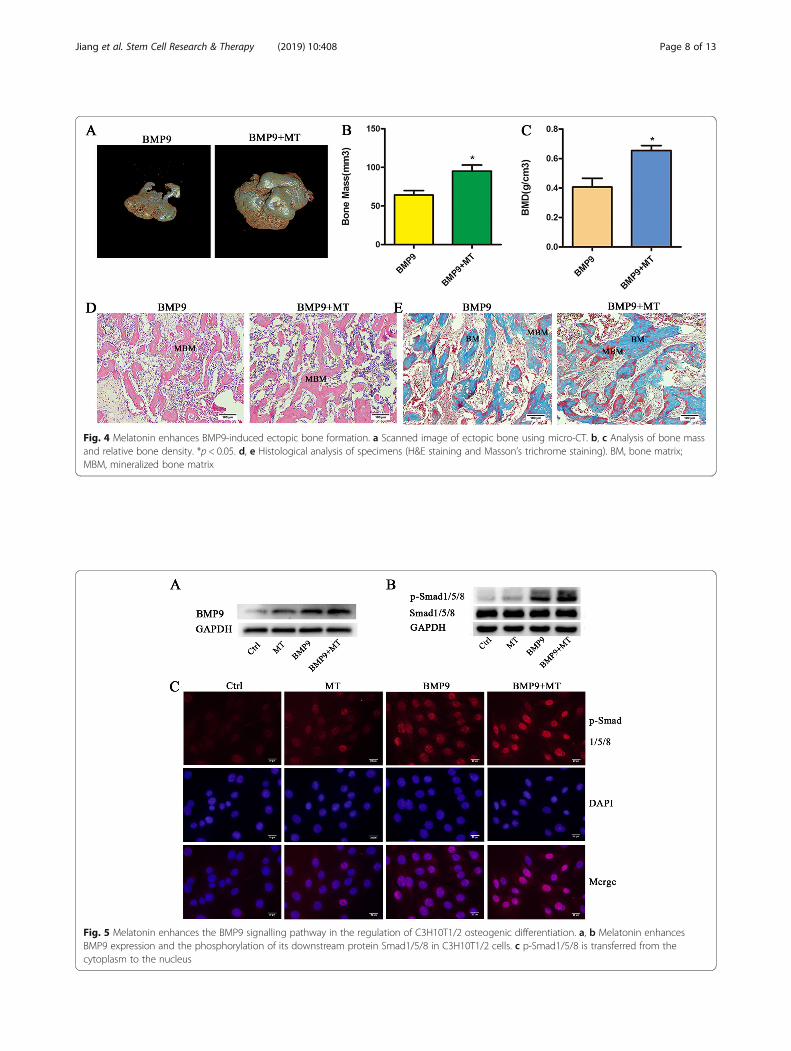
Fig. 5 Melatonin enhances the BMP9 signalling pathway in the regulation of C3H10T1/2 osteogenic differentiation. a, b Melatonin enhancesBMP9 expression and the phosphorylation of its downstream protein Smad1/5/8 in C3H10T1/2 cells. c p-Smad1/5/8 is transferred from thecytoplasm to the nucleus
Fig. 4 Melatonin enhances BMP9-induced ectopic bone formation. a Scanned image of ectopic bone using micro-CT. b, c Analysis of bone massand relative bone density. *p < 0.05. d, e Histological analysis of specimens (H&E staining and Masson’s trichrome staining). BM, bone matrix;MBM, mineralized bone matrix
Jiang et al. Stem Cell Research & Therapy (2019) 10:408 Page 8 of 13
bone metabolism by activating beta-catenin [29]. Mela-tonin can protect the osteogenic differentiation functionof MSCs by promoting the activation of AMPK or β-catenin [20, 30]. In multiple sources of MSCs, BMP9and Wnt/β-catenin signalling synergistically promoteosteogenic differentiation, and the knockdown of β-catenin eliminates the elevation of BMP9-induced osteo-genic marker ALP [4, 12]. Therefore, we examinedwhether melatonin and BMP9 have an effect on the acti-vation of AMPKα and β-catenin. Although there wereno significant changes in AMPKα and β-catenin proteinlevels in all groups, the phosphorylation of AMPKα atThr172 and the phosphorylation of β-catenin at Ser552were significantly increased (Fig. 6a). To further confirmthe role of AMPKα/β-catenin in melatonin- and BMP9-induced osteogenic differentiation, we treated C3H10T1/2 cells with AMPKα siRNA or β-catenin siRNA with agood silencing effect (Fig. 6c, d). Pretreatment withsiAMPKα blocked the β-catenin phosphorylation in-duced by co-treatment with melatonin and BMP9(Fig. 6b). Additionally, pretreatment with siAMPKα orsiβ-catenin significantly inhibited the increase in ALP,phosphorylation of Smad1/5/8 and expression of BMP9caused by the synergistic induction of melatonin andBMP9 (Fig. 6e–j). This finding suggests that the AMPK/β-catenin pathway is at least partially involved in mela-tonin/BMP9-induced osteogenic differentiation and thatAMPK may play a role, at least in the upstream signal-ling of β-catenin in MSCs.In summary, melatonin and BMP9 signalling show
crosstalk between the following pathways. Melatonin/BMP9 can directly upregulate bmp9 and its downstreamtargets and promote the transfer of p-Smad1/5/8 fromthe cytoplasm to the nucleus, which is more significantthan melatonin or BMP9 alone. By activating theAMPKα/β-catenin signalling pathway, melatonin/BMP9synergizes and leads to efficient bone formation.
DiscussionIn this study, we explored the possible synergistic rela-tionship between melatonin and BMP9 in inducing theosteogenic differentiation of mesenchymal stem cell lineC3H10T1/2 cells. The results indicate that melatonincan synergize with BMP9 to induce the osteogenic dif-ferentiation of C3H10T1/2 cells through the AMPK/β-catenin pathway. Currently, the role and mechanism ofmelatonin on the skeletal system has not been fully clari-fied, and the role of the melatonin and BMP pathwayinteraction in the process of MSC osteogenic differenti-ation has rarely been reported. The mechanism of actionbetween the two is very necessary.BMPs play an important regulatory role in cell prolif-
eration and differentiation during embryonic develop-ment and are key signalling pathways regulating the
osteogenic differentiation of mesenchymal stem cells [7].The interruption of BMP signalling leads to various boneand extra-bone abnormalities [31]. BMPs have the abilityto regulate the multilineage-specific differentiation ofMSCs. BMP2, BMP4, BMP6, BMP7 and BMP9 can effect-ively induce adipogenesis and osteogenic differentiationin vitro and in vivo, and BMP-induced osteogenesis oradipogenic differentiation has been shown to be mutuallyexclusive [12]. BMP9 is one of the most potent factors inthe BMP family to induce the osteogenic differentiation ofmesenchymal stem cells in vivo and in vitro, which havethe ability to form bone alone. However, many signallingpathways with different functions have been found to playa role in BMP9-mediated osteogenesis, such as Wnt/β-ca-tenin, ATRA and IGF [3, 4, 32].Studies have shown that melatonin may have a positive
effect on bone metabolism, but its osteogenesis mechan-ism remains unclear. Melatonin has widely distributedreceptors, including G protein-coupled melatonin recep-tors (MT1/MT2), nuclear receptors (ROR/RZR recep-tors), calmodulin and mitochondria. Among thesereceptors, the G protein receptor (MT1/MT2) is theclassical pathway for melatonin [33]. For perimenopausalwomen, nocturnal melatonin causes a time-dependentdecrease in the ratio of serum osteoclasts and osteoblasts[34]. Similarly, as age increases, melatonin levels de-crease. Bone mineral density (BMD) in the femoral neckincreases after treatment with melatonin in postmeno-pausal women [35]. Mechanistically, although melatoninhas no significant effect on the proliferation of MSCs[36, 37], melatonin has a regulatory effect on mesenchy-mal stem cell differentiation, which can promote osteo-genic and chondrogenic differentiation [38], inhibit lipiddifferentiation [36] and maintain the self-renewal anddifferentiation characteristics of MSCs [36]. Melatonincan also play a protective role in osteogenic differenti-ation disorders and ageing-related osteoporosis by acti-vating the antioxidant defence system, increasing boneformation or reducing bone resorption [13, 30, 39].Extracellular matrix (ECM) regulates the physiologicalfunction of hormones by providing binding sites andmodulating downstream signalling pathways. The inter-action of melatonin with ECM deposited by natural cellscan protect the osteogenic differentiation ability ofMSCs [40]. Therefore, melatonin may provide a new op-tion for the treatment of osteoporosis and fracture [19].Previous studies have shown that melatonin promotes
the expression of BMP4 or BMP6 in pituitary AtT20cells or rat granulosa cells and the phosphorylation ofSmad1/5/8 downstream of BMPs [22, 23]. Similarly,melatonin can activate osteogenic markers, such asRunx2, OCN and BMP2 and BMP4, in pre-osteoblastMC3T3-E1 cells in a dose-dependent manner [20].Therefore, we speculate that melatonin may induce
Jiang et al. Stem Cell Research & Therapy (2019) 10:408 Page 9 of 13
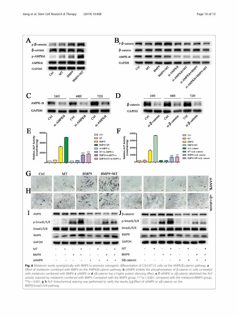
Fig. 6 Melatonin works synergistically with BMP9 to promote osteogenic differentiation of C3H10T1/2 cells via the AMPK/β-catenin pathway. aEffect of melatonin combined with BMP9 on the AMPK/β-catenin pathway. b siAMPK inhibits the phosphorylation of β-catenin in cells co-treatedwith melatonin combined with BMP9. c siAMPK or d siβ-catenin has a highly potent silencing effect. e, f siAMPK or siβ-catenin abolished the ALPactivity induced by melatonin combined with BMP9. Compared with the BMP9 group, ^^^p < 0.001; compared with the melatonin/BMP9 group,***p < 0.001. g, h ALP histochemical staining was performed to verify the results. i, j Effect of siAMPK or siβ-catenin on theBMP9/Smad1/5/8 pathway
Jiang et al. Stem Cell Research & Therapy (2019) 10:408 Page 10 of 13
BMP9 expression during the osteogenic differentiationof C3H10T1/2 cells, and there may be a mutual promot-ing effect between these two factors. Our results confirmthis idea, as melatonin induces BMP9 expression inC3H10T1/2 cells and enhances BMP9-induced Smad1/5/8 phosphorylation and Smad1/5/8 nuclear transloca-tion. Although both melatonin and BMP9 were able toinduce osteogenic marker expression alone, the combin-ation of the two stimuli significantly enhanced ALP ac-tivity as well as OCN and OPN expression.The osteogenic synthesis of collagen for bone model-
ling and bone remodelling requires the consumption oflarge amounts of adenosine triphosphate (ATP) [41].Disorders in circadian rhythms can alter a variety of pro-teins that regulate glucose balance and/or energy metab-olism in the body [42]. Melatonin may play a role in theregulation of energy metabolism by affecting the circa-dian rhythm [43, 44]. Leptin and adiponectin are adipo-kines that are closely related to glucose and lipidmetabolism and energy balance. Circadian rhythm disor-ders can interfere with the synthesis and secretion ofleptin and adiponectin, and melatonin supplementationnormalizes the expression and secretion patterns ofthese two adipokines [45]. AMPK is a key regulator ofenergy metabolism and is regulated by a variety of fac-tors, such as leptin, adiponectin and resistin [46, 47].Emerging evidence suggests that AMPK regulates celldifferentiation in addition to its role in metabolic pro-cesses [48, 49]: AMPK promotes osteogenesis and in-hibits lipogenesis in MC3T3-E1 cells [50]; and activeAMPK can directly phosphorylate RUNX2 and promoteosteogenesis [51]. Therefore, we speculate that mela-tonin may achieve synergistic osteogenesis with BMP9by activating AMPK. This idea is consistent with ourfindings that melatonin combined with BMP9 treatmentincreases phosphorylated AMPK levels.Both BMP and Wnt signalling are involved in the
regulation of osteoblast differentiation and bone forma-tion. In MSCs, Wnt3a and BMP9 regulate overlappingbut distinct downstream target gene expression [52, 53],suggesting that crosstalk may exist between their in-duced osteogenic signalling pathways, as demonstratedby subsequent studies [4, 54]. Crosstalk between AMPKand Wnt/β-catenin signalling was confirmed in bothin vitro and in vivo experiments [55, 56]. AMPK phos-phorylates β-catenin at Ser 552, stabilizes β-catenin andenhances β-catenin/TCF-mediated transcription, therebyregulating cell differentiation and developmental signal-ling pathways [46, 57]. Therefore, we propose that β-catenin may act downstream of AMPK and play a rolein the osteogenic differentiation induced by melatonin incombination with BMP9. To test this hypothesis, we ex-amined changes in AMPK/β-catenin in melatonin/BMP9-induced bone formation. The results indicate that
melatonin and BMP9 have a synergistic effect on the acti-vation of AMPK/β-catenin signalling. The knockout ofAMPK or β-catenin abolishes the stimulatory effect ofmelatonin on BMP9-induced alkaline phosphatase activ-ity. Therefore, we propose that melatonin combined withBMP9 may be an effective treatment for bone metabolicdiseases, such as osteoporosis, and fracture healing.However, our research also has some limitations. Stud-
ies have shown that the activation of the AMPK kinasecomplex induces mitochondrial division, increases apop-tosis and reduces the viability of MSCs. Knocking outAMPK may help reduce MSC damage in patients withmyelodysplastic syndrome [57]. Osteogenic differenti-ation is a multifactorial regulation process, and the mo-lecular mechanism of the AMPK/β-catenin pathway inosteogenic differentiation of C3H10T1/2 cells inducedby melatonin and BMP remains to be further studied.
ConclusionsIn summary, we investigated the effects of melatonin onBMP9-induced early and late osteogenic markers. Mech-anistically, we found that this process may be at leastpartially mediated by the AMPK/β-catenin pathway, andsilencing AMPK or β-catenin can effectively attenuateBMP9-induced osteogenesis. Therefore, the interactionbetween melatonin and the BMP9 pathway may play animportant role in regulating the osteogenic differenti-ation of MSCs. Exploring the combination of melatoninand BMP9 provides a new treatment for bone metabolicdiseases, such as osteoporosis.
AbbreviationsMSCs: Mesenchymal stem cells; BMP9: Bone morphogenetic protein (BMP)-9;MT: Melatonin; ALP: Alkaline phosphatase; OPN: Osteopotin;OCN: Osteocalcin
AcknowledgementsWe would like to thank Dr. He Baicheng (Chongqing Key Laboratory ofBiochemistry and Molecular Pharmacology, China) for providing therecombinant adenovirus of BMP9.
Authors’ contributionsAll authors read and approved the final manuscript.
FundingThis work was supported by the National Natural Science Foundation ofChina (81101360), the Shanghai Science and Technology Commission Fund(16ZR1422000 and 18411964500), the Shanghai Jiaotong University Medicaland Industrial Cross Fund (YG2015MS67) and the Shanghai Sailing Program(18YF1415700).
Availability of data and materialsThe datasets used and/or analysed during the current study are availablefrom the corresponding author on reasonable request.
Ethics approval and consent to participateThis research has been approved by the Ethics Committee of XinhuaHospital Affiliated to Shanghai Jiaotong University.
Consent for publicationNot applicable.
Jiang et al. Stem Cell Research & Therapy (2019) 10:408 Page 11 of 13
Competing interestsThe authors declare that they have no competing interests.
Author details1Department of Geriatrics, Xinhua Hospital, Shanghai Jiaotong UniversitySchool of Medicine, Shanghai 200092, China. 2Shanghai Institute for PediatricResearch, Shanghai Jiaotong University School of Medicine, Shanghai200092, China.
Received: 24 January 2019 Revised: 17 November 2019Accepted: 26 November 2019
References1. He X, He J, Shi Y, Pi C, Yang Y, Sun Y, et al. Nicotinamide
phosphoribosyltransferase (Nampt) may serve as the marker for osteoblastdifferentiation of bone marrow-derived mesenchymal stem cells. Exp CellRes. 2017;352(1):45–52.
2. Ho PJ, Yen ML, Tang BC, Chen CT, Yen BL. H2O2 accumulation mediatesdifferentiation capacity alteration, but not proliferative decline, in senescenthuman fetal mesenchymal stem cells. Antioxid Redox Signal. 2013;18(15):1895–905.
3. Liu X, Qin J, Luo Q, Bi Y, Zhu G, Jiang W, et al. Cross-talk between EGF andBMP9 signalling pathways regulates the osteogenic differentiation ofmesenchymal stem cells. J Cell Mol Med. 2013;17(9):1160–72.
4. Zhang H, Wang J, Deng F, Huang E, Yan Z, Wang Z, et al. Canonical Wntsignaling acts synergistically on BMP9-induced osteo/odontoblasticdifferentiation of stem cells of dental apical papilla (SCAPs). Biomaterials.2015;39:145–54.
5. Shi Y, Chen J, Karner CM, Long F. Hedgehog signaling activates a positivefeedback mechanism involving insulin-like growth factors to induceosteoblast differentiation. Proc Natl Acad Sci U S A. 2015;112(15):4678–83.
6. Bolamperti S, Signo M, Spinello A, Moro G, Fraschini G, Guidobono F, et al.GH prevents adipogenic differentiation of mesenchymal stromal stem cellsderived from human trabecular bone via canonical Wnt signaling. Bone.2018;112:136–44.
7. Mi LZ, Brown CT, Gao Y, Tian Y, Le VQ, Walz T, et al. Structure of bonemorphogenetic protein 9 procomplex. Proc Natl Acad Sci U S A. 2015;112(12):3710–5.
8. Cooper GS, Kou TD. Risk of cancer after lumbar fusion surgery withrecombinant human bone morphogenic protein-2 (rh-BMP-2). Spine. 2013;38(21):1862–8.
9. Khosla S, Westendorf JJ, Oursler MJ. Building bone to reverse osteoporosisand repair fractures. J Clin Invest. 2008;118(2):421–8.
10. Morine KJ, Qiao X, York S, Natov PS, Paruchuri V, Zhang Y, et al. Bonemorphogenetic protein 9 reduces cardiac fibrosis and improves cardiacfunction in heart failure. Circulation. 2018;138(5):513–526.
11. Ola R, Kunzel SH, Zhang F, Genet G, Chakraborty R, Pibouin-Fragner L, et al.SMAD4 prevents flow induced arterial-venous malformations by inhibitingcasein kinase 2. Circulation. 2018;138(21):2379–94.
12. Liu Y, Liu Y, Zhang R, Wang X, Huang F, Yan Z, et al. All-trans retinoic acidmodulates bone morphogenic protein 9-induced osteogenesis andadipogenesis of preadipocytes through BMP/Smad and Wnt/beta-cateninsignaling pathways. Int J Biochemistry Cell Biol. 2014;47:47–56.
13. Yang F, Yang L, Li Y, Yan G, Feng C, Liu T, et al. Melatonin protects bonemarrow mesenchymal stem cells against iron overload-induced aberrantdifferentiation and senescence. J Pineal Res. 2017;63(3):e12422.
14. Zhou L, Chen X, Liu T, Gong Y, Chen S, Pan G, et al. Melatonin reverses H2O2 -induced premature senescence in mesenchymal stem cells via theSIRT1-dependent pathway. J Pineal Res. 2015;59(2):190–205.
15. Luchetti F, Canonico B, Bartolini D, Arcangeletti M, Ciffolilli S, Murdolo G,et al. Melatonin regulates mesenchymal stem cell differentiation: a review. JPineal Res. 2014;56(4):382–97.
16. Nakade O, Koyama H, Ariji H, Yajima A, Kaku T. Melatonin stimulatesproliferation and type I collagen synthesis in human bone cells in vitro. JPineal Res. 1999;27(2):106–10.
17. Basoli V, Santaniello S, Cruciani S, Ginesu GC, Cossu ML, Delitala AP, et al.Melatonin and vitamin D interfere with the adipogenic fate of adipose-derived stem cells. Int J Mol Sci. 2017;18(5):981.
18. Shuai Y, Liao L, Su X, Yu Y, Shao B, Jing H, et al. Melatonin treatmentimproves mesenchymal stem cells therapy by preserving stemness duringlong-term in vitro expansion. J Pineal Res. 2016;6(11):1899–917.
19. Ping Z, Hu X, Wang L, Shi J, Tao Y, Wu X, et al. Melatonin attenuatestitanium particle-induced osteolysis via activation of Wnt/beta-cateninsignaling pathway. Acta Biomater. 2017;51:513–25.
20. Park KH, Kang JW, Lee EM, Kim JS, Rhee YH, Kim M, et al. Melatoninpromotes osteoblastic differentiation through the BMP/ERK/Wnt signalingpathways. J Pineal Res. 2011;51(2):187–94.
21. Radio NM, Doctor JS, Witt-Enderby PA. Melatonin enhances alkalinephosphatase activity in differentiating human adult mesenchymal stem cellsgrown in osteogenic medium via MT2 melatonin receptors and the MEK/ERK (1/2) signaling cascade. J Pineal Res. 2006;40(4):332–42.
22. Tsukamoto N, Otsuka F, Ogura-Ochi K, Inagaki K, Nakamura E, Toma K, et al.Melatonin receptor activation suppresses adrenocorticotropin productionvia BMP-4 action by pituitary AtT20 cells. Mol Cell Endocrinol. 2013;375(1–2):1–9.
23. Nakamura E, Otsuka F, Terasaka T, Inagaki K, Hosoya T, Tsukamoto-YamauchiN, et al. Melatonin counteracts BMP-6 regulation of steroidogenesis by ratgranulosa cells. J Steroid Biochem Mol Biol. 2014;143:233–9.
24. Han Y, Kim YM, Kim HS, Lee KY. Melatonin promotes osteoblastdifferentiation by regulating Osterix protein stability and expression. Sci Rep.2017;7(1):5716.
25. Cho YA, Noh K, Jue SS, Lee SY, Kim EC. Melatonin promotes hepaticdifferentiation of human dental pulp stem cells: clinical implications for theprevention of liver fibrosis. J Pineal Res. 2015;58(1):127–35.
26. Huang E, Zhu G, Jiang W, Yang K, Gao Y, Luo Q, et al. Growth hormonesynergizes with BMP9 in osteogenic differentiation by activating the JAK/STAT/IGF1 pathway in murine multilineage cells. J Bone Miner Res. 2012;27(7):1566–75.
27. Gao Y, Huang E, Zhang H, Wang J, Wu N, Chen X, et al. Crosstalk betweenWnt/beta-catenin and estrogen receptor signaling synergistically promotesosteogenic differentiation of mesenchymal progenitor cells. PLoS One. 2013;8(12):e82436.
28. Soleimani M, Nadri S. A protocol for isolation and culture of mesenchymalstem cells from mouse bone marrow. Nat Protoc. 2009;4(1):102–6.
29. Wang D, Ma W, Wang F, Dong J, Wang D, Sun B, et al. Stimulation of Wnt/beta-catenin signaling to improve bone development by Naringin viainteracting with AMPK and Akt. Cell Physiol Biochem. 2015;36(4):1563–76.
30. Lee S, Le NH, Kang D. Melatonin alleviates oxidative stress-inhibitedosteogenesis of human bone marrow-derived mesenchymal stem cellsthrough AMPK activation. Int J Med Sci. 2018;15(10):1083–91.
31. Lamplot JD, Qin J, Nan G, Wang J, Liu X, Yin L, et al. BMP9 signaling in stemcell differentiation and osteogenesis. Am J Stem Cells. 2013;2(1):1–21.
32. Chen L, Zou X, Zhang RX, Pi CJ, Wu N, Yin LJ, et al. IGF1 potentiates BMP9-induced osteogenic differentiation in mesenchymal stem cells through theenhancement of BMP/Smad signaling. BMB Rep. 2016;49(2):122–7.
33. Liu J, Zhou H, Fan W, Dong W, Fu S, He H, et al. Melatonin influencesproliferation and differentiation of rat dental papilla cells in vitro anddentine formation in vivo by altering mitochondrial activity. J Pineal Res.2013;54(2):170–8.
34. Maria S, Samsonraj RM, Munmun F, Glas J, Silvestros M, Kotlarczyk MP, et al.Biological effects of melatonin on osteoblast/osteoclast cocultures, bone,and quality of life: Implications of a role for MT2 melatonin receptors, MEK1/2, and MEK5 in melatonin-mediated osteoblastogenesis. J Pineal Res. 2018;64(3):e12465.
35. Amstrup AK, Sikjaer T, Heickendorff L, Mosekilde L, Rejnmark L. Melatoninimproves bone mineral density at the femoral neck in postmenopausalwomen with osteopenia: a randomized controlled trial. J Pineal Res. 2015;59(2):221–9.
36. Zhang L, Su P, Xu C, Chen C, Liang A, Du K, et al. Melatonin inhibitsadipogenesis and enhances osteogenesis of human mesenchymal stemcells by suppressing PPARgamma expression and enhancing Runx2expression. J Pineal Res. 2010;49(4):364–72.
37. Zhang L, Zhang J, Ling Y, Chen C, Liang A, Peng Y, et al. Sustained releaseof melatonin from poly (lactic-co-glycolic acid) (PLGA) microspheres toinduce osteogenesis of human mesenchymal stem cells in vitro. J PinealRes. 2013;54(1):24–32.
38. Wang B, Wen H, Smith W, Hao D, He B, Kong L. Regulation effects ofmelatonin on bone marrow mesenchymal stem cell differentiation. J CellPhysiol. 2019;234(2):1008–15.
39. Liu X, Gong Y, Xiong K, Ye Y, Xiong Y, Zhuang Z, et al. Melatonin mediatesprotective effects on inflammatory response induced by interleukin-1 betain human mesenchymal stem cells. J Pineal Res. 2013;55(1):14–25.
Jiang et al. Stem Cell Research & Therapy (2019) 10:408 Page 12 of 13
40. He F, Liu X, Xiong K, Chen S, Zhou L, Cui W, et al. Extracellular matrixmodulates the biological effects of melatonin in mesenchymal stem cells. JEndocrinol. 2014;223(2):167–80.
41. Lee WC, Guntur AR, Long F, Rosen CJ. Energy metabolism of the osteoblast:implications for osteoporosis. Endocr Rev. 2017;38(3):255–66.
42. Depner CM, Melanson EL, McHill AW, Wright KP Jr. Mistimed food intakeand sleep alters 24-hour time-of-day patterns of the human plasmaproteome. Proc Natl Acad Sci U S A. 2018;115(23):E5390–e9.
43. de Farias TS, de Oliveira AC, Andreotti S, do Amaral FG, Chimin P, deProenca AR, et al. Pinealectomy interferes with the circadian clock genesexpression in white adipose tissue. J Pineal Res. 2015;58(3):251–61.
44. Cipolla-Neto J, Amaral FG, Afeche SC, Tan DX, Reiter RJ. Melatonin, energymetabolism, and obesity: a review. J Pineal Res. 2014;56(4):371–81.
45. Szewczyk-Golec K, Wozniak A, Reiter RJ. Inter-relationships of thechronobiotic, melatonin, with leptin and adiponectin: implications forobesity. J Pineal Res. 2015;59(3):277–91.
46. Zhao J, Yue W, Zhu MJ, Sreejayan N, Du M. AMP-activated protein kinase(AMPK) cross-talks with canonical Wnt signaling via phosphorylation ofbeta-catenin at Ser 552. Biochem Biophys Res Commun. 2010;395(1):146–51.
47. Chen H, Liu X, Chen H, Cao J, Zhang L, Hu X, et al. Role of SIRT1 and AMPKin mesenchymal stem cells differentiation. Ageing Res Rev. 2014;13:55–64.
48. Shah M, Kola B, Bataveljic A, Arnett TR, Viollet B, Saxon L, et al. AMP-activated protein kinase (AMPK) activation regulates in vitro bone formationand bone mass. Bone. 2010;47(2):309–19.
49. Boufroura FZ, Le Bachelier C, Tomkiewicz-Raulet C, Schlemmer D, Benoist JF,Grondin P, et al. A new AMPK activator, GSK773, corrects fatty acidoxidation and differentiation defect in CPT2-deficient myotubes. Hum MolGenet. 2018;27(19):3417–33.
50. Wang YG, Qu XH, Yang Y, Han XG, Wang L, Qiao H, et al. AMPK promotesosteogenesis and inhibits adipogenesis through AMPK-Gfi1-OPN axis. CellSignal. 2016;28(9):1270–82.
51. Chava S, Chennakesavulu S, Gayatri BM, Reddy ABM. A novel phosphorylationby AMP-activated kinase regulates RUNX2 from ubiquitination in osteogenesisover adipogenesis. Cell Death Dis. 2018;9(7):754.
52. Luo Q, Kang Q, Si W, Jiang W, Park JK, Peng Y, et al. Connective tissuegrowth factor (CTGF) is regulated by Wnt and bone morphogeneticproteins signaling in osteoblast differentiation of mesenchymal stem cells. JBiol Chem. 2004;279(53):55958–68.
53. Si W, Kang Q, Luu HH, Park JK, Luo Q, Song WX, et al. CCN1/Cyr61 isregulated by the canonical Wnt signal and plays an important role inWnt3A-induced osteoblast differentiation of mesenchymal stem cells. MolCell Biol. 2006;26(8):2955–64.
54. Tang N, Song WX, Luo J, Luo X, Chen J, Sharff KA, et al. BMP-9-inducedosteogenic differentiation of mesenchymal progenitors requires functionalcanonical Wnt/beta-catenin signalling. J Cell Mol Med. 2009;13(8b):2448–64.
55. Wang L, Di LJ. Wnt/beta-catenin mediates AICAR effect to increase GATA3expression and inhibit adipogenesis. J Biol Chem. 2015;290(32):19458–68.
56. Zhao JX, Yue WF, Zhu MJ, Du M. AMP-activated protein kinase regulatesbeta-catenin transcription via histone deacetylase 5. J Biol Chem. 2011;286(18):16426–34.
57. Lee YH, Kim JS, Kim JE, Lee MH, Jeon JG, Park IS, et al. Nanoparticlemediated PPARgamma gene delivery on dental implants improvesosseointegration via mitochondrial biogenesis in diabetes mellitus ratmodel. Nanomed. 2017;13(5):1821–32.
Publisher’s NoteSpringer Nature remains neutral with regard to jurisdictional claims inpublished maps and institutional affiliations.
Jiang et al. Stem Cell Research & Therapy (2019) 10:408 Page 13 of 13


