MELAS - SciELO · MELAS Clinical features, muscle biopsy and molecular genetics Paulo José...
9
Arq Neuropsiquiatr 2009;67(3-A):668-676 668 MELAS Clinical features, muscle biopsy and molecular genetics Paulo José Lorenzoni 1 , Rosana H. Scola 1 , Cláudia S. Kamoi Kay 1 , Raquel C. Arndt 1 , Aline A. Freund 1 , Isac Bruck 2 , Mara Lúcia S.F. Santos 3 , Lineu C. Werneck 1 Abstract – Objective: The aim of the study was to analyze a series of Brazilian patients suffering from MELAS. Method: Ten patients with MELAS were studied with correlation between clinical findings, laboratorial data, electrophysiology, histochemical and molecular features. Results: Blood lactate was increased in eight patients. Brain image studies revealed a stroke-like pattern in all patients. Muscle biopsy showed ralled-red fibers (RRF) in 90% of patients on modified Gomori-trichrome and in 100% on succinate dehydrogenase stains. Cytochrome c oxidase stain analysis indicated deficient activity in one patient and subsarcolemmal accumulation in seven patients. Strongly succinate dehydrogenase-reactive blood vessels (SSV) occurred in six patients. The molecular analysis of tRNA Leu(UUR) gene by PCR/RLFP and direct sequencing showed the A3243G mutation on mtDNA in 4 patients. Conclusion: The muscle biopsy often confirmed the MELAS diagnosis by presence of RRF and SSV. Molecular analysis of tRNA Leu(UUR) gene should not be the only diagnostic criteria for MELAS. KEY WORDS: MELAS, mitochondrial myopathies, biopsy, mitochondrial DNA, genetics. MELAS: manifestações clínicas, biópsia muscular e estudo molecular Resumo – Objetivo: O objetivo deste estudo foi analisar uma série de pacientes brasileiros portadores de MELAS. Método: Dez pacientes com MELAS foram estudados com correlação entre manifestações clínicas, alterações laboratoriais, estudo eletrofisiológico, histoquímico e molecular. Resultados: O nível de lactato sérico estava aumentado em 8 pacientes. O estudo das imagens do crânio revelou padrão semelhante ao de AVC isquêmico em todos os pacientes. A biópsia muscular mostrou fibras rajadas vermelhas (RRF) em 90% dos pacientes na coloração pelo tricrômio de Gomori modificado e em 100% na reação histoquímica pela desidrogenase succicínica (SDH). A análise da coloração pela citocromo c oxidase indicou atividade deficiente em um paciente e acúmulo subsarcolemal em sete pacientes. Vasos com forte reação para SDH (SSV) ocorreram em seis pacientes. O estudo molecular do gene tRNA Leu(UUR) por PCR/RLFP e seqüenciamento direto mostrou a mutação A3243G no DNAmt de 4 pacientes. Conclusão: A biópsia muscular frequentemente confirma o diagnóstico de MELAS pela presença de RRF e SSV. O estudo molecular do gene tRNA Leu(UUR) não deve ser o único critério diagnóstico para MELAS. PALAVRAS-CHAVE: MELAS, miopatias mitocondriais, biópsia, DNA mitocondrial, genética. 1 Neurology/Neuromuscular Division, Internal Medicine Department, Universidade Federal do Paraná, Curitiba PR, Brazil; 2 Neuropediatric Division, Pediatric Department, Universidade Federal do Paraná, Curitiba PR, Brazil; 3 Pediatric Neurology Department, Hospital Pequeno Príncipe, Curitiba PR, Brazil. This study was supported by Fundação Araucária, CAPES and CNPq. Received 5 December 2008, received in final form 30 March 2009. Accepted 28 May 2009. Dra. Rosana Herminia Scola – Serviço de Doenças Neuromusculares / Hospital de Clínicas da UFPR - Rua General Carneiro 181 / 3 o andar - 80060-900 Curitiba PR - Brasil. E-mail: [email protected] Mitochondrial encephalomyopathies are a heteroge- neous group of clinical syndromes, with various biochem- ical defects of the respiratory chain due to primary de- fects in cellular mitochondria 1-5 . Mitochondrial myopathy, encephalopathy, lactic acidosis, and stroke-like episodes (MELAS) is one of occurring mitochondrial disease which includes in its definition encephalopathy, frequently with seizures and progressive dementia; stroke-like episodes at a young age; and biochemical evidence for mitochondrial defects, such as lactic acidosis or ragged-red fibers (RRF) in the muscle biopsy 3,4,6 . The pathogenesis of stroke-like episodes in MELAS is controversial; the two major hypotheses posit de- pendence on metabolic dysfunction or angiopathy in
Transcript of MELAS - SciELO · MELAS Clinical features, muscle biopsy and molecular genetics Paulo José...
Arq Neuropsiquiatr 2009;67(3-A):668-676
668
MELAS
Clinical features, muscle biopsy and molecular genetics
Paulo José Lorenzoni1, Rosana H. Scola1, Cláudia S. Kamoi Kay1, Raquel C. Arndt1, Aline A. Freund1, Isac Bruck2, Mara Lúcia S.F. Santos3, Lineu C. Werneck1
Abstract – Objective: The aim of the study was to analyze a series of Brazilian patients suffering from MELAS. Method: Ten patients with MELAS were studied with correlation between clinical findings, laboratorial data, electrophysiology, histochemical and molecular features. Results: Blood lactate was increased in eight patients. Brain image studies revealed a stroke-like pattern in all patients. Muscle biopsy showed ralled-red fibers (RRF) in 90% of patients on modified Gomori-trichrome and in 100% on succinate dehydrogenase stains. Cytochrome c oxidase stain analysis indicated deficient activity in one patient and subsarcolemmal accumulation in seven patients. Strongly succinate dehydrogenase-reactive blood vessels (SSV) occurred in six patients. The molecular analysis of tRNALeu(UUR) gene by PCR/RLFP and direct sequencing showed the A3243G mutation on mtDNA in 4 patients. Conclusion: The muscle biopsy often confirmed the MELAS diagnosis by presence of RRF and SSV. Molecular analysis of tRNALeu(UUR) gene should not be the only diagnostic criteria for MELAS.
KEY WORDS: MELAS, mitochondrial myopathies, biopsy, mitochondrial DNA, genetics.
MELAS: manifestações clínicas, biópsia muscular e estudo molecular
Resumo – Objetivo: O objetivo deste estudo foi analisar uma série de pacientes brasileiros portadores de MELAS. Método: Dez pacientes com MELAS foram estudados com correlação entre manifestações clínicas, alterações laboratoriais, estudo eletrofisiológico, histoquímico e molecular. Resultados: O nível de lactato sérico estava aumentado em 8 pacientes. O estudo das imagens do crânio revelou padrão semelhante ao de AVC isquêmico em todos os pacientes. A biópsia muscular mostrou fibras rajadas vermelhas (RRF) em 90% dos pacientes na coloração pelo tricrômio de Gomori modificado e em 100% na reação histoquímica pela desidrogenase succicínica (SDH). A análise da coloração pela citocromo c oxidase indicou atividade deficiente em um paciente e acúmulo subsarcolemal em sete pacientes. Vasos com forte reação para SDH (SSV) ocorreram em seis pacientes. O estudo molecular do gene tRNALeu(UUR) por PCR/RLFP e seqüenciamento direto mostrou a mutação A3243G no DNAmt de 4 pacientes. Conclusão: A biópsia muscular frequentemente confirma o diagnóstico de MELAS pela presença de RRF e SSV. O estudo molecular do gene tRNALeu(UUR) não deve ser o único critério diagnóstico para MELAS.
PALAVRAS-CHAVE: MELAS, miopatias mitocondriais, biópsia, DNA mitocondrial, genética.
1Neurology/Neuromuscular Division, Internal Medicine Department, Universidade Federal do Paraná, Curitiba PR, Brazil; 2Neuropediatric Division, Pediatric Department, Universidade Federal do Paraná, Curitiba PR, Brazil; 3Pediatric Neurology Department, Hospital Pequeno Príncipe, Curitiba PR, Brazil. This study was supported by Fundação Araucária, CAPES and CNPq.
Received 5 December 2008, received in final form 30 March 2009. Accepted 28 May 2009.
Dra. Rosana Herminia Scola – Serviço de Doenças Neuromusculares / Hospital de Clínicas da UFPR - Rua General Carneiro 181 / 3o andar - 80060-900 Curitiba PR - Brasil. E-mail: [email protected]
Mitochondrial encephalomyopathies are a heteroge-neous group of clinical syndromes, with various biochem-ical defects of the respiratory chain due to primary de-fects in cellular mitochondria1-5. Mitochondrial myopathy, encephalopathy, lactic acidosis, and stroke-like episodes (MELAS) is one of occurring mitochondrial disease which includes in its definition encephalopathy, frequently with
seizures and progressive dementia; stroke-like episodes at a young age; and biochemical evidence for mitochondrial defects, such as lactic acidosis or ragged-red fibers (RRF) in the muscle biopsy3,4,6.
The pathogenesis of stroke-like episodes in MELAS is controversial; the two major hypotheses posit de-pendence on metabolic dysfunction or angiopathy in
Arq Neuropsiquiatr 2009;67(3-A)
669
MelasLorenzoni et al.
the brain, but these two elements are thought to be related7.
Since its first description, few patients with this dis-ease have been identified in Brazil8-10. In this study we an-alyze the clinical and laboratory manifestations, brain im-ages, histological and molecular findings in 10 Brazilian pa-tients suffering from MELAS.
METHODA retrospective analysis of 4400 muscle biopsies performed
from January 1978 to January 2008 disclosed ten patients with a diagnosis of MELAS, according to the following requisite clinical features: (1) at least one of the following: seizures, dementia, re-current headache or vomiting; (2) stroke-like episodes at a young age (with computerized tomographic or magnetic resonance im-aging evidence of focal brain abnormalities); and (3) lactic acido-sis, RRF, or both. Relevant data, including clinical evaluation, age, gender, course of the disease, serum muscle enzyme levels, blood and cerebrospinal fluid (CSF) lactate levels, muscle cytochrome c oxidase (COX) activity, electrocardiogram (ECG), needle elec-tromyography (EMG), nerve conduction study, brain images and histological/histochemical aspects of the each muscle biopsy, were collected. The consent for muscle biopsy and mitochondri-al DNA (mtDNA) tests was obtained in the out-patient clinic or during hospital admission for diagnostic investigation. All studies were performed following the acquisition of informed consent.
Clinical evaluationThe time of progression of the disease was considered as the
interval between the stroke-like episodes and the definitive di-agnosis by muscle biopsy. Other relevant data, including clini-cal evaluation, age, gender, course of disease, and family histo-ry, were collected.
Laboratorial analysisAbnormal serum levels of creatine kinase (CK), aldolase (AL),
blood and CSF lactate were registered as a proportion reflecting their increase above normal limits. COX activity was measured in muscle biopsy extracts.
Electrophysiological findingsThe EMG pattern was classified as normal, myopathic, dener-
vated or mixed (myopathic with denervation findings), according to standard procedures. Motor and sensory nerve conduction studies were performed. The ECG abnormalities were reported.
Brain imagesThe radiologic findings (computed tomographic (CT) or mag-
netic resonance imaging (MRI)) were separated by lesion loca-tion and presence of basal ganglia calcifications.
Muscle biopsy analysisMuscle biopsies were frozen in liquid nitrogen and cryostat
sections were stained histologically; histochemical reactions were performed from the routine examination of the reaction, according to standard procedures11. The frequency of deficient muscle fibers on COX stain; and of RRF on modified Gomori-trichrome (MGT) and succinate dehydrogenase (SDH) stains were determined by counting in approximately 500 to 1000 muscle fibers of each specimen. In all specimens, the blood vessels (in-tramuscular arteries) were identified comparing the hematox-ylin-eosin, MGT and SDH stains. The blood vessel character-istics identified by SDH staining were categorized as follows: unstained or faintly stained vessels walls (normal); or strongly succinate dehydrogenase-reactive blood vessels (SSV) or darkly stained vessels walls with dense granules (abnormal).
Molecular analysisThe mtDNA was isolated from skeletal muscle from the
quadriceps or biceps brachii using a modified phenol/chloro-form method. The tRNALeu(UUR) gene of mtDNA was analyzed by a combination of polymerase chain reaction with restriction frag-ment length polymorphism (PCR/RFLP) and direct sequencing.
In the first PCR/RFLP, used to detect a point mutation, an A to G transition at nucleotide 3243 (A3243G), we used oligonucle-otide primers corresponding to positions nt 3116–3134 (forward) and nt 3353–3333 (backward) to amplify the mtDNA putative mu-tation by conventional polymerase chain reaction (PCR) with Taq DNA polymerase. In the second PCR/RLFP, used to detect a point mutation, a T to C transition at nucleotide 3271 (T3271C), we used oligonucleotide primers corresponding to positions nt 3148–3169 (forward) and nt 3295–3272 (backward) for conven-tional PCR amplification with Taq DNA polymerase.
The PCR conditions were as follows: 30 cycles of denatur-ation (94oC for 1 min), annealing (55oC for 1 min) and primer ex-tension (72oC for 1 min). The first PCR produced a 238-base-pair (bp) fragment which was digested for 16 hours at 37oC with the restriction endonuclease Hae III. The second PCR produced a 148 bp fragment which was digested for 6 hours at 37oC with the re-striction endonuclease Dde I.
These products from PCR/RFLP were analyzed on a 12% non-denaturing polyacrylamide gel (run at 200 V for 2 hours) and the bands were visualized by standard silver staining. In the first PCR/RLFP, the pattern observed with normal mtDNA consist of three bands (169, 37 and 32 bp), whereas, the presence of the A3243G mutation creates an additional Hae III restriction site and the 169 bp fragment is cleaved into two fragments of 97 and 72 bp. In the second PCR/RLFP, the pattern observed with nor-mal mtDNA consists of two bands (103 and 45 bp), whereas the presence of the T3271C mutation creates an additional Dde I re-striction site and the 103 bp fragment is cleaved into two frag-ments of 79 and 24 bp.
If any mutation was observed by both PCR/RLFP, the tRNALeu(UUR) gene was submitted to direct sequencing.
For direct sequencing we used the nt 3116–3134 and nt 3353–3333 oligonucleotide primers to amplify the tRNALeu(UUR) gene by
Arq Neuropsiquiatr 2009;67(3-A)
670
MelasLorenzoni et al.
conventional PCR with Taq DNA polymerase. The first PCR con-ditions were 30 cycles of denaturation (94oC for 1 min), annealing (55oC for 1 min) and primer extension (72oC for 1 min); followed by purification of the PCR fragment by enzymatic method. These purified PCR fragments were submitted to a second PCR using Big Dye Mix (Applied Biosystems). The second PCR conditions were 35 cycles of denaturation (96oC for 15 sec), annealing (50oC for 15 sec) and primer extension (60oC for 4 min); followed by PCR fragment purification by the isopropanolol method. These am-plified fragments were directly sequenced in forward direction on ABI PRISM 3100 Avant Genetic Analyzer and sequences were compared to the revised Cambridge reference sequence12.
RESULTSThe sample consisted of 10 patients (4 female and 6
male), aged 3 to 27 years, with a predominance of child patients in this series (in 6 patients onset was before age 15 years). The early development was normal in 9 of 10 pa-tients (90%). The time of the progression of the disease varied to a maximum of 90 months, with a mean time of 27.5 months. The families had no history of possible mi-tochondrial disorders (Table 1).
The stroke-like episodes were present in all cases. The
other symptoms reported were vomiting (10/10), headache (8/10), seizures (6/10), weakness (6/10), dementia (5/10), hearing loss (3/10), short stature (2/10), ocular symptoms (3/10), ataxia (1/10) and facial neuropathy (1/10) (Table 1).
Blood lactate levels varied from one to three times normal levels in eight patients. CSF lactate level doubled in one patient during the stroke-like episode (Table 1).
Serum CK levels varied from normal (6 patients) to twice normal (2 patients). Serum AL levels were normal in all patients (Table 1).
Analysis of COX activity from muscle extract was pos-sible in 4 cases, of which two indicated COX deficiency (Table 1).
The EMG pattern, observed in five patients, was my-opathic in two, neurogenic (only in facial muscles) in one and normal in two. Nerve conduction studies showed fa-cial neuropathy in one patient (case 5), but were normal in the other patients.
ECG, performed in 6 patients, was normal in three but revealed Wolf-Parkinson-White syndrome in one, right bundle branch conduction disturbance in one and left ventricular hypertrophy together with nonspecific T wave abnormality in another.
Table 1. Clinical and laboratorial description of MELAS patients.
Case 1 2 3 4 5 6 7 8 9 10
Age (years) 12 22 9 3 4 15 14 8 25 27
Gender M F M M F F M M F M
Evolution time (months) 36 6 90 12 10 1 48 12 33 0
Family history _ _ _ _ _ _ _ _ _ _
Developmental delay _ _ _ + _ _ _ _ _ _
Stroke_like episodes + + + + + + + + + +
Vomiting + + + + + + + + + +
Headache + + + + _ + + + _ +
Seizures + + + _ _ + + + _ _
Weakness + _ _ _ _ + + + + +
Dementia _ _ _ _ _ + + + + +
Hearing loss _ _ + _ _ _ + _ _ +
Short stature _ _ _ _ _ _ + _ _ +
Ocular symptons _ _ _ _ _ _ + _ + +
Ataxia _ _ + _ _ _ _ _ _ _
Facial neuropathy _ _ _ _ + _ _ _ _ _
Increased blood lactate + ND + + + + + + _ +
Increased CSF lactate ND ND ND ND ND ND + _ ND ND
Increased CK _ ND _ _ _ _ + _ ND +
Increased AL _ ND ND ND _ _ _ _ ND ND
COX deficiency* + + ND _ _ ND ND ND ND ND
F: female; M: male; CSF: cerebrospinal fluid; CK: creatine kinase; AL: aldolase; COX: cytocrome c oxidase; *COX deficient activity in muscle extract; RRF: ragged_red fiber; MGT: modified Gomori trichome; SDH: succinate dehydrogenase; +: presented; _: absent; ND: not done.
Arq Neuropsiquiatr 2009;67(3-A)
671
MelasLorenzoni et al.
Table 2. Imaging, histological and molecular characteristics of MELAS patients.
Case Brain abnormality localization
Muscle biopsy (%)
SSV %
tRNALeu(UUR) gene mutation
*MGT *SDH COX– A3243G# T3271C# Others##
1 CT: bilateral temporal-occipital 10.0 11.8 ND 33.3 – – –
2 CT: right frontal-temporal 0.5 2.1 ND 0 ND ND ND
3 MRI: bilateral basal ganglia 2.4 5.3 0 0 ND ND ND
4 CT: left temporal-occipital 0 0.7 0 0 – – –
5 MRI: bilateral occipital 6.3 7.5 0 0 ND ND ND
6 CT: left temporal-parietal-occipital 0.4 3.8 0 37.5 + ND ND
7 MRI: left temporal-parietal-occipital 2.2 5.4 0 70 + ND ND
8 MRI: bilateral frontal-parietal-temporal 6.9 9.2 0 75 + ND ND
9 MRI: right cerebelar 0.8 2.0 32.7 38 – – –
10 MRI: left basal ganglia 6.7 11.4 0 14.2 + ND ND
SSV: strongly succinate dehydrogenase-reactive blood vessels; *MGT: ragged-red fiber stained by modified Gomori trichome; *SDH: ragged-red fiber stained by succinate dehydrogenase; COX–: cytocrome c oxidase deficient on muscle fiber stain; CT: computed tomography; MRI: magnetic resonance imaging; #molecular analysis by PCR/RLFP; ##molecular analysis of others mutation in tRNALeu(UUR) gene by direct sequencing; +: present; –: absent; ND: not done.
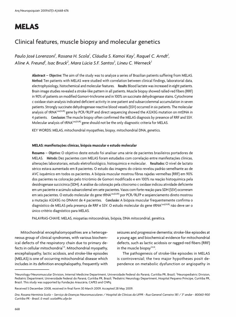
Fig 1. Brain magnetic resonance imag-ing confirms a unilateral stroke-like le-sion pattern in case 7 in axial T2 and FLAIR weighted images [A], and bilat-eral in case 8 in axial FLAIR weighted images [B].

Arq Neuropsiquiatr 2009;67(3-A)
672
MelasLorenzoni et al.
Brain image study, obtained from computed tomogra-phy in four patients and in six by magnetic resonance imag-ing, showed a stroke-like pattern in all patients. The lesion was unilateral in six patients and bilateral in four (Fig 1). The most common lesion locations were temporal-oc-cipital, frontal-temporal, basal ganglia, temporal, occip-ital, temporal-parietal-occipital, frontal-parietal-tempo-ral and cerebellar. The CT revealed basal ganglia calcifica-tions in two patients (Table 2).
The MGT and SDH stain were performed in all cases, but COX staining was performed only in 8 cases. The RRF occurred in nine cases by MGT stain and in all cases by SDH stain, but the proportion of RRF ranged from 0.4% to 11.6% on the MGT stain and from 0.7% to 11.8% on the SDH stain. Greater than 2% RRF was found in six of nine patients with the MGT stain and in eight cases with the SDH stain. COX deficient activity was found only in the muscle fibers of case 9, but COX subsarcolemmal accumu-lation occurred in 7 out 8 patients (Table 2 and Fig 2).
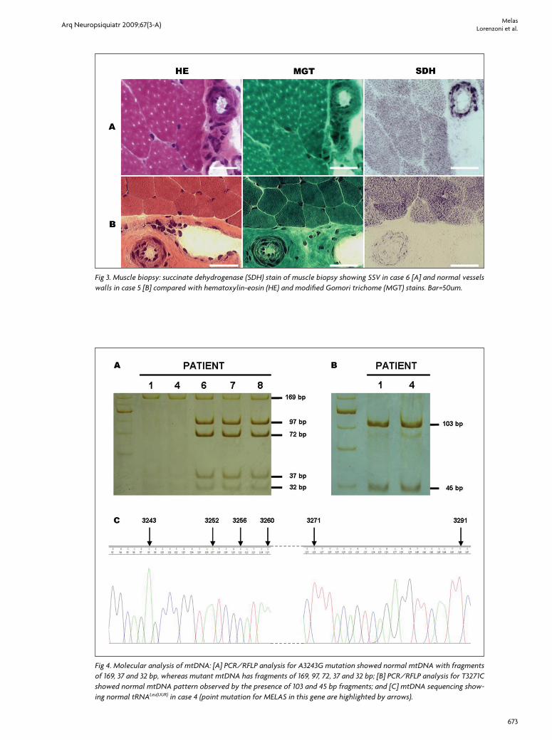
SSV occurred in six cases and its frequency ranged from 14.2% to 75% in these cases (Table 2 and Fig 3).
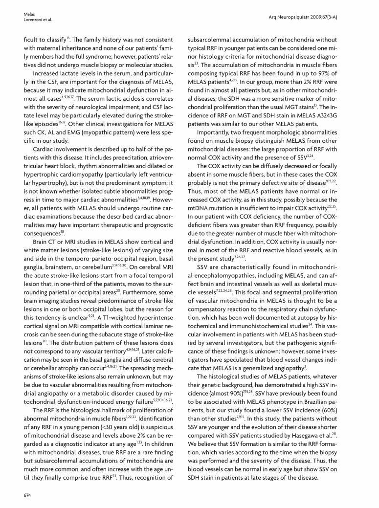
The molecular analysis was possible in the muscle bi-opsy from 7 patients (Table 2). The A3243G mutation was detected by PCR/RLFP in four (Fig 4A). Others mtDNA mutations were not found in tRNALeu(UUR) gene by PCR/RLFP to T3271C or direct sequence analysis (Fig 4B and 4C).
In the patients with MELAS related to the A3243G mu-tation, the most common findings were a mean time of progression of 15.2 months, normal levels of serum CK (two cases), normal levels of serum AL (three cases), EMG with myopathic pattern (two cases), normal COX activity (all cases) and SSV (all cases).
DISCUSSIONEarly disease development is normal in about 90% of
MELAS patients3,4,13. The onset of this disease occurs be-fore age 40 and is often seen in childhood, with grad-ual deterioration3,4,14. Clinical symptoms are highly vari-able across patients2. The most common feature in MELAS is episodic headaches with vomiting; this symptom was present in almost all patients3,4,15. Seizures occur in the vast majority of patients (95%)3, 4, 15. The other symptoms were not sufficiently severe to bring the patients to medical at-tention. Thus, the stroke-like episodes may appear abrupt-ly in otherwise normal-appearing children4,15.
Having an affected family member was a factor in 23% of patients with MELAS4,13. However, 86% of the oli-gosymptomatic and asymptomatic relatives of MELAS pa-tients are identified only by muscle biopsy or molecular studies4,13. In the absence of a member of the family with the full syndrome, and in the absence of molecular genet-ic studies, oligosymptomatic patients with MELAS are dif-
Fig 2. Muscle biopsy: [A] ragged-red fibers on modified Gomori-trichrome stain (case 8); [B] ragged-red fibers on succinate dehydrogenase stain (case 8); [C] deficient muscle fiber activity (*) on cytochrome c oxidase stain (case 9); and [D] normal muscle fiber activity on cytochrome c oxidase stain (case 8). Bar=50um.
Arq Neuropsiquiatr 2009;67(3-A)
673
MelasLorenzoni et al.
Fig 3. Muscle biopsy: succinate dehydrogenase (SDH) stain of muscle biopsy showing SSV in case 6 [A] and normal vessels walls in case 5 [B] compared with hematoxylin-eosin (HE) and modified Gomori trichome (MGT) stains. Bar=50um.
Fig 4. Molecular analysis of mtDNA: [A] PCR/RFLP analysis for A3243G mutation showed normal mtDNA with fragments of 169, 37 and 32 bp, whereas mutant mtDNA has fragments of 169, 97, 72, 37 and 32 bp; [B] PCR/RFLP analysis for T3271C showed normal mtDNA pattern observed by the presence of 103 and 45 bp fragments; and [C] mtDNA sequencing show-ing normal tRNALeu(UUR) in case 4 (point mutation for MELAS in this gene are highlighted by arrows).
Arq Neuropsiquiatr 2009;67(3-A)
674
MelasLorenzoni et al.
ficult to classify13. The family history was not consistent with maternal inheritance and none of our patients’ fami-ly members had the full syndrome; however, patients’ rela-tives did not undergo muscle biopsy or molecular studies.
Increased lactate levels in the serum, and particular-ly in the CSF, are important for the diagnosis of MELAS, because it may indicate mitochondrial dysfunction in al-most all cases4,9,16,17. The serum lactic acidosis correlates with the severity of neurological impairment, and CSF lac-tate level may be particularly elevated during the stroke-like episodes16,17. Other clinical investigations for MELAS such CK, AL and EMG (myopathic pattern) were less spe-cific in our study.
Cardiac involvement is described up to half of the pa-tients with this disease. It includes preexcitation, atrioven-tricular heart block, rhythm abnormalities and dilated or hypertrophic cardiomyopathy (particularly left ventricu-lar hypertrophy), but is not the predominant symptom; it is not known whether isolated subtle abnormalities prog-ress in time to major cardiac abnormalities3,4,18,19. Howev-er, all patients with MELAS should undergo routine car-diac examinations because the described cardiac abnor-malities may have important therapeutic and prognostic consequences18.
Brain CT or MRI studies in MELAS show cortical and white matter lesions (stroke-like lesions) of varying size and side in the temporo-parieto-occipital region, basal ganglia, brainstem, or cerebellum3,14,16,20. On cerebral MRI the acute stroke-like lesions start from a focal temporal lesion that, in one-third of the patients, moves to the sur-rounding parietal or occipital areas20. Furthermore, some brain imaging studies reveal predominance of stroke-like lesions in one or both occipital lobes, but the reason for this tendency is unclear9,21. A T1-weighted hyperintense cortical signal on MRI compatible with cortical laminar ne-crosis can be seen during the subacute stage of stroke-like lesions20. The distribution pattern of these lesions does not correspond to any vascular territory4,14,16,21. Later calcifi-cation may be seen in the basal ganglia and diffuse cerebral or cerebellar atrophy can occur3,4,16,21. The spreading mech-anisms of stroke-like lesions also remain unknown, but may be due to vascular abnormalities resulting from mitochon-drial angiopathy or a metabolic disorder caused by mi-tochondrial dysfunction-induced energy failure5,7,9,14,16,21.
The RRF is the histological hallmark of proliferation of abnormal mitochondria in muscle fibers1,22,23. Identification of any RRF in a young person (<30 years old) is suspicious of mitochondrial disease and levels above 2% can be re-garded as a diagnostic indicator at any age1,23. In children with mitochondrial diseases, true RRF are a rare finding but subsarcolemmal accumulations of mitochondria are much more common, and often increase with the age un-til they finally comprise true RRF23. Thus, recognition of
subsarcolemmal accumulation of mitochondria without typical RRF in younger patients can be considered one mi-nor histology criteria for mitochondrial disease diagno-sis23. The accumulation of mitochondria in muscle fibers composing typical RRF has been found in up to 97% of MELAS patients4,7,15. In our group, more than 2% RRF were found in almost all patients but, as in other mitochondri-al diseases, the SDH was a more sensitive marker of mito-chondrial proliferation than the usual MGT stains13. The in-cidence of RRF on MGT and SDH stain in MELAS A3243G patients was similar to our other MELAS patients.
Importantly, two frequent morphologic abnormalities found on muscle biopsy distinguish MELAS from other mitochondrial diseases: the large proportion of RRF with normal COX activity and the presence of SSV3,24.
The COX activity can be diffusely decreased or focally absent in some muscle fibers, but in these cases the COX probably is not the primary defective site of disease9,15,22. Thus, most of the MELAS patients have normal or in-creased COX activity, as in this study, possibly because the mtDNA mutation is insufficient to impair COX activity22,25. In our patient with COX deficiency, the number of COX-deficient fibers was greater than RRF frequency, possibly due to the greater number of muscle fiber with mitochon-drial dysfunction. In addition, COX activity is usually nor-mal in most of the RRF and reactive blood vessels, as in the present study7,26,27.
SSV are characteristically found in mitochondri-al encephalomyopathies, including MELAS, and can af-fect brain and intestinal vessels as well as skeletal mus-cle vessels7,22,24,28. This focal and segmental proliferation of vascular mitochondria in MELAS is thought to be a compensatory reaction to the respiratory chain dysfunc-tion, which has been well documented at autopsy by his-tochemical and immunohistochemical studies24. This vas-cular involvement in patients with MELAS has been stud-ied by several investigators, but the pathogenic signifi-cance of these findings is unknown; however, some inves-tigators have speculated that blood vessel changes indi-cate that MELAS is a generalized angiopathy3.
The histological studies of MELAS patients, whatever their genetic background, has demonstrated a high SSV in-cidence (almost 90%)7,15,28. SSV have previously been found to be associated with MELAS phenotype in Brazilian pa-tients, but our study found a lower SSV incidence (60%) than other studies7,9,15. In this study, the patients without SSV are younger and the evolution of their disease shorter compared with SSV patients studied by Hasegawa et al.28. We believe that SSV formation is similar to the RRF forma-tion, which varies according to the time when the biopsy was performed and the severity of the disease. Thus, the blood vessels can be normal in early age but show SSV on SDH stain in patients at late stages of the disease.
Arq Neuropsiquiatr 2009;67(3-A)
675
MelasLorenzoni et al.
The presence of SSV in muscle biopsy specimens pro-vides a relevant feature that can help to confirm the MELAS diagnosis, especially in patients without RRF in their muscle biopsy, but the lower incidence of SSV in our patients suggests that its absence should not be used as an exclusion criterion for MELAS7,15,28.
The degree of heteroplasmia (the proportion of normal and mutant mtDNA in each tissue) is an important factor influencing variability of phenotypical presentation9,15,22. Muscle biopsies from most MELAS cases, especially those associated with the most common mutation, A3243G in the tRNALeu(UUR) gene, have RRF with normal COX activity and SSV24. Quantitative analysis showed 80 to 90% mu-tant mtDNA in muscle25-27. The total mtDNA (both normal and mutant) are extremely increased in SSV and RRF25-27. The proportion of mutant mtDNA was significantly higher in RRF than in non-RRF, as well as in SSV than in non-SSV cases26,27. The similar morphological properties of these fibers and vessels suggest that increased mutant mtDNA is responsible for dysfunction and mitochondrial prolif-eration in both tissues of MELAS patients where COX is not a primarily defective enzyme25-27.
The present study suggested an association between SSV and A3243G mutation because all patients with this mutation had SSV. However, this study identified few cas-es with A3243G mutation.
The proportion of mutated mtDNA in blood cells is lower than in muscle (heteroplasmia), because of the turn-over of mitochondria in this tissue23. Therefore, the muscle was used in this study because genetic analysis of blood cells is not absolutely reliable as a diagnostic test and a negative result does not exclude mtDNA mutation23.
Only 6 years after its first clinical description, a point mutation in the mtDNA was associated with the MELAS syndrome29. A3243G mutation is the most frequent of the mtDNA mutations and causes a wide range of clinical dis-ease, of which MELAS is the most prevalent5,14. A3243G mu-tation occurs in approximately 80% of MELAS patients, but the present study showed association of A3243G muta-tion with MELAS in 57.1% of patients, possibly because this study had few cases for molecular analysis3,5,7,13,15. Due to the high frequency of this mutation, PCR/RLFP for the A3243G mutation must be the first molecular test when MELAS is suspected and is a simple molecular test for this genetic defect. The A3243G mutation accounts in approximately 80% of MELAS patients, but it is not the only genetic de-fect reported. Other point mutations in the same gene can be associated with MELAS9,13,15. The second most common mtDNA point mutation in MELAS patients is T3271C, which occurs in 7.5%. Brazilian patients were previously report-ed with this mutation, but we did not find this mutation in our group9. We suggest that the second genetic diag-nostic test should be the PCR/RLFP for T3271C7,9,13. MELAS
could be also caused by mtDNA point mutations in other genes, as the tRNAPhe, tRNAVal, tRNALys, COXIII, ND1, ND5 or rRNA, or by small-scale mtDNA deletions2,7. However, as the tRNALeu(UUR) mutations are still the most common-ly involved in the MELAS phenotype, we also speculate that molecular analysis of this gene by direct sequencing should be the third molecular test in MELAS patients7,24.
In our patients, the common mutation A3243G was found in 4 cases. Therefore, in MELAS with atypical bio-chemical or histological findings, such as COX deficiency, the putative mtDNA mutation might not involve one of the common tRNALeu(UUR) mutations24. In these cases, af-ter sequencing the tRNALeu(UUR) gene, the other possible “hot spots” for mtDNA mutation in MELAS were in the tRNAVal, tRNALys, ND1 or ND5 genes (all of which might be sequenced for diagnostic purpoes)3,5,14,24.
Our study reveals that muscle biopsy often confirms the MELAS diagnosis by presence of RRF on MGT and SDH stain, and SSV in muscle biopsy specimens. Molecu-lar analysis of tRNALeu(UUR) gene should not be the only di-agnostic criteria for MELAS.
REFERENCES 1. Walker UA, Collins S, Byrne E. Respiratory chain encephalomyopa-
thies: a diagnostic classification. Eur Neurol 1996;36:260-267. 2. Schmiedel J, Jackson S, Schafer J, Reichmann H. Mitochondrial cytop-
athies. J Neurol 2003;250:267-277. 3. Hirano M, Pavlakis SG. Mitochondrial myopathy, encephalopathy, lac-
tic acidosis, and stroke-like episodes (MELAS): current concepts. J Child Neurol 1994;9:4-13.
4. Hirano M, Ricci E, Koenigsberger MR, et al. MELAS: an original case and clinical criteria for diagnosis. Neuromuscul Disord 1992;2:125-135.
5. Zeviani M, Di Donato S. Mitochondrial disorders. Brain 2004;127:2153-2172.
6. Pavlakis SG, Phillips PC, DiMauro S, DeVivo DC, Rowland LP. Mito-chondrial myopathy, encephalopathy, lactic acidosis, and stroke-like ep-isodes: a distinctive clinical syndrome. Ann Neurol 1984;16:481-488.
7. Goto Y. Clinical features of MELAS and mitochondrial DNA mutations. Muscle Nerve 1995;20(Suppl 3):S107-S112.
8. Werneck LC, Abdalla H, Lohr A. MELAS (mitochondrial encephalopa-thy, lactic acidosis and stroke like episodes): case report. Arq Neurop-siquiatr 1987;45:288-294.
9. Marie SKN, Goto Y, Passos-Bueno MR, et al. A Caucasian family with the 3271 mutation in mitochondrial DNA. Biochem Med Metab Biol 1994;52:136-139.
10. Conforto AB, Yamamoto FI, Oba-Shinjo SM, et al. Screening for MELAS mutations in young patients with stroke of undetermined origin. Arq Neuropsiquiatr 2007;65:371-376.
11. Werneck LC. The value of muscle biopsy in neurology: a study of 290 biopsies. Rev Bras Clin Ter 1981;10(Suppl):S2-S24.
12. Anderson S, Bankier AT, Barrell BG, et al. Sequence and organization of the human mitochondrial genome. Nature 1981;290:457-465.
13. Ciafaloni E, Ricci E, Shanske S, et al. MELAS: clinical features, biochem-istry, and molecular genetics. Ann Neurol 1992;31:391-398.
14. Finsterer J. Genetic, pathogenetic, and phenotypic implications of the mitochondrial A3243G tRNALeu(UUR) mutation. Acta Neurol Scand 2007;116:1-14.
15. Goto Y, Horai S, Matsuoka T, et al. Mitochondrial myopathy, encephal-opathy, lactic acidosis, and stroke-like episodes (MELAS): a correlative study of the clinical features and mitochondrial DNA mutation. Neu-rology 1992;42:545-550.
Arq Neuropsiquiatr 2009;67(3-A)
676
MelasLorenzoni et al.
16. Finsterer J. Central nervous system manifestations of mitochondrial disorders. Acta Neurol Scand 2006;114:217-238.
17. Kaufmann P, Shungu DC, Sano MC, et al. Cerebral lactic acidosis cor-relates with neurological impairment in MELAS. Neurology 2004;62: 1297-1302.
18. Vydt TGC, Coo RFM, Soliman OI, et al. Cardiac involvement in adults with m.3243A>G MELAS gene mutation. Am J Cardiol 2007;99:264-269.
19. Okajima Y, Tanabe Y, Takayanagi M, Aotsuka H. A follow up study of myocardial involvement in patients with mitochondrial encephalomyo-pathy, lactic acidosis, and stroke-like episodes (MELAS). Heart 1998;80: 292-295.
20. Iizuka T, Sakai F, Kan S, Suzuki N. Slowly progressive spread of the stroke-like lesions in MELAS. Neurology 2003;61:1238-1244.
21. Haas R, Dietrich R. Neuroimaging of mitochondrial disorders. Mito-chondrion 2004;4:471-490.
22. Bourgeois JM, Tarnopolsky MA. Pathology of skeletal muscle in mito-chondrial disorders. Mitochondrion 2004;4:441-452.
23. Bernier FP, Boneh A, Dennett X, Chow CH, Cleary MA, Thorburn DR. Diagnostic criteria for respiratory chain disorders in adults and chil-dren. Neurology 2002;59:1406-1411.
24. Tanji K, Kaufmann P, Naini AB, et al. A novel tRNAVal mitochondrial mutation causing MELAS. J Neurol Sci 2008;270:23-27.
25. Petruzzella V, Moraes CT, Sano MC, Bonilla E, DiMauro S, Schon EA. Extremely high levels of mutant mtDNA co-localize with cytochrome c oxidase-negative ragged-red fibers in patients harboring a point mu-tation at nt 3243. Hum Mol Genet 1994;3:449-454.
26. Tokunaga M, Mita S, Sakuta R, Nonaka I, Araki S. Increased mito-chondrial DNA in blood vessels and ragged-red fibers in mitochondri-al myopathy, encephalopathy, lactic acidosis, and stroke-like episodes (MELAS). Ann Neurol 1993;33:275-280.
27. Tokunaga M, Mita S, Murakami T, et al. Single muscle fiber analysis of mitochondrial myopathy, encephalopathy, lactic acidosis, and stroke-like episodes (MELAS). Ann Neurol 1994;35:413-419.
28. Hasegawa H, Matsuoka T, Goto Y, Nonaka I. Strongly succinate dehy-drogenase-reactive blood vessels in muscle from patients with mito-chondrial myopathy, encephalopathy, lactic acidosis, and stroke-like episodes. Ann Neurol 1991;29:601-605.
29. Goto Y-I, Nonaka I, Horai S. A mutation in the tRNALeu(UUR) gene is as-sociated with the MELAS subgroup of mitochondrial encephalomyo-paties. Nature 1990;348:651-653.