Melanocytic Nevi and Neoplasms Andrew’s chapter 30 Michael Hohnadel, D.O. 4/20/04.
75
Melanocytic Nevi and Neoplasms Andrew’s chapter 30 Michael Hohnadel, D.O. 4/20/04 4/20/04
-
date post
22-Dec-2015 -
Category
Documents
-
view
216 -
download
2
Transcript of Melanocytic Nevi and Neoplasms Andrew’s chapter 30 Michael Hohnadel, D.O. 4/20/04.
Melanocytic Nevi and NeoplasmsAndrew’s chapter 30
Michael Hohnadel, D.O.
4/20/044/20/04
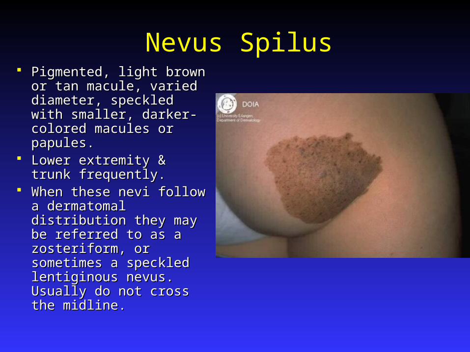
Nevus Spilus Pigmented, light brown or Pigmented, light brown or
tan macule, varied tan macule, varied diameter, speckled with diameter, speckled with smaller, darker-colored smaller, darker-colored macules or papules.macules or papules.
Lower extremity & trunk Lower extremity & trunk frequently.frequently.
When these nevi follow a When these nevi follow a dermatomal distribution dermatomal distribution they may be referred to as they may be referred to as a zosteriform, or a zosteriform, or sometimes a speckled sometimes a speckled lentiginous nevus. Usually lentiginous nevus. Usually do not cross the midline. do not cross the midline.

Nevus Spilus Syndromes: Syndromes:
Phakomatosis pigmentovascularis - nevus spilus Phakomatosis pigmentovascularis - nevus spilus is present with a nevus flammeus. is present with a nevus flammeus.
Phakomatosis pigmentokeratotica- Organoid Phakomatosis pigmentokeratotica- Organoid nevus with sebaceous differentiation, nevus with sebaceous differentiation, hemiatrophy with muscular weakness & other hemiatrophy with muscular weakness & other neurologic findings & speckled lentiginous neurologic findings & speckled lentiginous nevus.nevus.
The darker speckles usually contain nevus cells. The darker speckles usually contain nevus cells. Melanoma might arise with greater frequency than Melanoma might arise with greater frequency than in normal skin.in normal skin.
TX: Removal is not necessary. Q-switched ruby TX: Removal is not necessary. Q-switched ruby laser has been reported effective.laser has been reported effective.
Lentigo Simplex Sharply defined, rounded, brown or black macules Sharply defined, rounded, brown or black macules
found anywhere on body or mucosa. found anywhere on body or mucosa. No No predilection for sun exposed regions.predilection for sun exposed regions.
Usually arise in childhood but can arise anytime.Usually arise in childhood but can arise anytime. Histologically: Elongation of rete ridges, increase Histologically: Elongation of rete ridges, increase
in number of melanocytes in basal layer, increase in number of melanocytes in basal layer, increase of melanin in both melanocytes, and basal of melanin in both melanocytes, and basal keratinocytes, and melanophages in the upper keratinocytes, and melanophages in the upper dermis.dermis.
No therapy is needed/ there is No therapy is needed/ there is no predisposition to no predisposition to neoplastic change.neoplastic change.
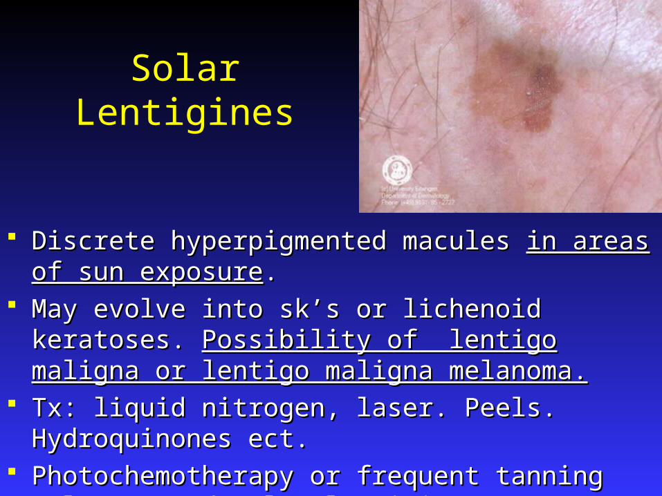
Solar Lentigines
Discrete hyperpigmented macules Discrete hyperpigmented macules in areas of sun exposurein areas of sun exposure.. May evolve into sk’s or lichenoid keratoses. May evolve into sk’s or lichenoid keratoses. Possibility of Possibility of
lentigo maligna or lentigo maligna melanoma.lentigo maligna or lentigo maligna melanoma. Tx: liquid nitrogen, laser. Peels. Hydroquinones ect.Tx: liquid nitrogen, laser. Peels. Hydroquinones ect. Photochemotherapy or frequent tanning salons may Photochemotherapy or frequent tanning salons may
develop lentigines on non-sun-exposed areas and these may develop lentigines on non-sun-exposed areas and these may show cellular atypiashow cellular atypia
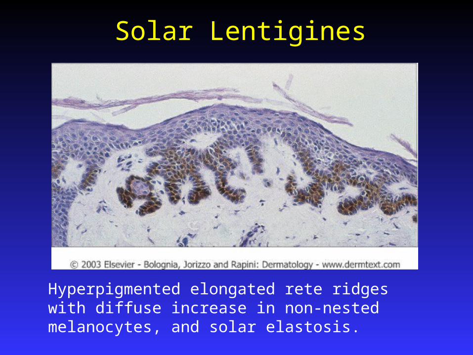
Solar Lentigines
Hyperpigmented elongated rete ridges with diffuse increase in non-nested melanocytes, and solar elastosis.

Penile and Vulvar Melanosis Localized pigmentary Localized pigmentary
alterationsalterations Most often show basilar Most often show basilar
hyperpigmentation hyperpigmentation May appear in large May appear in large
patches or in smaller, patches or in smaller, well-demarcated lesionswell-demarcated lesions
Present on the penis or Present on the penis or in women on the labia in women on the labia majora majora
Bannayan-Riley-Ruvalcaba Syndrome
Rare, AD disorder that manifests in Rare, AD disorder that manifests in childhood. 80% of the pts are malechildhood. 80% of the pts are male
Characterized by Characterized by genital lentiginosis, genital lentiginosis, macrocephaly, motor and speech delay, macrocephaly, motor and speech delay, mental retardation, lipomas, hemangiomas, mental retardation, lipomas, hemangiomas, verruca vulgaris, and many types of facial verruca vulgaris, and many types of facial papules.papules.
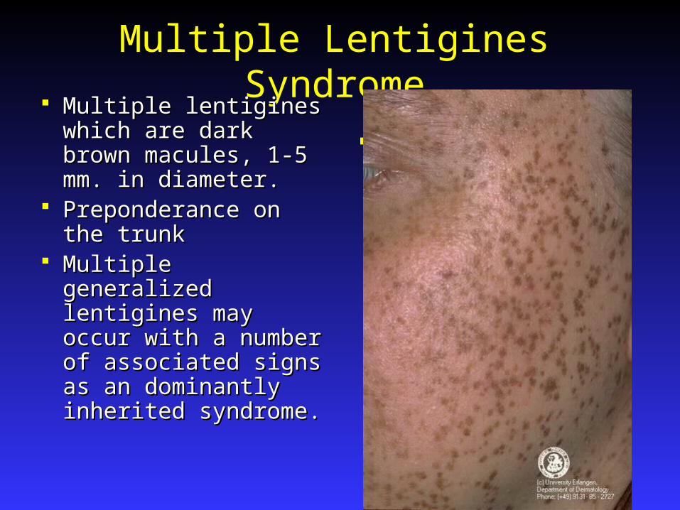
Multiple Lentigines Syndrome Multiple lentigines Multiple lentigines
which are dark brown which are dark brown macules, 1-5 mm. in macules, 1-5 mm. in diameter.diameter.
Preponderance on the Preponderance on the trunktrunk
Multiple generalized Multiple generalized lentigines may occur lentigines may occur with a number of with a number of associated signs as an associated signs as an dominantly inherited dominantly inherited syndrome.syndrome.
ll
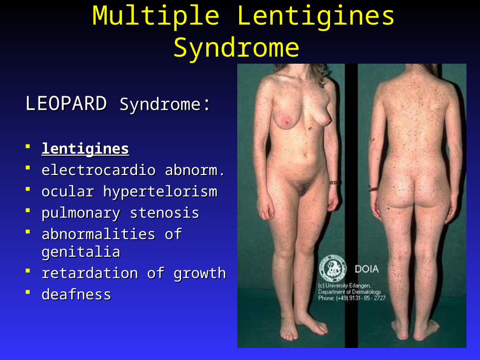
Multiple Lentigines Syndrome
LEOPARD LEOPARD SyndromeSyndrome::
lentigineslentigines electrocardio abnorm.electrocardio abnorm. ocular hypertelorismocular hypertelorism pulmonary stenosispulmonary stenosis abnormalities of genitalia abnormalities of genitalia retardation of growthretardation of growth deafnessdeafness
Moynahan SyndromeMoynahan Syndrome Multiple lentigines, Congenital mitral stenosis, Multiple lentigines, Congenital mitral stenosis,
Dwarfism, Genital hypoplasia and Mental Dwarfism, Genital hypoplasia and Mental deficiencydeficiency
Centrofacial LentiginosisCentrofacial Lentiginosis Characterized by lentigines on the nose, and Characterized by lentigines on the nose, and
adjacent cheeks adjacent cheeks Sometimes associated with status dysraphicus, Sometimes associated with status dysraphicus,
multiple skeletal anomalies, and CNS disordersmultiple skeletal anomalies, and CNS disorders Spares the mucous membranesSpares the mucous membranes Onset is first years of lifeOnset is first years of life
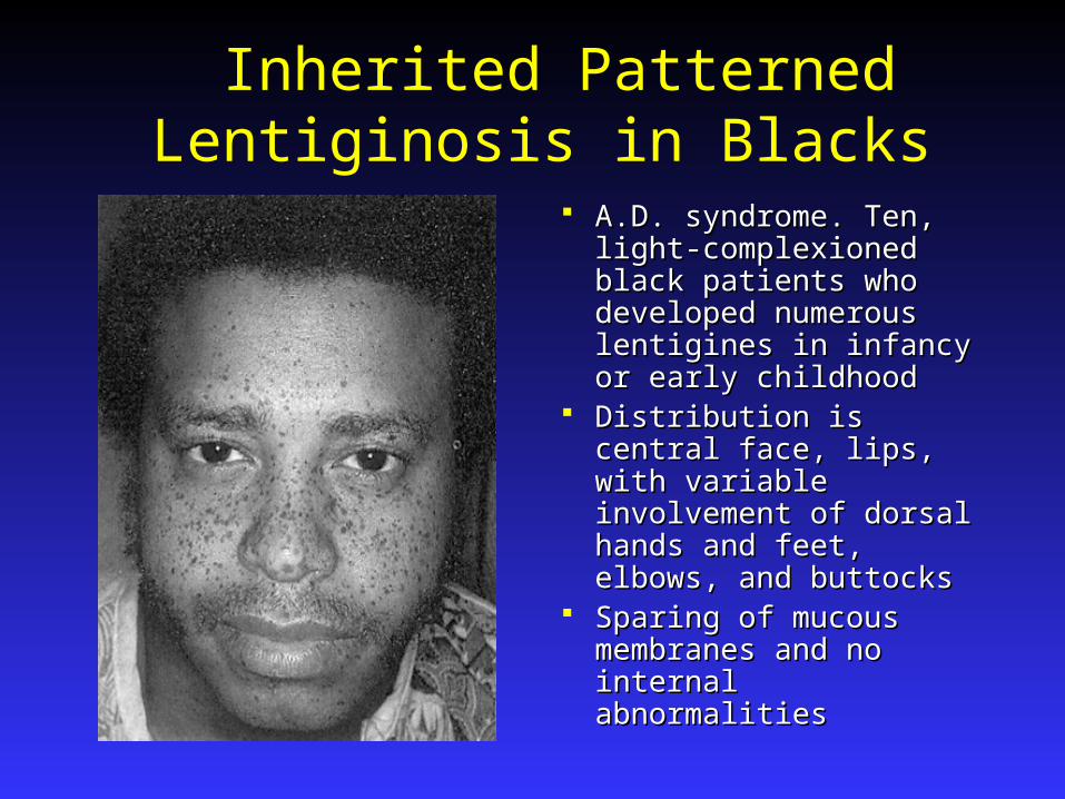
Inherited Patterned Lentiginosis in Blacks
A.D. syndrome. Ten, A.D. syndrome. Ten, light-complexioned black light-complexioned black patients who developed patients who developed numerous lentigines in numerous lentigines in infancy or early childhoodinfancy or early childhood
Distribution is central Distribution is central face, lips, with variable face, lips, with variable involvement of dorsal involvement of dorsal hands and feet, elbows, hands and feet, elbows, and buttocksand buttocks
Sparing of mucous Sparing of mucous membranes and no membranes and no internal abnormalitiesinternal abnormalities

Carney’s Syndrome AKA: NAME syndrome or LAMB syndromeAKA: NAME syndrome or LAMB syndrome Carney syndrome (2+ of following)Carney syndrome (2+ of following)
1.1.Cardiac Atrial Myxoma (79%) Can be life Cardiac Atrial Myxoma (79%) Can be life threatening.threatening.
2.2.Cutaneous myxomas (45%) <1 cm flesh colored Cutaneous myxomas (45%) <1 cm flesh colored papules which develop by the age of 18 and occur papules which develop by the age of 18 and occur on ears, eyelids and nipples.on ears, eyelids and nipples.
3.3.Mammary myxoid fibromas (30%)Mammary myxoid fibromas (30%)
4.4.Spotty mucocutaneous pigmentation (blue nevi) Spotty mucocutaneous pigmentation (blue nevi) (65%) or(65%) or lentigines lentigines
5.5.Prim. Pig. Nodular adrenocortical disease. (45%)Prim. Pig. Nodular adrenocortical disease. (45%)6.6.Testicular tumors (56%)Testicular tumors (56%)7.7.Pituitary G.H. secreting tumors. (10%)Pituitary G.H. secreting tumors. (10%)

Peutz-Jeghers Syndrome
A.D. A.D. Pigmented macules on Pigmented macules on
the lips, oral mucosa, the lips, oral mucosa, perioral acral areasperioral acral areas
Associated with Associated with gastrointestinal gastrointestinal polyps, especially polyps, especially prominent in the prominent in the jejunum.jejunum.
Melanoacanthoma
Uncommon, benign epidermal melanocytic Uncommon, benign epidermal melanocytic neoplasm, occurring on the headneoplasm, occurring on the head
Resembles a pigmented sk or a pigmented Resembles a pigmented sk or a pigmented BCC.BCC.
Predominantly seen in white men > 60 yrsPredominantly seen in white men > 60 yrs
Cellular Nevi Appear in first yrs of life, increases in number Appear in first yrs of life, increases in number
over the next 2-3 decades, after which there is a over the next 2-3 decades, after which there is a steady decline. F>M steady decline. F>M Less common in sun-protected areasLess common in sun-protected areas Maximum number is at age 20- 25 yrs, the average Maximum number is at age 20- 25 yrs, the average
number is 40number is 40 Sun exposure increases the number of nevi in the Sun exposure increases the number of nevi in the
exposed skin.exposed skin.
Eruptive nevi are rare, but may occur after severe Eruptive nevi are rare, but may occur after severe bullous disease such as TEN, EM, or severe bullous disease such as TEN, EM, or severe sunburn, Addison’s disease or immunosuppresion sunburn, Addison’s disease or immunosuppresion
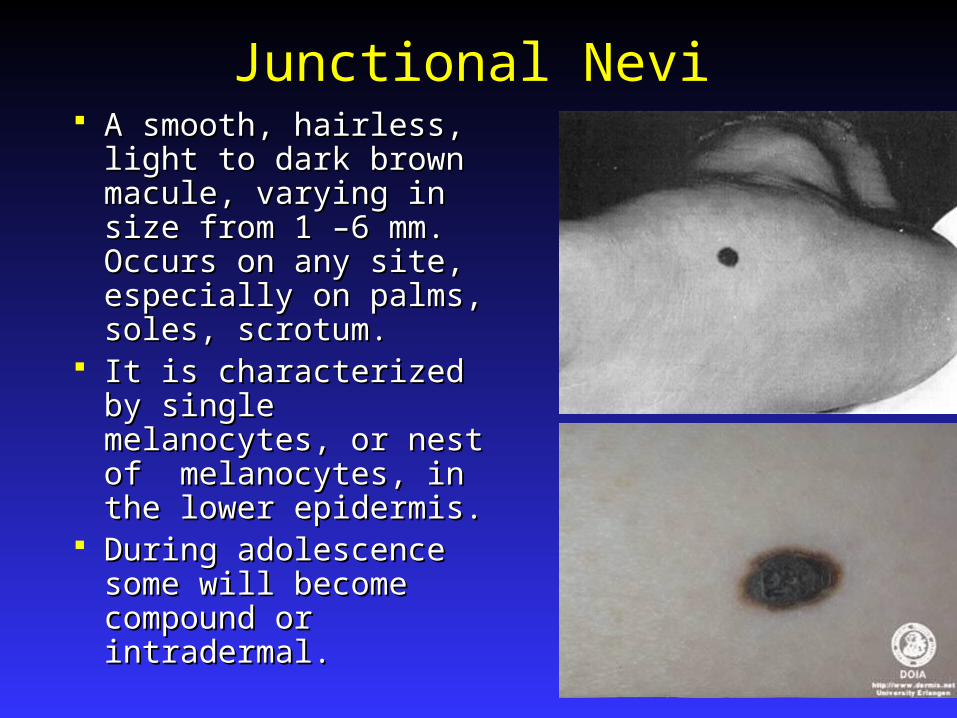
Junctional Nevi A smooth, hairless, light A smooth, hairless, light
to dark brown macule, to dark brown macule, varying in size from 1 –6 varying in size from 1 –6 mm. Occurs on any site, mm. Occurs on any site, especially on palms, especially on palms, soles, scrotum.soles, scrotum.
It is characterized by It is characterized by single melanocytes, or single melanocytes, or nest of melanocytes, in nest of melanocytes, in the lower epidermis.the lower epidermis.
During adolescence During adolescence some will become some will become compound or compound or intradermal. intradermal.
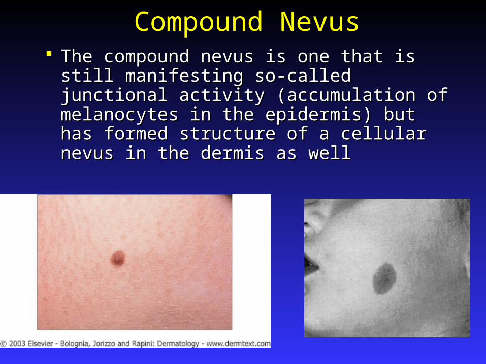
Compound Nevus The compound nevus is one that is still The compound nevus is one that is still
manifesting so-called junctional activity manifesting so-called junctional activity (accumulation of melanocytes in the (accumulation of melanocytes in the epidermis) but has formed structure of a epidermis) but has formed structure of a cellular nevus in the dermis as wellcellular nevus in the dermis as well
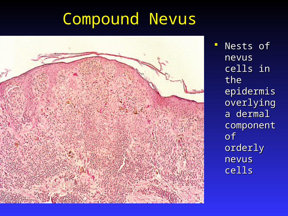
Compound Nevus
Nests of Nests of nevus cells in nevus cells in the epidermis the epidermis overlying a overlying a dermal dermal component component of orderly of orderly nevus cellsnevus cells
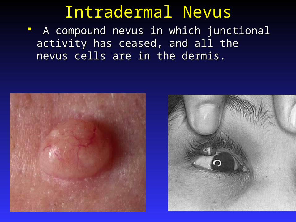
Intradermal Nevus A compound nevus in which junctional A compound nevus in which junctional
activity has ceased, and all the nevus cells activity has ceased, and all the nevus cells are in the dermis.are in the dermis.
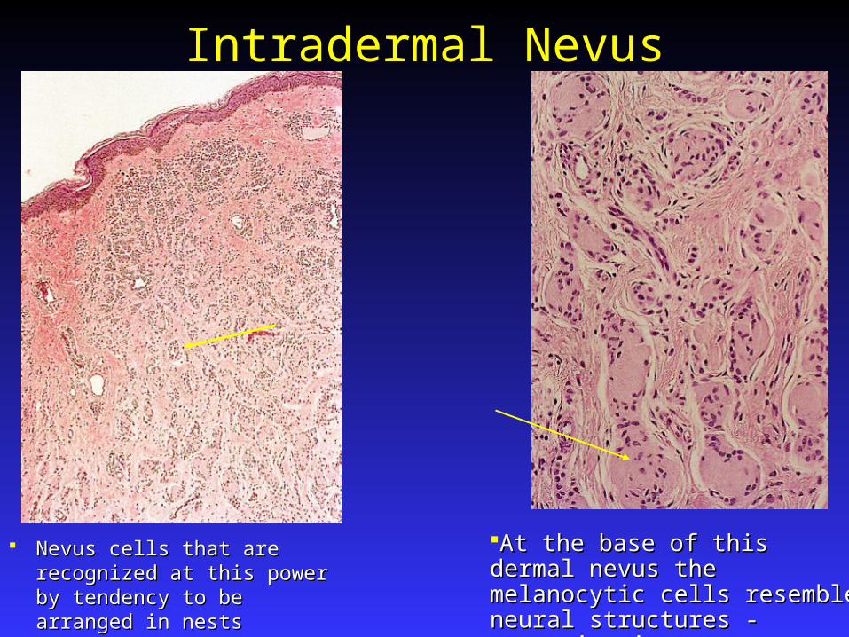
Intradermal Nevus
Nevus cells that are recognized Nevus cells that are recognized at this power by tendency to be at this power by tendency to be arranged in nestsarranged in nests
At the base of this dermal nevus the At the base of this dermal nevus the melanocytic cells resemble neural melanocytic cells resemble neural structures -neurotizationstructures -neurotization
Treatment of NeviSigns of malignant degenerationSigns of malignant degeneration
ABCDE changes. Thickening, ulceration, ABCDE changes. Thickening, ulceration, pain, ugly duckling sign. Dot extensions. New pain, ugly duckling sign. Dot extensions. New lesion in patient over 35 years. lesion in patient over 35 years.
Changes may occur during pregnancy or Changes may occur during pregnancy or with oral contraception.with oral contraception.
Biopsy if you think melanoma.Biopsy if you think melanoma.
Treatment: Andrews recommends biopsy Treatment: Andrews recommends biopsy and removal of scalp nevi if atypical due to and removal of scalp nevi if atypical due to difficulty in following. Single oral or difficulty in following. Single oral or vaginal lesions – biopsy. vaginal lesions – biopsy.

Balloon Cell Nevus A pigmented nevus, varying A pigmented nevus, varying
in size from 1 –5 mm, usually in size from 1 –5 mm, usually occurring on the head, neck, occurring on the head, neck, and trunk.and trunk.
Clinically indistinguishable Clinically indistinguishable from ordinary pigmented or from ordinary pigmented or nonpigmented nevusnonpigmented nevus
Histologically: Composed of Histologically: Composed of peculiar vesicular cells that peculiar vesicular cells that appear to be foamy and form appear to be foamy and form large pale polyhedral balloon large pale polyhedral balloon cells that may be cells that may be multinucleated giant cells in multinucleated giant cells in addition to nevus cells.addition to nevus cells.
Not considered potentially Not considered potentially malignant, and treatment is malignant, and treatment is same as other nevisame as other nevi
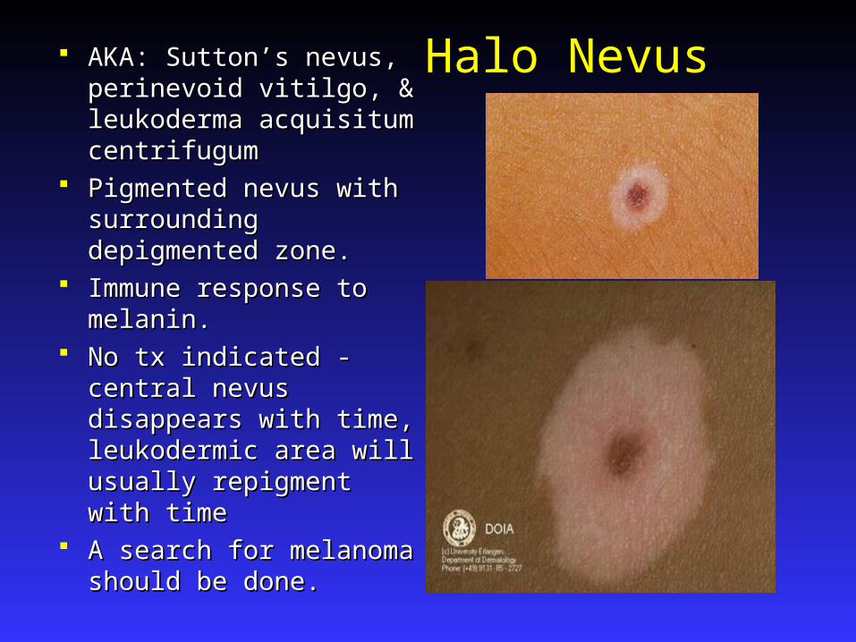
Halo Nevus AKA: Sutton’s nevus, AKA: Sutton’s nevus, perinevoid vitilgo, & perinevoid vitilgo, & leukoderma acquisitum leukoderma acquisitum centrifugumcentrifugum
Pigmented nevus with Pigmented nevus with surrounding depigmented surrounding depigmented zone.zone.
Immune response to Immune response to melanin. melanin.
No tx indicated - central No tx indicated - central nevus disappears with time, nevus disappears with time, leukodermic area will leukodermic area will usually repigment with timeusually repigment with time
A search for melanoma A search for melanoma should be done.should be done.
Halo Nevus
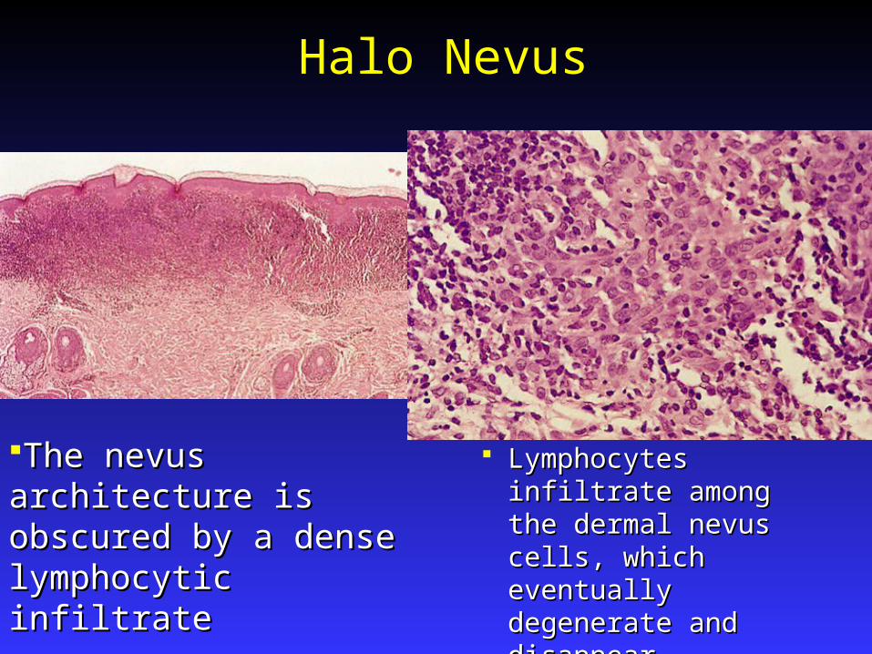
Lymphocytes infiltrate Lymphocytes infiltrate among the dermal among the dermal nevus cells, which nevus cells, which eventually degenerate eventually degenerate and disappearand disappear
The nevus architecture is The nevus architecture is obscured by a dense obscured by a dense lymphocytic infiltratelymphocytic infiltrate

Giant Pigmented NevusPresentation:Presentation: Characterized by a large, Characterized by a large,
darkly pigmented hairy darkly pigmented hairy patch in which smaller, patch in which smaller, darker patches are darker patches are interspersed or present as interspersed or present as small satellite lesions. small satellite lesions. Skin may be thickened or Skin may be thickened or verrucousverrucous
By definition >20cm.By definition >20cm. Has a tendency to follow Has a tendency to follow
a dermatome distributiona dermatome distribution Trunk favored site.Trunk favored site.
Giant Hairy Nevi Present at birth and grow proportionally to the site of Present at birth and grow proportionally to the site of
the body where they are located.the body where they are located. When a large congenital nevus involves the axial skin, When a large congenital nevus involves the axial skin,
there may be an associated there may be an associated neurocutaneous neurocutaneous melanocytosismelanocytosis Hydrocephalus, leptomingeal melanomaHydrocephalus, leptomingeal melanoma
Incidence of melanoma:Incidence of melanoma: Overall risk is 3% to 7%. (40% of melanoma in children.) Overall risk is 3% to 7%. (40% of melanoma in children.) Risk is greatest in axial lesions. Seldom in satellites.Risk is greatest in axial lesions. Seldom in satellites. Only 1/3 of melanoma arise from epidermal sites (makes) Only 1/3 of melanoma arise from epidermal sites (makes)
for more difficult surveillance.for more difficult surveillance.

Giant Hairy NeviTreatment:Treatment:
Most recommend total Most recommend total surgical excision and surgical excision and resurfacing autografts.resurfacing autografts.
Alternative treatments: Alternative treatments: dermabrasion, curettage, and dermabrasion, curettage, and laser ablation. These laser ablation. These eliminate some of the nevus eliminate some of the nevus cells, with theoretic lowering cells, with theoretic lowering of the risk of melanoma of the risk of melanoma
Monitoring. Serial MRI’s for Monitoring. Serial MRI’s for neurocutaneous melanosisneurocutaneous melanosis

Small and Medium-sized Congenital Nevocytic Nevus
Small - < 1.5 cm in greatest diameter.Small - < 1.5 cm in greatest diameter. Medium- > 1.5 cm but < 20 cm.Medium- > 1.5 cm but < 20 cm. Found in 1% of newborns.Found in 1% of newborns. Half eventually become hairy.Half eventually become hairy. Data indicates that those which do progress to Data indicates that those which do progress to
melanoma occur in Pts older than 18 yrs and in the melanoma occur in Pts older than 18 yrs and in the epidermis (Hence monitoring is effective)epidermis (Hence monitoring is effective)
Treatment: Excision is recommended for lesions Treatment: Excision is recommended for lesions of the hairy scalp, or those of great cosmetic of the hairy scalp, or those of great cosmetic concern or nevi with unusual clinical features. concern or nevi with unusual clinical features. Otherwise, observation.Otherwise, observation.
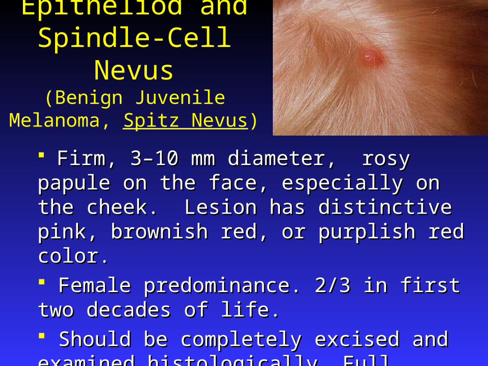
Epitheliod and Spindle-Cell Nevus
(Benign Juvenile Melanoma, Spitz Nevus)
Firm, 3–10 mm diameter, rosy papule on the face, Firm, 3–10 mm diameter, rosy papule on the face, especially on the cheek. Lesion has distinctive pink, especially on the cheek. Lesion has distinctive pink, brownish red, or purplish red color.brownish red, or purplish red color. Female predominance. 2/3 in first two decades of Female predominance. 2/3 in first two decades of life. life. Should be completely excised and examined Should be completely excised and examined histologically. Full excision recommended to prevent histologically. Full excision recommended to prevent confusion with melanoma at future date.confusion with melanoma at future date.
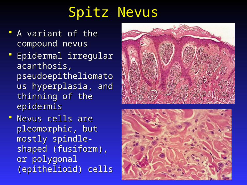
Spitz Nevus
A variant of the A variant of the compound nevuscompound nevus
Epidermal irregular Epidermal irregular acanthosis, acanthosis, pseudoepitheliomatous pseudoepitheliomatous hyperplasia, and hyperplasia, and thinning of the epidermisthinning of the epidermis
Nevus cells are Nevus cells are pleomorphic, but mostly pleomorphic, but mostly spindle-shaped spindle-shaped (fusiform), or polygonal (fusiform), or polygonal (epithelioid) cells(epithelioid) cells
Spitz Nevus
Immunohistochemical staining for MIB-1 Immunohistochemical staining for MIB-1 and bcl-2 will distinguish most Spitz nevi and bcl-2 will distinguish most Spitz nevi from melanoma:from melanoma: Melanomas Melanomas areare immunoreactive immunoreactive Spitz nevi Spitz nevi notnot reactive.are not reactive.are not
Differential diagnosis: Pyogenic Differential diagnosis: Pyogenic granuloma, mastocytoma, juvenile granuloma, mastocytoma, juvenile xanthogranuloma, or melanoma.xanthogranuloma, or melanoma.

Dysplastic Nevus Variegated tan, brown, Variegated tan, brown, pink coloration with pink coloration with pink hues seen in pink hues seen in macular portion. macular portion. Macular portion always Macular portion always present, frequently present, frequently surrounds a papular surrounds a papular center.center.
Generally larger than are Generally larger than are common nevi, usually common nevi, usually 5–12mm, with irregular 5–12mm, with irregular borders.borders.
Develop new lesions Develop new lesions over a lifetime. Sun over a lifetime. Sun protected areas. protected areas.
Dysplastic Nevus Occurrence: 5% -20% of pts have at least Occurrence: 5% -20% of pts have at least
one clinically dysplastic nevus.one clinically dysplastic nevus. Importance:Importance:
1.1. Careful history and evaluation of family Careful history and evaluation of family members.members.
2.2. DNs provide another risk factor for DNs provide another risk factor for melanoma predisposition. melanoma predisposition. >3 lesions >3 lesions increases the risk of melanoma from 3 increases the risk of melanoma from 3 to 43 times.to 43 times.
3. Increased risk of melanoma in the DN AND in the rest of epidermis
Dysplastic Nevus
Histologic features as per an NIH consensus:Histologic features as per an NIH consensus:
1.1. Basilar melanocytic hyperplasia with elongation Basilar melanocytic hyperplasia with elongation of rete ridges.of rete ridges.
2.2. Spindle-shaped or occasionally, epithelioid Spindle-shaped or occasionally, epithelioid melanocytes arranged horizontally and melanocytes arranged horizontally and aggregating in nests that fuse with adjacent rete aggregating in nests that fuse with adjacent rete ridges.ridges.
3.3. Lamellar and concentric superficial dermal Lamellar and concentric superficial dermal infiltrate. infiltrate.
4.4. Cytologic atypia, usually present but not Cytologic atypia, usually present but not essential for diagnosis.essential for diagnosis.
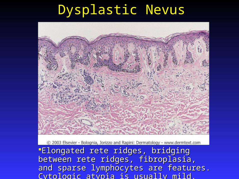
Dysplastic Nevus
Elongated rete ridges, bridging between rete ridges, Elongated rete ridges, bridging between rete ridges, fibroplasia, and sparse lymphocytes are features. fibroplasia, and sparse lymphocytes are features. Cytologic atypia is usually mild.Cytologic atypia is usually mild.
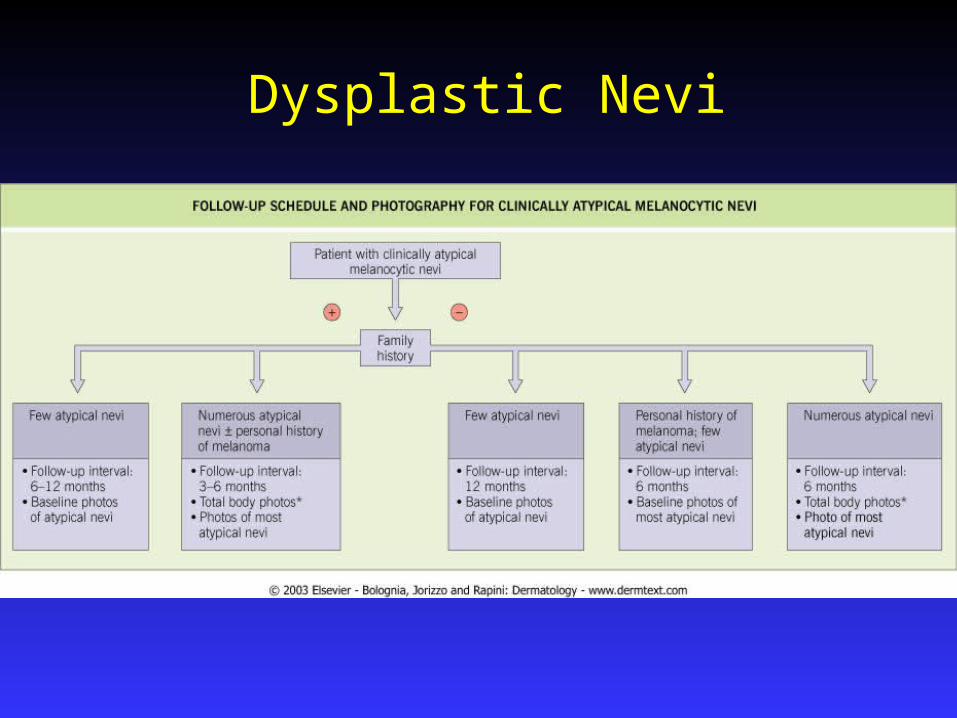
Dysplastic Nevi
Treatment:Treatment: Patients with dysplastic nevi and a positive Patients with dysplastic nevi and a positive
family or personal history of melanoma, family or personal history of melanoma, physician examination every 3 – 6 months physician examination every 3 – 6 months
Excision of those nevi that change clinically Excision of those nevi that change clinically or are located in difficult to monitor or are located in difficult to monitor locations such as the scalp.locations such as the scalp.
Photographs with measured scale is usefulPhotographs with measured scale is useful Sunscreens and Pt self exam are critical.Sunscreens and Pt self exam are critical.
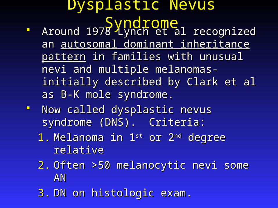
Dysplastic Nevus Syndrome Around 1978 Lynch et al recognized an Around 1978 Lynch et al recognized an
autosomal dominant inheritance patternautosomal dominant inheritance pattern in in families with unusual nevi and multiple families with unusual nevi and multiple melanomas-initially described by Clark et melanomas-initially described by Clark et al as B-K mole syndrome. al as B-K mole syndrome.
Now called dysplastic nevus syndrome Now called dysplastic nevus syndrome (DNS). Criteria:(DNS). Criteria:
1.1. Melanoma in 1Melanoma in 1stst or 2 or 2ndnd degree relative degree relative
2.2. Often >50 melanocytic nevi some ANOften >50 melanocytic nevi some AN
3.3. DN on histologic exam.DN on histologic exam.
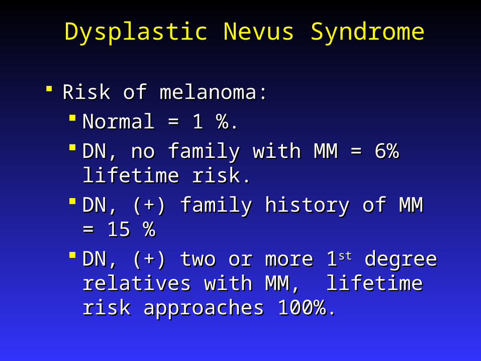
Dysplastic Nevus Syndrome
Risk of melanoma:Risk of melanoma: Normal = 1 %. Normal = 1 %. DN, no family with MM = 6% lifetime DN, no family with MM = 6% lifetime
risk.risk. DN, (+) family history of MM = 15 %DN, (+) family history of MM = 15 % DN, (+) two or more 1DN, (+) two or more 1stst degree relatives degree relatives
with MM, lifetime risk approaches with MM, lifetime risk approaches 100%.100%.
Dysplastic Nevi

Melanoma Originate from melanocytes at epidermal-dermal Originate from melanocytes at epidermal-dermal
junctionjunction 50% will develop in pre-existing nevi.50% will develop in pre-existing nevi. Prolonged, non invasive, horizontally oriented Prolonged, non invasive, horizontally oriented
growth phase. When tumor nodule develops the growth phase. When tumor nodule develops the vertical growth phase is occurring and the risk of vertical growth phase is occurring and the risk of metastatic disease increases dramatically.metastatic disease increases dramatically.
One in 80 Americans will develop melanoma.One in 80 Americans will develop melanoma. Incidence is low until after puberty.Incidence is low until after puberty.
During pregnancy pigmented nevi may enlarge in During pregnancy pigmented nevi may enlarge in size uniformly 2size uniformly 2ndnd to hormones. If changes of to hormones. If changes of irregular pigmentation or asymmetrical growth irregular pigmentation or asymmetrical growth occur ect, then a biopsy should be performed.occur ect, then a biopsy should be performed.
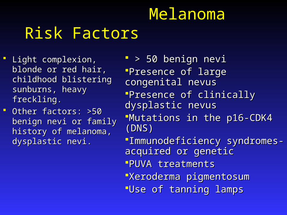
Melanoma Risk Factors
Light complexion, blonde Light complexion, blonde or red hair, childhood or red hair, childhood blistering sunburns, heavy blistering sunburns, heavy freckling.freckling.
Other factors: >50 benign Other factors: >50 benign nevi or family history of nevi or family history of melanoma, dysplastic melanoma, dysplastic nevi. nevi.
> 50 benign nevi> 50 benign neviPresence of large congenital nevusPresence of large congenital nevusPresence of clinically dysplastic nevusPresence of clinically dysplastic nevusMutations in the p16-CDK4 (DNS)Mutations in the p16-CDK4 (DNS)Immunodeficiency syndromes-acquired Immunodeficiency syndromes-acquired or geneticor geneticPUVA treatmentsPUVA treatmentsXeroderma pigmentosumXeroderma pigmentosumUse of tanning lampsUse of tanning lamps
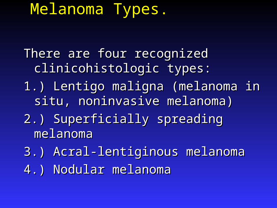
Melanoma Types.
There are four recognized clinicohistologic There are four recognized clinicohistologic types:types:
1.) Lentigo maligna (melanoma in situ, 1.) Lentigo maligna (melanoma in situ, noninvasive melanoma)noninvasive melanoma)
2.) Superficially spreading melanoma2.) Superficially spreading melanoma
3.) Acral-lentiginous melanoma3.) Acral-lentiginous melanoma
4.) Nodular melanoma4.) Nodular melanoma
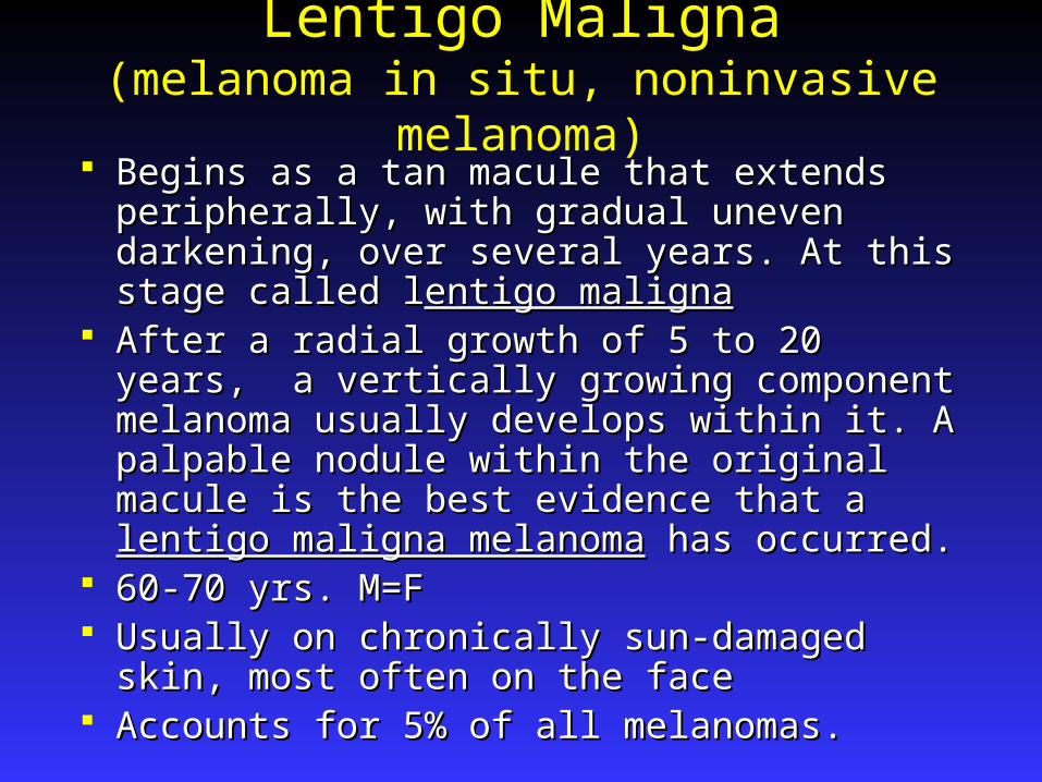
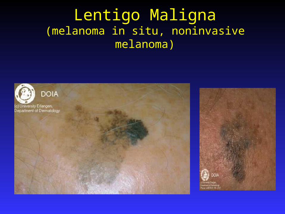
Lentigo Maligna(melanoma in situ, noninvasive melanoma)
Begins as a tan macule that extends peripherally, Begins as a tan macule that extends peripherally, with gradual uneven darkening, over several years. with gradual uneven darkening, over several years. At this stage called lAt this stage called lentigo malignaentigo maligna
After a radial growth of 5 to 20 years, a vertically After a radial growth of 5 to 20 years, a vertically growing component melanoma usually develops growing component melanoma usually develops within it. A palpable nodule within the original within it. A palpable nodule within the original macule is the best evidence that a macule is the best evidence that a lentigo maligna lentigo maligna melanomamelanoma has occurred. has occurred.
60-70 yrs. M=F60-70 yrs. M=F Usually on chronically sun-damaged skin, most Usually on chronically sun-damaged skin, most
often on the faceoften on the face Accounts for 5% of all melanomas.Accounts for 5% of all melanomas.
Lentigo Maligna(melanoma in situ, noninvasive melanoma)

Superficially Spreading Melanoma Most common, 70% of melanoma.Most common, 70% of melanoma. Adults of all ages. Median age 50 yrs.Adults of all ages. Median age 50 yrs. No preference for sun damaged skin..No preference for sun damaged skin.. Lesion has tendency for multicolored appearance Lesion has tendency for multicolored appearance
with notched borders and areas of regression.with notched borders and areas of regression. Faster growing than lentigo maligna.Faster growing than lentigo maligna. In a study by Bolognia et al 5% of lesions with an In a study by Bolognia et al 5% of lesions with an
eccentric foci of hyperpigmentation(a roundish area eccentric foci of hyperpigmentation(a roundish area of brown or black 3mm or less and located of brown or black 3mm or less and located peripherally) are melanomas arising from within a peripherally) are melanomas arising from within a nevus . It is necessary to ensure that the pathologist nevus . It is necessary to ensure that the pathologist sections through the black dot to make this early sections through the black dot to make this early diagnosisdiagnosis

Superficially Spreading Melanoma
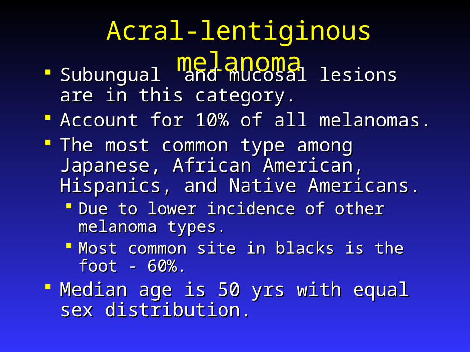
Acral-lentiginous melanoma Subungual and mucosal lesions are in this Subungual and mucosal lesions are in this
category.category. Account for 10% of all melanomas.Account for 10% of all melanomas. The most common type among Japanese, The most common type among Japanese,
African American, Hispanics, and Native African American, Hispanics, and Native Americans. Americans. Due to lower incidence of other melanoma Due to lower incidence of other melanoma
types.types. Most common site in blacks is the foot - 60%.Most common site in blacks is the foot - 60%.
Median age is 50 yrs with equal sex Median age is 50 yrs with equal sex distribution.distribution.
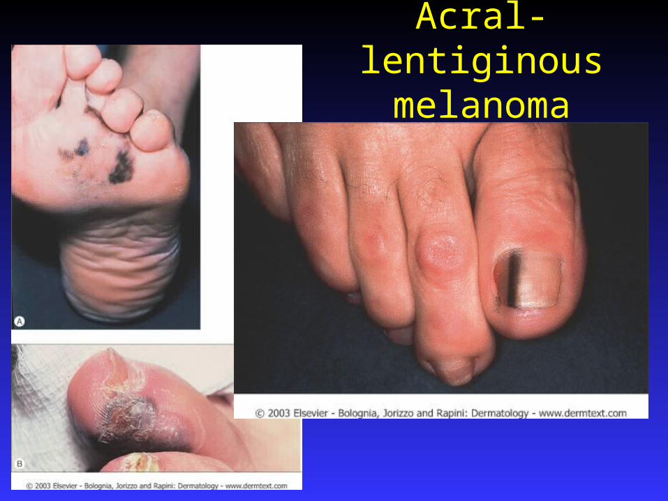
Acral-lentiginous melanoma
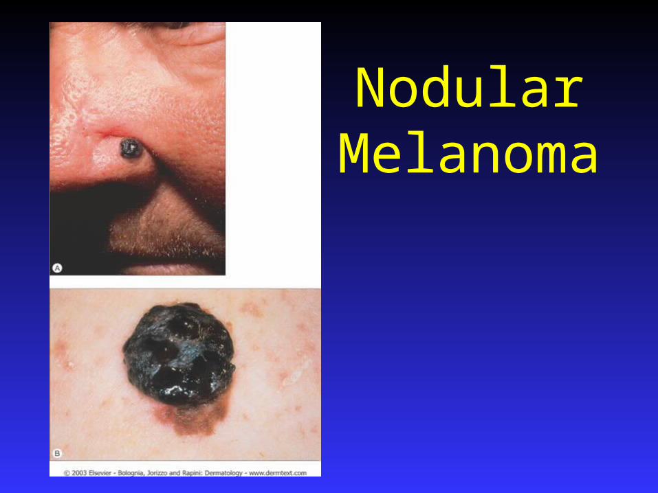
Nodular Melanoma
15 % of melanomas.15 % of melanomas. Pigmented papule or nodule of varying size.Pigmented papule or nodule of varying size. No apparent radial growth phase. No apparent radial growth phase. Histologically, lesion extends several rete Histologically, lesion extends several rete
ridges past the apparent margin.ridges past the apparent margin. Polypoid Variant: Does not appear to Polypoid Variant: Does not appear to
extend into dermis yet behaves like clark extend into dermis yet behaves like clark level IV or V. 42% vs 57% five year level IV or V. 42% vs 57% five year survival.survival.
Nodular Melanoma
Other Melanoma Types.
Desmoplastic Melanoma: Desmoplastic Melanoma: Deeply infiltratiing, spindle cell type. Deeply infiltratiing, spindle cell type. Most are Most are
amelanotic.amelanotic. Occur within lentigo maligna Occur within lentigo maligna often. Neurotropic spread. often. Neurotropic spread.
Inflammatory melanoma: Inflammatory melanoma: Inflammation surrounding melanoma = poor Inflammation surrounding melanoma = poor
prognosis.prognosis. Amelanocytic melanoma – pink or flesh colored. Amelanocytic melanoma – pink or flesh colored.
Mistaken for PG. Seen in Albinos.Mistaken for PG. Seen in Albinos.

Diagnosis of Melanoma
Surgical excision is the best method.Surgical excision is the best method. A shave may be better for less suspicious or A shave may be better for less suspicious or
broad, thin lesions.broad, thin lesions. For larger lesions an incisional or punch For larger lesions an incisional or punch
biopsy is the standard.biopsy is the standard. When melanoma is suspected in a melanotic When melanoma is suspected in a melanotic
freckle or a giant pigmented nevus, biopsy freckle or a giant pigmented nevus, biopsy should be done through the thickest and should be done through the thickest and most atypical area and multiply sectioned most atypical area and multiply sectioned to find thickest area of involvementto find thickest area of involvement

Histologic Diagnosis of Melanoma
Pinkus criteria:Pinkus criteria: Presence of mitosesPresence of mitoses Inflammatory reaction composed of Inflammatory reaction composed of
lymphocytes and possibly plasma cellslymphocytes and possibly plasma cells Dermoepidermal junctional activity. (Except in Dermoepidermal junctional activity. (Except in
giant nevus)giant nevus) Absence of dermal stromaAbsence of dermal stroma
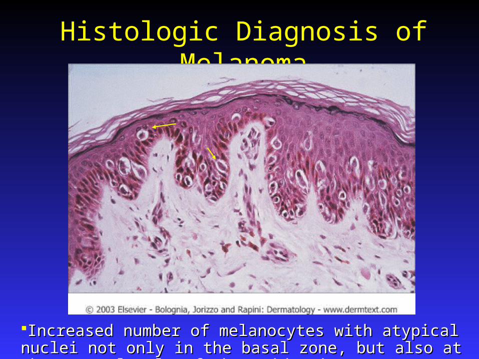
Histologic Diagnosis of Melanoma
Increased number of melanocytes with atypical nuclei not only in the Increased number of melanocytes with atypical nuclei not only in the basal zone, but also at the upper levels of the epidermis basal zone, but also at the upper levels of the epidermis
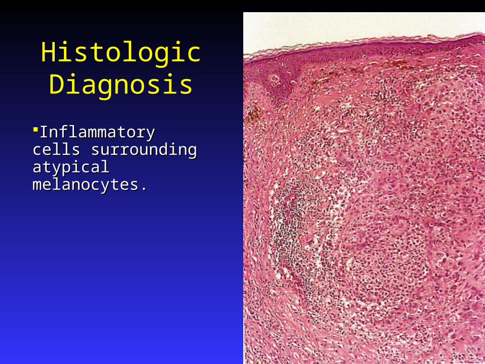
Histologic Diagnosis
Inflammatory cells Inflammatory cells surrounding atypical surrounding atypical melanocytes.melanocytes.
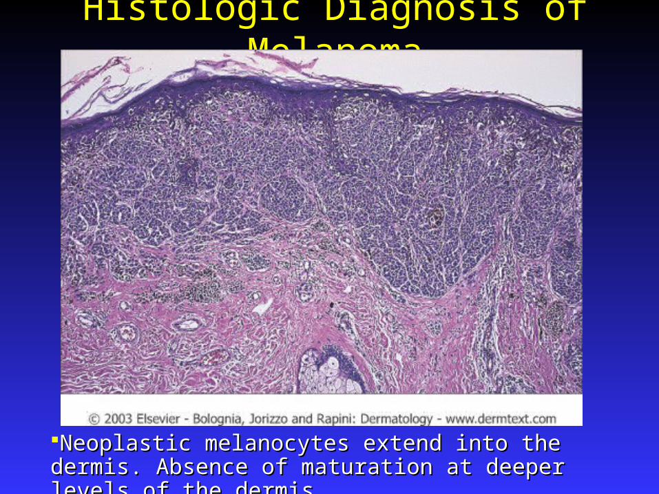
Histologic Diagnosis of Melanoma
Neoplastic melanocytes extend into the dermis. Absence Neoplastic melanocytes extend into the dermis. Absence of maturation at deeper levels of the dermis of maturation at deeper levels of the dermis
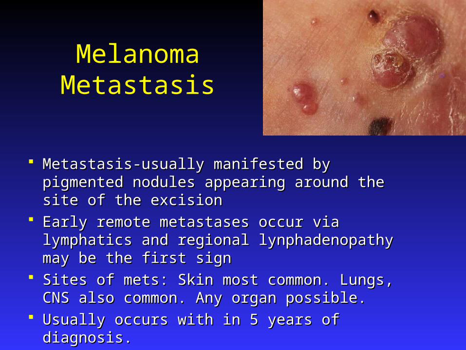
Melanoma Metastasis
Metastasis-usually manifested by pigmented Metastasis-usually manifested by pigmented nodules appearing around the site of the excisionnodules appearing around the site of the excision
Early remote metastases occur via lymphatics and Early remote metastases occur via lymphatics and regional lynphadenopathy may be the first signregional lynphadenopathy may be the first sign
Sites of mets: Skin most common. Lungs, CNS Sites of mets: Skin most common. Lungs, CNS also common. Any organ possible.also common. Any organ possible.
Usually occurs with in 5 years of diagnosis.Usually occurs with in 5 years of diagnosis.

Melanoma Workup and Treatment
Establish a family history, thorough review of Establish a family history, thorough review of systems and physical exam for all patients.systems and physical exam for all patients.
A consensus conference in 1992 concluded that a A consensus conference in 1992 concluded that a staging workup (including E.L.N.D.) was not staging workup (including E.L.N.D.) was not indicated for melanomas below 1.0 mm thickness.indicated for melanomas below 1.0 mm thickness.
Many physicians obtain a CXR and an LDH.Many physicians obtain a CXR and an LDH. Consultation with an oncologist is worthwhile for Consultation with an oncologist is worthwhile for
advanced cases.advanced cases. Sentinel node / ELND. Still evolving criteria. Sentinel node / ELND. Still evolving criteria.
Recommended for tumors >1.0 mm thick or with Recommended for tumors >1.0 mm thick or with ulceration.ulceration.

Melanoma Excision Margins
Melanoma - Other Treatments High-dose interferon alfa-2b therapy.High-dose interferon alfa-2b therapy.
Efficacy is equivocal and toxicity high.Efficacy is equivocal and toxicity high. May diminish the occurrence of mets and prolong May diminish the occurrence of mets and prolong
disease free survival with melanoma > 1.5 mm thickdisease free survival with melanoma > 1.5 mm thick
Chemotherapy is not effective.Chemotherapy is not effective. Adoptive immunotherapy with lymphokine-Adoptive immunotherapy with lymphokine-
activated killer cells + interleukin-2, or high dose activated killer cells + interleukin-2, or high dose interleukin-2 alone. Some patients are responsive.interleukin-2 alone. Some patients are responsive.
Perfusion chemotherapy has been used for Perfusion chemotherapy has been used for extremity melanoma and has almost eliminated the extremity melanoma and has almost eliminated the need for amputation.need for amputation.
Aldara and Tazorac for M.I.S. recently published. Aldara and Tazorac for M.I.S. recently published.

Survival Indicators
5 Year Survival 5 Year Survival Rates Based on Rates Based on Lesion Thickness:Lesion Thickness:
In situ ……= 100%In situ ……= 100% <0.76 mm. = 2-4%<0.76 mm. = 2-4% 0.76 to 1.49 = 90%0.76 to 1.49 = 90% 1.50 to 3.99 = 70%1.50 to 3.99 = 70% > 4 mm ….. = 50 % > 4 mm ….. = 50 %
Dermal Melanocytic Lesions
At birth, melanocytes may be present in the At birth, melanocytes may be present in the dermal portion of the skin of the scalp, the dermal portion of the skin of the scalp, the backs of the hands, and the sacrum.backs of the hands, and the sacrum.
These are large ameboid cells that normally These are large ameboid cells that normally disappear shortly after birth.disappear shortly after birth.
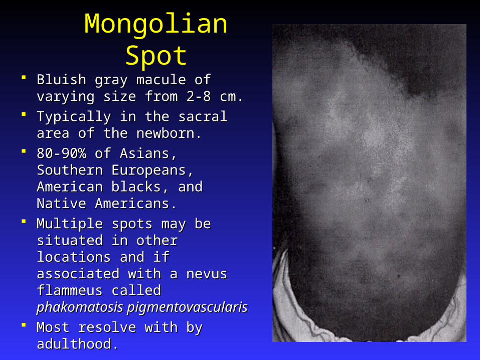
Mongolian Spot Bluish gray macule of varying Bluish gray macule of varying
size from 2-8 cm.size from 2-8 cm. Typically in the sacral area of Typically in the sacral area of
the newborn.the newborn. 80-90% of Asians, Southern 80-90% of Asians, Southern
Europeans, American blacks, Europeans, American blacks, and Native Americans.and Native Americans.
Multiple spots may be situated Multiple spots may be situated in other locations and if in other locations and if associated with a nevus associated with a nevus flammeus called flammeus called phakomatosis phakomatosis pigmentovascularispigmentovascularis
Most resolve with by adulthood.Most resolve with by adulthood.
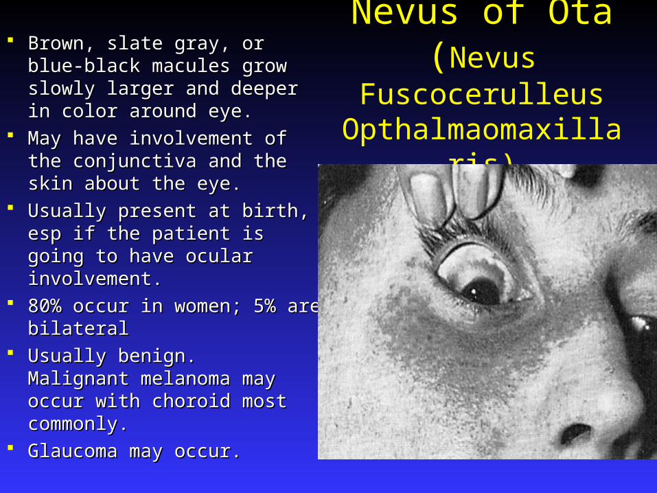
Nevus of Ota(Nevus Fuscocerulleus Opthalmaomaxillaris)
Brown, slate gray, or blue-black Brown, slate gray, or blue-black macules grow slowly larger and macules grow slowly larger and deeper in color around eye.deeper in color around eye.
May have involvement of the May have involvement of the conjunctiva and the skin about conjunctiva and the skin about the eye.the eye.
Usually present at birth, esp if Usually present at birth, esp if the patient is going to have the patient is going to have ocular involvement.ocular involvement.
80% occur in women; 5% are 80% occur in women; 5% are bilateralbilateral
Usually benign. Malignant Usually benign. Malignant melanoma may occur with melanoma may occur with choroid most commonly.choroid most commonly.
Glaucoma may occur.Glaucoma may occur.

Nevus of Ito(Nevus Fuscoceruleus Acromiodeltoideus)
Same features as nevus of Ota except that it Same features as nevus of Ota except that it occurs in the distribution of the posterior occurs in the distribution of the posterior supraclavicular and lateral cutaneous supraclavicular and lateral cutaneous brachial nerves.brachial nerves.
It involves the shoulder, side of the neck, It involves the shoulder, side of the neck, and supraclavicular areasand supraclavicular areas
TX: Q-switched ruby laser.TX: Q-switched ruby laser.
Blue Nevus
2 types:2 types:
1.1. Blue nevus of Jadassohn-Tiche (common Blue nevus of Jadassohn-Tiche (common blue nevus)blue nevus)
2.2. Cellular blue nevusCellular blue nevus
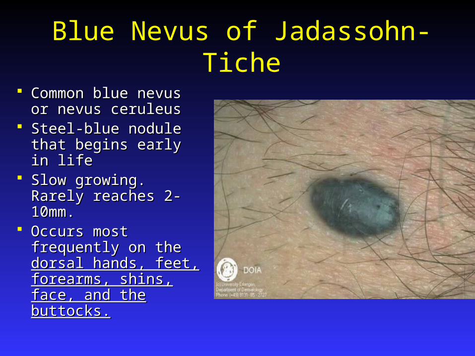
Blue Nevus of Jadassohn-Tiche
Common blue nevus Common blue nevus or nevus ceruleusor nevus ceruleus
Steel-blue nodule that Steel-blue nodule that begins early in lifebegins early in life
Slow growing. Rarely Slow growing. Rarely reaches 2-10mm.reaches 2-10mm.
Occurs most Occurs most frequently on the frequently on the dorsal hands, feet, dorsal hands, feet, forearms, shins, face, forearms, shins, face, and the buttocks.and the buttocks.

Blue Nevus
Within the dermis there is a poorly defined but Within the dermis there is a poorly defined but symmetric spindle cell proliferation that is dark symmetric spindle cell proliferation that is dark brown in colorbrown in color
No significant change in the overlying dermisNo significant change in the overlying dermis
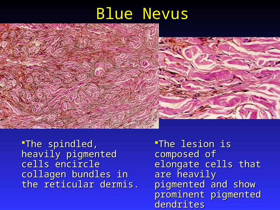
Blue Nevus
The spindled, heavily The spindled, heavily pigmented cells encircle pigmented cells encircle collagen bundles in the collagen bundles in the reticular dermis.reticular dermis.
The lesion is composed of The lesion is composed of elongate cells that are elongate cells that are heavily pigmented and show heavily pigmented and show prominent pigmented prominent pigmented dendritesdendrites
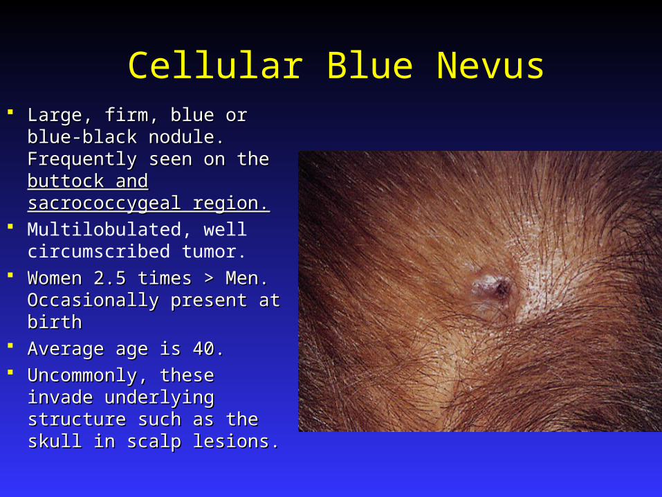
Cellular Blue Nevus Large, firm, blue or blue-Large, firm, blue or blue-
black nodule. Frequently black nodule. Frequently seen on the seen on the buttock and buttock and sacrococcygeal region.sacrococcygeal region.
Multilobulated, well circumscribed tumor.
Women 2.5 times > Men. Women 2.5 times > Men. Occasionally present at birthOccasionally present at birth
Average age is 40.Average age is 40. Uncommonly, these invade Uncommonly, these invade
underlying structure such as underlying structure such as the skull in scalp lesions.the skull in scalp lesions.
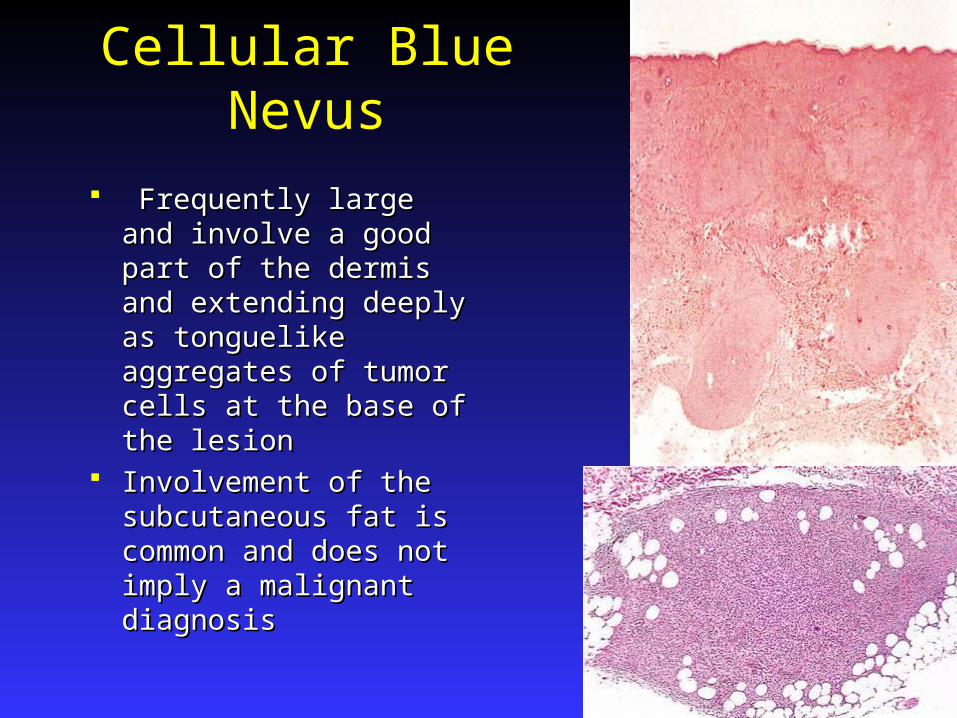
Cellular Blue Nevus
Frequently large and Frequently large and involve a good part of the involve a good part of the dermis and extending dermis and extending deeply as tonguelike deeply as tonguelike aggregates of tumor cells aggregates of tumor cells at the base of the lesionat the base of the lesion
Involvement of the Involvement of the subcutaneous fat is subcutaneous fat is common and does not common and does not imply a malignant imply a malignant diagnosisdiagnosis
Cellular Blue Nevus
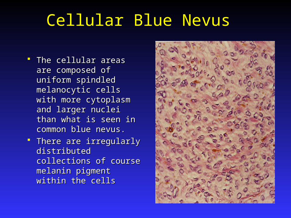
The cellular areas are The cellular areas are composed of uniform composed of uniform spindled melanocytic cells spindled melanocytic cells with more cytoplasm and with more cytoplasm and larger nuclei than what is larger nuclei than what is seen in common blue seen in common blue nevus. nevus.
There are irregularly There are irregularly distributed collections of distributed collections of course melanin pigment course melanin pigment within the cellswithin the cells

Epithelioid Blue Nevus
Newly described lesion with strong Newly described lesion with strong association with Carney’s complex association with Carney’s complex (myxomas, spotty skin pigmentation, (myxomas, spotty skin pigmentation, endocrine over activity, and schwannomas)endocrine over activity, and schwannomas)
Occur frequently on the head and neck, and Occur frequently on the head and neck, and are at times multipleare at times multiple
They are darkly pigmented, domed, and less They are darkly pigmented, domed, and less than 1 cm.than 1 cm.

Malignant Blue Nevus Cellular blue nevus may rarely undergo malignant Cellular blue nevus may rarely undergo malignant
transformation into malignant melanomatransformation into malignant melanoma Clinically sudden increase in size and ulceration.Clinically sudden increase in size and ulceration. Histologically: Pleomorphism of nuclei, mitotic Histologically: Pleomorphism of nuclei, mitotic
figures, and invasion of clusters of malignant cells figures, and invasion of clusters of malignant cells into the deep dermis and fatty tissue.into the deep dermis and fatty tissue.
Treatment: Treatment: Benign lesions - Excision has been mainstay of Benign lesions - Excision has been mainstay of
treatment. Q-switched ruby laser.treatment. Q-switched ruby laser. Malignant variety is the same as M.M.Malignant variety is the same as M.M.