MEET THE EXPERT SESSION CASE PRESENTATIONbsmedicine.org/congress/2014/Dr._Tamanna.pdf · •meet...
39
•MEET THE EXPERT SESSION – CASE PRESENTATION PRESENTED BY DR.CHOWDHURY TAMANNA TABASSUM INTERN DOCTOR, MU-XI,DMCH.
Transcript of MEET THE EXPERT SESSION CASE PRESENTATIONbsmedicine.org/congress/2014/Dr._Tamanna.pdf · •meet...

•MEET THE EXPERT SESSION – CASE PRESENTATION
PRESENTED BY
DR.CHOWDHURY TAMANNA TABASSUM
INTERN DOCTOR, MU-XI,DMCH.

A 36 YEARS OLD LADY WITH RECURRENT UPPER ABDOMINAL PAIN, FEVER & FATIGUE

PARTICULARS OF THE PATIENT
• NAME : Mrs. SHARMIN AKTAR
• AGE : 36 YEARS
• SEX : FEMALE
• RELIGION : ISLAM
• OCCUPATION : HOUSE WIFE
• MARITAL STATUS : MARRIED
• ADDRESS : KUSHTIA (PRESENT AND PERMANENT)
• DATE OF ADMISSION: 01.04.2013

CHIEF COMPLAINTS
•RECURRENT UPPER ABDOMINAL PAIN FOR 2 YEARS
•RECURRENT FEVER FOR 2 YEARS
•PROGRESSIVE FATIGUE AND LOSS OF APPETITE FOR SAME DURATION
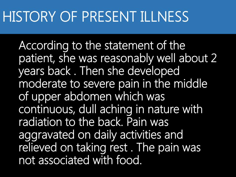
HISTORY OF PRESENT ILLNESS
According to the statement of the patient, she was reasonably well about 2 years back . Then she developed moderate to severe pain in the middle of upper abdomen which was continuous, dull aching in nature with radiation to the back. Pain was aggravated on daily activities and relieved on taking rest . The pain was not associated with food.

HISTORY OF PRESENT ILLNESS (CONTINUED)….
She also complained about fever which was initially high grade for few days , then followed by low grade with 2/3 peaks a day . It was associated with chills but no rigor and relieved with sweating . Her maximum recorded temparature was 103º F.
Her other complaints were progressive fatigue and joint pain involving both ankles ,elbows and hand joints .

HISTORY OF PRESENT ILLNESS (CONTINUED)….
She also noticed loss of appetite and disturbance in her sleep pattern . Her bowel and bladder habits were normal.
She had no history of vomiting, constipation, cough, weight loss or yellow discoloration of skin and urine. She is normotensive and diagnosed as a case of diabetis mellitus in 2011.

PAST ILLNESS AND PREVIOUS TREATMENT
The patient had occasional abdominal pain since she was a high school girl, which intensified in 1990 and laparotomy was done in Rajshahi but sufficient data is not available. Then 3 weeks later of this laparotomy, She became dyspnoic, transferred to BSMMU . She diagnosed as a case of tubercular pleural effusion and given anti-TB therapy for 9 months .

PAST ILLNESS AND PREVIOUS TREATMENT (CONTINUED)….
Then her next 20 years were uneventful except her diagnosis of diabetes in 2011 and she recieved insulin therapy and again she started having recurrent abdominal pain and irregular fever and one of the General physician prescribed her Anti-TB in the month of February, 2013.since she did not show any improvement ,she got herself admitted in DMCH in April ,2013.

•FAMILY HISTORY
Her brother also received anti-TB therapy.
•PERSONAL HISTORY She is non-alcoholic and non-smoker.
•IMMUNIZATION HISTORY Completed as per EPI schedule
•TRAVELLING HISTORY Nothing significant
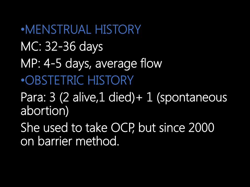
•MENSTRUAL HISTORY
MC: 32-36 days
MP: 4-5 days, average flow
•OBSTETRIC HISTORY
Para: 3 (2 alive,1 died)+ 1 (spontaneous abortion)
She used to take OCP, but since 2000 on barrier method.

PHYSICAL EXAMINATION
• GENERAL EXAMINATION
oANAEMIA-SEVERE
oLYMPHNODE-ENLARGED PARA-AORTIC GROUP
oJAUNDICE- ABSENT
oCYANOSIS -ABSENT
oOEDEMA-ABSENT
oCLUBBING-ABSENT
oKOILONYCHIA- ABSENT
oLEUCONYCHIA-ABSENT

•GENERAL EXAMINATION
(continued)…..
• Pulse-90/min ,regular
• Blood pressure-100/70 mmHg
• Temparature-98 F
• Respiratory Rate-16/min
• Thyroid Gland-not enlarged
• Jvp-not raised

SYSTEMIC EXAMINATION
(ABDOMEN)
Inspection:
• Shape –Normal,
•Umbilicus was Central & Vertical,
• Flanks were Normal,
•Scar Mark Of Upper Midline Incision.

SYSTEMIC EXAMINATION (CONTINUED)….
•Palpation:
• Abdomen was Non Tender,
• Hepatomegaly (16cm in size from the right costal margin along the mid clavicular line , Non tender, Firm in consistency, Smooth surface)
• Splenomegaly (6cm from left costal margin, Firm in consistency , smooth surface),
• Palpable Firm Para-aortic Lymph Nodes.
•Percussion: Percussion note -Tympanic
•Auscultation: Bowel Sound - Present,
Hepatic Bruit - Absent
Renal Bruit - Absent

SYSTEMIC EXAMINATION (CONTINUED)….
Examination of other systems reveal no abnormality

WHAT IS YOUR CLINICAL DIAGNOSIS???

DIFFERENTIAL DIAGNOSIS:
1.LYMPHOMA???
2.DISSEMINATED TUBERCULOSIS???

INVESTIGATION PROFILE
(Complete blood count )
Hemoglobin
(g/dl )
08.50
07.70
10.70
11.40
04.02.2013
30.03.2013
18.01.2012
13.05.2011
7,200
5,000
04.02.2013
30.03.2013
3,80,000
3,40,000
04.02.2013
30.03.2013

INVESTIGATION PROFILE
PBF RBC-normocytic,
normochromic
WBC-mature
Platelet-adequate
09.02.2013
ESR (mm in 1st
hr)
125
84
56
26
04.02.2013
30.03.2013
18.01.2013
13.05.2011

INVESTIGATIONS PROFILE
URINE R/M/E Normal study
Urine Micro-albumin
(mg/L)
46.6 (normal
range<300)

INVESTIGATION PROFILE Investigations Report Normal
Range
Date
S. Creatinine
(mg/dL)
0.6 0.6-1.5 03.04.2013
SGPT (IU/L) 16 10-45 30.03.2013
S. Bilirubin (mg/dL) 0.6 <1.00 30.03.2013
ALP (U/L) 152 40-125 21.01.2012
S. Albumin (g/dL) 4.1 3.5-5.00 21.01.2012
HBA1C (%) 6.9% 4.0-6.0% 04.02.2013

INVESTIGATION PROFILE
Investigations Report Normal
range
Date
S. Calcium
(mg/dL)
8.5
9.2
8.5-10.5 01.04.2013
21.01.2012
S. Phosphate
(mg/dL)
3.9 2.4-4.3 21.01.2012
Free T4 (pmol/L) 19.44 9-21 10.02.2013
6.64
5.69
0.2-5.0 10.02.2013
21.01.2012

INVESTIGATION PROFILE ICT for Malaria Negative 09.02.2013
ICT for Kala-Azar Negative 10.02.2013
Hemoglobin
Electrophoresis
Normal 10.02.2013
CRP (mg/L) 6.0 13.05.2011
RA test Negative 13.05.2011

INVESTIGATION PROFILE
Investigation Report Date
ECG Sinus
Tachycardia
04.04.2013
Upper GI
Endoscopy
Normal 15.05.2011
MT Negative 30.03.2013

INVESTIGATION PROFILE
X-ray of
Chest
(P/A view)
Right sided Pneumonitis
with small pleural
thickening
30.03.2013


INVESTIGATION PROFILE
USG of Whole
Abdomen
Hepato-splenomegaly,
irregular pancreas,
abdominal
lymphadenopathy
28.03.2013


DIAGNOSIS
?

INVESTIGATION PROFILE
Investigation Report Date
X-ray of Abdomen
(A/P view)
Pancreatic
Calcification
02.04.2013
USG of Whole
Abdomen
Hepato-
splenomegaly,
Chronic Pancreatitis
with Calcification,
Abdominal
Lymphadenopathy.
03.04.2013

INVESTIGATION PROFILE CT Scan of
Abdomen
and Pelvis
Chronic Pancreatitis with
pancreatic calcific foci,
chronic distal splenic vein
thrombosis and collaterals,
Spleen shows hypodense
lesions with extensive
abdominal
lymphadenopathy
(Suggestive of koch’s ?)
05.04.2013

Chronic Pancreatitis With Diabetes Mellitus was confirmed.
DIAGNOSTIC DILEMMA How can we explain
HEPATO-SPLENOMEGALY with ABDOMINAL LYMPHADENOPATHY ??

INVESTIGATION PROFILE FNAC from
Abdominal
Lymph node
Granulomatous
Inflammation marked
by clusters of Epithelioid
cells, Giant cells and
Lymphocytes, but no
caseation ,no malignant
cell is seen.(Suggestive
of Sarcoidosis??)
08.04.2013

INVESTIGATION PROFILE
Investigation Report Date
Serum ACE
(U/L)
144
(Normal
range-8-65
U/l)
10.04.2013

CONFIRMATORY DIAGNOSIS
Chronic Pancreatitis with Diabetes Mellitus with Abdominal Sarcoidosis.

Treatment in our unit
•Her Anti-Tb therapy was stopped. •For Chronic Pancreatitis: The patient was treated with Omeprazole and Pancreatic Enzymes . •For Abdominal Sarcoidosis: She received Prednisolone 1mg/kg/day for 2 weeks, then gradual tapering was done.

Follow up of the patient
• Patient has neither fever, nor abdominal pain on follow up.
•On 26,April 2013, the USG of whole abdomen showed a reduction in size of spleen, liver and abdominal lymph nodes.
•On 7, July 2013, the USG of whole abdomen showed mild hepatomegaly (suggestive of fatty change) with normal sized spleen and no enlarged abdominal lymph nodes.