Medico-legal wounds (Lecture 2018) - Scholar Idea...Medico-legal wounds (Lecture 2018) Dr/ Doha...
63
legal - Medico wounds ) 2018 Lecture ( Dr/ Doha Yahia Ahmed Associate professor of Forensic Medicine and Toxicology.
Transcript of Medico-legal wounds (Lecture 2018) - Scholar Idea...Medico-legal wounds (Lecture 2018) Dr/ Doha...
legal -Medicowounds
)2018Lecture (Dr/ Doha Yahia AhmedAssociate professor of Forensic
Medicine and Toxicology.
Objectives1- Different types of wounds
2- Medicolegal importance of wounds
3- Determination of the age of wounds
4- Antimortem and postmortem wounds.
5- Complications of wounds.
A wound is a disruption of the normal structure of tissues caused by the application of force to the body.
Legal definition of a wounda wound is where the whole skin is broken, thecontinuity of the skin is broken. - An abrasion of the surface is not sufficient- fractures or internal injuries are not included if the overlying skin is intact.
Definition of wound
Wound classification Wounds or injuries may be classified
according to their etiology and pathology into the following groups:
A- Legally ( من الناحيه القانونيه(
1- Slight or simple wound in which the lesions are not serious and heal rapidly in less than 20 days without leaving permanent infirmity عاهه مستديمه.
2- Dangerous or serious wound, it takes more than 20 days or less to heal but leaving a permanent infirmity.
3- Mortal or fatal wound which causes death either immediately or within a short time.
B- Medico-legallyI- Blunt wounds: (abrasions, bruises, lacerations)
II- Incised or cut wounds
III- Stab or punctured wounds
IV-Firearm wounds
- Description of wounds for medico-legal purposes
1. The type and nature: whether it is a bruise, abrasion or laceration etc,
2. Position and direction.3. Dimensions (length, width and depth). 4. The probable time of its occurrence may be
recorded.5. Is the wound ant-mortem or post- mortem??
in origin must be determined
6. The age of ante-mortem wound must be recognized.
7. Numbers should be assigned to each wound that is described.
8. It is helpful to take a photograph of the wound with an indication of dimension (e.g. a tape measure placed next to the wound).
9. Records of diagrams to chart the approximate situation of injuries found on examination during life or at autopsy are often of value.
For wound examination we have to answer the following questions
1- What is the nature of injury and the used instrument ???
2- Was the injury inflicted before or after death???
3- What was the cause of death??
1- Blunt woundsDefinition:They are injuries caused by a blunt force
(instrument).
They are divided into three types:a- Abrasions or scratches (سحجات أو خدوش)
b- Bruises or contusions (كدمات)
c- Lacerations.
a- AbrasionsDefinition
An abrasion is a destruction of the skin which
usually involves the superficial layers of the
epidermis only. Abrasions are caused by
friction of the skin against rough or sharp
surface resulting in scraping away the
superficial portions of the epidermis.
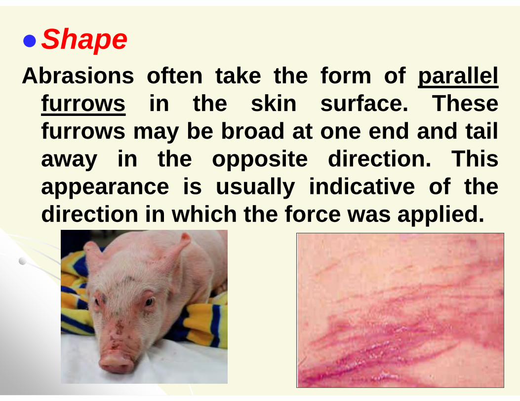
Shape Abrasions often take the form of parallel
furrows in the skin surface. Thesefurrows may be broad at one end and tailaway in the opposite direction. Thisappearance is usually indicative of thedirection in which the force was applied.
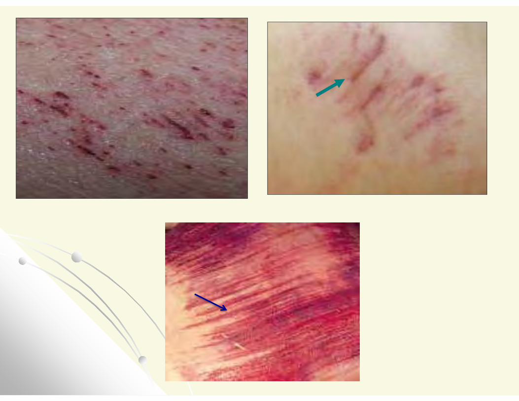
Abrasions- Surface injury
–) خدش( mild abrasions are called Grazeor scrapes (do not scar or bleed)- rough surface striking the body. - crushed epidermis, pressure or imprint abrasions- examples: ligature mark, fingernail scratches, tyre marks, ground or gravel injuries.
Causes:1- Blows from blunt instruments and from
falls. Such abrasions are commonly found on the head and face and over the bony prominences.
Abrasions are commonly accompanied by other injuries such bruises, fractures or internal injuries.
2.Finger nails: they appear either crescentic
marks or as relatively broad parallel
grooves which tail away at their end.
They are commonly found in the front or
at the sides of the neck in the case of
throttling and in the front of the neck, the
thighs, and the vulva in the cases of the
bestiality or rape.
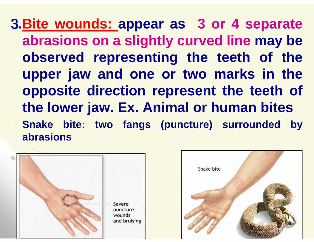
3.Bite wounds: appear as 3 or 4 separate abrasions on a slightly curved line may be observed representing the teeth of the upper jaw and one or two marks in the opposite direction represent the teeth of the lower jaw. Ex. Animal or human bites
Snake bite: two fangs (puncture) surrounded by abrasions
Medico-legal importance of abrasions:
1. They indicate that some force has been applied to the
body.
2. The features of abrasions may indicate the nature and
direction of the applied force and possibly the purpose
for which it was applied.
3. The site of the abrasions may refer to the type of the
crime.
4. The shape may refer to the used instrument.
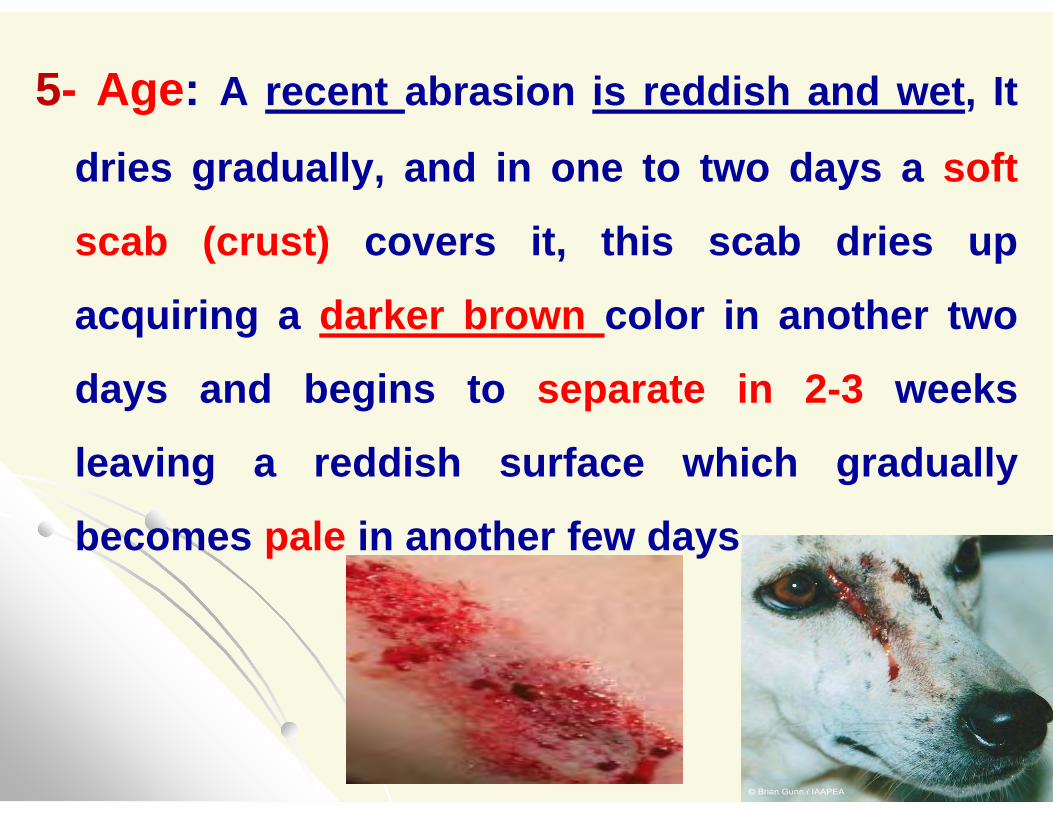
5- Age: A recent abrasion is reddish and wet, It
dries gradually, and in one to two days a soft
scab (crust) covers it, this scab dries up
acquiring a darker brown color in another two
days and begins to separate in 2-3 weeks
leaving a reddish surface which gradually
becomes pale in another few days.
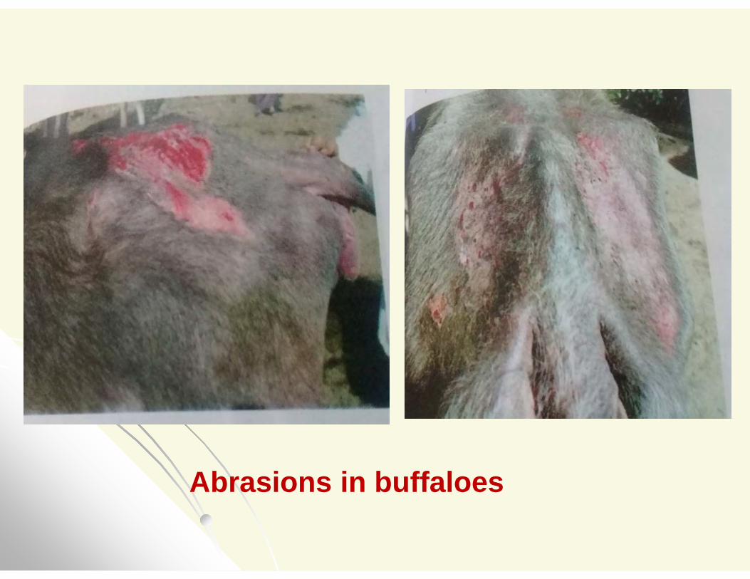
Abrasions in buffaloes
6- In the dead body; we have to differentiate between ante-mortem and post-mortem abrasions, so it is advisable to excise the abrasions and submit the tissue for histological examination.
On Microscopical examination: ante-mortem abrasions show signs of tissue reaction (hyperemia or extravasations of blood) while in post- mortem abrasions there is a white surface without any tissue reaction.
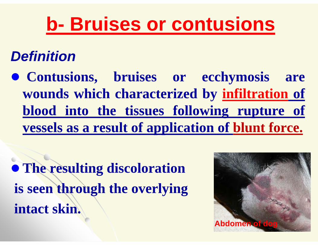
Bruises or contusions-bDefinition Contusions, bruises or ecchymosis are
wounds which characterized by infiltration of blood into the tissues following rupture of vessels as a result of application of blunt force.
The resulting discolorationis seen through the overlyingintact skin.
Abdomen of dog
The extent and the degree of bruising depend on:
1-The force applied to the body.
2-The structure and vascularity of the affected
tissue.
3- The thickness of the skin.
4- The texture of the subcutaneous tissues.
5- The relationship of these structures to the
deeper tissues which vary in different parts of
the body.
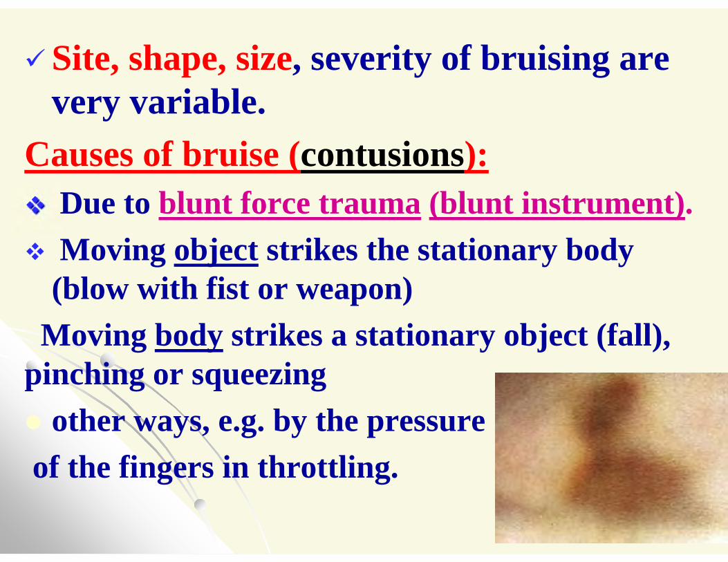
Site, shape, size, severity of bruising are very variable.
Causes of bruise (contusions): Due to blunt force trauma (blunt instrument). Moving object strikes the stationary body
(blow with fist or weapon)fall), strikes a stationary object (bodyMoving
pinching or squeezing other ways, e.g. by the pressureof the fingers in throttling.
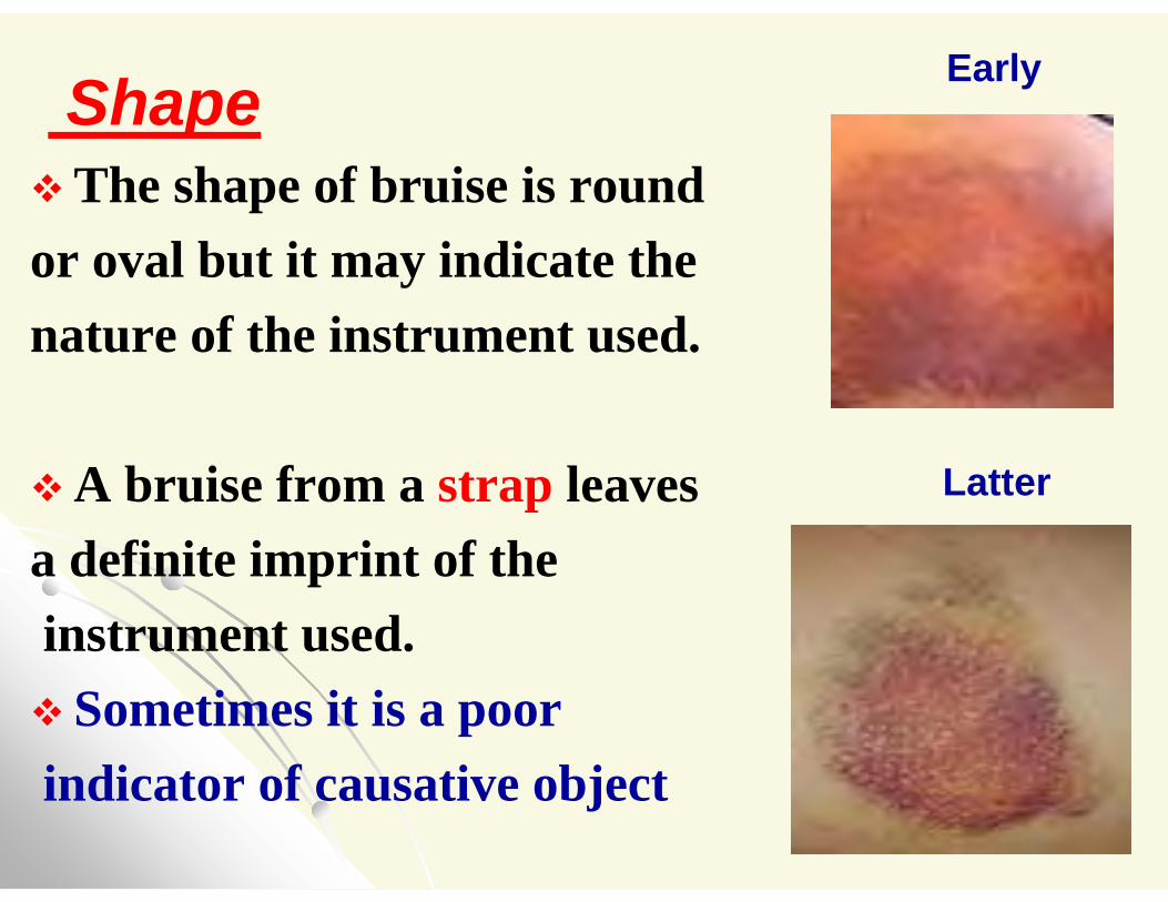
ShapeThe shape of bruise is round or oval but it may indicate the nature of the instrument used.
A bruise from a strap leaves a definite imprint of theinstrument used. Sometimes it is a poorindicator of causative object
Early
Latter
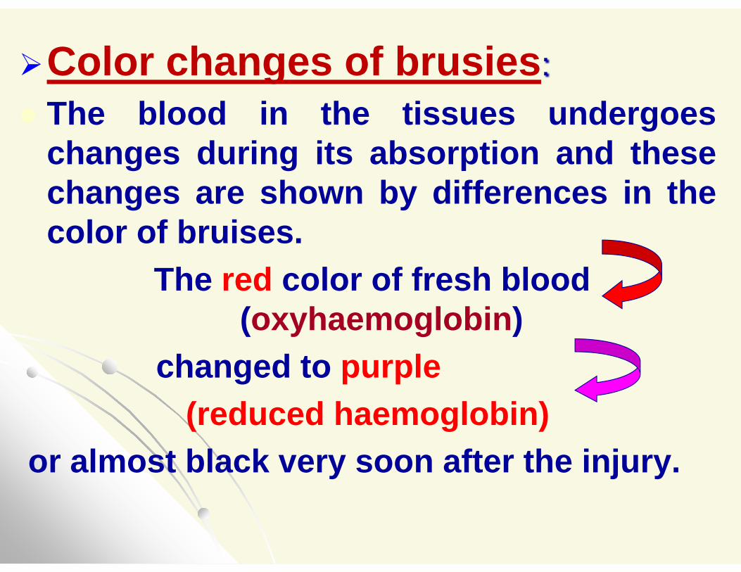
Color changes of brusies: The blood in the tissues undergoes
changes during its absorption and these changes are shown by differences in the color of bruises.
The red color of fresh blood (oxyhaemoglobin)
changed to purple (reduced haemoglobin)
or almost black very soon after the injury.
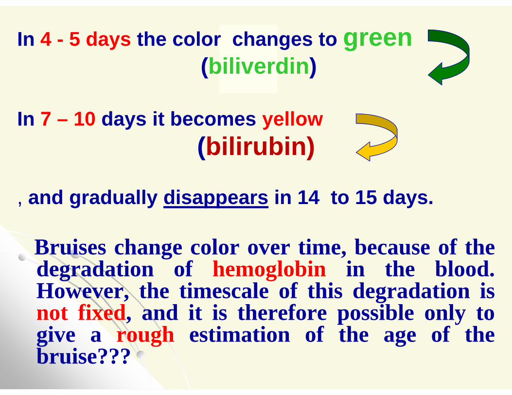
In 4 - 5 days the color changes to green(biliverdin)
In 7 – 10 days it becomes yellow(bilirubin)
, and gradually disappears in 14 to 15 days.
Bruises change color over time, because of thedegradation of hemoglobin in the blood.However, the timescale of this degradation isnot fixed, and it is therefore possible only togive a rough estimation of the age of thebruise???
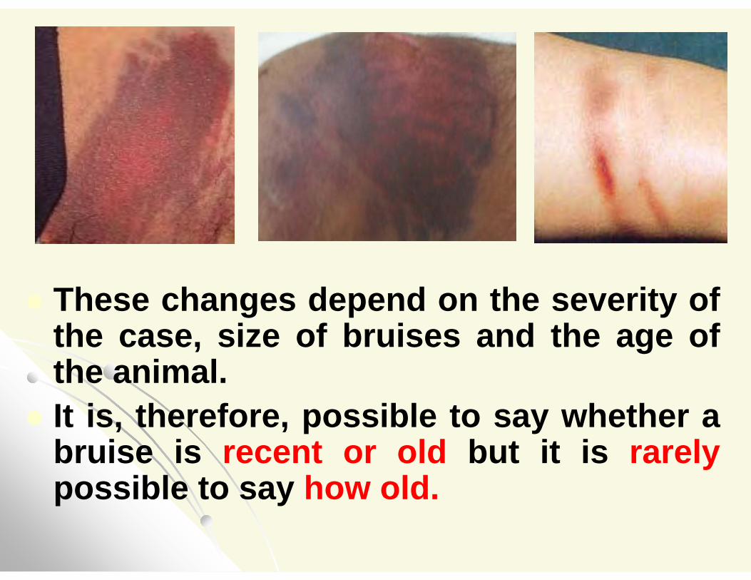
These changes depend on the severity of the case, size of bruises and the age of the animal.
It is, therefore, possible to say whether a bruise is recent or old but it is rarelypossible to say how old.
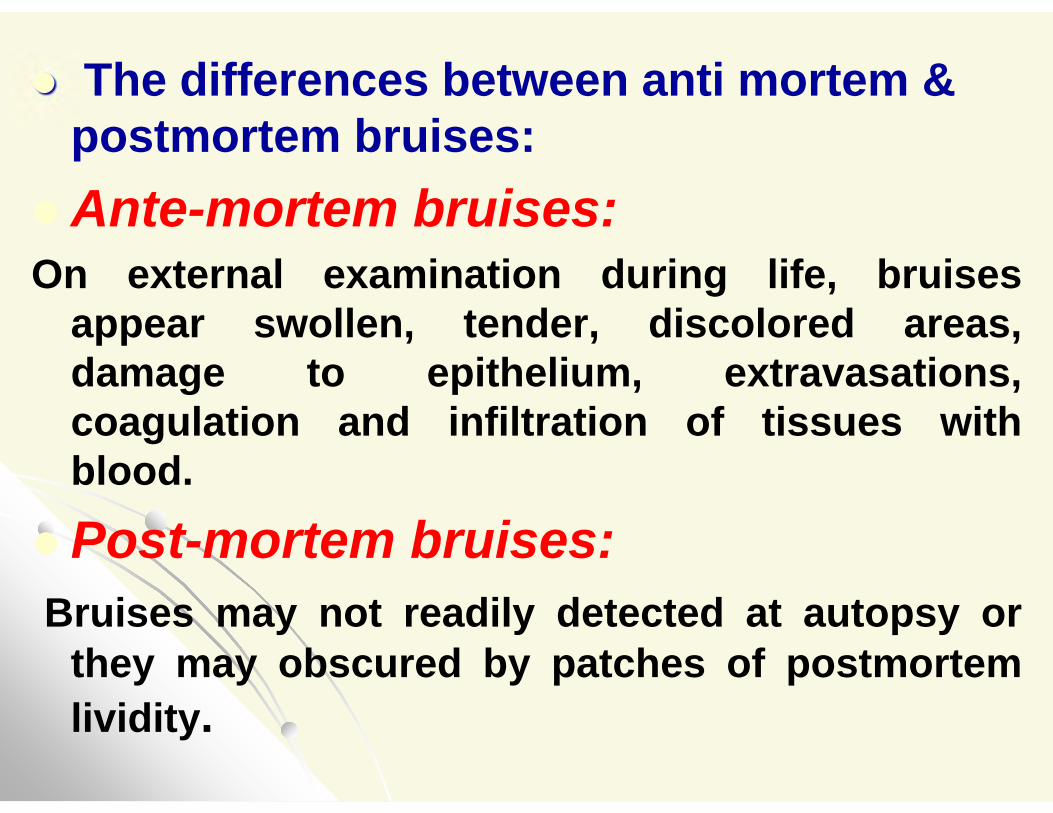
The differences between anti mortem & postmortem bruises:
Ante-mortem bruises:On external examination during life, bruises
appear swollen, tender, discolored areas, damage to epithelium, extravasations, coagulation and infiltration of tissues with blood.
Post-mortem bruises:Bruises may not readily detected at autopsy or
they may obscured by patches of postmortem lividity.
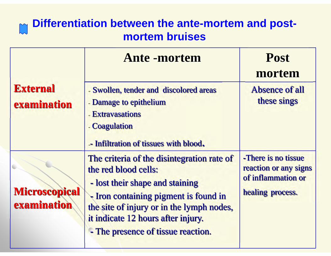
Differentiation between the ante-mortem and post-mortem bruises
Post mortem
Ante -mortem
External examination
Absence of all these sings
- Swollen, tender and discolored areas- Damage to epithelium - Extravasations - Coagulation
-- Infiltration of tissues with blood. -There is no tissue reaction or any signs of inflammation or
healing process.
The criteria of the disintegration rate of the red blood cells:- lost their shape and staining - Iron containing pigment is found in
the site of injury or in the lymph nodes, it indicate 12 hours after injury.- The presence of tissue reaction.
Microscopical examination
- Distinguishing between hypostasis and bruises
This can be done by cutting in the discolored area: In case of hypostasis the blood is present in blood
vessels, and easily washed away by a stream of water.
if the color is due to a bruises, extravasated blood will be seen infiltrating the tissues, this blood is firmlyclotted that it can not be washed away.
Also bruises are often accompanied by abrasions and /or signs of sepsis.
C- Lacerated wounds
DefinitionThey are wounds in which the tissues are torn as a result of the application of blunt force to the body.
The external lacerated wounds are caused by splitting of soft tissues against the underlying bone.
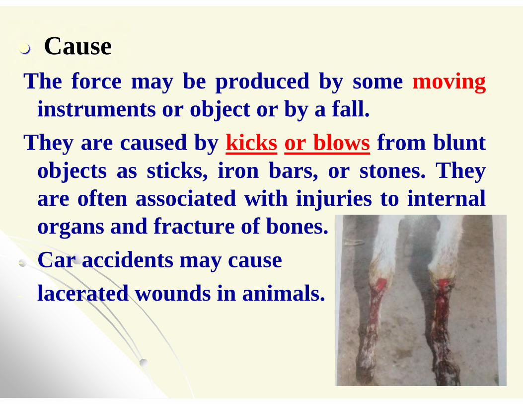
CauseThe force may be produced by some moving
instruments or object or by a fall. They are caused by kicks or blows from blunt
objects as sticks, iron bars, or stones. They are often associated with injuries to internal organs and fracture of bones.
- Car accidents may cause - lacerated wounds in animals.
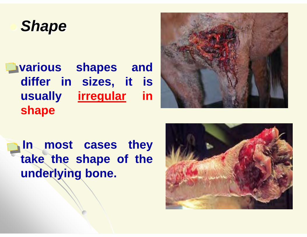
Shape
various shapes and differ in sizes, it is usually irregular in shape
In most cases they take the shape of the underlying bone.
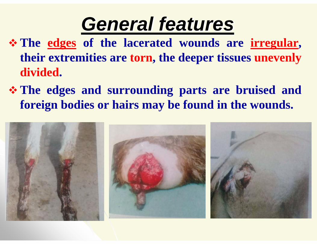
General featuresThe edges of the lacerated wounds are irregular,
their extremities are torn, the deeper tissues unevenly divided.
The edges and surrounding parts are bruised and foreign bodies or hairs may be found in the wounds.
If hair- bulbs are present they will be seen crushed instead of cut.
External bleeding from laceration is not pronounced (low) because the blood vessels are usually crushed.
Healing generally takes place by second intention results in a well-marked scarformation.
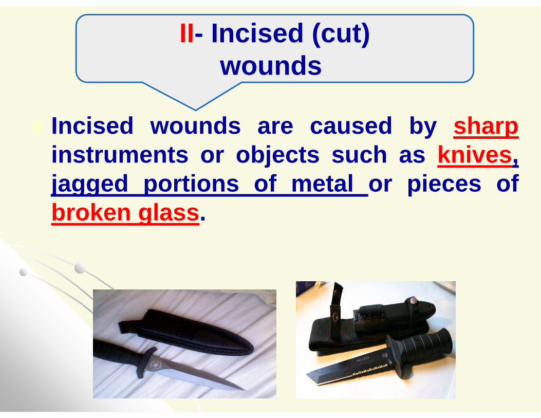
Incised wounds are caused by sharpinstruments or objects such as knives, jagged portions of metal or pieces of broken glass.
II- Incised (cut)wounds
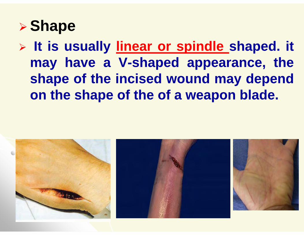
Shape It is usually linear or spindle shaped. it
may have a V-shaped appearance, the shape of the incised wound may depend on the shape of the of a weapon blade.
General featuresThe edges of incised wound are regular,
clean-cut and everted.
It is usually longer than deep, it is often gaping.
In deep incised wounds, the degree of gaping is greater when the muscles are cut transversely than when they are cut in the longitudinal plane of their fibers.
Bleeding is profuse especially if blood vessels are cut.
Bruising may or may not present.
Infection is relatively uncommon and usually heals with first intention with
minimal scar formation.
Age: Within 12 hours it becomes red with swollen
edges.
In about 24 hours, the proliferation of vascular endothelium occurs.
After 36 hours, incised wounds are covered with lymph.
After 3 days their edges are strongly adhered.
In about 7 days healing takes place leaving a red linear scar.
If infection or sepsis occurred, the wound may remain open for indefinite period and scars are larger and deeper.
The usual incised wound is linear except in the regions of the loose skin it takes zigzag course e.g. wound in the neck or axilla.
??????Compare between lacerated and incised wounds:- Cause, - Shape and edges- bleeding,-healing by,,,,,
- Scare formation.
Punctured or stab wounds are caused by long narrow instruments with blunt or pointed ends.
Punctured wounds are described as" penetrating" when they pierce deeply into tissues and as "perforating" when they transfix tissues and cause exit wounds.
An incised wound becomes stab when it is deeper than broad.
III-Stab and Punctured wounds
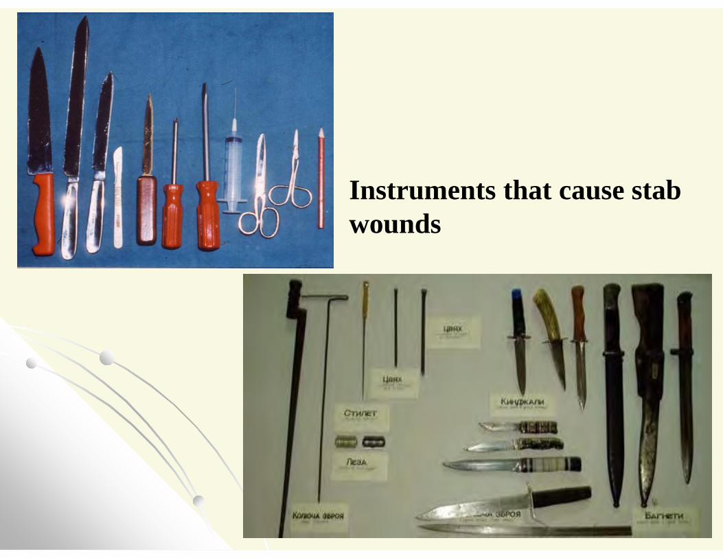
Instruments that cause stab wounds
CauseThey are caused by sharp - pointed objects as
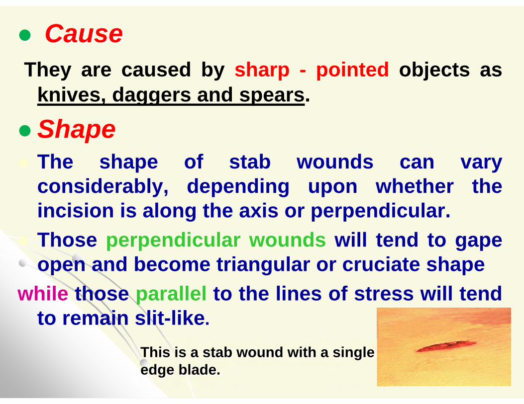
knives, daggers and spears.Shape The shape of stab wounds can vary
considerably, depending upon whether theincision is along the axis or perpendicular.
Those perpendicular wounds will tend to gapeopen and become triangular or cruciate shape
while those parallel to the lines of stress will tendto remain slit-like.
This is a stab wound with a single edge blade.
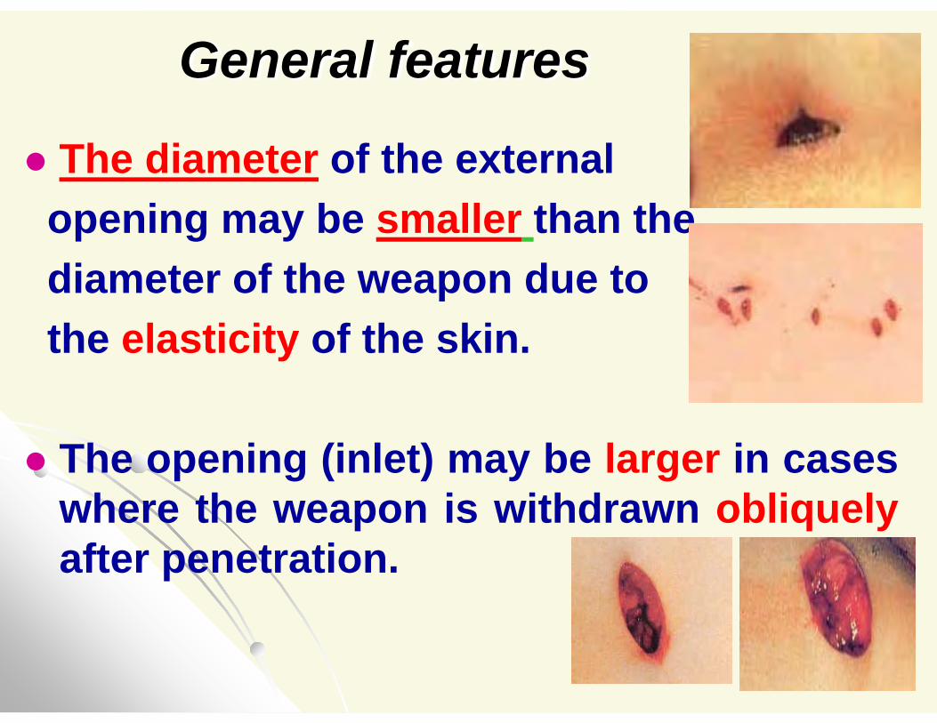
General features
The diameter of the externalopening may be smaller than the diameter of the weapon due to the elasticity of the skin.
The opening (inlet) may be larger in cases where the weapon is withdrawn obliquely after penetration.
The depth may be greater than the total length of the penetrating object because the tissues deep to the skin are often compressed during the process of penetration.
The size of the wound doesn'tnecessarily correspond to the width ofthe used weapon, because withdrawingthe instrument may cause the wound toenlarged.
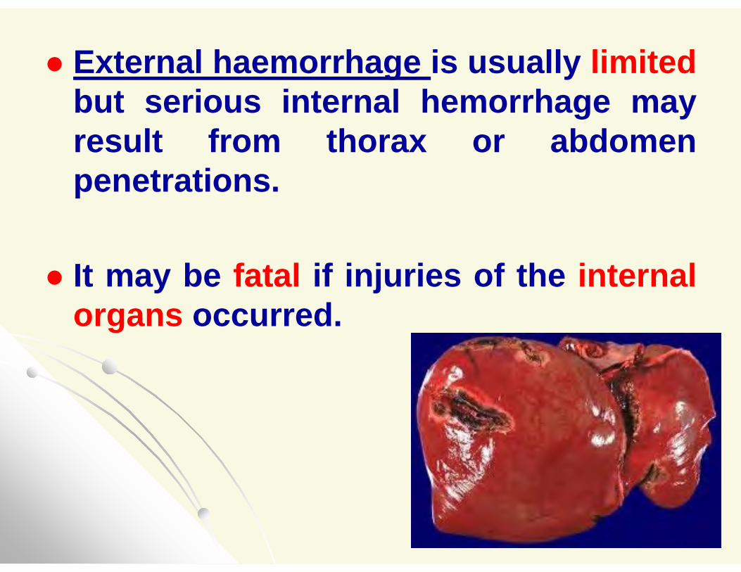
External haemorrhage is usually limitedbut serious internal hemorrhage may result from thorax or abdomen penetrations.
It may be fatal if injuries of the internal organs occurred.
The estimation of the approximate age of an ante-
mortem wound The age of the ante-mortem wound has
to be determined for medico-legal purposes.
The age of the ante-mortem wound was determined depending upon the time of occurrence of the reactive changes of inflammation to an aseptic injury as following:
Dilatation of the capillaries and migration of the leucocytes may be seen within few minutes of injury.
Emigration of leucocytes is usually observed within an hour, the first type of leucocytes is polymorph nuclear neutrophils.
Monocytes appear later after 12 hours.
The exudates reach the maximum intensity within 48 hours.
Fibroblasts present at the site of injury in few hours and the cells begin mitotic division through 15 hours.
The proliferation of the fibroblasts and vascularized granulation tissue takes 72 hours to develop the collagen formation.
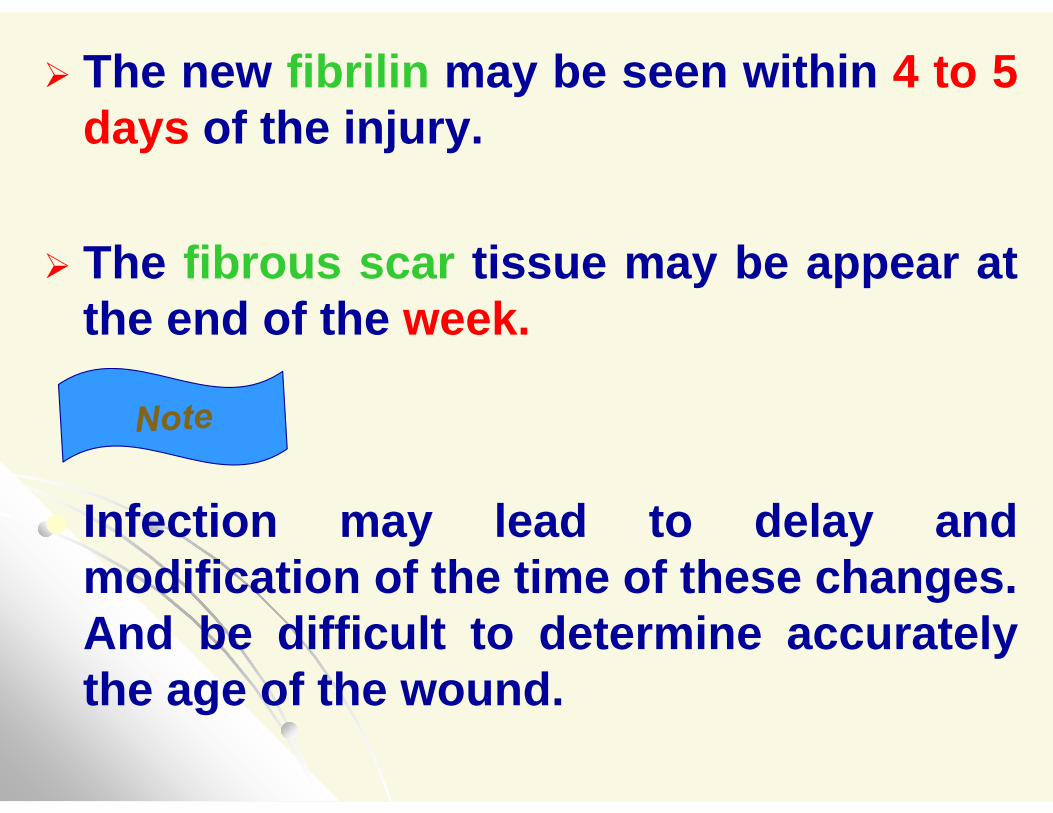
The new fibrilin may be seen within 4 to 5 days of the injury.
The fibrous scar tissue may be appear at the end of the week.
Infection may lead to delay and modification of the time of these changes. And be difficult to determine accurately the age of the wound.
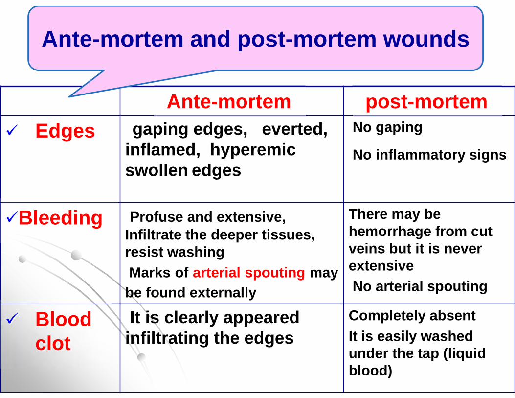
post-mortemAnte-mortemNo gaping
No inflammatory signsgaping edges, everted,
inflamed, hyperemic swollen edges
Edges
There may be hemorrhage from cut veins but it is never extensiveNo arterial spouting
Profuse and extensive, Infiltrate the deeper tissues, resist washingMarks of arterial spouting may
be found externally
Bleeding
Completely absent It is easily washed under the tap (liquid blood)
It is clearly appeared infiltrating the edges
Blood clot
Ante-mortem and post-mortem wounds
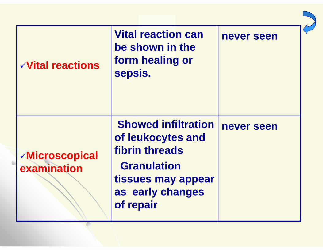
never seenVital reaction can be shown in the form healing or sepsis.
Vital reactions
never seenShowed infiltration of leukocytes and fibrin threadsGranulation
tissues may appear as early changes of repair
Microscopicalexamination
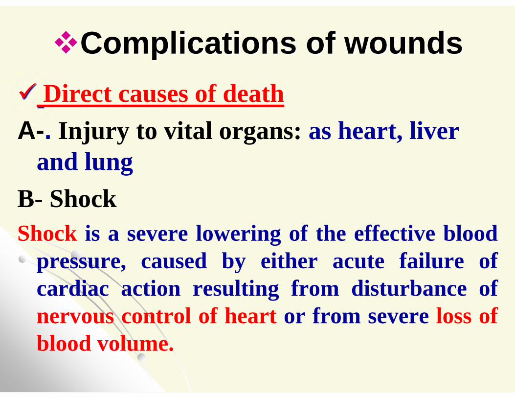
Complications of wounds Direct causes of deathA-. Injury to vital organs: as heart, liver
and lungB- ShockShock is a severe lowering of the effective blood
pressure, caused by either acute failure of cardiac action resulting from disturbance of nervous control of heart or from severe loss of blood volume.
Types of traumatic shock It is either neurogenic or hematogenic.I- Neurogenic shock: Two types:
A- Parasympathetic or vagus nerve leads to acute circulatory failure.
The PM: tissues and organs are pale; the right side of the heart is empty and collapsed.
B- Sympathetic nerves lead to ventricular fibrillation. It accompanied with painful wounds.
The PM showing congestion of organs and pulmonary edema.
II. Hematogenic (hemorrhagic) shock It is due to either external hemorrhage or
increases the capillary permeability and loss the capillary tone as a result of toxic histamine like substances absorbed from the site of trauma.
The PM showing engorgement of the capillaries and veins, peticheal haemorrhage in the tissue, empty heart and contraction of spleen.
Indirect causes of deathA-Embolism
It is a detached intravascular physical mass that is carried by the blood to a site distant from its point of origin it may be: Arterial embolism Pulmonary embolism Fat embolismAir embolismForeign body embolism
B-Septic infection As a result of microbial infection lead to
septicemia.
C- Scare formation Scare formation in the intestinal lumen
lead to obstruction and death.
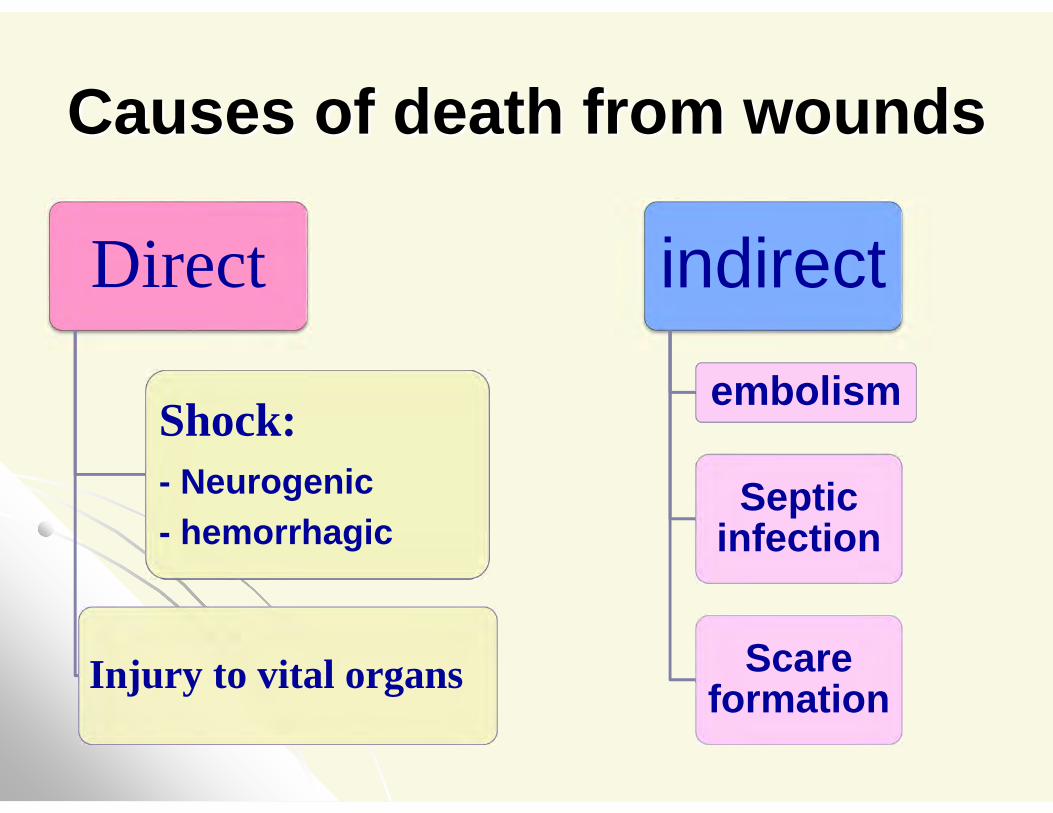
Causes of death from wounds
Direct
Shock:- Neurogenic- hemorrhagic
Injury to vital organs
indirectembolism
Septic infection
Scare formation
Firearm wounds

Thank you