Medical School Histology: Images of compact bone
16
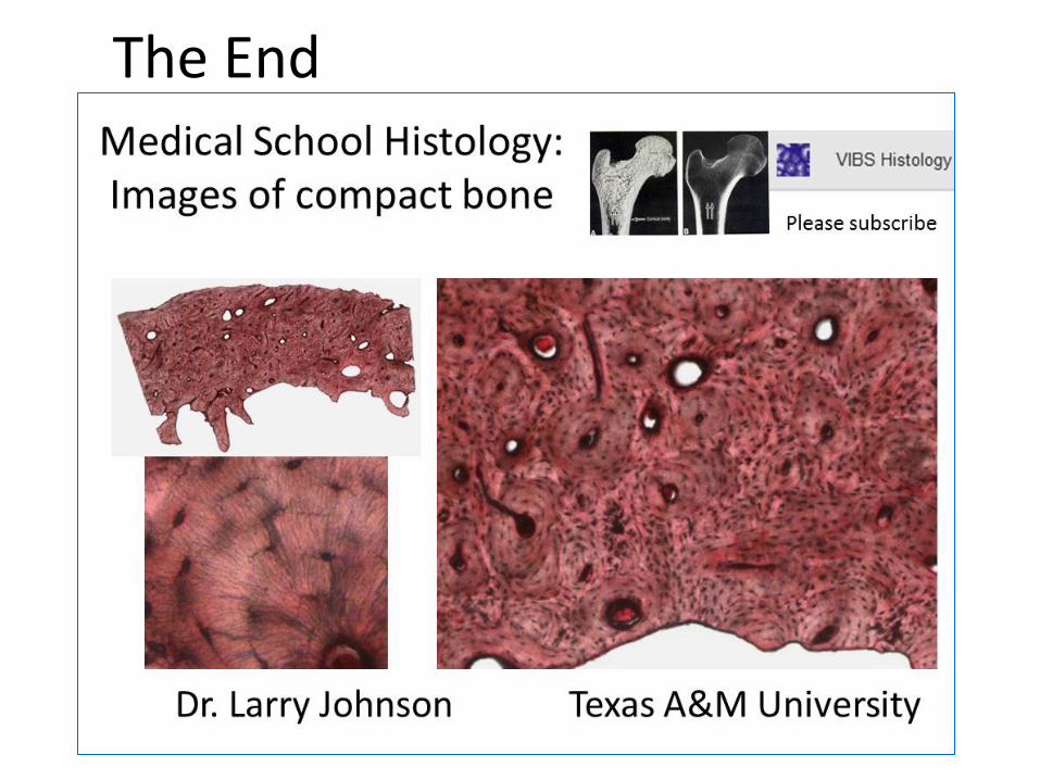
Dr. Larry Johnson Texas A&M University Medical School Histology: Images of compact bone Please subscribe
Transcript of Medical School Histology: Images of compact bone
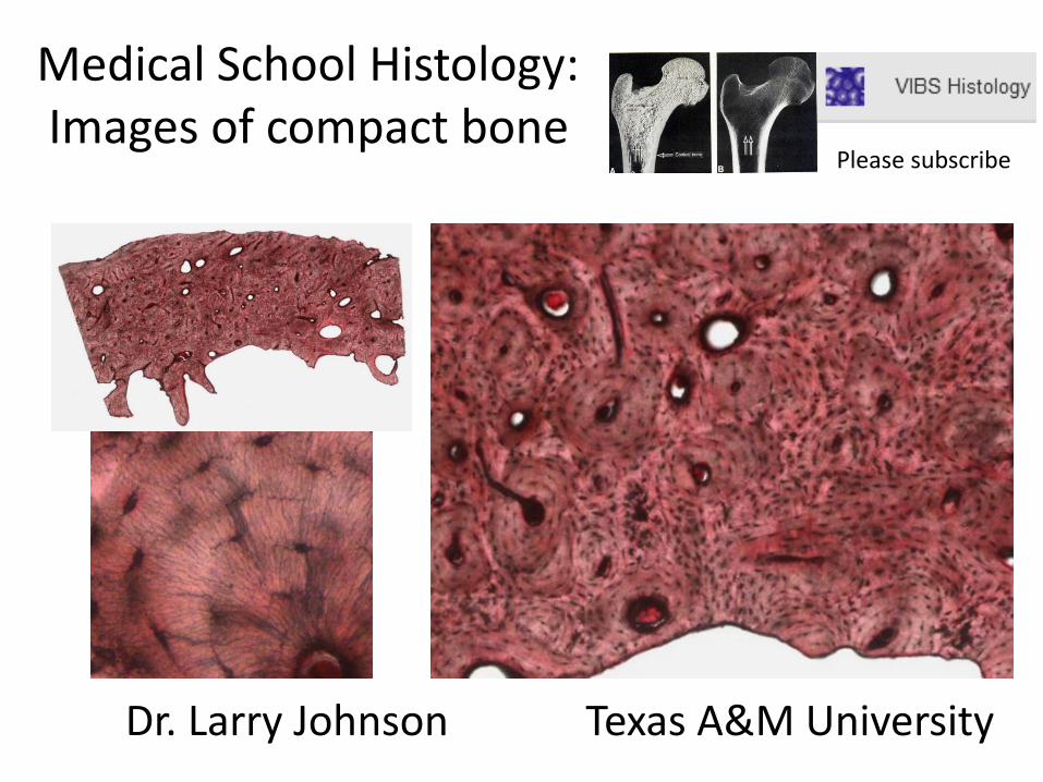
Dr. Larry Johnson Texas A&M University
Medical School Histology: Images of compact bone
Please subscribe

Observe/manipulate the images on your computer
PowerPoint slides on YouTube VIBS Histology and at http://peer.tamu.edu/histology.asp
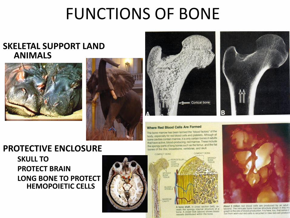
FUNCTIONS OF BONE SKELETAL SUPPORT LAND
ANIMALS PROTECTIVE ENCLOSURE
SKULL TO PROTECT BRAIN LONG BONE TO PROTECT
HEMOPOIETIC CELLS
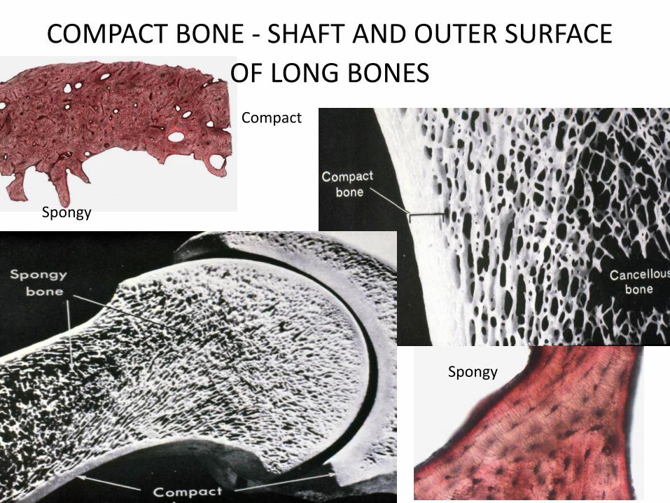
COMPACT BONE - SHAFT AND OUTER SURFACE
OF LONG BONES Compact
Spongy
Spongy

Vertebra
Fibrous periosteum containing fibroblasts
Osteoblasts lining bony trabeculae
Bone Osteocyte in lacunae
Compact bone
Spongy bone
Spongy bone
Endosteum
Periosteum
Endosteum
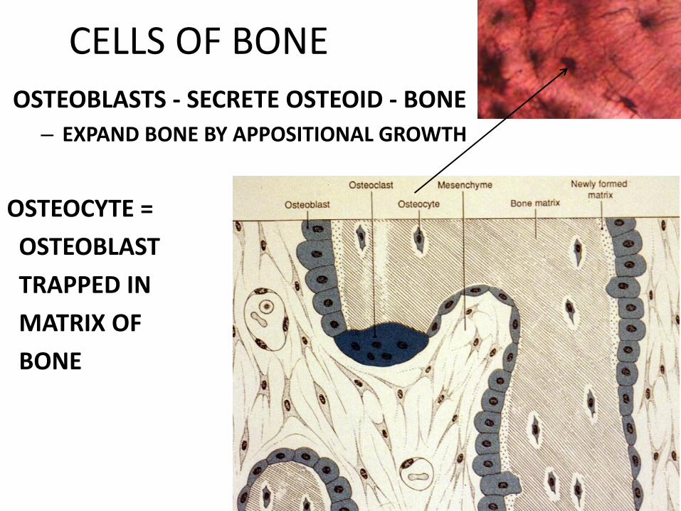
CELLS OF BONE
OSTEOBLASTS - SECRETE OSTEOID - BONE
– EXPAND BONE BY APPOSITIONAL GROWTH
OSTEOCYTE =
OSTEOBLAST
TRAPPED IN
MATRIX OF
BONE

EXTRACELLULAR MATRIX OF BONE
SECRETED by POLARIZED OSTEOBLASTs
CALCIFICATION - ADDS FIRMNESS, BUT PREVENTS DIFFUSION THROUGH MATRIX MATERIAL
Osteocytes
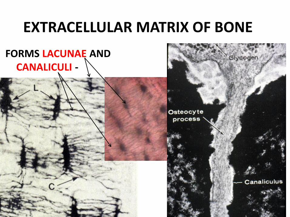
EXTRACELLULAR MATRIX OF BONE
FORMS LACUNAE AND CANALICULI -
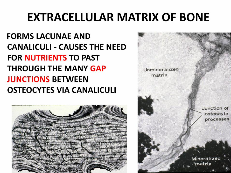
EXTRACELLULAR MATRIX OF BONE
FORMS LACUNAE AND CANALICULI - CAUSES THE NEED FOR NUTRIENTS TO PAST THROUGH THE MANY GAP JUNCTIONS BETWEEN OSTEOCYTES VIA CANALICULI

COMPACT BONE, REMODELING
(Spongy bone)
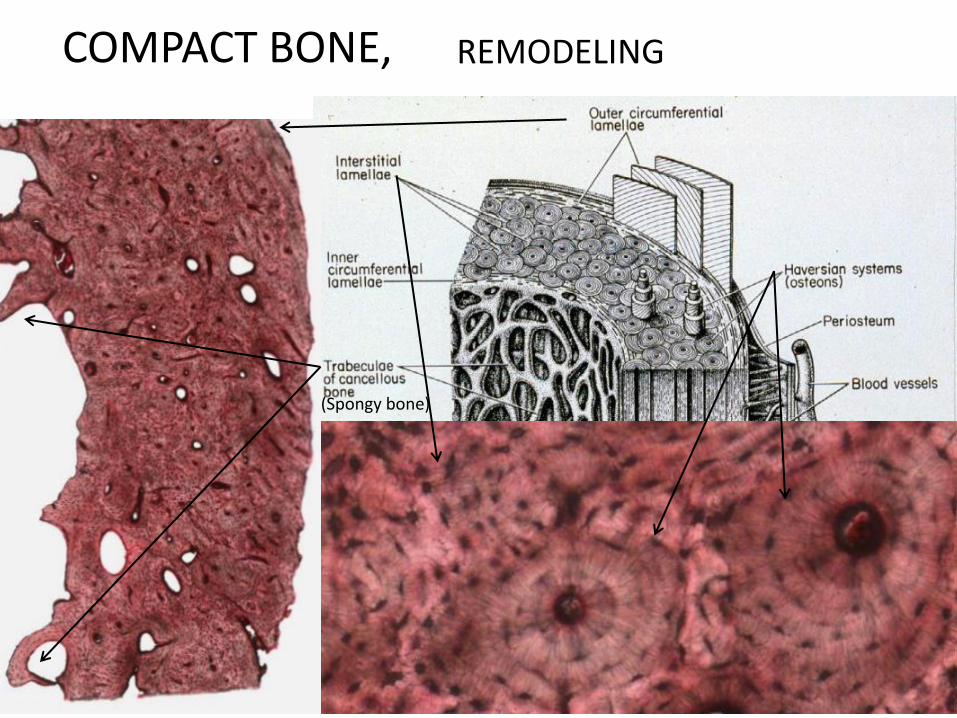
COMPACT BONE, REMODELING
(Spongy bone)
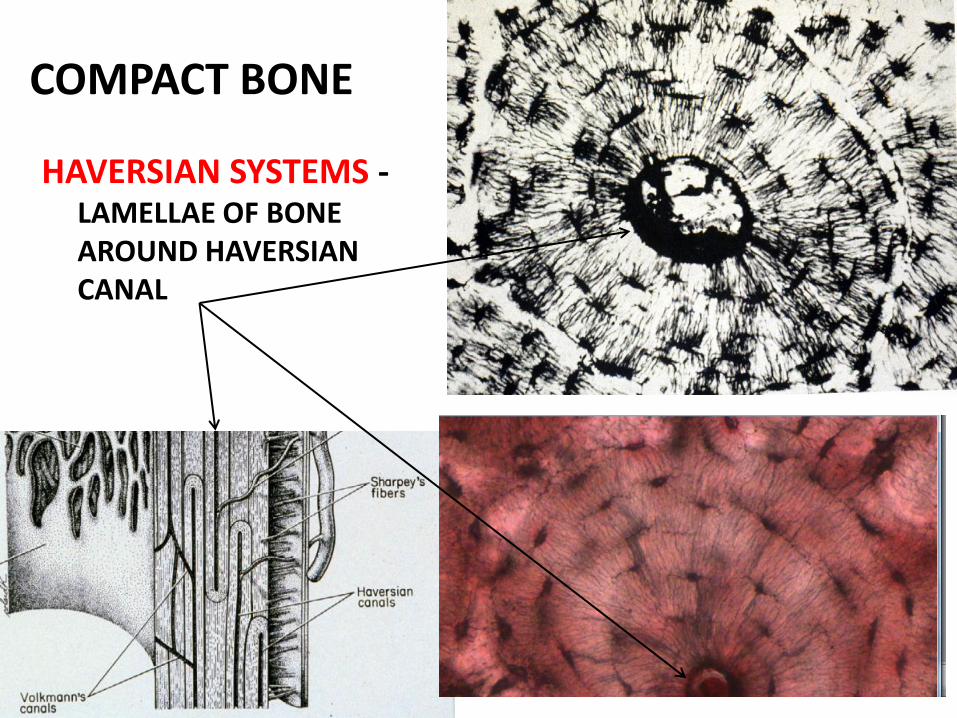
COMPACT BONE
HAVERSIAN SYSTEMS - LAMELLAE OF BONE AROUND HAVERSIAN CANAL
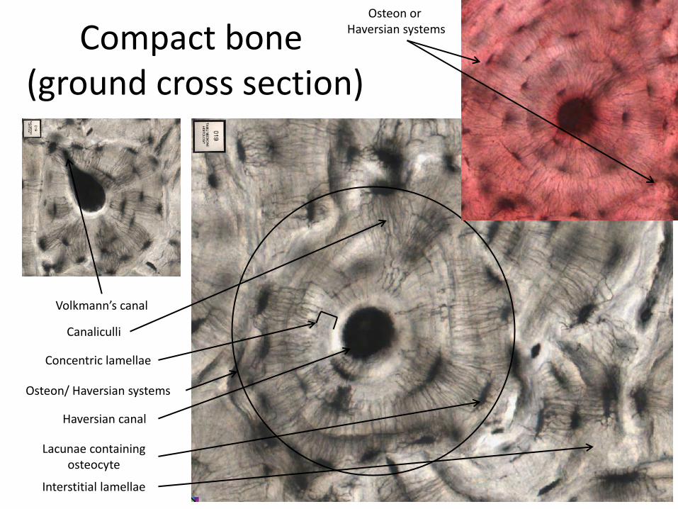
Compact bone (ground cross section)
Osteon/ Haversian systems
Haversian canal
Lacunae containing osteocyte
Concentric lamellae
Canaliculli
Interstitial lamellae
Volkmann’s canal
Osteon or Haversian systems
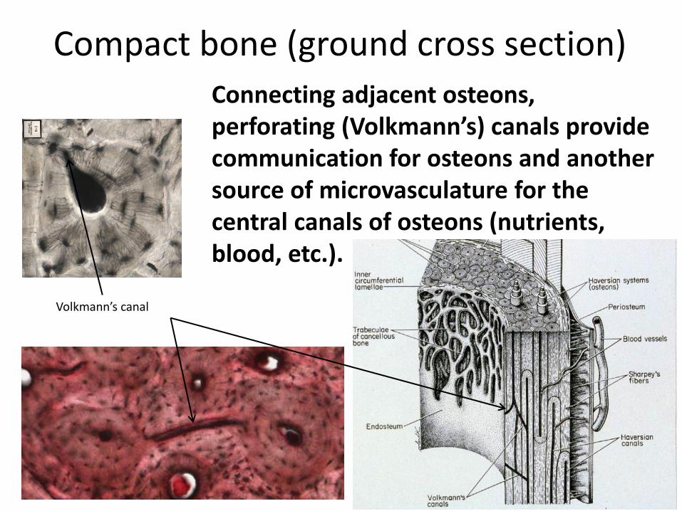
Compact bone (ground cross section)
Volkmann’s canal
Connecting adjacent osteons, perforating (Volkmann’s) canals provide communication for osteons and another source of microvasculature for the central canals of osteons (nutrients, blood, etc.).

• Bone 36726 – http://viewer.serenusview.com/LinkHandler.axd?LinkId=bfeae008-4430-49a9-8deb-200690e3f484
Download actual images at
The End