MEDICAL PHYSICS IN THE BALTIC STATES 11 (2013) … · MEDICAL PHYSICS IN THE BALTIC STATES 11 ......
4
MEDICAL PHYSICS IN THE BALTIC STATES 11 (2013) Proceedings of International Conference "Medical Physics in the Baltic States" 10- 12 October 2013, Kaunas, Lithuania GALLSTONES STUDIES BY EPR AND EDX SPECTROSCOPIES Maksims POLAKOVS 1 , Nina MIRONOVA-ULMANE 1 , Andrejs PAVLENK0 1 , Vera SKVORTSOVA 1 , Dmitrijs JAKOVLEVS 2 'Institute of solid State Physics, University of Latvia, l}.engaraga Street 8, Ri ga, Latvia, LV -I 063 2 Riga Technical University, Kalku Str. I, Riga, Latvia, LV-I 048 Abstract: In the present work we report results of investigations of gallstones. Gallstones can be separated into four main groups - cholesterol, mixed, pigment, and calcium carbonate stones. Gallstones were donated by patient after surgical removal. We have found C, 0, Na, Mg, AI, Si, S, Cl, K, Ca, Fe, Sb, I macro and micro elements into ga ll stones by X-ray fluorescence analysis (EDX). Electron paramagnetic resonance (EPR) spectra obtained for black pi gment have two EPR signals with g-factor=4.8 (hi gh spin state S= 5/2 of Fe3+) and EPR signal with g-factor=2.003 (low spin state S=l/2 of Fe3+). EPR spectra obtained for brown pigment have EPR signal with g-factor=2.003 (low spin state S= I/2 ofFe3+). Keywords: EPR (Electron paramagnet ic resonance), EDX (energy dispersive X-ray spectrometry), cholesterol, mixed, pigment, and calcium carbonate gallstones. 1. Introduction Gallstone formation in gallbladder is a common disease and constitutes a major health problem worldwide. Cholesterol stones account for 70% to 95% of adult ga ll stones and black pigment stones for most of the remainder. Calcium carbonate stones are exceptionally rare. Ga ll stones are hard masses can form in the gall bl adder and in bile ducts. They have variable compositions that can in clude cholesterol, bilirubin, calcite, fatty acids and other inorganic and organic compounds that may precipitate from bile. The morphology of ga ll stones is variable, and the following forms have been described: oviform, "mu lberry berries ", facetted (with number of s id es from 3 up to 20), aggregate (accretion of several primary stones), spherulite and " pearl " sand . Ga ll stones can be conveniently grouped into four major types depending on their predominant component: cholesterol stones (which are > 47% cholesterol by weight), mixed stones (which consist of varying concentrations of cholesterol and bilirubinate salts), pigment stones (which consist mainly of various bilirubinate salts) and calcium 24 carbonate stones (which are mainly various polymorphs of modification calcium carbonate, calcite, or aragonite, or vaterite) [ 1-9]. The formation of gallstones is characterized by a selectivity of crystallization of one or the other modification of calcium carbonate. This is connected with the crystal gro\vth influenced by the arrangement of atoms in crystal, and the constTaints imposed by coordination of cations and stabilization effect of amino acid in bile [I O-Il]. In the present work different types of gallstones were studied. We report results of gallstones investigations by EPR and EDX Spectroscopy. 2. Materials and methods The samples of gallstones were donated by private persons after surgical removal. Exterior view of investigated gallstones is shown in Fig. I. The EPR spectra of gallstones were measured on a BRUKER EMX-6/ I spectrometer equipped with an Aspect 2000 data system. An ER 4102 ST Universal X- Band Resonator (TE 102 mode) was used. The g-factor of EPR signals were determined by reference of the external magnet ic field value measured by a Bruker ER 035 Gaussmeter and of the microwave frequency measured by a Systron Donner 6235A frequency counter. Magnetic field varied between I 00 and 7000 Gauss. The EPR signal intensi ti es in ga ll stones were measured against fixed standard signals using the standard crystal MgO (Cr 3 +) pl aced in resonant cavity. The spectra were recorded at microwave power 6.2 mW, applying magnetic field modulation of 100 kHz and amplitude I mT. One spectrum is obtained averagin g 20 scans with receiver time constant 25 ms. The samples were observed in a scanning electron microscope (SEM, TESCAN Mira\LMUField- Emission-Gun) as well. The elemental composition of gall stones was assessed by energy dispersive X -ray spectrometry (EDS, Oxford instruments 7378) performed on the sca1ming electron microscope. The gall stones was separated into three groups by gall stone's component color. The samples were
Transcript of MEDICAL PHYSICS IN THE BALTIC STATES 11 (2013) … · MEDICAL PHYSICS IN THE BALTIC STATES 11 ......

MEDICAL PHYSICS IN THE BALTIC STATES 11 (2013)
Proceedings of International Conference "Medical Physics in the Baltic States" 10- 12 October 2013, Kaunas, Lithuania
GALLSTONES STUDIES BY EPR AND EDX SPECTROSCOPIES
Maksims POLAKOVS 1, Nina MIRONOVA-ULMANE1
, Andrej s PAVLENK01, Vera SKVORTSOVA1
,
Dmitrijs JAKOVLEVS2
'Institute of solid State Physics, University of Latvia, l}.engaraga Street 8, Riga, Latvia, LV -I 063 2Riga Technical University, Kalku Str. I, Riga, Latvia, LV-I 048
Abstract: In the present work we report results of investigations of gallstones. Gallstones can be separated into four main groups - cholesterol, mixed, pigment, and calcium carbonate stones. Gallstones were donated by patient after surgical removal. We have found C, 0, Na, Mg, AI, Si, S, Cl, K, Ca, Fe, Sb, I macro and micro elements into ga llstones by X-ray fluorescence analysis (EDX). Electron paramagnetic resonance (EPR) spectra obtained for black pigment have two EPR signals with g-factor=4.8 (high spin state S= 5/2 of Fe3+) and EPR signal with g-factor=2.003 (low spin state S= l /2 of Fe3+). EPR spectra obtained for brown pigment have EPR signal with g-factor=2.003 (low spin state S= I/2 ofFe3+).
Keywords: EPR (Electron paramagnetic resonance), EDX (energy dispersive X-ray spectrometry), cholesterol, mixed, pigment, and calcium carbonate gallstones.
1. Introduction
Gallstone formation in ga llbladder is a common disease and constitutes a major health problem worldwide. Cholesterol stones account for 70% to 95% of adult gallstones and black pigment stones for most of the remainder. Ca lcium carbonate stones are exceptionally rare. Gallstones are hard masses can form in the gall bladder and in bile ducts. They have variable compositions that can include cholestero l, bilirubin, calcite, fatty acids and other inorganic and organic compounds that may precipitate from bile. The morphology of gall stones is variable, and the following forms have been described: oviform, "mulberry berries", facetted (with number of sides from 3 up to 20), aggregate (accretion of several primary stones), spherulite and " pearl " sand . Gall stones can be conveniently grouped into four major types depending on their predominant component: cholesterol stones (which are > 47% cholesterol by weight), mixed stones (which consist of varying concentrations of cholesterol and bilirubinate salts), pigment stones (which consist mainly of various bilirubinate salts) and calcium
24
carbonate stones (which are mainly various polymorphs of modification calcium carbonate, calcite, or aragonite, or vaterite) [ 1-9]. The formation of gallstones is characterized by a selectivity of crystallization of one or the other modification of calcium carbonate. This is connected with the crystal gro\vth influenced by the arrangement of atoms in crystal, and the constTaints imposed by coordination of cations and stabilization effect of amino acid in bile [I O-Il]. In the present work different types of gallstones were studied. We report results of gallstones investigations by EPR and EDX Spectroscopy.
2. Materials and methods
The samples of gallstones were donated by private persons after surgical removal. Exterior view of investigated gallstones is shown in Fig. I. The EPR spectra of gallstones were measured on a BRUKER EMX-6/ I spectrometer equipped with an Aspect 2000 data system. An ER 4102 ST Universal XBand Resonator (TE102 mode) was used . The g-factor of EPR signals were determined by reference of the external magnetic field value measured by a Bruker ER 035 Gaussmeter and of the microwave frequency measured by a Systron Donner 6235A frequency counter. Magnetic field varied between I 00 and 7000 Gauss. The EPR signal intensities in gallstones were measured against fixed standard signals using the standard crystal MgO (Cr3+) placed in resonant cavity. The spectra were recorded at microwave power 6.2 mW, applying magnetic field modulation of 100 kHz and amplitude I mT. One spectrum is obtained averaging 20 scans with receiver time constant 25 ms. The samples were observed in a scanning electron microscope (SEM, TESCAN Mira\LMUFieldEmission-Gun) as well. The elemental composition of gallstones was assessed by energy dispersive X -ray spectrometry (EDS, Oxford instruments 7378) performed on the sca1ming electron microscope. The gallstones was separated into three groups by gallstone's component color. The samples were
M Polakovs et. al. I Medical Physics in the Baltic States 11 (201 3) 24-27
preparied in two diffrent ways. The gallstone was cutted into few layers for EDX spectroscopy studing and the layers were scraped for EPR sprectorscopy investigation. EDX spectroscopy technique let us to stud y the composite of gallstones and EPR spectroscopy give us information about electronic states of transition metal ions.
3. Experimental results and discussion
We observed gallstones from three different humans in our investigations. The Energy-dispersive X-ray (EDX) and Electronic paramagnetic resonance (EPR) spectroscopy methods were used for investigation of gallstones, scanning electron microscopy (SEM) was used for studying of gallstones surface morphology as well.
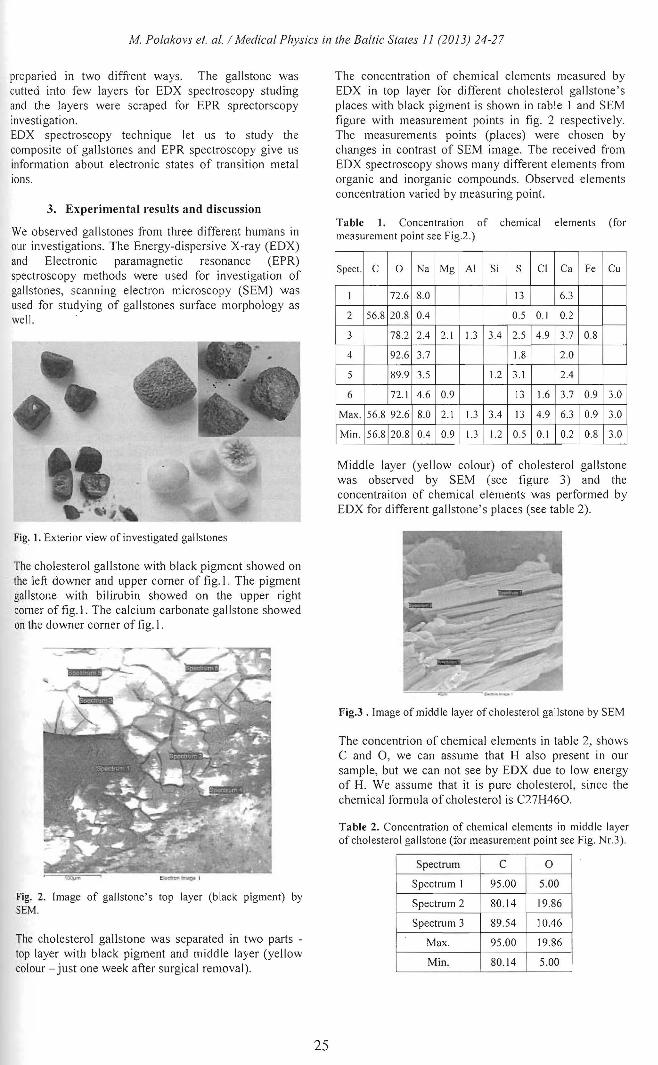
Fig~ 1. Exterior view of investigated gal I stones
The cholesterol gallstone with black pigment showed on the left downer and upper corner of fig. I. The pigment gallstone with bilirubin showed on the upper right comer of fig. I. The calcium carbonate ga llstone showed on the downer corner of fig. I.
Fig. 2. Image of ga llstone's top layer (black p igment) by SEM.
The cholestero l gallstone was separated in two parts -top layer with black pigment and middle layer (yellow colour - just one week after surgical remova l).
25
The concentration of chemical elements measured by EDX in top layer for different cholesterol gallstone's places with black pigment is shown in table 1 and SEM figure with measurement points in fig. 2 respectively. The measurements points (places) were chosen by changes in contrast of SEM image. The received from EDX spectroscopy shows many different elements from organic and inorganic compounds. Observed elements concentration varied by measuring point.
Table 1. Concentration of chemical elements (for measurement point see Fig.2.)
Spect. c 0 Na Mg AI Si s Cl Ca Fe Cu
I 72.6 8.0 13 6.3
2 56.8 20.8 0.4 0.5 0. 1 0.2
3 78.2 2.4 2.1 1.3 3.4 2.5 4.9 3.7 0 8
4 92.6 3.7 1.8 2.0
5 89.9 3.5 1.2 3.1 2.4
6 72. 1 4.6 0.9 13 1.6 3.7 0.9 3.0
Max. 56.8 92.6 8.0 2.1 1.3 3.4 13 4.9 6.3 0.9 3.0
Min. 56.8 20.8 0.4 0.9 1.3 1.2 0.5 0. 1 0.2 0.8 3.0
Middle layer (yellow colour) of cholesterol gallstone was observed by SEM (see figure 3) and the concentraiton of chemical e lements was performed by EDX for different gallstone's places (see table 2).
Fig.3. Image of middle layer of cholesterol gal lstone by SEM
The concentrion of chemical e lements in table 2, shows C and 0, we can assume that H also present in our sample, but we can not see by EDX due to low energy of H. We assume that it is pure cho lesterol, since the chemical formula of cholesterol is C27H460.
Table 2. Concentration of chemical elements in midd le layer of cho lesterol ga llstone (for measurement point see Fig. Nr.3 ).
Spectrum c 0
Spectrum I 95 .00 5.00
Spectrum 2 80.14 19.86
Spectrum 3 89.54 10.46
Max. 95 .00 19.86 J
M in. 80.14 5.oo 1

M Polakovs et. al. I Medical Physics in the Baltic States 11 (2013) 24-27
We can see on the fig. 3, that pure cholesterol has a layered structure. When cholesterol include other chemical elements then mentioned above, it is involve a changes in the structure of cholesterol. The changes in stucture· is showed in images (see fig. 4, fig. 5) recieved by Scanning Electron microscope. The recieved data by EDX spectroscopy for measurement points of 3,4 figures confirm in opinion that changes in structure is due to incorporated inorganic compounds.
Fig. 4 . Image of middle layer of cholesterol gallstone with additional chemical elements by SEM
It well known that heavy elements (high atomic number) backscatter electrons more strongly than light elements (low atomic number), and thus appear brighter in the image. The measurements points of Fig. 4 were chosen by changes in contrast ofSEM image.
Table 3. Concentration of chemical elements in middle layer of cholesterol gallstone with additional chemical elements (for measurement point see Fig. Nr.4, from right side).
Spect. c 0 Na AI C1 K I 75.39 8.76 0.74 7.3 7.8 2 88.51 11.38 0.11 3 87.78 7.67 0.24 2,0 2.3
Max. 88.51 11.38 0.74 0.11 7.3 7.8 Min. 75.39 7.67 0.24 0.11 2,0 2.3
Table 4. Concentration of chemical elements in middle layer of cholesterol gallstone with additional chemical elements (for measurement point see Fig. 4, from left side).
Spect. c 0 Na Mg AI Si s CJ K Ca Fe I 29.8 41.9 0.2 0.4 2.8 4.0 1.5 0.5 0.3 14.4 4.2 2 19.6 45.7 0.3 0.2 0.7 9.8 0.2 0.2 0.3 22.2 0.9 3 27.2 45.7 0.3 0.5 0.8 8.2 0.6 0.3 0.2 I 5.7 0.7 4 31.9 39.0 0.2 0.5 1.4 6.5 0.9 1.5 0.3 16.8 1.0
Max. 31.9 45.7 0.3 0.5 2.8 9.8 1.5 1.5 0.3 22.2 4.2 Min. 19.6 39.0 0.2 0.2 0.7 4.0 0.2 0.2 0.2 14.4 0.7
Table 5. Concentration of chemical elements (for measurement point see Fig. Nr.5)
Spect. c 0 Fe Ba
1 94.98 5.02 2 94.02 5.98 3 56.92 22.58 17.14 3.37 4 55.58 22.71 18.19 3.52 5 57.82 21.99 17.1 3.09
Max. 94.98 22.71 18.19 3.52 Min. 55.58 5.02 17.1 3.09
26
Analyze data received from patients donated the ga ll stone shows that the elements in gallstones is depending from the diet and implemented procedures to the patients before. Middle layer (brown colour) of calcium carbonate gallstone was observed by SEM (see figure 5) and the concentraiton of chemical elements was performed by EDX for different gallstone's places (see table 5).
Fig. 5. Image of brown pigment calcium carbonate ga llstone by SEM.
The EDX analyze of calcium carbonate gallstone with brown pigment shows that sample contains the Fe and Ba (see table 5). The human donated the mentioned gallstones informed us that she have to use BaO for stomach testing by X-ray diagnostic.
Fig. 6. Image of white pigment calcium carbonate gallstone by SEM.
The section with white pigment of calcium carbonate gallstone was observed by SEM (see figure 6) and the concentraiton of chemical elements was performed by EDX for different gallstone's places (see table 6). We can see from EDX measurement points of investigated sample consist from different chemical elements.
Table 6. Concentration of chemical elements section witl1 white pigment of ca lcium carbonate gallstone (for measurement point see Fig. Nr.6).
Spect. 0 Na Mo AI Si p Cl K Ca Sb I I 74.3 0.2 0.4 10.5 0.3 0.3 13.6 0.5 2 78.5 0.6 0.3 0.2 0.3 8.6 0.3 0.2 10.9 0.2
Max. 78.5 0.6 0.3 0.2 0.4 I 0.5 0.3 0.3 13.6 0.5 0.2 Min. 74.3 0.6 0.2 0.2 0.3 8.6 0.3 0.2 10.9 0.5 0.2

M Polakovs et. a!. I Medical Physics in the Baltic States 11 (2013) 24-27
The EPR spectra of top layer of cholesterol ga ll stone (black pigment) was obtained at room conditions. EPR spectrum of cholesterol gallstone with black pigment (see Fig. 7) was measured at magnetic field range 600 G- 4800 G.
_rn I Black J
"" ~
·;;; I " <1>
~ £ 0:
I Q. w
1000 2000 3000 4000 5000 Magnetic field, G
Fig. 7. The EPR spectra of black pigment in ga ll stone.
We observed EPR signal with g-factor=4.8 related to high spin state S= 5/2 of Fe3+ and EPR signa l with gfactor=2.003 related to low spin state S= 1/2 of Fe3+ (see Fig. 7)
Dq high spin < Dg low ------
e
eR ... t t g
t* ~. ~· t2R
++ t2R
+t High spin Low spin
Fig. 8. Ton Fe3+ low and high spin state Fe3+ (ferric) [Ar]3d5
Dq high spin < Dq low spin
The EPR spectrum of middle layer of gallstone with brown pigment on Fig. 7 was measured in magnetic field range 600 G - 4800 G. We observed EPR signal with g-factor=2.003 (low spin state S=l /2 of Fe3+) see Fig.9.
1000 2000 3000 4000 5000
Magnetic field, G
Fig. 9. The EPR spectra of brown pigment in gallstone.
4. Conclusions
Processed data of this work shows that chemical elements composition differ for different pigment gallstones types. Analyze data received from patients
27
donated the gallstone shows that the elements in gallstones is depending from the diet and implemented procedures to the patients before. EPR spectra obtained for black pigment have two EPR signals with g-factor=4.8 (high spin stateS= 5/2 ofFe3+) and EPR signal with g-factor=2.003 (low spin state S= 1/2 of Fe3+). EPR spectra obtained for brown pigment have EPR signal with g-factor=2.003 (low sp in state S= I /2 of Fe3+). EPR spectroscopy Jet us to receive information about electronic states of transition metal ions. Acknowledgement. This work was supported by the Latvian Science Council Grant No 402/2012 and the Latvian State Research Program "Development of innovative multifunctional materials, s ignal processing and information technology for competitive scienceintensive products" project No. 4 "New materials and technologies for evaluating biological ti ssue, and replace"
References
1. E.N. Chikvaidze, T.V. Gogoladze, I.N. Kirikashvili, G.l. Mamniashvili. Ternary complexes of albumin-Mn(ll)bilirubin and Electron Spin Resonance studies of gallstones. Georgian Medical News, ISSN 1512-0 I 12, No3( 168), march, 2009
2. M. Zaki, Al-Refeidi. A Histological Changes in the Human Gallbladder Epithelium associated with Gallstones. Oman Medical Journal 24 (2009) 269-273
3. S.J . Baig, S. Biswas, S. Das, K. Basu and G.Chattopadhyay. Histopathological changes 10
gallbladder mucosa in cholelithiasis: correlation with chemical compos ition of gallstones. Trop Gastroenterol. 23 (2002):25-27.
4 . Ganapathi Raman R, Selvaraju. RTH erma! analysis of human gallstone Int. J. ChemTech Res. 1(4) (2009) 1291-1296
5 . A.A. Korago, Introduction to Biomineralogy, Bowels Nedra, St. Peterburg, 1992 280pp.
6. E. Wentrup-Byrne, L. Rintoul , J.L. Smoth, P.M. Frederics, Appl. Spectrosc. 49 ( 1995) I 028-1036. Comparison of Vibrational Spectroscopic Techniques for the Characterization of Human Gallstones
7 . G.H. Nancollas, M.M. Sawada, J. Petroleum Techn. 34 5. (J 982) 76. Formation of Scales of Calcium Carbonate
Polymorphs: The Influence of Magnesium Ion and Inhibitors Journal of Petroleum Techno logy,Yolume 34, Number 3Pages 645-652 March 1982
6. N .A. Palchik, T.N. Moroz , Polymorph modifications of calcium carbonate in gallstones. Journal of Crystal Growth 283 (2005)450-456
7. Mark D. Stringera, Roger D. Soloway, Donald R. Taylor, Kallingal Riyad, Giles Toogood, Calcium carbonate gallstones in chi ldren Journal of Pediatric Surgery (2007) 42, 1677-1682
8. F. Manoli, J. Kanakis, P. Malkaj, E. Dalas, J. Crystal Growth 236 (2002) 363-370. The effect of aminoacids on the crystal growth of calci um carbonate
9. J. Kanakis, P. Malkaj , J. Petroheilos, E. Dalas, J 0. The crystallization of calcium carbonate on porcine and
human card iac valves and the antimineralization effect of sodium alginate . J.Crystal Growth 223 (2001) 557-564.