On Implicit Filter Level Sparsity in Convolutional Neural ...
IEEE SIGNAL PROCESSING LETTERS, VOL. 26, NO. 3, MARCH 2019 485
Medical Image Fusion via Convolutional SparsityBased Morphological Component AnalysisYu Liu , Xun Chen , Rabab K. Ward , Fellow, IEEE, and Z. Jane Wang , Fellow, IEEE
Abstract—In this letter, a sparse representation (SR) modelnamed convolutional sparsity based morphological componentanalysis (CS-MCA) is introduced for pixel-level medical image fu-sion. Unlike the standard SR model, which is based on single imagecomponent and overlapping patches, the CS-MCA model can si-multaneously achieve multi-component and global SRs of sourceimages, by integrating MCA and convolutional sparse representa-tion (CSR) into a unified optimization framework. For each sourceimage, in the proposed fusion method, the CSRs of its cartoon andtexture components are first obtained by the CS-MCA model us-ing pre-learned dictionaries. Then, for each image component, thesparse coefficients of all the source images are merged and the fusedcomponent is accordingly reconstructed using the correspondingdictionary. Finally, the fused image is calculated as the superpo-sition of the fused cartoon and texture components. Experimen-tal results demonstrate that the proposed method can outperformsome benchmarking and state-of-the-art SR-based fusion methodsin terms of both visual perception and objective assessment.
Index Terms—Medical image fusion, sparse representation (SR),morphological component analysis (MCA), convolutional sparserepresentation (CSR), dictionary learning.
I. INTRODUCTION
DUE to the diversity of image capturing mechanisms, med-ical images with different modalities may reflect very dif-
ferent categories of organ/tissue information. For example, thecomputed tomography (CT) images can clearly exhibit densestructures like bones and implants, while the magnetic reso-nance (MR) images provide high-resolution anatomical infor-mation for soft tissues. The aim of pixel-level medical imagefusion technique is to combine the complementary informationin multi-modality medical images by generating a composite
Manuscript received November 22, 2018; revised January 22, 2019; acceptedJanuary 24, 2019. Date of publication January 28, 2019; date of current versionFebruary 11, 2019. This work was supported in part by the National Natural Sci-ence Foundation of China under Grants 61701160 and 81571760, in part by theProvincial Natural Science Foundation of Anhui under Grant 1808085QF186,in part by the Fundamental Research Funds for the Central Universities underGrant JZ2018HGTB0228, and in part by the SenseTime Research Fund. Theassociate editor coordinating the review of this manuscript and approving it forpublication was Dr. Charles Kervrann. (Corresponding author: Xun Chen.)
Y. Liu is with the Department of Biomedical Engineering, Hefei Universityof Technology, Hefei 230009, China (e-mail:,[email protected]).
X. Chen is with Hefei National Laboratory for Physical Sciences at theMicroscale and Department of Electronic Science and Technology, Univer-sity of Science and Technology of China, Hefei 230026, China (e-mail:,[email protected]).
R. K. Ward and Z. J. Wang are with Department of Electrical and ComputerEngineering, University of British Columbia, Vancouver, BC V6T 1Z4, Canada(e-mail:,[email protected]; [email protected]).
This letter has supplementary downloadable material available athttp://ieeexplore.ieee.org, provided by the authors.
Digital Object Identifier 10.1109/LSP.2019.2895749
image, which is hoped to be more suitable for physician obser-vation or machine perception [1].
A variety of image fusion methods have been proposed inthe last few decades. According to a recent survey [2], thesemethods can be generally grouped into four categories basedon the image transform strategy adopted: multi-scale decom-position (MSD)-based methods [3]–[12], sparse representation(SR)-based methods [13]–[25], methods performed in other do-mains [26]–[34] and methods based on combination of differenttransforms [35], [36]. It is noted that the SR-based methods haverecently emerged as an influential branch in image fusion, al-though they have only appeared within the last decade since thepioneering work by Yang and Li [13]. A comprehensive reviewon this topic can be found in [37]. In this letter, we mainly focuson SR-based image fusion.
One of the most crucial issues in SR-based image fusion is theadoption of an SR model [37]. The early SR-based fusion meth-ods [13], [15] employ the standard sparse coding model [38]which is based on single image component and local patches.The source images are divided into a set of overlapping patchesin the original spatial domain for sparse coding. This practiceis followed by most existing SR-based fusion methods, whichgenerally attempt to the promote model performance by addingelaborate constraints into the model [19], [22], designing moreeffective dictionary learning strategies [20], employing multiplesub-dictionaries for representation [16], [18], etc.
To address the issue of single-component representation,Jiang and Wang [17] proposed a novel multi-component SR-based fusion method via morphological component analysis(MCA) [39], which can obtain the sparse representations ofcartoon and texture components of each source image. Thiscomponent separation process can significantly improve theflexibility for designing more effective fusion strategies. On theother hand, to overcome the drawbacks caused by patch-basedcoding, Liu et al. [21] proposed a global SR-based image fusionmethod by introducing the convolutional sparse representation(CSR) model [40], where sparse coding is performed over theentire image rather than on overlapping patches to achieve betterrepresentations.
In this letter, we study SR-based image fusion that simulta-neously addresses the above two issues: multi-component andglobal sparse representations. The contributions are two-fold:
1) We introduce an SR model, the convolutional sparsitybased morphological component analysis (CS-MCA),into the field of image fusion. By integrating MCA andCSR into a unified optimization framework, this modelcan achieve multi-component and global sparse represen-tations of source images at the same time.
2) We propose a new medical image fusion method based onthe CS-MCA model. Experimental results show that the
1070-9908 © 2019 IEEE. Personal use is permitted, but republication/redistribution requires IEEE permission.See http://www.ieee.org/publications standards/publications/rights/index.html for more information.
486 IEEE SIGNAL PROCESSING LETTERS, VOL. 26, NO. 3, MARCH 2019
proposed method outperforms several benchmarking andstate-of-the-art SR-based fusion methods in terms of bothvisual quality and objective assessment.
II. RELATED WORK AND MOTIVATION
A. Standard SR Model Based Image Fusion
In [13], Yang and Li first introduced SR into image fusion. Intheir method, a source images is divided into a set of overlappingpatches, and the standard sparse coding model [38] is appliedindependently on each image patch. Mathematically, the appliedSR model can be expressed as
minx
||x||0 s.t. ||y − Dx||2 < ε, (1)
where y ∈ Rn means a stacked vector version of an imagepatch of size
√n ×√
n. D ∈ Rn×m means an over-completedictionary. x ∈ Rm is the sparse vector to be calculated and thesparsity is measured by its l0-norm which counts the numberof non-zero entries. ε represents the tolerance of reconstructionerror. The orthogonal matching pursuit (OMP) algorithm is em-ployed to solve this optimization problem. In [15], Yang andLi improved this method by applying the simultaneous OMP(SOMP) algorithm, which can ensure that the source imagepatches at the same location are decomposed by identical dictio-nary atoms. For these patch-based SR methods, the target imageis finally obtained by aggregating all reconstructed patches andaveraging the overlapping pixels.
B. MCA-Based Image Fusion
In [17], Jiang and Wang proposed a multi-component SR-based image fusion method by adopting the MCA model [39],which can be formulated as
minxc ,xt
12‖y − Dcxc − Dtxt |‖2
2 + λc ||xc ||1 + λt ||xt ||1 , (2)
where xc and xt denote the sparse representations of the cartoonand texture components using dictionaries Dc and Dt , respec-tively. The l1-norm is used to constrain the sparsity, while λc andλt are regularization parameters.1 Clearly, the main advantageof the MCA model is that it can separate the cartoon and texturecomponents from the original image for individual fusion. Sincethe cartoon and texture components focus on distinct image con-tents (i.e., the cartoon component mainly contains piecewisesmooth contents such as geometric structures in large scales,while the texture component hold repeated/oscillating patternsand fine details in small scales), this separation can allow betterflexibility in designing more effective fusion strategies.
C. CSR-Based Image Fusion
In [21], Liu et al. introduced a CSR-based image fusion ap-proach to address the defects caused by patch-based represen-tation manner. The CSR model [40] is defined as
minXm
12
∥∥∥∥∥Y −
M∑
m=1
dm ∗ Xm
∥∥∥∥∥
2
2
+ λ
M∑
m=1
‖Xm‖1 , (3)
where Y is an entire image instead of a local image patch in Eq.(1). It is modeled as the sum over a set of M convolutions be-
1The standard SR model can also be expressed using an unconstrained formas minx
12 ‖y − Dx‖2
2 + λ||x||1 .
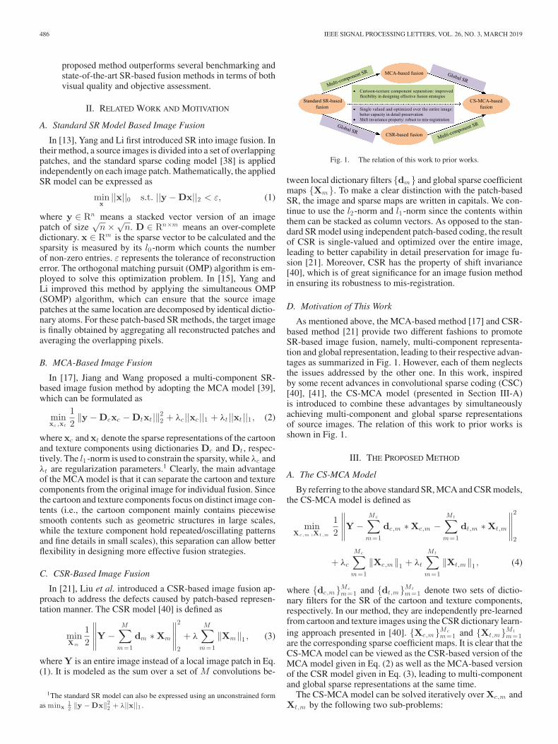
Fig. 1. The relation of this work to prior works.
tween local dictionary filters {dm} and global sparse coefficientmaps {Xm}. To make a clear distinction with the patch-basedSR, the image and sparse maps are written in capitals. We con-tinue to use the l2-norm and l1-norm since the contents withinthem can be stacked as column vectors. As opposed to the stan-dard SR model using independent patch-based coding, the resultof CSR is single-valued and optimized over the entire image,leading to better capability in detail preservation for image fu-sion [21]. Moreover, CSR has the property of shift invariance[40], which is of great significance for an image fusion methodin ensuring its robustness to mis-registration.
D. Motivation of This Work
As mentioned above, the MCA-based method [17] and CSR-based method [21] provide two different fashions to promoteSR-based image fusion, namely, multi-component representa-tion and global representation, leading to their respective advan-tages as summarized in Fig. 1. However, each of them neglectsthe issues addressed by the other one. In this work, inspiredby some recent advances in convolutional sparse coding (CSC)[40], [41], the CS-MCA model (presented in Section III-A)is introduced to combine these advantages by simultaneouslyachieving multi-component and global sparse representationsof source images. The relation of this work to prior works isshown in Fig. 1.
III. THE PROPOSED METHOD
A. The CS-MCA Model
By referring to the above standard SR, MCA and CSR models,the CS-MCA model is defined as
minX c , m ,X t , m
12
∥∥∥∥∥Y −
Mc∑
m=1
dc,m ∗ Xc,m −Mt∑
m=1
dt,m ∗ Xt,m
∥∥∥∥∥
2
2
+ λc
Mc∑
m=1
‖Xc,m‖1 + λt
Mt∑
m=1
‖Xt,m‖1 , (4)
where {dc,m}Mcm=1 and {dt,m}Mt
m=1 denote two sets of dictio-nary filters for the SR of the cartoon and texture components,respectively. In our method, they are independently pre-learnedfrom cartoon and texture images using the CSR dictionary learn-ing approach presented in [40]. {Xc,m}Mc
m=1 and {Xt,m}Mtm=1
are the corresponding sparse coefficient maps. It is clear that theCS-MCA model can be viewed as the CSR-based version of theMCA model given in Eq. (2) as well as the MCA-based versionof the CSR model given in Eq. (3), leading to multi-componentand global sparse representations at the same time.
The CS-MCA model can be solved iteratively over Xc,m andXt,m by the following two sub-problems:
LIU et al.: MEDICAL IMAGE FUSION VIA CONVOLUTIONAL SPARSITY BASED MORPHOLOGICAL COMPONENT ANALYSIS 487
Algorithm 1: Algorithm for Solving the CS-MCA model.
1: Input: Y, {dc,m}Mcm=1 , {dt,m}Mt
m=1 , λc , λt , L2: for i = 1: L3: Calculate Xc,m by solving Eq. (5);4: Calculate Xt,m by solving Eq. (6);5: end for6: Output: {Xc,m}Mc
m=1 and {Xt,m}Mtm=1 .
Sub-problem Xc,m (given the fixed Xt,m ):
minX c , m
12
∥∥∥∥∥Y′ −
Mc∑
m=1
dc,m ∗ Xc,m
∥∥∥∥∥
2
2
+ λc
Mc∑
m=1
‖Xc,m‖1 , (5)
where Y′ = Y − ∑Mt
m=1 dt,m ∗ Xt,m .Sub-problem Xt,m (given the fixed Xc,m ):
minX t , m
12
∥∥∥∥∥Y′′ −
Mt∑
m=1
dt,m ∗ Xt,m
∥∥∥∥∥
2
2
+ λt
Mt∑
m=1
‖Xt,m‖1 , (6)
where Y′′ = Y −Mc∑
m=1dc,m ∗ Xc,m .
These two sub-problems can be efficiently solved by theADMM-based CSC algorithm presented in [40]. The overallalgorithm for solving the CS-MCA model is summarized inAlgorithm 1.
B. Detailed Fusion Scheme
Let Ik , k ∈ {1, . . . ,K} denote a set of K pre-registeredsource images, the proposed CS-MCA-based medical imagefusion method consists of the following steps.
1) Sparse Coding Based on the CS-MCA Model: Apply theCS-MCA model to each source image Ik . Let {Xk
c,m}Mcm=1 and
{Xkt,m}Mt
m=1 denote the obtained sparse coefficient maps of thecartoon and texture components, respectively.
2) Fusion of Sparse Coefficient Maps: In accordance withthe related SR-based fusion methods [13], [15], [17], [21], thel1-norm of sparse coefficient vectors is employed as the activitylevel measurement of source images. For either Xk
c,m or Xkt,m ,
we use the same fusion strategy (the strategy used in the CSR-based fusion method [21] is adopted in this work to make a faircomparison) but with independent setting of free parameters.Thus, for notational simplicity, we adopt the character n (n ∈{c, t}) to universally denote the cartoon and texture componentswhen applicable. Specifically, let Xk
n,1:Mn(x, y) denote the Mn -
dimensional vector containing the coefficients of Xkn,m at pixel
(x, y). The initial activity level map Akn (x, y) is defined as
Akn (x, y) =
∥∥Xk
n,1:Mn(x, y)
∥∥
1 , n ∈ {c, t}. (7)
Further, a window-based strategy is applied to improve the ro-bustness to mis-registration and noise. The final activity levelmap is calculated as
Akn (x, y) =
∑rn
p=−rn
∑rn
q=−rnAk
n (x + p, y + q)
(2rn + 1)2 , n ∈ {c, t},(8)
where the parameters rc and rt are the window radius for thecartoon and texture components, respectively. Finally, the fused
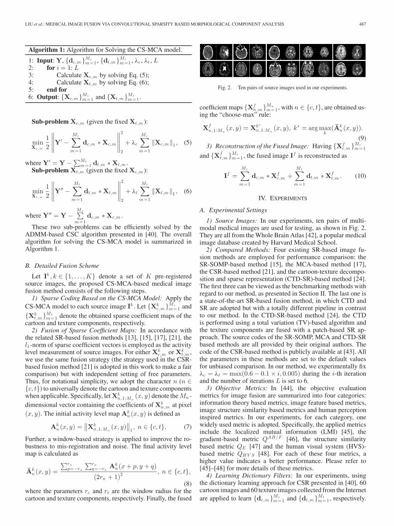
Fig. 2. Ten pairs of source images used in our experiments.
coefficient maps {Xfn,m}Mn
m=1 , with n ∈ {c, t}, are obtained us-ing the “choose-max” rule:
Xfn,1:Mn
(x, y) = Xk ∗n,1:Mn
(x, y), k∗ = arg maxk
(Akn (x, y)).
(9)3) Reconstruction of the Fused Image: Having {Xf
c,m}Mcm=1
and {Xft,m}Mt
m=1 , the fused image If is reconstructed as
If =Mc∑
m=1
dc,m ∗ Xfc,m +
Mt∑
m=1
dt,m ∗ Xft,m . (10)
IV. EXPERIMENTS
A. Experimental Settings
1) Source Images: In our experiments, ten pairs of multi-modal medical images are used for testing, as shown in Fig. 2.They are all from the Whole Brain Atlas [42], a popular medicalimage database created by Harvard Medical School.
2) Compared Methods: Four existing SR-based image fu-sion methods are employed for performance comparison: theSR-SOMP-based method [15], the MCA-based method [17],the CSR-based method [21], and the cartoon-texture decompo-sition and sparse representation (CTD-SR)-based method [24].The first three can be viewed as the benchmarking methods withregard to our method, as presented in Section II. The last one isa state-of-the-art SR-based fusion method, in which CTD andSR are adopted but with a totally different pipeline in contrastto our method. In the CTD-SR-based method [24], the CTDis performed using a total variation (TV)-based algorithm andthe texture components are fused with a patch-based SR ap-proach. The source codes of the SR-SOMP, MCA and CTD-SRbased methods are all provided by their original authors. Thecode of the CSR-based method is publicly available at [43]. Allthe parameters in these methods are set to the default valuesfor unbiased comparison. In our method, we experimentally fixλc = λt = max(0.6 − 0.1 × i, 0.005) during the i-th iterationand the number of iterations L is set to 6.
3) Objective Metrics: In [44], the objective evaluationmetrics for image fusion are summarized into four categories:information theory based metrics, image feature based metrics,image structure similarity based metrics and human perceptioninspired metrics. In our experiments, for each category, onewidely used metric is adopted. Specifically, the applied metricsinclude the localized mutual information (LMI) [45], thegradient-based metric QAB/F [46], the structure similaritybased metric QE [47] and the human visual system (HVS)-based metric QH V S [48]. For each of these four metrics, ahigher value indicates a better performance. Please refer to[45]–[48] for more details of these metrics.
4) Learning Dictionary Filters: In our experiments, usingthe dictionary learning approach for CSR presented in [40], 60cartoon images and 60 texture images collected from the Internetare applied to learn {dc,m}Mc
m=1 and {dt,m}Mtm=1 , respectively.
488 IEEE SIGNAL PROCESSING LETTERS, VOL. 26, NO. 3, MARCH 2019
TABLE IIMPACTS OF FREE PARAMETERS ON OBJECTIVE PERFORMANCE
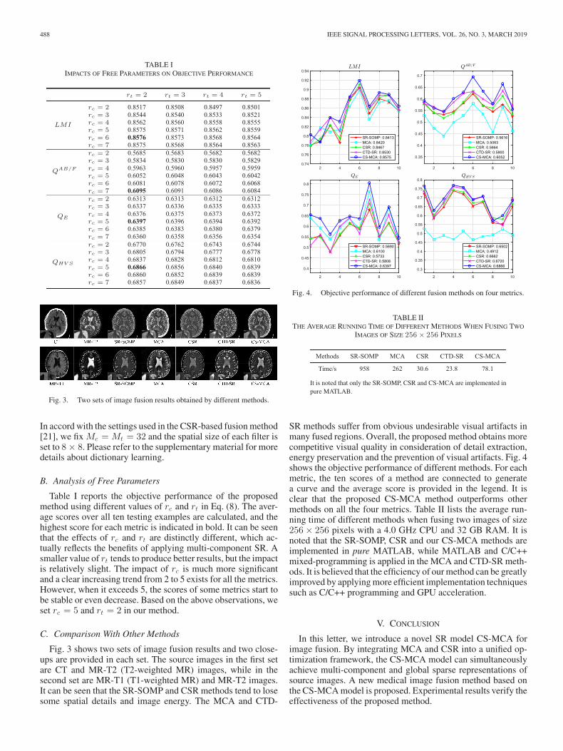
Fig. 3. Two sets of image fusion results obtained by different methods.
In accord with the settings used in the CSR-based fusion method[21], we fix Mc = Mt = 32 and the spatial size of each filter isset to 8 × 8. Please refer to the supplementary material for moredetails about dictionary learning.
B. Analysis of Free Parameters
Table I reports the objective performance of the proposedmethod using different values of rc and rt in Eq. (8). The aver-age scores over all ten testing examples are calculated, and thehighest score for each metric is indicated in bold. It can be seenthat the effects of rc and rt are distinctly different, which ac-tually reflects the benefits of applying multi-component SR. Asmaller value of rt tends to produce better results, but the impactis relatively slight. The impact of rc is much more significantand a clear increasing trend from 2 to 5 exists for all the metrics.However, when it exceeds 5, the scores of some metrics start tobe stable or even decrease. Based on the above observations, weset rc = 5 and rt = 2 in our method.
C. Comparison With Other Methods
Fig. 3 shows two sets of image fusion results and two close-ups are provided in each set. The source images in the first setare CT and MR-T2 (T2-weighted MR) images, while in thesecond set are MR-T1 (T1-weighted MR) and MR-T2 images.It can be seen that the SR-SOMP and CSR methods tend to losesome spatial details and image energy. The MCA and CTD-
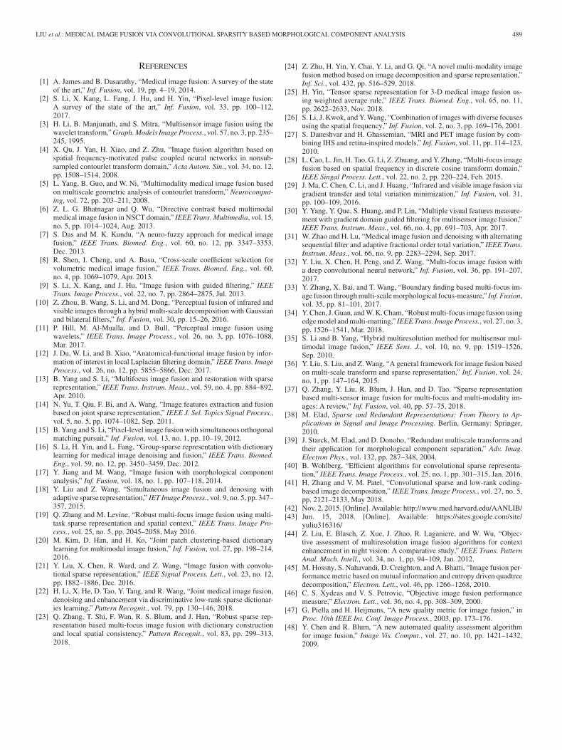
Fig. 4. Objective performance of different fusion methods on four metrics.
TABLE IITHE AVERAGE RUNNING TIME OF DIFFERENT METHODS WHEN FUSING TWO
IMAGES OF SIZE 256 × 256 PIXELS
It is noted that only the SR-SOMP, CSR and CS-MCA are implemented inpure MATLAB.
SR methods suffer from obvious undesirable visual artifacts inmany fused regions. Overall, the proposed method obtains morecompetitive visual quality in consideration of detail extraction,energy preservation and the prevention of visual artifacts. Fig. 4shows the objective performance of different methods. For eachmetric, the ten scores of a method are connected to generatea curve and the average score is provided in the legend. It isclear that the proposed CS-MCA method outperforms othermethods on all the four metrics. Table II lists the average run-ning time of different methods when fusing two images of size256 × 256 pixels with a 4.0 GHz CPU and 32 GB RAM. It isnoted that the SR-SOMP, CSR and our CS-MCA methods areimplemented in pure MATLAB, while MATLAB and C/C++mixed-programming is applied in the MCA and CTD-SR meth-ods. It is believed that the efficiency of our method can be greatlyimproved by applying more efficient implementation techniquessuch as C/C++ programming and GPU acceleration.
V. CONCLUSION
In this letter, we introduce a novel SR model CS-MCA forimage fusion. By integrating MCA and CSR into a unified op-timization framework, the CS-MCA model can simultaneouslyachieve multi-component and global sparse representations ofsource images. A new medical image fusion method based onthe CS-MCA model is proposed. Experimental results verify theeffectiveness of the proposed method.
LIU et al.: MEDICAL IMAGE FUSION VIA CONVOLUTIONAL SPARSITY BASED MORPHOLOGICAL COMPONENT ANALYSIS 489
REFERENCES
[1] A. James and B. Dasarathy, “Medical image fusion: A survey of the stateof the art,” Inf. Fusion, vol. 19, pp. 4–19, 2014.
[2] S. Li, X. Kang, L. Fang, J. Hu, and H. Yin, “Pixel-level image fusion:A survey of the state of the art,” Inf. Fusion, vol. 33, pp. 100–112,2017.
[3] H. Li, B. Manjunath, and S. Mitra, “Multisensor image fusion using thewavelet transform,” Graph. Models Image Process., vol. 57, no. 3, pp. 235–245, 1995.
[4] X. Qu, J. Yan, H. Xiao, and Z. Zhu, “Image fusion algorithm based onspatial frequency-motivated pulse coupled neural networks in nonsub-sampled contourlet transform domain,” Acta Autom. Sin., vol. 34, no. 12,pp. 1508–1514, 2008.
[5] L. Yang, B. Guo, and W. Ni, “Multimodality medical image fusion basedon multiscale geometric analysis of contourlet transform,” Neurocomput-ing, vol. 72, pp. 203–211, 2008.
[6] Z. L. G. Bhatnagar and Q. Wu, “Directive contrast based multimodalmedical image fusion in NSCT domain,” IEEE Trans. Multimedia, vol. 15,no. 5, pp. 1014–1024, Aug. 2013.
[7] S. Das and M. K. Kundu, “A neuro-fuzzy approach for medical imagefusion,” IEEE Trans. Biomed. Eng., vol. 60, no. 12, pp. 3347–3353,Dec. 2013.
[8] R. Shen, I. Cheng, and A. Basu, “Cross-scale coefficient selection forvolumetric medical image fusion,” IEEE Trans. Biomed. Eng., vol. 60,no. 4, pp. 1069–1079, Apr. 2013.
[9] S. Li, X. Kang, and J. Hu, “Image fusion with guided filtering,” IEEETrans. Image Process., vol. 22, no. 7, pp. 2864–2875, Jul. 2013.
[10] Z. Zhou, B. Wang, S. Li, and M. Dong, “Perceptual fusion of infrared andvisible images through a hybrid multi-scale decomposition with Gaussianand bilateral filters,” Inf. Fusion, vol. 30, pp. 15–26, 2016.
[11] P. Hill, M. Al-Mualla, and D. Bull, “Perceptual image fusion usingwavelets,” IEEE Trans. Image Process., vol. 26, no. 3, pp. 1076–1088,Mar. 2017.
[12] J. Du, W. Li, and B. Xiao, “Anatomical-functional image fusion by infor-mation of interest in local Laplacian filtering domain,” IEEE Trans. ImageProcess., vol. 26, no. 12, pp. 5855–5866, Dec. 2017.
[13] B. Yang and S. Li, “Multifocus image fusion and restoration with sparserepresentation,” IEEE Trans. Instrum. Meas., vol. 59, no. 4, pp. 884–892,Apr. 2010.
[14] N. Yu, T. Qiu, F. Bi, and A. Wang, “Image features extraction and fusionbased on joint sparse representation,” IEEE J. Sel. Topics Signal Process.,vol. 5, no. 5, pp. 1074–1082, Sep. 2011.
[15] B. Yang and S. Li, “Pixel-level image fusion with simultaneous orthogonalmatching pursuit,” Inf. Fusion, vol. 13, no. 1, pp. 10–19, 2012.
[16] S. Li, H. Yin, and L. Fang, “Group-sparse representation with dictionarylearning for medical image denoising and fusion,” IEEE Trans. Biomed.Eng., vol. 59, no. 12, pp. 3450–3459, Dec. 2012.
[17] Y. Jiang and M. Wang, “Image fusion with morphological componentanalysis,” Inf. Fusion, vol. 18, no. 1, pp. 107–118, 2014.
[18] Y. Liu and Z. Wang, “Simultaneous image fusion and denosing withadaptive sparse representation,” IET Image Process., vol. 9, no. 5, pp. 347–357, 2015.
[19] Q. Zhang and M. Levine, “Robust multi-focus image fusion using multi-task sparse representation and spatial context,” IEEE Trans. Image Pro-cess., vol. 25, no. 5, pp. 2045–2058, May 2016.
[20] M. Kim, D. Han, and H. Ko, “Joint patch clustering-based dictionarylearning for multimodal image fusion,” Inf. Fusion, vol. 27, pp. 198–214,2016.
[21] Y. Liu, X. Chen, R. Ward, and Z. Wang, “Image fusion with convolu-tional sparse representation,” IEEE Signal Process. Lett., vol. 23, no. 12,pp. 1882–1886, Dec. 2016.
[22] H. Li, X. He, D. Tao, Y. Tang, and R. Wang, “Joint medical image fusion,denoising and enhancement via discriminative low-rank sparse dictionar-ies learning,” Pattern Recognit., vol. 79, pp. 130–146, 2018.
[23] Q. Zhang, T. Shi, F. Wan, R. S. Blum, and J. Han, “Robust sparse rep-resentation based multi-focus image fusion with dictionary constructionand local spatial consistency,” Pattern Recognit., vol. 83, pp. 299–313,2018.
[24] Z. Zhu, H. Yin, Y. Chai, Y. Li, and G. Qi, “A novel multi-modality imagefusion method based on image decomposition and sparse representation,”Inf. Sci., vol. 432, pp. 516–529, 2018.
[25] H. Yin, “Tensor sparse representation for 3-D medical image fusion us-ing weighted average rule,” IEEE Trans. Biomed. Eng., vol. 65, no. 11,pp. 2622–2633, Nov. 2018.
[26] S. Li, J. Kwok, and Y. Wang, “Combination of images with diverse focusesusing the spatial frequency,” Inf. Fusion, vol. 2, no. 3, pp. 169–176, 2001.
[27] S. Daneshvar and H. Ghassemian, “MRI and PET image fusion by com-bining IHS and retina-inspired models,” Inf. Fusion, vol. 11, pp. 114–123,2010.
[28] L. Cao, L. Jin, H. Tao, G. Li, Z. Zhuang, and Y. Zhang, “Multi-focus imagefusion based on spatial frequency in discrete cosine transform domain,”IEEE Singal Process. Lett., vol. 22, no. 2, pp. 220–224, Feb. 2015.
[29] J. Ma, C. Chen, C. Li, and J. Huang, “Infrared and visible image fusion viagradient transfer and total variation minimization,” Inf. Fusion, vol. 31,pp. 100–109, 2016.
[30] Y. Yang, Y. Que, S. Huang, and P. Lin, “Multiple visual features measure-ment with gradient domain guided filtering for multisensor image fusion,”IEEE Trans. Instrum. Meas., vol. 66, no. 4, pp. 691–703, Apr. 2017.
[31] W. Zhao and H. Lu, “Medical image fusion and denoising with alternatingsequential filter and adaptive fractional order total variation,” IEEE Trans.Instrum. Meas., vol. 66, no. 9, pp. 2283–2294, Sep. 2017.
[32] Y. Liu, X. Chen, H. Peng, and Z. Wang, “Multi-focus image fusion witha deep convolutional neural network,” Inf. Fusion, vol. 36, pp. 191–207,2017.
[33] Y. Zhang, X. Bai, and T. Wang, “Boundary finding based multi-focus im-age fusion through multi-scale morphological focus-measure,” Inf. Fusion,vol. 35, pp. 81–101, 2017.
[34] Y. Chen, J. Guan, and W. K. Cham, “Robust multi-focus image fusion usingedge model and multi-matting,” IEEE Trans. Image Process., vol. 27, no. 3,pp. 1526–1541, Mar. 2018.
[35] S. Li and B. Yang, “Hybrid multiresolution method for multisensor mul-timodal image fusion,” IEEE Sens. J., vol. 10, no. 9, pp. 1519–1526,Sep. 2010.
[36] Y. Liu, S. Liu, and Z. Wang, “A general framework for image fusion basedon multi-scale transform and sparse representation,” Inf. Fusion, vol. 24,no. 1, pp. 147–164, 2015.
[37] Q. Zhang, Y. Liu, R. Blum, J. Han, and D. Tao, “Sparse representationbased multi-sensor image fusion for multi-focus and multi-modality im-ages: A review,” Inf. Fusion, vol. 40, pp. 57–75, 2018.
[38] M. Elad, Sparse and Redundant Representations: From Theory to Ap-plications in Signal and Image Processing. Berlin, Germany: Springer,2010.
[39] J. Starck, M. Elad, and D. Donoho, “Redundant multiscale transforms andtheir application for morphological component separation,” Adv. Imag.Electron Phys., vol. 132, pp. 287–348, 2004.
[40] B. Wohlberg, “Efficient algorithms for convolutional sparse representa-tion,” IEEE Trans. Image Process., vol. 25, no. 1, pp. 301–315, Jan. 2016.
[41] H. Zhang and V. M. Patel, “Convolutional sparse and low-rank coding-based image decomposition,” IEEE Trans. Image Process., vol. 27, no. 5,pp. 2121–2133, May 2018.
[42] Nov. 2, 2015. [Online]. Available: http://www.med.harvard.edu/AANLIB/[43] Jun. 15, 2018. [Online]. Available: https://sites.google.com/site/
yuliu316316/[44] Z. Liu, E. Blasch, Z. Xue, J. Zhao, R. Laganiere, and W. Wu, “Objec-
tive assessment of multiresolution image fusion algorithms for contextenhancement in night vision: A comparative study,” IEEE Trans. PatternAnal. Mach. Intell., vol. 34, no. 1, pp. 94–109, Jan. 2012.
[45] M. Hossny, S. Nahavandi, D. Creighton, and A. Bhatti, “Image fusion per-formance metric based on mutual information and entropy driven quadtreedecomposition,” Electron. Lett., vol. 46, pp. 1266–1268, 2010.
[46] C. S. Xydeas and V. S. Petrovic, “Objective image fusion performancemeasure,” Electron. Lett., vol. 36, no. 4, pp. 308–309, 2000.
[47] G. Piella and H. Heijmans, “A new quality metric for image fusion,” inProc. 10th IEEE Int. Conf. Image Process., 2003, pp. 173–176.
[48] Y. Chen and R. Blum, “A new automated quality assessment algorithmfor image fusion,” Image Vis. Comput., vol. 27, no. 10, pp. 1421–1432,2009.