1 HISTORY TAKING IN OBSTETRICS & GYNECOLOGY COLLEGE OF MEDICINE DEPT. OF OBSTETRICS AND GYNECOLOGY.
Upload
trinhtuongCategory
view
222download
0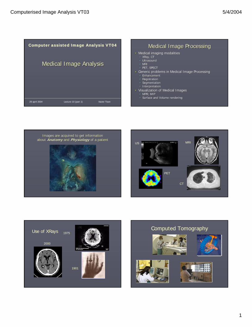
Computerised Image Analysis VT03 5/4/2004
1
Medical Image AnalysisMedical Image Analysis
Computer assisted Image Analysis VT04
Lecture 10 (part 1) Xavier Tizon29 april 2004
Medical Image ProcessingMedical Image Processing•• Medical imaging modalitiesMedical imaging modalities
•• XRayXRay, CT, CT•• UltrasoundUltrasound•• MRIMRI•• PET, SPECTPET, SPECT
•• Generic problems in Medical Image ProcessingGeneric problems in Medical Image Processing•• EnhancementEnhancement•• RegistrationRegistration•• SegmentationSegmentation•• InterpretationInterpretation
•• Visualization of Medical ImagesVisualization of Medical Images•• MPR, MIPMPR, MIP•• Surface and Volume renderingSurface and Volume rendering
Images are Images are acquiredacquired to to getget information information about about AnatomyAnatomy andand PhysiologyPhysiology ofof a patienta patient
US
PET
MRI
CT
1901
1975
2000
Use of XRays Computed Tomography
Computerised Image Analysis VT03 5/4/2004
2
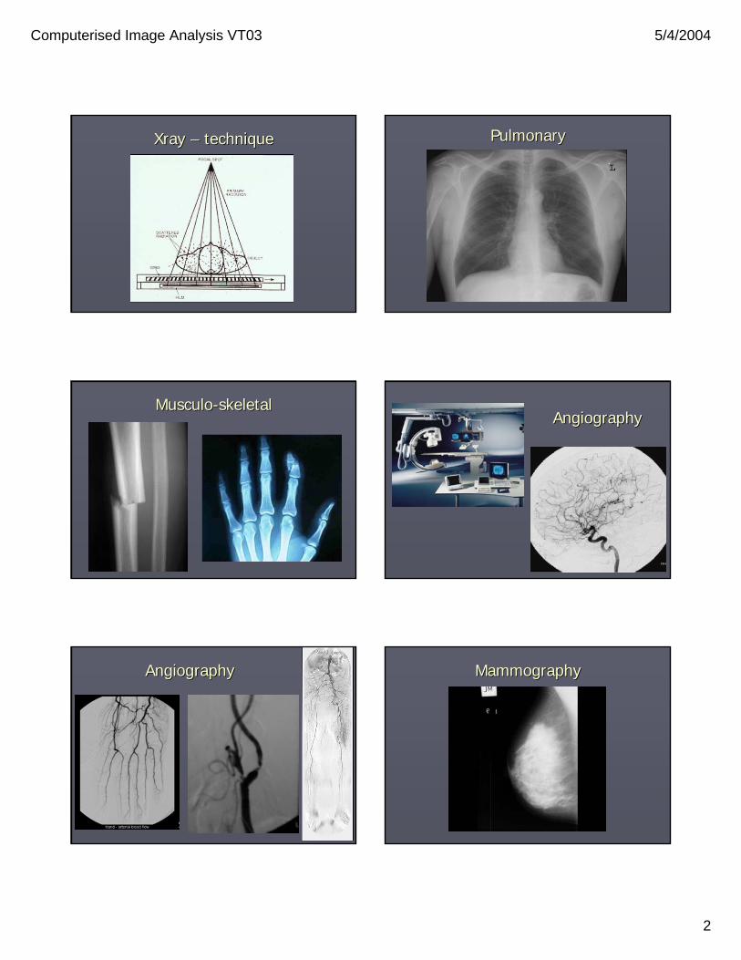
XrayXray –– techniquetechnique PulmonaryPulmonary
MusculoMusculo--skeletalskeletalAngiographyAngiography
AngiographyAngiography MammographyMammography
Computerised Image Analysis VT03 5/4/2004
3
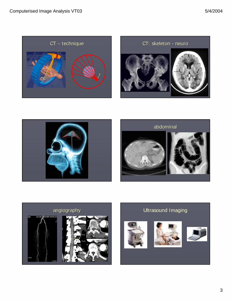
CT CT –– techniquetechnique CT: skeleton CT: skeleton -- neuroneuro
abdominalabdominal
angiographyangiography Ultrasound Imaging

Computerised Image Analysis VT03 5/4/2004
4
Cardiac imaging abdominal
imaging
AngiographyGynecologyObstetrics
GynecologyObstetrics
Positron Emission Tomography : the scanner

Computerised Image Analysis VT03 5/4/2004
5
PET: principlesPET: principles Positron Emission Tomography : production of radioactive products
PET : examples
Scintigraphy (SPECT): imaging device
Nuclear Imaging
Application to thedetection of bone
metastases

Computerised Image Analysis VT03 5/4/2004
6
T1 T2 PDW
MR tomograph
MusculoMusculo--skeletal (joints)skeletal (joints) neurologicalneurological
Multiple sclerosis
angiographyangiography angiographyangiography

Computerised Image Analysis VT03 5/4/2004
7
abdominalabdominalCardiac Cardiac imagingimaging
Functional imagingFunctional imaging Other medical imaging modalitiesOther medical imaging modalities
•• MicroscopyMicroscopy
Generic problems in Generic problems in Medical Image ProcessingMedical Image Processing
•• EnhancementEnhancement•• RegistrationRegistration•• SegmentationSegmentation•• InterpretationInterpretation
EnhancementEnhancement•• Noise (ex. MRI)Noise (ex. MRI)
•• Requires good knowledge of imaging physicsRequires good knowledge of imaging physics•• And a good approximation algorithmAnd a good approximation algorithm

Computerised Image Analysis VT03 5/4/2004
8
EnhancementEnhancement
•• BackgroundBackground•• Ex: MRIEx: MRI•• Field variations Field variations
produce nonproduce non--uniform uniform backgroundbackground
•• Corrected by fitting a Corrected by fitting a lowlow--order polynomial order polynomial to the imageto the image
RegistrationRegistration
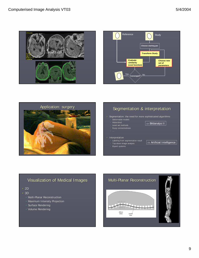
•• Registration = matching 2 volumes by applying Registration = matching 2 volumes by applying geometric correction to one of themgeometric correction to one of them
•• The need for registrationThe need for registration•• Study over timeStudy over time•• Fusion of different imaging modalitiesFusion of different imaging modalities•• Matching to an atlasMatching to an atlas•• Organs movementOrgans movement
Magnetic Resonance ImagingMagnetic Resonance Imaging
MRI gives anatomical information
Computed tomographyComputed tomography
CT gives anatomical information
Positron Emission TomographyPositron Emission Tomography
PET gives functional information
Single Photon Emission TomographySingle Photon Emission Tomography
SPECT gives functional information
Computerised Image Analysis VT03 5/4/2004
9
Choose starting par
Transform Study
Evaluate similarity(cost function)
Converged?
Choose newset ofparameters
Reference Study
NoYes
Application: surgeryApplication: surgery Segmentation & interpretationSegmentation & interpretation
•• Segmentation: the need for more sophisticated algorithmsSegmentation: the need for more sophisticated algorithms•• Deformable modelsDeformable models•• WatershedWatershed•• Level set methodsLevel set methods•• Fuzzy connectednessFuzzy connectedness•• ……
•• InterpretationInterpretation•• Labeling from segmentation resultLabeling from segmentation result•• TopTop--down image analysisdown image analysis•• Expert systemsExpert systems
⇒ Bildanalys II
⇒ Artificial Intelligence
Visualization of Medical ImagesVisualization of Medical Images
•• 2D2D•• 3D3D
•• MultiMulti--Planar ReconstructionPlanar Reconstruction•• Maximum Intensity ProjectionMaximum Intensity Projection•• Surface RenderingSurface Rendering•• Volume RenderingVolume Rendering
MultiMulti--Planar ReconstructionPlanar Reconstruction

Computerised Image Analysis VT03 5/4/2004
10
Maximum Intensity ProjectionMaximum Intensity Projection
Shaded Surface Display (SSD)Shaded Surface Display (SSD)
•• Preliminary segmentationPreliminary segmentation•• Voxel set Voxel set ⇒⇒ surface surface
( with for ex. marching cube)( with for ex. marching cube)•• Surface elements rendered according to Surface elements rendered according to
some illumination modelsome illumination model•• Optionally textureOptionally texture

Computerised Image Analysis VT03 5/4/2004
11
Volume RenderingVolume Rendering
•• ReflexionReflexion/transmission properties assigned to /transmission properties assigned to each voxeleach voxel
Volume rendering
Visualization of Computed Tomography volumetric data
Visualization of Ultrasound volumetric data Visualization of « The Visible Human »
Computerised Image Analysis VT03 5/4/2004
12
PerspectivePerspective
•• Fusion Fusion ofof techniques (MR, CT, PET,techniques (MR, CT, PET,……))•• Dual Dual imagingimaging devicesdevices•• Image Image ProcessingProcessing
•• InterventionalInterventional use use ofof MedicalMedical ImagesImages•• RealReal--timetime imagingimaging•• VirtualVirtual surgerysurgery
•• HigherHigher powerpower BUT BUT toughertougher problemsproblems•• IncreaseIncrease ofof computation computation capabilitiescapabilities•• IncreaseIncrease ofof resolutionresolution, , nbnb ofof dimensions, dimensions, physicianphysician’’ss demanddemand
•• CompleteComplete automation automation isis a DREAMa DREAM•• AmountAmount ofof data data tootoo importantimportant•• GoodGood interaction interaction isis more more reliablereliable
Going furtherGoing further
•• Visible Human ProjectVisible Human Project•• http://http://www.nlm.nih.gov/research/visible/visible_human.htmlwww.nlm.nih.gov/research/visible/visible_human.html•• http://visiblehuman.epfl.ch/http://visiblehuman.epfl.ch/
•• LinksLinks•• http://www.comp.leeds.ac.uk/comir/resources/links_c.htmlhttp://www.comp.leeds.ac.uk/comir/resources/links_c.html•• ……
•• Come pay us a visit!Come pay us a visit!
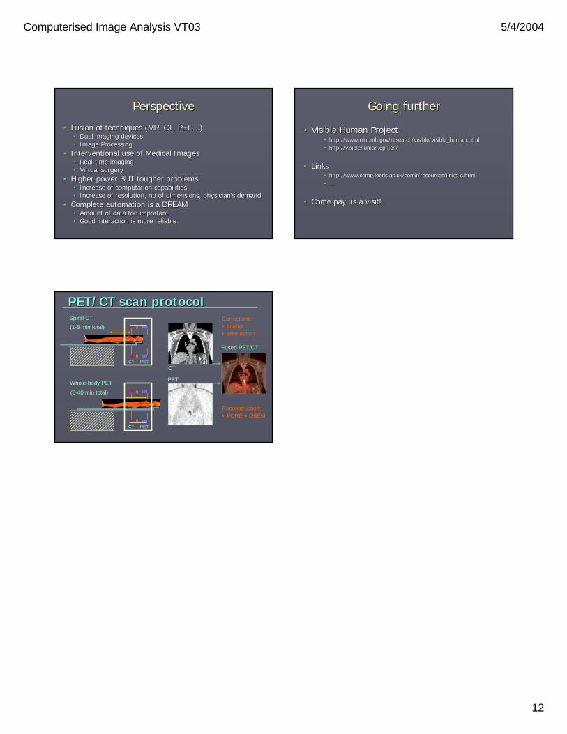
Spiral CT
Whole-body PET
(1-8 min total)
(6-40 min total)
Corrections:• scatter• attenuation
Fused PET/CT
PET
CT
PET/CT scan protocolPET/CT scan protocol
Reconstruction:• FORE + OSEM
CT PET
CT PET