Medical Background - Naas Physiotherapy€¦ · - whiplash trauma - late whiplash-associated...
52
1 All rights reserved. © DBC International Ltd 2009 Medical Background
Transcript of Medical Background - Naas Physiotherapy€¦ · - whiplash trauma - late whiplash-associated...

1 All rights reserved. © DBC International Ltd 2009
Medical Background

2All rights reserved. © DBC International Ltd 2009
3 All rights reserved. © DBC International Ltd 2009
Contents
1. Introduction . . . . . . . . . . . . . . . . . . . . . . . . . . . . . . . 5
Classification . . . . . . . . . . . . . . . . . . . . . . . . . . . . . . . . 5
Etiological models . . . . . . . . . . . . . . . . . . . . . . . . . . . . 6
The basic principles of Evidence-Based
Medicine (EBM) . . . . . . . . . . . . . . . . . . . . . . . . . . . . . . 7
2. Disorders of the back . . . . . . . . . . . . . . . . . . . . . . . 11
Definition . . . . . . . . . . . . . . . . . . . . . . . . . . . . . . . . . . 11
Prevalence, risk factors and determinants . . . . . . . . 12
Pathophysiology, prognosis and implications . . . . . 13
Diagnostics and the main treatment
approaches . . . . . . . . . . . . . . . . . . . . . . . . . . . . . . . . . 14
Evidence for efficacy of active treatments
in back disorders . . . . . . . . . . . . . . . . . . . . . . . . . . . . 15
3. Disorders of the neck . . . . . . . . . . . . . . . . . . . . . . . 19
Prevalence, risk factors and determinants . . . . . . . . 19
Pathophysiology, prognosis and sequelae . . . . . . . . 21
- Whiplash . . . . . . . . . . . . . . . . . . . . . . . . . . . . . . . . . 21
- Tension neck . . . . . . . . . . . . . . . . . . . . . . . . . . . . . . 21
Diagnostics and the main treatment
approaches . . . . . . . . . . . . . . . . . . . . . . . . . . . . . . . . . 21
The main approaches in the treatment
of neck disorders . . . . . . . . . . . . . . . . . . . . . . . . . . . . 22
Precautions and contraindications to exercise . . . . 22
Evidence for the efficacy of active treatments
in neck disorders . . . . . . . . . . . . . . . . . . . . . . . . . . . . 23
4. Shoulder disorders . . . . . . . . . . . . . . . . . . . . . . . . 25
Prevalence, risk factors and determinants . . . . . . . . 25
Physiology and pathophysiology . . . . . . . . . . . . . . . 26
Diagnostics and the main treatment
approaches . . . . . . . . . . . . . . . . . . . . . . . . . . . . . . . . . 28
- Dislocation of the shoulder joint . . . . . . . . . . . . . . 28
- Shoulder joint instability . . . . . . . . . . . . . . . . . . . . 28
- Shoulder impingement and rotator cuff tear . . . . . 29
- Arthrosis of the shoulder . . . . . . . . . . . . . . . . . . . . 29
- Adhesive capsulitis (frozen shoulder) . . . . . . . . . . 30
- Differential diagnostics . . . . . . . . . . . . . . . . . . . . . . 31
5. DBC treatment concept . . . . . . . . . . . . . . . . . . . . . 33
How does DBC apply evidence-based
medicine? . . . . . . . . . . . . . . . . . . . . . . . . . . . . . . . . . . 33
DBC treatment concept – general principles . . . . . . 34
Baseline assessment . . . . . . . . . . . . . . . . . . . . . . . . . 34
Individualized treatment program . . . . . . . . . . . . . . . 35
Cognitive and behavioural support . . . . . . . . . . . . . 35
Supporting elements . . . . . . . . . . . . . . . . . . . . . . . . . 36
Monitoring outcome . . . . . . . . . . . . . . . . . . . . . . . . . . 36
DBC in back disorders . . . . . . . . . . . . . . . . . . . . . . . . 36
- The therapist’s role . . . . . . . . . . . . . . . . . . . . . . . . . 37
- DBC back treatment results . . . . . . . . . . . . . . . . . . 37
- DBC back treatment action mechanisms . . . . . . . . 38
DBC in neck disorders . . . . . . . . . . . . . . . . . . . . . . . . 39
- DBC neck treatment results . . . . . . . . . . . . . . . . . . 39
- DBC neck treatment action mechanisms . . . . . . . . 40
DBC in shoulder disorders . . . . . . . . . . . . . . . . . . . . . 40
- DBC shoulder treatment results . . . . . . . . . . . . . . . 41
- DBC shoulder treatment action mechanisms . . . . 41
6. Literature . . . . . . . . . . . . . . . . . . . . . . . . . . . . . . . . 43

4All rights reserved. © DBC International Ltd 2009

5 All rights reserved. © DBC International Ltd 2009
1. Introduction
Musculoskeletal disorders are among the most preva-
lent long-term illnesses, and they account for more
pain and sickness absence from work than any other
medical condition. The most common condition of mus-
culoskeletal complaints is Low Back Pain (LBP). Pain in
the neck and shoulder area is almost as common. The
prevalence of disorders of the upper extremities has
received only limited attention in population-based
epidemiological research. Pain and restricted move-
ment of the shoulder joint are common symptoms, but
both medical definitions and diagnostic criteria still
vary.
The most common manifestations of musculoskeletal
disorders are pain, restrictions of physical function,
degeneration of tissue structures, absence from work
and early retirement. These symptoms are partly inter-
related, but to some extent they have differing causes.
It is important to understand which causes can and
should be addressed. In terms of public health and
national economy, the great significance of muscu-
loskeletal disorders is not primarily due to pain, physi-
cal impairment, functional deficits or tissue degenera-
tion, but rather to losses of working time. The treatment
costs of these conditions pale in comparison to those
related to sickness absence and premature retirement.
Manifestations of musculoskeletal disorders - Pain - Functional deficits - Structural tissue damage and degeneration - Absence from work and permanent disability
Classification
The classification of musculoskeletal disorders is dif-
ficult, as is evident from the widespread use of such
imprecise terms as ”back pain” and ”the neck and
shoulder area”. It is often assumed that most patients
with back or neck problems suffer from mechani-
cal pain. That is, their pain varies with mechanical
(physical) stresses to which the spine and the upper
extremities are subjected and where no general or non-
musculoskeletal illnesses (such as cancer or infection)
are involved. In some severe conditions of the back,
as well as the neck and shoulder area, the pathogen-
esis is relatively well known. These diagnoses include
spinal nerve-root compression and specific problems
of the shoulder joint and upper extremities (such as
shoulder dislocation). In these cases, the aetiology,
pathophysiology, prognosis and cause-specific treat-
ment are known. However, many of the “diagnoses” of

6All rights reserved. © DBC International Ltd 2009
Specific back disorders
- low-back trauma: vertebral fracture, contusion etc. - lumbar intervertebral disc herniation- lumbar spinal cord stenosis
Specific neck disorders
- cervical trauma: vertebral fracture, disc dislocation, ligament lesion etc. - cervical disc herniation- cervical spinal cord stenosis
Specific disorders of upper extremities
- shoulder joint dislocation - instability of shoulder joint - shoulder joint impingement syndrome- rotator cuff tear- various types of tendinitis, e.g. those of extensor tendons of finger or wrist- rheumatoid arthritis- osteoarthritis
Non-specific back disorders
- chronic back pain without a specific reason- degenerative back disorder
Non-specific neck disorders
- tension neck- torticollis- whiplash trauma- late whiplash-associated disorder, WAD
Non-specific disorders of upper extremities
- load-related pain- mixed shoulder disorders- adhesive capsulitis
Table 1. Specific and non-specific disorders of back, neck and upper extremities.
back, neck and shoulder conditions do not readily yield
similarly useful information.
It is important to understand that one can rarely pin-
point the tissue or segment from which pain in the
back or neck originates. Frequently, one fails to even
find the aetiology of the problem. It is similar in many
cases involving pain in the upper extremities, where
the precise cause remains unknown and the diagnoses
only describe the symptoms. In shoulder disorders, for
instance, one can often discern several overlapping
symptoms from different specific problems (instabil-
ity, impingement syndrome, rotator cuff tear). This is
fundamental problem in the prevention and treatment
of many chronic conditions of the back as well as neck
and shoulder area. However, this should not prevent
from successfully treating the symptoms.
Etiological models
In many musculoskeletal disorders, such as those of
the low-back and neck, it is seldom possible to deter-
mine the tissue from which pain emanates. Moreover,
as medical research is oriented towards ever more
minuscule scales - genes and molecules, it is unlikely
that pathogenesis of musculoskeletal pain will be fully
explained in the near future. In many musculoskeletal
disorders there is only a weak association between
tissue damage and subjective impairment or work dis-
ability. An explanation of this discrepancy requires a
wider perspective than a straightforward focus on tis-
sue-level problems.
Within musculoskeletal disorders, a biopsychosocial
explanation model was first applied for back disorders
to replace a model that made the simple inference
“structural problem causes disorder”. The biopsycho-
social model is also applicable to many disorders of
the neck and upper extremities. It differentiates pain,
subjective impairment, disability in work and disabil-
ity in everyday life into separate spheres, each involv-
ing partially distinct contributing factors.
The biopsychosocial model facilitates treatment
without necessitating a full explanation for the origin
of pain. In itself the model only provides a descrip-
tion of the patient’s situation; but as the focus is on
the subjective experience of the patient, the goal of
the treatment can be set at restoring physical function,
amelioration of pain and the adoption of new patterns
of behaviour.
The ideal of science is to discern “final, primary causes”
and “laws of nature” in their most minute details. This

7 All rights reserved. © DBC International Ltd 2009
Occupationaldisability
PainPhysical
impairment
Figure 1. Biopsychosocial model of musculoskeletal disorders.
approach, however, does not work in many disorders
of low-back, neck, shoulder and upper extremities. In
these conditions, subjectively experienced symptoms
and health-relevant behaviours are modified individu-
ally and in varying weights by biological, psychological
and social factors. A doctor or health care professional
may unintentionally induce typical illness-enhancing
behaviour by “discovering” that cervical disc degen-
eration seen on a radiological imaging or a “creaky”
tendon is “a reasonable biological explanation” for a
transient pain episode that would pass by itself. If a
healthy person with a transient pain episode is labelled
sick, and if the treatment prescribed prohibits exercise
and exertion, a recipe for disaster is created. Careless
comments such as “arthrosis of the joint is terrible, in
such a young person”; “don’t expect to do any work
with this hand” or “after a whiplash like this you will
need years of physiotherapy to keep symptoms at
bay”, may induce the patient to think that he or she is
afflicted by a difficult disorder and severe impairment.
In a worst case scenario this belief will last on its own
even after the original “trauma” has healed.
The basic principles of Evidence-Based Medicine (EBM)
“Evidence-based medicine is the conscientious,
explicit and judicious use of current best evidence
in making decisions about the care of the individual
patient. It means integrating individual clinical
expertise with the best available external clinical
evidence from systematic research.”
David Sackett
In this context “explicit” refers to the systematic use of
scientific and clinical knowledge with every patient to
whom it is applicable. “Judicious” denotes evaluation
of the advantages and disadvantages of diagnostic
tests and alternative treatments on the basis of clini-
cal expertise. Finally “conscientious” refers to taking
into account each patient’s baseline condition, clinical
status and preferences. The use of the best available
evidence presupposes that the clinician is capable
of distinguishing between trustworthy and unreliable
information (critical evaluation) and obtaining reliable,
up-to-date information as required.Therefore, evi-
dence-based medicine signifies a rational combination
of clinical experience with the best (up-to-date) scien-
tific evidence, whilst taking into account the patient’s
own values and preferences. Knowledge discovered in
high-quality research is an essential constituent of evi-
dence-based treatments. It has a bearing on the treat-
ment selection, selection of diagnostic tests (validity
of tests) and gives indications on the prognostic value
of factors affecting the course of illness (risk factors;
factors predicting and influencing treatment outcome).
Evidence-based medicine is not based solely on ran-
domized clinical trials, although the evidence they
produce is especially valuable. Evidence revealed by
systematic reviews of randomized clinical trials is only
one of criteria that need to be considered when select-
ing between treatment interventions.
Of the listed criteria “efficacy in research setting”
should especially be based on randomized clinical
trials whenever possible. As a rule, patients have been
chosen for such trials on the basis of predefined inclu-
sion and exclusion criteria and typically have only one
medical condition. The trials provide reliable evidence
on the efficacy of the intervention at its best. However,
follow-up studies with extensive population cohorts
may produce more reliable evidence on the safety and
effectiveness of treatments in normal circumstances
(in which patients’ backgrounds and motivation levels
vary more widely than in research settings). Also, the
skill level of caregivers and their resources may differ
from those that are available at specialized research
centres. It is especially difficult to carry out studies
on the efficacy of treatments when rare or multiple
illnesses are involved. This is why evidence based on
randomized trials is mostly lacking for such disorders.
In these cases, the most reliable information about
treatment efficacy and applicability can be obtained
with careful research in large population cohorts. Simi-
larly, cohort-based follow-up studies are required in
identifying prognostic factors, as the methodology of

8All rights reserved. © DBC International Ltd 2009
randomized trials offers no comparative advantages.
Discerning the efficacy of active treatments by obser-
vation and randomized studies is also problematic.
In observation studies, patients’ activity is influenced
to some extent by hereditary factors that also have a
bearing on many beneficial health-related habits. In
randomized trials it is not possible to carry out blinded
selections between patients who have participated in
active treatments and those who have not done so.
Investigation of the economic factors in health care is
not easy or unambiguous, and many decisions have
to be made on the basis of human values rather than
economy. Nevertheless, economic considerations
should not be dismissed altogether. One criterion of
Table 2. Therapies can be assessed according to different criteria.
Criteria Description
Efficacy (health benefit) in research setting
Is there evidence on the efficacy of treatment in the research setting?
Does the treatment work in “normal circumstances”?Effectiveness (health benefit) in normal circumstances
What kinds of side effects and risks does the treatment involve? Safety
Are the results commensurate with costs?Cost-effectiveness
Do the costs make the treatment unaccesible from the patient’s point of view?
Availability
economy is cost-effectiveness, which is defined as a
comparison of a treatment’s effectiveness (health ben-
efits) in normal circumstances with the costs that the
intervention involves. One of the ways health benefits
and benefits that can be measured in terms of money,
e.g. comparing the costs of intervention to produce a
saving in terms of sickness absence. may be measured
is with health-related quality of life. If it is possible to
define a generally acceptable monetary value to health
benefits, then that value can be used in an economic
analysis to signify effectiveness. Cost-benefit analysis
compares outlays. and benefits that can be measured
in terms of money, e.g. comparing the costs of interven-
tion to produce a saving in terms of sickness absence.

9 All rights reserved. © DBC International Ltd 2009

10All rights reserved. © DBC International Ltd 2009

11 All rights reserved. © DBC International Ltd 2009
2. Disorders of the back
Definition
Definitions of the “disorders of the back” vary. The
most commonly used concepts “low back pain”, “low
back trouble” and “low back disorder” are used as syn-
onyms although they often refer to different aspects of
the issue. None of the terms clarifies the structure that
has been damaged, or indeed if any structural abnor-
mality is involved at all.
With the exception of a herniated disc, trauma, spinal
stenosis and certain rheumatic diseases, it is rarely
possible to identify the definite causes of low back
pain and trouble. Most patients exhibit degenerative
changes on radiological imaging, but it is not possible
to draw definite conclusions to the origin of pain. That
is why the most commonly used classification divides
low back disorders into three groups: those related to
herniated disc, specific and rare conditions, and non-
specific conditions. The latter are the most prevalent.
On the basis of pain duration, low back disorders are
classified into acute (< 6 weeks), prolonged (suba-
cute) and chronic (> 3 months). Recent research has
raised questions about the duration-based grouping,
as novel low back pain and significant pain persisting
from day to day on a stable level are both relatively rare
in adults. The idea of constant, stable back pain was
associated with the belief that it would be possible to
achieve a permanent reduction in the level of pain with
Figure 2. Recurrent nature of spinal pain.
treatment
imagined course
“real” course

12All rights reserved. © DBC International Ltd 2009
suitable treatment. The current opinion is that low back
pain has a strong tendency to recur and that the clini-
cal picture of chronic back pain also follows a recurrent
course: i.e. there are periods of relative ease between
episodes of perhaps severe pain. Successful treatment
reduces the average intensity of pain, but it is likely
that there will be continued “ups” and “downs” in the
severity of pain. The prevalence of low back pain is
noticeably more common in individuals that have had
pain episodes in the past than in individuals that have
not experienced them before.
Most episodes of low back pain are transient phenom-
ena, and from the point of view of public health or
national economy, they do not cause significant prob-
lems by themselves. The situation becomes more prob-
lematic if the pain is prolonged and becomes chronic:
somatic phenomena become intertwined with various
psychological processes and the resulting syndrome
causes suffering, impairment and disability.
Prevalence, risk factors and determinants
According to the results of various surveys, three out of
four adults aged over 30 have experienced at least one
episode of low back pain during their lifetime. Back pain
is relatively common, even during adolescence. One
half of the adult population has had more than five epi-
sodes of low back pain. Over the last few decades, the
prevalence of experienced low back pain has remained
steady or slightly decreased in most countries, but it
should be noted that in different countries the trends
go in different directions. “Chronic back syndrome”,
as diagnosed by a doctor with a clinical examination,
has markedly declined. “Health 2000” was a Finnish
nationwide, cohort-based survey that relied on clini-
cal examinations. It was found that the prevalence of
the condition was 10% among males and 11% among
females, while 20 years earlier the corresponding
figures had been 18% and 16%.
There are significant differences between risk factors
related to the first episode of low back pain and those
that are involved with chronicity. Several concurring
and even interrelated risk factors may be involved in
inducing pain. Physical work, repeated lifting or carry-
ing of heavy loads, difficult postures at work, whole-
body vibration, weakness of trunk muscles, trauma,
obesity, smoking and stress are factors that have been
shown to have a bearing on the onset and prevalence
of low back disorders. Genetic predisposition may be
a significant risk factor for lumbar disc herniation in
young people and recurrent back pain associated with
disc degeneration.
Personal and behavioural traits are factors, which
cause differences in how people react to psychologi-
cal stress, for instance. They affect not only how pain is
experienced, but also the activation of back muscles.
However, too few studies have been carried out so
far and those that have been carried out have weak-
nesses.
Risk factors and acute back pain are both very common.
This makes it difficult to devise guidelines for the
primary prevention of low back pain, except the pro-
motion of a healthy lifestyle in general. One could even
question the need to take preventive action against
low back pain as it is such an insignificant, transient
condition and almost everyone experiences it at one
time or another.
However, the need to prevent frequent recurrence of
(debilitating) pain and chronicity, together with their
sequelae cannot be disputed. Yet the risk factors men-
tioned earlier seem to be of limited value in predicting
the chronicity of back pain. Factors that are extrinsic
(i.e., related to disorder but external to the individ-
ual) have recently been shown to be more significant
in predicting chronicity than those that are intrinsic
to the individual. Localized symptoms usually have
better prognosis and recur more rarely than radiating
No problem
Resolves
Resolves
Risk factors I- occupational loading- health behaviour
Risk factors II- severity of disorder- delayed treatment- personal and job- related factors
Risk factors III- perception of working ability- job-related factors- delayed treatment- severity of disorder
Healthyindividual
Musculoskeletalpain
Sicknessabsence
Earlyretirement
Figure 3. The course of musculoskeletal disorders may be seen as a chain of events where different outcomes have different risk factors.

13 All rights reserved. © DBC International Ltd 2009
Figure 4. The effect of pain, stress and fear on motor control. Modified from Hodges et al.
pain symptoms. The number of back pain episodes
experienced, severity of pain, severity of impairment
and wide referral of radiating pain increase the prob-
ability of recurrence and chronicity. In addition, the risk
of chronicity is also affected by external psychological
and psychosocial factors: back pain is more common
among lower social classes, those less educated and
those in employee position. Depression, fatigue and
distress also increase the danger of chronicity. It can
be assumed that early, efficacious treatment and sec-
ondary prevention reduce the probability of chronicity.
Pathophysiology, prognosis and implications
Experimental studies have attempted to locate the
origin of back pain by irritating the different spinal
tissues during local-anaesthesia surgery. Often these
studies have revealed soreness in the outer rim of
anulus fibrosus, in the vertebral end-plate located
between the disc and bony vertebra, in the anterior
spinal dura and ligamentum longitudinale posterior.
Pain is rare in ligamenta supraspinale and interspinalia,
zygapophyseal capsules and muscle-bone interfaces.
If not compressed or chemically irritated, the nerve
root is painless. A compressed nerve root or nerve root
exposed to nucleus pulposus tissue is painful. The epi-
dural space is well innervated and nucleus pulposus
tissue may cause an inflammatory reaction there. This
causes either local, radiating pain or a combination of
these depending on the irritated tissue structure: ante-
rior dura in local pain and nerve root or its surrounding
tissues in radiating pain.
Acute back pain quickly triggers spasm reactions and
reflex inhibition of the paravertebral muscles. This func-
tion of the paraspinal muscles is not always automati-
cally restored after pain recedes. If acute pain caused
by tissue damage becomes prolonged, widespread
deficits in motor control may develop. They may cause
excess activity of the paraspinal muscles during rest,
delay in the reaction reflexes of trunk muscles, deficits
in their co-ordination, in addition to deficits in balance
control. A “vicious circle of increasing disability” may
develop, in which (load-provoked) pain and functional
deficits in the paraspinal muscles lead to underutili-
zation of the back that in turn causes “disability” that
maintains pain: in due course this vicious circle results
in chronic low back disorder. When the situation per-
sists, the motion of the back becomes restricted and
muscle strength and endurance are weakened. In labo-
ratory and imaging studies it has been discovered that
patients with chronic low back pain exhibit a reduc-
tion in collagen synthesis and atrophy of paraspinal
muscles.
Prolonged low back disorder is regarded as a psycho-
physiological and psychosocial problem that is related,
not only to the physical factors mentioned earlier,
but also to psychological and social factors. Anxiety,
depression, stress reactions, fatigue, mistaken beliefs
and fear of pain are among the factors that have been
found to be more prevalent with chronic back pain
patients than in the general population. However, the
order in which these phenomena occur is not clear.
Cross-sectional studies do not reveal directions of
causal connections, and the few longitudinal studies
carried out so far have produced somewhat contradic-
tory results. Depression and distress seem to predict
the onset of pain, but on the other hand prolonged pain
seems to contribute to depression and stress. Never-
theless, a prolonged period of low back disorder does
not always aggravate psychological and psychosocial
problems. Psychological symptoms are not directly
associated with the duration of symptoms. Rather,
they are associated with the level of impairment expe-
rienced by the patient.
Typical symptoms of low back disorders include local
pain or pain radiating to lower extremities, back stiff-
ness or fatigue. In chronic disorders that cause work
disability, the role of psychological and psychosocial
factors is especially prominent. Symptoms of the back
Altered motorplanning
Spinal inhibition
Intention
Planning
Altered proprioceptive
input
Fear
Execution
Motor command
Corticalinhibition
AttentionStress
Pain

14All rights reserved. © DBC International Ltd 2009
rarely result in severe deficits in daily activities or loss
of independent coping, but the symptoms are a signifi-
cant cause of permanent occupational disability, espe-
cially in physically demanding jobs. Back disorders
also contribute significantly to short spells of sickness
absence from work, subjectively experienced impair-
ment, and use of pain medication and physiotherapy
services.
Diagnostics and the main treatment approaches
A doctor’s clinical examination, which is based on
a knowledge of functional anatomy and is repeated
when necessary, plays a central role in clarifying the
need for further tests and defining the course of treat-
ment. The first tasks are to exclude the possibility of
serious illness (malignant tumours, infections, etc.),
Protective guardingand spasms
Reflex inhibition
Changes in painmodulation & perception
Loss of musclecoordination
Weakening ofconnective tissue
Muscle atrophy
Diminished blood &nutrient flow
Reduced proteinsynthesis
ABNORMAL MOTORCONTROL
STRUCTURALCHANGES
Cumulativemicrotrauma
Relativeimmobility
More pain
PAIN
- Fear of pain- Depression- Distress and anxiety
- Reduction in - strength - mobility - endurance - connective tissue synthesis - size of muscles
Outcomes- Paraspinal muscle hyper-/hypoactivity- Absent flexion-reflexion- Delayed trunk muscle reaction to sudden loading- Insufficient anticipatory trunk stabilization- Abnormal postural control- Delayed psychomotor reaction times
Figure 5. Potential mechanisms involved in the chronicity of low back pain.
identify symptoms of possible severe nerve compres-
sion, which may require surgical treatment, and refer
such patients for further examinations and treatment.
In an overwhelming number (80%) of patients with
back pain the cause is functional (non-specific) and
does not automatically require laboratory tests or diag-
nostic imaging examinations. Diagnostic imaging is
necessitated if there is a reason to suspect the pres-
ence of a serious disorder, or if symptoms persist for
more than six weeks. Usually there are only weak causal
links between back pain and signs of degeneration or
other abnormalities seen in imaging examinations. As
the exact cause of back pain is usually not revealed,
the main reason to perform imaging examinations is
to exclude serious illnesses or to support the planning
of surgical treatment. In prolonged back disorders, it
is also important to uncover psychological and social
factors.
Serious conditions (malignancy, infections) induc-

15 All rights reserved. © DBC International Ltd 2009
ing low back pain and nerve-root compression, which
cause neurological deficits, should be diagnosed
early. These patients require further tests and cause-
specific treatment. In acute back pain, bed rest should
be avoided, and patients should be encouraged to
continue daily activities within the limits permitted by
pain. Physical exercise has not been shown to provide
benefits, nor has it been shown to do any harm. Early,
efficacious treatment of pain using anti-inflammatory
pain medication, for example, limiting pain-provoking
physical loading, but remaining active in daily living
reduces the risk of chronicity. Correct information about
the benign prognosis of the condition reduces anxiety
and increases satisfaction with treatment. If the disor-
der becomes prolonged, the patient should be encour-
aged to move the back, carry out physical activities and
perform exercises. Efficient rehabilitation treatment
should be started without delay. It is recommended
that comprehensive charting of the patient’s overall sit-
uation, active treatment and rehabilitation should be
launched after low back disorder involving significant
impairment has lasted for six weeks. In the treatment
of prolonged low back pain, physical exercises that
are sufficiently intensive, promote general fitness and
improve body co-ordination compliment other inter-
ventions that develop work capacity and function to
speed up recovery. However, it has to be kept in mind
that opportunities to improve coping at work diminish
rapidly as absence from work continues.
Is there a spinal problem?
General warning signs
Duration of the problem
Primary location and pattern
Lumbar Cervical
Post-traumatic spinal injury
Inflammatory Inflammatory
Post-traumatic
Postoperative
Nerve rootcompression
Narrowing ofspinal canal
Pelvic and LBP
Non-specific pain
SpondylolisthesisSpondylolysis
Whiplash associateddisorder, WAD
Postoperative
Narrowing ofspinal canal
Nerve rootcompression
Non-specific pain
Primary prevention
Actions
Pain managementAdvice to stay active
No
Yes
Acute
Prolonged/Recurrent/Postoperative/Post-traumatic
No
Yes
Outcome: diagnosis (pattern) and reconditioning programme mode
Figure 6. Treatment approaches in spine disorders.
Evidence for efficacy of active treatments in back disorders
Medical exercise therapy carried out independently
of other treatments is efficacious in the management
of chronic back disorders, but not in acute back pain.
However, the extent to which independent benefits
can be achieved by fitness/exercise therapy have to be
critically considered. In a systematic Cochrane-review
and meta-analysis, 61 randomized clinical trials that
included 6390 adult participants in total, were ana-
lyzed. The review revealed strong evidence for the
efficacy of exercise in the treatment of chronic back
disorder, but on the basis of the meta-analysis pain-
related benefits were small when considering all of the
trials (Table 3.). However, the impact on pain varied
according to the channel through which participants
had been selected into the studies. Patients that were
selected via normal healthcare displayed pain-related
benefit that was about 6 units better than the average
for all patients (some of whom were recruited by news-
paper advertisements and other such means).
A lack of evidence was observed on the efficacy of
exercise on pain and impairment in subacute low back
pain, although in two studies it was reported that pro-
gressive exercise reduced absence from work.
Efficacies of different forms of exercise were studied in
another meta-analysis. The aim was to identify partic-
ular exercise intervention characteristics that decrease
pain and improve function in adults with non-specific
chronic low back pain. 43 trials of 72 exercise treatment
and 31 comparison groups were included. Stretching
and strengthening demonstrated the largest improve-
ment over comparisons. The authors concluded that
exercise therapy consisting of individually designed
programs, including stretching or strengthening, and
delivered with supervision may improve pain and func-
tion in chronic non-specific low back pain. They also
concluded that strategies should be used to encourage
adherence.
Functional restoration / work conditioning is a course
of action in which progressive exercise (guided by
physiotherapists, for example) is combined with
the cognitive-behavioural approach. Here patients’
mistaken beliefs and conceptions are rectified and
patients are supported in modifying their behaviours
to directions that are beneficial for health. A systematic
Cochrane review and meta-analysis included 18 rand-
16All rights reserved. © DBC International Ltd 2009
(Best) 1
2
3
4
5
(Worst) 6
Mea
n Ra
nk o
n Fu
nctio
n Ou
tcom
e
Stretch
ing
Strength
ening
Other S
pecific
Aerobic
Mobilizing
Co-ord
ination
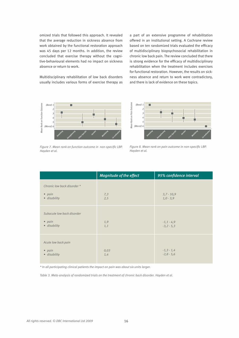
Figure 7. Mean rank on function outcome in non-specific LBP. Hayden et al.
Figure 8. Mean rank on pain outcome in non-specific LBP. Hayden et al.
* In all participating clinical patients the impact on pain was about six units larger.
Table 3. Meta-analysis of randomized trials on the treatment of chronic back disorder. Hayden et al.
(Best) 1
2
3
4
5
(Worst) 6
Mea
n Ra
nk o
n Pa
in O
utco
me
Stretch
ing
Strength
ening
Other S
pecific
Aerobic
Mobilizing
Co-ord
ination
Magnitude of the effect 95% confidence interval
Chronic low back disorder *
• pain• disability
7,32,5
3,7 - 10,91,0 - 3,9
Subacute low back disorder
• pain• disability
1,91,1
-1,1 - 4,9-3,2 - 5,3
Acute low back pain
• pain• disability
0,031,4
-1,3 - 1,4-2,8 - 5,6
omized trials that followed this approach. It revealed
that the average reduction in sickness absence from
work obtained by the functional restoration approach
was 45 days per 12 months. In addition, the review
concluded that exercise therapy without the cogni-
tive-behavioural elements had no impact on sickness
absence or return to work.
Multidisciplinary rehabilitation of low back disorders
usually includes various forms of exercise therapy as
a part of an extensive programme of rehabilitation
offered in an institutional setting. A Cochrane review
based on ten randomized trials evaluated the efficacy
of multidisciplinary biopsychosocial rehabilitation in
chronic low back pain. The review concluded that there
is strong evidence for the efficacy of multidisciplinary
rehabilitation when the treatment includes exercises
for functional restoration. However, the results on sick-
ness absence and return to work were contradictory,
and there is lack of evidence on these topics.

17 All rights reserved. © DBC International Ltd 2009

18All rights reserved. © DBC International Ltd 2009

19 All rights reserved. © DBC International Ltd 2009
3. Disorders of the neck
Prevalence, risk factors and determinants
Disorders of the neck and shoulder area are almost as
common as those of the lower back. Typical symptoms
include neck pain, stiffness and fatigue. Headache and
nausea are more prevalent among patients with neck
pain than in general population. When interviewed,
more than 60% of all adults recall having had experi-
enced pain in the neck and shoulder region at some
time. In the Finnish survey “Health 2000”, it was found
that 26% of males over 30 and 40% of females had
experienced neck pain during the last month. The cor-
responding figures for the shoulder were 23% in males
and 40% in females. In the survey, long-term neck
or shoulder syndrome (lasting more than 12 weeks)
was diagnosed in 5% of males and 7% of females. In
a similar survey carried out 20 years earlier, the syn-
drome had been diagnosed in 10% of men and 14%
of women; the prevalence of the long-term syndrome
has thus declined by a half in a period of 20 years.
However, there has been no corresponding change in
short-term symptoms of the neck and shoulder area.
In 1994 a comprehensive research project (Study on
Musculoskeletal disorders, Absenteeism, Stress and
Health, SMASH) on musculoskeletal disorders was
launched in Holland. The project was a prospective
three-year follow-up study of 1789 subjects lasting
three years. The participants were required to have
been in employment for more than one year, have
worked for at least 20 hours a week, and have had
no symptoms of neck pain for a period of one year
preceding the study. 1334 subjects fulfilled the cri-
teria. Once a year (from 1994 to 1997), an extensive
interview and video recording of working tasks was
carried out to quantify work-related and psychosocial
risk factors. The findings of the study were reported
in terms of prevalence of neck pain and sickness
absence caused by neck pain (Table 4.).
The most significant rate ratios (RR) were as follows:
Neck pain and physical variables
• sedentary work: RR 2.34, (95% CI 1.05-5.21)
• neck flexion and poor endurance of neck muscles:
RR 2.5, (95% CI 1.11-5.61)
Neck pain and psychological variables
• high quantitative job demands: RR 2.14, (95% CI
1.28-3.58)
• low co-worker support: RR 2.43, (95% CI 1.11-5.29)

20All rights reserved. © DBC International Ltd 2009
Sickness absence and physical variables
• neck flexion: RR 4.19, (95% CI 1.50-11.69)
• similar figures for neck flexion exceeding 45 degrees
and neck rotation exceeding 45 degrees
Sickness absence and psychological variables
• low decision authority at one’s job RR 3.66, (95% CI
1.44-9.26)
• discrepancy between skills and job demands RR
2.56, (95% CI 1.08-6.04)
On the basis of these results work-related physical
and psychological circumstances are independent
risk factors of neck pain and of comparable signifi-
cance. This was the first time it was shown that neck
pain and sickness absence due to neck disorders were
not explained by the same factors (similar differences
have earlier been shown in back trouble). That is why
preventive programmes should take into account both
physical and psychosocial work-related factors. On the
basis of the study, one should avoid uninterrupted sed-
entary work, avoid non-ergonomic neck positions and
exercise the neck muscles regularly. Work organization
management and society should pay attention to the
pace of work. In addition, workers should be provided
with a sufficient occupational support network and
opportunities for training to maintain their skills.
The improvement of the physical condition of the
joints and muscles (as a result of exercise) is related
to coping with the burden of static or monotonic work.
If the conditioning level is good, tissues can cope with
heavier loading. From the point of view of job perform-
ance it is essential to select suitable workers for each
task, take advantage of services that help to maintain
workers’ work capacities and monitor and develop
working conditions.
Symptoms of the neck and shoulder area rarely lead
to severe functional deficits or permanent disability.
However, they are of great significance as causes of
short-term absence, subjectively experienced impair-
ment, consumption of pain medication and physi-
otherapy services. Usually neck and shoulder pain is a
recurrent condition. Local symptoms have, in general,
a more benign prognosis and recur less often than radi-
ating pain symptoms.
recurrent or chronic neck pain during last 12 months.more than 3 days absence from work due to neck pain- no relationship; + increased relative risk, however not statistically significant; ++ statistically significant increased relative risk
a
b
Table 4. Summary results of the prospective cohort study examining the relationship between work-related physical and psychosocial variables and the occurence of neck pain and sickness absence due to neck pain. Modified from Ariëns.
Work-related physical and psychosocial variables
Occurence of neck pain
Sickness absence due to neck paina b
Neck flexion > 20 ° (over 40% of work time)
Neck flexion > 45 ° (over 5% of work time)
Neck rotation > 20 ° (over 25% of work time)
Sitting (over 95% of work time)
Quantitative job demands
Conflicting job demands
Decision authority
Co-worker support
Supervisor support
Job security
+ ++
- ++
- ++
++ -
++ +
- -
+ ++
++ -
- -
- +
21 All rights reserved. © DBC International Ltd 2009
Pathophysiology, prognosis and sequelae
A major proportion of neck disorders is thought to
originate from muscles or other soft tissues, facet
joints or discs. However, the exact pathophysiological
mechanisms of symptoms are not well known. For most
patients with neck or shoulder pain it is not possible to
give an exact pathological or anatomical diagnosis; the
situation is similar to low back disorders. It is assumed
that local tissue lesions, metabolic problems, muscle
fatigue, non-ergonomic working positions, general
posture and deficits in motor co-ordination contribute
to neck disorders.
Localized neck pain can, in principle, originate from all
structures that have nociceptors. Compression of the
nerve root or inflammation of the nerve root opening
may also cause radicular nerve root lesions and pain
radiating to the upper extremities.
Whiplash
Whiplash trauma refers to an injury in which the neck is
strained in a rapid, whip-like movement first into hyper-
extension and then hyperflexion. Following a whiplash
trauma, the signs and symptoms of the neck and upper
extremities may vary from no symptoms to unbearable
pain, forced neck position and comprehensive neu-
rological symptoms and signs. In these cases, one is
unlikely to identify a single specific cause of pain. Late
Whiplash-Associated Disorder (WAD) is a syndrome in
which the original whiplash problem does not resolve
within six months, but becomes a chronic problem
that is difficult to treat. In prolonged WAD, symptoms
may be polymorphous. Headaches, dizziness, nausea
(especially under exertion), depression and anxiety are
common. It has been shown in numerous studies that
WAD is also associated with an extensive decline in
psychomotoric and cognitive performance. This is indi-
cated by deficits in short-term memory, concentration
and eye-hand co-ordination.
Tension neck
By definition, tension neck is a syndrome in which
pain is associated with tension in the neck muscles.
It is assumed that the condition is related to excessive
biomechanical or psychological load, which affects the
musculature and the other tissues of neck and shoul-
der area. Typically people with tension neck work on
computers or have to maintain their arms in static, ele-
vated positions. In addition, the need to perform repet-
itive, monotonic movements in their jobs is typical of
tension neck patients. The complaint is more common
in females than males. It is thought that tension neck
may lead to metabolic dysfunction, microtrauma and
gradual muscular changes that do not reverse with
rest. The symptoms are manifested in a variety of ways,
including aching or stiffness throughout the neck and
shoulder area. As the trouble progresses, dizziness
and nausea may be involved.
Tenderness, pain, and tension in muscles do not nec-
essarily arise from the muscles themselves. Instead,
they can be a sign of segmental irritation in cervical
tissues or may reflect a more generalized dysfunction of
muscle balance. Pain research has produced clear evi-
dence that lesions in other tissues can increase muscle
tonus within the same myotome. Muscles themselves
have quite a large number of nociceptors that are par-
ticularly sensitive to the lack of oxygen. Recently, it has
been discovered that chronic neck pain involves defi-
cits in activation and co-ordination of the muscles in
the neck and shoulder area. Deep cervical muscles that
maintain posture react with delay to increased loads,
and secondary muscles have to compensate for this by
excessive activation. This dysfunction in co-ordination
may contribute to a continuation of functional neck dis-
order and pain, regardless of the original cause of pain.
Diagnostics and the main treatment approaches
As the exact aetiology and pathophysiology of neck
disorders is not well known, their classification also
varies. Clinical diagnosis is made mainly on the basis
of the patient’s history and clinical tests. The progno-
sis of neck disorders is in most cases benign, and that
is why symptoms can (should) be treated without the
need for a specific diagnosis. Serious and systemic ill-
nesses have to be excluded.
Neck disorders can be classified on the basis of history,
symptoms and clinical findings as follows:
1. Local (non-specific) neck pain.
2. Radiating neck pain.
3. Whiplash trauma.
4. Myelopathy (compression of spinal cord)
5. Other neck disorders: pain related to systemic ill-
nesses and tumours; sequelae of cervical fractures.

22All rights reserved. © DBC International Ltd 2009
On the basis of the duration of symptoms the first three
groups can be divided into acute (less than 12 weeks)
and chronic (more than 12 weeks) conditions.
The main approaches in the treatment of neck disorders
As has been explained, it is seldom possible to iden-
tify an exact cause for pain in the neck and shoul-
der area with clinical examination. If trauma, tumour
and myelopathy are excluded, ordinary cervical X-ray
images or MRI scans cannot be expected to yield sig-
nificant additional information about the causes of
neck pain. Therefore it makes sense to aim primarily
at excluding serious illnesses (i.e. tumours, infections
and fractures). The approach is similar to that followed
in low back disorders. Treatment can be planned on the
basis of a working hypothesis that can be made more
specific on the basis of patient’s response to treatment
and clinical follow-up. If necessary, the approach can
be modified.
The prognosis of local acute neck pain is usually good,
and in some patients the symptoms disappear or
resolve spontaneously. Pain can be treated by anti-
inflammatory pain medication. Action can be taken
to eliminate load factors that provoke pain, but the
patient should be encouraged to continue daily activi-
ties within the limits permitted by pain. Specific exer-
cise therapy is usually not necessary in acute neck
pain. Exercises that can be carried out by the patient
at home can be useful, especially in the prevention of
chronicity after whiplash trauma.
In the treatment of chronic neck pain, patients are
encouraged to stay active. Continuation of day-to-day
activities within the limits permitted by pain is impor-
tant. Active functional restoration treatment pro-
grammes that enhance strength, endurance and co-
ordination of the muscles provide benefits for a large
proportion of patients with chronic neck pain. Assess-
ment of working conditions and leisure time exposure
should be carried out, at the latest, when the neck dis-
order is becoming chronic. Predisposing factors should
be eliminated, respectively. Anti-inflammatory pain
medication must not be used for prolonged periods.
If pain involving significant disability has lasted six
weeks, the patient should be referred for an assess-
ment of treatment needs. When required, treatment
and rehabilitation must be started.
Precautions and contraindications to exercise
Neck disorders may involve rare diseases that demand
special caution when selecting form of exercise. In
patients with neck trauma, the possibility of cervi-
cal fracture has to be excluded. In elderly patients,
especially when osteoporosis or rheumatic diseases
are involved, even a minor cervical trauma can cause
a fracture (diagnosed in 3% of all patients with neck
trauma). An important clinical problem is a fracture of
the dens. Its diagnosis is often delayed and it causes
cervical instability and severe disability. Fracture in this
segment cannot be always identified even when MRI is
used, and cervical CT imaging needs to be applied as a
basis of diagnosis.
Pain that is continued, progressive, occurs at night, at
rest or is untypical may indicate some serious disease
of the cervical area. Often these patients exhibit sys-
temic symptoms such as weight loss, deterioration
of their overall physical condition and disability. Typi-
cally, patients are 50 years of age or older. Recovery
from cancer may be found in the patient’s history (in
females it is often breast cancer and in males prostate
cancer).
Cervical myelopathy is a rare condition and its diagno-
sis requires vigilance in clinical examination. Pain is
rarely the dominating symptom. Sensory symptoms,
numbness in upper and/or lower limbs, dizziness,
disturbances of balance, increased clumsiness, diffi-
culties in walking and ataxia are typical in myelopathy.
Thorough clinical examination may reveal a positive
Babinski reflex, hyperactive tendon reflexes and prov-
ocation of symptoms during cervical extension and/
or retraction. Timely surgical treatment may save the
patient from disability, so the possibility of myelopathy
has to be kept in mind when examining patients. MRI
is an important tool in diagnosis and it should be per-
formed without delay if myelopathy is suspected.
Rheumatic diseases often involve lesions in the upper
cervical spine. Even considerable dislocations of verte-
brae may cause just minor symptoms or no symptoms
at all. Often pain is involved and sometimes there
may be complications including neurological deficits,
or even quadriplegia and sudden death. Diagnosis is
made on the basis of flexion imaging. Usually treat-
ment is conservative, but surgery may also be needed.
Before planning mobility or exercise treatment the sta-

23 All rights reserved. © DBC International Ltd 2009
bility of the cervical spine has to be ascertained.
Severe neck disorders often involve headaches, visual
disturbances and nausea, especially if WAD is involved.
In these cases, starting mobility exercises and exercise
treatment may be especially challenging as even small
neck movements may provoke pain and associated
symptoms.
Evidence for the efficacy of active treatments in neck disorders
No known randomized studies have been carried out
on the significance of active interventions in the treat-
ment of acute non-specific neck disorder, and there-
fore the efficacy of exercise is unclear in this condition.
Continuation of normal daily activities within the limits
permitted by pain is recommended.
In acute whiplash trauma, randomized studies have
shown that self-administered exercise is more effica-
cious in treating pain than resting, medication or using
a supporting collar. Similar results have been reported
for disability, range of motion and chronicity. Mobil-
ity exercise carried out during recuperation seems to
prevent chronicity in whiplash trauma.
Efficacy of exercise therapy in the treatment and reha-
bilitation of chronic neck disorder has been studied
in 15 (or more) randomized controlled trials. Eight of
these studies had no control groups. The comparisons
were made between various types of exercise therapy,
or exercise therapy was compared with other forms of
physiotherapy (mobilization, manipulation, etc.). In
these studies efficacy differences between active treat-
ments were marginal. Seven trials included control
groups, and in most of these studies exercise therapy
was efficacious in treating pain and disability (at least
in the short term). However, the results of these rand-
omized trials were contradictory. In some studies, exer-
cise provided no benefits. In others, the benefits were
of short duration. Only a few studies showed that exer-
cise provided long-term benefits. Interpretation of the
results is made difficult by heterogeneity of methods
and the small sizes of participating patient groups.
What can be said, however, is that if the exercises did
not target neck specifically, no efficacy on neck pain
related to the neck disorder was observed. Exercise
has to be also of sufficient intensity and duration: light
exercises and stretching aimed at improving general
well-being had little effect on neck disorders in the
reviewed studies. On the other hand, the benefits of
even intensive exercise disappear in due course after
exercising is discontinued. In two randomized studies,
reductions in pain and disability were achieved with
proprioceptive training. Light loading was used, but
the exercise was targeted specifically at the neck and
repeated frequently. As a conclusion of the available
evidence, the potential benefits of exercise treatment
in neck disorders seems to depend on specific target-
ing of the neck and the patient’s compliance with the
treatment.

24All rights reserved. © DBC International Ltd 2009

25 All rights reserved. © DBC International Ltd 2009
4. Shoulder disorders
Prevalence, risk factors and determinants
Pain and restriction in the shoulder joint are common,
but the terminology used to describe shoulder dis-
orders is quite diverse. In addition, the diagnostic
criteria have been only partly established. About 5%
of patients visiting a general practitioner have shoul-
der complaints. About 5% of Finns aged 30 years or
over have long-term shoulder disorders. 20-50% of
all people experience troubling shoulder pain each
year. In many countries shoulder complaints are next
to back and neck conditions as the most prevalent
musculoskeletal disorders. The prevalence of shoulder
disorders is somewhat higher in females than males;
the prevalence grows slowly with age. The complaints
often emerge in the context of work. Shoulder trauma
comprises 3-20% of all trauma related to physical exer-
cise. In comparison with ailments of other joints, those
of shoulder are accentuated by their slower recovery
and relatively high prevalence in the working age pop-
ulation.
Shoulder disorders can be classified as follows:
• Dislocation
• Instability
• Impingement syndrome and rotator cuff tear
• Acromioclavicular (AC) joint injury
• Osteoarthritis
• Adhesive capsulitis
Among the working age population the most common
form of shoulder pain is a consequence of compressed
rotator cuff. This “impingement syndrome” may be
related to various background factors. Friction may
cause inflammation, scar tissue formation and rup-
tures of various sizes. Typical symptoms include pain
at night and with exertion, especially when upper
extremities are raised to shoulder level. Impingement
is rare among young people, but may occur especially
if inherited or acquired joint laxity is present and if the
muscles surrounding the shoulder joint are weak.
Terms such as “impingement”, “rotator cuff tendinitis”,
“rotator cuff syndrome”, “supraspinatus tendinitis”,
“periarthritis”, “rotator cuff tear (RCT)”, etc. describe
a variation on a continuum of disorders that originates
from tendinitis in one or more tendons and progresses
to tissue ruptures.
Friction-induced inflammation, swelling and finally
rupture are thought to develop most frequently in the

26All rights reserved. © DBC International Ltd 2009
presence of certain predisposing factors. Among them
are tightness in the posterior capsule, weak rotator
cuff and a narrowed subacromial space. If the joint
capsule is tight, it pushes the humeral bone towards
the acromion; a weak rotator cuff allows worsening of
the impingement, and the subacromial space may be
narrow because the acromion has a hooked morphol-
ogy. The latter may be an inherited condition or develop
as a result of osteoarthritis. Several other theories have
been presented, but without clear supporting research
evidence. The significance of these anatomical and
functional factors has not been ascertained in prospec-
tive follow-up studies.
Research on work-related risk factors of shoulder pain
has increased over recent years. A research group (at
The National Institute of Occupational Health and
Safety, NIOSH) in the United States published a review
article of 20 epidemiological studies. It was revealed
that work which involved frequent repetitive move-
ments of upper extremities (especially in flexion or
abduction of more than 60 degrees), increased the
risk of developing rotator cuff syndrome. The risk is
increased if the positions mentioned involve the use
of heavy tools. Exposure to vibration seems to be of
minor importance. Only three of the reviewed studies
gave a precise definition of shoulder pain. Therefore,
it was concluded that the evidence remains poor and
specific recommendations for prevention are not easily
justifiable.
Rotator cuff disorders are not a problem that is related
solely to ageing, although the prevalence of degen-
erative rotator cuff changes identified on radiological
imaging grows with age. Only a proportion of people
develop rotator cuff lesions related to ageing. A signifi-
cant number of these so-called degenerative lesions
are scars resulting from friction. Rotator cuff disorders
are not caused automatically by repetitive movements,
as they can also afflict the non-dominant hand and
people performing light sedentary work.
Trauma may also cause rotator cuff tears in healthy
tissue. However, trauma-related rotator cuff tears com-
prise less than 10% of all cases. Major ruptures usually
result only from massive traumas. In young people,
lesser traumas may cause partial ruptures that often
heal spontaneously. The same is true, for example, of
microtraumas with throwing athletes.
Follow-up studies are underway, but their results have
not yet been published. That is why recommendations
on preventive measures have to be based mainly on
knowledge gained through clinical experience. Main-
taining mobility, the muscle balance of the joint and
taking ergonomic factors into account at work is sup-
posed to reduce the risk of developing shoulder com-
plaints.
Traumatic dislocation of the shoulder is a common
injury, but it affects mainly young people. Among the
elderly, dislocation often causes injuries in bony struc-
tures and therefore these cases have to be treated
along separate lines. Laxity of the shoulder joint is a
significant cause of pain and above all a factor that
impairs performance. Laxity of the shoulder is also a
predisposing factor for other shoulder disorders. It has
been discovered that onset of the classical impinge-
ment syndrome is often preceded by various degrees of
laxity in the shoulder joint especially in young people.
With the individuals under the age of 21, the annual
incidence rates of shoulder dislocation are 20 in
females and 5 in males per 10000 individuals, respec-
tively. An earlier dislocation raises the risk of recurrence
by 2 or 3 fold. Follow-up studies show that shoulder
dislocations seldom heal and become asymptomatic
without treatment.
Risk factors for the first occurrence of shoulder disloca-
tion are not precisely known. Falls on the outstretched
arm and violent collisions, either at work or in leisure
time pursuits, may displace the head of the humerus
from its socket. Sports in which falls and collisions are
frequent increase the risk of shoulder injury, especially
if the rules of the game are not adhered to or if protec-
tive gear is not used. Contributing factors include a pre-
existing labral tear and ligaments strained in an earlier
dislocation. Muscle function can compensate reason-
ably well for injuries in passive structures. However,
in extreme positions the head of the humerus slips
easily out of the capsule, if its stability is not supported
by the labrum and the ligaments. The significance of
these injuries is heightened by the fact that recovery is
slow and incomplete.
Tissue injuries caused by shoulder dislocations tend to
vary from one age group to another. In individuals who
are less than 60 years of age, the most common soft
tissue injuries are lesions of the cartilaginous labrum.
In people aged 60 or over, rotator cuff tears are the
most significant injuries. In all age groups there are
about as many Bankart lesions (detachment of anterior
glenoid labrum) as Hill-Sachs depressions on the head

27 All rights reserved. © DBC International Ltd 2009
of the humerus.
The importance of preventing shoulder dislocation is
highlighted by a recent observation, which revealed
that the risk of shoulder osteoarthritis is increased by
a previous incidence of dislocation. According to the
study, patients who had experienced dislocation saw a
10-20 fold increase in the risk of shoulder osteoarthri-
tis compared to the normal level. Shoulder osteoarthri-
tis is an increasing problem, although it remains more
occasional than osteoarthritis of the knee or hip.
Physiology and pathophysiology
The shoulder joint and shoulder girdle form a system
consisting of three major joints: the shoulder joint
(articulatio humeri or glenohumeral joint), acromio-
clavicular joint (articulatio acromioclavicularis) and
scapulothoracic joint (articulatio scapulothoracalis).
The main bones are the upper arm bone (humerus),
shoulder blade (scapula) and collar bone (clavicula).
The shoulder joint is a ball-and-socket joint, in which
the joint cavity is significantly smaller than the head
of the humerus. The joint cavity is also flatter than the
more strongly rounded head of the humerus. That is
why the shoulder joint has a wider range of motion and
is less stable than other joints. The shoulder joint is
surrounded by the joint capsule and is also strength-
ened by ligaments. The capsule is attached to the
periphery of the joint cavity on the shoulder blade and
the neck of the humerus. The capsule is relatively loose
and facilitates the wide range of shoulder movements.
The capsule maintains a “vacuum” effect that contrib-
utes to the stability of the shoulder joint. The vacuum
is lost if the capsule ruptures or if it is opened during
surgery. Another function of the joint capsule-ligament
complex is to act as a proprioceptive sensory organ
that feeds back important sensory information needed
for the timely activation of muscles surrounding the
shoulder joint.
A rim of fibrous cartilage (labrum glenoidale) encir-
cles the joint cavity. The labrum increases the contact
surface of the joint by 50-70%. It also enhances the
stability of the joint by acting as a host for the ligament
endings. Together with the joint capsule the labrum
glenoidale also maintains the vacuum effect within
the joint. The vacuum is lost in lesions of the labrum.
Typical lesions include anterior dislocations of the
shoulder joint, in which the upper extremity is in exter-
nal rotation and abduction, causing detachment of
anterior labrum. This is what is called a Bankart lesion.
A rupture of the tendon of the long head of the biceps
muscle is in turn called a Superior Labrum Anterior Pos-
terior (SLAP) lesion.
The four muscles of the rotator cuff (supraspinatus,
infraspinatus, subscapularis and teres minor) are
primarily responsible for the dynamic stability of the
shoulder joint. The rotator cuff is also known as humer-
oscapular muscle group. The tendons of these muscles
are tightly connected to the capsule of the shoulder
joint and form a capsule of tendons around the ana-
tomical neck of the humerus. Functionally the tendon
of the long head of the biceps muscle is regarded as a
part of the rotator cuff.
The term “rotator cuff” does not clearly indicate its most
important function, which is to control and adjust the
position of the head of the humerus in the joint cavity
during arm movements. The muscles of the rotator cuff
keep the contact in the shoulder joint stable in all posi-
tions. The muscles controlling the shoulder joint act in
agonist-antagonist pairs (such as the deltoid and infra-
spinatus in abduction). The rotator cuff includes move-
ment-sensing nerves that participate in the control of
the shoulder’s multidimensional arcs of movement.
GLENOHUM
ERAL ABD
UCTIO
N 120°
UT
MT
LTSA
DEL
SCAPULOTHORACIC UPWARD ROTATION 60 °
Figure 9. Humeroscapular rhythm. SA=serratus anterior, DEL= deltoideus, UT=upper trapezius, MT=middle trapezius and LT=lower trapezius. Rotator cuff fine tunes the alignment of the head of humerus in relation to scapular fossa.

28All rights reserved. © DBC International Ltd 2009
The co-coordinated movements of the humerus and
shoulder blade is called the humeroscapular rhythm.
When the upper extremity is elevated along the plane
of the shoulder blade, the joint cavity moves medially,
turns upwards and slides up as the shoulder blade
rotates. In over 90° of elevation, external rotation of
the humerus is needed. Otherwise tuberculum major
would collide with acromion. The humeroscapular
rhythm is co-coordinated by tonic muscles, rotator cuff
and rotators of the shoulder blade.
For optimal performance of the shoulder joint, the func-
tion of scapulothoracic muscles should be as perfect
as possible. Often the ability of serratus anterior and
parts of trapezius to turn the shoulder blade upwards
has been weakened, resulting in the overloading of the
glenohumeral joint when arm is raised. Strengthened
and often shortened pectoralis major and latissimus
dorsi also have a negative impact on the function of
shoulder joint. The increased activation of these tho-
racohumeral muscles tends to twist the shoulder blade
medially and in addition they push the humerus anteri-
orly. The muscles of the rotator cuff offset the variation
in these forces. They keep the head of the humerus in
the joint cavity in as optimal a position as possible.
Dysfunction of the shoulder blade may be primary or
secondary. Winging of the shoulder blade, its incom-
plete retraction or protraction and disturbances in the
movement rhythm are common. Pain in the neck-shoul-
der girdle may inhibit the tonic muscles of the shoul-
der blade. Trapezius, serratus anterior, rhomboids,
and levator scapulae are all exposed to overloading
and damage. Loss of the protracted movement of the
shoulder blade may lead to the rotator cuff impinge-
ment syndrome. Its permanent protracted position may
restrict the subacromial space and result in impinge-
ment symptoms. The correct humeroscapular rhythm is
an important prerequisite for the healthy functioning
of the shoulder joint; however, this can easily be dis-
turbed by pain in the neck and shoulder area. Typically,
chronic shoulder disorders involve (secondary) func-
tional disorders
Diagnostics and the main treatment approache
Dislocation of the shoulder joint
The shoulder joint may dislocate anteriorly (>90%
of the cases) or posteriorly (< 10% of the cases). The
typical injury mechanism in shoulder dislocation is col-
lision or twisting when the upper arm is in abduction
with external rotation.
More than 90% of all surgically treated shoulder dislo-
cations involve, not only a strained or ruptured capsule
complex, but also a Bankart lesion, i.e., the detach-
ment of anterior glenoid labrum. In elderly patients,
the dislocation may be complicated by a fracture as
well. About 80% of dislocations involve compression
of the head of the humerus (Hill-Sachs lesion), but it
seldom affects the course of treatment or long-term
prognosis. Neural damages arise in about 5% of shoul-
der joint dislocations. Most shoulder dislocations in
people over 40 also involve rotator cuff tears.
A first shoulder dislocation can take place at any age,
but dislocations mainly occur before the age of 20, or
between 50 and 60 years of age. At a young age, labral
lesions usually complicate dislocations; among the
elderly the most common complication is a rotator cuff
tear.
Painful, post-traumatic deformity of the shoulder
joint is an indication of dislocation; in differentiating
diagnostics fracture is to be considered if the injury
mechanism is unknown. Repositioning of the joint
should be performed as soon as possible in order to
limit damage to soft tissues and nerves. Usually pain
resolves forthwith after repositioning. Immediate sur-
gical treatment is rarely needed. Clinical examination
and diagnostic imaging ascertain the diagnosis. After
repositioning the dislocated joint, the arm is placed
in a sling to create the best possible condition for the
healing of soft tissues. Estimates of immobilization
times required vary in the literature, from a few days
to six weeks.
When treated with prompt repositioning, immobiliza-
tion and gradually progressive rehabilitation, the shoul-
der can be expected to recover and even allow a return
to sports in 10-16 weeks. Active treatment aiming at
strengthening the stability of the shoulder joint signifi-
cantly reduces the risk of recurrent dislocation.
Shoulder joint instability
Shoulder joint instability is a condition in which the
stability of the shoulder joint is defective and the head
of the humerus moves excessively relative to the joint
cavity. It results in recurrent dislocations or subluxa-
tions, with which the head of the humerus moves to
the edge of the joint cavity. Instability accelerates joint

29 All rights reserved. © DBC International Ltd 2009
degeneration and may lead to premature arthrosis.
Instability may develop as a result of trauma, general
laxity of the ligaments, congenital factors or other
medical conditions, if the structures that are respon-
sible for the stability of the joint have been damaged
or are abnormal. If the first traumatic dislocation in
an under 20-year-old is left untreated, instability will
follow with a probability of 90%.
The diagnosis is based on the patient’s history and
clinical examination. For exclusion purposes diagnos-
tic imaging can be carried out as well. Chronic shoul-
der instability typically occurs in young people who are
actively involved in sports.
Shoulder impingement and rotator cuff tear
The rotator cuff should slide below the acromion
smoothly and without interference. Shoulder impinge-
ment is a condition in which the rotator cuff is com-
pressed or rubs against the acromion and/or the cora-
coacromial ligament. The condition can be complicated
by subacromial bursitis and various tendinitises. The
result is pain, tenderness and restricted movement.
Tendinitises are common stress injuries of the shoul-
der joint. Tendinitis of the supraspinatus insertion is
common, but repetitive strain may also involve tend-
initis of the long head of biceps as well as luxation of
the biceps tendon.
Subacromial bursitis may involve strong symptoms.
Calcified bursitis may elevate the blood sedimentation
rate and create a strong clinical picture of inflamma-
tion.
Impingement syndrome may lead to a rotator cuff tear.
The rotator cuff tear may also result from trauma or
excessive loading. A rotator cuff tear usually presents
in middle-aged or elderly patients; it is rare in individu-
als who are less than 40-years of age. The prevalence
is similar between males and females.
Shoulder impingement syndromes and rotator cuff
tears are classified as follows:
I degree Swelling and painII degree Inflammation and scarringIII degree Partial tear of the rotator cuffIV degree Complete tear of the rotator cuff
The typical age of patients with impingement syndrome
or a torn rotator cuff is over 50 years.
Patient history and clinical examination may give indi-
cations of an impingement or tear. However, the symp-
toms and clinical findings are often similar to those in
patients with frozen shoulder syndrome, arthrosis or
neural lesions.
The clinical examination should include measurement
of range of motion and specific diagnostic tests (Neer’s;
Hawkin’s); differences between active and passive
ranges of motion and positive findings in provocation
tests indicate the diagnosis. It has to be confirmed by
imaging findings. Plain x-ray imaging can be used to
exclude fractures and serious illnesses and verify the
shape of the acromion. Ultrasound may reveal swell-
ing or lesions in the rotator cuff. Magnetic resonance
imaging depicts the status of soft tissues more accu-
rately than ultrasound.
Treatment is started with rest, activities that provoke
symptoms are limited and anti-inflammatory medica-
tion (NSAIDs, cold therapy, corticoid injections) are
used to ease the pain. Progressive active rehabilita-
tion programmes are efficacious in the treatment of the
impingement syndromes as well as of small or partial
tears of the rotator cuff.
Surgical treatment is only rarely required. This is espe-
cially true in elderly and other patients with increased
risk of complications in this instance; the benefits and
risks of surgery should be weighted carefully. Surgical
treatment is recommended for active young and mid-
dle-aged patients with the IV degree trauma (complete
tear). The operation aims at restoring the integrity of
the rotator cuff; a complete tear will not heal by itself.
In young patients suspected of having the shoulder
impingement syndrome, instability as a primary cause
for the shoulder disorder should be excluded first. The
repair of an extensive tear does not always succeed; in
some of the patients debridement of the shoulder joint
should be considered. One option in the treatment of
the impingement syndrome is acromioplasty, in which
the subacromial space is extended by bone resection
and/or excision of coracoacromial ligament.
Arthrosis of the shoulder
Arthrosis may occur in the shoulder joint and the
acromioclavicular (AC) joint. Progressive pain related to
loading is a typical symptom. Arthrotic pain is usually

30All rights reserved. © DBC International Ltd 2009
felt behind the shoulder or deep within it. Arthrosis
pain in the AC joint localizes in the frontal shoulder area
and in the area of the AC joint. Restricted movement is
another typical sign; crepitus may also be heard during
shoulder movements. As the disorder progresses, pain
at night may become a significant problem.
Typical clinical examination findings include muscle
atrophy, palpation tenderness, restricted movement,
crepitus and provocation of pain when loading the
joint. Narrowing of the joint space and osteophytes
may be observed in native radiography. If an injection
of local anaesthetic removes the pain, the diagnosis
can be considered certain.
Treatment is usually conservative: rest, medication and
cold therapy. Progressive physiotherapy may be useful
in maintaining functions. If the pain cannot be brought
under control, surgical treatment may be considered.
Prosthetic surgery is rapidly becoming more common
in the treatment of shoulder arthrosis; the most usual
procedure in the treatment of the arthrosis of the AC
joint is resection of the collar bone.
Adhesive capsulitis (frozen shoulder)
Frozen shoulder is a condition in which the shoulder
movements are restricted. The exact aetiology of the
disorder is not known, but it is thought that inflamma-
tion plays a central role in its development. The shoul-
der joint capsule becomes thicker and diminishes in
size, restricting shoulder mobility. Usually only one
shoulder is afflicted, but every third patient has the
restriction in both shoulders.
In an idiopathic presentation of this disorder, the his-
tology of the joint capsule is abnormal. The aetiology
of the change has not been fully explained. Diabetes
is known to be a predisposing factor. Frozen shoulder
may also develop as a result of trauma or prolonged
immobilization.
Age Aetiology Inspection Active range of motion
Passive range of motion
Shoulder dislocation
Young TraumaDeformation in the shoulder joint area
No movement Restricted
NormalNormal or excessive
Normal or excessive
Instabilityprovoked bymovement
YoungInstability
Shoulder blade protraction
Restricted in abduction and rotation
PainfulPain after loading and weakness
Middle-aged/ elderly
Impingementsyndrome/Rotator cuff tear
AC-joint deformity
Restricted by pain
Restricted by pain
Direct hit on the shoulder (trauma)
YoungAC-trauma
Muscle atrophy possible
Restricted RestrictedPost-traumatic or rheumatic
ElderlyArthrosis
Muscle atrophy possible
Clearly restricted
Clearly restricted
Idiopathic or post-traumatic
Middle-aged/Elderly
Adhesive capsulitis
Table 5. Findings related to the most common shoulder disorders.

31 All rights reserved. © DBC International Ltd 2009
The main symptom is restricted movement. The syn-
drome develops gradually, and three phases may be
discerned in its progression:
• Pain phase. Pain associated with movement and
restriction progresses gradually; first abduction
and external rotation are restricted. Pain at night
may occur. The phase lasts between two and nine
months.
• Stiffness phase. Pain starts to lessen and shoulder
movements become less painful, although move-
ment becomes increasingly constrained. The range
of motion may be less than half of that in a healthy
shoulder. The phase lasts about 4 – 12 months.
• Healing phase. The disorder begins to heal. In most
patients, the range of motion improves over the next
few years.
In the beginning, pain can be treated with anti-inflam-
matory pain medication, cold therapy and injections.
At a later stage, progressive mobility therapy is planned
for the treatment of the movement restriction.
Differential diagnostics
Table 5. shows probable diagnoses on the basis of
different anamneses and clinical findings; the table
should be regarded as an approximate guide for diag-
nosis.

32All rights reserved. © DBC International Ltd 2009

33 All rights reserved. © DBC International Ltd 2009
5. DBC treatment concept
How does DBC apply evidence-based medicine?
As a fundamental, DBC functional rehabilitation
includes systematic quantification of both the physical
function and psychological factors, which “drive” the
therapeutic process. The baseline assessment yields a
bio-psychosocial profile of the patient and establishes
initial parameter levels, which are monitored later as
progress and outcome indicators. The profile is used in
the design of an individualized treatment program and
in assessing prognostic factors. The tests that measure
range of motion, endurance/strength and question-
naires on pain, function and psychological well-being
of the patient are periodically repeated during the
rehabilitation treatment. This allows the scheduling of
a correct therapeutic approach on an individual basis,
and gives the patient feedback on the improvement of
his/her physical capacity and well-being.
DBC functional rehabilitation programs aim to:
• Restore the range of motion.
• Restore muscle co-ordination and movement
control.
• Improve muscle endurance.
• Improve general condition.
• Re-educate patients in the difference between normal
physical loading and pain.
• Reduce fears and avoidance behaviour.
• Tackle the psychological/social/occupational obsta-
cles to return-to-work.
The individualized treatment program combines spe-
cific exercises together with cognitive-behavioural
support. The exercises progress gradually and are
carried out under close supervision of trained staff.
Cognitive-behavioural support includes individual
education and “learning by doing”. Towards the end of
the program, a home exercise program is introduced to
maintain the results.
The DBC treatment sessions’ average duration is 60
minutes and treatment is carried out twice per week.
During the treatment program there are assessment
sessions of 90 minutes evaluating the patient’s base-
line condition, progress and/or outcome. The monitor-
ing of outcomes provide information on the progress
and outcome of treatment and allows for long-term
follow up.

34All rights reserved. © DBC International Ltd 2009
The clinic-specific DBC quality assurance system
includes ongoing clinical support, training workshops
and patient data analysis. The quality assurance reports
are produced twice a year on the basis of patient data
from each clinic. These reports help to demonstrate the
effectiveness of the treatments at local level. All this
results in a treatment program, which is strongly based
on the principles of EBM:
1. The DBC treatment concept is based on knowl-
edge obtained from scientific research. The validity
of measurements and efficacy of treatments have
been evaluated in several studies. The test and
treatment results gathered on tens of thousands of
treated patients in DBC databanks have been used
in the development of the treatment programmes.
The treatment results are good when the treatment
method is applied correctly.
2. Diligence in the reproducibility of the treatment
method at different locations is achieved as a result
of personnel training and careful documentation of
treatments given to patients.
3. Individualized treatment programmes are planned
on the basis of initial measurements and take into
account the patients’ individual preferences and
needs.
4. The QA process enables the monitoring of clini-
cal treatment results, which are obtained at the local
level.
DBC treatment concept – general principles
The DBC treatment concept is formed by a combina-
tion of DBC assessments, treatments and outcome
monitoring protocols. The protocols cover all of the
main phases of the treatment process. They comprise
of uniform procedures for initial patient assessment,
methods that enable the design of individualized treat-
ment programs, techniques for monitoring treatment
progress and outcome, and tailored guidelines that
help patients to maintain the results achieved during
the rehabilitation phase.
Baseline assessment
Standardized clinical examinationClinical examination forms the basis of planning indi-
vidualized treatment programs and constitutes an
important screening phase for the detection of possi-
ble contraindications for active treatment. The stand-
ardized DBC clinical examination comprises of items
proven to be of primary importance for back, neck and
shoulder patients.
QuestionnairesThe evaluation includes questionnaires charting
the patient’s complete history and present status of
the back, neck or shoulder. The questionnaires also
provide a comprehensive description of the patient’s
functional and psychosocial status, general health and
working conditions.
Medical backgroundThis module of the questionnaire collects information
on the number and duration of pain episodes, the
onset of the pain, as well as the length of absence from
work. Information on the medical background is used
in predicting the duration of the treatment program.
Pain intensity, pain duration and pain drawing
Pain intensity is measured using a 100 mm Visual
Analogue Scale tool. Pain drawing and pain frequency
information indicate the severity of the condition.
These modules of the questionnaire influence the pre-
dicted duration of the treatment, initial loading level of
the DBC exercise devices and load progression.
Psychological questionnairesThe Fear Avoidance Behaviour Questionnaire assesses
the patient’s beliefs about the relationship between
physical activity (including work) and pain. Rimon´s
Brief Depression Scale is used to screen for depressive
symptoms and the Recovery Locus of Control probes
the patient’s attitudes towards treatment. Usually, the
results of the psychological modules are favourable,
and the therapist can concentrate on giving cognitive
and behavioural support. An additional psychologi-
cal module can be added to the individual treatment
program, if a patient needs it.
Physical impairment indexThe physical impairment indexes are used for assess-
ing the level of self-experienced physical impairment
and disability. The shoulder impairment index is based
on the subjective shoulder scale of the American
Shoulder and Elbow Surgeons (ASES).
Job description, working statusThe level of physical workload, and psychological
factors at the workplace are screened. Work-related
factors can be taken into account in the planning of the

35 All rights reserved. © DBC International Ltd 2009
individual treatment program as well as in selecting
the means by which the patient is supported to con-
tinue his or her work.
Physical activityPhysical activity is measured by obtaining a Metabolic
Equivalents of Task (MET) score.
Stress VASSubjective stress is measured with a 100 mm Visual
Analogue Scale tool.
Personal goalsThe patient’s personal goals and expectations of getting
better with the DBC treatment are also mapped. It is
important to know to what extent the patient’s expec-
tations are realistic. If the expectations are unrealistic,
the danger is that the patient will not be satisfied even
if the results achieved will be good in relation to the
patient’s initial state.
Pattern of the disorderOn the basis of the patient’s medical history and clini-
cal examination, a physician or therapist carries out
the identification of the individual disorder pattern.
Imaging and laboratory findings sometimes provide
useful additional information. The best available treat-
ments can be selected for each type of disorder by
taking advantage of evidence accumulated within the
DBC network.
Range of motionRange of Motion (ROM) can be measured in each DBC
rehabilitation device. ROM measurements indicate the
severity of the patient’s condition and are important in
the planning of the treatment.
Fatigue and EMG activity in low back musclesThe DBC Muscle Monitor can be used to measure
“muscle fatigue” during exercise with the DBC Lumbar
Extension device. Absent flexion-relaxation (a condi-
tion often present in low back pain patients) can also
be detected. The DBC Muscle Monitor provides reliable
information on progress achieved during and after the
treatment and gives valuable feedback for the planning
of individualized treatment programs.
Isometric strengthIsometric strength testing system is applicable with
back and shoulder treatments.
Individualized treatment program
Duration of treatmentThe duration of the treatment is predicted on the basis
of the patient’s pain severity and the level of decondi-
tioning.
ExercisesThe special features of the DBC devices are a major factor
in the success of DBC treatment programs. The DBC
devices guide patients’ movements through planned,
targeted, controlled and physiologically correct pat-
terns. The movement patterns have been designed
on the basis of thorough biomechanical research with
the aim of restoring the natural arcs of movements in
the lumbar, cervical and shoulder regions. The recon-
struction of physiological movements takes place on
the basis of repeated, isolated exercises with variable
resistance levels using three-dimensional arcs of move-
ment as required. The exercise patterns are individual-
ized for every patient.
Safety (ROM limiters)
Targeting of loading
Iso-inertial (variable resistance)
Low friction (end-feel)
Adjustability
Throughput
Table 6. Why do we use devices?
Cognitive and behavioural support
Cognitive and behavioural support is essential for
achieving good results. Support is given by the DBC
therapist during treatment sessions in the form of dis-
cussions, where the ”benign nature and good progno-
sis” of the patient’s condition is emphasized.

36All rights reserved. © DBC International Ltd 2009
Supporting elements
Relaxation between exercises and adequate resting
periods are included in the DBC treatment protocols
to relieve muscle tension. Protocols also integrate
functional exercises that improve the patients’ physi-
cal capabilities in day-to-day activities. Individualized
functional exercises form the basis of the home exer-
cise programs for each patient. Throughout the DBC
treatment, patients are encouraged to keep active fol-
lowing the conclusion of the programs.
On the basis of the initial patient assessment, psycho-
logical and workplace interventions can be added to
the protocol as supplementary modules.
Monitoring outcome
The progress in physical function and pain reduction
is monitored during the treatment. An evaluation of
function, pain and impairment levels and overall treat-
ment satisfaction is carried out at the conclusion of the
program. Periodical follow-up evaluations can be per-
formed. This is not only to monitor the condition of the
patient, but also to verify the results obtained with the
home programs.
If the patient continues to lead an active lifestyle
and exercises regularly, the results obtained with the
DBC treatment can be maintained for years to come.
The continuing benefits are evident in terms of avoid-
ing absence from work in addition to living free from
chronic pain.
DBC in back disorders
The DBC treatment method for back disorders is reha-
bilitation aimed at restoring function. The physical ele-
ments of the method consist of exercises and stretch-
ing/relaxation intervals specifically targeting the back.
These are combined with the cognitive-behavioural
components that redress the patients’ mistaken con-
ceptions and support changing their behaviour towards
a healthier outlook. This is why a specific focus on the
back differentiates the DBC treatments from more
general exercise training programmes. The fact that
the DBC approach takes into account the psychologi-
cal perspective differentiates it from other therapeutic
exercise forms. This is crucial in cases with chronicity
and work absenteeism. It is important to realize that
the DBC treatment method includes all the elements
that have been shown to have the highest efficacy in
the treatment of chronic back pain (see above, page
13, for the evidence).
The contents and duration of the physical rehabilita-
tion program are defined on the basis of the severity
of pain, the severity of deconditioning, psychological
profile and social needs. These are assessed with vali-
dated questionnaires and measurements. The ques-
tionnaires and assessments are essential not only in
defining the needs, but they are also used in monitor-
ing the progress and documenting the outcomes.
The physical reconditioning program includes progres-
sive co-ordination, mobility and muscle endurance
exercises in combination with stretching and relaxa-
tion. Specially trained therapists guide the physi-
cal reconditioning program. The treatment is prima-
rily based on exercises in iso-inertial rehabilitation
devices; correct loading and range limiters ensure
that exercises are performed in a painless range of
motion and ensure that they find their right target in
the lumbar spine. Treatment includes controlled move-
ments in lumbar/thoracic flexion, extension, rotation
and lateral flexion. Treatment is planned on the basis
of initial endurance test, mobility measurements,
clinical examination and interviews. Records are kept
about the level of progress. The treatment begins
with low loads for the first weeks. Here the object is
to improve mobility and to teach proper co-ordination
and control of the lumbar spine. The load is gradually
increased with subjectively strenuous loading applied
within the pain tolerance of the individual patient only
later in the program. The load is further increased in
a gradual and controlled manner until, at the end of
the program, the patients are instructed to continue
an individual secondary-prevention program. This is
carried out with or without guidance depending on the
patient’s individual needs. The inclusions of exercises,
rate of progression of loading and ranges of motion
are individualized based on the type (diagnosis) and
severity of the back problem.
One issue to be considered in assessing the possi-
ble efficacy of exercises for the treatment of low back
trouble is whether the training effects are back specific.
This is achieved with pelvic stabilization techniques
using devices employing a “hip lock mechanism”.
Lumbar posture and the involvement of pelvic sagittal

37 All rights reserved. © DBC International Ltd 2009
rotation produce a large variation in strength produc-
tion. Pelvic stabilization excluding strong gluteus and
hamstring muscles is required to specifically test and
train the lumbar extensor function.
Trunk extension involves strong gluteal and hamstring
muscles and in an erect position, the back extensors
are only slightly involved, and even then in a static way.
In isolated spinal extension, the aim is to exclude the
function of the gluteus and hamstring muscles with a
specific “hip lock”. The lock system aims at preventing
pelvic sagittal rotation and subsequently, the dynamic
movement of the muscles involved. Specific devices
are required for this function. Electromyographic find-
ings confirm, for example, that static loading of upper
body extension primarily targets the gluteal and ham-
string muscles, rather than spine erector muscles.
The key difference between back-specific and non-spe-
cific exercises is that the loading and, subsequently,
the effect can be targeted in an isolated and safe way
to the lumbar spine.
The therapist’s roleThe skills of the therapists to target the loading accu-
rately and in the right place(s), especially at the early
phase of the active treatment, play a crucial role in the
success of the treatment program. The aim is to achieve
segmental motion of the lumbar spine in a controlled
manner. Very few individuals are able to produce the
desired motion without the hip lock system and exter-
nal guidance from the therapist. Later on, after the
correct movements have been learned, the role of the
therapist in active treatment is primarily in guiding the
progress of loading and movement ranges, and teach-
ing a functional (home) exercise program. The patient
is advised to follow this program in order to maintain
the results.
An elementary part of the treatment program is cog-
nitive and behavioural support and motivation given
by the therapists and the other members of the reha-
bilitation team. This is given using reinforcement of
the “benign nature and good prognosis” of low back
pain during treatment sessions. Written handouts
that describe the back problem in an understandable
language are distributed to the patient. In addition,
the evaluation results concerning pain, disability, the
objective measurements and their changes are used
as a tool to convince the patient of progress. All this
results in diminished fear of pain and increased self-
efficacy beliefs.
DBC back treatment results
ResearchThe efficacy of the DBC protocol has been studied in
a randomized setting. 57 middle-aged patients with
a non-specific, chronic LBP were randomly assigned
to either a 12 week DBC treatment program, or to a
4-week passive control treatment program, which was
focusing on pain relief with the means of physical and
thermal therapy. 19 men and 11 women completed
the active program, and 16 men and 8 women com-
pleted the passive treatment program. After the inter-
vention patients were followed-up and re-measured at
six months and one year.
Several variables were recorded before and after the
interventions, in addition to a follow-up at 6 months
and one year: Pain and Disability Index (PDI), low back
pain (100 mm VAS), and paraspinal muscle fatiga-
bility (spectral EMG) in the DBC 90 sec submaximal
isoinertial back endurance test. The changes in back
pain intensity (VAS scale), disability (PDI score) and
lumbar fatigability (Mean Power Frequency Slope,
MPFSLOPE) were significantly larger in the active DBC
group than in the passive control treatment program.
The changes were not significantly different between
men and women. Pain intensity, disability and lumbar
fatigability all decreased significantly during the active
program. No significant changes were observed during
the passive treatment program in any of these outcome
variables. The change in fatigability (MPFSLOPE) did not
correlate with changes in either pain intensity (VAS) or
disability (PDI). The difference between groups in all
outcome measures remained significant during the
one year of follow up.
Thus, the DBC treatment was successful in reducing
pain, self-experienced disability and lumbar fatigabil-
ity compared to the passive treatment program, which
was focused on pain relief. The study also revealed that
the benefits regarding reduction in pain and physical
impairment and the improvement in lumbar endurance
still remained at the one-year follow-up.
Long-term outcomesA follow-up study investigating the long-term results
of DBC treatment was conducted in Luxembourg. 125
consecutive chronic or recurrent low back pain patients
(76 women, 49 men) participated in a 12-week active
low-back rehabilitation program at an outpatient DBC
unit. They were followed up at an average of 14 months
before their back symptoms and function were reas-
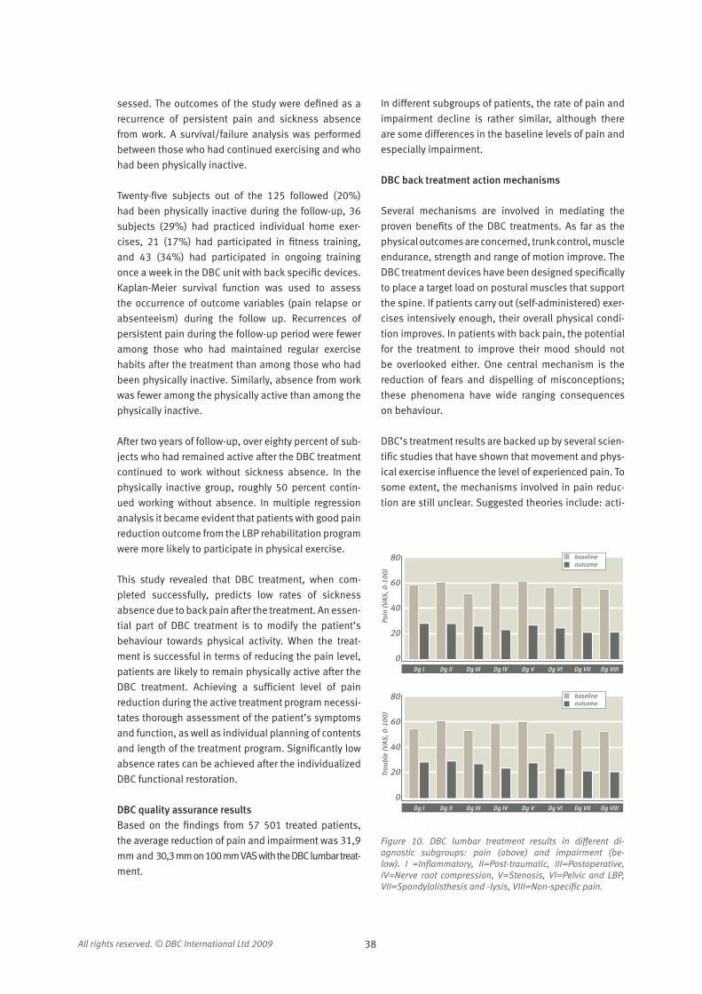
38All rights reserved. © DBC International Ltd 2009
sessed. The outcomes of the study were defined as a
recurrence of persistent pain and sickness absence
from work. A survival/failure analysis was performed
between those who had continued exercising and who
had been physically inactive.
Twenty-five subjects out of the 125 followed (20%)
had been physically inactive during the follow-up, 36
subjects (29%) had practiced individual home exer-
cises, 21 (17%) had participated in fitness training,
and 43 (34%) had participated in ongoing training
once a week in the DBC unit with back specific devices.
Kaplan-Meier survival function was used to assess
the occurrence of outcome variables (pain relapse or
absenteeism) during the follow up. Recurrences of
persistent pain during the follow-up period were fewer
among those who had maintained regular exercise
habits after the treatment than among those who had
been physically inactive. Similarly, absence from work
was fewer among the physically active than among the
physically inactive.
After two years of follow-up, over eighty percent of sub-
jects who had remained active after the DBC treatment
continued to work without sickness absence. In the
physically inactive group, roughly 50 percent contin-
ued working without absence. In multiple regression
analysis it became evident that patients with good pain
reduction outcome from the LBP rehabilitation program
were more likely to participate in physical exercise.
This study revealed that DBC treatment, when com-
pleted successfully, predicts low rates of sickness
absence due to back pain after the treatment. An essen-
tial part of DBC treatment is to modify the patient’s
behaviour towards physical activity. When the treat-
ment is successful in terms of reducing the pain level,
patients are likely to remain physically active after the
DBC treatment. Achieving a sufficient level of pain
reduction during the active treatment program necessi-
tates thorough assessment of the patient’s symptoms
and function, as well as individual planning of contents
and length of the treatment program. Significantly low
absence rates can be achieved after the individualized
DBC functional restoration.
DBC quality assurance resultsBased on the findings from 57 501 treated patients,
the average reduction of pain and impairment was 31,9
mm and 30,3 mm on 100 mm VAS with the DBC lumbar treat-
ment.
In different subgroups of patients, the rate of pain and
impairment decline is rather similar, although there
are some differences in the baseline levels of pain and
especially impairment.
DBC back treatment action mechanisms
Several mechanisms are involved in mediating the
proven benefits of the DBC treatments. As far as the
physical outcomes are concerned, trunk control, muscle
endurance, strength and range of motion improve. The
DBC treatment devices have been designed specifically
to place a target load on postural muscles that support
the spine. If patients carry out (self-administered) exer-
cises intensively enough, their overall physical condi-
tion improves. In patients with back pain, the potential
for the treatment to improve their mood should not
be overlooked either. One central mechanism is the
reduction of fears and dispelling of misconceptions;
these phenomena have wide ranging consequences
on behaviour.
DBC’s treatment results are backed up by several scien-
tific studies that have shown that movement and phys-
ical exercise influence the level of experienced pain. To
some extent, the mechanisms involved in pain reduc-
tion are still unclear. Suggested theories include: acti-
Figure 10. DBC lumbar treatment results in different di-agnostic subgroups: pain (above) and impairment (be-low). I =Inflammatory, II=Post-traumatic, III=Postoperative, IV=Nerve root compression, V=Stenosis, VI=Pelvic and LBP, VII=Spondylolisthesis and -lysis, VIII=Non-specific pain.
80
60
40
20
0
Pain
(VAS
, 0-1
00)
Dg I Dg VIIDg VIDg VDg IVDg IIIDg II Dg VIII
baselineoutcome
80
60
40
20
0
Trou
ble
(VAS
, 0-1
00)
Dg I Dg VIIDg VIDg VDg IVDg IIIDg II Dg VIII
baselineoutcome

39 All rights reserved. © DBC International Ltd 2009
vation of pain gate control, release of endorphins with
intensive exercise and suppression of the brain’s pain
centres by the activation of the motor cortex. Whatever
the mechanism is, the reduction is undeniable. Also
“learning by doing” can be a strong factor in inducing
a person to unlearn the role of being a patient. Here,
general activation and mood improvement may lower
the patient’s pain experience, as the patient realizes
that his or her wellbeing increases during the therapy
or rehabilitation. A definite organic (periferic nocicep-
tive) reason for pain cannot be identified for most of
the patients with back disorders.
DBC in neck disorders
The DBC treatment method for neck disorders is reha-
bilitation aimed at restoring function. Its physical ele-
ments comprise of progressive exercises targeting
the cervical spine, thoracic spine, and their adjacent
tissues and includes stretching/relaxation intervals.
These elements are combined with the cognitive-
behavioural approach, in which patients’ mistaken
conceptions and beliefs are redressed. Patients are
given support in changing their behaviour towards a
healthier outlook.
The contents and duration of the physical rehabilita-
tion program are defined on the basis of the severity of
pain and impairment, psychological profile, and social
needs. These are assessed with validated question-
naires and measurements. The questionnaires and
assessments are essential, not only in defining the
needs, but also used in the monitoring of progress and
documenting outcomes.
The physical reconditioning program includes progres-
sive co-ordination, mobility and muscle endurance
exercises combined with stretching and relaxation.
Specially trained therapists guide the physical recon-
ditioning program. The treatment is primarily based
on exercises in specific rehabilitation devices; correct
loading and range limiters ensure that exercises are
performed in a painless range of motion and that
they target the right area. Treatment includes control-
led movements in cervical/thoracic extension and
rotation, and shoulder blade adduction. Treatment is
planned on the basis of initial range-of-motion meas-
urements, questionnaires and interviews, and records
are kept of the progress. The treatment begins with low
loads for the first weeks with the object of improving
mobility and especially teaching proper co-ordination
and posture of the cervical spine. The load is gradu-
ally increased so that only later subjectively strenuous
loading is applied within the pain tolerance of the indi-
vidual patient. The load is further increased in a gradual
and controlled manner until, at the end of the program,
the patients are instructed to continue an individual
secondary-prevention program with or without guid-
ance depending on their individual needs. The inclu-
sions of exercises, rate of progression in loading and
ranges of motion are individualized based on the type
(diagnosis) and severity of the neck problem.
DBC neck treatment results
ResearchA randomized comparative study with single-blind
outcome assessments compared the efficacy of a mul-
timodal treatment emphasizing proprioceptive train-
ing (DBC) with activated home exercises (HOME) and
recommendation of exercise (CONTROL) in patients
with non-specific chronic neck pain. The study group
consisted of seventy-six patients (22 men, 54 women)
with chronic, non-specific neck pain. Sixty-two subjects
participated in the 1-year follow-up. Subjective pain
and disability, cervical ranges of motion, and pressure
pain threshold in the shoulder region were measured
at baseline, at 3 months, and at 12 months. The DBC
treatment consisted of 24 sessions of progressive exer-
cises, relaxation and behavioural support. The HOME
regimen included a neck lecture and two sessions of
practical training for home exercises and instructions
for maintaining a diary of progress. The CONTROL treat-
ment included a lecture regarding care of the neck with
a recommendation to exercise. According to the exer-
cise diaries, the actual amount of exercise was largest
in HOME group and smallest in CONTROL group.
The average self-experienced total benefit was highest
in the DBC group, and the HOME group rated above
the CONTROL group. Differences between the groups
in favour of the DBC treatment were recorded in the
reduction of neck symptoms and improvements in
general health and self-experienced working ability.
Changes in measures of mobility and pressure-pain
threshold were minor. Since no major differences were
noted in the objective measurements of cervical func-
tion between the groups, it can be assumed that neck
pain and especially its chronicity comprises a condi-
tion where motivation and perception of the problem
plays a significant role. These findings support the idea
that multimodal treatment, which integrates both pro-
prioceptive and endurance exercises as well as behav-

40All rights reserved. © DBC International Ltd 2009
ioural support is more efficacious in treating chronic
neck pain patients than solitary training.
DBC quality assurance resultsBased on the findings of 9 244 treated patients, the
average reductions of pain and impairment were 33,4
mm and 32,6 mm on 100 mm VAS with the DBC cervi-
cal treatment. In different subgroups of patients, the
rate of pain and impairment decline was slightly differ-
ent. There were also differences in the baseline levels
of pain and impairment.
DBC neck treatment action mechanisms
There are potentially several mechanisms that mediate
the benefits of exercise and mobility therapy in
patients with pain in the neck and shoulder area. As
exercise targets the neck, mobility and exercise therapy
enhances muscle strength, co-ordination and mobil-
ity as well as reduces pain in the neck and shoulder
area. DBC treatments differ from other mobility thera-
pies in that the 3-dimensional arch of movement that
they “force” upon the neck is physiologically correct.
The DBC mobility exercises have been specifically
designed to restore the correct coupled motions of
the cervical spine. As a result of the cognitive-behav-
Figure 11. DBC neck treatment results in different diagnos-tic subgroups: pain (above) and impairment (below). I =In-flammatory, II=Post-traumatic, III=WAD, IV=Postoperative, V=Stenosis, VI=Nerve root compression, VII=Non-specific pain.
80
60
40
20
0
Pain
(VAS
, 0-1
00)
Dg I Dg VIIDg VIDg VDg IVDg IIIDg II
baselineoutcome
80
60
40
20
0
Trou
ble
(VAS
, 0-1
00)
Dg I Dg VIIDg VIDg VDg IVDg IIIDg II
baselineoutcome
ioural approach, mistaken beliefs are systematically
redressed, fears are relieved and support/motivation is
given to the patient. The purpose is to help the patient
reduce the experienced impairment and inhibitions,
which prevent the normal use and loading of the neck.
Furthermore, the potential impact of physical exercise
on the patient’s mood should not be underestimated.
DBC in shoulder disorders
The primary aim of reconditioning is to restore the
normal control of scapular movements and humero-
scapular rhythm. Later, it aims to reverse the effects of
physical deconditioning, especially in the rotator cuff.
This is achieved by improving postural control and co-
ordination, and by producing adaptive changes in the
tissues in a progressive, controlled way. Strengthening
of the muscles and other soft tissues is based on the
progression (increase) in loading. This is gained either
by increasing the weight, number of repetitions, range
of motion used, or by applying increasingly demand-
ing functional exercises. Improvements of neural
control and co-ordination are based on repetition of
the correct movement under guidance. The contents of
the reconditioning program are chosen on the basis of
the anatomic/ aetiopathologic diagnosis, i.e., the pain
pattern defines the components (exercises) and the
progression of restoration.
The DBC Shoulder treatment system is based on the
notion of different exercise levels. The level of exer-
cises is chosen based on the healing process. Each
treatment can be divided into different phases, each
containing exercises that are safe at that stage of the
healing process. Treatment is typically carried out in
6-week modules. The recommended length of treat-
ment for most problems is 12 weeks (24 sessions).
The more severe the problem, the longer the program
needs to be.
Treatment is carried out twice a week in order to give
the structures enough time to recover in between ses-
sions. In special cases and in the early phase of the
treatment (low loading) 3-4 sessions per week can be
carried out. If more than 2 sessions are done, one must
be very careful not to overload the patient. One treat-
ment session lasts an average of one hour. However,
this might vary from country to country.
Progression of the program mainly happens by moving
in to more demanding exercises. For range of motion,
the main principle is that the preferred range of motion
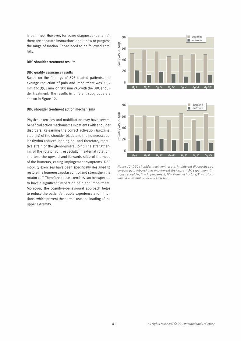
41 All rights reserved. © DBC International Ltd 2009
is pain free. However, for some diagnoses (patterns),
there are separate instructions about how to progress
the range of motion. Those need to be followed care-
fully.
DBC shoulder treatment results
DBC quality assurance resultsBased on the findings of 893 treated patients, the
average reduction of pain and impairment was 35,2
mm and 39,5 mm on 100 mm VAS with the DBC shoul-
der treatment. The results in different subgroups are
shown in Figure 12.
DBC shoulder treatment action mechanisms
Physical exercises and mobilization may have several
beneficial action mechanisms in patients with shoulder
disorders. Relearning the correct activation (proximal
stability) of the shoulder blade and the humeroscapu-
lar rhythm reduces loading on, and therefore, repeti-
tive strain of the glenohumeral joint. The strengthen-
ing of the rotator cuff, especially in external rotation,
shortens the upward and forwards slide of the head
of the humerus, easing impingement symptoms. DBC
mobility exercises have been specifically designed to
restore the humeroscapular control and strengthen the
rotator cuff. Therefore, these exercises can be expected
to have a significant impact on pain and impairment.
Moreover, the cognitive-behavioural approach helps
to reduce the patient’s trouble-experience and inhibi-
tions, which prevent the normal use and loading of the
upper extremity.
80
60
40
20
0
Pain
(VAS
, 0-1
00)
Dg I Dg VIIDg VIDg VDg IVDg IIIDg II
baselineoutcome
80
60
40
20
0
Trou
ble
(VAS
, 0-1
00)
Dg I Dg VIIDg VIDg VDg IVDg IIIDg II
baselineoutcome
Figure 12. DBC shoulder treatment results in different diagnostic sub-groups: pain (above) and impairment (below). I = AC separation, II = Frozen shoulder, III = Impingement, IV = Proximal fracture, V = Disloca-tion, VI = Instability, VII = SLAP lesion.

42All rights reserved. © DBC International Ltd 2009

43 All rights reserved. © DBC International Ltd 2009
6. Literature
1. Addison R, Schultz A. Trunk strengths in patients
seeking hospitalization for chronic low-back disorders.
Spine 1980;5:539-44.
2. Agency for Health Policy and Research UDoHHS. Man-
agement Guidelines for Acute Low- Back Pain. Washing-
ton, DC: US Government Printing Office, 1994.
3. Alaranta H, Luoto S, Heliovaara M, et al. Static back
endurance and the risk of low-back pain. Clin Biomech
(Bristol, Avon) 1995;10:323-4.
4. Alaranta H, Moffroid M, Elmqvist L, et al. Postural
control of adults with musculoskeletal impairment.
Critical Reviews in Physical& Rehabilitation Medicine
1994:337-70.
5. Alaranta H, Rytokoski U, Rissanen A, et al. Intensive
physical and psychosocial training program for patients
with chronic low back pain. A controlled clinical trial.
Spine 1994;19:1339-49.
6. American College of Sports Medicine position stand.
The recommended quantity and quality of exercise for
developing and maintaining cardiorespiratory and mus-
cular fitness in healthy adults. Medicine and Science in
Sports and Exercicise 1990;22:265-74.
7. Andersson GB. Epidemiologic aspects on low-back
pain in industry. Spine 1981;6:53-60.
8. Andersson JL, Lilja A, Hartvig P, et al. Somatotopic
organization along the central sulcus, for pain localiza-
tion in humans, as revealed by positron emission tomog-
raphy. Exp Brain Res 1997;117:192-9.
9. Assendelft WJ, Koes BW, van der Heijden GJ, et al.
The effectiveness of chiropractic for treatment of low
back pain: an update and attempt at statistical pooling. J
Manipulative Physiol Ther 1996;19:499-507.
10. Bagg SD, Forrest WJ. A biomechanical analysis of
scapular rotation during arm abduction in the scapular
plane. Am J Phys Med Rehabil 1988;67:238-45.
11. Bak K. Nontraumatic glenohumeral instability and
coracoacromial impingement in swimmers. Scand J Med
Sci Sports 1996;6:132-44.
12. Barbe MF, Barr AE. Inflammation and the pathophys-
iology of work-related musculoskeletal disorders. Brain
Behav Immun 2006;20:423-9.
13. Battie MC, Videman T, Gibbons LE, et al. 1995 Volvo
Award in clinical sciences. Determinants of lumbar disc
degeneration. A study relating lifetime exposures and
magnetic resonance imaging findings in identical twins.
Spine 1995;20:2601-12.
14. Bearcroft PW, Blanchard TK, Dixon AK, et al. An
assessment of the effectiveness of magnetic resonance
imaging of the shoulder: literature review. Skeletal Radiol
2000;29:673-9.
15. Bendix AF, Bendix T, Vaegter K, et al. Multidisciplinary
intensive treatment for chronic low back pain: a rand-

44All rights reserved. © DBC International Ltd 2009
omized, prospective study. Cleve Clin J Med 1996;63:62-
9.
16. Biering-Sorensen F. Physical measurements as risk
indicators for low-back trouble over a one-year period.
Spine 1984;9:106-19.
17. Biering-Sorensen F. A prospective study of low back
pain in a general population. I. Occurrence, recurrence
and aetiology. Scand J Rehabil Med 1983;15:71-9.
18. Biering-Sorensen F, Thomsen CE, Hilden J. Risk
indicators for low back trouble. Scand J Rehabil Med
1989;21:151-7.
19. Bigliani LU, Levine WN. Subacromial impingement
syndrome. J Bone Joint Surg Am 1997;79:1854-68.
20. Bigos SJ, Battie MC, Spengler DM, et al. A prospec-
tive study of work perceptions and psychosocial factors
affecting the report of back injury. Spine 1991;16:1-6.
21. Boden SD, Davis DO, Dina TS, et al. Abnormal mag-
netic-resonance scans of the lumbar spine in asympto-
matic subjects. A prospective investigation. J Bone Joint
Surg Am 1990;72:403-8.
22. Boden SD, McCowin PR, Davis DO, et al. Abnor-
mal magnetic-resonance scans of the cervical spine in
asymptomatic subjects. A prospective investigation. J
Bone Joint Surg Am 1990;72:1178-84.
23. Bogdanffy M, Taimela S, Rashbaum R, et al. Backspe-
cific physical reconditioning after laminectomy or fusion
spine surgery: A controlled intervention. 8th Annual
Meeting of the European Spine Society. Kos, Greece,
1997.
24. Bombardier C, Kerr MS, Shannon HS, et al. A guide
to interpreting epidemiologic studies on the etiology of
back pain. Spine 1994;19:2047S-56S.
25. Boos N, Rieder R, Schade V, et al. 1995 Volvo Award
in clinical sciences. The diagnostic accuracy of magnetic
resonance imaging, work perception, and psychoso-
cial factors in identifying symptomatic disc herniations.
Spine 1995;20:2613-25.
26. Bouchard C, Shephard R. Physical activity, fitness
and health: the model and key concepts. In Bouchard C,
Shephard R, Stephens T eds. Physical activity, fitness and
health. Consensus statement. Champaign, Ill: Human
Kinetics Publishers, 1993:11-23.
27. Brown JJ, Wells GA, Trottier AJ, et al. Back pain in a
large Canadian police force. Spine 1998;23:821-7.
28. Brox JI, Brevik JI. Prognostic factors in patients with
rotator tendinosis (stage II impingement syndrome) of
the shoulder. Scand J Prim Health Care 1996;14:100-5.
29. Brox JI, Gjengedal E, Uppheim G, et al. Arthroscopic
surgery versus supervised exercises in patients with
rotator cuff disease (stage II impingement syndrome):
a prospective, randomized, controlled study in 125
patients with a 2 1/2-year follow-up. J Shoulder Elbow
Surg 1999;8:102-11.
30. Bullock-Saxton JE, Janda V, Bullock MI. Reflex activa-
tion of gluteal muscles in walking. An approach to resto-
ration of muscle function for patients with low-back pain.
Spine 1993;18:704-8.
31. Burton AK, Tillotson KM, Main CJ, et al. Psychosocial
predictors of outcome in acute and subchronic low back
trouble. Spine 1995;20:722-8.
32. Byl N, Sinnott P. Variations in balance and body sway
in middle-aged adults: subjects with healthy backs com-
pared with subjects with low-back dysfunction. Spine
1991;16:325-30.
33. Campello M, Nordin M, Weiser S. Physical exercise
and low back pain. Scand J Med Sci Sports 1996;6:63-
72.
34. Cartas O, Nordin M, Frankel VH, et al. Quantification
of trunk muscle performance in standing, semistanding
and sitting postures in healthy men. Spine 1993;18:603-
9.
35. Cherkin DC, Deyo RA, Loeser JD, et al. An international
comparison of back surgery rates. Spine 1994;19:1201-
6.
36. Cholewicki J, Panjabi MM, Khachatryan A. Stabiliz-
ing function of trunk flexor-extensor muscles around a
neutral spine posture. Spine 1997;22:2207-12.
37. Clinical Standards Advisory Group. Report on Back
Pain. London: Her Majesty´s Stationery Office, 1994.
38. Cooper RG, St Clair Forbes W, Jayson MI. Radiographic
demonstration of paraspinal muscle wasting in patients
with chronic low back pain. Br J Rheumatol 1992;31:389-
94.
39. Craig AD, Reiman EM, Evans A, et al. Functional
imaging of an illusion of pain. Nature 1996;384:258-60.
40. Croft PR, Papageorgiou AC, Ferry S, et al. Psychologic
distress and low back pain. Evidence from a prospective
study in the general population. Spine 1995;20:2731-7.
41. Davis KD, Taylor SJ, Crawley AP, et al. Functional MRI
of pain- and attention-related activations in the human
cingulate cortex. J Neurophysiol 1997;77:3370-80.
42. Decker MJ, Hintermeister RA, Faber KJ, et al. Serra-
tus anterior muscle activity during selected rehabilitation
exercises. Am J Sports Med 1999;27:784-91.
43. Derbyshire SW, Vogt BA, Jones AK. Pain and Stroop
interference tasks activate separate processing modules
in anterior cingulate cortex. Exp Brain Res 1998;118:52-
60.
44. Devinsky O, Morrell MJ, Vogt BA. Contributions of
anterior cingulate cortex to behaviour. Brain 1995;118 (
Pt 1):279-306.
45. Deyo RA. Fads in the treatment of low back pain. N
Engl J Med 1991;325:1039-40.
46. Deyo RA, Diehl AK, Rosenthal M. How many days of

45 All rights reserved. © DBC International Ltd 2009
bed rest for acute low back pain? A randomized clinical
trial. N Engl J Med 1986;315:1064-70.
47. Deyo RA, Tsui-Wu YJ. Descriptive epidemiology of
low-back pain and its related medical care in the United
States. Spine 1987;12:264-8.
48. Dickenson AH. Central acute pain mechanisms. Ann
Med 1995;27:223-7.
49. Dvorak V, Taimela S. Trainability of back muscles in
chronic low back pain patients: the effect of sex and age.
EuroSpine 96. Zurich, Switzerland, 1996.
50. Eisenstein SM, Ashton IK, Roberts S, et al. Innervation
of the spondylolysis “ligament”. Spine 1994;19:912-6.
51. Erkintalo MO, Salminen JJ, Alanen AM, et al. Develop-
ment of degenerative changes in the lumbar interverte-
bral disk: results of a prospective MR imaging study in
adolescents with and without low-back pain. Radiology
1995;196:529-33.
52. Estlander AM, Takala EP, Viikari-Juntura E. Do psycho-
logical factors predict changes in musculoskeletal pain?
A prospective, two-year follow-up study of a working pop-
ulation. J Occup Environ Med 1998;40:445-53.
53. Faas A, Chavannes AW, van Eijk JT, et al. A rand-
omized, placebo-controlled trial of exercise therapy in
patients with acute low back pain. Spine 1993;18:1388-
95.
54. Faas A, van Eijk JT, Chavannes AW, et al. A randomized
trial of exercise therapy in patients with acute low back
pain. Efficacy on sickness absence. Spine 1995;20:941-
7.
55. Falla D, Jull G, Hodges PW. Feedforward activity of
the cervical flexor muscles during voluntary arm move-
ments is delayed in chronic neck pain. Exp Brain Res
2004;157:43-8.
56. Falla DL, Jull GA, Hodges PW. Patients with neck pain
demonstrate reduced electromyographic activity of the
deep cervical flexor muscles during performance of the
craniocervical flexion test. Spine 2004;29:2108-14.
57. Farfan HF. Form and function of the musculoskel-
etal system as revealed by mathematical analysis of the
lumbar spine. An essay. Spine 1995;20:1462-74.
58. Fidler MW, Jowett RL, Troup JD. Myosin ATPase activ-
ity in multifidus muscle from cases of lumbar spinal
derangement. J Bone Joint Surg Br 1975;57:220-7.
59. Frymoyer JW. Back pain and sciatica. N Engl J Med
1988;318:291-300.
60. Fu FH, Ticker JB, Imhoff AB. An Atlas of Shoulder Sur-
geryed: Appleton & Lange, 1998.
61. Gallagher RM, Rauh V, Haugh LD, et al. Determinants
of return-to-work among low back pain patients. Pain
1989;39:55-67.
62. Gatchel RJ, Polatin PB, Mayer TG. The dominant role
of psychosocial risk factors in the development of chronic
low back pain disability. Spine 1995;20:2702-9.
63. Goldberg L, Elliot D. Exercise for prevention and treat-
ment of illnessed. Philadelphia: F.A. Davis Company,
1994.
64. Graves JE, Pollock ML, Carpenter DM, et al. Quantita-
tive assessment of full range-of-motion isometric lumbar
extension strength. Spine 1990;15:289-94.
65. Graves JE, Pollock ML, Foster D, et al. Effect of training
frequency and specificity on isometric lumbar extension
strength. Spine 1990;15:504-9.
66. Graves JE, Webb DC, Pollock ML, et al. Pelvic stabili-
zation during resistance training: its effect on the devel-
opment of lumbar extension strength. Arch Phys Med
Rehabil 1994;75:210-5.
67. Green S, Buchbinder R, Glazier R, et al. Interven-
tions for shoulder pain. Cochrane Database Syst Rev
2000:CD001156.
68. Green S, Buchbinder R, Glazier R, et al. Systematic
review of randomised controlled trials of interventions
for painful shoulder: selection criteria, outcome assess-
ment, and efficacy. Bmj 1998;316:354-60.
69. Guzman J, Esmail R, Karjalainen K, et al. Multidiscipli-
nary bio-psycho-social rehabilitation for chronic low back
pain. Cochrane Database Syst Rev 2002:CD000963.
70. Guzman J, Esmail R, Karjalainen K, et al. Multidiscipli-
nary rehabilitation for chronic low back pain: systematic
review. Bmj 2001;322:1511-6.
71. Haahr JP, Ostergaard S, Dalsgaard J, et al. Exercises
versus arthroscopic decompression in patients with sub-
acromial impingement: a randomised, controlled study
in 90 cases with a one year follow up. Ann Rheum Dis
2005;64:760-4.
72. Hadar H, Gadoth N, Heifetz M. Fatty replacement of
lower paraspinal muscles: normal and neuromuscular
disorders. AJR Am J Roentgenol 1983;141:895-8.
73. Hansen FR, Bendix T, Skov P, et al. Intensive, dynamic
back-muscle exercises, conventional physiotherapy,
or placebo-control treatment of low-back pain. A rand-
omized, observer-blind trial. Spine 1993;18:98-108.
74. Harkapaa K. Relationships of psychological distress
and health locus of control beliefs with the use of cog-
nitive and behavioral coping strategies in low back pain
patients. Clin J Pain 1991;7:275-82.
75. Harkapaa K, Jarvikoski A, Mellin G, et al. Health locus
of control beliefs and psychological distress as predictors
for treatment outcome in low-back pain patients: results
of a 3-month follow-up of a controlled intervention study.
Pain 1991;46:35-41.
76. Hayden JA, van Tulder MW, Malmivaara A, et al. Exer-
cise therapy for treatment of non-specific low back pain.
Cochrane Database Syst Rev 2005:CD000335.
77. Hayden JA, van Tulder MW, Malmivaara AV, et al.

46All rights reserved. © DBC International Ltd 2009
Meta-analysis: exercise therapy for nonspecific low back
pain. Ann Intern Med 2005;142:765-75.
78. Hayes K, Ginn KA, Walton JR, et al. A randomised
clinical trial evaluating the efficacy of physiotherapy after
rotator cuff repair. Aust J Physiother 2004;50:77-83.
79. Hazard RG. Spine update. Functional restoration.
Spine 1995;20:2345-8.
80. Heliovaara M. Risk factors for low back pain and sci-
atica. Ann Med 1989;21:257-64.
81. Heliövaara M. Risk factors of low back pain - A review.
In Ernst J JM, Pope MH, Porter RW, eds ed. Advances in idi-
opathic low back pain. Wien: Blackwell- MZW, 1993:41-
51.
82. Hestbaek L, Leboeuf-Yde C, Kyvik KO. Are lifestyle-
factors in adolescence predictors for adult low back pain?
A cross-sectional and prospective study of young twins.
BMC Musculoskelet Disord 2006;7:27.
83. Hestbaek L, Leboeuf-Yde C, Kyvik KO, et al. The course
of low back pain from adolescence to adulthood: eight-
year follow-up of 9600 twins. Spine 2006;31:468-72.
84. Hestbaek L, Leboeuf-Yde C, Manniche C. Low back
pain: what is the long-term course? A review of studies
of general patient populations. Eur Spine J 2003;12:149-
65.
85. Hides JA, Richardson CA, Jull GA. Multifidus muscle
recovery is not automatic after resolution of acute, first-
episode low back pain. Spine 1996;21:2763-9.
86. Hides JA, Stokes MJ, Saide M, et al. Evidence of
lumbar multifidus muscle wasting ipsilateral to symp-
toms in patients with acute/subacute low back pain.
Spine 1994;19:165-72.
87. Hildebrandt J, Pfingsten M, Saur P, et al. Prediction
of success from a multidisciplinary treatment program for
chronic low back pain. Spine 1997;22:990-1001.
88. Hodges PW, Eriksson AE, Shirley D, et al. Intra-
abdominal pressure increases stiffness of the lumbar
spine. J Biomech 2005;38:1873-80.
89. Hodges PW, Moseley GL. Pain and motor control of
the lumbopelvic region: effect and possible mechanisms.
J Electromyogr Kinesiol 2003;13:361-70.
90. Hodges PW, Moseley GL, Gabrielsson A, et al.
Experimental muscle pain changes feedforward pos-
tural responses of the trunk muscles. Exp Brain Res
2003;151:262-71.
91. Hodges PW, Richardson CA. Delayed postural con-
traction of transversus abdominis in low back pain asso-
ciated with movement of the lower limb. J Spinal Disord
1998;11:46-56.
92. Hodges PW, Richardson CA. Inefficient muscular
stabilization of the lumbar spine associated with low
back pain. A motor control evaluation of transversus
abdominis. Spine 1996;21:2640-50.
93. Holroyd CB, Dien J, Coles MG. Error-related scalp
potentials elicited by hand and foot movements: evi-
dence for an output-independent error-processing
system in humans. Neurosci Lett 1998;242:65-8.
94. Howell SM, Galinat BJ. The glenoid-labral socket.
A constrained articular surface. Clin Orthop Relat Res
1989:122-5.
95. Hultman G, Nordin M, Saraste H, et al. Body com-
position, endurance, strength, cross-sectional area, and
density of MM erector spinae in men with and without
low back pain. J Spinal Disord 1993;6:114-23.
96. Hurri H. The Swedish back school in chronic low back
pain. Part II. Factors predicting the outcome. Scand J
Rehabil Med 1989;21:41-4.
97. Huskisson EC, Jones J, Scott PJ. Application of visual-
analogue scales to the measurement of functional capac-
ity. Rheumatol Rehabil 1976;15:185-7.
98. Härkäpää K, Taimela S, . Pain reduction and changes
in depression and perceived competence during active
functional restoration for chronic low- back pain: a 12-
week follow- up. EuroSpine 96. Zurich, Switzerland:
Schulthess Clinic, 1996:76.
99. Indahl A, Kaigle A, Reikeras O, et al. Electromyo-
graphic response of the porcine multifidus musculature
after nerve stimulation. Spine 1995;20:2652-8.
100. Indahl A, Kaigle AM, Reikeras O, et al. Interac-
tion between the porcine lumbar intervertebral disc,
zygapophysial joints, and paraspinal muscles. Spine
1997;22:2834-40.
101. Institute PotNPH. Health and Functional Capacity
in Finland. Baseline Results of the Health 2000 Health
Examination Survey. In Aromaa A, Koskinen S eds. Hel-
sinki: KTL- National Public Health Institute, Finland,
2004:175.
102. Jarvik JG, Hollingworth W, Heagerty PJ, et al. Three-
year incidence of low back pain in an initially asympto-
matic cohort: clinical and imaging risk factors. Spine
2005;30:1541-8; discussion 9.
103. Jarvikoski A, Mellin G, Estlander AM, et al. Outcome
of two multimodal back treatment programs with and
without intensive physical training. J Spinal Disord
1993;6:93-8.
104. Jensen MC, Brant-Zawadzki MN, Obuchowski N, et
al. Magnetic resonance imaging of the lumbar spine in
people without back pain. N Engl J Med 1994;331:69-
73.
105. Jiang H, Moreau M, Raso J, et al. Identification of
the location, extent, and pathway of sensory neurologic
feedback after mechanical stimulation of a lateral spinal
ligament in chickens. Spine 1997;22:17-25.
106. Jiang H, Raso JV, Moreau MJ, et al. Quantitative mor-
phology of the lateral ligaments of the spine. Assessment

47 All rights reserved. © DBC International Ltd 2009
of their importance in maintaining lateral stability. Spine
1994;19:2676-82.
107. Jiang H, Russell G, Raso VJ, et al. The nature and
distribution of the innervation of human supraspinal and
interspinal ligaments. Spine 1995;20:869-76.
108. Jones AK, Derbyshire SW. Reduced cortical
responses to noxious heat in patients with rheumatoid
arthritis. Ann Rheum Dis 1997;56:601-7.
109. Jones AK, Friston K, Dolan R. Positron emission tom-
ography as a research tool in the investigation of psychi-
atric and psychological disorders. Baillieres Clin Endocri-
nol Metab 1991;5:187-203.
110. Kaigle AM, Wessberg P, Hansson TH. Muscular and
kinematic behavior of the lumbar spine during flexion-
extension. J Spinal Disord 1998;11:163-74.
111. Kalaska JF, Drew T. Motor cortex and visuomotor
behavior. Exerc Sport Sci Rev 1993;21:397-436.
112. Kankaanpaa M, Taimela S, Airaksinen O, et al. The
efficacy of active rehabilitation in chronic low back pain.
Effect on pain intensity, self-experienced disability, and
lumbar fatigability. Spine 1999;24:1034-42.
113. Kankaanpaa M, Taimela S, Laaksonen D, et al.
Back and hip extensor fatigability in chronic low back
pain patients and controls. Arch Phys Med Rehabil
1998;79:412-7.
114. Kankaanpaa M, Taimela S, Webber CL, Jr., et al.
Lumbar paraspinal muscle fatigability in repetitive
isoinertial loading: EMG spectral indices, Borg scale
and endurance time. Eur J Appl Physiol Occup Physiol
1997;76:236-42.
115. Kannus P, Josza L, Renström P, et al. The effects
of training, immobilization and remobilization on mus-
culoskeletal tissues. Training and immobilization. Scan-
dinavian Journal of Medicine and Science in Sports
1992;2:100-8.
116. Karjalainen K, Malmivaara A, Mutanen P, et al.
Outcome determinants of subacute low back pain. Spine
2003;28:2634-40.
117. Karjalainen K, Malmivaara A, Mutanen P, et al. Mini-
intervention for subacute low back pain: two-year follow-
up and modifiers of effectiveness. Spine 2004;29:1069-
76.
118. Karjalainen K, Malmivaara A, Pohjolainen T, et al.
Mini-intervention for subacute low back pain: a rand-
omized controlled trial. Spine 2003;28:533-40; discus-
sion 40-1.
119. Karjalainen K, Malmivaara A, van Tulder M, et al.
Biopsychosocial rehabilitation for upper limb repetitive
strain injuries in working age adults. Cochrane Database
Syst Rev 2000:CD002269.
120. Karjalainen K, Malmivaara A, van Tulder M, et al.
Multidisciplinary biopsychosocial rehabilitation for neck
and shoulder pain among working age adults. Cochrane
Database Syst Rev 2003:CD002194.
121. Karjalainen K, Malmivaara A, van Tulder M, et al.
Multidisciplinary biopsychosocial rehabilitation for suba-
cute low back pain among working age adults. Cochrane
Database Syst Rev 2003:CD002193.
122. Karjalainen K, Malmivaara A, van Tulder M, et al.
Multidisciplinary rehabilitation for fibromyalgia and mus-
culoskeletal pain in working age adults. Cochrane Data-
base Syst Rev 2000:CD001984.
123. Keltikangas-Jarvinen L, Rimon R. Rimon’s Brief
Depression Scale, a rapid method for screening depres-
sion. Psychol Rep 1987;60:111-9.
124. Kibler WB. The role of the scapula in athletic shoul-
der function. Am J Sports Med 1998;26:325-37.
125. Kirkley A, Griffin S, Richards C, et al. Prospective
randomized clinical trial comparing the effectiveness of
immediate arthroscopic stabilization versus immobiliza-
tion and rehabilitation in first traumatic anterior disloca-
tions of the shoulder. Arthroscopy 1999;15:507-14.
126. Klenerman L, Slade PD, Stanley IM, et al. The pre-
diction of chronicity in patients with an acute attack
of low back pain in a general practice setting. Spine
1995;20:478-84.
127. Koes BW, Assendelft WJ, van der Heijden GJ, et
al. Spinal manipulation for low back pain. An updated
systematic review of randomized clinical trials. Spine
1996;21:2860-71; discussion 72-3.
128. Koes BW, Bouter LM, van der Heijden GJ. Methodo-
logical quality of randomized clinical trials on treatment
efficacy in low back pain. Spine 1995;20:228-35.
129. Koes BW, Scholten RJ, Mens JM, et al. Efficacy of
epidural steroid injections for low-back pain and sciatica:
a systematic review of randomized clinical trials. Pain
1995;63:279-88.
130. Koes BW, Scholten RJ, Mens JM, et al. Efficacy of
non-steroidal anti-inflammatory drugs for low back pain:
a systematic review of randomised clinical trials. Ann
Rheum Dis 1997;56:214-23.
131. Kohles S, Barnes D, Gatchel RJ, et al. Improved phys-
ical performance outcomes after functional restoration
treatment in patients with chronic low-back pain. Early
versus recent training results. Spine 1990;15:1321-4.
132. Komi P. Strength and power in sported. London:
Blackwell Publishing, 1992.
133. Kujala UM, Taimela S, Viljanen T, et al. Physical
loading and performance as predictors of back pain in
healthy adults. A 5-year prospective study. Eur J Appl
Physiol Occup Physiol 1996;73:452-8.
134. Laasonen EM. Atrophy of sacrospinal muscle groups
in patients with chronic, diffusely radiating lumbar back
pain. Neuroradiology 1984;26:9-13.

48All rights reserved. © DBC International Ltd 2009
135. Leboeuf-Yde C, Klougart N, Lauritzen T. How common
is low back pain in the Nordic population? Data from a
recent study on a middle-aged general Danish popula-
tion and four surveys previously conducted in the Nordic
countries. Spine 1996;21:1518-25; discussion 25-6.
136. Letham J, Slade P, Troup J, et al. Outline of a feara-
voidance model of exaggerated pain perception. Part 1.
Behavioral Research and Therapy 1983;21:401-8.
137. Lindstrom I, Ohlund C, Eek C, et al. The effect of
graded activity on patients with subacute low back
pain: a randomized prospective clinical study with an
operant-conditioning behavioral approach. Phys Ther
1992;72:279-90; discussion 91-3.
138. Lindstrom I, Ohlund C, Eek C, et al. Mobility, strength,
and fitness after a graded activity program for patients
with subacute low back pain. A randomized prospective
clinical study with a behavioral therapy approach. Spine
1992;17:641-52.
139. Litaker D, Pioro M, El Bilbeisi H, et al. Returning
to the bedside: using the history and physical exami-
nation to identify rotator cuff tears. J Am Geriatr Soc
2000;48:1633-7.
140. Luopajarvi T, Kuorinka I, Virolainen M, et al. Preva-
lence of tenosynovitis and other injuries of the upper
extremities in repetitive work. Scand J Work Environ
Health 1979;5 suppl 3:48-55.
141. Luoto S, Aalto H, Taimela S, et al. One-footed
and externally disturbed two-footed postural control in
patients with chronic low back pain and healthy control
subjects. A controlled study with follow-up. Spine
1998;23:2081-9; discussion 9-90.
142. Luoto S, Taimela S, Hurri H, et al. Psychomotor speed
and postural control in chronic low back pain patients A
controlled follow-up study. Spine 1996;21:2621-7.
143. Luoto S, Taimela S, Hurri H, et al. Mechanisms
explaining the association between low back trouble and
deficits in information processing. A controlled study
with follow-up. Spine 1999;24:255-61.
144. Magnusson ML, Aleksiev A, Wilder DG, et al.
European Spine Society--the AcroMed Prize for Spinal
Research 1995. Unexpected load and asymmetric
posture as etiologic factors in low back pain. Eur Spine
J 1996;5:23-35.
145. Main CJ, Wood PL, Hollis S, et al. The Distress and
Risk Assessment Method. A simple patient classification
to identify distress and evaluate the risk of poor outcome.
Spine 1992;17:42-52.
146. Malkia E, Ljunggren AE. Exercise programs for sub-
jects with low back disorders. Scand J Med Sci Sports
1996;6:73-81.
147. Malmivaara A, Hakkinen U, Aro T, et al. The treat-
ment of acute low back pain--bed rest, exercises, or ordi-
nary activity? N Engl J Med 1995;332:351-5.
148. Manniche C, Asmussen K, Lauritsen B, et al. Inten-
sive dynamic back exercises with or without hyperexten-
sion in chronic back pain after surgery for lumbar disc
protrusion. A clinical trial. Spine 1993;18:560-7.
149. Manniche C, Hesselsoe G, Bentzen L, et al. Clini-
cal trial of intensive muscle training for chronic low back
pain. Lancet 1988;2:1473-6.
150. Manniche C, Lundberg E, Christensen I, et al. Inten-
sive dynamic back exercises for chronic low back pain: a
clinical trial. Pain 1991;47:53-63.
151. Mannion AF, Connolly B, Wood K, et al. The use of
surface EMG power spectral analysis in the evaluation of
back muscle function. J Rehabil Res Dev 1997;34:427-
39.
152. Mannion AF, Dolan P, Adams MA. Psychological
questionnaires: do “abnormal” scores precede or follow
first-time low back pain? Spine 1996;21:2603-11.
153. Mannion AF, Muntener M, Taimela S, et al. A rand-
omized clinical trial of three active therapies for chronic
low back pain. Spine 1999;24:2435-48.
154. Markenson JA. Mechanisms of chronic pain. Am J
Med 1996;101:6S-18S.
155. Marx RG, Bombardier C, Wright JG. What do we
know about the reliability and validity of physical exami-
nation tests used to examine the upper extremity? J Hand
Surg [Am] 1999;24:185-93.
156. Mayer TG, Gatchel RJ, Kishino N, et al. Objective
assessment of spine function following industrial injury.
A prospective study with comparison group and one-year
follow-up. Spine 1985;10:482-93.
157. Mayer TG, Gatchel RJ, Mayer H, et al. A prospective
two-year study of functional restoration in industrial low
back injury. An objective assessment procedure. Jama
1987;258:1763-7.
158. Mayer TG, Polatin P, Smith B, et al. Spine rehabili-
tation. Secondary and tertiary nonoperative care. Spine
1995;20:2060-6.
159. Mayer TG, Smith SS, Keeley J, et al. Quantification of
lumbar function. Part 2: Sagittal plane trunk strength in
chronic low-back pain patients. Spine 1985;10:765-72.
160. McKinnon ME, Vickers MR, Ruddock VM, et al. Com-
munity studies of the health service implications of low
back pain. Spine 1997;22:2161-6.
161. McLain RF. Mechanoreceptor endings in human cer-
vical facet joints. Spine 1994;19:495-501.
162. McMaster WC, Roberts A, Stoddard T. A correlation
between shoulder laxity and interfering pain in competi-
tive swimmers. Am J Sports Med 1998;26:83-6.
163. McMaster WC, Troup J. A survey of interfering shoul-
der pain in United States competitive swimmers. Am J
Sports Med 1993;21:67-70.

49 All rights reserved. © DBC International Ltd 2009
164. McNeill T, Warwick D, Andersson G, et al. Trunk
strengths in attempted flexion, extension, and lateral
bending in healthy subjects and patients with low-back
disorders. Spine 1980;5:529-38.
165. Mellin G, Harkapaa K, Vanharanta H, et al. Outcome
of a multimodal treatment including intensive physical
training of patients with chronic low back pain. Spine
1993;18:825-9.
166. Michener LA, Walsworth MK, Burnet EN. Effective-
ness of rehabilitation for patients with subacromial
impingement syndrome: a systematic review. J Hand Ther
2004;17:152-64.
167. Miyamoto H, Doita M, Nishida K, et al. Effects of
cyclic mechanical stress on the production of inflamma-
tory agents by nucleus pulposus and anulus fibrosus
derived cells in vitro. Spine 2006;31:4-9.
168. Mok NW, Brauer SG, Hodges PW. Hip strategy for
balance control in quiet standing is reduced in people
with low back pain. Spine 2004;29:E107-12.
169. Mooney V, Kenney K, Leggett S, et al. Relationship of
lumbar strength in shipyard workers to workplace injury
claims. Spine 1996;21:2001-5.
170. Myers RR, Campana WM, Shubayev VI. The role of
neuroinflammation in neuropathic pain: mechanisms
and therapeutic targets. Drug Discov Today 2006;11:8-
20.
171. Nachemson A. Chronic pain--the end of the welfare
state? Qual Life Res 1994;3 Suppl 1:S11-7.
172. Nachemson A. The lumbar spine: an orthopedic
challenge. Spine 1976;1:59- 71.
173. Nachemson A, Lindh M. Measurement of abdomi-
nal and back muscle strength with and without low back
pain. Scand J Rehabil Med 1969;1:60-3.
174. Nelson JM, Walmsley RP, Stevenson JM. Relative
lumbar and pelvic motion during loaded spinal flexion/
extension. Spine 1995;20:199-204.
175. O’Brien SJ, Pagnani MJ, Fealy S, et al. The active
compression test: a new and effective test for diagnosing
labral tears and acromioclavicular joint abnormality. Am J
Sports Med 1998;26:610-3.
176. Osterman H, Seitsalo S, Karppinen J, et al. Effec-
tiveness of microdiscectomy for lumbar disc herniation:
a randomized controlled trial with 2 years of follow-up.
Spine 2006;31:2409-14.
177. Ozaki J, Fujimoto S, Nakagawa Y, et al. Tears of the
rotator cuff of the shoulder associated with pathological
changes in the acromion. A study in cadavera. J Bone
Joint Surg Am 1988;70:1224-30.
178. Paajanen H, Erkintalo M, Kuusela T, et al. Magnetic
resonance study of disc degeneration in young low-back
pain patients. Spine 1989;14:982-5.
179. Parkkola R, Kujala U, Rytokoski U. Response of the
trunk muscles to training assessed by magnetic reso-
nance imaging and muscle strength. Eur J Appl Physiol
Occup Physiol 1992;65:383-7.
180. Parnianpour M, Nordin M, Kahanovitz N, et al. 1988
Volvo award in biomechanics. The triaxial coupling of
torque generation of trunk muscles during isometric exer-
tions and the effect of fatiguing isoinertial movements
on the motor output and movement patterns. Spine
1988;13:982-92.
181. Rahme H, Solem-Bertoft E, Westerberg CE, et al. The
subacromial impingement syndrome. A study of results
of treatment with special emphasis on predictive factors
and pain-generating mechanisms. Scand J Rehabil Med
1998;30:253-62.
182. Rainville P, Duncan GH, Price DD, et al. Pain affect
encoded in human anterior cingulate but not somatosen-
sory cortex. Science 1997;277:968-71.
183. Ready AE, Boreskie SL, Law SA, et al. Fitness and
lifestyle parameters fail to predict back injuries in nurses.
Can J Appl Physiol 1993;18:80-90.
184. Richards R, An K, Bigliani L, et al. A standardized
method for the assessment of shoulder function. Journal
of Shoulder and Elbow Surgery 1994;3:347-52.
185. Riihimaki H, Mattsson T, Zitting A, et al. Radiograph-
ically detectable degenerative changes of the lumbar
spine among concrete reinforcement workers and house
painters. Spine 1990;15:114-9.
186. Riihimäki H, Luoma K, Raininko R, et al. Low-back
pain and disc degeneration of the lumbar spine- A MRI
study. ISSLS 23rd Annual Meeting. Burlington, Vermont,
1996:170.
187. Riipinen M, Niemisto L, Lindgren KA, et al. Psycho-
social differences as predictors for recovery from chronic
low back pain following manipulation, stabilizing exer-
cises and physician consultation or physician consulta-
tion alone. J Rehabil Med 2005;37:152-8.
188. Risch SV, Norvell NK, Pollock ML, et al. Lumbar
strengthening in chronic low back pain patients. Physio-
logic and psychological benefits. Spine 1993;18:232-8.
189. Rissanen A, Kalimo H, Alaranta H. Effect of inten-
sive training on the isokinetic strength and structure of
lumbar muscles in patients with chronic low back pain.
Spine 1995;20:333-40.
190. Roberts S, Eisenstein SM, Menage J, et al. Mech-
anoreceptors in intervertebral discs. Morphology, distri-
bution, and neuropeptides. Spine 1995;20:2645-51.
191. Rose MJ, Klenerman L, Atchison L, et al. An appli-
cation of the fear avoidance model to three chronic pain
problems. Behav Res Ther 1992;30:359-65.
192. Rothwell J. Control of human voluntary movement
(2 ed.) ed. Cambridge: University Press, 1994.
193. Roy SH, De Luca CJ, Casavant DA. Lumbar muscle

50All rights reserved. © DBC International Ltd 2009
fatigue and chronic lower back pain. Spine 1989;14:992-
1001.
194. Roy SH, De Luca CJ, Emley M, et al. Spectral elec-
tromyographic assessment of back muscles in patients
with low back pain undergoing rehabilitation. Spine
1995;20:38-48.
195. Sackett DL. Evidence-based medicine. Spine
1998;23:1085-6.
196. Salminen JJ, Erkintalo-Tertti MO, Paajanen HE. Mag-
netic resonance imaging findings of lumbar spine in the
young: correlation with leisure time physical activity,
spinal mobility, and trunk muscle strength in 15-year-
old pupils with or without low-back pain. J Spinal Disord
1993;6:386-91.
197. Schmidt R. Motor control and learning. A behavio-
ral emphasis. (2nd ed.) ed. Champaign, Illinois: Human
Kinetics Publishers, 1988.
198. Scott J, Huskisson EC. Accuracy of subjective meas-
urements made with or without previous scores: an
important source of error in serial measurement of sub-
jective states. Ann Rheum Dis 1979;38:558-9.
199. Seidel H, Beyer H, Brauer D. Electromyographic
evaluation of back muscle fatigue with repeated sus-
tained contractions of different strengths. Eur J Appl
Physiol Occup Physiol 1987;56:592-602.
200. Sihvonen T. Flexion relaxation of the hamstring
muscles during lumbar-pelvic rhythm. Arch Phys Med
Rehabil 1997;78:486-90.
201. Sihvonen T, Lindgren KA, Airaksinen O, et al. Move-
ment disturbances of the lumbar spine and abnormal
back muscle electromyographic findings in recurrent low
back pain. Spine 1997;22:289-95.
202. Sihvonen T, Partanen J, Hanninen O, et al. Elec-
tric behavior of low back muscles during lumbar pelvic
rhythm in low back pain patients and healthy controls.
Arch Phys Med Rehabil 1991;72:1080-7.
203. Snijders CJ, Slagter AH, van Strik R, et al. Why leg
crossing? The influence of common postures on abdomi-
nal muscle activity. Spine 1995;20:1989-93.
204. Solem-Bertoft E, Lundh I, Westerberg CE. Pain is a
major determinant of impaired performance in standard-
ized active motor tests. A study in patients with fracture of
the proximal humerus. Scand J Rehabil Med 1996;28:71-
8.
205. Soslowsky LJ, Thomopoulos S, Esmail A, et al.
Rotator cuff tendinosis in an animal model: role of extrin-
sic and overuse factors. Ann Biomed Eng 2002;30:1057-
63.
206. Speer KP, Hannafin JA, Altchek DW, et al. An evalu-
ation of the shoulder relocation test. Am J Sports Med
1994;22:177-83.
207. Stocker D, Pink M, Jobe FW. Comparison of shoulder
injury in collegiate- and master’s-level swimmers. Clin J
Sport Med 1995;5:4-8.
208. Suzuki N, Endo S. A quantitative study of trunk
muscle strength and fatigability in the low-back-pain
syndrome. Spine 1983;8:69-74.
209. Svensson HO, Andersson GB. Low-back pain in 40-
to 47-year-old men: work history and work environment
factors. Spine 1983;8:272-6.
210. Svensson HO, Andersson GB. The relationship of
low-back pain, work history, work environment, and
stress. A retrospective cross-sectional study of 38- to
64-year-old women. Spine 1989;14:517-22.
211. Taimela S, Diederich C, Hubsch M, et al. The role
of physical exercise and inactivity in pain recurrence and
absenteeism from work after active outpatient rehabilita-
tion for recurrent or chronic low back pain: a follow-up
study. Spine 2000;25:1809-16.
212. Taimela S, Harkapaa K. Strength, mobility, their
changes, and pain reduction in active functional res-
toration for chronic low back disorders. J Spinal Disord
1996;9:306-12.
213. Taimela S, Kankaanpaa M, Luoto S. The effect of
lumbar fatigue on the ability to sense a change in lumbar
position. A controlled study. Spine 1999;24:1322-7.
214. Taimela S, Osterman K, Alaranta H, et al. Long
psychomotor reaction time in patients with chronic low-
back pain: preliminary report. Arch Phys Med Rehabil
1993;74:1161-4.
215. Taimela S, Takala EP, Asklof T, et al. Active treatment
of chronic neck pain: a prospective randomized interven-
tion. Spine 2000;25:1021-7.
216. Tait RC, Pollard CA, Margolis RB, et al. The Pain Dis-
ability Index: psychometric and validity data. Arch Phys
Med Rehabil 1987;68:438-41.
217. Takeda Y, Kashiwaguchi S, Endo K, et al. The most
effective exercise for strengthening the supraspinatus
muscle: evaluation by magnetic resonance imaging. Am J
Sports Med 2002;30:374-81.
218. Thorstensson A, Arvidson A. Trunk muscle strength
and low back pain. Scand J Rehabil Med 1982;14:69-75.
219. Triano JJ, McGregor M, Cramer GD, et al. A compari-
son of outcome measures for use with back pain patients:
results of a feasibility study. J Manipulative Physiol Ther
1993;16:67-73.
220. Tucci JT, Carpenter DM, Pollock ML, et al. Effect of
reduced frequency of training and detraining on lumbar
extension strength. Spine 1992;17:1497-501.
221. Uhthoff HK, Sarkar K. An algorithm for shoulder
pain caused by soft-tissue disorders. Clin Orthop Relat
Res 1990:121-7.
222. Waddell G, Newton M, Henderson I, et al. A Fear-
Avoidance Beliefs Questionnaire (FABQ) and the role of

51 All rights reserved. © DBC International Ltd 2009
fear-avoidance beliefs in chronic low back pain and dis-
ability. Pain 1993;52:157-68.
223. Wallston KA. ‘Hocus-pocus, the focus isn’t strictly
on locus: Rotter’s social learning theory modified for
health.’ Cognitive Therapy and Research 1992;16:189-
99.
224. Wallston KA, Wallston BS, DeVellis R. Development
of the Multidimensional Health Locus of Control (MHLC)
Scales. Health Educ Monogr 1978;6:160-70.
225. van der Heijden GJ. Shoulder disorders: a state-of-
the-art review. Baillieres Clin Rheumatol 1999;13:287-
309.
226. van der Windt DA, Koes BW, de Jong BA, et al.
Shoulder disorders in general practice: incidence,
patient characteristics, and management. Ann Rheum
Dis 1995;54:959-64.
227. van der Windt DA, Thomas E, Pope DP, et al. Occupa-
tional risk factors for shoulder pain: a systematic review.
Occup Environ Med 2000;57:433-42.
228. van der Windt DA, van der Heijden GJ, Scholten
RJ, et al. The efficacy of non-steroidal anti-inflammatory
drugs (NSAIDS) for shoulder complaints. A systematic
review. J Clin Epidemiol 1995;48:691-704.
229. van Poppel MN, Koes BW, Smid T, et al. A systematic
review of controlled clinical trials on the prevention of
back pain in industry. Occup Environ Med 1997;54:841-
7.
230. van Tulder M, Becker A, Bekkering T, et al. Chapter
3. European guidelines for the management of acute
nonspecific low back pain in primary care. Eur Spine J
2006;15 Suppl 2:S169-91.
231. van Tulder MW, Assendelft WJ, Koes BW, et al.
Method guidelines for systematic reviews in the Cochrane
Collaboration Back Review Group for Spinal Disorders.
Spine 1997;22:2323-30.
232. van Tulder MW, Koes B, Malmivaara A. Outcome of
non-invasive treatment modalities on back pain: an evi-
dence-based review. Eur Spine J 2006;15 Suppl 1:S64-
81.
233. van Tulder MW, Koes B, Seitsalo S, et al. Outcome of
invasive treatment modalities on back pain and sciatica:
an evidence-based review. Eur Spine J 2006;15 Suppl
1:S82-92.
234. van Tulder MW, Koes BW, Bouter LM. Conservative
treatment of acute and chronic nonspecific low back pain.
A systematic review of randomized controlled trials of the
most common interventions. Spine 1997;22:2128-56.
235. Vangsness CT, Jr., Ennis M, Taylor JG, et al. Neural
anatomy of the glenohumeral ligaments, labrum, and
subacromial bursa. Arthroscopy 1995;11:180-4.
236. Warner JJ, Micheli LJ, Arslanian LE, et al. Scapulot-
horacic motion in normal shoulders and shoulders with
glenohumeral instability and impingement syndrome.
A study using Moire topographic analysis. Clin Orthop
Relat Res 1992:191-9.
237. Watson M. Rotator cuff function in the impingement
syndrome. J Bone Joint Surg Br 1989;71:361-6.
238. Videman T, Sarna S, Battie MC, et al. The long-term
effects of physical loading and exercise lifestyles on
back-related symptoms, disability, and spinal pathology
among men. Spine 1995;20:699-709.
239. Wilder DG, Aleksiev AR, Magnusson ML, et al. Mus-
cular response to sudden load. A tool to evaluate fatigue
and rehabilitation. Spine 1996;21:2628-39.
240. Viljanen M, Malmivaara A, Uitti J, et al. Effectiveness
of dynamic muscle training, relaxation training, or ordi-
nary activity for chronic neck pain: randomised controlled
trial. Bmj 2003;327:475.
241. Wilk KE, Andrews JR, Arrigo CA. The physical exami-
nation of the glenohumeral joint: emphasis on the stabi-
lizing structures. J Orthop Sports Phys Ther 1997;25:380-
9.
242. Wilk KE, Meister K, Andrews JR. Current concepts in
the rehabilitation of the overhead throwing athlete. Am J
Sports Med 2002;30:136-51.
243. Vleeming A, Pool-Goudzwaard AL, Hammudoghlu
D, et al. The function of the long dorsal sacroiliac liga-
ment: its implication for understanding low back pain.
Spine 1996;21:556-62.
244. Vogt BA, Derbyshire S, Jones AK. Pain process-
ing in four regions of human cingulate cortex localized
with co-registered PET and MR imaging. Eur J Neurosci
1996;8:1461-73.
245. Von-Korff M. Studying the natural history of back
pain. Spine 1994;19:2041S-6S.
246. Wood KB, Garvey TA, Gundry C, et al. Magnetic reso-
nance imaging of the thoracic spine. Evaluation of asymp-
tomatic individuals. J Bone Joint Surg Am 1995;77:1631-
8.
247. Vuori I, Taimela S, Kujala U, et al. Liikuntalääketie-
deed. Helsinki: Duodecim, 2005.
248. Yamashita T, Cavanaugh JM, el-Bohy AA, et al.
Mechanosensitive afferent units in the lumbar facet joint.
J Bone Joint Surg Am 1990;72:865-70.
249. Ylinen J, Takala EP, Nykanen M, et al. Active
neck muscle training in the treatment of chronic neck
pain in women: a randomized controlled trial. Jama
2003;289:2509-16.
250. Yoganandan N, Maiman DJ, Pintar F, et al. Micro-
trauma in the lumbar spine: a cause of low back pain.
Neurosurgery 1988;23:162-8.
251. Yu J, McGarry MH, Lee YS, et al. Biomechanical
effects of supraspinatus repair on the glenohumeral
joint. J Shoulder Elbow Surg 2005;14:65S-71S.

52All rights reserved. © DBC International Ltd 2009
DBC Internationalwww.dbc.fi