
MEDIAN NERVE SOMATOSENSORY EVOKED POTENTIALS CORRELATION ...ijpp.com/IJPP...
5
Indian J Physiol Pharmacol1996; 40(2): 175-179 MEDIAN NERVE SOMATOSENSORY EVOKED POTENTIALS CORRELATION WITH PHYSICAL PARAMETERS NEE LAM VANEY, SUNEET GUPTA, SHAILENDER AGGARWAL AND O. P. TANDON* Department of Physiology, U.C.M.S. and G.T.B. Hospital, Dilshad Garden, Delhi - 110 095 ( Received on August 8, 1995 ) Abstract : Somatosensory evoked potentials (SEPs) following median nerve stimulation of 25 medical students were recorded along with their height, age and upper limb length. Three major positive and negative peaks were recorded. PI (16 msec), NI (20 msec), P2 (28 msec), N2 (33 msec), P3 (43 msec), N3 (50 msec). Significant correlation (P value < 0.05) of NI and PI with height and limb length was observed. It is therefore suggested that studies involving SEPs must include physical parameters like age, height and limb length. Our future studies will indicate the accurate correction factor for these physical parameters. Key words: somatosensory evoked potential height median nerve upper limb length INTRODUCTION Somatosensory Evoked Potentials (SEPs) are the electrical manifestations of the neural response to an external stimulus. These serve as non-invasive clinical tools used in characterizing the electrophysiological phenomenon of neural excitation, conduction and transmission across the sensory pathways. It is generally accepted that the complexity of the potentials characterizes the impulse transmission and its generators. The different peaks and components predominantly reflect the sequential activation of neural generators excited by the ascending volley (1). Reliable identification of any abnormalities in the SEPs, requires their statistical evaluation in neurologically normal subjects in relation to various physical variables like age, sex, height, limb length etc. As sufficient data on SEPs in normal young Indian adults is not available, it was of interest to record SEPs, and compare *Corresponding Author them with the already available western data to see whether there were any ethnic variations. Further this data can also serve as a reference for future studies. METHODS The study was conducted on 25 medical students aged 18-21 years, in a sound proof, air conditioned room. They were breifed about the technical procedure and physical parameters like height, age, and upper limb length (2) were measured. The SEPs were recorded by the methods used in the previous studies (3, 4). The right median nerve was stimulated at the wrist, using an electronic stimulator delivering a constant square wave current of 0.1 msec duration at a frequency of 2 Hz. The intensity of stimulation was enough to produce a detectable twitch of the right thumb. The evoked responses were recorded by using silver-silver chloride disc electrodes on the contralateral parietal scalp 2 cm posterior to the C3 position
Transcript of MEDIAN NERVE SOMATOSENSORY EVOKED POTENTIALS CORRELATION ...ijpp.com/IJPP...

Indian J Physiol Pharmacol1996; 40(2): 175-179
MEDIAN NERVE SOMATOSENSORY EVOKED POTENTIALSCORRELATION WITH PHYSICAL PARAMETERS
NEE LAM VANEY, SUNEET GUPTA, SHAILENDER AGGARWALAND O. P. TANDON*
Department of Physiology,U.C.M.S. and G.T.B. Hospital,Dilshad Garden, Delhi - 110 095
( Received on August 8, 1995 )
Abstract : Somatosensory evoked potentials (SEPs) following mediannerve stimulation of 25 medical students were recorded along withtheir height, age and upper limb length. Three major positive andnegative peaks were recorded. PI (16 msec), NI (20 msec), P2(28 msec), N2 (33 msec), P3 (43 msec), N3 (50 msec). Significantcorrelation (P value < 0.05) of NI and PI with height and limb lengthwas observed. It is therefore suggested that studies involving SEPsmust include physical parameters like age, height and limb length. Ourfuture studies will indicate the accurate correction factor for thesephysical parameters.
Key words: somatosensory evoked potential heightmedian nerve upper limb length
INTRODUCTION
Somatosensory Evoked Potentials (SEPs)are the electrical manifestations of theneural response to an external stimulus. Theseserve as non-invasive clinical tools usedin characterizing the electrophysiologicalphenomenon of neural excitation, conductionand transmission across the sensory pathways.It is generally accepted that the complexity ofthe potentials characterizes the impulsetransmission and its generators. The differentpeaks and components predominantly reflectthe sequential activation of neural generatorsexcited by the ascending volley (1).
Reliable identification of any abnormalitiesin the SEPs, requires their statistical evaluationin neurologically normal subjects in relation tovarious physical variables like age, sex, height,limb length etc. As sufficient data on SEPs innormal young Indian adults is not available, itwas of interest to record SEPs, and compare
*Corresponding Author
them with the already available western datato see whether there were any ethnic variations.Further this data can also serve as a referencefor future studies.
METHODS
The study was conducted on 25 medicalstudents aged 18-21 years, in a sound proof, airconditioned room. They were breifed about thetechnical procedure and physical parameterslike height, age, and upper limb length (2) weremeasured. The SEPs were recorded by themethods used in the previous studies (3, 4). Theright median nerve was stimulated at the wrist,using an electronic stimulator delivering aconstant square wave current of 0.1 msecduration at a frequency of 2 Hz. The intensityof stimulation was enough to produce adetectable twitch of the right thumb. The evokedresponses were recorded by using silver-silverchloride disc electrodes on the contralateralparietal scalp 2 cm posterior to the C3 position
176 Vaney et al Indian J Physiol Pharmacol 1996; 40(2)
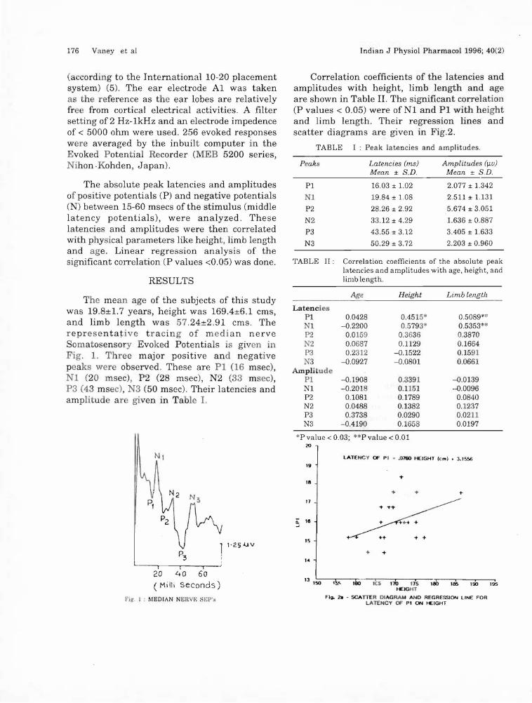
Correlation coefficients of the latencies andamplitudes with height, limb length and ageare shown in Table II. The significant correlation(P values < 0.05) were of Nl and PI with heightand limb length. Their regression lines andscatter diagrams are given in Fig.2.
TABLE I: Peak latencies and amplitudes.
(according to the International 10-20 placementsystem) (5). The ear electrode Al was takena the reference as the ear lobes are relativelyfree from cortical electrical activities. A filtersetting of 2 Hz-1kHz and an electrode impedenceof < 5000 ohm were used. 256 evoked responseswere averaged by the inbuilt computer in theEvoked Potential Recorder (MEB 5200 series,Nihon.Kohden, Japan). Peaks Latencies (ms)
Mean ± S.D.Amplitudes O.w)
Mean ± S.D.
Correlation coefficients of the absolute peaklatencies and amplitudes with age, height, andlimb length.
The absolute peak latencies and amplitudesof positive potentials (P) and negative potentials(N) between 15-60 msecs of the stimulus (middlelatency potentials), were analyzed. Theselatencies and amplitudes were then correlatedwith physical parameters like height, limb lengthand age. Linear regression analysis of thesignificant correlation (P values <0.05) was done.
RESULTS
PI
1
P2
N2
P3
N3
TABLE II:
16.03 ± 1.02
19.84 ± 1.08
28.26 ± 2.92
33.12 ± 4.29
43.55 ± 3.12
50.29 ± 3.72
2.077 ± 1.342
2.511 ± 1.131
5.674 ± 3.051
1.636 ± 0.887
3.405 ± 1.633
2.203 ± 0.960
........
+... -t -t
...
+ -t
'k!' ,110 ,6 nb .is ,ab 185 100 195HEIGHT
F11J. 2a - SCATTER DlAGRA.. AND REGRESSION LINE FORLATENCY OF PI ON HEIGHT
11
0: 111..J
'5
"13 1
150
Age Height Limb length
LatenciesPI 0.0428 0.4515* 0.5089**N1 -0.2200 0.5793* 0.5353*'P2 0.0159 0.3636 0.3870N2 0.0687 0.1129 0.1664P3 0.2312 -0.1522 0.1591N3 -0.0927 -0.0801 0.0661
AmplitudePI -0.1908 0.3391 -0.0139N1 -0.201 0.1151 -0.0096P2 0.1081 0.1789 0.0840N2 0.0488 0.1382 0.1237P3 0.3738 0.0290 0.0211N3 -0.4190 0.1658 0.0197
*Pvalue<0.03; **Pvalue<O.OI20
LATENCY (F PI ••ll7OO HEIGHT lem) • 3.1556
19
...III
1'250 V
f t ,20 40 60
(Milli seconds)
Fig : MEDIAN NERVE Sf;!",
The mean age of the subjects of this studywas 19.8±1.7 years, height was 169.4±6.1 ems,and limb length was 57.24±2.91 ems. Therepresentative tracing of median nerveSomatosensory Evoked Potentials is given inFig. 1. Three major positive and negativepeaks were observed. These are PI (16 msec),Nl (20 msec), P2 (28 msec), N2 (33 msec),P3 (43 msec), N3 (50 msec). Their latencies andamplitude ar given in Ta Ie I.

Indian J Physiol Pharmacal 1996; 40(2) Median Nerve SEPs 177
150 155 160 165 170 175 180 185 190 195HEIGHT
Fig. 2c - SCATTER DIAGRAM AND REGRESSION LINE FORLATENCY Of' Nl ON HEIGHT
LATENCY Of' Nl ••1036 HEIGHT (em) • 2.2902
The data on SEPs vary from lab to labdepending upon the various techniques used.Hence each lab should spell out various technicalfactors for recording normative data on SEPs.In light of this our data can serve as a referencefor normative data on SEPs.
When the median nerve is stimulated, twotypes of fibres are excited, Motor and Ia andtype II sensory fibres. The impulses from typeII afferent fibres ascends through the followingpathway : Median nerve - Brachial plexus Dorsal ganglia of Spinal nerves - DorsalColumns - Cuneate nucleus - Medialleminiscus- Contralateral Thalamus - Thalamo-corticalprojections - Cortex.
The SEPs are divided into short latency« 15 msecs), middle latency (15-60 msecs), andlong latency (> 60 msecs). In our study themiddle latency SEPs were recorded. These SEPsshowed mainly three positive and negativewaves.
DISCUSSION
The N1 peak is the first cortical response tothe external stimulus (1, 4, 12, 14) and is usuallythe largest negative peak of the SomatosensoryEvoked Potentials. The later peaks of this
Pl (16 msec) : A peak similar to our PI hasbeen recorded in many studies at 13 msec (6),15 msec (4, 7) and 16 msec (8, 9). However, thispeak is most widely accepted as P 14 and hasbeen so recorded by many authors (10, 11, 12).
This peak is suggested to be generated inthe dorsal column nuclei or medical leminiscus(7, 11, 13). This is supported by the fact thatpatients with thalamic lesions show the presenceof P
14(12) while it is abolished in patients with
spinal lesions at the cervical level.
Nl (20 msec) - P2 (28 msec) - N2 (33 msec)
complex: This complex is reported as a whole inmany studies because all the components aredue to the activation of the parietalsomatosensory cortex. Latencies of this complexin our study are comparable with others(4, 8, 9, 11, 12).
+*
+
+
+
+ +
++
+
+
+
+
LATENCY OF PI ••17110 LIMB LENGTH (colo) • SJM2i
50 52 ~ ~ ~ 80 ~ ~ MLIMB LENGTH
FIg. 2d - SCATTER DIAGRAM N¥:J REGRESSION LINE FORLATENCY Of' NI ON UPPER LIMB LENGTH
LATENCY Of' Nl ••1_ LIMB LENGTH (em) • 8.4550
+
50 52~ 5& 58 60 6264LIMB LENGTH
Fill. 2b - SCATTER DIAGRAM AND REGRESSION L_" FORLATENCY OF PI ON Ul'f't:R liMn LlNGlI.
2_
23
22
21
20Z-'
19
III
17
16
48
20
19
18
17
Ii: 16...J
15
I~
13~
24
23
22
21
Z 20...J
19
III
17
16

178 Vaney et al
complex are also generated by the activation ofthe somatosensory cortex (4). This complex isfound absent in cases of brain death andthalamic lesions (12).
P3 (44 msec) - N3 (50 msec) complex: Thiscomplex is generally recorded as a P
4o-N
60(1, 9,
12) complex. The P3-N3 complex is a generalizedcomplex, and is due to the activation of nucleiother than the primary sensory nuclei (15).
Comparison of our data with the previousstudies is given in Table III. It shows onlyminor variations most of which can be accountedfor by the differences in our stimulating andrecording techniques, and/or by the variationsin physical parameters of the subjects.
In our study quite an early N3 is reportedcompared to the other studies. This differencecould be due to ethnic variations.
The significant correlations found in ourstudy were between PI and N1 with Heightand Limb length. The significance was morewith limb length than that with the height.
Indian J Physiol Pharmacol 1996; 40(2)
Since limb length and height are significantlycorrelated (r value = 0.6162, P value < .001)the correlation to height may be secondary tothe correlation with limb length. Regressionanalysis showed that Nl is more dependent onthe physical parameters than PI.
Thus our study has provided normativedata in Indian males, and could serve as a basefor future studies. We suggest that the heightand limb length should also be taken intoconsideration while recording SomatosensoryEvoked Potentials, and if need be, correctionfactors can be worked out and applied. Morenumber of subjects have to be used to find outthe exact correction factors.
ACKNOWLEDGEMENTS
We are thankful to the Computer Center,Department of Biostatistics for the invaluablesuggestions and assistance in the statisticalanalysis of the data. The technical assistancerendered by Mr. Manjhi is gratefullyacknowledged.
TABLE III : Comparison of our results with the results of previous studies.
Author YearLatencies (msecs)
PI NI P2 N2 P3 N3
GiblinD.R. 1964 16 19 28 36
Abbruzzese M. 1978 15
YamadaT. 1980 14
KimuraJ. 1982 14 19 27 32
Allison T. 1983 15 20 26
YamadaT. 1984 14 20 26 34 44 64
Kakigi R. 1989 13
GoffP.S. 1990 16 20 25 48 65
Our study 1995 16 20 28 33 44 50

Indian J Physiol Pharmacol 1996; 40(2)
REFERENCES
Median Nerve SEPs 179
1. Aminoff MJ. Electrodiagnosis in clinical Neurology.Churchill Livingstone, 3rd Ed, 1992; 580-581.
2. Dass S. A manual on clinical surgery. S. Dass, 3rd Ed,1988; 134.
3. Kumar A, Tandon OP, Bhattacharya A, Gupta RK,Dhar D. Somatosensory evoked potential changesfollowing electroacu puncture therapy in chronic painpatients. Anaesthesia 1994; 50 : 1-4.
4. Allison T, Wood CC, Goff WR. Brain Stem auditory,Pattern reversal visual, and short-latencysomatosensory evoked potentials : Iatencies inrelation to age, sex, and brain and body size.Electroencephalography and Clinical Neurophysiology1983; 55 : 619-636.
5. Jasper HH. The ten-twenty electrode system ofinternational federation. Electroencephalography andClinical Neurophysiology 1958; 10 : 371-375.
6. Kakigi R. Short Latency Somatosensory EvokedPotentials following median nerve stimulation inDown's syndrome. Electronecephalography and ClinicalNeurophysiology 1989; 74 : 88-94.
7. Abbruzzese M, FavaIe E, Leandri M, Ratto S. Newsub-cortical components of the cerebrall somatosensoryevoked potential in man. Acta Neurolog Scandinau1978; 57 : 325-332.
8. Giblin DR. Somatosensory Evoked Potentials inhealthy subjects and in patients with lesions of thenervous system. Annals of New York Academy ofSciences 1964; 112 : 93-142.
9. Goff PS, Karnaze DS, Fisher N. Assessment of mediannerve Somatosensory Evoked Potentials in cerebralischemia. Stroke 1990; 21 : 1167.
10. Yamada T, Kimura J, Nitz DM. Short LatencySomatosensory Evoked Potentials following MedianNerve stimulation in man. Electroencephalographyand Clinical Neurophysiology 1980; 48 : 367-376.
11. Kimura J, Yamada T. Short latency SEPs followingmedian nerve stimulation. Annals of New YorkAcademy of Sciences 1982; 388 : 689-694
12. Yamada T, Kayamori R, Kimura J, Beck DO.Topography of Somatosensory Evoked Potentialsaft·er stimulation of the median nerve.Electroencephalography and Clinical Neurophysiology1984; 59 : 29-43.
13. Chiapa KH, Ropper AH. Evoked Potentials in clinicalmedicine. The New England Journal of Medicine1982; 306 (20) : 1205-1211.
14. Fross N, Hari R, Salmelin R, Ahonen A, HamalajnenM, Rajola M, Knuutila J, Simola J. Activation ofthe human posterior parietal cortex by mediannerve stimulation. Experimental Brain Research 1994;99 : 309-315.
15. Yamada T. The Anatomic and Physiologic Basis ofMedian nerve Somatosensory Evoked Potentials.Neurologica Clinica 1988; 6 : 705.


![MEDIAN NERVE - gmch.gov.in lectures/Anatomy/UL-median nerve.pdf · MEDIAN NERVE • Formation:from two roots from lateral cord [C(5),6,7]& from medial cord(C8,T1) of brachial plexus](https://static.fdocuments.net/doc/165x107/5ed4b477d718f333af5c5484/median-nerve-gmchgovin-lecturesanatomyul-median-nervepdf-median-nerve-a.jpg)