
Mechanism of the reductive half reaction in cellobiose dehydrogenase
35
1 Mechanism of the reductive half reaction in cellobiose dehydrogenase* B. Martin Hallberg ‡§ , Gunnar Henriksson ¶ , Göran Pettersson | , Andrea Vasella || , and Christina Divne ‡# From the ‡Department of Biotechnology, Albanova University Center SCFAB, KTH, SE-106 91 Stockholm, Sweden; the §Department of Cell and Molecular Biology, Structural Biology, Uppsala University, SE-751 24 Uppsala, Sweden; the ¶Department of Fiber and Polymer Technology, KTH, SE-100 44 Stockholm, Sweden; the | Department of Biochemistry, Uppsala University, SE-751 23 Uppsala, Sweden; and the ||Laboratorium für Organische Chemie, ETH Hönggerberg, CH-8093 Zürich, Switzerland # To whom correspondence should be addressed: Dr. Christina Divne Department of Biotechnology, KTH Albanova University Center SCFAB SE-106 91 Stockholm, Sweden. Phone: +46-8-5537 8296 Fax: +46-8-5537 8468 E-mail: [email protected]. Running Title: Mechanism of Cellobiose Dehydrogenase Copyright 2002 by The American Society for Biochemistry and Molecular Biology, Inc. JBC Papers in Press. Published on December 19, 2002 as Manuscript M210961200 by guest on February 2, 2018 http://www.jbc.org/ Downloaded from
Transcript of Mechanism of the reductive half reaction in cellobiose dehydrogenase

1
Mechanism of the reductive half reaction in cellobiose dehydrogenase*
B. Martin Hallberg‡§
, Gunnar Henriksson¶, Göran Pettersson
|, Andrea Vasella
||, and Christina
Divne‡#
From the ‡Department of Biotechnology, Albanova University Center SCFAB, KTH, SE-106
91 Stockholm, Sweden; the §Department of Cell and Molecular Biology, Structural Biology,
Uppsala University, SE-751 24 Uppsala, Sweden; the ¶Department of Fiber and Polymer
Technology, KTH, SE-100 44 Stockholm, Sweden; the |Department of Biochemistry, Uppsala
University, SE-751 23 Uppsala, Sweden; and the ||Laboratorium für Organische Chemie,
ETH Hönggerberg, CH-8093 Zürich, Switzerland
# To whom correspondence should be addressed:
Dr. Christina Divne
Department of Biotechnology, KTH
Albanova University Center SCFAB
SE-106 91 Stockholm, Sweden.
Phone: +46-8-5537 8296
Fax: +46-8-5537 8468
E-mail: [email protected].
Running Title: Mechanism of Cellobiose Dehydrogenase
Copyright 2002 by The American Society for Biochemistry and Molecular Biology, Inc.
JBC Papers in Press. Published on December 19, 2002 as Manuscript M210961200 by guest on February 2, 2018
http://ww
w.jbc.org/
Dow
nloaded from

2
SUMMARY
The extracellular flavocytochrome cellobiose dehydrogenase (CDH; EC 1.1.99.18)
participates in lignocellulose degradation by white-rot fungi with a proposed role in the early
events of wood degradation. The complete hemoflavoenzyme consists of an catalytically
active dehydrogenase fragment (DHcdh) connected to a b-type cytochrome domain via a linker
peptide. In the reductive half reaction, DHcdh catalyzes the oxidation of cellobiose to yield
cellobiono-1,5-lactone. The active site of DHcdh is structurally similar to that of glucose
oxidase and cholesterol oxidase with a conserved histidine residue positioned at the re face of
the flavin ring close to the N5 atom. The mechanisms of oxidation in glucose oxidase and
cholesterol oxidase are still poorly understood, partly because of lack of experimental
structure data or difficulties in interpreting existing data for enzyme-ligand complexes. Here
we report the crystal structure of the Phanerochaete chrysosporium DHcdh with a bound
inhibitor, cellobiono-1,5-lactam, at 1.8 Å resolution. The distance between the lactam C1 and
the flavin N5 is only 2.9 Å, implying that in an approximately planar transition state, the
maximum distance for the axial 1-hydrogen to travel for covalent addition to N5 is 0.8-0.9 Å.
The lactam O1 interacts intimately with the side chains of His689 and Asn732. Our data lends
substantial structural support to a reaction mechanism where His689 acts as a general base by
abstracting the O1 hydroxyl proton in concert with transfer of the C1 hydrogen as hydride to
the re face of the flavin N5.
by guest on February 2, 2018http://w
ww
.jbc.org/D
ownloaded from
3
INTRODUCTION
Cellobiose dehydrogenases (CDHs; EC 1.1.99.18) are extracellular fungal
flavocytochromes that are believed to participate in lignocellulose degradation by fungi. They
are oxidoreductases carrying protoheme and FAD cofactors bound to separate domains. In
vitro, CDH from the white-rot basidiomycete Phanerochaete chrysosporium depolymerizes
cellulose, hemicelluloses and lignin (1-3, review 4), as well as other polymers (5). The exact
biological function of CDH has been a subject of lively debate, but recent results suggest that
the enzyme is important for invasion and colonization of wood (6).
The catalytic site is located in the flavoprotein domain where the reductive half
reaction proceeds by oxidation of β-cellobiose (apparent kcat 15.7 s-1 and Km 0.11 mM, 7) to
yield cellobiono-1,5-lactone (Fig. 1) and the concomitant two-electron reduction of FAD. In
dilute aqueous solution, cellobionolactone hydrolyzes to cellobionic acid. Results from 1H
NMR spectroscopy have shown that the product from cellobiose oxidation by CDH is
unequivocally cellobionolactone, and thus, cellobionic acid is not formed on the enzyme (8).
During the ensuing oxidative half reaction, the flavin is re-oxidized by an electron acceptor,
either directly or via the cytochrome domain (9). At present, the most favored mechanism for
in vivo degradation of biopolymers by CDH is the reduction of ferric compounds present in
wood in the presence of hydrogen peroxide to form hydroxyl radicals through a Fenton-type
reaction (2, 10). The cytochrome domain has been implicated in this reaction (10) since the
generation of hydroxyl radicals proceeds by one-electron reduction, however, the flavin
by guest on February 2, 2018http://w
ww
.jbc.org/D
ownloaded from

4
domain is also able to generate hydroxyl radicals (1). Besides cellobiose, soluble
cellodextrins, mannobiose and lactose are good, or acceptable substrates for CDH, whereas
monosaccharides are poor substrates (7). CDH can use a large number of electron acceptors,
however, reduction of oxygen is slow (11).
We have recently reported the crystal structure of the dehydrogenase fragment of
CDH without ligand (DHcdh) showing that it is closely related to that of members of the
glucose-methanol-choline (GMC) family of oxidoreductases (12). The GMC oxidoreductases
glucose oxidase (GOx; 13, 14) and cholesterol oxidase (ChOx; 15, 16) are similar in overall
structure and active-site architecture to DHcdh (12). GOx catalyzes the oxidation of β-D-
glucose to D-glucono-1,5-lactone. Thus, GOx and CDH perform similar chemistry in the
reductive half reaction. The bifunctional enzyme ChOx, however, catalyzes the oxidation and
isomerization of cholesterol to 4-cholesten-3-one. Although CDH and GOx catalyze similar
chemical reactions, the structural details of the catalytic site in the immediate vicinity of the
flavin ring in CDH is more similar to those in ChOx: two conserved residues (His and Asn) at
the re face of the flavin ring in a similar conformation, and with near identical geometry
relative to the flavin N5 (12). In GOx, however, the asparagine is replaced by a histidine
residue. Based on modeling of cellobiose in the DHcdh active site, we suggested two glucosyl-
binding sites at the re face of the isoalloxazine ring with the reducing end of cellobiose bound
to the innermost site C close to the flavin ring, and the non-reducing end of cellobiose
residing in the distant site B (12).
Over the years, the most widely accepted reaction mechanisms for flavin-assisted
by guest on February 2, 2018http://w
ww
.jbc.org/D
ownloaded from

5
dehydrogenation include the carbanion mechanism (17, 18), the radical mechanism (19, 20),
and the hydride-transfer mechanism (21-23). In general, crystallographic data are not
sufficient per se to conclusively discriminate between these mechanisms. However, with the
increasing number of available ligand complexes for redox-active enzymes, the hydride-
transfer mechanism is gaining in popularity as a general mechanism for dehydrogenation,
although the radical mechanism has not been disproved. The only structure available for a
GMC enzyme-ligand complex is that of Brevibacterium sterolicum ChOx with bound
dehydroisoandrosterone (16). The authors propose a radical mechanism, although the
structural details of ligand binding did not exclude any of the possible mechanisms. The
situation is, at least partly, complicated by the dual activity of ChOx.
We have used the inhibitor 5-amino-5-deoxy-cellobiono-1,5-lactam (Cblm; Ki ~0.25
mM at 35°C, data not shown), which has a geometry similar to that of the product and of a
possible transition state (TST), to study the active-site interactions and delineate a possible
reaction mechanism for CDH. This is the first crystal structure of a CDH with a ligand bound
in the active site. The reaction mechanism for enzymatic oxidation of cellobiose by CDH is
discussed in the light of the present structure of a complex between the dehydrogenase
fragment of CDH and Cblm determined at 1.8 Å resolution.
by guest on February 2, 2018http://w
ww
.jbc.org/D
ownloaded from
6
EXPERIMENTAL PROCEDURES
Crystallization, Data Collection and Refinement–DHcdh was purified and crystallized
as described elsewhere (7, 12). Data were collected at 100°K from crystals soaked overnight
in reservoir solution containing 1 mM Cblm. Data collection and model refinement statistics
are summarized in Table 1. Data reduction and scaling were carried out using MOSFLM (24)
and SCALA (25), respectively. Our reported structure of DHcdh (PDB ID code 1KDG, 12)
was used as starting model for crystallographic refinement against DHcdh-Cblm amplitudes.
Initial refinement was done with CNS (26), and manual re-building was done with the
program O (27). Starting coordinates for Cblm were generated using CORINA (28), followed
by manual fitting of the model to the electron density. Final refinement was done with
REFMAC5 (29) at 1.8 Å resolution using anisotropic scaling, hydrogens in their riding
positions, and atomic displacement parameter refinement using the “translation, libration,
screw-rotation” (TLS) model. The flavin cofactor, inhibitor, glycosylation adducts, and the
substrate and flavin-binding domains of DHcdh (12) were defined as rigid bodies during TLS
refinement. The model contains two protein molecules (residues 215 to 755); two 6-
hydroxylated FAD molecules; five N-acetylglucosamine residues (three in molecule A and
two in B); two Cblm molecules; and 1007 water molecules (533 for A, 473 for B, and one
located on a non-crystallographic symmetry 2-fold axis). The bending angle of the
isoalloxazine ring was calculated as described previously (12).
by guest on February 2, 2018http://w
ww
.jbc.org/D
ownloaded from
7
Modeling of Cellobiose–Cellobiose was modeled manually in the active site of DHcdh
guided by the observed binding pattern for Cblm. To relieve geometric strain and impose
favorable van der Waals contacts, the model was subjected to energy minimization with CNS
(26) without the X-ray pseudo-energy term. During the energy minimization, only atoms
within a sphere of 5.5 Å from the linking oxygen in the glycosidic bond were allowed to
move. Atoms within a cushion of 3.5 Å around the sphere were refined with harmonic
restraints.
by guest on February 2, 2018http://w
ww
.jbc.org/D
ownloaded from
8
RESULTS
Overall Structure–The 1.5 Å crystal structure of DHcdh (PDB ID code 1KDG) has
been reported elsewhere (12). In brief, DHcdh consists of an FAD-binding subdomain and a
substrate-binding subdomain (Fig. 2a). The entrance to the active site is located at the
subdomain interface where a 12 Å long tunnel leads down to the flavin ring. The structures of
DHcdh and DHcdh-Cblm are nearly identical with rms deviation values (all atoms) of 0.35 and
0.34 Å for pair-wise least-squares comparisons of A and B molecules, respectively. The
electron density for the protein, as well as for the inhibitor, is of excellent quality (Fig. 2b).
The average residual B factor for the ligand (all atoms) is 8.2 and 8.4 Å2 for the A and B
molecule, respectively.
The Flavin Cofactor–As observed for DHcdh, the non-covalently bound flavin cofactor
in DHcdh-Cblm is present as 6-hydroxylated FAD. The butterfly-bending angle of the flavin
ring is less pronounced in DHcdh-Cblm (mol A, 7°, mol B, 11°) compared with DHcdh (22°).
The flattening of the flavin ring appears to result from an induced fit where the N5 moves into
the isoalloxazine plane to accommodate the incoming C1 atom of the lactam ring. The angle
defined by the flavin N5 and N10 with the backbone-nitrogen atom of Gly310 is 136°, and the
N5-Gly310 N distance is 3.2 Å.
Binding of Cellobionolactam–Inhibitor interactions are outlined in Fig. 3a, 3b. The
by guest on February 2, 2018http://w
ww
.jbc.org/D
ownloaded from
9
glucosyl-binding sites and the substrate-binding residues are located at the re face of the
isoalloxazine ring. The lactam moiety of Cblm, corresponding to the reducing end of
cellobiose, is bound in site C with four protein residues and three solvent molecules within
hydrogen-bonding distance of its exocyclic carbonyl and hydroxyl groups: O1-His689 Nε2,
O1-Asn732 Nδ2, O2-Ser687 O, O2-His689 Nε2, and O3-Asn688 Nδ2. The C3 and C6 hydroxyl
groups can form one and two water-mediated hydrogen (H) bonds, respectively. Thus, a total
of eight H-bonds are possible in site C. The endocyclic lactam nitrogen is positioned near the
flavin N5 (3.2 Å) and O4 (2.9 Å). The C1 atom of the lactam moiety, which corresponds to
the site of oxidative attack in cellobiose, binds in a position 2.9 Å in front of and below the
N5-C4a locus of the isoalloxazine ring defining an angle of 108° (mol A) and 110° (mol B)
with the N5-N10 atoms of the flavin ring. These values are in agreement with those typically
observed in flavoenzymes (30). It should be noted that the lactam C1 and O1 are almost
perfectly aligned with the flavin N5 and C4a, respectively (C1-N5, 2.9 Å; O1-C4a, 2.9 Å).
The short distance (mol A, 2.5 Å; mol B, 2.6 Å) of the H-bond formed between the lactam O1
and His689 Nε2 suggests that this interaction is strong, and that His689 is suitably positioned
to deprotonate the substrate hydroxyl group.
The glucosyl moiety of the ligand resides in site B where it forms a total of five
ligand-protein H-bonds: O2-Glu279 Oε2, O2-Arg586 Nε, O3-Glu279 Oε1, O3-Arg586 Nη2,
and O6-Asn688 Nδ2. The aromatic ring of Phe282 has rotated (mol A, 7°; mol B, 11°)
compared with the DHcdh structure to stack more planar with the B-site pyranose ring. Five
solvent-mediated H-bonds are observed, one each for O2, O3 and O6, and two for O4. A total
by guest on February 2, 2018http://w
ww
.jbc.org/D
ownloaded from

10
of ten H-bonds and one planar hydrophobic stacking interaction contribute to binding in site
B. Judged solely from the number of possible interactions, site B is likely to make substantial
contribution to the binding of cellobiose which is in line with reported kinetic constants for
di- and monosaccharides (7).
Coupled tyrosine-serine flipping–Only two residues undergo conformational change
in response to inhibitor binding (Fig. 3b). In the non-liganded DHcdh structure, Tyr609 is kept
out of the C-site by H-bonds formed by its hydroxyl group with backbone atoms of Asn732
and the Gln582 side chain. The active-site tunnel is filled with solvent molecules, of which
one water molecule binds in front of the flavin ring within H-bonding distance from His689
Nε2.
In the DHcdh-Cblm complex however, the tyrosine side chain has discarded its H-
bonding partners and moved into site C (Fig. 3b). In the new position, Tyr609 resides below
the lactam ring where Oη coordinates two ordered water molecules not present in the non-
liganded structure. One water is located below the lactam O6 where it is involved in a H-bond
network with Tyr609 Oη, Thr581 Oγ, Asn732 Nδ2 and the C6 hydroxyl group. The second
water molecule is below the lactam C3 hydroxyl group and forms H-bonds to Tyr609 Oη, the
lactam O3 and another water molecule. The tyrosine and its two coordinated water molecules
(colored yellow in Fig. 3b, 3c) elevates the “floor” of site C.
The flipping of the tyrosine side chain imposes two additional changes in the active
site: i) to accommodate the Tyr609 side chain in its new position, the side chain of Ser519
by guest on February 2, 2018http://w
ww
.jbc.org/D
ownloaded from

11
also flips and thereby exchanges its two H-bonding partners (Ser519 Oγ-Ser687 Oγ, and
Ser519-water) for two new ones (Ser519 Oγ-Met685 O, and Ser519 Oγ-new water). The
concomitant flips of Tyr609 and Ser519 give rise to a local backbone-Cα displacement of 0.6
Å and 0.8 Å at residue 609 and 519, respectively; ii) the re-positioning of the Tyr609 side
chain, together with the presence of the ligand, effectively traps a water molecule (Wat1366)
in a position 3 Å from the Cε1-Nδ1 edge of the His689 imidazole ring (Fig. 3a-3c), i.e., the
imidazole side opposite to that interacting with the C1 hydroxyl in cellobiose. This water
molecule is not present in the non-liganded structure.
by guest on February 2, 2018http://w
ww
.jbc.org/D
ownloaded from
12
DISCUSSION
Comparison of Cellobionolactam with Modeled Cellobiose–Structurally,
cellobionolactam resembles the product, cellobionolactone, with the only difference that the
endocyclic O5 oxygen is replaced by an N-H function (Fig. 1). In both compounds C1 is
coplanar with O1, C5 and O5 (or lactam nitrogen). We thus assume that the tetrahedral
configuration of the anomeric center of cellobiose is changing towards a planarized
intermediate during its transition to a sp2-hybridized carbon in cellobionolactone.
Based on the Cblm binding pattern, we have produced a docking model of cellobiose
in DHcdh. Minor steric restraints near the flavin N5 and His689 Nε2 enable the planar lactam
C1=O to penetrate somewhat farther (0.3-0.4 Å) into site C compared with the equatorial O1
group of cellobiose (Fig. 3c). The C1-N5 distance is 2.9 Å in the cellobiose model, and the
shortest distance that the C1 hydrogen (1-H) would need to traverse to bind covalently to N5
is thus roughly 0.9-1.0 Å. In the Cblm complex, the corresponding transfer distance
(imagining a 1-H as in the TST) would be slightly less, 0.8-0.9 Å. Although these distances
do not differ significantly, the precise geometry of the 1-H relative to N5 is slightly different
in cellobiose, as compared with a TST, in that 1-H in a TST would be closer to being aligned
so as to interact with the lowest unoccupied molecular orbital (LUMO) of the flavin (Fig. 3c).
The assumption that the observed inhibitor binding is also valid for a TST implies that upon
approaching the TST the substrate slides slightly deeper into the active site. This results in a
better alignment of the 1-H with the flavin LUMO. Similar to what has been suggested for
by guest on February 2, 2018http://w
ww
.jbc.org/D
ownloaded from
13
several other flavoproteins (30), the resulting negatively charged flavin hydroquinone may be
stabilized at the N1-C2=O locus of the isoalloxazine ring by the positive dipole of the C-
terminal α helix. These small but distinct differences in binding may be interpreted as the
oxidative site favoring a partially planar TST mimicked by the lactam ring in our structure.
Mechanistic Implications–Similar specific relative geometry of the substrate and
cofactor reactive groups has been observed in crystal structures of ligand complexes for
nicotinamide-dependent (31), flavin-dependent (32) and quinone-dependent (33)
oxidoreductases, all of which have been assigned a hydride-transfer mechanism. From a
purely structural viewpoint, the close proximity between C1 and N5 together with the relative
geometry of the atoms appears to favor a general-base catalyzed hydride-transfer mechanism
(Scheme 1, panel a). General base-assisted deprotonation of the C1 hydroxyl group by His689
in concert with the expulsion of 1-H as hydride via a planar or nearly planar TST would be
entirely consistent with the experimentally observed binding of Cblm and modeled cellobiose.
The carbanion mechanism in its classical implementation requires that His689
abstracts 1-H as a proton, resulting in a substrate carbanion. The carbanion then performs a
nucleophilic attack at N5 to form a covalent C1-N5 adduct. The subsequent elimination
reaction proceeds by the concomitant formation of a double bound between C1-O1 and
uptake of the O1 hydroxyl proton by the flavin O4, and ultimately by N5. In order to
accommodate the carbanion reaction, the C-site glucosyl residue would need to tilt forward
towards His689 by at least 45-90° to position the 1-H for proton abstraction. Such a
by guest on February 2, 2018http://w
ww
.jbc.org/D
ownloaded from

14
conformational change in the spatially restricted active site is unlikely, structurally and
energetically. The non-reducing end of the substrate is anchored in site B (Fig. 3b), and the
conformational change would introduce unreasonable strain in the glycosidic bond between
sites B and C. The intimate interaction between His689 and O1 makes it difficult to find any
reasonable incentive for this residue to abstract the more distant 1-H. The carbanion
mechanism is also highly unlikely from a purely chemical point of view in that the generated
substrate carbanion would be conjugatively destabilized, as has been well established by work
of Eliel (34). Thus, we suggest that the carbanion mechanism is incompatible with cellobiose
oxidation by CDH.
Apart from a hydride-transfer mechanism, a radical mechanism is compatible with the
present structure (Scheme 1, panel b). In this reaction, one electron may be transferred from
the substrate O1 to the flavin C4a or N5 concomitantly with the abstraction of the O1-H as a
proton by His689, resulting in a flavin radical and a substrate radical. The subsequent step
involves a transfer of the 1-H as a hydrogen radical to N5. Although the structure is
compatible with the radical mechanism, the two radical species implied have not been
demonstrated. The failure to detect the radical species spectroscopically does not, however,
provide conclusive evidence against the radical mechanism. The formation of a cellobiosyl
radical may be slow, and its subsequent decomposition rapid, making detection of an ESR
signal difficult. We may conclude that a physically meaningful difference between the
hydride and the electron-transfer mechanisms hinges upon the temporal sequence, or
concertedness of events.
by guest on February 2, 2018http://w
ww
.jbc.org/D
ownloaded from

15
Studies on Structurally Unrelated Enzymes with Similar Substrate Specificity–The
structure of soluble glucose dehydrogenase (s-GDH) from Acinetobacter calcoaceticus in
complex with its substrate, β-D-glucose, has been reported by Oubrie et al. (33). This enzyme
catalyzes the oxidation of β-D-glucose to gluconolactone, but uses pyrroloquinoline quinone
(PQQ) as cofactor. Similar to what is discussed here for CDH, a hydride-transfer mechanism
was assigned to s-GDH (33) based on the specific orientation of 1-H relative to the C5 in
PQQ (corresponding to the flavin N5 in CDH). In s-GDH, the distance between the substrate
C1 and PQQ C5 is 3.2 Å, implicating a transfer distance of 1.2 Å for 1-H. The overall
structure of CDH and s-GDH (PDB ID code 1CQ1) and their active sites display no obvious
similarity, but nevertheless, interesting details emerge when superimposing the active sites. A
superposition with reference to C1 in the two enzyme complexes aligns the C5 of the PQQ
cofactor in s-GDH within 0.7 Å of the flavin N5 in CDH. The Nε2 atoms of the proposed
active base in s-GDH (His144) and CDH (His689) are only 0.4 Å apart; and the distance
between Asn732 Nδ2 in CDH and Arg228 Nη2 in s-GDH is 1.7 Å. Thus, the precise geometry
of the tetrad defined by the hydrogen acceptor of the cofactor, the C1, the proton acceptor of
the general base catalyst, and the assisting residue coincide remarkably well, despite different
structure and cofactor dependence.
Studies on Structurally Related GMC Oxidoreductases–Although no results are
available from site-directed mutagenesis studies on CDH, the residues proposed to participate
by guest on February 2, 2018http://w
ww
.jbc.org/D
ownloaded from

16
in catalysis (12) have been mutated in the related enzymes GOx and ChOx. In B. sterolicum
ChOx (35), Streptomyces ChOx (36) and Penicillium amagasakiense GOx (37), replacement
of the proposed catalytic base (His689 in CDH) resulted in enzyme variants with drastically
reduced, or abolished catalytic performance as measured by kcat whereas Km values were
practically unaffected, thus supporting the assignment of His689 in CDH as a general base
catalyst. On the other hand, mutation of the Asn732 counterpart in Streptomyces ChOx
(N480A, N480Q) and P. amagasakiense GOx (H563A, H563V) resulted in inactive enzymes.
In the light of the present structure and the mutant data for GOx and ChOx we propose a dual
role for Asn732 where it i) helps to position the substrate with respect to the flavin, and ii) by
offering a H-bond to O1 also facilitates proton abstraction by His689.
The only crystal structure available for a GMC oxidoreductase-ligand complex is that
of B. sterolicum ChOx with bound dehydroisoandrosterone (16). In ChOx, His447 (His689 in
CDH) has been proposed to activate a water molecule (Wat541) for nucleophilic attack on the
substrate. This water molecule occupies the position of the substrate C1-O1 group in CDH.
Thus, CDH and ChOx share the same reaction geometry, although the reaction in ChOx is
suggested to be relayed through a water molecule. For GOx, no experimentally determined
complex with substrate or substrate analogue is available, but β-D-glucose has been modeled
in the active site (14, 37), resulting in a position of the substrate relatively similar to that of
the C-site glucosyl moiety of Cblm in CDH: the substrate and the catalytic residues are
positioned at the re face of the flavin ring, and the C1 hydroxyl group is equidistantly
positioned between His689 and Asn732 (His520 and His563 P. amagasakiense GOx).
by guest on February 2, 2018http://w
ww
.jbc.org/D
ownloaded from

17
Molecular-dynamics calculations of a glucose-GOx complex with a water-mediated
interaction between the substrate O1 and the active histidine similar to that observed for
ChOx resulted in expulsion of the water molecule, suggesting that a water-relayed mechanism
is unlikely in GOx (14). Thus, direct interaction between the proposed catalytic base and the
substrate is in agreement with our observed mode of Cblm binding to CDH.
Induced Fit and Water Trapping–Before another reductive half reaction can occur, a
total of two electrons acquired by the flavin needs to be transferred to an electron acceptor
during the ensuing oxidative half reaction, and the hydrogens transferred to N5 and His689
Nε2 have to be suitably disposed of. For the N5 hydrogen, the most probable destination of a
proton is to bulk water concomitantly with two single-electron, or a two-electron transfer
upon flavin re-oxidation (depending on the electron acceptor used). In the case of the proton
withdrawn from the substrate 1-hydroxyl group by His689 Nε2, the structure provides some
hypothetical, but interesting scenarios.
As described above, the conformational change in Tyr609, assisted by a concomitant
flip of the Ser519 side chain, orchestrates the formation of a highly ordered network of H-
bonds below the ligand in site C. This imposes an effective restriction of this site to perfectly
accommodate the lactam ring and thereby induce an optimal fit of the TST-like ligand to the
protein. The movement of the Tyr609 side chain into site C appears to occur in response to
Cblm binding, and results in the entrapment of a water molecule (Wat1366) close to His689
Cε1 (Fig. 3a-3c). In the non-liganded structure, the position of Wat1366 is occupied by the
by guest on February 2, 2018http://w
ww
.jbc.org/D
ownloaded from
18
CZ-OH group of Tyr609. The water 1366 is particularly interesting in that it is the only water
molecule within H-bonding distance to the imidazole group of the proposed catalytic base in
the ligand structure, and it may thus serve as a secondary proton acceptor. The substrate
proton acquired by His689 can be transferred to Wat1366 by a 180° rotation about χ2 of the
imidazole. In DHcdh-Cblm, Wat1366 interacts at the center of the aromatic ring of Tyr609
which is likely to increase its affinity for the extra proton at His689. A H3O+-π interaction
(38) with the tyrosyl ring may thus promote proton transfer to Wat1366 and stabilize the
resulting oxonium ion. The next step may be: i) that the oxonium ion triggers the Tyr609 side
chain to swing out from site C and resume its original position and thereby displacing the
product from the active site; or ii) that product departure itself triggers the tyrosine side chain
to leave the active site and release the oxonium ion to exchange a proton with bulk water; or
iii) that protonation of the histidine and the subsequent flip of the imidazole ring forces the
product to leave due to an emerging unfavorable contact between the product carbonyl
oxygen and the ε-1 carbon of His689 as a consequence of Cε1 assuming the position of Nε2.
Nevertheless, the tyrosine flip clearly helps to induce an optimal fit of the catalytic site
for the inhibitor, and it generates a cavity for a solvent molecule that may accept a proton
from the catalytic base. Wat1366 makes no direct contact with the site of oxidative attack, and
hence, the C1-N5 pathway of a presumed hydride ion is completely shielded from water. It
should be stressed that the position of Wat1366 in DHcdh-Cblm is completely different from
that of the water molecule bound in front of His689 in the non-liganded structure. This
stresses the inherent difficulty in assigning catalytic roles to active-site water molecules in the
by guest on February 2, 2018http://w
ww
.jbc.org/D
ownloaded from
19
absence of ligand, or when non-authentic binding of ligand occurs. Thus, the water molecule
implicated in catalysis by ChOx (16) should be carefully evaluated, although the dual
function of ChOx may actually justify the presence of a catalytic water positioned between
the substrate and the catalytic histidine.
CONCLUSIONS
The crystal structure of the CDH flavoprotein with bound inhibitor provides support
for a hydride-transfer mechanism for dehydrogenation with His689 acting as a general base
catalyst, deprotonating the equatorial 1-hydroxyl group. The axial 1-H is aligned for a
concerted hydride transfer from C1 to N5, via a transition state characterized by partial
planarization of C1. The binding of the inhibitor is in agreement with hydrogen transfer at the
re side of the flavin ring. We also suggest that Asn732 is bifunctional in that it both
participates in the formation of a productive enzyme-substrate complex, and that it supports
deprotonation by His689 by serving as a H-bond donor to the 1-hydroxyl group. From a
structural and biochemical viewpoint, the reaction mechanism is simple and requires only
minor structural changes in the substrate and protein in order for the 1-H to be expelled. The
1-hydrogen would then need to traverse a distance of less than 1 Å for covalent attachment to
the flavin N5 atom. Although the perfect setup of the active site for hydride transfer is
evident, we cannot rule out the possibility of a radical mechanism using structural data alone.
The results provide a structural platform for the use of conventional biochemical and
by guest on February 2, 2018http://w
ww
.jbc.org/D
ownloaded from

20
biophysical techniques as well as quantum mechanical and classical molecular mechanics
approaches to further investigate the molecular mechanism of cellobiose oxidation by CDH.
Acknowledgments–We thank the beamline staff at ESRF ID14-EH4 (Grenoble,
France) for assistance during data collection.
by guest on February 2, 2018http://w
ww
.jbc.org/D
ownloaded from
21
REFERENCES
1 Henriksson, G., Ander, P., Pettersson, B., and Pettersson, G. (1995) Appl. Microbiol.
Biotechnol. 42, 790-796
2 Mansfield, S. D., de Jong, E., and Saddler, J. N. (1997) Appl. Environ. Microbiol. 63,
3804-3809
3 Vallim, M. A., Janse, B. J. H., Gaskell, J., Pizzirani-Kleiner, A. A., and Cullen, D.
(1998) Appl. Environ. Microbiol. 64, 1924-1928
4 Henriksson, G., Johansson, G., and Pettersson, G. (2000) J. Biotechnol. 78, 93-113
5 Cameron, M. D., and Aust, S. D. (1999) Arch. Biochem. Biophys. 367, 115-121
6 Dumonceaux, T., Bartholomew, K., Valeanu, L., Charles, T., and Archibald, F. (2001)
Enz. Microbial Technol. 29, 478-489
7 Henriksson, G., Sild, V., Szabo, I. J., Pettersson, G., and Johansson, G. (1998) Biochim.
Biophys. Acta 1383, 48-54
8 Higham, C. W., Gordon-Smith, D., Dempsey, C. E., and Wood, P. M. (1994) FEBS
Lett. 351, 128-132
9 Cohen, J. D., Bao, W., Renganathan, V., Subramaniam, S. S., and Loehr, T. M. (1997)
Arch. Biochem. Biophys. 341, 321-328
10 Kremer, S. M., and Wood, P. M. (1992) Eur. J. Biochem. 208, 807-814
11 Bao, W., Usha, S. N., and Renganathan, V. (1993) Arch. Biochem. Biophys. 300, 705-
713
by guest on February 2, 2018http://w
ww
.jbc.org/D
ownloaded from
22
12 Hallberg, B. M., Henriksson, G., Pettersson, G., and Divne, C. (2002) J. Mol. Biol. 315,
421-434
13 Hecht, H. J., Kalisz, H. M., Hendle, J,. Schmid, R. D., and Schomburg, D. (1993) J.
Mol. Biol. 229, 153-172
14 Wohlfahrt, G., Witt, S., Hendle, J. Schomburg, D., Kalisz, H. M., and Hecht, H.-J.
(1999) Acta Crystallogr. Sect. D Biol. Crysallogr. 55, 969-977
15 Vrielink, A., Lloyd, L. F., and Blow, D. M. (1991) J. Mol. Biol. 219, 533-554
16 Li, J., Vrielink, A., Brick, P., and Blow, D. M. (1993) Biochemistry 32, 11507-11515
17 Walsh, C. T., Schonbrunn, A., and Abeles, R. H. (1971) J. Biol. Chem. 248, 6855-6866
18 Porter, D. J. T., Voet, J. G., and Bright, H. J. (1973) J. Biol. Chem. 248, 4400-4416
19 Silverman, R. B., Hoffman, S. J., and Catus, W. B., III. (1980) J. Am. Chem. Soc. 102,
7126-7128
20 Sherry, B., and Abeles, R. H. (1985) Biochemistry 24, 2594-2605
21 Neims, A. H., Deluca, D. C., and Hellerman, L. (1966) Biochemistry 5, 203-213
22 Hersch, L. B., and Schuman-Jorns, M. (1975) J. Biol. Chem. 250, 8728-8734
23 Ghisla, S., and Massey, V. (1989) Eur. J. Biochem. 181, 1-17
24 Powell, H. R. (1999). Acta Crystallogr. Sect. D Biol. Crysallogr. 55, 1690-1695
25 Evans, P. R. (1993) in Proceedings of CCP4 Study Weekend on Data Collection and
Processing, eds. Sawyer, L., Isaacs, N. and Bailey, S. (SERC Daresbury Lab.,
Warrington, U.K.), pp. 114-122
26 Brünger, A. T., Adams. P. D., Clore, G. M., DeLano, W. L., Gros, P., Grosse-
by guest on February 2, 2018http://w
ww
.jbc.org/D
ownloaded from

23
Kunstleve, R. W., Jiang, J. S., Kuszewski, J., Nilges, M., Pannu, N. S., et al. (1998)
Acta Crystallogr. Sect. D Biol. Crysallogr. 54, 905-921
27 Jones, T. A., Zou, J.-Y., Cowan, S. W., and Kjeldgaard, M. (1991) Acta Crystallogr.
Sect. A Foundations Crystallogr. 47, 110-119
28 Gasteiger, J., Rudolph, C., and Sadowski, J. (1990) Tetrahedron Comp. Method. 3, 537-
547
29 Murshudov, G. N., Vagin, A. A., and Dodson E. J. (1997) Acta Crystallogr. Sect. D
Biol. Crysallogr. 53, 240-255
30 Fraaije, M. W., and Mattevi, A. (2000) Trends Biochem. Sci. 25, 126-132
31 Mattevi, A., Vanoni, M. A., Todone, F., Rizzi, M., Teplyakov, A., Coda, A., Bolognesi,
M., and Curti, B. (1996) Proc. Natl. Acad. Sci. USA 93, 7496-7501
32 Karplus, P. A., and Schulz, G. E. (1989) J. Mol. Biol. 210, 163-180
33 Oubrie, A., Rozeboom, H. J., Kalk, K. H., Olsthoorn, A. J. J., Duine, J. A., and Dijkstra,
B. W. (1999) EMBO J. 18, 5187-5194
34 Abatjoglou, A. G., Eliel, E. L., and Kuyper, L. F. (1977) J. Am. Chem. Soc. 99, 8262-
8269
35 Kass, I. J., and Sampson, N. S. (1998) Biochemistry 37, 17990-18000
36 Yamashita, M., Toyama, M., Ono, H., Fujii, I., Hirayama, N., and Murooka, Y. (1998)
Protein Eng. 11, 1075-1081
37 Witt, S., Wohlfahrt, G., Schomburg, D., Hecht, H.-J., and Kalisz, H. M. (2000)
Biochem. J. 347, 553-559
by guest on February 2, 2018http://w
ww
.jbc.org/D
ownloaded from

24
38 Montes-Morán, M. A., Menéndez, J. A., Fuente, E., and Suárez, D. (1998) J. Phys.
Chem. B 102, 5595-5601
39 Kleywegt, G. J., and Jones, T. A. (1996) Structure 4, 1395-1400
40 DeLano, W. L. (2002) The PyMOL User's Manual. DeLano Scientific, San Carlos, CA,
USA.
41 Wallace, A. C., Laskowski, R. A., and Thornton, J. M. (1995) Prot. Eng. 8, 127-134
42 Guex, N., and Peitsch, M. C. (1997) Electrophoresis 18, 2714-2723
by guest on February 2, 2018http://w
ww
.jbc.org/D
ownloaded from

25
FOOTNOTES
* This work was funded by grants from the Swedish Research Council for Environment,
Agricultural Sciences and Spatial Planning, and the Swedish Research Council (to CD).
1 The abbreviations used: Cblm, 5-amino-5-deoxy-cellobiono-1,5-lactam; CDH, cellobiose
dehydrogenase; ChOx, cholesterol oxidase; DH, dehydrogenase fragment; DHcdh, CDH
flavoprotein without ligand; GMC, glucose-methanol-choline; GOx, glucose oxidase; s-GDH,
soluble glucose dehydrogenase; TST, transition state.
by guest on February 2, 2018http://w
ww
.jbc.org/D
ownloaded from
26
LEGENDS TO FIGURES
Figure 1. Molecules discussed in the text. (a) Cellobiose, (b) cellobionolactone, and (c)
cellobionolactam.
Figure 2. Structure of the DHcdh with bound cellobionolactam.
(a) Overall structure of the DHcdh fragment with bound cellobionolactam in the active site.
The polypeptide chain has been color ramped from the N terminus (blue) to the C terminus
(red). α helices and β strands are shown as spirals and arrows, respectively. The FAD
cofactor and the ligand are shown as ball-and-stick representations. Atom colors: nitrogen,
blue; oxygen, red; carbon, yellow (FAD) or green (Cblm). The picture was made with the
program PyMOL, official URL, http://www.pymol.org/ (40). (b) σA-weighted Fo-Fc electron-
density map calculated to 2.0 Å resolution using the model from the first simulated-annealing
refinement with CNS where only protein atoms had been included and refined. The electron
density for the ligand is therefore free from model bias.
Figure 3. Ligand interactions of the DHcdh active site.
(a) Schematic representation showing the active-site interactions. Atom colors: carbon,
black; nitrogen, blue; oxygen, red. Covalent bonds are colored yellow (protein) or green
(ligand), and H-bonds are drawn as green, dashed lines. The H-bond indicated between
Wat1366 (colored violet) and His689 Cε1 is hypothetical and requires a rotation of 180° about
by guest on February 2, 2018http://w
ww
.jbc.org/D
ownloaded from

27
χ2 in the histidyl side chain. The water molecules that form the floor below the lactam moiety
are drawn as yellow spheres. For clarity, H-bonds between ligand and water molecules other
than those shown have been omitted, as well as intramolecular H-bonds in the
cellobionolactam molecule. The C1 atom in the ligand is labelled. The hydrophobic stacking
interaction with Phe282 is depicted with a red crest. The drawing was made with the program
LIGPLOT (41). (b) Ligand interactions and the ligand-induced changes in the protein. The
non-reducing end of the inhibitor is bound to site B (to the left), and the lactam ring in site C
(right side). Interatomic distances that satisfy those of H-bonds (< 3.2 Å) are depicted as
dashed lines. For comparison, the DHcdh structure without inhibitor (green) has been
superimposed with DHcdh-Cblm showing the ligand-induced changes in the protein discussed
in the text. The water molecules coordinated by Tyr609 and the H-bonds formed with the
tyrosine are colored yellow. The water molecule (Wat1366) close to His689 Cε1 is shown in
violet color. A dashed line (violet) has been drawn to highlight the possible formation of a H-
bond between Wat1366 and His689 Nε2 given a rotation of 180° about χ2. (c) Superposition
of cellobionolactam (yellow) with modeled cellobiose (green). The 1-H and O1-H atoms
(grey) are shown for the modeled cellobiose. For comparison, the position of 1-H in a TST
has been drawn as a light-blue sphere connected to the lactam C1 by a dashed line. The water
molecule close to His689 Nε2 is shown as in (b). The two tyrosyl-coordinated water molecules
that form a floor below the lactam moiety of the ligand are drawn as yellow spheres. For
clarity, only selected interactions are shown in (a)-(c). The drawings in (b) and (c) were made
with the program Swiss-Pdb Viewer (42) and rendered with POV-Ray™ (official URL,
by guest on February 2, 2018http://w
ww
.jbc.org/D
ownloaded from

28
http://www.povray.org/).
by guest on February 2, 2018http://w
ww
.jbc.org/D
ownloaded from
29
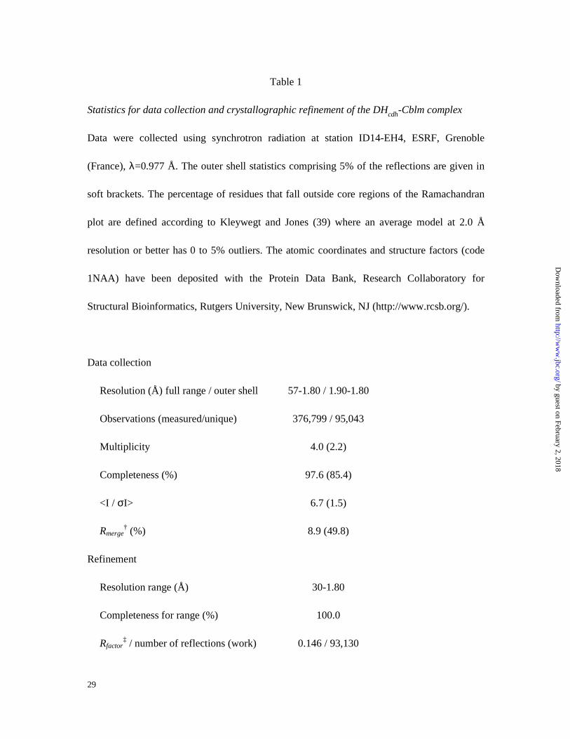
Table 1
Statistics for data collection and crystallographic refinement of the DHcdh-Cblm complex
Data were collected using synchrotron radiation at station ID14-EH4, ESRF, Grenoble
(France), λ=0.977 Å. The outer shell statistics comprising 5% of the reflections are given in
soft brackets. The percentage of residues that fall outside core regions of the Ramachandran
plot are defined according to Kleywegt and Jones (39) where an average model at 2.0 Å
resolution or better has 0 to 5% outliers. The atomic coordinates and structure factors (code
1NAA) have been deposited with the Protein Data Bank, Research Collaboratory for
Structural Bioinformatics, Rutgers University, New Brunswick, NJ (http://www.rcsb.org/).
Data collection
Resolution (Å) full range / outer shell 57-1.80 / 1.90-1.80
Observations (measured/unique) 376,799 / 95,043
Multiplicity 4.0 (2.2)
Completeness (%) 97.6 (85.4)
<I / σI> 6.7 (1.5)
Rmerge† (%) 8.9 (49.8)
Refinement
Resolution range (Å) 30-1.80
Completeness for range (%) 100.0
Rfactor‡ / number of reflections (work) 0.146 / 93,130
by guest on February 2, 2018http://w
ww
.jbc.org/D
ownloaded from

30
Rfree / number of reflections (free) 0.185 / 1,894
Number of non-hydrogen atoms 9,365
Mean B values (Å2) protein all atoms (A/B) 8.8 / 8.9
NCS rmsd (Å) Cα / all protein atoms 0.20 / 0.42
NCS rms ∆B (Å2) Cα / all protein atoms 0.7 / 1.8
Rmsd bond lengths (Å) / angles (°) 0.022 / 1.92
Ramachandran plot outliers (%) 1.8
† Rmerge = [ Σhkl Σi |I–<I>| /Σhkl Σi |I| ] x 100 %.
‡ Rfactor = Σhkl | |Fo|–|Fc| | / Σhkl |Fo|
by guest on February 2, 2018http://w
ww
.jbc.org/D
ownloaded from
DivneB. Martin Hallberg, Gunnar Henriksson, Göran Pettersson, Andrea Vasella and Christina
Mechanism of the reductive half reaction in cellobiose dehydrogenase
published online December 19, 2002J. Biol. Chem.
10.1074/jbc.M210961200Access the most updated version of this article at doi:
Alerts:
When a correction for this article is posted•
When this article is cited•
to choose from all of JBC's e-mail alertsClick here
by guest on February 2, 2018http://w
ww
.jbc.org/D
ownloaded from