Mechanical complications of MI
46
Mechanical complications of MI Smitha Anilkumar Echo Lab HH, HMC
Transcript of Mechanical complications of MI
Mechanical complications of MI
Smitha Anilkumar
Echo Lab
HH, HMC

No disclosures

Electrical Complications
Mechanical Complications
Heart failure
Pericarditis
Post infarction angina or Reinfarction
The major mechanical complications after acute myocardial
infarction (AMI) include;
- Rupture of the left ventricular free wall,
- Acute severe MR
- papillary muscle displacement
- papillary muscle rupture
- Ventricular septal rupture.
- Pseudo aneurysm formation
- RV infarction
- Dynamic LV outflow obstruction
- LV thrombus
Primary percutaneous coronary intervention has
significantly reduced major mechanical complications
since its introduction as a treatment strategy in AMI.
Echocardiography with color-flow Doppler is the
investigation of choice in the diagnosis and differentiation
of the conditions.

Free wall rupture
Most common least recognized complication:
< 1% to 6.2% patients with acute MI
Accounts for 14 to 26% of Infarct related mortality
7% in hospital deaths
Time Course :
First 5 days post MI in 50%
90% occur within 2 weeks

Mid ventricle and lateral wall are most common sites
May affect any wall
Can involve RV
Atria may be affected rarely
Adjacent to junction of normal with infarcted tissue
Presentation
Acute free wall rupture
Rapid PEA /death
Sub acute free wall rupture 30%
Slow ooze with warning signs and symptoms

Clinical signs :
Pericarditis , emesis and agitation
Recurrent chest pain
Transient hypotension and bradycardia
Deviation from expected T-wave evolution

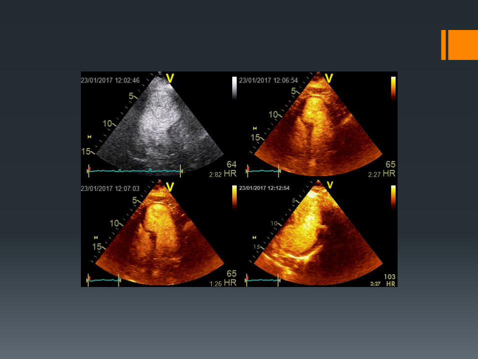
Echocardiographic features include;
Pericardial effusion in end –diastole > 5mm
High density intrapericardial echoes (thrombus)
RV/RA compression (Tamponade)
Direct 2D identification of tear is unusual
- Contrast may be helpful


Suspected LVFWR
Echo confirmation
Hypotension
No EMD
IV fluids, Inotropes, IABP,
Pericardiocentesis(?)
Emergency Surgery

LV pseudo aneurysm
Incomplete rupture
Sealed by pericardium and hematoma
Lacks elements of myocardial wall
Echo- lucent space external to LV
Narrow neck
Ratio of the diameter of the entry point to the
maximal diameter is <40 -50%
May contain thrombus
Characteristic Doppler Profile
Bidirectional ( “to- and –fro”) flow pattern

Narrow base
Walls composed of
pericardium and
thrombus
High risk of rupture
Wide base
Walls composed of
myocardium
Low risk of rupture
AneurysmsPseudo-aneurysms


Aneurysms Pseudo-aneurysms
Location 3% posterior Posterior or inferior
Echocardiography
Anatomy Thinned myocardium Ruptures
Contractility Non contractile Dyskinesia
Consequences/Complica
tions
Congestive heart failure
Embolic events
Ventricular arrhythmias
Congestive heart failure
Embolic events
Ventricular arrhythmias
Therapy Medical or Surgical
therapy
Surgery
Surgical risk Dubious Lower than medical
therapy

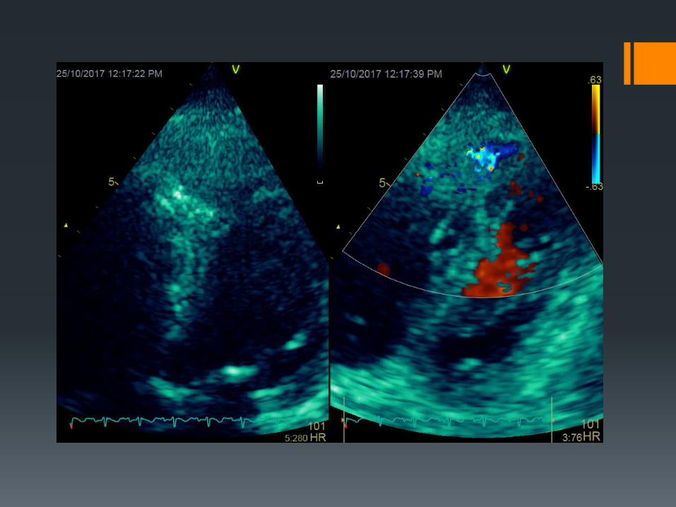
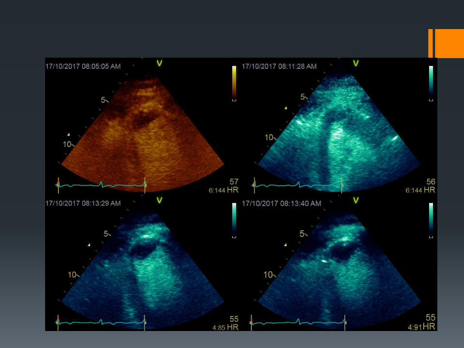
Ventricular septal rupture
Occurs in 0.2 to 1% patients with MI
- Bimodal distribution – 24 hrs. and days 3-5
Any portions of the septum may be involved margin between
necrotic and non necrotic myocardium
Anterior VSR s tend to located distally with defects that perforate
the septum at the same level - “simple”
Inferior VSRs located more toward the base and follows
a serpingenious course - “ complex”

Presentation
Holosystolic murmur (often loud)
Thrill
Heart failure
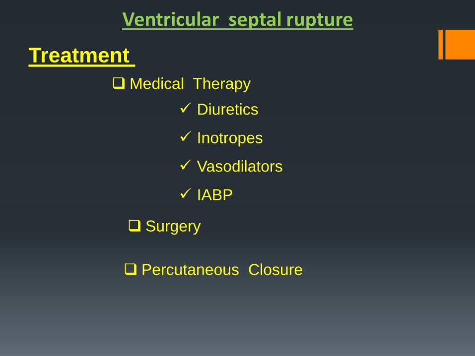
Treatment
Medical Therapy
Diuretics
Inotropes
Vasodilators
IABP
Surgery
Percutaneous Closure

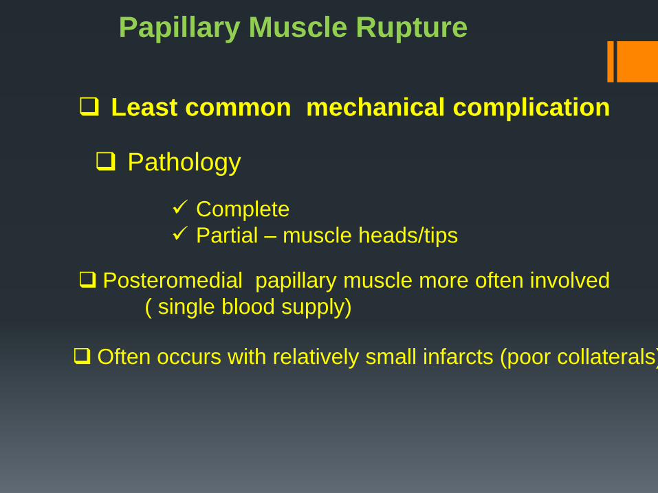
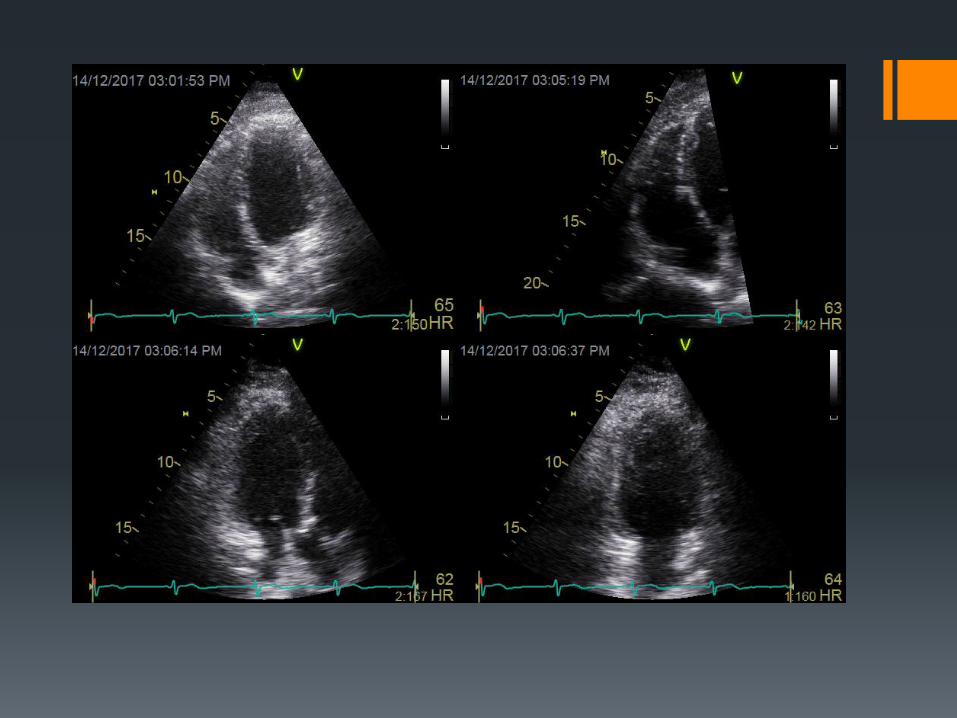
Papillary Muscle Rupture
Least common mechanical complication
Pathology
Complete
Partial – muscle heads/tips
Posteromedial papillary muscle more often involved
( single blood supply)
Often occurs with relatively small infarcts (poor collaterals)

Presentation
1 to 7 days after MI
Heart failure
Shock
MR murmur
- may be soft/indistinct
- often no thrill present

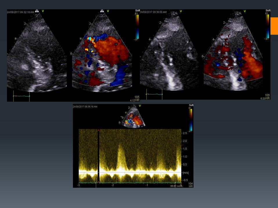
Echocardiographic features
Flail mitral leaflet with systolic cusp prolapse to LA
Mobile echogenic mass attached to the chordae tendinae and to
the mitral valve
No prolapse of papillary head to LA is observed in 35%
Abnormal cutoff of one papillary muscle
Severe MR
- Color –flow disturbance area can be small
- Cut-off sign in CW spectral profile
Hyper dynamic LV function

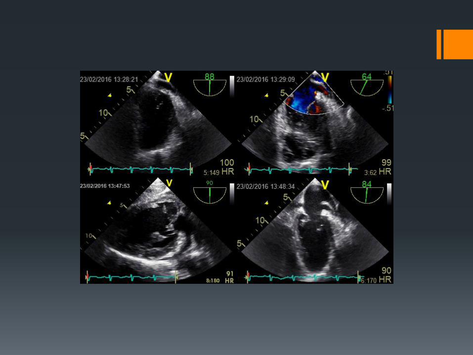
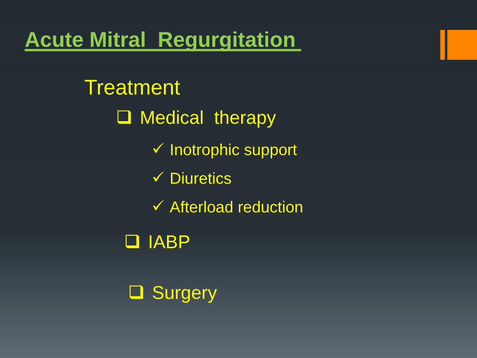
Acute Mitral Regurgitation
Treatment
Medical therapy
Inotrophic support
Diuretics
Afterload reduction
IABP
Surgery

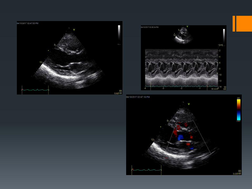
Right ventricular MI Common association with inferior MI
RV apical segments may be involved in LAD occlusions
ST segment elevation in V1and or V4R- V6R
Mortality high vs inferior MI
Clinical presentation
Hypotension (preload sensitive)
Clear lung fields
Increased JVP (Kussumaul sign may be present)
Lack of pulmonary congestion
Hypoxemia
Right to left shunting via PFO

Echocardiographic features
Focal RV wall motion abnormalities – McConnell's sign
Paradoxical septal motion due to acute volume overload
Dilation of RV (RA)
Small LV
Bowing of interatrial septum from right to left
RV thrombus
Tricuspid regurgitation
TAPSE and RVS’ reduced
IVC plethora
Right to left shunting via PFO


Dynamic LV outflow obstruction
In setting of apical infarction sparing the base
Basal hyper kinesis
Systolic anterior motion of mitral leaflet
Dynamic LVOT obstruction
Hypotension
Systolic murmur
Exacerbation by inotropic agents and IABP


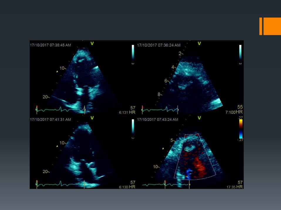
Left ventricular thrombus
Anterior wall infarcts
20-40% (60% in large anterior-wall AMIs, not treated
with anticoagulant therapy)
High risk of systemic embolization (Anticoagulant
therapy ↓ rate of embolic events by 33%
anticoagulation)
Most common presentation is Stroke (within the first
10 days after AMI)

Transthoracic echocardiography is modality
of choice (92% sensitivity, 88% specificity)
Management - heparin treatment
followed by oral warfarin therapy for 3-6
months, lifelong anticoagulation if a clot
persists.
Left ventricular thrombus
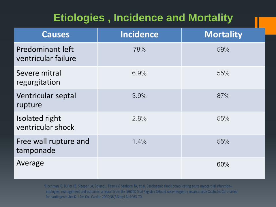
Etiologies , Incidence and Mortality

Summary for LAD Infarct
Left anterior descending artery
40% of LV myocardium His-Purkinje System
Cardiogenic shock due to loss
of large amount of myocardium
Intraventricular septum
(upper two – thirds)
Antero-apical wall
Acute VSD
Apical LV aneurysm
Ventricular
arrhythmias
Apical thrombus
formation
Arterial embolism originating in
the LV
Advanced Heart Block
(LBBB, 3rd degree A-V
block
And Mobitz II 2nd degree)

Summary for RCA (or Circumflex) Infarct Right coronary artery
RV Infarct
Hypotension due to
Decreased LV filling SA –nodal infarct
A-V nodal infarct
Brady arrhythmias
1st degree A-V block
Mobitz 2nd degree
block
A-V dissociation
Posteromedial
Papillary muscle
infarct
Acute MR
(with or without papillary
muscle rupture )