Mechanical Behavior of Individual Type I Collagen Fibrils
95
MECHANICAL BEHAVIOR OF INDIVIDUAL TYPE I COLLAGEN FIBRILS BY JULIA HONG LIU THESIS Submitted in partial fulfillment of the requirements for the degree of Master of Science in Aerospace Engineering in the Graduate College of the University of Illinois at Urbana-Champaign, 2016 Urbana, Illinois Adviser: Professor Ioannis Chasiotis
Transcript of Mechanical Behavior of Individual Type I Collagen Fibrils

MECHANICAL BEHAVIOR OF INDIVIDUAL TYPE I COLLAGEN FIBRILS
BY
JULIA HONG LIU
THESIS
Submitted in partial fulfillment of the requirements
for the degree of Master of Science in Aerospace Engineering
in the Graduate College of the
University of Illinois at Urbana-Champaign, 2016
Urbana, Illinois
Adviser:
Professor Ioannis Chasiotis

ii
ABSTRACT
Despite the plethora of studies on the mechanical response of collagen, especially
at the molecular scale or at much larger length scales, such as of those of fibers, tendons,
and fascicles, there is still limited information about the mechanics of collagen fibrils (50
- 500 nm diameter) that serve as mesoscale building blocks in tissues. In this dissertation
research the mechanical behavior of dry individual reconstituted collagen fibrils with
different diameters were investigated via monotonic tests at 0.004 s-1, cyclic loading and
cyclic loading/recovery experiments, and strain rate tests spanning six decades of strain
rates in the range 10-3 – 102 s-1. Experiments were conducted with reconstituted type I
calfskin collagen fibrils which were isolated from buffer and after drying were adhesively
attached to a microelectromechanical systems (MEMS) device. Experiments were carried
out using high-resolution optical microscopy under dry conditions at 20-30% relative
humidity (RH) and laboratory temperature.
Eight fibrils with diameters of 165±77 nm tested under monotonic loading yielded
an average tensile strength of 752±186 MPa, ultimate stretch ratio of 1.3±0.06, and initial
stiffness of the stress (σ) vs. stretch ratio (λ) curves, E1 = 5.7±2.3 GPa. These results
depended on fibril diameter: fibrils with larger diameters showed increased maximum
stretch ratio, λmax, and decreased E1 and decreased stiffness, E2, of the softening regime in
the σ - λ curves. Normalizing the applied stress with E1, removed the diameter size effect
and provided great consistency in the softening regime of different σ - λ curves. The same
process was applied to fibrils tested at nominal strain rates of 10-2 - 102 s-1 showing good
agreement between σ/E1 - λ curves obtained at the same strain rate from different fibrils,
but also showed a clear increase in E2 with the applied strain rate without a reduction in λ
at failure, which implies a gradual linearization of σ-λ curves at higher rates.
The mechanical behavior under cyclic loading was studied via experiments in
each of the three regimes, with target λmax ~ 1.05 in regime I, λmax ~ 1.25 in regime II, and
λmax ~ 1.3 in regime III. In regime I, E1 was unaffected by cycling loading or recovery.
The residual strain increased in every cycle, but ~80% of λmax was recovered after resting
for 1 hr at zero stress. Regime II was characterized by constant E1, after an initial drop

iii
between cycles 1 and 2, a slightly increasing value of E2 in every cycle, and increasing
residual strain with cycling. Cycling in regime III also resulted in constant E1 and E2 after
an initial reduction between cycles 1 and 2 and increased residual strain with cycle order.
The experimental results point out to a process of damage accumulation during
cycling, as manifested by the very consistent hysteresis loops and the gradually
accumulated residual strain, which however, does not affect the mechanical stiffness of
regimes I and II. The latter points out to a cross-link network within the collagen fibril
that maintains molecular connectivity, as well as material regions that allow for viscous
sliding (supported by the increase in E2 and E2/E1 with applied strain rate) in the
softening regime of the σ - λ curves without disrupting the cross-link network. The rapid
recovery and restoration of the three-regime shape of the σ - λ curves of collagen fibrils
also supports the existence of sacrificial bonds which reform upon recovery that is driven
by residual stresses in the fiber.

iv
ACKNOWLEDGMENTS
Completing a thesis requires a great deal of support and guidance, and I would
like to thank here the people who guided, mentored, and assisted me throughout my
research here at the University of Illinois.
First and foremost, I would like to thank my primary research advisor Professor
Ioannis Chasiotis. After I was accepted into the University of Illinois he offered me the
opportunity to work on a project that straddled both biology and nanomechanics. His
expertise in nanomechanics has helped me understand my research and develop methods
for performing my experiments, and his understanding and support allowed me to create
a solid base on which to pursue my dreams. He guided me not only as his student, but
also as a person. I cannot express how grateful I am for his mentorship.
I would like to acknowledge the funding support by the National Institutes of
Health (NIH) under award numbers 1U01EB016422-01A1, 5U01EB016422-02 and
5U01EB016422-03 Revised. I would also like to thank Professor Guy Genin from
Washington University in St. Louis and Professor Stavros Thomopoulos from Columbia
University in New York City. Their advice was indispensable and their knowledge of
biological materials guided me in the directions necessary to move forward with my
research. I would also like to thank Professor Thomopoulos’s student, Dr. Annie
Schwartz, for reconstituting the collagen used in these experiments and for teaching me
how to handle the fibrils.
I want to show my gratitude to my colleagues in the Nanomechanics and
Materials Research Lab, who taught me everything I know about using the lab and
pushed me to perform my best. Mr. Jan Clawson was my first mentor and taught me the
basics of experimental nanomechanics. With his assistance, my transition from student to
researcher was seamless. Dr. Pavan Kolluru has also been an incredible mentor. I am
grateful to have been under his tutelage, as his knowledge and guidance have made me
strive for perfection. I would also like to thank Mr. Debashish Das for helping me run
experiments and performing data analysis when I was unable. His support and friendship
has been instrumental in completing this thesis and in my development as a researcher.

v
Mr. Fan Yang and Mr. Korhan Sahin worked with me on this project, and I would like to
thank them for being by my side and helping me these last few years. I am also grateful
to Mr. Dimitrios Antartis and Mr. Ryan Mott for their friendship and assistance.
An incredible portion of my time was spent in Frederick Seitz Materials Research
Laboratory Central Facilities at UIUC, and I would like to thank the staff there for
training and assisting me. I would like to thank Ms. Honghui Zhou for training me on the
Hitachi S4700 and S4800 SEMs, as well as for ensuring that these machines were always
operational. Thank you to Mr. Matt Bresin for training me on the FEI DB235 FIB and for
being quick to respond to my questions and issues at all hours of the day. I am grateful to
the rest of the staff at MRL for assisting me with non-technical questions and issues.
The Aerospace Department also deserves a large thanks. Mr. Greg Milner and the
others at the Aerospace Department’s machine shop were extremely helpful. I brought
them several drawings and they always provided me with perfect materials and parts. I
am grateful for their hard work, friendship, and patience. I would also like to thank Ms
Staci McDannel and Mrs. Kendra Lindsey for keeping me informed about the state of my
degree and addressing any administrative issues for me. Finally, I would like to thank my
family and friends. Thank you for always encouraging and supporting me. I would not be
here without you.

vi
TABLE OF CONTENTS
INTRODUCTION............................................................................................................. 1
1.1. Structure of Collagenous Tissues ........................................................................ 2
Effect of Mineralization on Mechanical Behavior of Collagen Fibrils .............. 6
Effect of Hydration on Mechanical Behavior of Collagen Fibrils ...................... 9
Formation and Mechanical Effects of Chemical Cross-Links in Collagen ...... 11
1.2. Mechanical Properties of Collagen Molecules and Fibers .............................. 14
Mechanical Stiffness and Strength.................................................................... 14
Viscoelastic and Strain Rate Dependent Behavior of Collagen Fibrils ............ 18
1.3. Computational Modeling of Mechanical Behavior of Collagen Fibrils ......... 21
1.4. Objectives of this Dissertation Research........................................................... 24
MATERIALS AND EXPERIMENTAL METHODS ……………………………….26
2.1. Materials .............................................................................................................. 27
Synthesis of Reconstituted Collagen ................................................................ 27
MEMS Devices for Mechanical Testing of Individual Collagen Fibrils .......... 29
2.2. Experimental Methods ....................................................................................... 31
Isolation and Mounting of Individual Collagen Fibrils .................................... 31
Mechanical Experiments with Individual Collagen Fibrils .............................. 34
Mechanical Hysteresis and Recovery of Collagen Fibrils ................................ 35
MECHANICAL BEHAVIOR OF DRY COLLAGEN FIBRILS …………………..38
3.1. Tensile Testing of Individual Collagen Fibrils ................................................. 38
3.2. Strain Rate Dependence of Mechanical Behavior of Collagen Fibrils ........... 45
3.3. Deformation and Failure of Individual Collagen Fibrils ................................ 50
3.4. Conclusions .......................................................................................................... 56
CYCLIC LOADING AND RECOVERY OF COLLAGEN FIBRILS ……………..57
4.1. Cyclic Behavior and Recovery of Collagen Fibrils in Regime I ..................... 57
4.2. Cyclic Behavior and Recovery of Collagen Fibrils in Regime II .................... 63
4.3. Cyclic Behavior and Recovery of Collagen Fibrils in Regime III .................. 68
4.4. Comparison of Cyclic Mechanical Behavior in Regimes I, II, and III .......... 73
4.5. Conclusions .......................................................................................................... 78
CONCLUSIONS ............................................................................................................. 79
REFERENCES ................................................................................................................ 82

1
CHAPTER 1
INTRODUCTION
Collagen is the most abundant protein in the animal kingdom providing
mechanical stability, elasticity, and strength to biological tissues. The protein itself is
extremely diverse, spanning over 16 different types and is capable of forming both fibrils
and two-dimensional networks [1-3]. However, despite this diversity, 80-90% of collagen
in nature is either type I, II, or III [1,2,4]. Each collagen type has a specific function or set
of functions and interacts extensively with other connective tissues [3]. Type I collagen
forms skin, tendon, organs, bone, and interstitial tissues, while type II is comprised of
50% all cartilage protein. Type II collagen is also essential in order to properly form
cartilage, bones, teeth, and the vitreous humor of the eye [2]. The diversity, complexity,
and interactions of collagen synthesis result in a mutation-sensitive system [3]. A single
mutation in the amino acid sequence of the procollagen1 molecule may be lethal. Over 70
different mutations in type I procollagen genes alone have been found in probands2 with
osteogenesis imperfecta 3 . Other diseases produced by collagen mutations include
osteoporosis, Alport’s syndrome, Marfan syndrome, Ehlers-Danlos syndrome, Stickler
syndrome, and osteoarthritis [3,5,6]. At a larger scale, changes in the structure of collagen
fibrils and tissues are associated with the ageing process and tissue failure. Intermolecular
cross-linking is the primary cause of stiffening of collagenous tissues, while increased
mineralization results in increased brittleness [7,8].
Because of its highly hierarchical structure, mutations at the molecular level
heavily influence the collagen structure and properties at higher length scales. For
1 Procollagen: Precursor of the collagen molecule that is cleaved to form collagen molecules.
2 Probands: Individual affected with a disorder, and is the first subject in a genetic study.
3 Osteogenesis imperfecta: Congenital bone disorder usually characterized by defective connective tissue.

2
example, increased mineralization at the fibrillar level, a symptom of osteogenesis
imperfecta, is a direct result of a mutation in its molecular structure [7]. In order to fully
understand the mechanical properties of biological tissues, it is necessary to analyze the
behavior of collagen at all levels. Even at its fibrillar level, the mechanical behavior of
collagen has shown tremendous diversity. In the following Sections of this Chapter,
several parameters that effect the behavior of a collagen fibril will be discussed,
including: hydration, mineralization, and cross-link density. Within the body of a live
animal, these parameters are adjusted to fit the fibril’s function, resulting in an extremely
complex weave with other connective tissues.
1.1. Structure of Collagenous Tissues
Although dozens of collagen types have been identified to date, all of them have
the same structural elements: Specific amino acids in an Xaa-Yaa-Gly repeat sequence
(amino acid - amino acid - Glycine) form a polypeptide strand, three of these strands are
bonded to form a collagen molecule, which is stacked into fibrils of 50-500 nm diameter
and then packed into collagen fibers. These fibers are then combined with bioapatite
mineral and other materials such as elastin to form biological tissues [9]. An overview of
this structural hierarchy is depicted in Figure 1.1 [10].
The structure of a collagen molecule, called tropocollagen in animals, is
comprised of triplets: three left-hand coiled polypropylene strands are offset by one
amino acid residue4 and twisted into a single right-handed triple-helix. Because of the
tight packing of the polypeptide strands in the triple-helix, every third amino acid residue
in the sequence must be glycine. The other two amino acids may vary, but the most
common repeat is Proline-Hydroxyproline-Glycine (~10% of all Xaa-Yaa-Gly repeats).
This Xaa-Yaa-Gly repeat is consistent in all collagen, although it may be disrupted
occasionally in non-fibrillar types. However, regardless of type and source, the collagen
4 Amino acid residue: An amino acid that has lost a water molecule by bonding with another amino acid.

3
molecule always has a diameter of ~1.5 nm and a length of 300 nm. This consistency
assists with the molecule’s organized packing structure into fibrils [9,11].
Figure 1.1. Hierarchy of collagen: Amino acids are the building blocks of the collagen
molecule. The molecules are stacked into fibrils, which are then packed into fibers.
Tissues, such as tendon and bone, are at the top of the hierarchy consisting of collagen
fibers, bioapatite mineral, and other connective materials such as elastin [10]. Adopted
with permissions from Nano Letters.
Collagen fibrillogenesis 5 begins only after tropocollagen is cleaved out of
procollagen. When collagen is first constructed, lengthy propeptide6 chains are attached
5 Fibrillogenesis: Formation of a fibril.
6 Propeptide: Protein precursor.

4
at either end of the collagen triple helix. The propeptides on the C-terminal7 of the
monomer are necessary for proper triple helix formation, but prevent fibrillogenesis. N-
and C-proteinases cleave the propetides at each triple helix terminus to generate
tropocollagen molecules, which then self-assemble into fibrils [9]. Fibrils of type I
collagen have a characteristic 67 nm repeat period, called the D-period. Under various
forms of microscopy, this repeat period has been imaged as bands with a dark 27 nm gap
regions alternating with light 40 nm overlap regions [4,9,11]. The monomers are packed
in a quasi-hexagonal unit cell and form supertwisted, right-handed microfibrils of 4 nm
diameter [9,12]. Neighboring telopeptides8 within the tropocollagen monomer interact
with each other and are covalently cross-linked by lysyl oxidase. These cross-links occur
both within an individual microfibril and between microfibrils. Some studies have
suggested that collagen fibrils found in cartilage have a 10+4 heterotypic microfibril
structure, namely a collagen fibril has a core of 4 microfibrils surrounded by a skin of 10
microfibrils [9,10]. However, this microfibrillar structure is not apparent in all fibril
types. In the cases where the monomers directly form a fibril, the packing structure varies
with collagen type [9,11]. The formation of type I collagen is shown in Figure 1.2 [9].
Tropocollagen molecules are held together by two types of cross-links:
enzymatic9 and nonenzymatic. Enzymatic cross-linking in fibrillar collagen is based on
aldehyde formation to form a single telopeptide lysine or hydroxylysine residue. Lysyl
oxidase deaminates the residues only when bound to a specific Hyl-Gly-His-Arg amino
acid sequence. This prevents premature deamination of individual collagen molecules.
Because of the packing structure, collagen monomers may have up to three enzymatic
cross-links with nearby monomers. Nonenzymatic cross-linking, or glycation, increases
with age and is a nonenzymatic reaction with glucose. Glucose reacts with a lysine
peptide in the collagen monomer, and continues to react until it ultimately forms
intermolecular cross-links. Although several other proteins experience glycation, collagen
7 C-terminal: End of an amino acid chain terminated by a free carboxyl group.
8 Telopeptides: Peptides at the ends of an amino acid sequence that are proteolytically removed.
9 Enzyme: Biological molecule that acts as a catalyst.

5
has a considerable biological half-life so this cross-linking mechanism has an important
role in ageing [8].
Figure 1.2. Molecular structure of collagen: the overlap and gap regions in fibrils are a
result of the quarter-length stagger of molecules. In several forms of microscopy, these
regions are shown as 27 nm dark regions alternating with 40 nm light regions [9]. Used
with permission from Annual Review of Biochemistry.
Upon forming fibers and tissues, collagen fibrils align not only longitudinally, but
also transversely and horizontally. They cross each other, forming spirals, cross-plies,
and spiral type plaits [11]. Fibrils form fibers with diameters of 3-7 µm, and fibers
combine into fascicles, which have diameters in the range 150-300 µm. At the next level
of hierarchy, fascicles form tendons, which are millimeters in diameter [4,12]. Collagen
fibrils are not spaced regularly along a fiber. Their distribution in fibers or fibril bundles
is rather a function of the mechanical characteristics of the tissue. These larger structures
of collagen often provide the necessary interface for binding with more elastic
biopolymers, such as elastin and fibrillin that complement the stiffer base of collagen
structure and define the overall properties of the tissue [11].

6
Effect of Mineralization on Mechanical Behavior of Collagen Fibrils
Mineral formation in the skeleton and dental tissues of nearly all vertebrae and
otoliths10 uses collagen as the matrix macromolecule. Type I collagen is the dominant
matrix protein in bone, dental dentin and cementum, but several other types of collagen
comprise different calcified tissues such as cartilage. The active sites of apatite nucleation
in the interior of a collagen fiber are believed to be microchannels with ~2 nm diameter
produced by the arrangement of microfibrils [13]. These channels, which appear in both
the overlap and hole zones, permit the influx of Ca2+ and PO43+ ions and the nucleation of
calcium-phosphate crystallites without significantly deforming the microfibrillar
arrangement. Mineralization primarily occurs on the fiber surface, with the
crystallographic c-axis being parallel to the length of the fibrous matrix macromolecules
and growth being equal in both directions along the length of the mineral strands [13].
The amount of mineral within the collagen matrix increases with age. An analysis
performed on equine metacarpal tissue showed little variation in mineral content between
gender, but a consistent increase with age across all specimens [14].
A particularly sensitive collagenous structure is the tendon-to-bone attachment, in
which collagen fibrils have a gradient with 0% mineralization near the tendon and 50%
mineralization near the bone. The natural attachment rarely fails, but surgical
replacements do not sufficiently mimic this gradient and may have re-tear rates of ~94%
[15]. Mathematical models have been created to characterize the effect of the gradient of
mineral volume fraction on the effective stiffness tensor, and one of these models [12]
predicts an increase in the elastic modulus by a factor of 100 upon fiber mineralization.
The authors in [12] compared the tensile and shear moduli of mineralized collagen fibers
to unmineralized collagen fibers and extended their analysis to the mechanics of collagen
tissue. The general trends of their results were consistent with several other models,
including those by Halpin-Tsai [16], Gao [17], and analytical models such as that in [18]
in which the moduli was shown to increase nonlinearly with the mineral volume fraction.
However, none of these models completely aligned with the experimental results [18].
10 Otoliths: Calcium carbonate structures in the inner ear of vertebrates used to sense gravity and
movement.

7
Figure 1.3. Longitudinal section of a mineralized collagen fibril. Arrows mark the
apatitic strands composed of nanometer-sized particles (1) inside, and (2) on the surface
of the fibril [13]. Adopted with permissions from the International Review of Cytology.
According to the limited experiments presented in [18], there was no substantial
increase in the effective modulus of mineralized collagen when the mineral volume
fraction increased from 0% to 70% [18]. However, the experimental data were limited to
only those two conditions. To our knowledge, most, if not all, of the experimental reports
on mineralized collagen have been limited to a few extreme conditions without
intermediate mineral densities. It is interesting to note, however, that the stress vs. strain
curves demonstrated dramatic changes between different levels of mineralization. This
change in behavior is depicted in Figure 1.4 for experiments performed on deer beams
[19]. As the mineral volume increased the fibrils (approximately 90 nm in diameter)
exhibited more brittle behavior. Molecular dynamics (MD) simulations did not predict
the behavior in Figure 1.4(a) because the models assumed uncrimped, uniaxially aligned

8
tropocollagen networks. In reality, crimping is removed from the tropocollagen
molecules with increasing strain, which results in increased tensile modulus. Instead, MD
simulations have predicted a mechanical behavior that is similar to that in Figure 1.4(b).
This leads to the belief that the deformation in region II in Figure 1.4(b) is due to
intermolecular slip and failure at the mineral-tropocollagen macromolecule interface [19].
Figure 1.4. Stress vs. strain curves of mineralized dear beam fibrils demonstrating two
distinct behaviors: (a) Enhanced elastic modulus in region II is due to mineral that
increases the stress transfer between tropocollagen molecules, (b) sliding between
tropocollagen molecules due to low mineral density [19]. Used with permission from
Interface.
In another study [20], in situ tensile testing of fibrolamellar bone was combined
with simultaneous small-angle X-ray scattering (SAXS) and wide-angle X-ray diffraction
(WAXD) to measure the tissue, fibrillar, and mineral strain during tensile loading. Before
yielding, the elastic response resulted in nearly constant fibril-to-tissue and mineral-to-
tissue strain ratios, and the mineral strain had a linear correlation with fibril strain. This
was true for both dry and wet samples [20]. The dry samples experienced higher mineral-
to-fibril and mineral-to-tissue strains, and larger elastic moduli than the wet samples in
ratios of approximately 5:3, 6:4, and 7:6 respectively. The mechanism behind this
behavior is explained in Section 1.1.2. At the tissue, fibrillar, and mineral particle levels

9
of hierarchy, the strains decreased in a ratio of approximately 12:5:2. This is consistent
with measurements of fibrillar strain in partially mineralized tendon and parallel fiber
bone and is the result of hierarchical gradation, where stiffer elements at smaller length
scales assume less strain than those at larger scales. The 5:2 ratio of fibrillar-to-mineral
strain found in the experiments in [20] supports the hypothesis that load transfer between
mineral platelets occurs by shear transfer in the collagen matrix, which is facilitated by
the high aspect ratio and large mismatch in mechanical properties between the mineral
particles and the collagen [20].
Effect of Hydration on Mechanical Behavior of Collagen Fibrils
The function of water as stabilizing agent in collagen has been studied extensively
vis-à-vis the influence of hydration on collagen structure and its properties [21,22].
Although the potential of water bridges with stabilizing properties has been demonstrated
for both native collagen and collagen-like peptides, the necessity of these water bridges in
keeping the collagen structure has been questioned [22]. Assuming dehydration does not
reach a threshold value, the flexibility of hydrated collagen could be recovered from
dehydrated collagen by rewetting it [23].
The primary hydration model for collagen has been proposed by Ramachandran
and Chandrasekharan [24], which is based on x-ray analysis and proposes that one water
molecule forms a hydrogen–bonded bridge between two-peptide carbonyl oxygens on
two different protein chains in the collagen triple helix. Another water molecule forms a
hydrogen-bonded bridge between a peptide N-H and a carbonyl oxygen on separate
chains. This model allows for one water molecule every three amino acid residues, and is
consistent with results obtained by magnetic resonance and dielectric measurements [21].
Magnetic resonance and dielectric measurements have also been used to determine the
hydration properties of partially dried collagen. In this case, two of the three amino acids
are firmly bound to water molecules which are hydrogen bonded to the macromolecular
backbone. The remaining water in collagen interacts weakly with a number of sites and

10
forms a multilayer with more liquid-like properties. These “free” water molecules are in
rapid exchange with each other with rotational correlation times under 10-10 s [21].
In order to investigate the aforementioned model, a simulation was carried out
using the GROMOS package [21] on a collagen-like peptide molecule surrounded by five
water shells. The results were compared to collagen crystals in vacuo. The effect of
hydration state on the overall structure of collagen is shown in Figure 1.5. The results of
the simulation in vacuo show large deviations from the reference crystal structure. In
comparison to experimental data, dehydration caused more rigidity and increased the
thermal stability of collagen fibers. As such, further work was done to model and analyze
protein mobility via MD simulations [22] that agreed with experimental data and showed
that, in the absence of solvent, the molecules indeed become stiffer. The MD simulation
also made clear that dry collagen had more hydrogen bonds than hydrated or crystal
collagen, which contributes to its increased stiffness [22].
Figure 1.5. MD simulation of collagen molecules in crystal form, in aqueous solution,
and in vacuo [22]. Adopted with permissions from the Journal of Molecular Graphics
and Modelling.

11
Again, the existence of water bridges that was suggested by Ramachandran and
Chandrasekran [24] and later confirmed by NMR [21] was verified by the MD
simulations. Furthermore, more recent X-ray diffraction and NMR studies [25] have
shown that water bridges are not only inter-chain, but also intra-chain, and may be
connected to the rest of the water network. Interestingly, the molecular conformation of
collagen remains quite stable, even at the extreme that the number of water bridges is
small. This suggests that hydration of a collagen molecule as a whole may play a role in
the stabilization process [22].
Formation and Mechanical Effects of Chemical Cross-Links in Collagen
Cross-linking can be distinguished into two major types: non-enzymatic
(glycation) and enzymatic. Non-enzymatic cross-linking occurs via a non-enzymatic
reaction with glucose and decreases the critical flexibility and permeability of tissues.
During glycation, the aldehyde of the open chain form of glucose reacts with a free ε-
amino group of a peptide bound lysine. After spontaneous Amadori rearrangement11 [8]
and further reactions with other amino acid residues, advanced glycation end-products, or
AGEs, form. Long-term glycation of fibrous collagen occurs during ageing, and the
collagen becomes less soluble, more resistant to enzymes, and less flexible in vivo.
Eventually, this process of cross-linking may become intermolecular within collagen
fibers. This is consistent with decreasing elasticity of retinal capillaries, called renal
glomeruli, and arterial vessel walls, which is characteristic of ageing [8].
Enzymatic cross-linking, as its name implies, is a cross-linking mechanism
dependent on the enzyme lysyl oxidase [8]. In fibrillar collagen, enzymatic cross-linking
is based on aldehyde formation from a single telopeptide lysine or hydroxylysine residue,
which is oxidatively deaminated by lysyl oxidase [8]. After this initial enzymatic step all
following reactions are spontaneous. The number of these cross-links, called immature
cross-links12 , declines with age. Immature reducible 13cross-links become irreducible
11 Amadori rearrangement: A type of acid or base catalyzed rearrangement reaction. 12 Immature cross-links: Cross-links formed spontaneously after aldehyde formation using lysyl oxidase.

12
mature cross-links, and become tri-functional14 and more stable [8]. Immature cross-links
are initially divalent, meaning that they link two different collagen molecules. Once they
become mature cross-links, they become trivalent, namely link three collagen molecules
together. Because of the staggered collagen structure, there are sets of lone residues on
either side of the collagen molecule. In this case, divalent and trivalent cross-links
covalently bond either to one or two adjacent molecules, respectively, as the larger
distance between collagen molecules makes the formation of cross-links difficult [26].
The transformation of divalent to trivalent, or immature to mature, cross-linking explains
the increase in both insolubility and strength of older collagenous tissues [8].
A direct comparison of the effect of different types of cross-linking on the
mechanical behavior of collagen is shown in Figure 1.6 where collagen from several
sources was used to obtain the various cross-links. Specifically, the collagen sources were
human patellar tendon (HPT) for mature trivalent enzymic cross-links, native rat tail
tendon (N-RTT) for immature enzymic cross-links, reduced rat tail tendon (R-RTT) for
reduced divalent enzymic cross-links, and Zucker diabetic fat rat tail tendon (ZDF-RTT)
for AGEs [27]. The results of that work showed that mature cross-links have considerably
higher strength than immature cross-links, Figure 1.6(b). Furthermore, simply reducing
immature enzymic cross-links is insufficient to mimic mature cross-linking: the cross-
links remained divalent and did not have the additional covalent bond of mature cross-
links. In fact, there was little difference between the natural and reduced rat tail tendons
that were used to analyze immature cross-linking, Figure 1.6(c). It is observed in Figure
1.6(d) that AGEs have a similar effect on the strength of a collagen fibril as divalent
cross-links, but tend to fail at lower strains [27].
The effects of cross-linking were later modeled via an MD simulation [26],
yielding similar results: trivalent cross-links were capable of reaching higher stresses than
divalent cross-links. Furthermore, MD simulations, Figure 1.7 [26], showed that
increasing cross-link density leads to larger failure stresses and toughness.
13 Reducible cross-links: Cross-links capable of forming one more bond and are reduced in the process. 14 Tri-functional cross-links: Cross-links that have reacted at three sites.

13
(a) (b)
(c) (d)
Figure 1.6. Mechanical response of (a) HPT collagen fibrils, (b) native RTT fibrils
compared to the HPT fibrils, (c) reduced RTT compared to native RTT, and (d) ZDF
RTT compared to native RTT [27]. Adopted with permissions from Biophysics Journal.

14
(a) (b)
Figure 1.7. Stress vs. strain curves of a collagen fibril containing different densities of
(a) divalent and (b) trivalent cross-links [26]. Used with permission from the Journal of
Mechanical Behavior of Biomedical Materials.
1.2. Mechanical Properties of Collagen Molecules and Fibers
Mechanical Stiffness and Strength
Historically, the collagen monomer has been described by the wormlike chain
(WLC) model, a classical entropic model commonly used to characterize the mechanical
properties of other biological molecules, such as DNA and titin [ 28 ]. To test this
assumption, recombinant human procollagen II and human procollagen I purified from
fibroblast cultures were tested using optical tweezers and their force vs. extension curves
were fit to the WLC model. Both procollagen types were described well by the WLC
model, showing similar persistence lengths of 11.2 ± 8.4 nm and similar force vs.
extension curves [28, 29]. However, according to Bozec and Horton [6], the model
accurately fits only the initial half of the force vs. extension curve. Their experiments
performed on type I rat tail monomers via atomic force microscopy (AFM) showed
instabilities in approximately 18% of their data set. These instabilities suggested that the
collagen molecule has two distinct regimes in its mechanical behavior, of which only one
can be accurately modeled using the WLC model [6]. The reported instabilities may be

15
due to the maximum force applied in their experiments: the human procollagen I and II
molecules were loaded to a maximum of only 12 pN, while the work in [6] used forces of
~300 pN [6,28,29].
The first region of the force vs. extension curve that lied below the instability
could be modeled by WLC, and is the regime in which the monomer experiences true
elastic stretching [6]. This region of the curve is also consistent with the data by Sun et al
[28]. It is believed that this half of the curve represents the stretching and/or unwinding of
the monomer [6,28,29]. The second regime of the force-extension curve occurs when the
AFM probe applies an increasing force to the molecule, despite that the molecule has
already unwound. The molecule is inelastic and the WLC model does not fit the
experimental data. This implies that, given a strong enough binding force between the
probe and molecule, the internal structure of the monomer may undergo irreversible
changes before complete failure [6]. The instability in the force vs. extension curve as
well as the fitting using the WLC model are shown in Figure 1.8.
While the shape of the force vs. extension curve of a collagen molecule varies
little between different testing techniques, research groups, and sources, the stress vs.
strain curves of collagen fibrils reported in literature have been extremely diverse. The
mechanical properties of collagen molecules are already dependent on several factors
(source, type, and hydration), but at one level up in the hierarchy additional parameters,
such as mineralization, cross-linking, and diameter, can further affect the mechanical
properties. Due to the large number of pertinent material parameters, this dissertation
research focuses specifically on the stiffness and strength of dry, unmineralized type I
collagen fibrils. It should be reiterated that the source of collagen has a considerable
effect on its mechanical properties and comparisons between fibrils from different
sources, such as rat tail or bovine Achilles tendon, should be made with this consideration
in mind.

16
Figure 1.8. Force vs. extension curve of a collagen monomer, fitted with the WLC model
[6]. The model accurately describes the region below the instability. Used with
permission from Biophysics Journal.
One of the most common methods to test individual collagen fibrils is with the aid
of an AFM. In this method, collagen is usually deposited onto a surface and one end is
bonded to the substrate via an epoxy adhesive. The tip of an AFM probe is then dipped
into a small amount of epoxy and used to pick up the free end (or a section) of a collagen
fibril. The fibril is mechanically extended after the epoxy is cured [4,27,30]. With this
approach, tests have been performed on collagen fibrils from bovine Achilles tendon at
low strains and stresses [6]. For low stresses, the stress vs. strain relationship has been
found to be almost perfectly linear, and a Young’s modulus value of 5±2 GPa has been
derived [30]. This modulus is higher than that determined by other research groups that
used similar testing methods, commonly reported in the range of 1.2 GPa - 3.9 GPa
[4,27].
A less common method to test collagen fibrils employs with
microelectromechanical systems (MEMS) devices. Liu et al [31-33] performed extensive
testing on Type I collagen isolated from sea cucumber with fibril diameters ranging
between 10 and 500 nm. They reported that their collagen fibrils could have one of four
different stress vs. strain responses: linear to failure, perfectly plastic, perfectly plastic-

17
strain hardening, and nonlinear strain softening [32]. As shown in Figure 1.9, unloading
resulted in plastic strain, followed by strain recovery such that the reloading curves began
at strains that were smaller than the plastic strain accumulated in the first loading-
unloading cycle. This hysteresis was present in subsequent loading-unloading cycles. The
exact reasons for this mechanical behavior are unknown [32]. All experiments provided
similar results for the ultimate strength and train. The fibrils consistently yielded at
220±140 MPa (mean±SD) corresponding to 21±13% strain, while fracture occurred at
230±120 MPa and 80±44% strain [32,33]. The elastic modulus reported in the first
papers by this group ranged between 60 and 880 MPa, but their later studies reported a
modulus of 140±50 MPa, which is within the lower bound of the originally reported
range [31,34]. Most recently, the same group reported on the mechanical behavior of
mammalian collagen isolated from rat patellar tendon [35]. These tests resulted in much
lower stresses and strains with ultimate strength values ranging from 39 to 130 MPa and
strains averaging 63%.
While these data were obtained using collagen directly isolated from its source,
alternative methods to acquire collagen fibrils are available. Reconstituted collagen is
frequently used [36-38], and more recently electrospinning has also become an option
[39]. However, the molecules in electrospun collagen do not cross-link, and an additional
cross-linking process in which the collagen is rested in glutamaldehyde vapor should be
added to approximate the collagen behavior. Unmodified electrospun calfskin collagen
nanofibers reported in [ 39 ] did not exhibit the distinct 63 nm banding structure
characteristic of collagen and had extremely low mechanical properties: a tensile strength
of 25±3 MPa and a failure strain of 33±3%. In comparison, reconstituted type I collagen
from mouse tail has been shown to have the banding pattern, a tensile strength of ~600
MPa, and tensile strain of ~50% [40]. Type I calfskin fabrics that have been cross-linked
in glutameldahyde for 24 hr also had a clear banding structure, but the strength of these
fabrics was only 1.5±0.2 MPa [41]. This lower failure stress may originate in the fact
that, in the structural hierarchy, the fabrics are several orders of magnitude larger than the
collagen nanofibers, which although may have been cross-linked, they still lacked
glycosaminoglycans (GAGs) and other interfibrillar interactions found in larger
collagenous structures.

18
Figure 1.9. Loading-unloading curves of four fibrils demonstrating quite different
mechanical behavior. Each fibril was subjected to four loading-unloading cycles [32].
Used with permission from Biophysics Journal.
Viscoelastic and Strain Rate Dependent Behavior of Collagen Fibrils
To date, very limited research has been conducted on the viscoelastic and rate
dependent behavior of individual collagen fibrils. The only study of the time-dependent
behavior of collagen fibrils has been reported in [34]. In that study, collagen isolated

19
from sea cucumber was mounted onto a MEMS device and immersed in Phosphate
Buffered Saline (PBS) buffer. Due to the compliance of MEMS devices, tests were
limited to coupled creep and stress relaxation. Specifically, tests were performed on eight
fibrils with diameters ranging from 110 nm to 260 nm and gauge lengths from 9.5 µm to
10.6 µm, to initial strains between 14% and 30%. A Maxwell-Weichert model was
applied to interpret the mechanical response of the collagen fibrils. As shown in Figure
1.10, the stress followed an initial rapid relaxation rate towards an asymptotic
equilibrium, while the engineering strain reached a steady-state creep rate after an initial
creep regime of ~50s [34].
This test was then repeated three times with a 100 min rest time between tests, as
shown in Figure 1.11. The elastic modulus was always higher in the first test than in
subsequent tests, and the characteristic relaxation time changed very little among the
three tests, indicating that the mechanical parameters did not change as a result of the first
loading/unloading cycle. The authors further hypothesized that the observed viscoelastic
behavior of collagen fibrils was due to transport of water molecules. This assumption
may be verified via Raman spectroscopy or computational modeling, but no such results
are available in literature.
Figure 1.10. Stress relaxation and creep strain curves of a collagen fibril, extracted from
coupled creep-relaxation tests [34]. Adapted with permission from Biophysics Journal.

20
Figure 1.11. Stress vs. strain curves of a fibril tested three times in the order of plots
shown in images. The yield stress (indicated by the arrow) decreased between plots,
possibly indicating the accumulation of damage [34]. Adapted with permission from
Biophysics Journal.
Experiments on the viscoelastic behavior of collagen fibers, tendons, and other
larger scale structures are much more common. Fibers of reconstituted rat tail of ~50 µm

21
diameter were tested with several hold times, and then modeled using a four element
viscoelastic model [ 42 ]. The model included the microfibrils, the contribution of
crosslinks among microfibrils, the interfibrillar matrix, and the interaction between the
microfibrils and the interfibrillar matrix. The elastic modulus E0 was set to 12 GPa, a
value determined by MD, and all other values were obtained from experimental data.
Incremental stress relaxation tests were performed at strain levels of 2.4%, 4.8%, 7.3%,
9.7%, and 12.1% from a total of 14 specimens. The stress relaxation curve for 12.1%
applied strain is shown in Figure 1.12.
Figure 1.12. Stress relaxation at 12.1% applied strain of reconstituted rat tail tendon
collagen fibers. A four-element viscoelastic model is fitted to the experimental data [42].
Used with permission from Journal of Theoretical Biology.
1.3. Computational Modeling of Mechanical Behavior of Collagen Fibrils
MD modeling of a single collagen fibril is challenging; simulating the exact
behavior of all the elements in its amino acid structure requires a considerable amount of
computational power. As such, MD simulations either focus on an individual collagen
molecule or employ a “Bead and Spring” model. However, if modeled correctly, these
simulations may lead to better understanding of the deformation mechanisms and
molecular interactions that occur in a collagen fibril during mechanical loading. Several

22
MD simulations have already been performed on collagen [9,18,21,26,27, 43 ,44],
including one to calculate the Young’s modulus [43], while in another work [9] the role
of hydroxylated prolines15 in the collagen triple helix was investigated. In a different
study a force vs. pulling distance curve of type III collagen from humans, another type of
fibrillar collagen, was simulated [44]. The shape of that curve agreed with that reported
by Gutsmann et al. [4], and Bozec and Horton [6], whose results showed an initial plateau
followed by a dramatic increase in stiffness. Bozec and Horton [6] speculated that the
triple helix uncoils during tensile loading, and the MD simulations reported in [44]
verified that the magnitude of forces within the molecule is high enough to induce
molecular uncoiling. The magnitude of the force applied in the MD simulation [44] was
in line with data from AFM experiments [4,6].
The bead-spring model is used to model larger collagen assemblies, but lacks
several of the intermolecular interactions in collagen fibrils. To avoid this, DePalle et al.
[26] used homology modeling16 to obtain a complete atomic structure with the human
collagen sequence. Simulations using this model, which is based off experimental data
from [27], compared several properties of dry and wet fibrils, and the effects of various
degrees of cross-linking along with the deformation mechanisms active during fibril
extension. From these simulations, the stress vs. strain curve of hydrated microfibrils was
shown to be comprised of two regimes. The first small deformation regime corresponds
to molecular straightening of the kinks in the gap regions, while the second regime has a
higher slope due to molecular stretching [26]. In contrast, the stress vs. strain curve of a
dry microfibril is linear until failure, the molecular end-to-end distance increases linearly,
and the dihedral angle decreases [26]. This implies that mechanical deformation in the
second regime is primarily the result of straightening the molecules and is distributed
between both the gap and the overlap regions.
15 Prolines: A type of amino acid.
16 Homology modeling: An atomic resolution model of a protein from its amino acid sequence and an
experimental 3D structure of a related homologous protein.

23
Simulations of cross-linking in collagen fibrils have provided more details about
the failure mechanism of hydrated collagen, with the ability to distinguish five regions in
the stress vs. strain curves: alignment, molecular uncoiling, molecular uncoiling and
interfibrillar sliding, backbone stretching and interfibrillar sliding, and bond fracture,
Figure 1.13 [26]. The initial 2% of deformation corresponds to alignment of the fibril in
the loading direction, and is the initial low compliance region in the stress vs. strain
curve. Regime I is the elastic regime. Here, the strain in the gap region is higher than the
overlap region because of the lower molecular density. In regime II the deformation
transitions into molecular sliding, and to molecules that are connected with cross-links to
form the core of the fibril, experiencing most of the load. In regime III, if the cross-link
strength is sufficient, the molecular backbone begins to stretch before the bonds break in
regime IV. Details about the aforementioned five regions are given in the schematics in
Figure 1.14 [26].
Figure 1.13. Representative stress vs. strain response of a cross-linked collagen fibril.
Regimes 0 - IV designate the deformation mechanisms exhibited by a fibril and shown in
Figure 1.14 [26]. Used with permission from the Journal of the Mechanical Behavior of
Biomedical Materials.

24
Figure 1.14. Deformation mechanism in each regime of the representative stress vs.
strain response of a cross-linked collagen fibril shown in Figure 1.13 [26]. Used with
permission from the Journal of the Mechanical Behavior of Biomedical Materials.
1.4. Objectives of this Dissertation Research
Despite the aforementioned research on collagen, little is definitely known about
the mechanical response of individual collagen fibrils. The stress vs. strain behavior
under tensile loading has been given the most attention in literature, but little work has
been performed on diameter effects and strain rate dependency. Statistical data on the
viscoelastic behavior and mechanical hysteresis are also lacking. This research focuses on
some of these important parameters that affect the mechanical response of dry collagen
fibrils subjected to tension. The specific objectives of this dissertation research are:
Obtain for the first time the mechanical properties of individual reconstituted collagen
fibrils in dry form, and investigate the effect of diameter on the details of the stress
vs. stretch ratio response.

25
Understand the hysteresis and recovery of the mechanical behavior of individual dry
collagen fibrils subjected to cyclic loading.
Examine the effect of strain rate on the mechanical behavior of individual
reconstituted collagen fibrils in dry form.
These objectives are pursued by testing individual dry collagen fibrils with the aid
of MEMS devices and experimental methods developed by this group in the past to study
the mechanics of polymeric nanofibers at a broad spectrum of strain rates.

26
CHAPTER 2
MATERIALS AND EXPERIMENTAL METHODS
The experiments conducted for this dissertation research were designed to
compare the mechanical response of individual reconstituted collagen fibrils under
various loading profiles, namely monotonic, cyclic, cyclic with recovery, and monotonic
under different strain rates. The mechanical behavior of collagen dependents strongly on
its physical state. Consequently, a strict protocol was followed to ensure consistency
between the test specimens. Each isolated collagen fibril was dried at ambient conditions
for two hours before mounted onto a MEMS device using an epoxy adhesive. The epoxy
adhesive was then let to cure for several hours before testing the fibril at room
temperature and relative humidity below 30%. The temperature and humidity were
closely monitored with a pair of hygro-thermometers. The experimental data of dry
collagen fibrils that were loaded monotonically at 0.004 s-1 were used to determine the
elastic modulus, tensile strength, and ultimate tensile strain by computing via Digital
Image Correlation (DIC) the extension and force exerted by MEMS testing devices.
Collagen fibrils were also cycled in different regimes of the stress vs. stretch ratio curves
determined in the monotonic tests. For some of these tests, a recovery interval of 1 hr was
added between 10 cycles. Finally, the strain rate dependence of dry collagen fibrils was
examined with experiments conducted at nominal (computed based on the displacement
of the actuator) strain rates in the range 10-2 - 102 s-1. All experiments in this dissertation
research were performed using MEMS devices that were designed by this group and
fabricated at commercial foundries. The experimental method employed and the specific
MEMS designs were developed by this group in the past and were adapted for the needs
of this research [45-52].

27
2.1. Materials
Synthesis of Reconstituted Collagen
Reconstituting collagen is a delicate process, as it depends not only on the
collagen to buffer ratio, but also on the buffer concentration, pH, and incubation time.
The buffer used to synthesize reconstituted collagen for this research was 2×
triethylsaline (TES), prepared in a beaker by mixing 30mM of TES, 135 mM NaCl, and
30 mM Na2HPO4. Because the pH of this solution is higher than that of standard bodily
conditions, drops of 10 N NaOH were added to bring the buffer to pH ≈ 7.4 - 7.5. 500 μL
of this 2× TES buffer were mixed with 400 μL dH2O in a 1.5 mL centrifuge tube and
placed on ice before a separate solution with lyophilized collagen17 was added. This
procedure was adopted from a method created by Marvin L. Tanzer in 1967 [53].
Lyophilized collagen was purchased from Elastin Products Co. and polymerized
at the Washington University in St. Louis. This type I calfskin collagen was lyophilized
according to a modified method by Gallop and Seifter developed in 1963 [54]: Fresh
calfskin was extracted with 0.5 M NaOAc to remove non-collagen proteins, and the
soluble collagen extracted with 0.075 M sodium citrate (pH = 3.7). The extracted
collagen was then precipitated by dialysis against 0.02 M Na2HPO4 [54]. To polymerize
the collagen, 35 mg of lyophilized collagen were dissolved in 10 mL 0.2 N acetic acid
and mixed in a vortex mixer (Fisher Scientific™). After solubilizing in acetic acid, 100
μL of 3.5 mg/mL collagen solution was added on ice to the centrifuge tube containing the
buffer. The solution was then placed in the vortex mixer and mixed briefly before placed
in a 28 °C water bath for 5-6 hr. Approximately 2 hr after placed in the bath, the collagen
solution was again briefly mixed in the vortex mixer and monitored for gel formation.
Extensive research has proven that reconstituted collagen is a viable source of
collagen and more representative of collagen in vivo than from some other methods of
assembling collagen [55-57]. Studies have shown that fibrils and macroscopic fibers that
assemble from acid-soluble collagen, or the collagen used in the reconstitution process,
17 Lyophilized collagen: Freeze-dried collagen.

28
have the characteristic native banding and, in general, are more similar to collagen in vivo
than fibers formed from enzyme digested collagen [56]. Furthermore, a direct comparison
between reconstituted collagen fibers and rat tail tendon fibers showed that the two had
comparable ultimate tensile strength and elastic modulus values [57]. It is generally
agreed that as long as reconstituted collagen has the 67 nm periodic banding structure, it
can be used as a substitute for collagen fibrils that are directly isolated from the original
source. Before testing, all batches of reconstituted collagen used in this dissertation
research were imaged with a TEM or an AFM, and all batches had a clear 67 nm periodic
banding, which verified that the collagen tested in this research had the correct structure,
Figure 2.1.
Figure 2.1. Isolated reconstituted collagen fibril with distinct 67 nm banding.
Isolation of individual collagen fibrils was carried out under a high-resolution
optical microscope. Due to the diameter range of the resulting collagen fibrils (50 - 500

29
µm), the diffraction-limited resolution of the optical microscope did not allow isolating
individual fibrils with exact diameters. As such, the diameters for most of the isolated
fibrils were between 90 and 270 nm in their dry state.
MEMS Devices for Mechanical Testing of Individual Collagen Fibrils
Reconstituted collagen fibrils were tested following the experimental methods and
analyses described in [45-52]. A typical MEMS device used in this research is shown in
Figure 2.2. During an experiment, the large pedal shown in Figure 2.2(a) was held
stationary with a glass grip. A piezoelectric actuator was used to drive the substrate of the
MEMS device to the left in Figure 2.2(b), which, in the presence of a fibril bridging parts
[2] and [3], also opens the moving portions [1] and [2] of the load cell. A segment of the
fibril is mounted across the gap in the circled area and used post mortem to determine the
diameter of the fibril via an SEM. Before mounting a collagen fibril onto a MEMS
device, the four tethers suspending the pedal were notched using a FEI Dual Beam 235
Focused Ion Beam (FIB). As the piezoelectric actuator translated the substrate to the left,
the four tethers of the pedal grip failed with minimal effect on the test specimen.
The eight load cell beams shown in Figure 2.2(a) are designed to a specific
stiffness, and their deflections change the loadcell gap that is indicated by the dashed box
in Figure 2.2(b). The change in this gap multiplied by the total stiffness of the folded
beam loadcell provides the applied force as:
𝐹 = 𝑘Δ𝑥 (2.1)
where F is the applied force in [N], k is the total stiffness of the system of folded beams
in [N·m], and Δx is the change in the gap opening in [m]. All displacements are
determined in pixels by using DIC and converted into [nm] using the conversion factors
of 178 nm/pixel and 143 nm/pixel for tests performed at 40× and 50× magnification,
respectively. Thus, Equation (2.1) can be modified as:
𝐹 = 𝑘(𝑑[2] − 𝑑[1]) · 𝐶 (2.2)

30
(a)
(b)
Figure 2.2. (a) A MEMS device used for mechanical testing of individual fibrils. (b)
Detail of the device in (a). A glass grip holds the pedal so that [1] remains stationary
while the substrate is translated to the left. When a fibril bridges the gap [2] - [3] then the
loadcell gap [1] - [2] opens during testing. The pad [3] is attached to the substrate.
Pedal for adhesive
gripping
[1] [2]
Folded beam
loadcell Grips for fiber
specimen
[3]
Suspending tethers

31
where d[2] is the displacement of segment [2], d[1] is the displacement of segment [1], and
C is the conversion factor from pixels to [m]. The fiber stress and the stretch ratio are
then calculated as:
𝜎 =
𝐹
𝐴=
𝑘(𝑑[2] − 𝑑[1]) ∗ 𝐶
𝐴
𝜆 =𝑙 + (𝑑[3] − 𝑑[2])
𝑙
(2.3)
where A is the cross-sectional area of the collagen fibril in [m2] and l is the initial gage
length in [pixels]. Thus, the calculation of the fibril stretch ratio or strain does not require
a conversion from pixels, which is the unit used in DIC, to physical dimensions.
Depending on the required force range, load cells with three different stiffness
values were used to ensure good force resolution for fibers with small and large
diameters, so that deflection of the loadcell beams did not exceed the linear deflection
regime. The calibrated loadcell stiffness values used in the tests reported in this
dissertation were 1.2 N·m, 3.5 N·m, and 5.9 N·m.
2.2. Experimental Methods
Isolation and Mounting of Individual Collagen Fibrils
The collagen solutions received in vials from Washington University in St. Louis
were kept in a hydrated state and suspended in TES buffer upon arrival. The collagen
remained refrigerated at 37 ºF until testing when 3 µL of the collagen/TES buffer mixture
were drawn with a pipette onto a non-reflective silicon surface and observed under dark
field optical microscopy at 50× magnification. When suspended in buffer, the collagen
fibrils were not visible under light field conditions or at magnifications lower than 50×.
While higher magnifications may increase the image quality of the collagen fibrils, the
reduced field of view and depth of focus did not allow locating and isolating fibrils.

32
(a)
(b)
Figure 2.3. A collagen fibril dried while (a) being freestanding and (b) resting on a Si
surface.
A tungsten probe with 0.5 μm tip diameter was lowered into a droplet of
collagen/TES buffer and a collagen fibril was extracted from the buffer. The fibril
adhered onto the tip of the tungsten probe and was allowed to hang freely and dry for

33
three hours. While there is a risk in leaving a fibril hanging subjected to air currents in the
lab, placing a collagen fibril on a flat surface to dry was impractical as it collapsed and
flattened on the surface. Free-hanging collagen fibrils, on the other hand, retained their
circular cross-section as they dried: Figure 2.3(a) and Figure 2.3(b) show sections of the
same fibril, dried while hanging freely and while resting flat on a surface, respectively.
Figure 2.4. Epoxy droplets placed in the three marked locations.
Approximately 2.5 hr after beginning the process to dry a collagen fibril, equal
parts of an epoxy adhesive and a hardener (5 Minute® Epoxy, ITW Devcon) were
deposited onto a non-absorbent surface, followed by the addition of epoxy-modified
silicon oxide nanoparticles (SkySpring Nanomaterials, Inc.). The three components were
mixed together for one minute before a small droplet was picked up with tungsten probe
with a hook-like tip and carefully deposited onto the MEMS device in the locations
circled in Figure 2.4. Next, a collagen fibril was placed such that the free end landed on

34
the load cell. The other end was placed on the other two droplets and the epoxy was
allowed to cure for 30 min before the tungsten probe was detached. Setting the collagen
on the epoxy in this manner ensured an undamaged gage section for testing and a pristine
section for post mortem diameter measurement with an SEM.
Mechanical Experiments with Individual Collagen Fibrils
Both dry and humid conditions applied in this dissertation research were achieved
by simply using the current laboratory humidity levels. Dry conditions are defined
henceforth as relative humidity (RH) < 30%, while humid conditions were 40-60% RH.
The collagen fibrils mounted onto MEMS devices were allowed to dry and the epoxy to
cure under these conditions for 13 hr. Subsequently, the tip of a glass beam (cut from a
glass slide) that was narrower than 300 μm was gently attached to the pedal of the MEMS
device with fresh epoxy by letting the epoxy droplet on the glass to spread on the pedal.
This epoxy was allowed to cure for another two hours before a test was run. The glass
grip held the pedal stationary, while a linear piezoelectrically driven actuator was used to
translate the substrate of the MEMS device to the left in Figure 2.2(a). In both dry and
humid tensile tests, MEMS devices with calibrated load cell stiffness values of 1.2 N·m
and 3.5 N·m were employed.
In monotonic tests, the piezoelectric actuator was run at 125 nm/sec, which for a
collagen fibril with gauge length of 30 μm, resulted in a nominal strain rate of
approximately 0.004 sec-1. The initial stiffness, E1, was calculated as the linear fit of the
curve up to 2% strain. A typical stress vs. stretch ratio curve for a fibril is shown in
Figure 2.5. The initial diameter of each fiber was determined after testing with the use of
an SEM. The shapes of the monotonic stress vs. stretch ratio curves of the collagen fibrils
were quite identical, and could be divided into three distinct regimes, as shown in Figure
2.5: (I) an initial elastic regime, (II) a softening regime, and (III) a hardening regime. In a
similar manner, mechanical tests were carried out at different strain rates. A LabVIEW
program controlled the PZT actuator to nominal strain rates between 10-2 and 102 s-1.

35
While testing, optical images were taken with a Redlake MotionPro HS-4 high-speed
camera at up to 30,000 fps.
Figure 2.5. Typical stress vs. stretch ratio curve divided into three distinct regimes of
deformation: an initial elastic regime (I), a softening regime (II), and a final hardening
regime (III).
Mechanical Hysteresis and Recovery of Collagen Fibrils
Dry collagen fibrils subjected to cyclic mechanical loading demonstrated clear
hysteresis loops. In cyclic loading experiments the maximum value for the stretch ratio in
every cycle was manually maintained to a target value that was calculated as
𝑙 = 𝑙0 · 𝜆𝑚𝑎𝑥 (2.4)
where 𝑙0 is the initial gage length, λmax is the target stretch ratio, and l is the target length
of the specimen. Although the collagen fibrils failed at different stretch ratios, the range
for each of the three regimes was quite consistent: Regime I was took place for λ <1.1,
regime II occurred for 1.1 < λ < 1.3, and Regime III for λ > 1.3 and until fibril failure.
0
100
200
300
400
500
600
700
800
900
1 1.1 1.2 1.3 1.4
Str
ess (
MP
a)
Stretch Ratio, λ
I III II

36
(a)
(b)
Figure 2.6. Cyclic loading test comprised of 20 loading-unloading cycles (a) showing
the first 10 cycles, (b) showing all 20 cycles with the first 10 cycles in gray.
0
100
200
300
400
500
600
1 1.05 1.1 1.15 1.2 1.25 1.3 1.35
Str
ess (
MP
a)
Stretch Ratio, λ
Cycle 1
Cycle 2
Cycle 3
Cycle 4
Cycle 5
Cycle 6
Cycle 7
Cycle 8
Cycle 9
Cycle 10
0
100
200
300
400
500
600
1 1.05 1.1 1.15 1.2 1.25 1.3 1.35
Str
ess (
MP
a)
Stretch Ratio, λ
Cycle 11
Cycle 12
Cycle 13
Cycle 14
Cycle 15
Cycle 16
Cycle 17
Cycle 18
Cycle 19
Cycle 20
Cycle 21

37
Cyclic loading was not automated and a variance of ±0.05 in the applied λmax is expected.
In order to stay within the bounds of the three regimes, the target λmax for each regime
were 0.05, 0.2, and 0.3, respectively. Because several fibrils broke at λ~1.3 under
monotonic loading, stretch ratios larger than that value were avoided. All collagen fibrils
were subjected to 20 loading-unloading cycles in one of these three regimes before
extended to failure. An example is shown in Figure 2.6.
In recovery tests, individual collagen fibrils were subjected to 10 loading-
unloading cycles. At the end of the 10th cycle the gage section was unloaded to its
original length and allowed to recover for 1 hr before undergoing another 10 loading-
unloading cycles. Henceforth, each set of 10 loading-unloading cycles in recovery tests is
referred to as iteration. After the last loading cycle of the second iteration the collagen
fibrils were loaded to failure. One fibril was tested in four iterations to assess the effect of
a large number of cycles (40) with intermediate recovery breaks.

38
CHAPTER 3
MECHANICAL BEHAVIOR OF DRY COLLAGEN
FIBRILS
3.1. Tensile Testing of Individual Collagen Fibrils
The results of tensile tests performed on dry collagen fibrils (RH = 20 - 28%) are
shown in Figure 3.1. In total, eight fibrils with dry diameters, as measured in the SEM, of
were 137±62 nm (mean ± standard deviation) were tested at the rate of ~0.004 s-1
resulting in average tensile strength of 752±186 MPa, ultimate stretch ratio of 1.3±0.06,
and an initial stiffness of stress vs. stretch ratio curves, E1 = 5.7±2.3 GPa, and stiffness in
regime 2, E2 = 2.19±1.06 GPa. These values depended on fibril diameter. In particular,
the ultimate stretch ratio, λf, and tensile strength, σf, values could vary depending on
whether a fibril could reach regime III or fail in regime II. Rehydrated rat tail fibrils were
much weaker with tensile strengths of 71±23 MPa and E1 of 326±112 MPa [35], but the
tensile strengths measured in this research were closer to those of rehydrated human
patellar collagen fibrils (540±140 MPa) as calculated using the dry fibril diameter [27].
Similarly, the dry state of the fibrils tested in this work resulted in smaller λf (1.31±0.06)
compared to rehydrated collagen from rat tail tendon with ultimate strain of 63±21%, and
similar characteristics as rehydrated collagen from human patellar tendon with ultimate
strain of 20±1% [35].

39
(a)
(b)
Figure 3.1. (a) σ - λ curves of five dry collagen fibrils showing an initial elastic and
stiff regime (I), followed by a softening regime (II), and terminated by a final
hardening regime (III). The initial fibril diameters are listed next to each curve. (b)
Detail of the curves in (a) in the initial 10% strain.
0
100
200
300
400
500
600
700
800
900
1000
1 1.1 1.2 1.3 1.4 1.5
Str
ess (
MP
a)
Stretch Ratio, λ
Test 1
Test 2
Test 3
Test 4
Test 5
0
50
100
150
200
250
300
350
400
1 1.02 1.04 1.06 1.08 1.1
Str
ess (
MP
a)
Stretch Ratio, λ
Test 1
Test 2
Test 3
Test 4
Test 5
76 nm
103 nm
170 nm
110 nm 226 nm

40
The shape of stress vs. stretch ratio (σ - λ) curves agreed with previous literature
reports [27,32,33,35] and MD simulations [26], but lacked the initial segment of high
compliance that MD simulations have associated with an initial molecular alignment
within the fibril. The initially high stiffness recorded in monotonic tests was further
verified in cyclic experiments repeated for 20 or more cycles with complete unloading
between cycles. It is plausible that an initial softening is only typical in hydrated collagen
fibrils and will be the focus of future experiments. Literature reports have shown such an
initial softening of rehydrated collagen fibrils that were immersed in PBS buffer for
several hours before testing [27]. Notably, there was no evidence of an initial softening
region in experiments reported in literature where dry collagen fibrils were tested in a
relatively humid environment (RH = 30 - 60%) [32-34].
Experiments were also performed under humid conditions (40-53% RH) similar
to those reported in literature [31,32], and the results are plotted in Figure 3.2. The steps
in the curve for Test 3 were induced by device adhesion and are not due to the intrinsic
fibril behavior. The stresses were calculated using the dry fibril diameters measured with
an SEM after testing, thus, depending on the degree of hydration, the stress values in
Figure 3.2(a,b) could be an overestimate. The shape of the σ - λ plots of collagen tested
under humid conditions was the same as that of dry collagen. This implies that a high
level of humidity or hydration conditions are necessary for water molecules within the
fibril to assist with distributing stresses and facilitate the molecular alignment reported
for wet collagen.
While the σ - λ curves from fibrils with diameters 76 nm, 110 nm, 170, nm, and
226 nm in Figure 3.1 demonstrated all three regimes, the curve of the fibril with diameter
103 nm did not. In regime III the majority of the load is transmitted through cross-linked
molecules that form the core of the fibril; this regime is reached only if these cross-links
are strong enough to stretch the molecular backbone [26] and no major defects exist to
limit the fiber strength. The fibril with 103 nm diameter was either damaged or its cross-
link density was not high enough. The stretch ratio at failure, λf, tensile strength, σf, initial
stiffness, E1, and the stiffness in regime II, E2, are plotted vs. the fibril diameter in Figure
3.3. Initially, second-order polynomial fitting was used to calculate the tangent moduli

41
but the results were found to be identical to the fitted slopes. In addition to the data
derived from the five plots in Figure 3.1(a), values for E1, and E2 were obtained from the
very first cycle of the very first iteration in cyclic tests and in recovery tests. As shown in
Figure 3.3(a,b), increasing the fibril diameter increased λf but had no discernible effect on
σf. Within the scatter of the experimental data, decreasing trends for E1 and E2 with fibril
diameter could be deduced from Figure 3.3(c,d). Previous studies reported inconclusive
results regarding the role of fibril diameter on the mechanical response of biological
tissues as additional factors such as mineralization also influence the mechanical
behavior [58-61]. The only reported correlation between fibril diameter and mechanical
response pointed to an increase in E1 with increased diameter [ 62] for a range of
diameters (20 - 40 nm) that are outside those tested in this dissertation (80 - 220 nm).
Due to the correlation between E1 and fibril diameter, the applied stress, σ, was
normalized by the initial modulus E1 to remove the bias of different initial fibril
stiffnesses on the rest of the curves. Notably, the plot of σ/E1 vs. λ in Figure 3.4(a) shows
that four of the five tests, initially reported in Figure 3.1(a), fall on the same curve, except
for the plot of the fibril with diameter of 103 nm. Figure 3.4(b) shows the good overlap in
regime I for 1 < λ < 1.05.
Finally, it should be noted that regimes I-III did not represent the same fraction of
the total fibril extension in all experiments conducted in this study. This can be better
illustrated by the plots of normalized stress, σ/σf vs. λ/λf in Figure 3.5, which clearly show
the disproportionate regimes between the different curves. The three regimes did not
appear to obey a particular stress or strain threshold, or extend for a specific amount of
strain or stress. However, the normalization by σf in Figure 3.5 has shown that it is
possible to obtain a master curve that could approximately describe the behavior of
collagen fibrils with different diameters.

42
(a)
(b)
Figure 3.2. (a) σ - λ curves of three fibrils tested at RH = 40 - 60%. The initial fibril
diameters are listed next to each curve. (b) Detail of the curve in (a) in the initial 10%
strain.
0
100
200
300
400
500
600
700
800
900
1000
1 1.1 1.2 1.3 1.4 1.5
Str
ess (
MP
a)
Stretch Ratio, λ
Test 1
Test 2
Test 3
0
50
100
150
200
250
300
350
1 1.02 1.04 1.06 1.08 1.1
Str
ess (
MP
a)
Stretch Ratio, λ
Test 1
Test 2
Test 3
280 nm
90 nm 183 nm
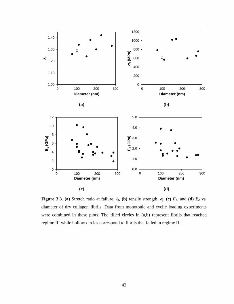
43
(a) (b)
(c) (d)
Figure 3.3. (a) Stretch ratio at failure, λf, (b) tensile strength, σf, (c) E1, and (d) E2 vs.
diameter of dry collagen fibrils. Data from monotonic and cyclic loading experiments
were combined in these plots. The filled circles in (a,b) represent fibrils that reached
regime III while hollow circles correspond to fibrils that failed in regime II.
1.00
1.10
1.20
1.30
1.40
0 100 200 300
λf
Diameter (nm)
0
200
400
600
800
1000
1200
0 100 200 300
σf(M
Pa)
Diameter (nm)
0
2
4
6
8
10
12
0 100 200 300
E1
(GP
a)
Diameter (nm)
0.0
1.0
2.0
3.0
4.0
5.0
0 100 200 300
E2
(GP
a)
Diameter (nm)

44
(a) (b)
Figure 3.4. (a) σ/E1 vs λ for the five dry collagen fibrils initially plotted in Figure 3.1(a).
(b) Detail of (a) in the initial 5% strain regime, essentially confirming the correct
determination of E1 for each curve since normalizing by the corresponding E1 values
collapsed all curves onto one near the origin.
Figure 3.5. σ/σf vs. λ/λf curves of the five dry collagen fibrils whose original plots are
shown in Figure 3.1(a).
0
0.04
0.08
0.12
0.16
0.2
1 1.1 1.2 1.3 1.4
σ/E
1
Stretch Ratio, λ
Test 1
Test 2
Test 3
Test 4
Test 5
0
0.01
0.02
0.03
0.04
1 1.01 1.02 1.03 1.04
σ/E
1
Stretch Ratio, λ
Test 1
Test 2
Test 3
Test 4
Test 5
0
0.2
0.4
0.6
0.8
1
1.2
0 0.2 0.4 0.6 0.8 1 1.2
σ/σ
f
λ/λf
Test 1
Test 2
Test 3
Test 4
Test 5

45
3.2. Strain Rate Dependence of Mechanical Behavior of Collagen Fibrils
In monotonic experiments with controlled strain rate, six nominal strain rates
were considered in the range 10-3 - 102 s-1. Despite efforts to select fibrils with diameters
in the range 90 - 120 nm, some tested fibrils had diameters >200 nm. Example plots from
strain rate tests are presented in Figure 3.6: Three tests were performed at the nominal
rate 0.01 s-1 and two more tests at nominal strain rates 0.001 s-1 and 100 s-1. The actual
strain rates are given in parentheses next to the nominal values. The σ - λ curves for
fibrils tested at 0.01 s-1 are bracketed by the curves of fibrils tested at 0.001 s-1 and 100 s-
1. The σ - λ curves for fibrils loaded at the nominal rate of 0.01 s-1 were divided by the
initial slope E1 and a comparison before and after normalization is shown in Figure
3.7(a,b). Clearly, the σ/E1 - λ curves demonstrated considerably better consistency and
showed a good agreement in regime II, namely a consistent ratio of the fibril stiffness in
regime II over the stiffness in regime I, E2/E1.
Figure 3.6. σ - λ curves of three fibrils tested at the nominal strain rate 0.01 s-1
compared to fibrils tested at nominal strain rates 0.001 s-1 and 100 s-1. The actual strain
rates are given in parentheses.
0
400
800
1200
1600
2000
1 1.1 1.2 1.3 1.4
Str
ess (
MP
a)
Stretch Ratio, λ
0.001 1/s (0.004 1/s)
0.1 1/s (0.24 1/s)
0.1 1/s (0.4 1/s)
0.1 1/s (0.12 1/s)
100 1/s (34 1/s)

46
(a) (b)
Figure 3.7. (a) Mechanical behavior of fibrils tested at 0.01 s-1, (b) curves in (a)
normalized by E1.
Figure 3.8(a) shows σ - λ curves from strain rate tests of fibrils with similar
diameters. In the legend of Figure 3.8 the true strain rate is followed by the fibril
diameter. All fibrils had comparable diameters in the range of 110 - 116 nm except for
those tested at 0.004 s-1 and 0.02 s-1, with diameters of 90 and 100 nm, respectively. The
six curves were normalized by E1, as shown in Figure 3.8(b). The main difference
between the two plots is the specimen tested at 0.02 s-1, whose curve moved from being
between those for 5.6 and 21 s-1 to those for 0.004 s-1 and 0.4 s-1. After normalization
with E1 in Figure 3.8(b) the σ - λ curves followed very consistent trends and order in
regime II according to the applied strain rate. According to the deformation mechanisms
outlined in Figure 1.14, molecular uncoiling and sliding dominating regime II [26] is
strain rate sensitive even for dry collagen, as shown in the experimental results in Figure
3.8(b).
Finally, Figure 3.9(a-e) shows plots of σm, σf/E1, E1, E2, and E2/E1 vs. strain rate.
From the plots in Figure 3.9(a,b) it clearly emerges that the mechanical strength of
collagen fibrils failing in regime III is strain rate sensitive, reaching tensile strengths that
exceed 2.0 GPa, values that are possible for high strength man-made fibers. In terms of
0
400
800
1200
1600
2000
1.00 1.10 1.20 1.30 1.40
Str
ess (
MP
a)
Stretch Ratio, λ
90 nm
110 nm
250 nm
0.00
0.05
0.10
0.15
0.20
0.25
0.30
1.00 1.10 1.20 1.30 1.40
σ/E
1
Stretch Ratio, λ
90 nm
110 nm
250 nm

47
stiffness values in regimes I and II, Figure 3.9(c) shows a weak dependence of E1 on the
applied strain rate, while Figure 3.9(d) shows strong dependence of E2 on the applied
rate. As explained earlier, variations in the value of E1 may originate in the original state
of the collagen fibril and its diameter (which may be related) and therefore any weak
trends in E1 are overshadowed by other effects. Furthermore, E1 is a measure of the initial
elastic response and a weak dependence on strain rate indicates weak viscous effects in
the mechanical behavior in regime I, which may stem from the limited presence of H2O
molecules in the collagen fibrils. On the other hand, regime II corresponds to a
combination of molecular uncoiling and sliding that manifest as viscous behavior and
would control the value of E2. As expected, E2 increased with the applied strain rate. This
trend is more clear in Figure 3.9(e) where the ratio E2/E1 vs. strain rate is plotted. The
normalization by E1 allows to reduce the effect of the initial material state on the value of
E2 and to reveal a more pronounced trend with the applied strain rate.
Other research groups have only studied the viscoelastic behavior of collagen
tissues [63,64]. Incremental strain tests were performed on rat tail tendon fibers
(diameters of 38-70 μm) [63] and high rate cycling to 4% strain of human patellar tendon
fibrils [64]. The curves from these studies were separated into their elastic and viscous
stress vs. strain curves and the results showed that the elastic slopes were similar between
all rates. The elastic component of the stress vs. strain curves, to a first approximation,
was found to be strain rate independent in both of studies, confirming that the strain rate
dependence is due primarily to the viscous component of deformation [63,64]. It was
further hypothesized that the thixotropy18 of collagen fibrils depends on the movement of
water in subfibrillar components [63]. Low shear rates allow time for water to rearrange,
thus collagen-collagen interactions are in their majority viscous. At high strain rates, the
subfibrillar elements “hydroplane” over each other during deformation because water
does not have enough time to flow between elements [63]. However, both studies [63,64]
were performed using rehydrated collagen and it is unclear whether this mechanism
applies to fibrils at low hydration state.
18 Thixotropy: Time-dependent decrease in viscosity under loading.

48
(a)
(b)
Figure 3.8. (a) σ - λ curves of fibrils with similar diameters loaded at different strain rates.
(b) σ/E1 vs. λ plots constructed using the plots in (a).
0
400
800
1200
1600
2000
1 1.1 1.2 1.3 1.4
Str
ess (
MP
a)
Stretch Ratio, λ
0.004 1/s (D=100 nm)
0.02 1/s (D=87 nm)
0.4 1/s (D=110 nm)
1.3 1/s (D=115 nm)
5.6 1/s (D=116 nm)
21 1/s (D=115 nm)
0.00
0.05
0.10
0.15
0.20
0.25
0.30
1 1.1 1.2 1.3 1.4
σ/E
1
Stretch Ratio, λ
0.004 1/s (D=100 nm)
0.02 1/s (D=87 nm)
0.4 1/s (D=110 nm)
1.3 1/s (D=115 nm)
5.6 1/s (D=116 nm)
21 1/s (D=115 nm)

49
(a) (b)
(c) (d)
(e)
Figure 3.9. Plots of (a) σf, (b) σf/E1, (c) E1, (d) E2, and (e) E2/E1 vs. strain rate.
0
500
1000
1500
2000
2500
0.001 0.01 0.1 1 10 100
σf(M
Pa)
Strain Rate (s-1)
0.00
0.10
0.20
0.30
0.40
0.001 0.01 0.1 1 10 100
σf/E
1
Strain Rate (s-1)
0.0
3.0
6.0
9.0
12.0
15.0
0.001 0.01 0.1 1 10 100
E1
(GP
a)
Strain Rate (s-1)
0.0
1.0
2.0
3.0
4.0
5.0
6.0
0.001 0.01 0.1 1 10 100
E2
(GP
a)
Strain Rate (s-1)
0
0.1
0.2
0.3
0.4
0.5
0.6
0.7
0.001 0.01 0.1 1 10 100
E2/E
1
Strain Rate (s-1)

50
3.3. Deformation and Failure of Individual Collagen Fibrils
After testing, all collagen fibrils were imaged in an SEM for diameter
measurements (using a segment that was not part of the gage section) and to study their
failure sections. Due to collagen’s sensitivity to the electron beam, the SEM imaging
voltage was set to 2 kV, the beam current to 5 μA, and the working distance at 5 mm. In
order to assess the mode of deformation of the fibrils, a collagen fibril was stretched to λ
≈ 1.25 (regime II) and then unloaded, the test was interrupted, and the load cell was fixed
in place. A composite image of this fibril is shown in Figure 3.11. The diameter
sporadically varied in a limited fashion along the length of the fibril. However, given the
constant slope of regime II, Figure 3.10, no strain localizations (fibril sections that are
undergo plastic thinning while the majority of the fiber remains elastic) could have
occurred, and the deformation could be considered rather uniform.
Previous research supported that collagen fails at a 45° angle by circumferential
rupture [35] but the experimental data at hand did not support this. Specifically, Figure
3.12 shows the SEM images of matching fibril failure sections of three different fibrils,
tested at a slow (0.005 s-1) and a fast strain rate (1 s-1). All three image sets point to
fibrillar failure along a longitudinal plane, which was irrespective of the applied strain
rate. This type of failure could also be due to a core-shell fibril structure (Figure 3.12(f)
could point to such structure), although core-shell type of fiber failure is associated with
low strength values, which was not the case in the present study. Such fibrillar fracture
surfaces were evidenced in fibrils with tensile strengths ranging from 550 tested at slow
strain rates, to 2000 MPa for fibrils tested at high rates, such as the failure section shown
in Figure 3.13(a,b) for 100 s-1 and the failure section in Figure 3.14(a,b) for fibrils tested
at strain rate ~0.001 s-1, thus suggesting no correlation between tensile strength, strain
rate and failure mode. This consistency in failure mode is clouded by examples of
different failure modes, such as the recoiled matching failure ends shown in Figure
3.13(c,d), the reminiscent core-shell failure of the fibrils in Figure 3.14(c,d), the brittle
failure in Figure 3.14(e) and the slanted failure section in Figure 3.14(f).

51
(a)
(b)
Figure 3.10. Plots of (a) σ and (b) σ/E1 vs. λ in regime II for fibrils whose original curves
are shown in Figure 3.1(a). Note the agreement in the slope of the majority curves in
regime II, implying a rather constant E2/E1 for fibrils with different diameters or initial
material state. The consistency in the value of the ratio E2/E1 provides further support to
the previous discussion on the effect of strain rate on the fibril response in regime II.
0
100
200
300
400
500
600
700
1.05 1.1 1.15 1.2 1.25
Str
ess (
MP
a)
Stretch Ratio, λ
Test 1Test 2Test 3Test 4Test 5
0
0.02
0.04
0.06
0.08
0.1
1.05 1.1 1.15 1.2 1.25
σ/E
1
Stretch Ratio, λ
Test 1
Test 2
Test 3
Test 4
Test 5

52
Figure 3.11. Composite image of unloaded collagen fibril after deformed to λ = 1.25.

53
(a) (b)
(c) (d)
(e) (f)
Figure 3.12. Matching fibril failure sections (a,b) and (c,d) from two experiments, which
are reminiscent of core-shell fiber failure tested at nominal strain rate 0.005 s-1. (e,f)
Matching fracture sections of a fibril tested at nominal strain rate 1 s-1.

54
(a) (b)
(c) (d)
Figure 3.13. Failure section of a fibril tested at nominal strain rate (a) 0.01 s-1, and (b)
100 s-1, both showing fibrillar failure. (c,d) Matching fracture sections of a fibril tested at
nominal strain rate 1 s-1.

55
(a) (b)
(c) (d)
(e) (f)
Figure 3.14. (a,b) Examples of two different fibrils with fibrillar failure. (c,d) Failure
ends of two different fibrils reminiscent of core-shell failure, (e) brittle failure, (f) slanted
failure. All were tested at approximate strain rates 0.004 - 0.006 s-1.

56
3.4. Conclusions
Dry, reconstituted, type I collagen fibrils tested at 20-30% RH exhibited σ - λ
curves with three distinct regimes, consistently with previous studies [26,27]. The initial
regime of high compliance reported in MD simulations and experiments with hydrated
fibrils [26,27,63,64] was not present in the collagen fibrils tested at 20-60% RH in this
research or other literature reports on dry collagen [32-34]. Hence, this initial high
compliance is potentially the result of the interaction between the fibril structure and
water molecules. Increased fibril diameter resulted in increased λ and reduced E1, E2, E3.
Normalization of the applied stress, σ, with E1 helped to remove the effect of fibril
diameter on the measured σ - λ curves. Similarly, the variation in σ - λ curves as a
function of strain rate tests was reduced in σ/E1 vs. λ curves, and a clear strain rate effect
emerged in regime II. Increasing the applied strain rate clearly increased σf and E2,
potentially due to increased viscous effects in molecular sliding and uncoiling, but had no
clear effect on λf (although the few data points imply no effect on λf) and a weak
influence on E1. Literature on collagenous tissues hypothesized that collagen-collagen
interactions dominate the viscous response at low rates, while hydroplaning in subfibrillar
elements dictates the viscous response at high rates [63].
Although prior research has reported that failure of rehydrated collagen fibrils
occurs at a 45° angle with respect to the fibril axis [35] the experimental data at hand
showed fibrillar failure in the majority of cases that was independent of strain rate and
may stem from a core-shell or fibrillar structure and properties.

57
CHAPTER 4
CYCLIC LOADING AND RECOVERY OF COLLAGEN
FIBRILS
Cyclic loading experiments were conducted at 20-30% RH to quantify damage
accumulation and recovery of individual collagen fibrils in each of the three regimes
defined in Figure 2.5. The available experimental apparatus lacked closed loop feedback
and, therefore, it was operated manually so that, in every cycle, approximately the same
strain was imposed to a fibril. Because of the manual approach, the target maximum
stretch ratios, λmax (corresponding to a value σmax), to which the fibrils were cycled were
selected to be near the middle of each regime, namely λmax ~ 1.05 in regime I, λmax ~ 1.25
in regime II, and λmax ~ 1.3 in regime III. Actuation was reversed when the fibril reached
λmax, and the fibrils were completely unloaded to produce a slight slack. The stretch ratio
upon complete unloading may be higher than that at the onset of the following cycle due
to slight recovery. The fibrils were extended to failure after the end of the last cycle. All
experiments were conducted at ~0.004 s-1. In this Chapter, the mechanical behavior under
cyclic loading and under the cyclic/recovery is discussed in connection with each of the
three deformation regimes.
4.1. Cyclic Behavior and Recovery of Collagen Fibrils in Regime I
The majority of fibrils cycled in regime I were brought to maximum strain of ~5%,
except for one specimen that was extended to λ ~ 1.13 but still lied in regime I. The σ - λ
curves of a fibril that was cycled twenty times are plotted in Figure 4.1. The first two
cycles are shown in Figure 4.1(a), and the last two cycles are compared to the first two in

58
Figure 4.1(b). The shape of the first loading curve is distinct from that of all subsequent
cycles, but the unloading curves were consistent throughout all cycles including the first.
After a few cycles, and within the limits of applying the same strain between cycles, the
mechanical behavior of the fibrils followed the same hysteresis loops. Figure 4.2(a,b)
shows plots of E1 and residual strain per cycle, respectively. Cycling loading did not
affect E1, but in most cases the residual strain increased linearly with cycle order. The
sudden jump in the residual strain in the plot for Test 2 between cycles 14 and 15 is due
to an overextension applied in cycle 14, shown in Figure 4.2(c,d), which, however, did
not affect the value of E1. Therefore, is likely that despite the large hysteresis recorded in
the cyclic loading σ - λ curves in regime I, the deformation taking place is limited to
simple stretching of the original network without major molecular rearrangements.
(a) (b)
Figure 4.1. (a) First two cycles, and (b) last two cycles compared to the first two cycles
(in gray) of a collagen fibril subjected to 20 loading-unloading cycles in regime I.
0
50
100
150
200
250
1.00 1.02 1.04 1.06 1.08 1.10
Str
ess (
MP
a)
Stretch Ratio, λ
0
50
100
150
200
250
1 1.02 1.04 1.06 1.08 1.1
Str
ess (
MP
a)
Stretch Ratio, λ

59
(a) (b)
(c) (d)
Figure 4.2. (a) E1, (b) residual strain, (c) σmax, and (d) λmax vs. cycle number for four
fibrils. Tests 1 and 2 are from cyclic loading tests extended to 20 cycles. Tests 3 and 4 are
results from the first 10 cycles of fibrils subjected to loading/recovery tests.
0
1
2
3
4
5
6
7
0 5 10 15 20 25
E1 (G
Pa)
Cycle
Test 1
Test 2
Test 3
Test 4
0
1
2
3
4
5
6
7
0 5 10 15 20 25
Resid
ual
Str
ain
(%
)
Cycle
Test 1
Test 2
Test 3
Test 4
0
50
100
150
200
250
300
0 5 10 15 20 25
σm
ax
(MP
a)
Cycle
Test 1Test 2Test 3Test 4
1.00
1.04
1.08
1.12
1.16
1.20
0 5 10 15 20 25
λm
ax
Cycle
Test 1Test 2Test 3Test 4

60
In strain recovery tests, the collagen fibrils underwent 10 loading cycles, followed
by 1 hr of recovery, with an additional 10 loading cycles, Figure 4.3. In this series of
plots, the cycles are numbered according to each iteration of 10 cycles and the cycle
number; namely “cycle 2-3”, refers to the third loading-unloading imposed in the second
iteration of ten cycles. As shown in Figure 4.3(c), the shape of cycle 2-1 was identical to
that of cycle 1-1, but shifted slightly to the right. The behavior of the collagen fibril after
recovery eventually followed that before recovery, which is shown in Figure 4.3(d) as
cycles 2-9 and 2-10 overlapped not only with each other, but also with cycles 1-9 and 1-
10, as shown in Figure 4.3(e).
According to the plot in Figure 4.4(a), the initial slope, E1, was not affected by
recovery. Similarly, the residual strain trended to a plateau in iteration 2, as shown in
Figure 4.4(b). According to DePalle et al [26], intermolecular adhesion allows uniform
deformation within the fibril, and the main form of deformation in regime I is due to
uncoiling of tropocollagen molecules, which contributes to approximately 80% of the
total deformation. The remaining 20% is attributed to surface effects and molecular
sliding [26]. The results presented here agree to some extent with [26]: For instance, a
fibril deformed to λmax ~1.12, Figure 4.4(d), resulted in ~ 4-6% residual strain, Figure
4.4(b), which was reduced to ~2% residual strain after 1 hr of recovery. Similarly,
another fibril deformed to λmax ~1.04, resulted in ~0.8% residual strain which was
reduced to ~0.8% strain after 1 hr of recovery time, namely in each case there was ~20%
residual strain after recovery. Interestingly, the fibril that was cycled to λmax ~1.04 had a
residual strain value less than < 1% throughout all of its cycles in iteration #1 and after
recovery in iteration #2.

61
(a) (b)
(c) (d)
(e)
Figure 4.3. σ - λ curves of a collagen fibril subjected to cyclic loading/recovery in regime
I: (a) cycles 1-1 and 1-2, (b) cycles 1-9 and 1-10 superposed on 1-1 and 1-2, (c) cycles 2-
1 and 2-2 superposed on iteration 1, (d) the last two full cycles of the test superposed on
the second iteration of cycling, and (e) cycles 1-9, 1-10, 2-1, and 2-10 superposed on the
first two cycles of both iterations.
0
50
100
150
200
250
1.00 1.05 1.10 1.15
Str
ess (
MP
a)
Stretch Ratio, λ
Cycle 1-1
Cycle 1-2
0
50
100
150
200
250
1.00 1.05 1.10 1.15
Str
ess (
MP
a)
Stretch Ratio, λ
Cycle 1-9
Cycle 1-10
0
50
100
150
200
250
1.00 1.05 1.10 1.15
Str
ess (
MP
a)
Stretch Ratio, λ
Cycle 2-1
Cycle 2-2
0
50
100
150
200
1.00 1.05 1.10 1.15
Str
ess (
MP
a)
Stretch Ratio, λ
Cycle 2-9
Cycle 2-10
0
50
100
150
200
250
1.00 1.05 1.10 1.15
Str
ess (
MP
a)
Stretch Ratio, λ
Cycle 1-9Cycle 1-10Cycle 2-9Cycle 2-10

62
(a) (b)
(c) (d)
Figure 4.4. Comparison before and after recovery for two fibrils, stretched to 4% (Test 1)
and 10% (Test 2) in regime I: (a) E1, (b) residual strain, (c) σmax, and (d) λmax vs. cycle
number.
0.0
1.0
2.0
3.0
4.0
5.0
0 2 4 6 8 10 12
E1 (G
Pa)
Cycle
Test 1 Iteration 1 Test 1 Iteration 2
Test 2 Iteration 1 Test 2 iteration 2
0.0
2.0
4.0
6.0
8.0
0 2 4 6 8 10 12
Resid
ual
Str
ain
(%
)
Cycle
Test 1 Iteration 1 Test 1 Iteration 2
Test 2 Iteration 1 Test 2 Iteration 2
0
50
100
150
200
250
0 2 4 6 8 10 12
σm
ax
(MP
a)
Cycle
Test 1 Iteration 1 Test 1 Iteration 2
Test 2 Iteration 1 Test 2 Iteration 2
1.00
1.04
1.08
1.12
1.16
0 2 4 6 8 10 12
λm
ax
Cycle
Test 1 Iteration 1 Test 1 Iteration 2
Test 2 Iteration 1 Test 2 Iteration 2

63
4.2. Cyclic Behavior and Recovery of Collagen Fibrils in Regime II
Fibrils were cycled in Regime II to λmax ~1.25. An example is shown in Figure 4.5.
The initial stiffness, E1, in cycle 2 was not as high as in cycle 1 but the stiffness of regime
II, E2, in cycle #2 was higher than in the first cycle. In Figure 4.5(b), cycles 19 and 20 are
shown to have reached a steady-state with complete overlap while E1 is rather constant,
according to the plots in Figure 4.6(a): E1 did not change after a reduction between cycles
1 and 2, while E2 showed steady increase with cycle number, Figure 4.6(b). Similarly, the
residual strain, Figure 4.6(c), increased with cycle number. The increase in plastic strain
is may originate in hardening induced in each cycle which did not affect E1 but increased
E2 and the amount of residual strain. Internal sliding could provide an explanation for the
gradual increase in residual strain. Note that, E1 remained rather constant for tests 1 and 2,
Figure 4.6(a), while tests 3 and 4 demonstrated fluctuations in E1 due to an inaccuracy in
maintaining the same λmax between cycles, Figure 4.6(d,e). Thus the small fluctuations in
residual strain and E1 could be attributed to an error in imposing the same λmax in each
cycle, but the gradual increase in residual strain may also include damage accumulation.
(a) (b)
Figure 4.5. Cyclic loading of a fibril in regime II: (a) Cycles 1 and 2, (b) comparison of
cycles 19 and 20 (color) with cycles 1 and 2 (gray).
0
100
200
300
400
1.0 1.1 1.2 1.3
Str
ess (
MP
a)
Stretch Ratio, λ
Cycle 1
Cycle 2
0
100
200
300
400
1.0 1.1 1.2 1.3
Str
ess (
MP
a)
Stretch Ratio, λ
Cycle 19
Cycle 20

64
(a) (b)
(c) (d)
(e)
Figure 4.6. (a) E1, (b) E2, (c) residual strain, (d) λmax, and (e) σmax vs. cycle number for
four different fibrils. Tests 1 and 2 are from fibrils that experienced 20 mechanical loading
cycles. The 21st datum point in these two sets is the initial modulus in the final loading to
failure. Tests 3 and 4 have fewer points because they are the first 10 cycles of cyclic
loading/recovery tests.
0.0
2.0
4.0
6.0
8.0
10.0
12.0
0 5 10 15 20 25
E1
(GP
a)
Cycle
Test 1Test 2Test 3Test 4
0.0
1.0
2.0
3.0
4.0
5.0
0 5 10 15 20 25
E2
(GP
a)
Cycle
Test 1Test 2Test 3Test 4
0.0
4.0
8.0
12.0
16.0
0 5 10 15 20 25
Resid
ual
Str
ain
(%
)
Cycle
Test 1Test 2Test 3Test 4
1.10
1.15
1.20
1.25
1.30
0 5 10 15 20 25
λm
ax
Cycle
Test 1
Test 2
Test 3
Test 4
0
200
400
600
800
0 5 10 15 20 25
σm
ax
(MP
a)
Cycle
Test 1Test 2Test 3Test 4

65
Figure 4.7(a) shows cycles 1-1 and 1-2 of a recovery test, and Figure 4.7(b) shows
cycles 1-9 and 1-10 (in color) together with cycles 1-1 and 1-2 (in gray). Only a fraction
of the total residual strain at the end of the first iteration was recovered during the 1 hr of
recovery, and cycle 2-1 began at a strain value that was marginally larger than the
residual strain at the end of cycle 1-1, Figure 4.7(c). Furthermore, both E1 and E2 in cycle
2-1 were similar to those of cycles 1-1 and 1-2. Cycles 2-9 and 2-10 completely
overlapped in Figure 4.7(d) and, for the sake of clarity, only the second iteration of
cycling is shown in the plot. Figure 4.8(a-e) show E1, E2, residual strain, σmax, and λmax as
a function of cycle number for two different fibrils, with similar conclusions as those for
Figure 4.6, except for E2 that remained rather constant in the first iteration.
According to MD simulations [26], the mechanical deformation in regime II is a
combination of molecular sliding and uncoiling. The energy applied to a fibril exceeds
that of intermolecular interactions and the molecules begin to slide with respect to each
other while also being restrained by cross-links [26]. In this regard, changes in the value
of E2 may be a combination of uncoiling of tropocollagen molecules and collagen triple
helix, and may also depend on the exact value of σmax or λmax, while the increasing
residual strain is caused primarily by intermolecular sliding.

66
(a) (b)
(c) (d)
Figure 4.7. σ - λ plots for a fibril subjected to cyclic loading/recovery in regime II: (a)
Cycles 1-1 and 1-2, (b) cycles 1-9 and 1-10 (color) compared to cycles 1-1 and 1-2
(gray), (c) cycles 2-1 and 2-2 superposed to a plot of all cycles of the first iteration (gray),
and (d) last two full cycles (2-9 and 2-10) vs. cycles 2-1, and 2-2 (in gray).
0
100
200
300
400
500
600
1.0 1.1 1.2 1.3
Str
ess (
MP
a)
Stretch Ratio, λ
Cycle 1-1
Cycle 1-2
0
100
200
300
400
500
600
1.0 1.1 1.2 1.3
Str
ess (
MP
a)
Stretch Ratio, λ
Cycle 1-9
Cycle 1-10
0
100
200
300
400
500
600
1.0 1.1 1.2 1.3
Str
ess (
MP
a)
Stretch Ratio, λ
Cycle 2-1
Cycle 2-2
0
100
200
300
400
500
600
1.0 1.1 1.2 1.3
Str
ess (
MP
a)
Stretch Ratio, λ
Cycle 2-9
Cycle 2-10

67
(a) (b)
(c) (d)
(e)
Figure 4.8. Experimental results from two different fibrils before and after recovery: (a)
E1 and (b) E2 (c) residual strain (d) σmax, and (e) λmax vs. cycle number.
0
2
4
6
8
10
12
0 2 4 6 8 10 12
E1
(GP
a)
CycleTest 1 Iteration 1 Test 1 Iteration 2Test 2 Iteration 1 Test 2 Iteration 2
0.0
1.0
2.0
3.0
4.0
5.0
0 2 4 6 8 10 12
E2
(GP
a)
Cycle
Test 1 Iteration 1 Test 1 Iteration 2Test 2 Iteration 1 Test 2 Iteration 2
0
4
8
12
16
20
0 2 4 6 8 10 12
Resid
ual
Str
ain
(%
)
CycleTest 1 Iteration 1 Test 1 Iteration 2Test 2 Iteration 1 Test 2 Iteration 2
0
200
400
600
800
0 2 4 6 8 10 12
σm
ax
(MP
a)
CycleTest 1 Iteration 1 Test 1 Iteration 2
Test 2 Iteration 1 Test 2 Iteration 2
1.15
1.20
1.25
1.30
0 2 4 6 8 10 12
λm
ax
Cycle
Test 1 Iteration 1 Test 1 Iteration 2
Test 2 Iteration 1 Test 2 Iteration 2

68
4.3. Cyclic Behavior and Recovery of Collagen Fibrils in Regime III
Similarly to regime II, fibrils cycled into regime III experienced a drastic change
in the shape of σ - λ curves right after the very first cycle, as shown in Figure 4.9(a). Each
cycle was characterized by an increase in residual strain and a gradual reduction in the
area of the hysteresis loops, Figure 4.9(b). Additional plots from a different fibril in
Figure 4.9(c,d) show that the hysteresis loops overlapped after a few cycles. Similarly to
regimes I and II, although the loading curves of the first few cycles did not show perfect
agreement with each other (especially the loading curve in the very first cycle), the
unloading curves perfectly agreed with each other starting from the very first unloading
cycle. Figure 4.10(a,b,c) show plots of E1, E2, and residual strain vs. cycle for three fibrils
cycled into regime III: Test 1 corresponds to the first 10 cycles of a cyclic
loading/recovery test, in Test 2 a fibril was cycled for only 10 cycles, and the fibril in
Test 3 was cycled 20 times. As shown in Figure 4.10(a,b), E1 and E2 remained rather
unchanged with cycling after an initial reduction between cycles 1 and 2. This is
especially true for Test 3 that demonstrated a slowly increasing residual strain, Figure
4.10(c), while the large fluctuations between cycles shown in the results for Tests 1 and 2
were due to the inability to achieve the same value of λmax, Figure 4.10(d), between cycles.
Figure 4.11(a,b) show the progression in the hysteresis loops between cycles 1-1
and subsequent cycles for a fibril subjected to two sets of cyclic loads separated by 1 hr
of recovery. During recovery, the fibril recovered most of the residual strain, and began
cycle 2-1 with residual strain lower than that of cycle 1-2, Figure 4.11(c). The curve of
cycle 2-1 is reminiscent of that of a fibril cycled into regime II, perhaps because the fibril
recovered most of the residual strain accumulated in the first iteration. Finally, the σ - λ
curves for cycles 2-9 and 2-10 completely overlapped, Figure 4.11(d). In Figure 4.12 E1
E2, residual strain, λmax, and σmax are plotted vs. the cycle number before (iteration 1) and
after (iteration 2) recovery. Despite the local fluctuations in value triggered by small
errors in maintaining constant λmax between cycles, E1 and E2 are consistent in each
iteration and between cycles with E2 gradually reaching a plateau value. The residual
strain reached higher values in the second iteration compared to the first, Figure 4.12(c),
despite a 75% recovery between cycle 1-10 and 2-1. In cycle 2-3, the residual strain was

69
at a similar value as in cycle 1-10, and then increased to reach comparable values as in
the first iteration. Notably, although λmax was rather constant, σmax decreased with cycling.
(a) (b)
(c) (d)
Figure 4.9. σ - λ curves of fibrils cycled into regime III. (a) Cycles 1 and 2, (b) cycles 9
and 10 (in color) compared to the first two cycles (shown in gray) for Test 2 in Figure
4.10. (c) Cycles 1 and 2, (d) cycles 19 and 20 (in color) compared to the first two cycles
(shown in gray) for Test 3 in Figure 4.10. The discontinuity until λ~1.06 in the loading
curves of cycle 1 in (c,d) was due to an obstruction to the load cell.
0
100
200
300
400
500
600
1.00 1.05 1.10 1.15 1.20 1.25
Str
ess (
MP
a)
Stretch Ratio, λ
Cycle 1
Cycle 2
0
100
200
300
400
500
600
1.00 1.05 1.10 1.15 1.20 1.25
Str
ess (
MP
a)
Stretch Ratio, λ
Cycle 9
Cycle 10
0
100
200
300
400
500
600
1.0 1.1 1.2 1.3
Str
ess (
MP
a)
Stretch Ratio, λ
Cycle 1Cycle 2
0
100
200
300
400
500
600
1.0 1.1 1.2 1.3
Str
ess (
MP
a)
Stretch Ratio, λ
Cycle 19Cycle 20

70
(a) (b)
(c) (d)
(e)
Figure 4.10. (a) E1, (b) E2, (c) residual strain, (d) λmax, and (e) σmax vs. cycle number for
three fibrils cycled into regime III.
0
2
4
6
8
0 5 10 15 20 25
E1
(GP
a)
Cycle
Test 1
Test 2
Test 3
0
1
2
3
4
0 5 10 15 20 25
E2
(GP
a)
Cycle
Test 1
Test 2
Test 3
0
2
4
6
8
10
12
14
0 5 10 15 20 25
Resid
ual
Str
ain
Cycle
Test 1
Test 3
Test 2
1.10
1.15
1.20
1.25
1.30
1.35
0 5 10 15 20 25
λm
ax
Cycle
Test 1
Test 2
Test 3
0
200
400
600
800
1000
0 5 10 15 20 25
σm
ax
(MP
a)
Cycle
Test 1
Test 2
Test 3

71
(a) (b)
(c) (d)
Figure 4.11. σ - λ curves for a fibril subjected to cyclic loading and recovery in regime
III. (a) Cycles 1-1 and 1-2, (b) cycles 1-9 and 1-10 (in color) compared to cycles 1-1 and
1-2 (in grey), (c) cycles 2-1 and 2-2 compared to iteration 1 (in grey), and (d) last two full
cycles compared to 2-1 and 2-2.
0
200
400
600
800
1000
1.0 1.1 1.2 1.3 1.4
Str
ess (
MP
a)
Stretch Ratio, λ
Cycle 1-1
Cycle 1-2
0
200
400
600
800
1000
1.0 1.1 1.2 1.3 1.4
Str
ess (
MP
a)
Stretch Ratio, λ
Cycle 1-9
Cycle 1-10
0
200
400
600
800
1000
1.0 1.1 1.2 1.3 1.4
Str
ess (
MP
a)
Stretch Ratio, λ
Cycle 2-1
Cycle 2-2
0
200
400
600
800
1.0 1.1 1.2 1.3 1.4
Str
ess (
MP
a)
Stretch Ratio, λ
Cycle 2-9
Cycle 2-10

72
(a) (b)
(c) (d)
(e)
Figure 4.12. (a) E1, (b) E2, (c) residual strain, (d) λmax, and (e) σmax vs. cycle number for a
cyclic loading/recovery test in regime III.
0.0
2.0
4.0
6.0
8.0
0 2 4 6 8 10 12
E1
(GP
a)
Cycle
Iteration 1
Iteration 2
0.0
1.0
2.0
3.0
4.0
0 2 4 6 8 10 12
E2
(GP
a)
Cycle
Iteration 1
Iteration 2
0
4
8
12
16
0 2 4 6 8 10 12
Resid
ual
Str
ain
(%
)
Cycle
Iteration 1
Iteration 2
1.00
1.10
1.20
1.30
1.40
0 2 4 6 8 10 12
λm
ax
Cycle
Iteration 1
Iteration 2
0
200
400
600
800
1000
0 2 4 6 8 10 12
σm
ax
(MP
a)
Cycle
Iteration 1
Iteration 2

73
4.4. Comparison of Cyclic Mechanical Behavior in Regimes I, II, and III
Compared to regime I in which large fluctuations in λmax had no effect on E1, the
value of the latter in regime II was more sensitive. Furthermore, ~1% increase in residual
strain due to fluctuations in regime II corresponded to 0.1 GPa increase in E1, and regime
III was the most sensitive as a 2% increase in residual strain due to fluctuations resulted
in ~0.5 GPa increase in E1. A comparison of the σ - λ curves in Figure 4.13(b-f) with
those in Figure 3.1, shows that mechanical cycling in any of the three regimes (without
recovery) results in linearization of the σ - λ curves up to λmax. This is evidenced in Figure
4.13(a) that shows the final loading of a fibril cycled 20 times in regime I, and Figure
4.13(b) that shows the final loading curve of iteration 2 of a fibril cycled in regime I (note
that the fibril whose curve is shown in Figure 4.13(b) did not reach regime III). Due to
the large overlap between cycles and the small range in which regime I was cycled, the
earlier cycles are not shown in the two plots. A comparison between the curves in Figure
3.1 and Figure 4.13(a) shows no discernible difference in the shape of the σ - λ curve
beyond regime I, except for a slight linearization in the behavior of regime I, which does
not impact the mechanical behavior of the fibril in regimes II and III. A similar
conclusion can be deduced from the plot in Figure 4.13(b).
Unlike in regime I, there is a clear difference in the mechanical behavior of a
fibril after cycling into regime II. The linearization of the mechanical behavior is more
pronounced in Figure 4.13(c) for a fibril only cycled in regime II, and in Figure 4.13(d)
for a fibril cycled in regime II, but let to recover for 1 hr, and then cycled again in regime
II. In both plots, regime I is not distinguished any longer after cycling in regime II, and
the final loading curves appear linear at failure. Regime III might be distinguished in
Figure 4.13(d) but it was not reached in the final loading curve in Figure 4.13(c). After
cycling in regime III, Figure 4.13(e,f), the overall curve was quite linear and steeper than
that of cycle 11 in Figure 4.13(e) and 1-1 in Figure 4.13(f). After cycling, the fibril whose
curves are shown in Figure 4.13(e) failed at a stress similar to the maximum stress in the
first cycle, and although this did not occur in many fibrils, it implies that mechanical
cycling does not necessarily result in mechanical drawing and strengthening. The fibril
that was allowed to recover before a second iteration of 10 cycles, Figure 4.13(f),

74
demonstrated all 3 regimes in the final loading curve (with regime I rather suppressed)
and failed at quite high stress after experiencing over 30% strain, supporting the
conclusion that cycling in regime III does not result in significant damage accumulation.
Finally, the mechanical response of a fibril that was cycled four times into regime
II is presented in Figure 4.14. This test was performed differently from previous cyclic
loading/recovery tests: The first cycle of each iteration was brought to a designated initial
σmax and in subsequent cycles the fibril was brought to λmax associated with the initial σmax.
The hysteresis curves shifted with increased residual strain, and this process continued
into the fourth iteration of cycling reaching a great consistency in iteration 4 as shown in
Figure 4.14(d). The actual residual strain, Figure 4.15(c), increased slightly within each
iteration but more significantly between iterations, reaching a plateau in iterations 1, 3
and 4. Achieving a plateau in residual stress depended on the ability to cycle the fibril in
each iteration by accurately reaching the same λmax in each cycle. Figures Figure
4.14(a,c,d) attest to this condition. Similarly to previous experiments in regime II, the
fibril did not recover all of its strain between iterations but reached very consistent values
for E1, Figure 4.15(a) and notably the value of E1 did not change after a reduction in the
first cycle despite the total of 40 cycles, Figure 4.15(a). On the other hand, E2 showed
larger variability between iterations and a consistent reduction to a steady-state value in
iterations 3 and 4, Figure 4.15(b).

75
(a) (b)
(c) (d)
(e) (f)
Figure 4.13. Regime I: (a) Final loading until failure after 20 cycles, (b) final loading
until failure after recovery and an additional 10 cycles. Regime II: (c) Initial and final
loading until failure after 10 cycles, (d) final loading until failure after recovery, also
showing cycles 1-1 and 2-1. Regime III: (e) Initial and final loading until failure after
10 cycles, (f) final loading until failure after recovery, also showing cycles 1-1 and 2-1.
0
100
200
300
400
500
1.0 1.1 1.2 1.3 1.4 1.5
Str
ess (
MP
a)
Stretch Ratio, λ
Cycle 21
0
100
200
300
400
500
600
700
1.0 1.1 1.2 1.3 1.4 1.5
Str
ess (
MP
a)
Stretch Ratio, λ
Cycle 2-11
0
100
200
300
400
500
600
1.0 1.1 1.2 1.3 1.4 1.5
Str
ess (
MP
a)
Stretch Ratio
Cycle 1
Cycle 21
0
200
400
600
800
1000
1200
1400
1.0 1.1 1.2 1.3 1.4 1.5
Str
ess (
MP
a)
Stretch Ratio, λ
Cycle 1-1Cycle 2-1Cycle 2-11
0
100
200
300
400
500
600
1.0 1.1 1.2 1.3 1.4 1.5
Str
ess (
MP
a)
Stretch Ratio, λ
Cycle 1
Cycle 11
0
200
400
600
800
1000
1200
1400
1.0 1.1 1.2 1.3 1.4 1.5
Str
ess (
MP
a)
Stretch Ratio, λ
Cycle 1-1
Cycle 2-1
Cycle 2-11

76
(a) (b)
(c) (d)
Figure 4.14. σ - λ curves of a fibril cycled four times in regime II. (a) Cycles 1-1, and 1-2,
vs. 1-9, and 1-10, (b) cycles 1-1 and 1-10 (in grey) plotted together with cycles 2-1, 2-2,
and 2-9, 2-10, (c) cycles 2-1 and 2-10 (in gray) plotted together with the first and the last
two cycles of iteration 3, and (d) cycles 3-1 and 3-10 (in grey) plotted together with the
first and last two cycles of iteration 4.
0
100
200
300
400
500
600
1 1.1 1.2 1.3
Str
ess (
MP
a)
Stretch Ratio, λ
Cycle 1-1Cycle 1-2Cycle 1-9Cycle 1-10
0
100
200
300
400
500
600
1 1.1 1.2 1.3
Str
ess (
MP
a)
Stretch Ratio, λ
Cycle 2-1Cycle 2-2Cycle 2-9Cycle 2-10
0
100
200
300
400
500
600
700
1 1.1 1.2 1.3 1.4
Str
ess (
MP
a)
Stretch Ratio, λ
Cycle 3-1
Cycle 3-2
Cycle 3-9
Cycle 3-10
0
100
200
300
400
500
600
700
1 1.1 1.2 1.3 1.4
Str
ess (
MP
a)
Stretch Ratio, λ
Cycle 4-1
Cycle 4-2
Cycle 4-9
Cycle 4-10

77
(a) (b)
(c) (d)
(e)
Figure 4.15. (a) E1, (b) E2, (c) residual strain, (d) λmax, and (e) σmax vs. cycle for a fibril
that was cycled four times into regime II with 1 hr of recovery between iterations.
0.0
2.0
4.0
6.0
8.0
10.0
0 2 4 6 8 10 12
E1
(GP
a)
Cycle
Iteration 1Iteration 2Iteration 3Iteration 4
0.0
1.0
2.0
3.0
4.0
5.0
0 2 4 6 8 10 12
E2
(GP
a)
Cycle
Iteration 1Iteration 2Iteration 3Iteration 4
0
5
10
15
20
25
0 2 4 6 8 10 12
Resid
ual
Str
ain
(%
)
Cycle
Iteration 1
Iteration 2
Iteration 3
Iteration 41.0
1.1
1.2
1.3
1.4
0 2 4 6 8 10 12
λm
ax
Cycle
Iteration 1
Iteration 2
Iteration 3
Iteration 4
0
200
400
600
800
0 2 4 6 8 10 12
σm
ax
(MP
a)
Cycle
Iteration 1Iteration 2Iteration 3Iteration 4

78
4.5. Conclusions
This Chapter examined the effect of cyclic loading and recovery in each of the
three deformation regimes distinguished in Figure 2.5. Collagen fibrils cycled into regime
I did not experience drastic changes in their σ - λ behavior. Interestingly, they
consistently recovered ~80% of the total applied strain, in agreement with MD
simulations predicting that 80% of the deformation in regime I originates in uncoiling of
tropocollagen molecules [26]. This suggests that deformation from uncoiling of
tropocollagen is recoverable, while deformation from molecular sliding in regime II is
not. According to the same MD simulations [26], the deformation in regime II is a
combination of molecular sliding and uncoiling and is restrained by cross-links, which
explains the constant value for E1 after cycling (even after 4 iterations and a total of 40
cycles) in regime II and the increase in residual strain. However, fibrils cycled into
regime II also experienced an increased overall stiffness. After cycling in each of the
three regimes, the σ - λ curves showed a more linear and stiffer response up to λmax in
comparison to monotonic loading. This effect was removed during recovery. When most
of the residual strain was removed during recovery, the recovered fibrils consistently
reached maximum strain at failure exceeding 35%.

79
CHAPTER 5
CONCLUSIONS
Reconstituted type I collagen fibrils with diameters in the range 70-280 nm tested
monotonically at 20-30% RH and ~0.004 s-1 strain rate demonstrated a three regime σ - λ
curves that were consistent in shape with previous studies [26,27]. The experimental
results from dry collagen fibrils did not uphold the existence of an initial compliant
regime at <2% strain that has been reported before for hydrated fibrils [26,27,63,64].
Additional experiments conducted at 40-53% RH, similarly to previous studies at 30-60%
RH [32-34], demonstrated the same σ - λ curves as dry collagen (20-30% RH), thus
implying that the initial compliance may be a characteristic of fully hydrated fibrils. As
computed from the σ - λ curves, the dry fibrils had an initial stiffness E1 = 5.7±2.3 GPa, a
regime II stiffness E2 = 2.19±1.06 GPa, tensile strength of 752±186 MPa, and ultimate
stretch ratio of 1.3±0.06. In comparison, rehydrated rat tail fibrils were shown to be much
weaker with tensile strength of 71±23 MPa and elastic modulus of 326±112 MPa [35],
but the tensile strengths measured in this research were closer to rehydrated human
patellar collagen fibrils with a tensile strength of 540±140 MPa as calculated using the
dry fibril diameter [27]. The dry state of the fibrils tested in this research resulted in
engineering failure strains (31±6%) that were lower than rehydrated collagen that has
been reported to have 63±21% fracture strain in rat tail tendon but only 20±1% in human
patellar tendon [35].
While the difference in the results from this dissertation research and prior studies
may originate in the specific details of the collagen tested, sample preparation, and
measurement techniques, the data reported by all studies to date has been characterized
by large variations. The results of this research clearly show that, despite following strict
protocols to ensure consistency between specimens, there are still variations in the
recorded mechanical response. The lack of precise control on fibril diameter is a major

80
contributing factor to the scatter of the experimental data. Since E1 correlated with the
fibril diameter, the effect of the initial material state or fibril diameter on the σ - λ curves
could be reduced by normalizing with E1. The σ/E1 - λ curves showed very consistent
trends with the applied strain rate in the range 0.001 - 100 s-1, agreement between
different plots obtained in the range at ~0.001 s-1, a clear increase in E2 and the ultimate
tensile strength that exceeded 2.0 GPa at high strain rates. E2/E1 increased with increasing
strain rate, implying a linearization of the effective σ - λ behavior with increasing rate.
The study of failure sections yielded mixed results, with most of the fibrils failing in a
fibrillar manner possibly influenced by the internal fibril structure of the collagen fibrils.
The mechanical behavior under cyclic loading was characterized in each of the
three regimes of deformation via cyclic and cyclic/recovery experiments at target λmax ~
1.05 in regime I, λmax ~ 1.25 in regime II, and λmax ~ 1.3 in regime III. Fibrils cycled into
regime I did not experience any drastic changes in the σ - λ curves and recovered more
than half of the residual strain accumulated by the last cycle before recovery time, and
~80% of λmax. According to DePalle et al [26], intermolecular adhesion allows uniform
deformation within the fibril, with deformation in regime I driven by uncoiling of the
tropocollagen molecules. Notably, mechanical cycling in regime II increased E2 and the
residual strain, but left E1 largely unaffected despite a distinct change in the shape of the
σ - λ curves between cycles 1 and 2. This was also true for fibrils tested before and after 1
hr of rest at zero stress and for a fibril subjected to four iterations of 10 cycles each with 1
hr of recovery between iterations. Despite experiencing 40 loading-unloading cycles, E1
was approximately the same in cycle 4-10 as in cycle 1-2. However, although E2
increased with cycle order, it decreased with iteration order, and E2 in iteration 4 cycling
was comparable to E2 in iteration 3. Deformation in regime II is attributed to molecular
sliding and uncoiling of the collagen triple helix, with the majority of molecular sliding
contained within the gap regions [35]. Applied loads are also distributed throughout the
fibril via water [63,64], resulting in uniform deformation. These mechanisms explain the
reasonably uniform diameter after loading to λ ~1.25, the increase in E2 and residual
strain with mechanical cycling, and the rather constant value of E1 throughout many
loading cycles.

81
Cycling in regime III resulted in similar results as regime II, with an overall
increase in the effective total stiffness of the fibrils, as fitted across all three regimes, and
an effective linearization of the σ - λ curves. Most notable was the change in the shape of
the σ - λ curve before and after recovery: Iteration 1 demonstrated all three regimes, but
only regimes I and II were present in the σ - λ curves in iteration 2. The amount of
possible deformation imparted in regime III is controlled by cross-link strength that can
enable stretching of the molecular backbone [26].
In summary, the experimental results presented in this dissertation point out to a
process of damage accumulation during cycling, as manifested by the very consistent
hysteresis loops and the gradually accumulated residual strain, which however, does not
affect the mechanical stiffness of regimes I and II. The latter points out to a cross-link
network within the collagen fibril that maintains molecular connectivity, as well as
material regions that allow for viscous sliding (supported by the increase in E2 and E2/E1
with applied strain rate) in the softening regime of the σ - λ curves without disrupting the
cross-link network. The rapid recovery and restoration of the three-regime shape of the σ
- λ curves of collagen fibrils also supports the existence of sacrificial bonds that reform
upon recovery that is driven by residual stresses in the fiber.

82
REFERENCES
[1] "Section 22.3 Collagen: The Fibrous Proteins of the Matrix," in Molecular Cell
Biology. 4th Edition, New York, W. H. Freeman, (2000).
[2] Illinois Institute or Technology, Packing It In: A New Look at Collagen Fibers,
Argonne National Laboratory, (2010).
[3] R. Dalgleish, "The human type I collagen mutation database," Oxford University
Press: Nucleic Acids Research, 25(1), pp. 181-187, (1997).
[4] T. Gutsmann, G. E. Fantner, J. H. Kindt, M. Venturoni, S. Danielsen and P. K.
Hansma, "Force Spectroscopy of Collagen Fibers to Investigate Their Mechanical
Properties and Structural Organization," Biophysics Journal, 86, pp. 3186-3193,
(2004).
[5] H. Kuivaniemi, G. Tromp and D. J. Prockop, "Mutations in collagen genes:
causes of rare and some common diseases in humans," The FASEB Journal, 5,
April, pp. 2052-2060, (1991).
[6] L. Bozec and M. Horton, "Topography and Mechanical Properties of Single
Molecules of Type I Collagen Using Atomic Force Microscopy," Biophysics
Journal, 88, pp. 4223-4231, (2005).
[7] A. L. Boskey and S. B. Doty, "Mineralized Tissue: Histology, Biology and
Biochemistry in Osteogenesis Imperfecta: A Translational Approach to Brittle
Bone Disease,” J. R. Shapiro, P. H. Byers, F. H. Glorieux and P. D. Sponseller,
Eds., Academic Press, 2013, pp. 31-43.
[8] A. J. Bailey, R. G. Paul and L. Knott, "Mechanisms of maturation and ageing of
collagen," Mechanisms of Ageing and Development, 106, pp. 1-56, (1998).

83
[9] M. D. Shoulders and R. T. Raines, "Collagen Structure and Stability," Annual
Review Biochem, 78, pp. 929-958, (2009).
[10] A. Gautieri, S. Vesentini, A. Redaielli and M. J. Buehler, "Hierarchical Structure
of Nanomechanics of Collagen Microfibrils from the Atomistic Scale Up," Nano
Letters, 11(2), pp. 757-766, (2011).
[11] T. J. Wess, "Collagen Fibrillar Structure and Hierarchies," in Collagen: Structure
and Mechanics, P. Fratzl, Ed., Springer US, 2008, pp. 49-80.
[12] G. M. Genin, A. Kent, V. Birman, B. Wopenka, J. D. Pasteris, P. J. Marquez and
S. Thomopoulos, "Functional Grading of Mineral and Collagen in the Attachment
of Tendon to Bone," Biophysics Journal, 97, pp. 976-985, (2009).
[13] H. P. Wiesmann, U. Meyer, U. Plate and H. J. Hohling, "Aspects of Collagen
Mineralization in Hard Tissue Formation," International Review of Cytology, vol.
242, pp. 121-156, 2005.
[14] S. L. Lees, "Mineralization of Type I Collagen," Biophysics Journal, vol. 85, pp.
204-207, 2003.
[15] Y. Liu, V. Birman, C. Chen, S. Thomopoulos and G. M. Genin, "Mechanisms of
Bimaterial Attachment at the Interface of Tendon to Bone," J Eng Mater Technol.,
no. 133, 2011.
[16] J. C. Halpin and J. L. Kardos, “The Halpin-Tsai equations: a review,” Polymer
Engineering and Science, 16(5), pp. 344-352, (352)
[17] H. Gao, B. Ji, L. I. Jager, E. Arzt and P. Fratzl, “Materials become insensitive to
flaws at nanoscale: Lessons from nature.” Proceedings of the National Academy
of Sciences of the United States of America, 100(10), pp. 5597–5600, (2003).
[18] A. K. Nair, A. Gautieri, S.W. Chang and M.J. Buehler, “Molecular mechanics of
mineralized collagen fibrils in bone,” Nature Communications, 4, pp. 1-9, (2013).

84
[19] F. Hang and A. Barber, "Nano-mechanical properties of individual mineralized
collagen fibrils from bone tissue," Journal of the Royal Society, 8(57), pp. 457-
471, (2011).
[20] H. S. Gupta, J. Seto, W. Wagermaier, P. Zaslansky, P. Boesecke and P. Fratzl,
"Cooperative deformation of mineral and collagen in bone at the nanoscale,"
PNAS, 103(47), pp. 17741-17746, (2006).
[21] J. R. Girgera and H. J. Berendsen, "The Molecular Details of Collagen
Hydration," Biopolymers, 18, pp. 47-57, (1979).
[22] I. G. Mogilner, C. Ruderman and J. R. Grigera, "Collagen stability, hydration and
native state," Journal of Molecular Graphics and Modelling, 21, pp. 209-213,
(2002).
[23] M. Luscher-mattli and M. Ruegg, "Thermodynamic functions of biopolymer
hydration. I. Their determination by vapor pressure studies, discussed in an
analysis of the primary hydration process," Biopolymers, 21(2), pp. 403-418,
(1982).
[24] G. N. Ramchandran and R. Chandrasekharan, "Interchain Hydrogen Bonds via
Bound Water Molecules in the Collagen Triple Helix," Biopolymers, 6, pp. 1649-
1658, (1968).
[25] J. Bella, M. Eaton, B. Brodsky and H. H. Berman, "Crystal and molecular
strucutre of a collagen-like peptide at 1.9 A resolution," Science, 266, pp. 75-81,
(1994).
[26] B. Depalle, Z. Qin, S. J. Shelbine and M. J. Buehler, "Influence of cross-link
structure, density and mechanical properties in the mesoscale deformation
mechanisms of collage fibrils," Journal of the Mechanical Behavior of Biomedical
Materials, 52, pp. 1-13, (2015).

85
[27] R. B. Svensson, H. Mulder, K. Vuokko and P. S. Magnusson, "Fracture
Mechanics of Collagen Fibrils: Influence of Natural Cross-Links," Biophysics
Journal, 104, pp. 2476-2484, (2013).
[28] Y.-L. Sun, Z.-P. Luo, A. Fertala and K.-N. An, "Direct quantification of the
flexibility of type I collagen monomer," Biochemical and Biophysical Research
Communications, 295, pp. 382-386, (2002).
[29] Y.-L. Sun, Z.-P. Luo, A. Fertala and K.-N. An, "Stretching type II collagen with
optical tweezers," Journal of Biomechanics, 37, pp. 1665-1669, (2004).
[30] J. A. J. van der Rijt, K. O. van der Werf, M. L. Bennink, P. J. Dijkstra and J.
Feijen, "Micromechanical Testing of Individual Collagen Fibrils,"
Macromolecular Bioscience, 6, pp. 697-702, (2006).
[31] Z. Liu, B. N. Smith, H. Kahn, R. Ballarini and S. J. Eppell, "Mechanical Testing
of Hydrated Collagen Nanofibrils Using MEMS Technology," IEEE, 1, pp. 177-
180, (2006).
[32] Z. L. Shen, M. R. Dodge, H. Kahn, R. Ballarini and S. J. Eppell, "Stress-Strain
Experiments on Individual Collagen Fibrils," Biophysics Journal, v95, pp. 3956-
3963, (2008).
[33] Z. L. Shen, M. R. Dodge, H. Kahn, R. Ballarini and S. J. Eppell, "In Vitro
Fracture Testing of Submicron Diameter Collagen Fibril Specimens," Biophysics
Journal, 99, pp. 1986-1995, (2010).
[34] Z. L. Shen, H. Kahn, R. Ballarini and S. J. Eppell, "Viscoelastic Properties of
Isolated Collagen Fibrils," Biophysics Journal, 100, pp. 3008-3015, (2011).
[35] Y. Liu, R. Ballarini, S. J. Eppell, “Tension tests on mammalian collagen fibrils,”
The Royal Society Publishing, 6(1), pp. 1-7, (2016).

86
[36] C. V. Kampen, S. Arnoczky, P. Parks, E. Hackett, D. Ruehlman, A. Turner, T.
Schlegel, “Tissue-engineered augmentation of a rotator cuff tendon using a
reconstituted collagen scaffold: a histological evaluation in sheep,” Muscles
Ligamens Tendons Journal, 3(3), pp. 229-235, (2013).
[37] D. V. Shepherd, J. H. Shepherd, S. Ghose, S. K. Kew, R. E. Cameron, S. M. Best,
“The process of EDC-NHS cross-linking of reconstituted collagen fibres increases
collagen fibrillar order and alignment,” APL Materials, 3(1), pp. 014902-2 – 8,
(2015).
[38] Y. Li, E. P. Douglas, “Effects of various salts on structural polymorphism of
reconstituted type I collagen fibrils,” Colloids and Surfaces B: Biointerfaces, 112,
pp. 42-50, (2013).
[39] C. R. Carlisle, C. Coulais and M. Guthold, "The mechanical stress-strain
properties of single electrospun collagen type I nanofibers," Acta Biomaterialia, 6,
pp. 2997-3003, (2010).
[40] P. Panwar, G. Lamour, N. C. W. Machenzie, H. Yang, F. Ko, H. Li and D.
Bromme, "Changes in structural-mechanical properties and degradability of
collagen during ageing-associaed modification," JBS Papers in Press, (2015).
[41] J. A. Matthews, G. E. Wneck, D. G. Simpson and G. L. Bowlin, "Electrospinning
of Collagen Nanofibers," Biomacromaterials, 2, pp. 232-238, (2002).
[42] R. Sopakayang, R. De Vita, A. Kwansa and J. W. Freeman, "Elastic and
viscoelastic properties of a type I collagen fiber," Journal of Theoretical Biology,
293, pp. 197-205, (2012).
[43] F. Yuan, S.R. Stock, D.R. Haeffner, J. D. Almer, D.C. Dunand, L. C. Brinson “A
new model to simulate the elastic properties of mineralized collagen fibril,”
Biomechanics and Modelling in Mechanobiology, 10, pp. 147-160, (2011).

87
[44] P. J. in 't Veld and M. J. Stevens, "Simulation of the Mechanical Strength of a
Single Collagen Molecule," Biophysics Journal, 95, pp. 33-39, (2008).
[45] M. Naraghi, I. Chasiotis, Y. Dzenis, Y. Wen, and H. Kahn, “Novel Method for
Mechanical Characterization of Polymeric Nanofibers,” Review of Scientific
Instruments 78, pp. 0851081-6, (2007).
[46] Naraghi, I. Chasiotis, Y. Dzenis, Y. Wen, H. Kahn, “Mechanical deformation and
failure of electrospun polyacrylonitrile nanofibers as a function of strain rate”,
Applied Physics Letters 91, pp. 151901-3, (2007).
[47] M. Naraghi and I. Chasiotis, “Optimization of Comb-driven Devices for
Mechanical Testing of Polymeric Nanofibers Subjected to Large Deformations”
Journal of Microelectromechanical Systems 18 (5), pp. 1032-1046, (2009).
[48] M. Naraghi, S. Arshad, and I. Chasiotis, “Molecular Orientation and Mechanical
Property Size Effects in Electrospun Polyacrylonitrile Nanofibers”, Polymer 52,
pp. 1612-1618, (2011).
[49] P. V. Kolluru, J. Lipner, L. Wenying, Y. Xia, S. Thomopoulos, G.M. Genin and I.
Chasiotis, “Strong and Tough Mineralized PLGA Nanofibers for Tendon-to-bone
Scaffolds”, Acta Biomaterialia 9, pp. 9442–9450, (2013).
[50] M. Naraghi, P.V. Kolluru and I. Chasiotis, Invited: “Time and Strain Rate
Dependent Mechanical Behavior of Polymeric Nanofibers”, Journal of the
Mechanics and Physics of Solids 62, pp. 257–275, (2014).
[51] P.V. Kolluru, I. Chasiotis, “Interplay of Molecular and Specimen Length Scales in
the Large Deformation Mechanical Behavior of Polystyrene Nanofibers”,
Polymer 56, pp. 507-515, (2015).

88
[52] P.V. Kolluru, I. Chasiotis, “A Master Curve for the Molecular Size and Strain
Rate Dependent Large Deformation Response of Glassy PS Nanofibers”, to
appear in Polymer, (2016).
[53] Marvin L. Tanzer, “Intermolecular Cross-links in Reconstituted Collagen Fibrils,”
The Journal of Biological Chemistry, 243(15), pp. 4045-4054, (1968).
[54] Elastin Products Company, "Collagen Type I: No. C857," Owensville, Missouri,
(2005).
[55] Y.P. Kato, F. H. Silver, “Formation of continuous collagen fibres: Evaluation of
biocompatibility and mechanical properties,” Biomaterials, 11, pp. 169-175, 1989.
[56] D. I. Zeugolis, R. G. Paul and G. Attenburrow, "Factors influencing the properties
of reconstituted collagen fibers rior to self-assembly: Animal species and collagen
extraction method," Journal of Biomedical Materials Research Part A, pp. 892-
904, (2007).
[57] P. Y. Kato, D. L. Christiansen, R. A. Hahn, S.-J. Shieh, J. D. Goldstein and F. H.
Silver, "Mechanical properties of collagen fibres: a comparison of reconstituted
and rat tail tendon fibres," Biomaterials, 10, January, pp. 38-42, (1989).
[58] M. Lavagnino, S. P. Arnoczky, K. Frank, T. Tian, “Collagen fibril diameter
distribution does not reflect changes in the mechanical properties of in vitro
stress-deprived tendons,” Journal of Biomechanics, 38(1), pp. 69-75, (2005).
[59] K.A. Derwin, L.J. Soslowsky, “A quantitative investigation of structure-function
relationships in a tendon fascicle model,” Journal of Biomechanical Engineering,
121, pp. 598-604, (1999).
[60] J.M. Binkley, M. Peat “The effects of immobilization on the ultrastructure and
mechanical properties of the medial collateral ligaments of rats,” Clinical
Orthopaedics and Related Research, 203, pp. 301-308, (1986).

89
[61] D.A.D. Parry, G. R. G. Barnes, A. D. Craig, “A comparison of the size
distribution of collagen fibrils in connective tissues as a function of age and a
possible relation between fibrils size and distribution and mechanical properties,”
Proceedings of the Royal Society of London. Series B, Biological Sciences,
20(1152), pp. 305-321, (1978).
[62] D. L. Christiansen, E. K Huang, F. H. Silver, “Assembly of type I collagen: fusion
of fibril subunits and the influence of fibril diameter on mechanical properties,”
Matrix Biology, 19, pp. 409-4220, (2000).
[63] F. H. Silver, A. Ebrahimi, P. B. Snowhill, “Viscoelastic Properties of Type I
Collagen Fibers: Molecular Basis of Elastic and Viscous Behaviors,” Connective
Tissue Research, 43, pp. 569-580, (2002).
[64] R. B. Svensson, T. Hassenkam, P. Hansen, S. P. Magnusson, “Viscoelastic
behavior of discrete human collagen fibrils,” Journal of the Mechanical Behavior
of Biomedical Materials, 3, pp. 112-115, (2010).