Mechanical and histological evaluation of a titanium ... · The device for orthodontic anchorage...
8
© 2019 Dental Press Journal of Orthodontics Dental Press J Orthod. 2019 May-June;24(3):71-8 71 original article Mechanical and histological evaluation of a titanium device for orthodontic anchorage, placed with or without cyanoacrylate adhesive Anderson Antonio Mamede 1 , Elizabeth Ferreira Martinez 1 , Roberta Tarkany Basting 1 Objective: The objective of the present study was to perform a histological evaluation of a titanium mini-implant for orth- odontic anchorage. Shear strength and fracture patterns that occurred immediately, 30 and 60 days after insertion with or without N-2-butyl-cyanoacrylate adhesive were evaluated. Methods: Ninety-six mini-implants (Arrow, Peclab, Brazil) were placed in the tibia of 9 male rabbits, with or without an adhesive (Vetbond™, 3M, USA). Histological evaluation was done by optical light microscope. Shear strength testing was performed, followed by fracture analysis with visual inspection. Results: Close contact between the newly formed bone and the device was evidenced in the group without adhesive, whereas gaps in the group with adhesive were found. Tukey test showed similar values in both groups at the immediate time point (20.70 N without adhesive and 24.69 N with adhesive), and higher values for the non-adhesive group, after 30 and 60 days (43.98 N and 78.55 N, respectively). The values for the adhesive group were similar for the immediate time point (24.69 N), 30 days (18.23 N) and 60 days (31.98 N). The fractures were adhesive for both groups at the immediate time point. The fractures were cohesive in bone for the non-adhesive group after 30 and 60 days. Conclusions: The mini-implants showed close bone con- tact and required higher shear strength for removal at 30 and 60 days for the non-adhesive group. Further studies are needed to assess the proper way to remove the orthodontic anchorage without cohesive fractures in bone. Keywords: Cortical bone. Orthodontic anchorage procedures. Fractures. 1 Instituto e Centro de Pesquisas São Leopoldo Mandic (Campinas/SP, Brazil). » The authors report no commercial, proprietary or financial interest in the products or companies described in this article. Submitted: February 21, 2018 - Revised and accepted: July 15, 2018 DOI: https://doi.org/10.1590/2177-6709.24.3.071-078.oar How to cite: Mamede AA, Martinez EF, Basting RT. Mechanical and histologi- cal evaluation of a titanium device for orthodontic anchorage, placed with or with- out cyanoacrylate adhesive. Dental Press J Orthod. 2019 May-June;24(3):71-8. DOI: https://doi.org/10.1590/2177-6709.24.3.071-078.oar Contact address: Roberta Tarkany Basting Rua José Rocha Junqueira, 13. bairro Swift, Campinas/SP – Brasil CEP: 13.045-755 – E-mail: [email protected] Objetivos: este estudo teve como objetivo realizar uma avaliação histológica de um mini-implante para ancoragem em Ortodon- tia. Avaliou-se, também, a carga de cisalhamento e o padrão de fratura imediatamente e após 30 e 60 dias da sua inserção, com ou sem o uso do adesivo N-butil-2-cianoacrilato. Métodos: noventa e seis mini-implantes (Arrow, Peclab, Brasil) foram instalados na tíbia de nove coelhos machos, com ou sem adesivo (Vetbond™, 3M, EUA). A avaliação histológica foi realizada com uso de microscópico de luz óptica. Realizou-se o teste de resistência ao cisalhamento, seguido pela análise da fratura, por meio de inspeção visual. Resultados: um contato íntimo entre o novo osso formado e o dispositivo foi evidenciado no grupo sem adesivo, enquanto espaços foram encontrados no grupo com adesivo. O teste de Tukey mostrou valores semelhantes em ambos os grupos no tempo imediato (20,70 N sem adesivo e 24,69 N com adesivo), e valores maiores para o grupo sem adesivo após 30 e 60 dias (43,98 N e 78,55 N, respectivamente). Os valores para o grupo com adesivo foram semelhantes para os tempos imediato (24,69 N), 30 dias (18,23 N) e 60 dias (31,98 N). As fraturas foram adesivas para ambos os grupos, no tempo imediato. As fraturas foram coesivas no osso para os grupos sem adesivo, após 30 e 60 dias. Conclusões: os mini-implantes mostraram um contato íntimo com o osso e requereram alta carga de cisalhamento para sua remoção após 30 e 60 dias nos grupos sem adesivo. Estudos adicionais são necessá- rios para avaliar um método para remoção do dispositivo ortodôntico sem fratura coesiva no osso. Palavras-chave: Osso cortical. Procedimentos de ancoragem ortodôntica. Fraturas.
Transcript of Mechanical and histological evaluation of a titanium ... · The device for orthodontic anchorage...
© 2019 Dental Press Journal of Orthodontics Dental Press J Orthod. 2019 May-June;24(3):71-871
original article
Mechanical and histological evaluation of a titanium
device for orthodontic anchorage, placed with or
without cyanoacrylate adhesive
Anderson Antonio Mamede1, Elizabeth Ferreira Martinez1, Roberta Tarkany Basting1
Objective: The objective of the present study was to perform a histological evaluation of a titanium mini-implant for orth-odontic anchorage. Shear strength and fracture patterns that occurred immediately, 30 and 60 days after insertion with or without N-2-butyl-cyanoacrylate adhesive were evaluated. Methods: Ninety-six mini-implants (Arrow, Peclab, Brazil) were placed in the tibia of 9 male rabbits, with or without an adhesive (Vetbond™, 3M, USA). Histological evaluation was done by optical light microscope. Shear strength testing was performed, followed by fracture analysis with visual inspection. Results: Close contact between the newly formed bone and the device was evidenced in the group without adhesive, whereas gaps in the group with adhesive were found. Tukey test showed similar values in both groups at the immediate time point (20.70 N without adhesive and 24.69 N with adhesive), and higher values for the non-adhesive group, after 30 and 60 days (43.98 N and 78.55 N, respectively). The values for the adhesive group were similar for the immediate time point (24.69 N), 30 days (18.23 N) and 60 days (31.98 N). The fractures were adhesive for both groups at the immediate time point. The fractures were cohesive in bone for the non-adhesive group after 30 and 60 days. Conclusions: The mini-implants showed close bone con-tact and required higher shear strength for removal at 30 and 60 days for the non-adhesive group. Further studies are needed to assess the proper way to remove the orthodontic anchorage without cohesive fractures in bone.
Keywords: Cortical bone. Orthodontic anchorage procedures. Fractures.
1 Instituto e Centro de Pesquisas São Leopoldo Mandic (Campinas/SP, Brazil).
» The authors report no commercial, proprietary or financial interest in the products or companies described in this article.
Submitted: February 21, 2018 - Revised and accepted: July 15, 2018
DOI: https://doi.org/10.1590/2177-6709.24.3.071-078.oar
How to cite: Mamede AA, Martinez EF, Basting RT. Mechanical and histologi-cal evaluation of a titanium device for orthodontic anchorage, placed with or with-out cyanoacrylate adhesive. Dental Press J Orthod. 2019 May-June;24(3):71-8. DOI: https://doi.org/10.1590/2177-6709.24.3.071-078.oar
Contact address: Roberta Tarkany BastingRua José Rocha Junqueira, 13. bairro Swift, Campinas/SP – BrasilCEP: 13.045-755 – E-mail: [email protected]
Objetivos: este estudo teve como objetivo realizar uma avaliação histológica de um mini-implante para ancoragem em Ortodon-tia. Avaliou-se, também, a carga de cisalhamento e o padrão de fratura imediatamente e após 30 e 60 dias da sua inserção, com ou sem o uso do adesivo N-butil-2-cianoacrilato. Métodos: noventa e seis mini-implantes (Arrow, Peclab, Brasil) foram instalados na tíbia de nove coelhos machos, com ou sem adesivo (Vetbond™, 3M, EUA). A avaliação histológica foi realizada com uso de microscópico de luz óptica. Realizou-se o teste de resistência ao cisalhamento, seguido pela análise da fratura, por meio de inspeção visual. Resultados: um contato íntimo entre o novo osso formado e o dispositivo foi evidenciado no grupo sem adesivo, enquanto espaços foram encontrados no grupo com adesivo. O teste de Tukey mostrou valores semelhantes em ambos os grupos no tempo imediato (20,70 N sem adesivo e 24,69 N com adesivo), e valores maiores para o grupo sem adesivo após 30 e 60 dias (43,98 N e 78,55 N, respectivamente). Os valores para o grupo com adesivo foram semelhantes para os tempos imediato (24,69 N), 30 dias (18,23 N) e 60 dias (31,98 N). As fraturas foram adesivas para ambos os grupos, no tempo imediato. As fraturas foram coesivas no osso para os grupos sem adesivo, após 30 e 60 dias. Conclusões: os mini-implantes mostraram um contato íntimo com o osso e requereram alta carga de cisalhamento para sua remoção após 30 e 60 dias nos grupos sem adesivo. Estudos adicionais são necessá-rios para avaliar um método para remoção do dispositivo ortodôntico sem fratura coesiva no osso.
Palavras-chave: Osso cortical. Procedimentos de ancoragem ortodôntica. Fraturas.
© 2019 Dental Press Journal of Orthodontics Dental Press J Orthod. 2019 May-June;24(3):71-872
Mechanical and histological evaluation of a titanium device for orthodontic anchorage, placed with or without cyanoacrylate adhesiveoriginal article
INTRODUCTIONThe search for stable anchorage in orthodontics has
been sought over the years, particularly because of low compliance by patients. Traditional extra- and intraoral devices are being replaced by osseointegrated implants, including palatal implants, onplants, miniplates and mini-implants, in an effort to achieve higher success rates and shorter treatment time.1 Despite the clinical advantages of mini-implants, there are some disadvan-tages, such as risk of root damage, nasal floor or max-illary sinus perforation,2 tooth ankylosis,3 and lack of space for insertion, among others.1
Seeking a simple and safer way to achieve skeletal anchorage, Xie et al4 presented the concept of bone-bonding anchorage, using an adhesive to attach the de-vice to the bone. They demonstrated the fixation of a stainless steel piece to the cortical bone surface using N-butyl-2-cyanoacrylate adhesive. Synthetic cyanoac-rylate adhesives are a family of liquid monomers that cure at room temperature, in an exothermic reaction on contact with a small amount of water or basic flu-id. In 1988, the FDA approved 2-octyl-cyanoacrylate (Dermabond®) for topical use as a substitute for sutures, and, later, 2-butyl-cyanoacrylate (Hystoacryl®, Inder-mil®).5,6 These cyanoacrylate adhesives have been found to be biocompatible, bacteriostatic and hemostatic, in addition to offering easy handling7 and shorter operative time during surgical procedures.8
In an endeavor to overcome some of the disadvan-tages during mini-implant placement, the present group of authors developed a novel device with reduced di-mensions and an active anchoring part in the shape of an arrow. The ‘Arrow’ device was designed to be placed in the cortical portion of the bone and facilitates its place-ment, enhancing mechanical anchorage. Furthermore, the use of a N-butyl-2-cyanoacrylate-based adhesive may result in better primary stability and a higher suc-cess rate. The ultimate purpose of this innovative con-cept is to create a skeletal anchorage system that ensures good stability and decreases the risk of damage to im-portant structures during insertion.
Therefore, using an animal model, the aims of this study were: to perform a histological assessment of the interlocking of the Arrow device to the cortical bone, by optical microscopy observation; to mechani-cally evaluate the maximum loading capacity by shear strength test; and to study the types of fractures that oc-
curred during the mechanical test, by visual inspection immediately, 30 and 60 days after its insertion, with or without N-2-butyl-cyanoacrylate adhesive.
MATERIAL AND METHODSThis study was approved by the Ethics Committee
on Animal Research of Federal University of Minas Gerais (protocol no. 330/2014).
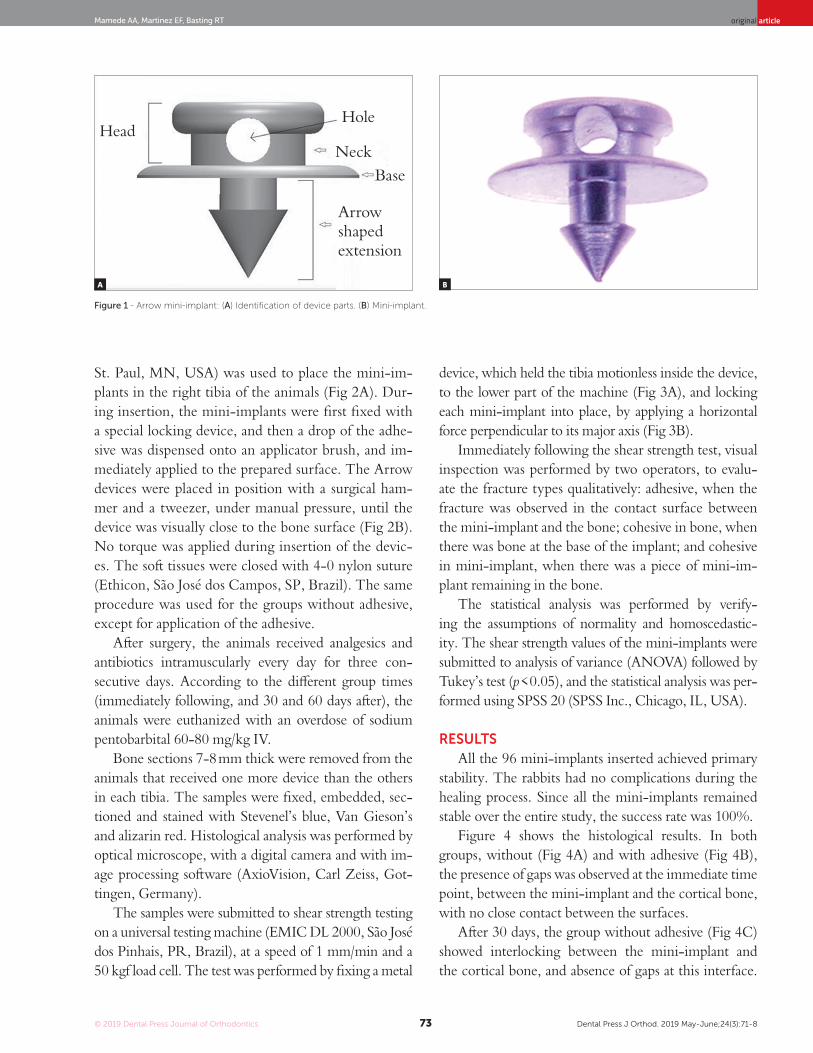
The device for orthodontic anchorage (Arrow) was made with commercially pure titanium grade 4 (ASTM F67) (PecLab, Belo Horizonte, Brazil). The Arrow is com-posed by three parts: head, base and an arrow-shaped extension. The head is 2.4 mm wide and 1.2 mm high, with a 0.7 mm diameter hole for orthodontic wire fixa-tion (Fig 1). The base is 3.5 mm in diameter, with an ar-row-shaped extension, 1.3 mm high and 1.25 mm wide.
Ninety-six Arrow devices were placed bilaterally in the tibia of nine male New Zealand male white rabbits (similar to those used by Xie et al4), aged 5 to 6 months old and weighing 3 to 3.5 kg. This experimental de-sign was based in that performed by Xie et al,4 to re-duce the number of animals used in the study, as sug-gested by the Ethics Committee on Animal Research. From a total of 96 devices placed in the tibias, 90 were used to evaluate the shear strength, and the other six devices were used for histological evaluation. The fac-tors studied were: (1) use or non-use of an adhesive for mini-implants fixation; (2) evaluation time points: immediately following, and 30 and 60 days after. Nine animals were divided into three groups. Twelve devic-es were placed bilaterally (six in each tibia) on one rab-bit from each group, and ten devices (five in each tibia) were placed on the other animals. The devices were placed with adhesive on the right tibia of all animals, and without adhesive on the left tibia.
The surgical procedures began one week after the ani-mals arrived at the laboratory. The animals were anesthe-tized with intramuscular ketamine (30 mg/kg) and xyla-zine (5 mg/kg). Following skin preparation, trichotomy and antisepsis with povidone-iodine, 2% lidocaine with epinephrine 1:100.000 was applied as the local anesthet-ic. All the surgeries were conducted under sterile condi-tions, in an operating room specific for animals.
The skin was incised and dissected, and the tibia was exposed. Five or six holes were made in the cor-tical bone with a 1.1 mm manual drill, at intervals of 8 mm. N-2-butyl-cyanoacrylate (Vetbond™, 3M,
© 2019 Dental Press Journal of Orthodontics Dental Press J Orthod. 2019 May-June;24(3):71-873
original articleMamede AA, Martinez EF, Basting RT
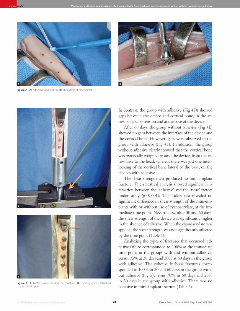
St. Paul, MN, USA) was used to place the mini-im-plants in the right tibia of the animals (Fig 2A). Dur-ing insertion, the mini-implants were first fixed with a special locking device, and then a drop of the adhe-sive was dispensed onto an applicator brush, and im-mediately applied to the prepared surface. The Arrow devices were placed in position with a surgical ham-mer and a tweezer, under manual pressure, until the device was visually close to the bone surface (Fig 2B). No torque was applied during insertion of the devic-es. The soft tissues were closed with 4-0 nylon suture (Ethicon, São José dos Campos, SP, Brazil). The same procedure was used for the groups without adhesive, except for application of the adhesive.
After surgery, the animals received analgesics and antibiotics intramuscularly every day for three con-secutive days. According to the different group times (immediately following, and 30 and 60 days after), the animals were euthanized with an overdose of sodium pentobarbital 60-80 mg/kg IV.
Bone sections 7-8 mm thick were removed from the animals that received one more device than the others in each tibia. The samples were fixed, embedded, sec-tioned and stained with Stevenel’s blue, Van Gieson’s and alizarin red. Histological analysis was performed by optical microscope, with a digital camera and with im-age processing software (AxioVision, Carl Zeiss, Got-tingen, Germany).
The samples were submitted to shear strength testing on a universal testing machine (EMIC DL 2000, São José dos Pinhais, PR, Brazil), at a speed of 1 mm/min and a 50 kgf load cell. The test was performed by fixing a metal
device, which held the tibia motionless inside the device, to the lower part of the machine (Fig 3A), and locking each mini-implant into place, by applying a horizontal force perpendicular to its major axis (Fig 3B).
Immediately following the shear strength test, visual inspection was performed by two operators, to evalu-ate the fracture types qualitatively: adhesive, when the fracture was observed in the contact surface between the mini-implant and the bone; cohesive in bone, when there was bone at the base of the implant; and cohesive in mini-implant, when there was a piece of mini-im-plant remaining in the bone.
The statistical analysis was performed by verify-ing the assumptions of normality and homoscedastic-ity. The shear strength values of the mini-implants were submitted to analysis of variance (ANOVA) followed by Tukey’s test (p < 0.05), and the statistical analysis was per-formed using SPSS 20 (SPSS Inc., Chicago, IL, USA).
RESULTSAll the 96 mini-implants inserted achieved primary
stability. The rabbits had no complications during the healing process. Since all the mini-implants remained stable over the entire study, the success rate was 100%.
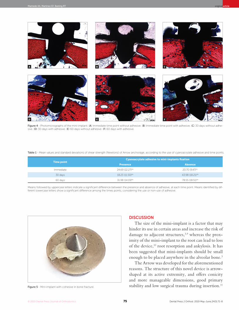
Figure 4 shows the histological results. In both groups, without (Fig 4A) and with adhesive (Fig 4B), the presence of gaps was observed at the immediate time point, between the mini-implant and the cortical bone, with no close contact between the surfaces.
After 30 days, the group without adhesive (Fig 4C) showed interlocking between the mini-implant and the cortical bone, and absence of gaps at this interface.
A B
Figure 1 - Arrow mini-implant: (A) Identification of device parts. (B) Mini-implant.
Arrow shaped extension
HeadHole
Neck
Base
© 2019 Dental Press Journal of Orthodontics Dental Press J Orthod. 2019 May-June;24(3):71-874
Mechanical and histological evaluation of a titanium device for orthodontic anchorage, placed with or without cyanoacrylate adhesiveoriginal article
A
A
B
B
Figure 2 - A) Adhesive application. B) Mini-implant placement.
Figure 3 - A) Metal device fixed in the machine. B) Locking device attached to the mini-implant.
In contrast, the group with adhesive (Fig 4D) showed gaps between the device and cortical bone, in the ar-row-shaped extension and at the base of the device.
After 60 days, the group without adhesive (Fig 4E) showed no gaps between the interface of the device and the cortical bone. However, gaps were observed in the group with adhesive (Fig 4F). In addition, the group without adhesive clearly showed that the cortical bone was practically wrapped around the device, from the ar-row base to the head, whereas there was just one inter-locking of the cortical bone lateral to the base, on the devices with adhesive.
The shear strength test produced no mini-implant fracture. The statistical analysis showed significant in-teraction between the ‘adhesive’ and the ‘time’ factors under study (p < 0.001). The Tukey test revealed no significant difference in shear strength of the mini-im-plants with or without use of cyanoacrylate, at the im-mediate time point. Nevertheless, after 30 and 60 days, the shear strength of the device was significantly higher in the absence of adhesive. When the cyanoacrylate was applied, the shear strength was not significantly affected by the time point (Table 1).
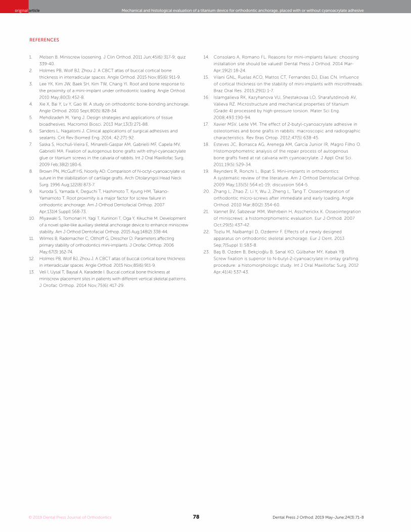
Analyzing the types of fractures that occurred, ad-hesive failure corresponded to 100% at the immediate time point in the groups with and without adhesive, versus 75% at 30 days and 30% at 60 days in the group with adhesive. The cohesive in bone fractures corre-sponded to 100% in 30 and 60 days in the group with-out adhesive (Fig 5), versus 70% in 60 days and 25% in 30 days in the group with adhesive. There was no cohesive in mini-implant fracture (Table 2).
© 2019 Dental Press Journal of Orthodontics Dental Press J Orthod. 2019 May-June;24(3):71-875
original articleMamede AA, Martinez EF, Basting RT
DISCUSSIONThe size of the mini-implant is a factor that may
hinder its use in certain areas and increase the risk of damage to adjacent structures,3,9 whereas the prox-imity of the mini-implant to the root can lead to loss of the device,10 root resorption and ankylosis. It has been suggested that mini-implants should be small enough to be placed anywhere in the alveolar bone.3
The Arrow was developed for the aforementioned reasons. The structure of this novel device is arrow-shaped at its active extremity, and offers conicity and more manageable dimensions, good primary stability and low surgical trauma during insertion.11
A
D
B
E
C
F
Figure 4 - Photomicrographs of the mini-implant. (A) Immediate time point without adhesive. (B) Immediate time point with adhesive. (C) 30 days without adhe-sive. (D) 30 days with adhesive. (E) 60 days without adhesive. (F) 60 days with adhesive.
Figure 5 - Mini-implant with cohesive in bone fracture.
Time pointCyanoacrylate adhesive to mini-implants fixation
Presence Absence
Immediate 24.69 (12.27)Aa 20.70 (9.47)Ac
30 days 18.23 (11.50)Ba 43.98 (18.21)Ab
60 days 31.98 (14.19)Ba 78.55 (18.51)Aa
Table 1 - Mean values and standard deviations of shear strength (Newtons) of Arrow anchorage, according to the use of cyanoacrylate adhesive and time points.
Means followed by uppercase letters indicate a significant difference between the presence and absence of adhesive, at each time point. Means identified by dif-ferent lowercase letters show a significant difference among the times points, considering the use or non-use of adhesive.

© 2019 Dental Press Journal of Orthodontics Dental Press J Orthod. 2019 May-June;24(3):71-876
Mechanical and histological evaluation of a titanium device for orthodontic anchorage, placed with or without cyanoacrylate adhesiveoriginal article
Table 2 - Distribution of fracture modes by study group.
Time point and adhesiveFracture mode
Adhesive Cohesive in bone Cohesive in mini-implant
Immediate with adhesive 100% 0 0
Immediate without adhesive 100% 0 0
30 days with adhesive 75% 25% 0
30 days without adhesive 0 100% 0
60 days with adhesive 30% 70% 0
60 days without adhesive 0 100% 0
The device is placed in such a way that its active part is located almost entirety in the cortical bone due to its dimensions12,13 (Fig 4), thereby reducing the risk of perforating the dental root or other impor-tant anatomical structures. Because of the reduced dimensions of its design, Arrow was manufactured with commercially pure titanium Grade 4, to enable osseointegration. As shown in the present study, this novel mini-implant did not fracture during the shear strength test performed at different time points, de-spite the finding that osseointegration may increase the possibility of mini-implant fracture.14 Although the reduced length of the device may contribute to fracture resistance, as observed by Vilani et al,15 who reported that shorter mini-implants have a lower risk of fracture, this occurrence was not reported in the present study. The absence of fractures may be related to the mechanical properties of the titanium Grade 4, which has high strength, with minimum yield strength of 480 MPa. This alloy combines ex-cellent corrosion resistance and good formability and weldability.16
In the present study, 100% of the devices showed osseointegration and cohesive bone fracture after 30 and 60 days in the group without adhesive. In both the adhesive and non-adhesive groups, there was ad-hesive failure only at the immediate time point, since there was no time for osseointegration. Although Arrow design could provide immediate stability and provided osseointegration over time, cohesive bone fracture is related as one of the limitations of this anchorage device, and methods to minimize bone loss during removal of this device need to be devel-oped. Furthermore, it was observed that the use of adhesive led to a lower prevalence of cohesive bone
fractures (25% and 70% in 30 and 60 days, respec-tively), and that the presence of the adhesive hin-dered osseointegration.
Analyzing images of the histological sections of the group without adhesive at the immediate time point, gaps were observed between the bone and the device (Fig 4A), since there was no time for osseoin-tegration. At the 30- and 60-day time points (Fig 4C and 4E), interlocking of newly formed cortical bone with mini-implant was observed without gaps, show-ing osseointegration. On the other hand, gaps were noticed between the newly formed cortical bone and the device, along the arrow-shaped extension (Fig 4B, 4D and 4F) in the group with adhesive, at all the time points. This is probably due to the adhesive present on these sites, as also observed by Xie et al.4
As shown in Table 1, there was no significant dif-ference in shear strength between the groups with or without adhesive, at the immediate time point. How-ever, it should be considered that the application of adhesive could have some effect on mini-implant to bone adherence. This could explain the tendency of higher average load values (24.69 N) of the adhesive versus non-adhesive group (20.70 N). All the groups with adhesive showed no significant difference in shear strength average values, and trended toward decreasing values at 30 days (18.23 N) and increas-ing values at 60 days (31.98 N). The shear strength of the device was significantly higher in the absence of adhesive (43.98 N at 30 days, and 78.55 N at 60 days), compared with the adhesive group. The lower average values observed in the adhesive group may be related to the cyanoacrylate not being metabo-lized,17 thereby hindering the interlocking of newly formed bone with the titanium device.18

© 2019 Dental Press Journal of Orthodontics Dental Press J Orthod. 2019 May-June;24(3):71-877
original articleMamede AA, Martinez EF, Basting RT
A systematic review19 reported that the shear strength levels used in orthodontic treatments ranged from 50 to 400 g, but the majority of studies have used 200 g or less.20 Once rigid fixation (secondary stabil-ity) is achieved, orthodontic forces are not a threat to mini-implants and bone integration.21 In this study, mechanical evaluation conducted with the shear strength test indicated that all the groups with and without adhesive achieved average values above the optimum values required for use in orthodontic skel-etal anchorage, at all the tested time points.
Comparing the Arrow device used in the adhe-sive group with the device used by Xie et al,4 the lat-ter had lower values (10.84 N) at the immediate time point, and also exhibited a decreasing trend in aver-age values at 15 days (6.23 N) and 30 days (1.8 N), and an increasing trend at 60 days (45.69 N), all in-dicating a significant difference. According to these authors, the decreasing trend occurred because of reduced adhesion strength, due to adhesive hydro-lysis. In the adhesive group, higher average shear strength values were verified at the immediate time point and at 30 days, compared with those by Xie et al4 at the same time points. This is probably due to the arrow-shaped structure extending from the base, which anchors the device and keeps it in posi-tion. Regarding the results observed after 60 days, Xie et al4 recorded higher values than the present study at this same time point, using a device with a flat base, probably due to the amount of new bone tissue around the device.
The thickness of the cortical bone has a major im-pact on primary stability, especially in dolichofacial patients, with high mandibular and gonial angles, be-cause their cortical bone is very thin.11,12,13 Alternative anchorage devices should be used on these patients to achieve primary stability. Other considerations for these devices include the use of biological adhe-sive, a greater device diameter, a surface treatment or the combined insertion of mini-implant and auxil-iary accessories with indentations facing the cortical bone.3,12,22 The application of small amounts of the adhesive lateral to the device23 should be evaluated for primary stability in patients with thin cortical bone.
Better results for the mini-implants, especially in patients with thin cortical bone, could be obtained by developing an adhesive agent, together with a
method for its application, which ensures primary stability in almost 100% of the cases, thus promot-ing an increase in bone implant contact, achiev-ing secondary stability, so that it can be used as an orthodontic anchorage. Further studies are needed to assess the best location of the biological adhesive during device insertion, use of other types of adhe-sive, a shear strength test that simulates orthodontic active treatment, and a way of removing the device in a clinical trial.
CONCLUSIONSThere was new bone formation, achieving close
contact with the device in 30-60 days in the nonad-hesive group. In the adhesive group, there were gaps between the device and the bone. At the immediate time point, the shear strength was similar between the groups with and without adhesive. Higher shear strength was found for the group without adhesive at 30 and 60 days, increasing over time. The Arrow showed no mini-implant fracture.
Authors’ contribution (ORCID )
Anderson A. Mamede (AAM): 0000-0001-9305-876XElizabeth F. Martinez (EFM): 0000-0002-4991-1185Roberta T. Basting (RTB): 0000-0002-5345-5776
Conception or design of the study: AAM, EFM, RTB. Data acquisition, analysis or interpretation: AAM, EFM, RTB. Writing the article: AAM, RTB. Critical revision of the article: AAM, EFM, RTB. Final approval of the arti-cle: AAM, EFM, RTB. Obtained funding: AAM. Overall responsibility: RTB.
© 2019 Dental Press Journal of Orthodontics Dental Press J Orthod. 2019 May-June;24(3):71-878
Mechanical and histological evaluation of a titanium device for orthodontic anchorage, placed with or without cyanoacrylate adhesiveoriginal article
1. Melsen B. Miniscrew loosening. J Clin Orthod. 2011 Jun;45(6):317-9; quiz
339-40.
2. Holmes PB, Wolf BJ, Zhou J. A CBCT atlas of buccal cortical bone
thickness in interradicular spaces. Angle Orthod. 2015 Nov;85(6):911-9.
3. Lee YK, Kim JW, Baek SH, Kim TW, Chang YI. Root and bone response to
the proximity of a mini-implant under orthodontic loading. Angle Orthod.
2010 May;80(3):452-8.
4. Xie X, Bai Y, Lv Y, Gao W. A study on orthodontic bone-bonding anchorage.
Angle Orthod. 2010 Sept;80(5):828-34.
5. Mehdizadeh M, Yang J. Design strategies and applications of tissue
bioadhesives. Macromol Biosci. 2013 Mar;13(3):271-88.
6. Sanders L, Nagatomi J. Clinical applications of surgical adhesives and
sealants. Crit Rev Biomed Eng. 2014; 42:271-92.
7. Saska S, Hochuli-Vieira E, Minarelli-Gaspar AM, Gabrielli MF, Capela MV,
Gabrielli MA. Fixation of autogenous bone grafts with ethyl-cyanoacrylate
glue or titanium screws in the calvaria of rabbits. Int J Oral Maxillofac Surg.
2009 Feb;38(2):180-6.
8. Brown PN, McGuff HS, Noorily AD. Comparison of N-octyl-cyanoacrylate vs
suture in the stabilization of cartilage grafts. Arch Otolaryngol Head Neck
Surg. 1996 Aug;122(8):873-7.
9. Kuroda S, Yamada K, Deguchi T, Hashimoto T, Kyung HM, Takano-
Yamamoto T. Root proximity is a major factor for screw failure in
orthodontic anchorage. Am J Orthod Dentofacial Orthop. 2007
Apr;131(4 Suppl):S68-73.
10. Miyawaki S, Tomonari H, Yagi T, Kuninori T, Oga Y, Kikuchie M. Development
of a novel spike-like auxiliary skeletal anchorage device to enhance miniscrew
stability. Am J Orthod Dentofacial Orthop. 2015 Aug;148(2):338-44.
11. Wilmes B, Rademacher C, Olthoff G, Drescher D. Parameters affecting
primary stability of orthodontics mini-implants. J Orofac Orthop. 2006
May;67(3):162-74.
12. Holmes PB, Wolf BJ, Zhou J. A CBCT atlas of buccal cortical bone thickness
in interradicular spaces. Angle Orthod. 2015 Nov;85(6):911-9.
13. Veli I, Uysal T, Baysal A, Karadede I. Buccal cortical bone thickness at
miniscrew placement sites in patients with different vertical skeletal patterns.
J Orofac Orthop. 2014 Nov;75(6):417-29.
REFERENCES
14. Consolaro A, Romano FL. Reasons for mini-implants failure: choosing
installation site should be valued! Dental Press J Orthod. 2014 Mar-
Apr;19(2):18-24.
15. Vilani GNL, Ruelas ACO, Mattos CT, Fernandes DJ, Elias CN. Influence
of cortical thickness on the stability of mini-implants with microthreads.
Braz Oral Res. 2015;29(1):1-7.
16. Islamgalieva RK, Kazyhanova VU, Shestakovaa LO, Sharafutdinovb AV,
Valieva RZ. Microstructure and mechanical properties of titanium
(Grade 4) processed by high-pressure torsion. Mater Sci Eng.
2008;493:190-94.
17. Xavier MSV, Leite VM. The effect of 2-butyl-cyanoacrylate adhesive in
osteotomies and bone grafts in rabbits: macroscopic and radiographic
characteristics. Rev Bras Ortop. 2012;47(5):638-45.
18. Esteves JC, Borrasca AG, Arenega AM, Garcia Junior IR, Magro Filho O.
Histomorphometric analysis of the repair process of autogenous
bone grafts fixed at rat calvaria with cyanoacrylate. J Appl Oral Sci.
2011;19(5):529-34.
19. Reynders R, Ronchi L, Bipat S. Mini-implants in orthodontics:
A systematic review of the literature. Am J Orthod Dentofacial Orthop.
2009 May;135(5):564.e1-19; discussion 564-5.
20. Zhang L, Zhao Z, Li Y, Wu J, Zheng L, Tang T. Osseointegration of
orthodontic micro-screws after immediate and early loading. Angle
Orthod. 2010 Mar;80(2):354-60.
21. Vannet BV, Sabzevar MM, Wehrbein H, Asscherickx K. Osseointegration
of miniscrews: a histomorphometric evaluation. Eur J Orthod. 2007
Oct;29(5):437-42.
22. Tozlu M, Nalbantgil D, Ozdemir F. Effects of a newly designed
apparatus on orthodontic skeletal anchorage. Eur J Dent. 2013
Sep;7(Suppl 1):S83-8.
23. Baş B, Ozden B, Bekçioğlu B, Sanal KO, Gülbahar MY, Kabak YB.
Screw fixation is superior to N-butyl-2-cyanoacrylate in onlay grafting
procedure: a histomorphologic study. Int J Oral Maxillofac Surg. 2012
Apr;41(4):537-43.