Bundle of fibers Muscle fiber (muscle cell) Myofibril Sarcomere.

Meat proteins

Levels of complexity
C, H, O, N
Amino acids
Actin Myosin
Thin filaments Thick filaments
Myofibrils

Levels of complexity
Muscle fiber
Primary bundle
Secondary bundle
Muscle
MeatChanges happen
Myofibrils

Meat composition
• Water– ~75%
• Protein– ~18%
• Lipid– ~3%
• Non-protein nitrogen– ~1.6%

Meat composition
• Carbohydrate– ~1.2%
• Minerals– ~0.7%
• Traces of vitamins, etc.

Muscle cells
• Muscle cells are unique in that they allow the conversion of chemical energy in the form of ATP (high energy phosphate bonds) into mechanical energy and, hence, the ability to do work
• The basic unit of muscle tissue is the muscle cell (10-100 m x several centimeters long)

Muscle structureImage courtesy of M. W. King,Ind. State (web.indstate.edu:80/thcme/mwking/muscle.html#sarcomere)

Sarcolemma
• Each muscle fiber is surrounded by a thin membrane called the sarcolemma
• Motor nerve endings terminate on the sarcolemma
• This is how the signal gets to the muscle cell that it is supposed to contract

Sarcoplasm
• Inside the muscle fiber are the myofibrils• These are suspended in a fluid called the
sarcoplasm• There are about 2000 myofibrils per
muscle fiber


Muscle fiber divisions
Image courtesy of savell-j.tamu.edu/structure.html.

Endomysium
Image courtesy of www.aps.uoguelph.ca/swatland/html/ch5.0.html.
Endomysium

Myofibrils
• Some protein in the myofibril forms the thick filaments
• Other proteins comprise the thin filaments
• The particular arrangement of thick and thin filaments in the myofilament produces a distinct light/dark repeating pattern in electron microscopy

Muscle--electron micrograph
Image courtesy of ortho84-13.ucsd.edu/MusIntro/Fibril.html

Another micrograph
Image courtesy of www.aps.uoguelph.ca/swatland/html/ch5.0.html

Thick filaments
• The A band is principally thick filament protein
• This is almost entirely myosin– Myosin comprises about 45% of muscle
protein• MW of myosin = 470,000-480,000• There are about 400 myosin molecules
per thick filament

Myosin
Image courtesy of M. W. King, Ind. State (web.indstate.edu:80/thcme/mwking/muscle.html#sarcomere)
ATPaseactivity

Thin filaments
• The I band is almost completely thin filament protein
• This protein is called actin, and it constitutes about 20-25% of muscle protein
• The actin comes in two forms, G-actin and F-actin

Thin and thick filaments
Image courtesy of www.aps.uoguelph.ca/swatland/html/ch5.1.html#aging.

Actin
• G-actin– Globular– 42,000-48,000 MW
• F-actin– Fibrous– 92% alpha helix

Fibrous actin
Image courtesy of www.pdb.bnl.gov/PPS/course/9_quaternary/aggregs.html
Each sphere represents a globular actin monomer

Tropomyosin
• Tropomyosin– 2 stranded alpha helix– About 5% of muscle protein– 33,000-37,000 MW– Length 400 Å– Lies in actin double helix groove– Each tropomyosin interacts with seven G-
actin monomers

Troponin
• Troponin C– 17,000-18,000 MW– Contains many acidic amino acids,
responsible for calcium ion binding• Troponin I
– 20,000-24,000 MW– Strongly inhibits ATPase activity of
actomyosin

Troponin
• Troponin T– 37,000-40,000 MW– Provides a strong association site for binding
troponin to tropomyosin

Thin filaments
Image courtesy of www.pdb.bnl.gov/PPS/course/9_quaternary/aggregs.html

Contraction of skeletal muscle
Image courtesy of www.aps.uoguelph.ca/swatland/html/ch5.1.html#aging.

Image courtesy ofwww.pdb.bnl.gov/PPS/course/10_interactions/a_m_contract.gif
Muscle contraction animation

Role of creatine and creatine kinase in relaxation
ADP + Creatine phosphate
ATP + Creatine
Creatine kinase

Connective tissue
• Binds muscle fibers in bundles to form muscle– Amount and kind of connective tissue
determines tenderness in meat• Reticulin
– Small structural fibers around cells, blood vessels, and neural structures

Connective tissue
• Elastin– Gristle
• Collagen– Principal connective tissue protein. Built up
from tropocollagen units. The more collagen in a muscle tissue, the tougher it is. Is converted into (tender) gelatin by treatment with moist heat.

False colored SEM of connective tissue-individual collagen fibers can be seen
London Research Institute

Adipose tissue
• Adipocytes– Fat cells
• Surface fat– Subcutaneous
• Intramuscular fat– Marbling
• Related to tenderness and flavor in high grades of meat

Marbling•Prime •Choice
•Choice •Select

Heavily marbled Kobe beef

Post-mortem changes in muscle
• On the death of the animal, a series of complex biochemical reactions occur that bring about the conversion of muscle into meat
• As the animal is bled at slaughter, the aerobic pathway for energy metabolism can no longer function

Post-mortem changes in muscle
• For a time, an anaerobic pathway takes over
• This results in the conversion of D-glucose into lactic acid, which builds up in the muscle and causes a drop in tissue pH
• This pH drop affects meat quality
O2

Post-mortem changes in muscle
• For a time, an anaerobic pathway takes over
• This results in the conversion of D-glucose into lactic acid, which builds up in the muscle and causes a drop in tissue pH
• This pH drop affects meat quality
O2 No O2
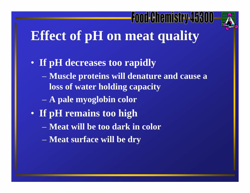
Effect of pH on meat quality
• If pH decreases too rapidly– Muscle proteins will denature and cause a
loss of water holding capacity– A pale myoglobin color
• If pH remains too high– Meat will be too dark in color– Meat surface will be dry
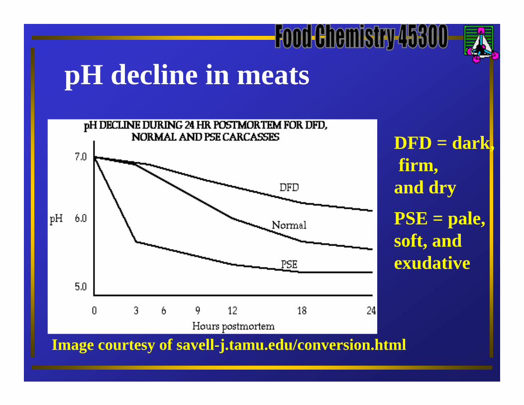
pH decline in meats
Image courtesy of savell-j.tamu.edu/conversion.html
DFD = dark,firm,
and dry
PSE = pale,soft, andexudative

Effect of pH on water holding capacity
Image courtesy of savell-j.tamu.edu/conversion.html
IEP ~ 5.2

Effect of pH on meat color (pH too high)
Image courtesy of savell-j.tamu.edu/conversion.html
An exampleof darkcutting beefor lamb (DFD meat)

Effect of pH on meat color (pH too low)
An exampleof pale, soft and exudative chicken breast(PSE meat)
•OK •pH too low

Generation of meat flavor potentiators during aging• As meat ages ATP is converted into ADP,
then AMP, and finally in IMP (inosine monophosphate) and ammonia
• These molecules provide some of the characteristic flavors of cooked meat

Rigor mortis and aging
• Rigor is a stiffening of the carcass a short time post mortem
• As muscle ATP is depleted and cannot be replaced, actin and myosin react to form the contracted muscle protein actomyosin

Rigor mortis and aging
• Accompanying rigor are– Loss of muscle elasticity and extensibility– Increase in muscle tension– Sarcomere shortening
• Note that meat cooked in rigor tends to be quite tough

Effect of rigor on meat tenderness
Rigor mortis and aging
• Aging is the process of simply holding the meat so as to allow for resolution of rigor (muscle relaxation)
• Alterations in the structure of the myofibrils is noted during this process

Rigor mortis and aging
• The microscopic appearance of the sarcomere changes– Some actin filaments become dissociated
from the Z line– The Z line itself becomes less distinct– Sarcomere length increases during aging– Muscle protein increases its water holding
capacity