mbbs ims msu
20
-
Upload
mbbs-ims-msu -
Category
Health & Medicine
-
view
1.478 -
download
1
Transcript of mbbs ims msu


Muscles of the scalp: Occipito-frontalis
•Origin: occipital belly from the highest
nuchal line. Frontal belly from the skin
and superficial fascia of the
eyebrows.
•Insertion: both bellies are inserted into
the epicranial aponeurosis.
•Nerve supply: occipital belly from
posterior auricular branch of facial
nerve and frontal belly from temporal
branch of facial nerve.
•Action: moves the superficial 3 layers
together, and raises the eyebrows.Superficial temporal artery-
frontal Superficial temporal
artery

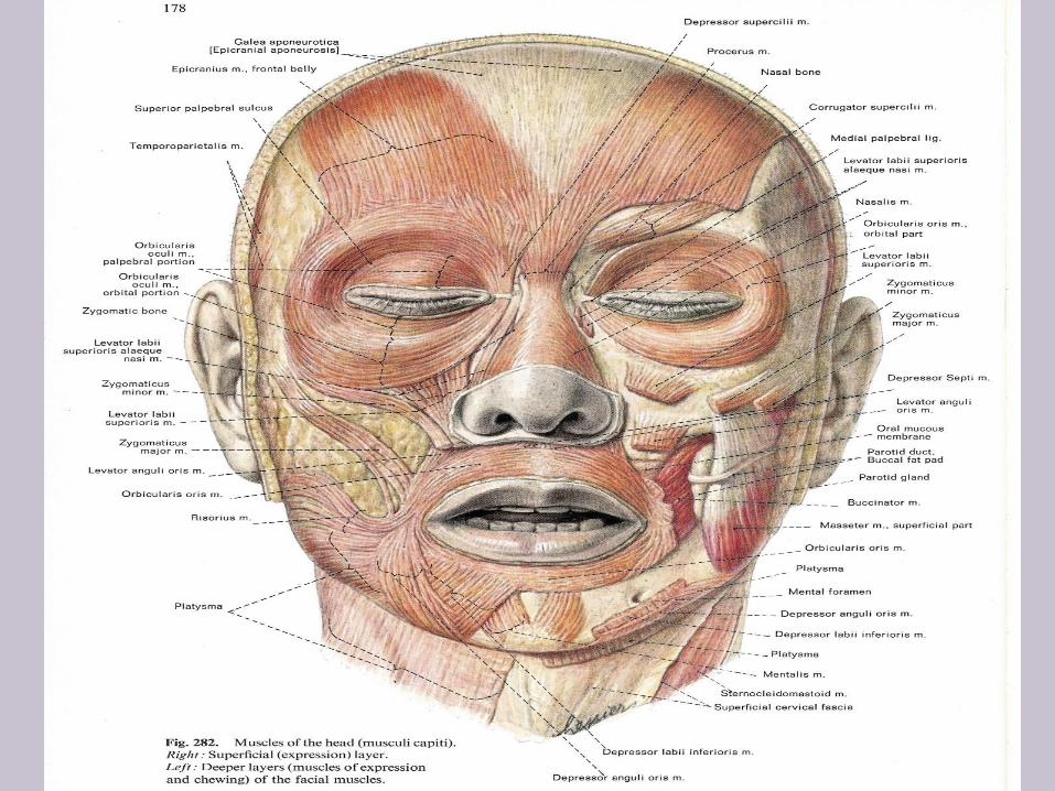
Muscles of facial expression• Present in the superficial fascia of the face.
• Inserted into the skin of the face.
• Related to the three main orifices of the face (either sphincters or dilator).
• Most important three are:
• Orbicularis oculi.
• Orbicularis oris.
• Buccinator.

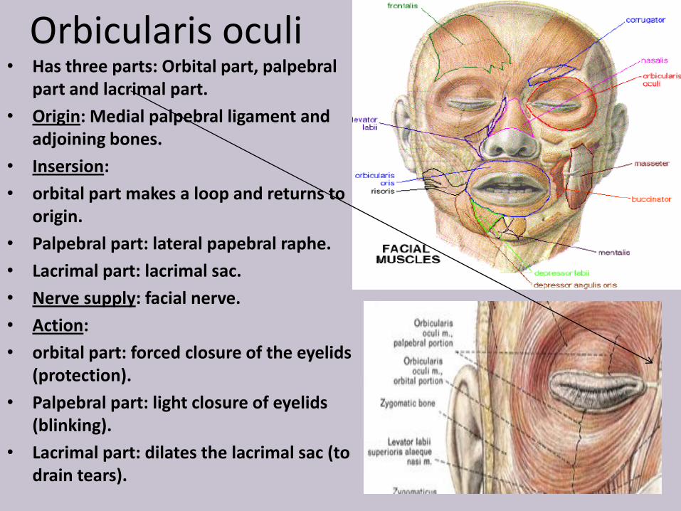
Orbicularis oculi• Has three parts: Orbital part, palpebral
part and lacrimal part.
• Origin: Medial palpebral ligament and adjoining bones.
• Insersion:
• orbital part makes a loop and returns to origin.
• Palpebral part: lateral papebral raphe.
• Lacrimal part: lacrimal sac.
• Nerve supply: facial nerve.
• Action:
• orbital part: forced closure of the eyelids (protection).
• Palpebral part: light closure of eyelids (blinking).
• Lacrimal part: dilates the lacrimal sac (to drain tears).

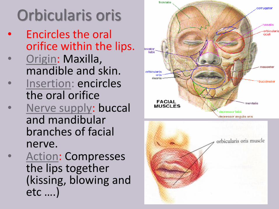
Orbicularis oris• Encircles the oral
orifice within the lips.• Origin: Maxilla,
mandible and skin.• Insertion: encircles
the oral orifice• Nerve supply: buccal
and mandibularbranches of facial nerve.
• Action: Compresses the lips together (kissing, blowing and etc ….)

Buccinators• Origin: outer surface of the alveolar
margins of maxilla and mandible
opposite the molar teeth and pterygomandibular raphe.
• Insertion:
• Upper fibers enter the upper lip
to be attached to fibers of opposite side.
• Lower fibers enter the lower lip to be attached to fibers of opposite side.
• Middle fibers decussate at the angle of the mouth and form the orbicularis oris muscle.
• Nerve supply: buccal branch of facial nerve.
Action: Compresses the cheeks lips against the teeth.
This muscle is pierced by the duct of the parotid salivary gland

External
carotid artery
Superficial
temporal
artery- frontal
Superficial
temporal
artery

The arterial and venous supply to the
face is seen in the diagram. They are
the: Facial artery
inferior labial
superior labial
angular
Facial vein
Superficial temporal artery
Superficial temporal vein
The facial vein is important clinically
because it has a direct connection to
the ophthalmic vein and then to a deep
venous sinus within the cranial cavity,
the cavernous sinus.
Bacteria can enter the facial vein and
gain access to internal cranial structures
resulting in infection there.
This is probably the reason our mothers
always said not to squeeze our pimples.


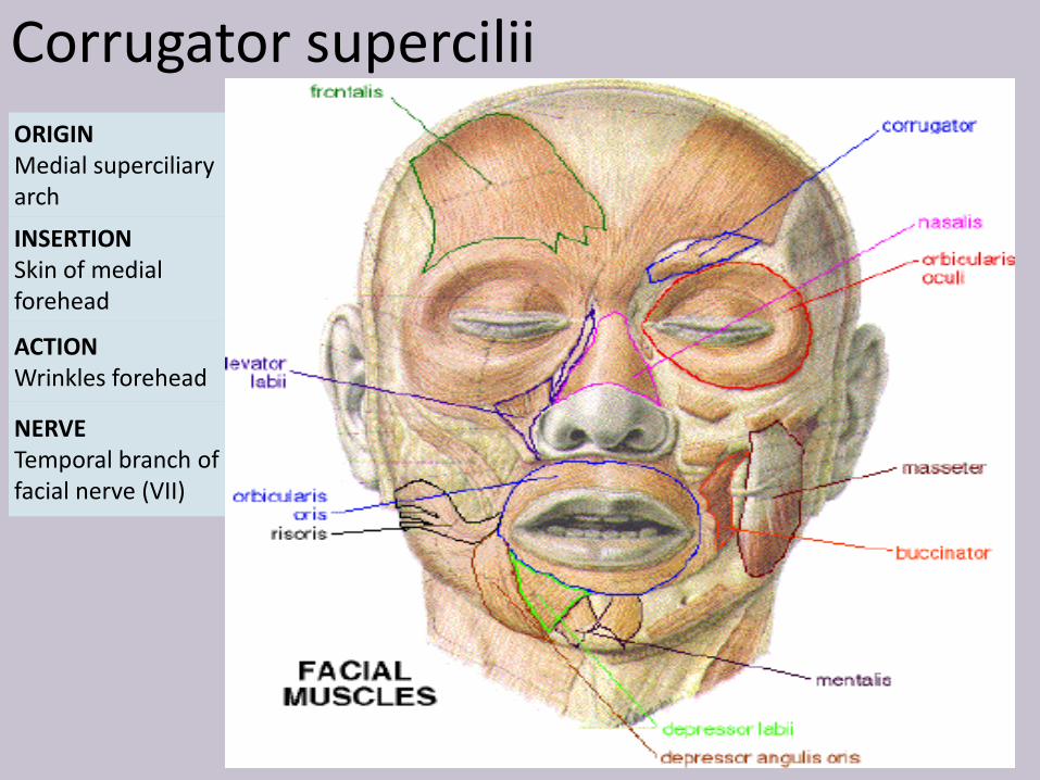
Corrugator supercilii
ORIGINMedial superciliaryarch
INSERTIONSkin of medial forehead
ACTIONWrinkles forehead
NERVETemporal branch of facial nerve (VII)