Maxillofacial and Ocular Injuries - A surgical community ...€¦ · • Symphysis Mandible –100...
62
Transcript of Maxillofacial and Ocular Injuries - A surgical community ...€¦ · • Symphysis Mandible –100...
Maxillofacial and Ocular Injuries
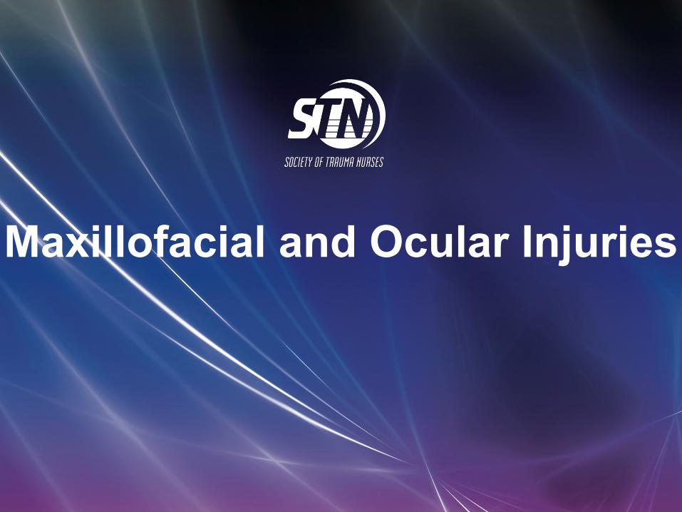
Objectives
At the conclusion of this presentation the participant will be able to:
• Identify the key anatomical structures of the face and eye and the impact of force on those structures
• Discuss assessment priorities for a patient with maxillofacial and ocular injuries
• Prioritize the care of a patient with facial and ocular injuries
• Discuss psychosocial support for a patient with maxillofacial and ocular injuries

Mechanism of Injury
Low velocity
High velocity

Pathophysiology
• Bones of face make up the most complex skeletal area of the body
• Maxillofacial fractures result from either blunt or penetrating trauma

Pathophysiology• ‘G’ force is a measure of acceleration
not produced by gravity • High Impact:
• Supraorbital rim – 200 G • Symphysis Mandible –100 G • Frontal – 100 G • Angle mandible – 70 G
• Low Impact: • Zygoma – 50 G • Nasal bone – 30 G


Etiology
• 60% of patients with severe facial trauma have multisystem trauma and the potential for airway compromise
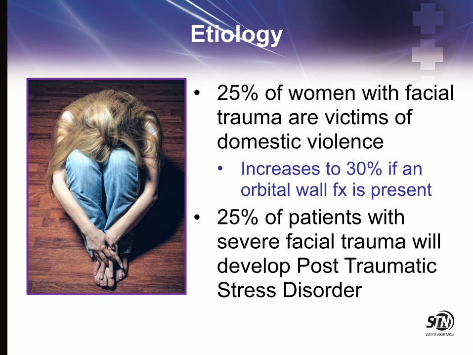
Etiology
• 25% of women with facial trauma are victims of domestic violence • Increases to 30% if an
orbital wall fx is present • 25% of patients with
severe facial trauma will develop Post Traumatic Stress Disorder

Ocular StructuresHuman Eye Anatomy

Bony Orbit• Roof
• Frontal bone • Sphenoid
• Medial wall • Maxilla, • lacrimal, ethmoid • body of sphenoid
• Floor • Maxilla • Palatine • Zygoma
• Lateral • Zygoma and greater
sphenoid
Frontal
MaxillaZygoma
Sphenoid
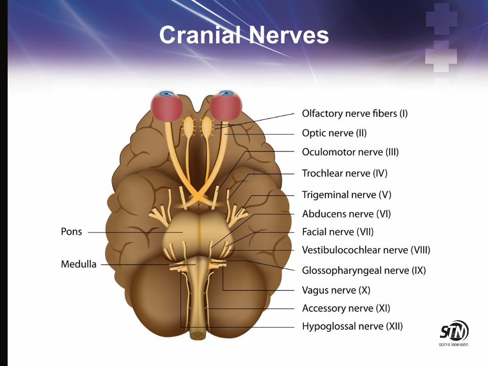
Cranial Nerves

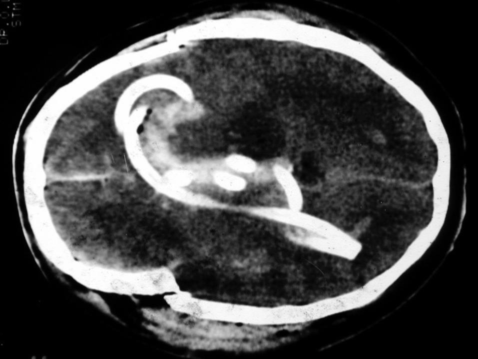
Orbital Fractures
Image found on Wikimedia.com
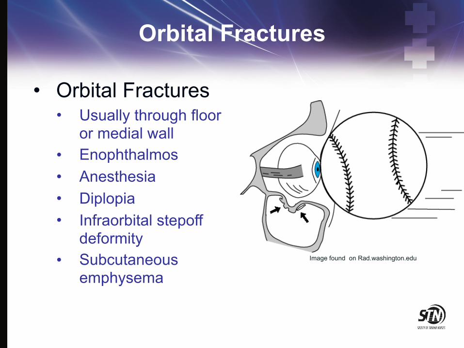
Orbital Fractures
• Orbital Fractures • Usually through floor
or medial wall • Enophthalmos • Anesthesia • Diplopia • Infraorbital stepoff
deformity • Subcutaneous
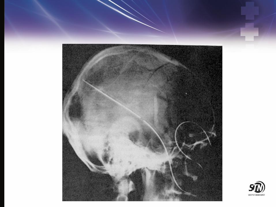
emphysemaImage found on Rad.washington.edu
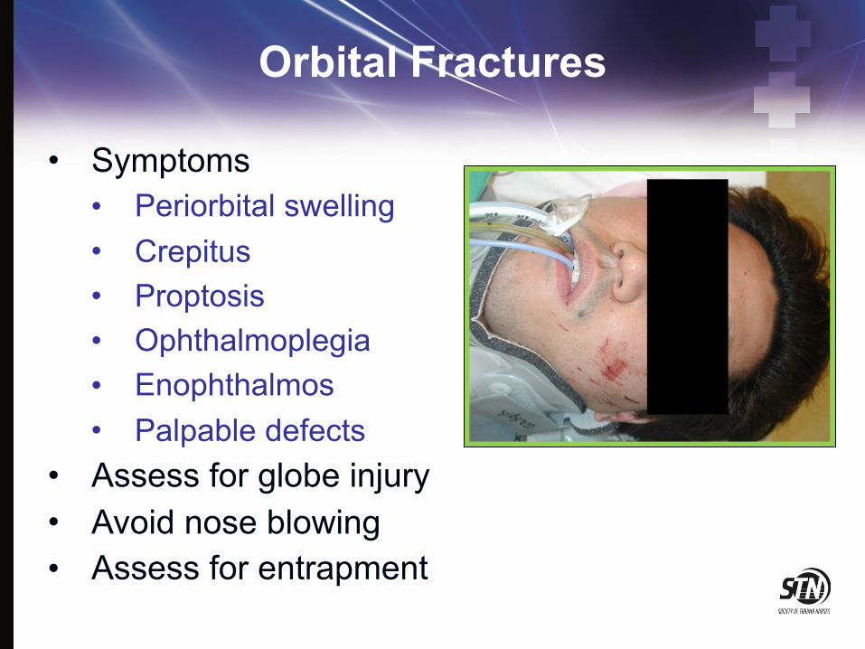
Orbital Fractures
• Symptoms • Periorbital swelling • Crepitus • Proptosis • Ophthalmoplegia • Enophthalmos • Palpable defects
• Assess for globe injury • Avoid nose blowing • Assess for entrapment
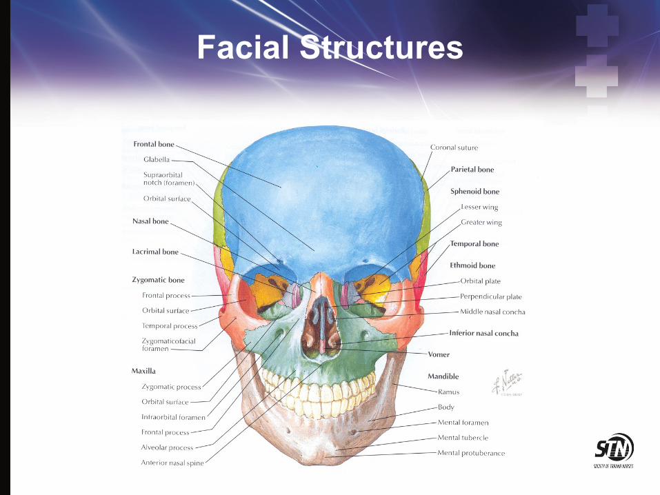
Facial Structures

LeFort I Fracture
Image found on Wikimedia.com

LeFort II Fracture
Image found on Wikimedia.com

LeFort III Fracture
Image found on Wiimedia.com

Le Fort Fractures
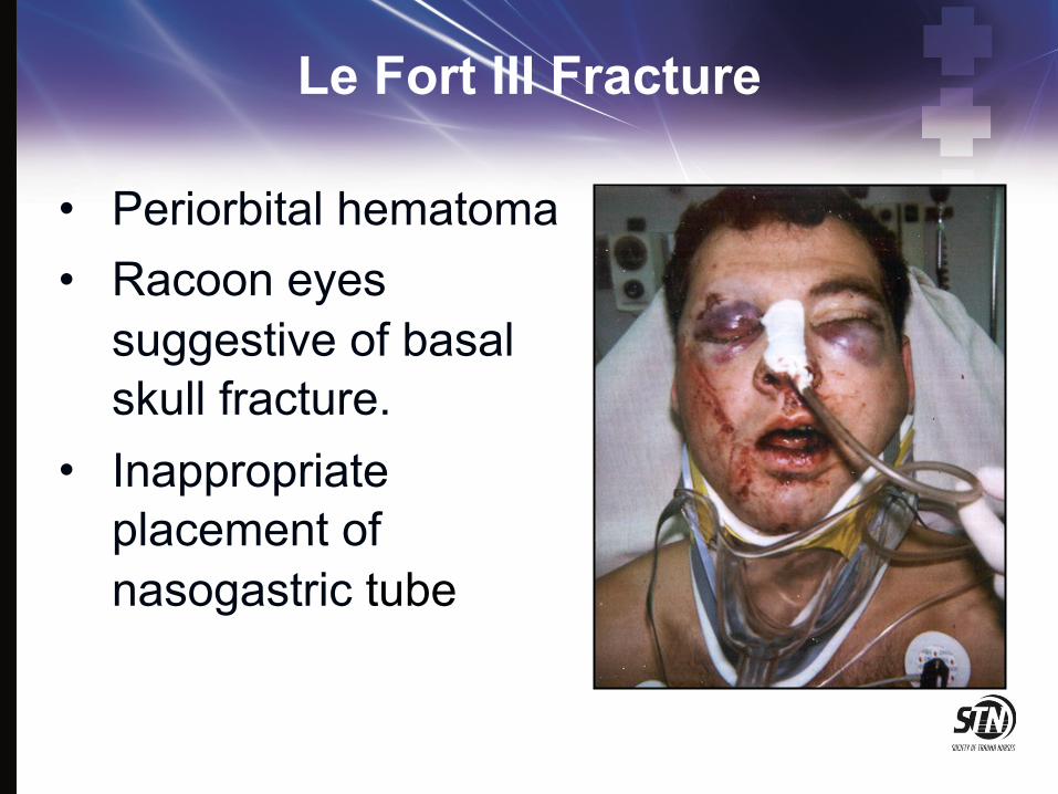
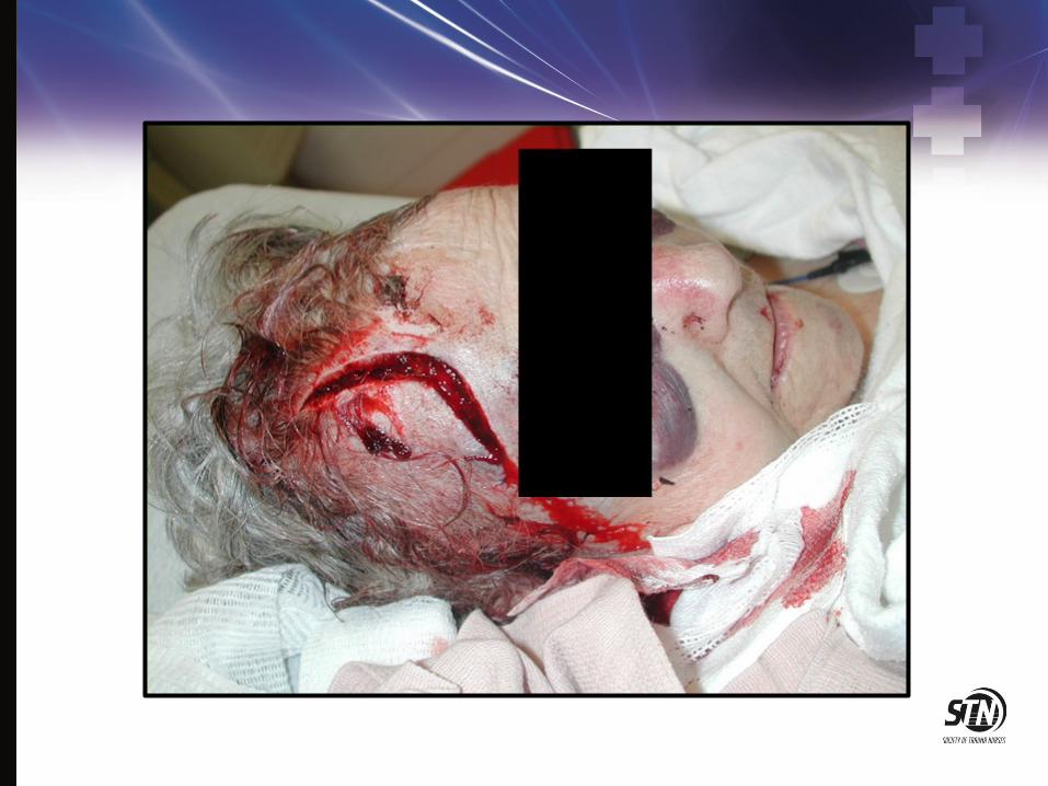
Le Fort III Fracture
• Periorbital hematoma • Racoon eyes
suggestive of basal skull fracture.
• Inappropriate placement of nasogastric tube
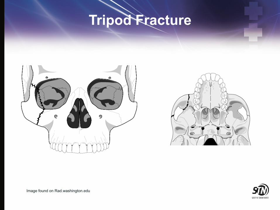
Tripod Fracture
Image found on Rad.washington.edu

Orbitozygomatic Fractures
• Complex fractures of the zygoma and orbital floor
• May have double vision, ocular proptosis or enophthalmos
• Must assess for entrapment of extraocular muscles
• Surgical management directed at decompression of entrapped muscles and anatomic realignment of zygoma

Naso-Ethmoidal-Orbital Fracture
• Fractures that extend into the nose through the ethmoid bones.
• Associated with lacrimal disruption and dural tears.
• Suspect if there is trauma to the nose or medial orbit.
• Patients complain of pain on eye movement.

Mandibular Fractures
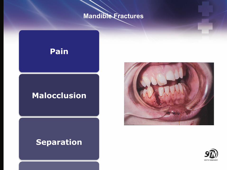
Mandible Fractures
Pain
Malocclusion
Separation
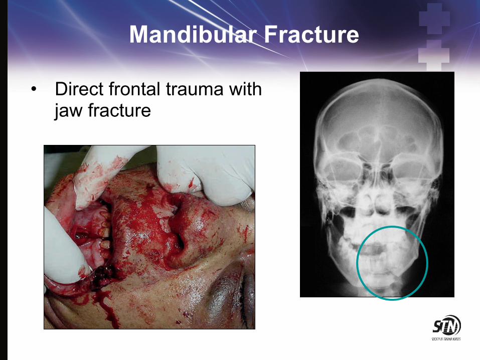
Mandibular Fracture
• Direct frontal trauma with jaw fracture

Mandibular Fractures Treatment
• Nondisplaced fractures: • Analgesics • Soft diet • oral surgery referral in 1-2 days
• Displaced fractures, open fractures and fractures with associated dental trauma • Urgent oral surgery consultation
• All fractures should be treated with antibiotics and tetanus prophylaxis.

Maxillofacial Injuries General Assessment
• ABC’s • Assess for symmetry of facial
structures • Assess for paresthesias • Assess symmetry of facial
movements • Assess the ears, nose and
oral cavity for occult lacerations, hematomas
• Palpate for crepitus, tenderness or deformity
• Assess sense of smell

Ocular Assessment
• Visual acuity • Pupil assessment • Extraocular movements • Eye position and
movement • Intraocular pressure • Fundoscopic exam

Physical Examination
• Inspect open wounds for foreign bodies
• Palpate the entire face • Supraorbital and
Infraorbital rim • Zygomatic-frontal suture • Zygomatic arches

Physical Examination
• Inspect the nose for asymmetry, telecanthus, widening of the nasal bridge
• Inspect nasal septum for septal hematoma, CSF or blood
• Palpate nose for crepitus, deformity and subcutaneous air
• Palpate the zygoma along its arch and its articulations with the maxilla, frontal and temporal bone
Physical Examination
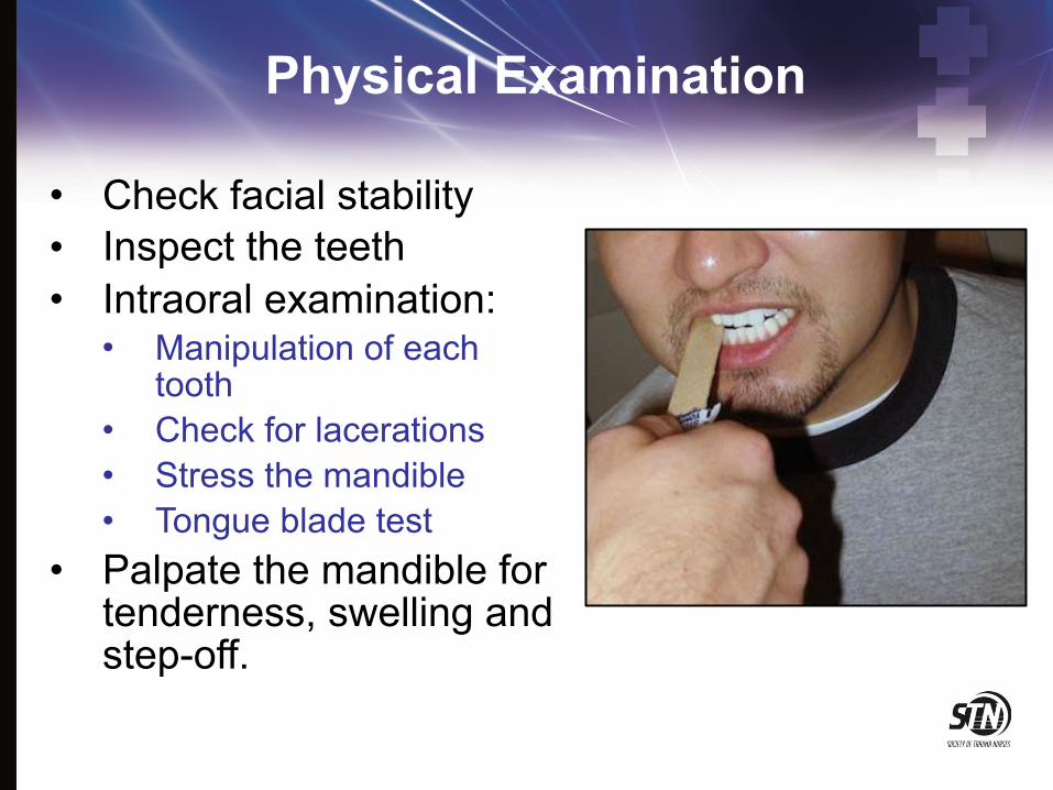
• Check facial stability • Inspect the teeth • Intraoral examination:
• Manipulation of each tooth
• Check for lacerations • Stress the mandible • Tongue blade test
• Palpate the mandible for tenderness, swelling and step-off.

Physical Examination
• Check visual acuity • Check pupils for
roundness and reactivity • Examine the eyelids for
lacerations • Test extra ocular
muscles • Palpate around the
entire orbits
Physical Examination
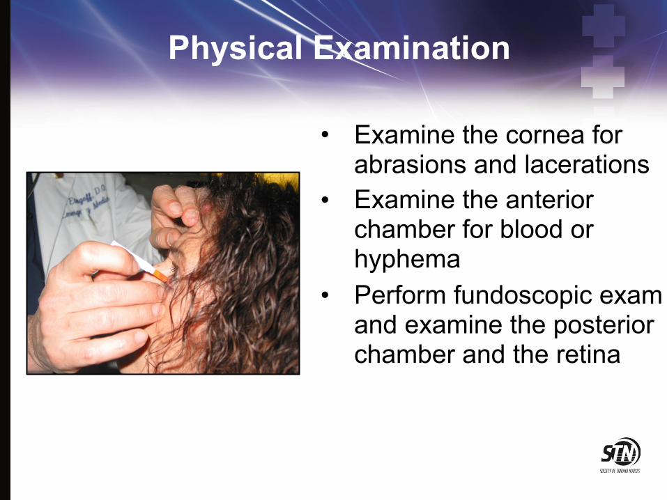
• Examine the cornea for abrasions and lacerations
• Examine the anterior chamber for blood or hyphema
• Perform fundoscopic exam and examine the posterior chamber and the retina

Airway Management
• Protect and maintain airway • Pull tongue forward with
padded forceps or sutures • Endotracheal intubation • Anticipate need for
cricothyroidotomy • Prevent aspiration • Ensure adequate
oxygenation and ventilation
Airway Management
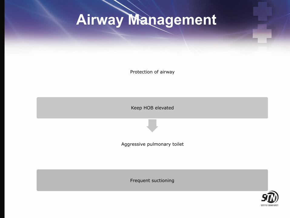
Protection of airway
Keep HOB elevated
Aggressive pulmonary toilet
Frequent suctioning
Management
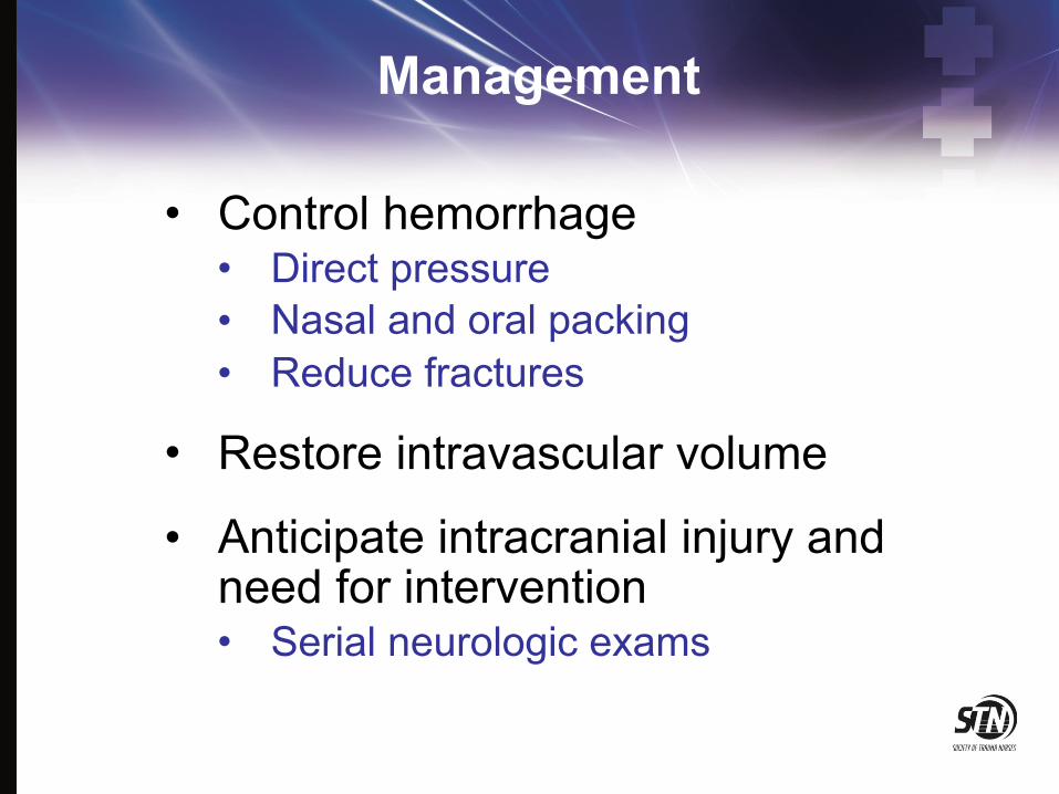
• Control hemorrhage • Direct pressure • Nasal and oral packing • Reduce fractures
• Restore intravascular volume
• Anticipate intracranial injury and need for intervention • Serial neurologic exams
Management
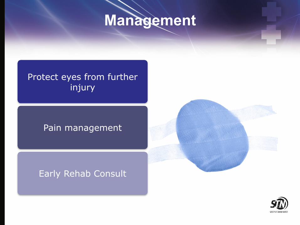
Protect eyes from further injury
Pain management
Early Rehab Consult

Management
• Nutrition management • Early initiation of enteral
feeding
• Keep HOB elevated
• Evaluate for swallowing dysfunction prior to oral feeding
• Wire cutters at bedside at all times

Management
• Prevention of infection • Perioperative antibiotics
• Frequent oral lavage
• Minimize nasal packing and tubes
• Decongestants
• Avoid blowing nose
• Avoid foreign bodies or instrumentation in nares or ear canal

Direct Eye Trauma

Blast Injury: Thermal Injury

Thermal Injury
• Eye is usually spared • Corneal exposure
may occur as burn heals and skin contracts

Corneal Abrasion

Chemical Burns

Traumatic Hyphema
Image courtesy of EyeMac Development

Traumatic Hyphema
• Limit activity • Keep HOB elevated • Protect the eye • Cycloplegic agents • Monitor for re-bleeding • Avoid NSAIDS and
anticoagulants • Aminocaproic acid
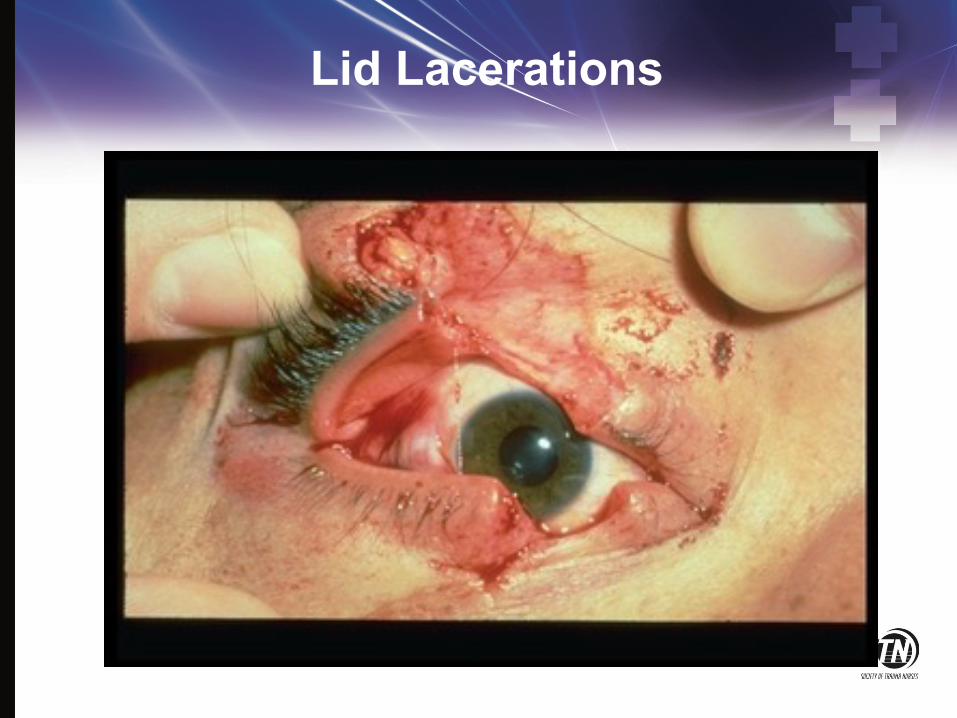
Lid Lacerations

Lid Laceration
• REFER for • Depth • Extensive tissue loss
• REFER for location • medial • margin
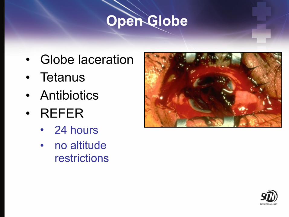
Open Globe
• Globe laceration • Tetanus • Antibiotics • REFER
• 24 hours • no altitude
restrictions

Open Globe
• Minimize additional damage • Make sure a shield is used • Do not use a patch which
applies pressure • Avoid bearing down • Be prepared for patient to
go to the OR
• NPO

Complications Sympathetic Ophthalmia
• Inflammatory condition • Common after penetrating injury or
ruptured globe • Occurs 5 days to many years after injury • Results in loss of vision of uninjured eye • Prevented by early enucleation of
injured eye
Psychosocial Support
• Provide communication aids • Frequent positive reinforcement • Early referrals to psychiatric liaisons or
counselors • Early referrals to community agencies
for the blind • Referrals for home safety evaluations • Referrals to local and state agencies for
financial assistance
Patient and Family Education
• Reinforce surgical plan of care • Medications • Nutrition management • Wound care • Tracheostomy care • Avoid direct sunlight for 6-12 months • Use of cosmetics

Summary
• Facial and ocular trauma requires a comprehensive multidisciplinary team to maximize outcomes
• Early incorporation of rehabilitation services is necessary for functional recovery
• Overall prognosis of reconstruction may take months or years