MAXILLARY SINUSITIS CHILDREN · maxillary sinusitis in children differs fromthe adult infection by...
9
CHRONIC MAXILLARY SINUSITIS IN CHILDREN BY J. F. BIRRELL From the Ear, Nose and Throat Department, the Royal Hospitalfor Sick Children, Edinburgh (RECEIVED FOR PUBLICATION APRIL 4, 1951) There has been a considerable volume of literature on the subject of sinusitis in childhood during the past 30 years; much of it deals with the results obtained in routine proof puncture or antral lavage. For example, Mollison and Kendall (1922) found evidence of sinus infection in 22% of 102 routine tonsil and adenoid cases. Crooks and Signy (1936) reported 24 positive proof punctures in 100 routine tonsil and adenoid cases, while Gerrie (1939) puts the percentage at 29 in 300 such patients. Carrying this further, we find that Carmack (1931) reports positive findings in 14-2% of a series of 211 tonsil and adenoid cases from which he had excluded all known sinusitic and allergic patients. There are two reports of proof puncture results in known or suspected sinus cases. Crooks (1947) found evidence of infection in 59-6% of 570 cases with opaque antra seen during 1946, while Walker (1947) obtained positive findings in 76 % of 442 cases with sufficient signs and symptoms to warrant puncture out of 1,779 tonsil and adenoid patients. Lastly, Ebbs (1938) has obtained post-mortem evidence of sinus infection in 30- 6% of 496 routine necropsies. It is small wonder that sinusitis has attracted considerable attention during this period, and the importance given to this condition is reflected in the space devoted to it in StClair Thomson's book. In the second edition, published in 1916, the condition is dismissed as rare, but in the current edition, published in 1948, several pages are given to it. The implications are obvious. Ebbs' figures suggest that nearly one-third of all children suffer from sinusitis, while other reports imply that one in every four or five children requiring removal of tonsils and adenoids also needs attention to the maxillary antra. If we accept this premise whole- heartedly, we must also be willing to accept the consequences, or be prepared to explain any anomalies in the deductions arising from this premise. The first question to be answered is how chronic sinusitis arises. It is generally conceded that, in adults at least, chronic sinusitis does not arise per se, but develops from an acute maxillary sinusitis in which the infected material cannot, or does not, drain away. Among adults, many more people suffer from acute maxillary sinusitis than ever develop chronic sinusitis, as most cases of acute infection return to normal. It is natural to assume that in children there will be many more cases of acute maxillary sinusitis for the same reason. Yet it is generally stated that acute maxillary sinusitis in children is an uncommon condition, and the records of the E.N.T. Department of the Royal Hospital for Sick Children in Edinburgh tend to support this opinion, as during the past five years, when over 10,000 new patients were examined, only one case of acute maxillary sinusitis was seen. How is this to be explained ? It may be that, as with acute tonsillitis, the young child makes no complaint of infra-orbital pain, accepting it as part of an acute coryza, or ascribing it to toothache. If this be so, it is possible that the great majority of children suffer from acute maxillary sinusitis, which results in one child in every three, four or five having chronic maxillary sinusitis. Or again it may be that chronic maxillary sinusitis in children differs from the adult infection by arising per se in the antrum. Might it not be that chronic sinusitis arises from acute sinusitis, and is not as common as one is led to believe ? The second question to be considered is concerned with the fact that during childhood the maxillary sinus, in common with other sinuses, is in a state of active development. It grows to fill the maxilla as the teeth descend to the alveolar margin and finally erupt. If there is chronic infection lying close to these developing teeth, why is there no evidence of the eruption of unhealthy teeth, such as one finds after osteomyelitis of the maxilla ? And, again, taking as a corollary the development. of the mastoid air cells, why is there no arrested aeration and growth of the maxillary air sinus ? The lack of development of a normally cellular 2 copyright. on June 1, 2020 by guest. Protected by http://adc.bmj.com/ Arch Dis Child: first published as 10.1136/adc.27.131.1 on 1 February 1952. Downloaded from
Transcript of MAXILLARY SINUSITIS CHILDREN · maxillary sinusitis in children differs fromthe adult infection by...
CHRONIC MAXILLARY SINUSITIS IN CHILDRENBY
J. F. BIRRELLFrom the Ear, Nose and Throat Department, the Royal Hospitalfor Sick Children, Edinburgh
(RECEIVED FOR PUBLICATION APRIL 4, 1951)
There has been a considerable volume of literatureon the subject of sinusitis in childhood during thepast 30 years; much of it deals with the resultsobtained in routine proof puncture or antral lavage.For example, Mollison and Kendall (1922) foundevidence of sinus infection in 22% of 102 routinetonsil and adenoid cases. Crooks and Signy (1936)reported 24 positive proof punctures in 100 routinetonsil and adenoid cases, while Gerrie (1939) putsthe percentage at 29 in 300 such patients. Carryingthis further, we find that Carmack (1931) reportspositive findings in 14-2% of a series of 211 tonsiland adenoid cases from which he had excluded allknown sinusitic and allergic patients. There are tworeports of proof puncture results in known orsuspected sinus cases. Crooks (1947) found evidenceof infection in 59-6% of 570 cases with opaqueantra seen during 1946, while Walker (1947)obtained positive findings in 76% of 442 cases withsufficient signs and symptoms to warrant punctureout of 1,779 tonsil and adenoid patients. Lastly,Ebbs (1938) has obtained post-mortem evidence ofsinus infection in 30- 6% of 496 routine necropsies.
It is small wonder that sinusitis has attractedconsiderable attention during this period, and theimportance given to this condition is reflected in thespace devoted to it in StClair Thomson's book.In the second edition, published in 1916, thecondition is dismissed as rare, but in the currentedition, published in 1948, several pages are givento it. The implications are obvious. Ebbs' figuressuggest that nearly one-third of all children sufferfrom sinusitis, while other reports imply that onein every four or five children requiring removal oftonsils and adenoids also needs attention to themaxillary antra. If we accept this premise whole-heartedly, we must also be willing to accept theconsequences, or be prepared to explain anyanomalies in the deductions arising from thispremise.The first question to be answered is how chronic
sinusitis arises. It is generally conceded that, in
adults at least, chronic sinusitis does not ariseper se, but develops from an acute maxillarysinusitis in which the infected material cannot, ordoes not, drain away. Among adults, many morepeople suffer from acute maxillary sinusitis thanever develop chronic sinusitis, as most cases of acuteinfection return to normal. It is natural to assumethat in children there will be many more cases ofacute maxillary sinusitis for the same reason. Yetit is generally stated that acute maxillary sinusitisin children is an uncommon condition, and therecords of the E.N.T. Department of the RoyalHospital for Sick Children in Edinburgh tend tosupport this opinion, as during the past five years,when over 10,000 new patients were examined, onlyone case of acute maxillary sinusitis was seen. Howis this to be explained ? It may be that, as withacute tonsillitis, the young child makes no complaintof infra-orbital pain, accepting it as part of an acutecoryza, or ascribing it to toothache. If this be so,it is possible that the great majority of children sufferfrom acute maxillary sinusitis, which results in onechild in every three, four or five having chronicmaxillary sinusitis. Or again it may be that chronicmaxillary sinusitis in children differs from the adultinfection by arising per se in the antrum. Might itnot be that chronic sinusitis arises from acutesinusitis, and is not as common as one is led tobelieve ?The second question to be considered is concerned
with the fact that during childhood the maxillarysinus, in common with other sinuses, is in a stateof active development. It grows to fill the maxillaas the teeth descend to the alveolar margin andfinally erupt. If there is chronic infection lyingclose to these developing teeth, why is there noevidence of the eruption of unhealthy teeth, such asone finds after osteomyelitis of the maxilla ? And,again, taking as a corollary the development. ofthe mastoid air cells, why is there no arrestedaeration and growth of the maxillary air sinus ?The lack of development of a normally cellular
2
copyright. on June 1, 2020 by guest. P
rotected byhttp://adc.bm
j.com/
Arch D
is Child: first published as 10.1136/adc.27.131.1 on 1 F
ebruary 1952. Dow
nloaded from
A4RCHI1-ES OF DISE.4SE l\ CHILDHOOD
mastoid has been associated xxith the presence ofotitis media in infancv and earl! childhood. Is onenot ju\titied in as,suming that similar changes shouldbe found in the antra of adults. and that one ine\erx- three. four. or fixe adults should shoxw arresteddexelopment of the paranasal air cells. eitherunilaterallv or bilaterallv '
Thirdix. if such a great percentage of childrenhas been shox'n in the past 20 or 30 vears to haxehad such a degree of sinusitis as to require energeticsurgical correction. one must ask xhat happened tosinus infections in children in the past ' Thetreatment of otitis media in childhood is beingprosecuted x-ith zeal in the hope of materiallyreducing the numbers of adults xsith chronic otitismedia. \\ ill these enerzetic measures no"- adoptedin the treatment of sinusitis in children similarlvreduce the numbers of chronic sinusitis patients in20 vears ? If so. the measures xvill be Justified. butif not, one must assume that chronic sinusitis inchildhood does not persist into adult life. In othervkords. so-called chronic sinusitis in children mustbe considered a self-limiting disease. thus differingfrom chronic infection elsexkhere. If it is self-limitin . >-hen does it clear up. and xshx ' Theopinion of Dean (1925) and others that remoxal ofthe adenoids and tonsils xsill cure 80'. of cases ofchronic sinusitis lends support to the theory thatthe natural regression of lymphoid tissue in thenasopharynx about puberty may determine thislimitation of infection in the sinuses. The 'iexk iscurrentlv held in many quarters. on the other hand,that it is the sinusitis which causes and maintainsthe infection in. and the enlargement of. adenoids.If this xiexw is correct, how- can one explain eitherthe disappearance of infection or the atrophy of theadenoids ' I doubt if it can be explained on thishypothesis. unless infection does not. in fact.disappear. but is really verv uncommon.The impression has been zained in the operatina
theatre that true chronic maxillar- sinusitis is indeeduncommon. and the case notes of all the proofpunctures. 240 in all. performed betveen Januar- 1.1946. and October 31. 1949. wsere reviexved early-in 19'0. Arisina from these findines certainin-'estigations haxe since been made. and this reportis concemed. firstly. xsith the results obtained.secondly %vith the in\estigations. and thirdly xsitha discussion of the problem in relation to theliterature on the subject. It should. hosexver. beemphasized that this paper does not represent acontrolled or planned inxestigation. but is a baldreport of the findings.One other point should be stressed at this juncture.
About half-xway through the period under rexie"-.it occurred to me that as these cases av-aited their
turn on the ordinary tonsil list. the sinus infectionmiaht haxe undergone resolution betmxeen the dateof diagnosis and the date on xshich the child xvassent for. Accordingly. these patients \%ere takenout of their tum at an extra operatina session so thatbefore the end of the series cases diagnosed assinusitis xsere operated upon %kithin a xseek or tvxo.The result of this procedure did not. in any material,x ay. alter the percentage of positixe results. andthe series may thus be treated as a xshole.
Analvsis of Case Notes
Sex. Of 240 case sheets examined. 160 relatedto boys and 80 to girls. a ratio of boys to zirls of 2:1.
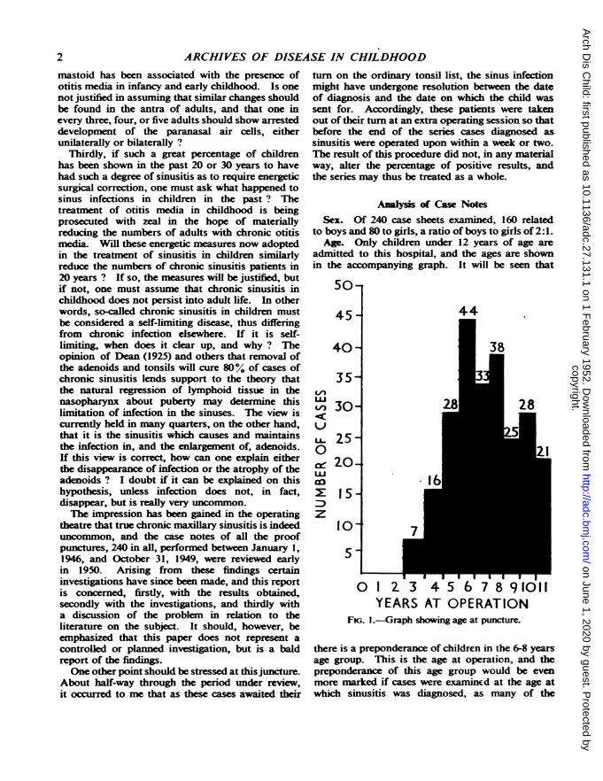
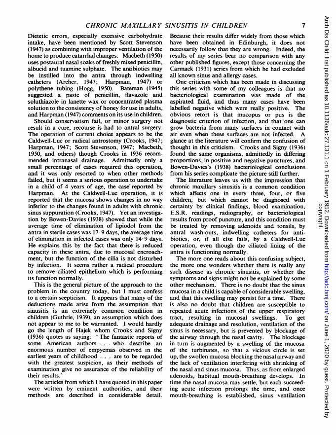
Age. Only children under 12'ears of age areadmitted to this hospital. and the ages are shoxwnin the accompanying graph. It xxill be seen that
50-
45 -
40-
35-VUL/30-
U± 25-0c: 20-LU
DEy 15 -
z10-
5-
0 1 2 3YEARS
4 5 6 7 P 91011AT C0ERATION
Fic,. 1.-Graph shoxx ing age pllncture.
there is a preponderance of chiIdrcn in the 6-s yearsage group. This is the age at cperauicn. and thepreponderance of this age group %xxcu'd be exenmore marked if cases xsere examincd at the age atx-hich sinusitis xxasa diaanosed. as nman'- of the
copyright. on June 1, 2020 by guest. P
rotected byhttp://adc.bm
j.com/
Arch D
is Child: first published as 10.1136/adc.27.131.1 on 1 F
ebruary 1952. Dow
nloaded from
CHRONIC MAXILLARY SINUSITIS IN CHILDREN
9 to 12-year-old children had waited up to two yearsfor admission.
Symptoms. It is very difficult in a review such as
this to obtain an accurate picture of the presentingsymptoms, as case histories have been taken by anumber of different people-by residents, out-patient sisters, secretaries and surgeons. There isthus no unanimity of view, as would be obtainedin a planned investigation, such as that of Birdsall(1939).
For what they are worth, the following symptomshave been noted: nasal catarrh, nasal discharge, or
running nose (60 cases); frequent colds (40 cases);nasal obstruction or mouth breathing (35 cases);persistent cough (23 cases); bronchitis (10 cases);bronchiectasis (11 cases); headaches (19 cases);asthma (22 cases); hay fever (one case); allergicrhinitis (one case); discharging ears or deafness(27 cases); laryngitis (five cases); pharyngitis (onecase); epistaxis (two cases); night sweats (onecase); and acidosis (one case). In 39 case sheetsthere was insufficient history for the predominatingsymptom to be listed.
SigDs. On examination of the nose, the findingshave varied with the descriptive power of theexaminer, and I have aggregated these into groupsas follows: nose clear, i.e. a dry nose with no
swelling of the turbinates (58 cases); congested nose
(17 cases); allergic type of nose, i.e. pale, swollenturbinates (35 cases); nose containing unspecifieddischarge (50 cases); mucoid discharge (four cases);mucopurulent discharge (19 cases); purulent dis-charge (29 cases); crusted discharge (21 cases);and deflected septum (15 cases). No notes were
made on eight case sheets.Tonsils and Adenoids. Tonsils and adenoids were
present in 117 cases, and had been removed in117 cases. In the remaining six case sheets thereare no notes. When tonsils and adenoids were
present, their removal was carried out at the same
time as the puncture in 42 cases. Of the patientswhose tonsils had previously been removed,tonsillectomy was deemed inadequate in nine cases.
Thirty-four patients were recommended to haveadenoids (either the primary growth or the recur-
rence) removed at the same time as the puncture,and this is distinct from the 42 tonsil and adenoidoperations listed above.
Radiology. Positive radiological findings were
obtained in every case subjected to proof puncture,and no case was clinically labelled sinusitis until a
radiograph revealed either mucosal thickening or
opacity of the antrum. This aspect of the diagnosiswill be discussed in greater detail later.
Technique. The children were all dealt with as
in-patients. After pre-operative luminal the child
was anaesthetized with ethyl chloride vapourized ina Guy's inhaler. No local cocaine anaesthesia wasused. An opportunity was thus afforded ofpalpatingthe nasopharynx and removing any adenoid massrequiring surgery, and palpation and curettage isnow part of the routine procedure ofa proofpuncture.A Lichwitz trocar and cannula were used, and
sterile saline solution injected into the antrum froma 5 ml. syringe carrying a blunt-pointed needleinserted through the cannula. A 2-3 ml. quantityof the solution was injected into the antrum, thehead being rolled through 45- to the correspondingside. The solution was then reaspirated into thesyringe and examined macroscopically for mucopusor pus. If this were present, further wash-outs ofthis kind were used, or a Higginson's syringe wasattached to the cannula, and the- classical wash-outemployed. With this injection and reaspirationtechnique no accidents have occurred. This methodis thought to be the only accurate means of deter-mining the contents of an antrum, as the simpleantral wash-out, without preliminary injection andaspiration, carries with it all secretions lying in thenasal cavities and nasopharynx, and, indeed, inbronchiectatic subjects, much pulmonary secretionis added to the specimen during the cough associatedwith recovery from anaesthetic. No reliance can beplaced on the antral wash-out as a diagnostic guide.It is a therapeutic measure alone.
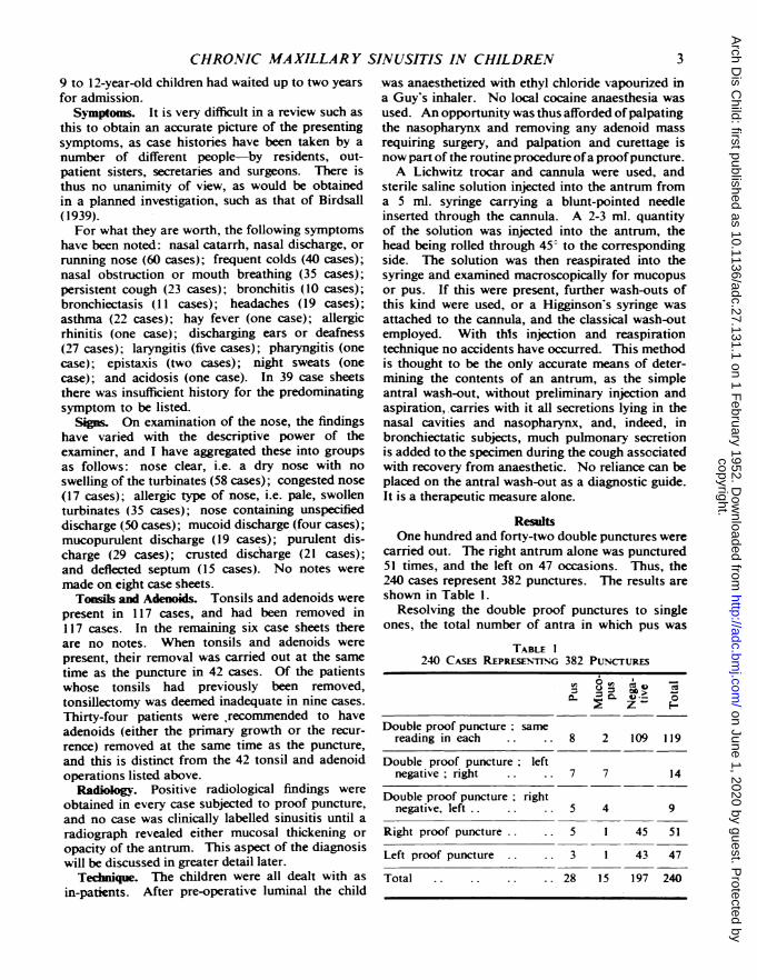
ResultsOne hundred and forty-two double punctures were
carried out. The right antrum alone was punctured51 times, and the left on 47 occasions. Thus, the240 cases represent 382 punctures. The results areshown in Table 1.
Resolving the double proof punctures to singleones, the total number of antra in which pus was
TABLE 1240 CASES REPRESENrING 382 PuNcruREs
tt0
Double proof puncture. samereading in each .. 8 2 109 119
Double proof puncture; leftnegative ; right .. 7 7 14
Double proof puncture rightnegative, left .. 5 4 9
Right proof puncture .. 5 1 45 51
Left proof puncture .. .. 3 1 43 47
Total .. .. .. .. 28 15 197 240
3
copyright. on June 1, 2020 by guest. P
rotected byhttp://adc.bm
j.com/
Arch D
is Child: first published as 10.1136/adc.27.131.1 on 1 F
ebruary 1952. Dow
nloaded from
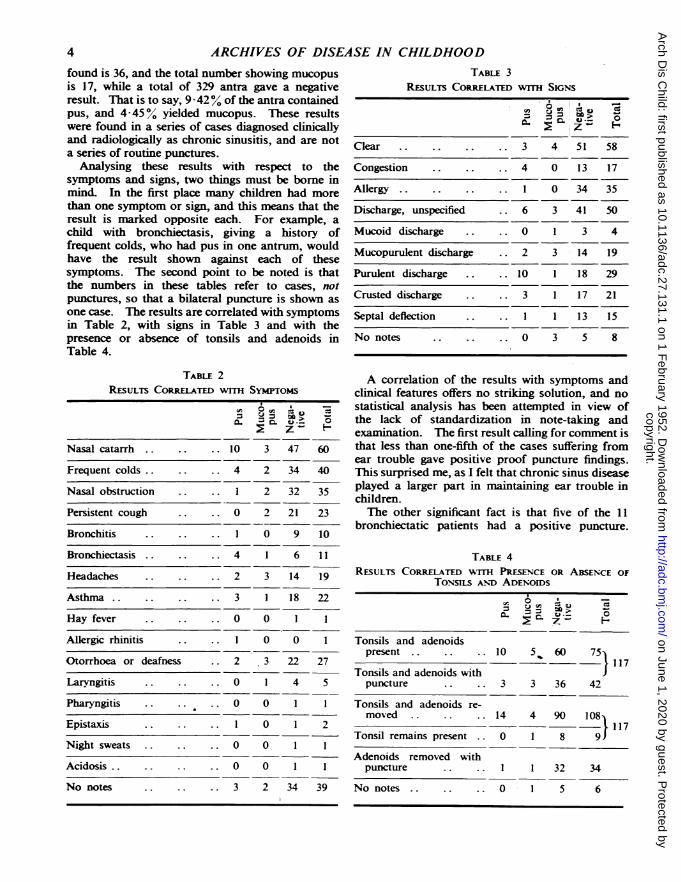
ARCHIVES OF DISEASE IN CHILDHOODfound is 36, and the total number showing mucopusis 17, while a total of 329 antra gave a negativeresult. That is to say, 9 42% of the antra containedpus, and 4 45o% yielded mucopus. These resultswere found in a series of cases diagnosed clinicallyand radiologically as chronic sinusitis, and are nota series of routine punctures.
Analysing these results with respect to thesymptoms and signs, two things must be borne inmind. In the first place many children had morethan one symptom or sign, and this means that theresult is marked opposite each. For example, achild with bronchiectasis, giving a history offrequent colds, who had pus in one antrum, wouldhave the result shown against each of thesesymptoms. The second point to be noted is thatthe numbers in these tables refer to cases, notpunctures, so that a bilateral puncture is shown asone case. The results are correlated with symptomsin Table 2, with signs in Table 3 and with thepresence or absence of tonsils and adenoids inTable 4.
TABLE 2RESULTS CoRRELATm wrrH SYMTroMs
Z-
Nasal catarrh .. .. .. 10 3 47 60
Frequent colds.. .. .. 4 2 34 40
Nasal obstruction .. .. 1 2 32 35
Persistent cough .. .. 0 2 21 23
Bronchitis .. .. .. 1 0 9 10
Bronchiectasis .. .. .. 4 1 6 11
Headaches .. .. .. 2 3 14 19
Asthma.. .. .. .. 3 1 18 22
Hay fever .. .. .. 0 0 1 1
Allergic rhinitis .. .. 1 0 0 1
Otorrhoea or deafness .. 2 3 22 27
Laryngitis .. .. .. 0 1 4 5
Pharyngitis .. .. .. 0 0 1 1
Epistaxis .. .. .. 1 0 1 2
Night sweats .. .. .. 0 0 1 1
Acidosis.. .. .. .. 0 0 1 1
No notes .. .. .. 3 2 34 39
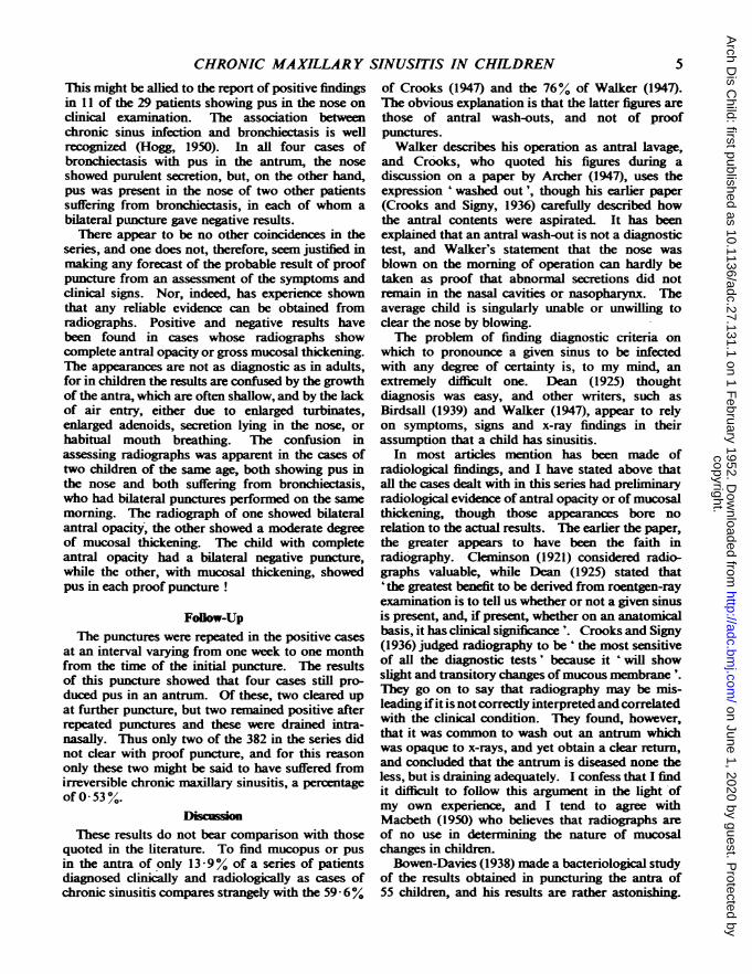
TABLE 3RESULTs CoRRELATED WITH SIGNS
C. H
Clear .. 3 4 51 58
Congestion .. 4 0 13 17
Allergy 1 0 34 35
Discharge, unspecified 6 3 41 50
Mucoid discharge 0 1 3 4
Mucopurulent discharge .. 2 3 14 19
Purulent discharge .. 10 1 18 29
Crusted discharge 3 1 17 21
Septal deflection I.. 1 13 15
No notes 0 3 5 8
A correlation of the results with symptoms andclinical features offers no striking solution, and nostatistical analysis has been attempted in view ofthe lack of standardization in note-taking andexamination. The first result calling for comment isthat less than one-fifth of the cases suffering fromear trouble gave positive proof puncture findings.This surprised me, as I felt that chronic sinus diseaseplayed a larger part in maintaining ear trouble inchildren.The other significant fact is that five of the 11
bronchiectatic patients had a positive puncture.
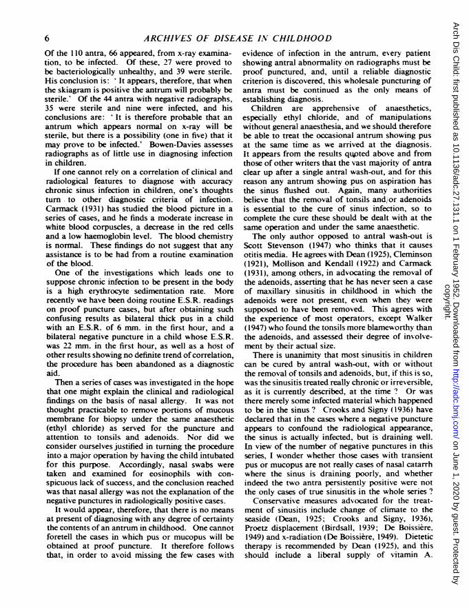
TABLE 4RESULTS CORRELATED WrrH PRESENCE OR ABSENCE OF
TONSILS AND ADENOIDSo
Tonsils and adenoidspresent .. .. .. 10 5, 60 75}
* S ~117Tonsils and adenoids with
puncture .. .. 3 3 36 42
Tonsils and adenoids re-moved .. .. .. 14 4 90 108
117Tonsil remains present .. 0 1 8 9
Adenoids removed withpuncture I.. .. 1 32 34
No notes .. .. .. 0 1 5 6
4
copyright. on June 1, 2020 by guest. P
rotected byhttp://adc.bm
j.com/
Arch D
is Child: first published as 10.1136/adc.27.131.1 on 1 F
ebruary 1952. Dow
nloaded from
CHRONIC MAXILLARY SINUSITIS IN CHILDRENThis might be allied to the report of positive findingsin 11 of the 29 patients showing pus in the nose onclinical examination. The association betweenchronic sinus infection and bronchiectasis is wellrecognized (Hogg, 1950). In all four cases ofbronchiectasis with pus in the antrum, the noseshowed purulent secretion, but, on the other hand,pus was present in the nose of two other patientssuffering from bronchiectasis, in each of whom abilateral puncture gave negative results.There appear to be no other coincidences in the
series, and one does not, therefore, seem justified inmaking any forecast of the probable result of proofpuncture from an assessment of the symptoms andclinical signs. Nor, indeed, has experience shownthat any reliable evidence can be obtained fromradiographs. Positive and negative results havebeen found in cases whose radiographs showcomplete antral opacity or gross mucosal thikening.The appearances are not as diagnostic as in adults,for in children the results are confused by the growthof the antra, which are often shallow, and by the lackof air entry, either due to enlarged turbinates,enlarged adenoids, secretion lying in the nose, orhabitual mouth breathing. The confusion inassessing radiographs was apparent in the cases oftwo children of the same age, both showing pus inthe nose and both sufferig from bronchiectasis,who had bilateral punctures performed on the samemorning. The radiograph of one showed bilateralantral opacity, the other showed a moderate degreeof mucosal thickening. The child with completeantral opacity had a bilateral negative puncture,while the other, with mucosal thickening, showedpus in each proof puncture !
FoBow-UpThe punctures were repeated in the positive cases
at an interval varying from one week to one monthfrom the time of the initial puncture. The resultsof this puncture showed that four cases still pro-duced pus in an antrum Of these, two cleared upat further puncture, but two remained positive afterrepeated punctures and these were drained intra-nasally. Thus only two of the 382 in the series didnot clear with proof puncture, and for this reasononly these two might be said to have suffered fromirreversible chronic maxillary sinusitis, a percentageof 0-53%.
DiscussionThese results do not bear comparison with those
quoted in the literature. To find mucopus or pusin the antra of only 13 9% of a series of patientsdiagnosed clinically and radiologically as cases ofchronic sinusitis compares sbtangely with the 59-6%
of Crooks (1947) and the 76% of Walker (1947).The obvious explanation is that the latter figures arethose of antral wash-outs, and not of proofpunctures.Walker describes his operation as antral lavage,
and Crooks, who quoted his figures during adiscussion on a paper by Archer (1947), uses theexpression 'washed out', though his earlier paper(Crooks and Signy, 1936) carefully described howthe antral contents were aspirated. It has beenexplained that an antral wash-out is not a diagnostictest, and Walker's statement that the nose wasblown on the morning of operation can hardly betaken as proof that abnormal secretions did notremain in the nasal cavities or nasopharynx. Theaverage child is singularly unable or unwilling toclear the nose by blowing.The problem of finding diagnostic criteria on
which to pronounce a given sinus to be infectedwith any degree of certainty is, to my mind, anextremely difficult one. Dean (1925) thoughtdiagnosis was easy, and other writers, such asBirdsall (1939) and Walker (1947), appear to relyon symptoms, signs and x-ray findings in theirassumption that a child has sinusitis.
In most articles mention has been made ofradiological findings, and I have stated above thatall the cases dealt with in this series had preliminaryradiological evidence of antral opacity or of mucosalthickening, though those appearances bore norelation to the actual results. The earlier the paper,the greater appears to have been the faith inradiography. Cleminson (1921) considered radio-graphs valuable, while Dean (1925) stated that'the greatest benefit to be derived from roentgen-rayexamination is to tell us whether or not a given sinusis present, and, if present, whether on an anatomicalbasis, it has clinical signnc '. Crooks and Signy(1936) judged radiography to be 'the most sensitiveof all the diagnostic tests' because it ' will showslight and transitory changes of mucous membrane'.They go on to say that radiography may be mis-leading ifit is not correctly interpreted and correlatedwith the clinical condition. They found, however,that it was common to wash out an antrum whichwas opaque to x-rays, and yet obtain a clear return,and concluded that the antrum is diseased none theless, but is draining adequately. I confess that I findit difficult to follow this argument in the light ofmy own experience, and I tend to agree withMacbeth (1950) who believes that radiographs areof no use in determining the nature of mucosalchanges in children.Bowen-Davies (1938) made a bacteriological study
of the results obtained in puncturing the antra of55 children, and his results are rather astonihing.
5
copyright. on June 1, 2020 by guest. P
rotected byhttp://adc.bm
j.com/
Arch D
is Child: first published as 10.1136/adc.27.131.1 on 1 F
ebruary 1952. Dow
nloaded from
ARCHIVES OF DISEASE IN CHILDHOOD
Of the 110 antra, 66 appeared, from x-ray examina-tion, to be infected. Of these, 27 were proved tobe bacteriologically unhealthy, and 39 were sterile.His conclusion is: ' It appears, therefore, that whenthe skiagram is positive the antrum will probably besterile.' Of the 44 antra with negative radiographs,35 were sterile and nine were infected, and hisconclusions are: ' It is therefore probable that anantrum which appears normal on x-ray will besterile, but there is a possibility (one in five) that itmay prove to be infected.' Bowen-Davies assessesradiographs as of little use in diagnosing infectionin children.
If one cannot rely on a correlation of clinical andradiological features to diagnose with accuracychronic sinus infection in children, one's thoughtsturn to other diagnostic criteria of infection.Carmack (1931) has studied the blood picture in aseries of cases, and he finds a moderate increase inwhite blood corpuscles, a decrease in the red cellsand a low haemoglobin level. The blood chemistryis normal. These findings do not suggest that anyassistance is to be had from a routine examinationof the blood.One of the investigations which leads one to
suppose chronic infection to be present in the bodyis a high erythrocyte sedimentation rate. Morerecently we have been doing routine E.S.R. readingson proof puncture cases, but after obtaining suchconfusing results as bilateral thick pus in a childwith an E.S.R. of 6 mm. in the first hour, and abilateral negative puncture in a child whose E.S.R.was 22 mm. in the first hour, as well as a host ofother results showing no definite trend of correlation,the procedure has been abandoned as a diagnosticaid.Then a series of cases was investigated in the hope
that one might explain the clinical and radiologicalfindings on the basis of nasal allergy. It was notthought practicable to remove portions of mucousmembrane for biopsy under the same anaesthetic(ethyl chloride) as served for the puncture andattention to tonsils and adenoids. Nor did weconsider ourselves justified in turning the procedureinto a major operation by having the child intubatedfor this purpose. Accordingly, nasal swabs weretaken and examined for eosinophils with con-spicuous lack of success, and the conclusion reachedwas that nasal allergy was not the explanation of thenegative punctures in radiologically positive cases.
It would appear, therefore, that there is no meansat present of diagnosing with any degree of certaintythe contents of an antrum in childhood. One cannotforetell the cases in which pus or mucopus will beobtained at proof puncture. It therefore followsthat, in order to avoid missing the few cases with
evidence of infection in the antrum, every patientshowing antral abnormality on radiographs must beproof punctured, and, until a reliable diagnosticcriterion is discovered, this wholesale puncturing ofantra must be continued as the only means ofestablishing diagnosis.
Children are apprehensive of anaesthetics,especially ethyl chloride, and of manipulationswithout general anaesthesia, and we should thereforebe able to treat the occasional antrum showing pusat the same time as we arrived at the diagnosis.It appears from the results qu,pted above and fromthose of other writers that the vast majority of antraclear up after a single antral wash-out, and for thisreason any antrum showing pus on aspiration hasthe sinus flushed out. Again, many authoritiesbelieve that the removal of tonsils and/or adenoidsis essential to the cure of sinus infection, so tocomplete the cure these should be dealt with at thesame operation and under the same anaesthetic.The only author opposed to antral wash-out is
Scott Stevenson (1947) who thinks that it causesotitis media. He agrees with Dean (1925), Cleminson(1921), Mollison and Kendall (1922) and Carmack(1931), among others, in advocating the removal ofthe adenoids, asserting that he has never seen a caseof maxillary sinusitis in childhood in which theadenoids were not present, even when they weresupposed to have been removed. This agrees withthe experience of most operators, except Walker(1947) who found the tonsils more blameworthy thanthe adenoids, and assessed their degree of involve-ment by their actual size.There is unanimity that most sinusitis in children
can be cured by antral wash-out, with or withoutthe removal of tonsils and adenoids, but, if this is so,was the sinusitis treated really chronic or irreversible,as it is currently described, at the time ? Or wasthere merely some infected material which happenedto be in the sinus ? Crooks and Signy (1936) havedeclared that in the cases where a negative punctureappears to confound the radiological appearance,the sinus is actually infected, but is draining well.In view of the number of negative punctures in thisseries, I wonder whether those cases with transientpus or mucopus are not really cases of nasal catarrhwhere the sinus is draining poorly, and whetherindeed the two antra persistently positive were notthe only cases of true sinusitis in the whole series ?
Conservative measures advocated for the treat-ment of sinusitis include change of climate to theseaside (Dean, 1925; Crooks and Signy, 1936),Proetz displacement (Birdsall, 1939; De Boissiere,1949) and x-radiation (De Boissiere, 1949). Dietetictherapy is recommended by Dean (1925), and thisshould include a liberal supply of vitamin A.
6
copyright. on June 1, 2020 by guest. P
rotected byhttp://adc.bm
j.com/
Arch D
is Child: first published as 10.1136/adc.27.131.1 on 1 F
ebruary 1952. Dow
nloaded from
CHRONIC MAXILLARY SINUSITIS IN CHILDREN 7Dietetic errors, especially excessive carbohydrateintake, have been mentioned by Scott Stevenson(1947) as combining with improper ventilation of thehome to produce catarrhal changes. Macbeth (1950)uses postaural nasal soaks of freshly mixed penicillin,albucid and tuamine sulphate. The antibiotics maybe instilled into the antra through indwellingcatheters (Archer, 1947; Harpman, 1947) orpolythene tubing (Hogg, 1950). Bateman (1945)suggested a paste of penicillin, flavazole andsoluthiazole in lanette wax or concentrated plasmasolution to the consistency of honey for use in adults,and Harpman (1947) comments on its use in children.
Should conservatism fail, or minor surgery notresult in a cure, recourse is had to antral surgery.The operation of current choice appears to be theCaldwell-Luc or radical antrostomy (Crooks, 1947;Harpman, 1947; Scott Stevenson, 1947; Macbeth,1950, and others) though Crooks in 1936 recom-mended intranasal drainage. Admittedly only asmall percentage of cases required this operation,and it was only resorted to when other methodsfailed, but it seems a serious operation to undertakein a child of 4 years of age, the case' reported byHarpman. At the Caldwell-Luc operation, it isreported that the mucosa shows changes in no wayinferior to the changes found in adults with chronicsinus suppuration (Crooks, 1947). Yet an investiga-tion by Bowen-Davies (1938) showed that while theaverage time of elimination of lipiodol from theantra in sterile cases was 17 9 days, the average timeof elimination in infected cases was only 14 9 days.He explains this by the fact that there is reducedcapacity in these antra, due to mucosal encroach-ment, but the function of the cilia is not disturbedby infection. It seems rather a radical procedureto remove ciliated epithelium which is performingits function normally.
This is the general picture of the approach to theproblem in the country today, but I must confessto a certain scepticism. It appears that many of thedeductions made arise from the assumption thatsinusitis is an extremely common condition inchildren (Guthrie, 1939), an assumption which doesnot appear to me to be warranted. I would hardlygo the length of Hajek whom Crooks and Signy(1936) quotes as saying: 'The fantastic reports ofsome Amnerican authors . . . who describe anenormous number of empyemas observed in theearliest years of childhood . . . are to be regardedwith the greatest suspicion, as their methods ofexamination give no assurance of the reliability oftheir results.'The articles from which I have quoted in this paper
were written by eminent authorities, and theirmethods are described in considerable detail.
Because their results differ widely from those whichhave been obtained in Edinburgh, it does notnecessarily follow that they are wrong. Indeed, theresults of my series bear no comparison with anyother published figures, except those concerning theCarmack (1931) series from which he had excludedall known sinus and allergy cases.One criticism which has been made in discussing
this series with some of my colleagues is that nobacteriological examination was made of theaspirated fluid, and thus many cases have beenlabelled negative which were really positive. Theobvious retort is that mucopus or pus is thediagnostic criterion of infection, and that one cangrow bacteria from many surfaces in contact withair even when these surfaces are not infected. Aglance at the literature will confirm the confusion ofthought in this criticism. Crooks and Signy (1936)found the same organisms, admittedly in differingproportions, in positive and negative punctures, andBowen-Davies's (1938) bacteriological conclusionsfrom his series complicate the picture still further.The literature leaves us with the impression that
chronic maxillary sinusitis is a common conditionwhich affects one in every three, four, or fivechildren, but which cannot be diagnosed withcertainty by clinical findings, blood examination,E.S.R. readings, radiography, or bacteriologicalresults from proof puncture, and this condition mustbe treated by removing adenoids and tonsils, byantral wash-outs, indwelling catheters for anti-biotics, or, if all else fails, by a Caldwell-Lucoperation, even though the ciliated lining of theantra is functioning normally.The more one reads about this confusing subject,
the more one wonders whether there is really anysuch disease as chronic sinusitis, or whether thesymptoms and signs might not be explained by someother mechanism. There is no doubt that the sinusmucosa in a child is capable of considerable swelling,and that this swelling may persist for a time. Thereis also no doubt that children are susceptible torepeated acute infections of the upper respiratorytract, resulting in mucosal swellings. To getadequate drainage and resolution, ventilation of thesinus is necessary, but is prevented by blockage ofthe airway through the nasal cavity. The blockagein turn is augmented by a swelling of the mucosaof the turbinates, so that a vicious circle is setup, the swollen mucosa blocking the nasal airway andthe lack of ventilation interfering with shrinking ofthe nasal and sinus mucosa. Thus, as from enlargedadenoids, habitual mouth-breathing develops. Intime the nasal mucosa may settle, but each succeed-ing acute infection prolongs the time, and oncemouth-breathing is established, sinus ventilation
7
copyright. on June 1, 2020 by guest. P
rotected byhttp://adc.bm
j.com/
Arch D
is Child: first published as 10.1136/adc.27.131.1 on 1 F
ebruary 1952. Dow
nloaded from
ARCHIVES OF DISEASE IN CHILDHOODremains impaired, and the sinus mucosa remainsthickened.The reason why removing tonsils and adenoids
helps these cases is not so much that infection iseliminated, as that, after removal, there is anincreased chance that nasal respiration will beresumed, especially during sleep. If the child's roomis well ventilated durng the night, nasal breathingshould be restored, but if there is insufficient freshair, catarrh and mouth-breathing may persist. Thiswould serve to explain why the greatest percentageof so-called sinusitis cases are found in the 6 to 8years group, for this period embraces the physio-logical enlargement of tonsils and possibly ofadenoids. Such enlargement will cause a sense ofstuffiness, quite apart from the superadded acuteinfections.
Secretion lying in the nose, which the child is nottaught to blow out properly, further impedes airway.The discharge is either present at the anterior nares,from which it is wiped, not blown, or is continuouslysniffed backwards, and the nose is never completelycleared.
If one accepts these facts, it serves to explain whyso many so-called sinusitis cases are relieved byremoving tonsils and adenoids, by Proetz displace-ments, by mechanial douching, or antral lavage.It also serves to explain why no reliance is to beplaced on cinical features, radiographs and proofpuncture results. No sinus can remain free fromsecretion when the nasal cavity, with which itcommunicates, contains a plentiful supply ofsemetion. A certain amount will undoubtedlyescape back into the nose from the antra, but somemay be left in place, and would explain the para-doxical statement that the sinus is infected, but isdr g well. The sinus is not infected. It merelyreceives a certain amount of spillage from the nasalcavity, and the cilia get rid of this quickly against anobstnrcted ostium. How much more easily wouldthe cilia deal with the secretion if the nose wereproperly aerated and cleaned.The correct way to aerate a nose is not to use
vasoconstrictor drops or sprays, nor to douche itout via the antrum each week under local or generalanaesthesia, but to train the child in nose-blowingand nose-breathing. No quantity of ephedrine andno multiplicity of wash-outs will ever clear the noseof a child who is afraid to use it for breathing. Oneconstantly meets children who have a clear nasalairway, free from secretion, and whose adenoidsand tonsils have been adequately removed, but whoapproach panic when told to close their lips andbreathe through their noses. It is ten times worsein those whose adenoids are present, andwhose noses,lined by swollen mucosa, are filed with secretion.
In a discussion on an upper respiratory clinic forchildren held at the Royal Society of Medicine in1945, Gwynne-Evans (1945) showed radiographsdemonstrating how the failure of the palato-lingualreflex to close the faucial isthmus resulted in mouth-breathing, while an active palato-lingual reflexpreserves nasal respiration. But the radiographsalso showed that the antra were hazy in the childwho breathed through the mouth, and clear in thechild who used the nose for breathing.
In the same discussion Nove (1945) mentioned thatrelaxation of lingual musculature resulted in a lowlevel of the hyoid bone, and stated that this wasassociated with mal-related jaws, post-nasal catarrhand respiratory abnormalities. After orthodontictreatment the tone of the muscles improved, thehyoid assumed its normal level, and the tongueceased sagging. This resulted, among other things,in an improvement in respiration. Again, theradiographs showed blurring of the antra beforetreatment, and clear antra thereafter.These two papers lend unwitting weight to the
theory that the clinical and radiological appearancesof sinusitis in children may be found with mouth-breathing, and will disappear when nasal respirationis established. If, then, we disabuse our minds ofthe idea that chronic sinusitis in children is common,though undiagnosable, and search for one factorwhich will produce all the signs and symptomsassociated with the condition, we find the answerin mouth-breathing. lhe treatment, therefore,should not be directed against a non-existentsinusitis, but against mouth-breathing. The childrenand their parents must be taught the importance ofusing the nose for its proper function. The childrenmust be shown that it is possible to breatheadequately with the lips shut. Physiotherapists mayhave to issue the initial instructions on breathingexercises, and I venture to think that if theseexercises are continued until nose-breathing hasbecome unconscious, the clinical features willdisappear. It may be necessary to remove adenoidsand tonsils in the first instance, and to use drops of2-5% argyrol to reduce any rhinitis, but these maybe quickly dispensed with, and the dying habit of' nose-drill' continued.
In this way we may find that the so-called chronicsinusitis is really as uncommon as acute sinusitisin children, and that the figures of 0 53% of allso-called sinusitis cases is a correct one.
Cohd.uiousThe results of 382 proof punctures in 240 children,
clinically and radiologically diagnosed as chronicsinusitis, showed positive results in 13 87% but
8
copyright. on June 1, 2020 by guest. P
rotected byhttp://adc.bm
j.com/
Arch D
is Child: first published as 10.1136/adc.27.131.1 on 1 F
ebruary 1952. Dow
nloaded from
CHRONIC MAXILLARY SINUSITIS IN CHILDREN 9only two children (0 53%YO) were considered to betrue cases of chronic maxillary sinusitis. Anexamination of the literature in conjunction withthese results leads one to the conclusion that thecondition cannot be diagnosed with accuracy byany means at our disposal. It is felt that thegenerally accepted theory that sinusitis is commonin children is false, and does not withstand criticalexamination. If the problem is approached with anunbiased mind it will be appreciated that identicalclinical and radiological findings may be producedby habitual mouth-breathing. Treatment should bedirected in the first instance towards re-establishingnasal respiration rather than against a non-existentsinus infection.A proportion of the cases reported in this paper were
punctured by my senior colleague, Dr. C. E. Scott, towhom I am indebted for permission to include them inthe series.
REFERENCESArcher, G. E. (1947). Proe. R. Soc. Med., 40, 854.Bateman, G. H. (1945). J. Larvng., 60, 1 10.
BirdsalL S. E. (1939). Ibid., 54, 549.Bowen-Davies, A. . (1938). Proc. R. Soc. Med., 31,
1411.Camack, J. W. (1931). Ann. Otol., etc., St. Louis, 40,
515.Cleminson, F. J. (1921). J. Laryng., 36, 505.Crooks, J. (1947). Proc. R. Soc. Med., 40, 857.
and Signy, A. G. (1936). Archives of Disease inChildhood, 11, 281.
Dean, L. W. (1925). J. Amer. med. Ass., 85, 317.De Boissire, V. (1949). Canad. med. Ass. J., 60, 14.Ebbs, J. H. (1938). Brit. med. J., 1, 385.Gerrie, J. (1939). Ibid., 2, 364.Guthrie, D. (1939). Ibid., 2, 363.Gwynne-Evans, E. (1945). Proc. R.- Soc. Med., 38,
536.Harpman, J. A. (1947). Brit. med. J., 2, 1054.Hogg, J. C. (1950). Proc. R. Soc. Med., 43, 1089.Macbeth, R. (1950). Irish J. med. Sci., no. 296, p. 362.Mollison, W. M. and Kendall, N. E. (1922). Guy's
Hosp. Rep., 72, 225.Nove, A. A. (1945). Proc. R. Soc. Med., 38, 540.Stevenson, R. Scott (1947). Ibid., 40, 858.Walker, F. M. (1947). Brit. Med. J., 2, 908.
copyright. on June 1, 2020 by guest. P
rotected byhttp://adc.bm
j.com/
Arch D
is Child: first published as 10.1136/adc.27.131.1 on 1 F
ebruary 1952. Dow
nloaded from