Maxillary bone necrosis following the use of formaldehyde ...
6
Serbian Dental Journal, vol. 67, N o 3, 2020 165 CASE REPORT DOI: https://doi.org/10.2298/SGS2003165R PRIKAZ BOLESNIKA UDC: 616-089.5-06; 616.716.1-002.42 Maxillary bone necrosis following the use of formaldehyde containing paste – case report Igor Radović 1 , Lado Davidović 1 , Smiljka Cicmil 2 , Slavoljub Tomić 2 , Dragan Ivanović 3 , Ljiljana Bjelović 1 1 University of East Sarajevo, Faculty of Medicine, Department of Dental Pathology, Foča, Republika Srpska, Bosnia and Herzegovina; 2 University of East Sarajevo, Faculty of Medicine, Department of Oral Rehabilitation, Foča, Republika Srpska, Bosnia and Herzegovina; 3 University of East Sarajevo, Faculty of Medicine, Department of Pediatric and Preventive Dentistry and Orthodontics, Foča, Republika Srpska, Bosnia and Herzegovina SUMMARY Many of medicaments used historically in root canal treatment have been shown to be cytotoxic. Paraformaldehid agents (such as Toxavit and Depulpin) are used to devitalize inflamed pulp when local anesthesia is ineffective. The misuse of pulp devitalizing agents may cause damage to gingiva and alveolar bone. This case report demonstrates com- plications arising after application of paraformaldehyde containing paste, necrosis of the gingiva and alveolar cortical bone, which resulted in great loss of supporting bone. Surgical intervention was required wherein necrotic bone was removed and bone defect was filled with xenograft of bovine origin. After three months endodontic treatment was performed. After the treatment, the patient’s complaints were resolved. The use of paraformaldehyde-based agents during endodontic therapy requires special caution. Keywords: root canal treatment; paraformaldehyde; bone necrosis Address for correspondence: Lado DAVIDOVIĆ, University of East Sarajevo, Faculty of Medicine, Studentska 5, 73 300 Foča, Bosnia and Hercegovina; [email protected] INTRODUCTION Successful local anesthesia and performing pain-free root canal treatment may be a challenge for dentists. Cohen et al. reported that 39% of patients who had irreversible pulpitis of the mandibular molar remained sensitive to a cold test after administration of an inferior alveolar nerve block with 2% lidocaine [1]. In cases of acute symptom- atic pulpitis, particularly in the mandibular molar, where profound anesthesia was previously difficult to achieve because of technical or anatomical problems, dental clini- cians used arsenic or paraformaldehyde paste to devitalize inflamed painful pulp. Pulp necrotizing agents are toxic and exhibit non-selective action. Effect of paraformalde- hyde paste is not confined to the pulp but it also affects surrounding tissues if it comes into contact, directly or by diffusion through dentin. If not placed correctly, it may lead to local complications such as damage to interdental papilla, destruction of periodontal ligament, or the for- mation of periapical, interradical and lateral damage of periodontal bone tissue [2, 3]. The purpose of this article is to present the compli- cations caused by inadequate application of paraformal- dehyde-based pulp devitalizer on periodontal and bone tissues and its treatment. CASE REPORT A 23 year old male patient visited our clinic asking for ad- vice and complained about pain in upper first right molar region. He reported that root canal treatment has been started by his dentist 3 month earlier. Due to extreme pain during access preparation caused by ineffective an- esthesia, the patient was told that the pulp devitalizing paste was applied. It was later confirmed (by phone) that it was a paraformaldehyde paste (Depulpin, Voco, GmbH, Cuxhaven, Germany). Shortly after the placement of the paraformaldehyde paste, the patient experienced pain and this feeling persisted even after endodontic treatment was completed. The patient called his dentist and he was told to take some antibiotics and analgesics. However, the patient’s complaints did not resolve and he came to our clinic for further evaluation and management. Clinical examination revealed a crater-like lesion locat- ed on the interdental gingiva between upper right second premolar and first molar (tooth #15 and 16), as well as periodontal pocket in the same region (Figure 1). Second class composite fillings (mesio-occlusal) were observed on both teeth. A radiograph revealed incomplete root canal treatment of tooth 16 (only the palatal canal was obturat- ed) and severe intraosseous defect between teeth 15 and 16 (Figure 2). Sensitivity of the tooth 15 to the electric pulp test was absent.
Transcript of Maxillary bone necrosis following the use of formaldehyde ...

Serbian Dental Journal, vol. 67, No 3, 2020
ORIGINAL ARTICLE DOI: ORIGINALNI RAD UDC:
165
CASE REPORT DOI: https://doi.org/10.2298/SGS2003165RPRIKAZ BOLESNIKA UDC: 616-089.5-06; 616.716.1-002.42
Maxillary bone necrosis following the use of formaldehyde containing paste – case report
Igor Radović1, Lado Davidović1, Smiljka Cicmil2, Slavoljub Tomić2, Dragan Ivanović3, Ljiljana Bjelović1
1University of East Sarajevo, Faculty of Medicine, Department of Dental Pathology, Foča, Republika Srpska, Bosnia and Herzegovina;2University of East Sarajevo, Faculty of Medicine, Department of Oral Rehabilitation, Foča, Republika Srpska, Bosnia and Herzegovina;3University of East Sarajevo, Faculty of Medicine, Department of Pediatric and Preventive Dentistry and Orthodontics, Foča, Republika Srpska, Bosnia and Herzegovina
SUMMARYMany of medicaments used historically in root canal treatment have been shown to be cytotoxic. Paraformaldehid agents (such as Toxavit and Depulpin) are used to devitalize inflamed pulp when local anesthesia is ineffective. The misuse of pulp devitalizing agents may cause damage to gingiva and alveolar bone. This case report demonstrates com-plications arising after application of paraformaldehyde containing paste, necrosis of the gingiva and alveolar cortical bone, which resulted in great loss of supporting bone. Surgical intervention was required wherein necrotic bone was removed and bone defect was filled with xenograft of bovine origin. After three months endodontic treatment was performed. After the treatment, the patient’s complaints were resolved. The use of paraformaldehyde-based agents during endodontic therapy requires special caution. Keywords: root canal treatment; paraformaldehyde; bone necrosis
Address for correspondence: Lado DAVIDOVIĆ, University of East Sarajevo, Faculty of Medicine, Studentska 5, 73 300 Foča, Bosnia and Hercegovina; [email protected]
INTRODUCTION
Successful local anesthesia and performing pain-free root canal treatment may be a challenge for dentists. Cohen et al. reported that 39% of patients who had irreversible pulpitis of the mandibular molar remained sensitive to a cold test after administration of an inferior alveolar nerve block with 2% lidocaine [1]. In cases of acute symptom-atic pulpitis, particularly in the mandibular molar, where profound anesthesia was previously difficult to achieve because of technical or anatomical problems, dental clini-cians used arsenic or paraformaldehyde paste to devitalize inflamed painful pulp. Pulp necrotizing agents are toxic and exhibit non-selective action. Effect of paraformalde-hyde paste is not confined to the pulp but it also affects surrounding tissues if it comes into contact, directly or by diffusion through dentin. If not placed correctly, it may lead to local complications such as damage to interdental papilla, destruction of periodontal ligament, or the for-mation of periapical, interradical and lateral damage of periodontal bone tissue [2, 3].
The purpose of this article is to present the compli-cations caused by inadequate application of paraformal-dehyde-based pulp devitalizer on periodontal and bone tissues and its treatment.
CASE REPORT
A 23 year old male patient visited our clinic asking for ad-vice and complained about pain in upper first right molar region. He reported that root canal treatment has been started by his dentist 3 month earlier. Due to extreme pain during access preparation caused by ineffective an-esthesia, the patient was told that the pulp devitalizing paste was applied. It was later confirmed (by phone) that it was a paraformaldehyde paste (Depulpin, Voco, GmbH, Cuxhaven, Germany). Shortly after the placement of the paraformaldehyde paste, the patient experienced pain and this feeling persisted even after endodontic treatment was completed. The patient called his dentist and he was told to take some antibiotics and analgesics. However, the patient’s complaints did not resolve and he came to our clinic for further evaluation and management.
Clinical examination revealed a crater-like lesion locat-ed on the interdental gingiva between upper right second premolar and first molar (tooth #15 and 16), as well as periodontal pocket in the same region (Figure 1). Second class composite fillings (mesio-occlusal) were observed on both teeth. A radiograph revealed incomplete root canal treatment of tooth 16 (only the palatal canal was obturat-ed) and severe intraosseous defect between teeth 15 and 16 (Figure 2). Sensitivity of the tooth 15 to the electric pulp test was absent.

166
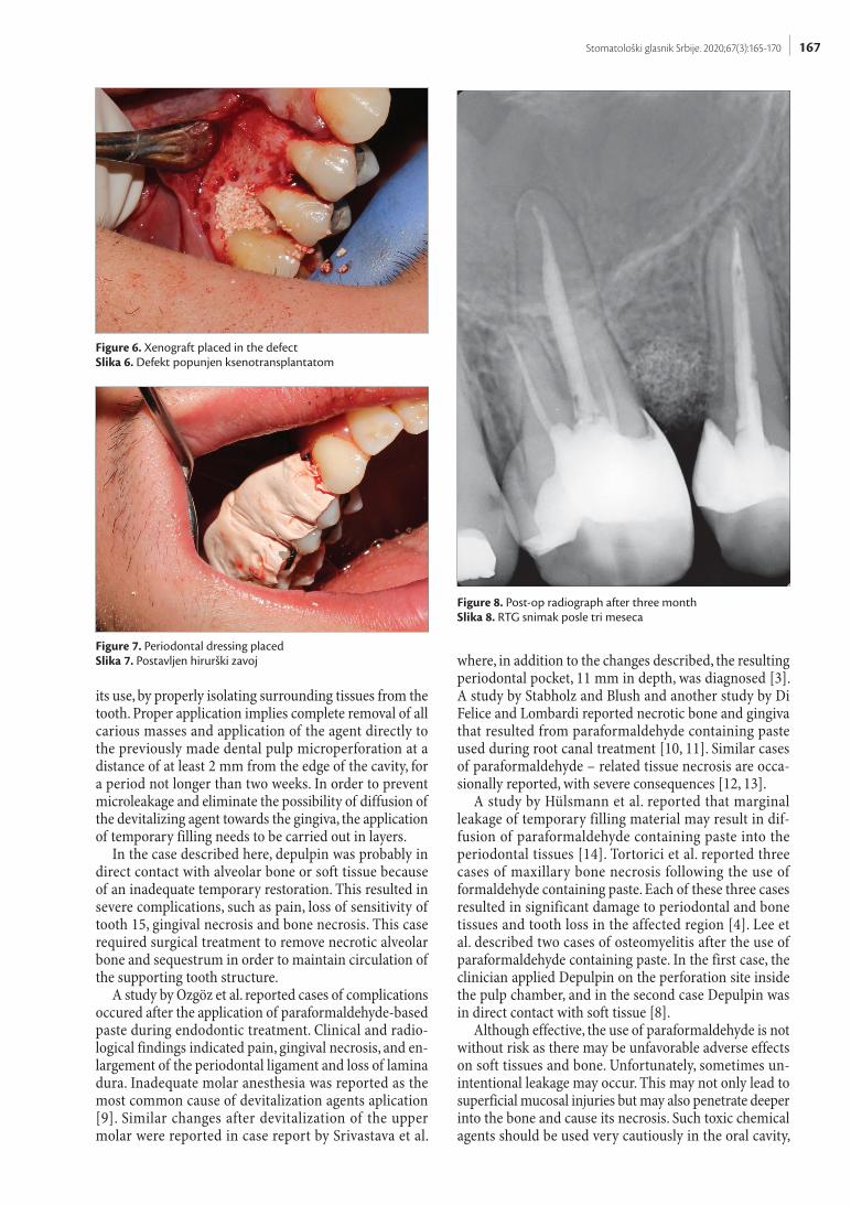
Disinfection of the mouth was performed with ch-lorhexidine 0,2%, and under local anaesthesia, the full thickness periodontal flap was raised both buccally and palatally, necrotic bone was removed and curettage of the cavity was carried out (Figure 3, 4, 5). The surgical site was irrigated with sterile physiological saline. After curettage and irrigation of the area, the defect was filled with xeno-graft of bovine origin (Figure 6). The flap was sutured in place and periodontal dressing was given (Figure 7). The
patient was prescribed antibiotics for seven days (amoxi-cillin 500 mg 3×1 and metronidazole 400 mg 3×1).
After three months, endodontic treatment was per-formed on tooth 15, as well as retreatment on tooth 16 (Figure 8). After surgical procedure and endodontic treat-ment completion all symptoms disappeared.
DISCUSSION
Dentists often face difficulties during endodontic treat-ment, when there is failure of anesthesia in teeth diag-nosed with irreversible pulpitis. Various agents are used to devitalize extremely painful pulps prior to extirpation. Paraformaldehyde-containing products are commonly used for this purpose. Paraformaldehyde leads to co-agulation and denaturation of cell wall proteins, which results in the arrest of all vital cell functions. The tissue becomes “fixed,” and this state of fixation is irreversible [3, 4]. Despite of its clinical benefit, the use of paraformal-dehyde containing paste in such circumstances may lead to many noxious effects on the host tissue. In addition to damage of interdental papilla, these agents can diffuse deeper into the bone and with their effect, lead to circu-latory disorders and consequent necrosis. These changes may create a precondition for bone infection or localized osteomyelitis [5–8]. Caution should be exercised during
Figure 1. Lesion localized in the area of interdental papilla between teeth 15 and 16.Slika 1. Lezija lokalizovana u području interdentalne papile između zuba 15 i 16.
Figure 2. Diagnostic radiographSlika 2. Dijagnostički RTG snimak
Figure 3. Removal of necrotic boneSlika 3. Uklanjanje nekrotične kosti
Figure 4. Necrotic bone sequestersSlika 4. Sekvestri kosti
Figure 5. Defect seen after necrotic bone removalSlika 5. Defekt posle uklanjanja nekrotične kosti
Radović I. et al. Maxillary bone necrosis following the use of formaldehyde containing paste – case report
167Stomatološki glasnik Srbije. 2020;67(3):165-170
its use, by properly isolating surrounding tissues from the tooth. Proper application implies complete removal of all carious masses and application of the agent directly to the previously made dental pulp microperforation at a distance of at least 2 mm from the edge of the cavity, for a period not longer than two weeks. In order to prevent microleakage and eliminate the possibility of diffusion of the devitalizing agent towards the gingiva, the application of temporary filling needs to be carried out in layers.
In the case described here, depulpin was probably in direct contact with alveolar bone or soft tissue because of an inadequate temporary restoration. This resulted in severe complications, such as pain, loss of sensitivity of tooth 15, gingival necrosis and bone necrosis. This case required surgical treatment to remove necrotic alveolar bone and sequestrum in order to maintain circulation of the supporting tooth structure.
A study by Ozgöz et al. reported cases of complications occured after the application of paraformaldehyde-based paste during endodontic treatment. Clinical and radio-logical findings indicated pain, gingival necrosis, and en-largement of the periodontal ligament and loss of lamina dura. Inadequate molar anesthesia was reported as the most common cause of devitalization agents aplication [9]. Similar changes after devitalization of the upper molar were reported in case report by Srivastava et al.
where, in addition to the changes described, the resulting periodontal pocket, 11 mm in depth, was diagnosed [3]. A study by Stabholz and Blush and another study by Di Felice and Lombardi reported necrotic bone and gingiva that resulted from paraformaldehyde containing paste used during root canal treatment [10, 11]. Similar cases of paraformaldehyde – related tissue necrosis are occa-sionally reported, with severe consequences [12, 13].
A study by Hülsmann et al. reported that marginal leakage of temporary filling material may result in dif-fusion of paraformaldehyde containing paste into the periodontal tissues [14]. Tortorici et al. reported three cases of maxillary bone necrosis following the use of formaldehyde containing paste. Each of these three cases resulted in significant damage to periodontal and bone tissues and tooth loss in the affected region [4]. Lee et al. described two cases of osteomyelitis after the use of paraformaldehyde containing paste. In the first case, the clinician applied Depulpin on the perforation site inside the pulp chamber, and in the second case Depulpin was in direct contact with soft tissue [8].
Although effective, the use of paraformaldehyde is not without risk as there may be unfavorable adverse effects on soft tissues and bone. Unfortunately, sometimes un-intentional leakage may occur. This may not only lead to superficial mucosal injuries but may also penetrate deeper into the bone and cause its necrosis. Such toxic chemical agents should be used very cautiously in the oral cavity,
Figure 6. Xenograft placed in the defectSlika 6. Defekt popunjen ksenotransplantatom
Figure 7. Periodontal dressing placedSlika 7. Postavljen hirurški zavoj
Figure 8. Post-op radiograph after three monthSlika 8. RTG snimak posle tri meseca

168
so that they do not come in contact with gingiva or other parts of oral mucosa during placement.
REFERENCES
1. Cohen HP, Cha BY, Spångberg LS. Endodontic anesthesia in mandibular molars: a clinical study. J Endod. 1993;19(7):370–3. [DOI: 10.1016/S0099-2399(06)81366-X] [PMID: 8245762]
2. Lin LM, Rosenberg PA, Lin J. Do procedural errors cause endo-dontic treatment failure? J Am Dent Assoc. 2005;136(2):187–231. [DOI: 10.14219/jada.archive.2005.0140] [PMID: 15782522]
3. Srivastava A, Gupta KK, Tandon P, Rajpal J. Necrosis of alveolar bone secondary to endodontic treatment and its management. J Interdiscip Dentistry 2011;1(1):41–4.
4. Tortorici S, Burruano F, Difalco P. Maxillary bone necrosis following the use of formaldehyde containing paste: management and case series. Br Dent J. 2007;203(9):511–2. [DOI: 10.1038/bdj.2007.995]
5. Block R. Are you still using formocresol? An update. J Tenn Dent Assoc. 2009;89(4):14–9.
6. Lewis B. The obsolescence of formocresol. J Calif Dent Assoc. 2010;38(2):102–7.
7. Hargreaves, K. M. & Berman, L. H. Cohen’s pathways of the pulp. 11th ed. St. Louis, MO: Elsevier, 2016.
8. Lee CH, Choi Y, Park S. Mandibular bone necrosis after use of paraformaldehyde-containing paste. Restor Dent
Endod. 2016;41(4):332–7. [DOI: 10.5395/rde.2016.41.4.332] [PMID: 27847756]
9. Ozgöz M, Yagiz H, Ciçek Y, Tezel A. Gingival necrosis follow-ing the use of a paraformaldehyde-containing paste: a case report. Int Endod J. 2004;37(2):157–61. [DOI: 10.1111/j.0143-2885.2004.00770.x] [PMID: 14997897]
10. Stabholz A, Blush MS. Necrosis of the crestal bone caused by the use of Toxavit. J Endod. 1983;9(3):110–3. [DOI: 10.1016/S0099-2399(83)80107-1] [PMID: 6590772]
11. Di Felice R, Lombardi T. Gingival and mandibular bone necrosis caused by a paraformaldehyde-containing paste. Endod Dent Traumatol. 1998;14(4):196–8. [DOI: 10.1111/j.1600-9657.1998.tb00837.x] [PMID: 9796485]
12. Cambruzzi JV, Greenfeld RS. Necrosis of crestal bone related to the use of excessive formocresol medication during endodontic treatment. J Endod. 1983;9(12):565–7. [DOI: 10.1016/S0099-2399(83)80062-4] [PMID: 6581262]
13. Tal M, Kaufman AY, Buchner A. Bone necrosis and dentine re-sorption caused by Toxavit: a case report. J Br Endod Soc. 1978;11(2):77–9. [DOI:10.1111/j.1365-2591.1978.tb00665.x]
14. Hülsmann M, Hornecker E , Redeker M. Periodontal de-struction and tooth loss following pulp devitalization with Toxavit: report of a case. Dent Traumatol. 1993;9(5):216–21. [DOI:10.1111/j.1600-9657.1993.tb00277.x]
Received: 24.6.2020 • Accepted: 27.8.2020
Radović I. et al. Maxillary bone necrosis following the use of formaldehyde containing paste – case report
169Stomatološki glasnik Srbije. 2020;67(3):165-170
Nekroza maksilarne kosti posle primene devitalizacione paste na bazi formaldehida – prikaz bolesnika
Igor Radović1, Lado Davidović1, Smiljka Cicmil2, Slavoljub Tomić2, Dragan Ivanović3, Ljiljana Bjelović1
1Univerzitet u Istočnom Sarajevu, Medicinski fakultet, Odeljenje za dentalnu patologiju, Foča, Republika Srpska, Bosna i Hercegovina;2Univerzitet u Istočnom Sarajevu, Medicinski fakultet, Odeljenje za oralnu rehabilitaciju, Foča, Republika Srpska, Bosna i Hercegovina;3Univerzitet u Istočnom Sarajevu, Medicinski fakultet, Odeljenje za dečju i preventivnu stomatologiju, Foča, Republika Srpska, Bosna i Hercegovina
KRATAK SADRŽAJVeliki broj medikamenata koji su se kroz istoriju koristili za tretman kanala korena ispoljavali su citotoksičnost. Sredstva na bazi paraformaldehida (kao što su toksavit i depulpin) koriste se za devitalizaciju zubne pulpe u slučajevima kada je primena lokalnih anastetika neefikasna. Nepravilna primena sredstava za devitalizaciju pulpe može prouzrokovati oštećenja desni i alveolarne kosti. Ovaj prikaz iz prakse ukazuje na ozbiljne komplikacije nastale posle primene paste na bazi paraformaldehida, nekrozu desni i alve-olarne kosti, što je rezultiralo velikim gubitkom koštanog tkiva. Bila je potrebna hirurška intervencija da se ukloni nekrotična kost i defekt popuni ksenotransplantatom bovinog porekla. Tri meseca posle operacije izvedena je endodontska terapija. Nakon tretmana pacijentove tegobe su prestale. Primena sredstava na bazi paraformaldehida tokom endodontske terapije zahteva poseban oprez.Ključne reči: tretman kanala korena; paraformaldehid; nekroza kosti
UVOD
Uspešna primena lokalne anestezije i izvođenje bezbolnog endodontskog tretmana u pojedinim situacijama može pred-stavljati izazov za stomatologa. Cohen i saradnici su u svojoj studiji pokazali da 39% pacijenata sa ireverzibilnim pulpitisom mandibularnih molara pokazuje osetljivost na hladne nadražaje polse blok-anestezije donjeg alveolarnog nerva 2% rastvorom lidokaina [1]. U slučajevima akutnog simptomatskog pulpitisa, posebno mandibularnih molara, kada je postizanje adekvatne anestezije bilo otežano zbog tehničkih ili anatomskih faktora, stomatolozi su koristili paste na bazi arsena ili paraformalde-hida za devitalizaciju inflamirane, bolne zubne pulpe. Sredstva za nekrotizaciju pulpe su toksična i ispoljavaju neselektivno delovanje. Paraformaldehid ne deluje isključivo na pulpu već i na okolna tkiva sa kojima dođe u kontakt – direktno ili di-fuzijom kroz dentinske tubule. Ukoliko se ne postavi pravilno, može dovesti do lokalnih komplikacija kao što su oštećenje in-terdentalne papile, destrukcija parodonta, odnosno nastanak periapeksnih, interradiksnih i lateralnih oštećenja u koštanom tkivu parodoncijuma [2, 3].
Cilj ovog članka je da prikaže komplikacije nastale nakon neadekvatne primene sredstva za devitalizaciju pulpe na bazi paraformaldehida i terapiju komplikacija.
PRIKAZ BOLESNIKA
Pacijent star 23 godine, muškog pola, primljen je na našu klini-ku sa molbom za konsultaciju i mišljenje u vezi sa tegobama u vidu bola u regiji prvog gornjeg desnog molara. Pacijent u ana-mnezi navodi da je tri meseca pre dolaska zub 16 bio podvrgnut endodontskoj terapiji. Zbog izraženih bolova tokom preparacije pristupnog kaviteta pacijentu je tada ukazano na neefikasnost lokalne anestezije i aplikovana je devitalizaciona pasta. Kasnije je potvrđeno da je u pitanju pasta na bazi paraformaldehida (Depulpin, Voco, GmbH, Cuxhaven, Germany). U periodu posle aplikacije paste pacijent je osećao bolove, koji su perzistirali i posle završene endodontske terapije na zubu 16. Prilikom
ponovnog javljanja svom stomatologu pacijentu su propisani antibiotici i analgetici. Ipak, pacijentove tegobe nisu prestale i on se upravo zbog toga javio na našu kliniku.
Kliničkim pregledom je utvrđeno postojanje lezije u obliku kratera, lokalizovane u području interdentalne papile između zuba 15 i 16, kao i postojanje parodontalnog džepa u pomenutoj regiji (Slika 1). Na oba zuba su uočeni kompozitni ispuni druge klase, mezio-okluzalno. Radiografijom je ustanovljena nepot-puna endodontska terapija na zubu 16 (opturisan je jedino pa-latinalni kanal) i izražen intrakoštani defekt između zuba 15 i 16 (Slika 2). Zub 15 je bio negativan na elektro test.
Potom se pristupilo parodontološkoj intervenciji. Za dezin-fekciju usta korišten je 0,2% hlorheksidin. Pod dejstvom lokalne anestezije podignut je mukoperiostalni režanj pune debljine i nekrotična kost je pažljivo uklonjena uz kiretažu šupljine (slike 3, 4, 5). Operaciono polje je irigirano sterilnim fiziološkim ra-stvorom. Posle kiretaže i irigacije područja defekt je popunjen ksenotransplantatom bovinog porekla (Slika 6). Režanj je ušiven i postavljen je hirurški zavoj (Slika 7). Pacijentu su propisani antibiotici u trajanju od sedam dana (amoksicilin 500 mg 3×1 i metronidazol 400 mg 3×1).
Posle tri meseca izvršeni su endodontska terapija na zubu 15 i retretman na zubu 16 (Slika 8). Na kontrolnom pregledu posle hirurške terapije i endodontskog tretmana pacijent nije imao nikakve simptome.
DISKUSIJA
Stomatolozi često imaju poteškoće tokom endodontske inter-vencije u slučajevima kada je lokalna anestezija neuspešna kod zuba sa dijagnostikovanim ireverzibilnim pulpitisom. Za devitalizaciju bolnih zuba tokom ekstirpacije zubne pulpe pri-menjuju se različita sredstva. U ovu svrhu se najčešće koriste preparati na bazi paraformaldehida. Paraformaldehid dovodi do koagulacije i denaturacije proteina ćelijskog zida, što uzrokuje prestanak svih vitalnih ćelijskih funkcija. Na ovaj način tkivo postaje „fiksirano“ i ovo stanje je ireverzibilno [3, 4]. I pored kliničkih prednosti, primena pasti na bazi paraformaldehida

170
može dovesti do toksičnog delovanja na okolna tkiva sa kojima dođu u kontakt. Pored oštećenja interdentalne papile, ova sred-stva mogu difundovati dublje u kost te svojim efektom dovesti do poremećaja cirkulacije i posledične nekroze. Ove promene mogu stvoriti preduslov za nastanak infekcije kosti ili lokalizo-vani osteomijelitis [5–8]. Prilikom njegove aplikacije treba biti veoma oprezan, u smislu izolacije i zaštite okolnog tkiva. Pravil-na aplikacija podrazumeva potpuno uklanjanje svih karijesnih masa i aplikaciju primenjenog sredstva direktno na prethodno napravljenu mikroperforaciju zubne pulpe, udaljenju najmanje 2 mm od ivice kaviteta, na period ne duži od dve sedmice. Kako bi se sprečilo mikrocurenje i otklonila mogućnost difundovanja sredstva za devitalizaciju prema gingivi, aplikaciju privremenog ispuna potrebno je sprovesti u slojevima.
U prikazu bolesnika koji je opisan u ovom radu, depulpin je verovatno bio u direktnom kontaktu sa koštanim tkivom ili gingivom zbog neadekvatnog privremenog ispuna. Ovo je re-zultiralo pojavom teških komplikacija, kao što su bol, nekroza gingive i koštanog tkiva te formiranje parodontalnog džepa. U ovom slučaju je bila neophodna hirurška intervencija ukla-njanja nekrotične kosti sa ciljem održavanja cirkulacije potpor-nog aparata zuba.
Ozgöz i saradnici u svom radu navode slučajeve komplikacija nastalih posle primene paste na bazi paraformaldehida prili-kom endodontskog tretmana zuba. Klinički i radiološki nalazi ukazali su na bol, nekrozu gingive te proširenje periodontalnog ligamenta i gubitak lamine dure. Neadekvatna anestezija molara se navodi kao najčešći uzrok primene sredstava za devitalizaciju
zuba [9]. Slične promene posle devitalizacije gornjeg molara navode u svom prikazu slučaja Srivastava i saradnika, gde je pored gore opisanih promena dijagnostikovana i dubina na-stalog parodontalnog džepa od 11 mm [3]. U svojim radovima Stabholz i Blush, kao i Di Felice i Lombardi opisuju nekrozu koštanog tkiva i gingive prouzrokovanu sredstvima za avitali-zaciju na bazi paraformaldehida [10, 11]. Povremeno se objav-ljuju slični slučajevi tkivnih nekroza sa teškim posledicama izazvanim aplikacijom sredstava na bazi paraformaldehida [12, 13]. Hülsmann i saradnici su pokazali da marginalno curenje privremenog ispuna može dovesti do difuzije paraformalde-hida u parodoncijum [14]. Tortorici i saradnici su opisali tri slučaja nekroze maksilarne kosti posle primene paste na bazi formaldehida. Svaki od ova tri slučaja je rezultirao značajnim oštećenjima parodontalnog tkiva i gubitkom zuba u zahvaćenoj regiji [4]. Lee i saradnici su opisali dva slučaja osteomijelitisa koji je nastao kao posledica primene paste na bazi formaldehi-da. U prvom slučaju stomatolog je aplikovao pastu u području perforacije pulpne komore, a u drugom slučaju depulpin je bio u direktnom kontaktu sa mekim tkivima [8].
Iako je efikasna, primena devitalizacionih sredstava na bazi formaldehida nije bez rizika jer može izazvati neželjena dejstva na meka tkiva i kost. Nažalost, nekada može doći do difuzije u okolna tkiva, što može prouzrokovati ne samo oštećenje sluznice već i nekrozu koštanog tkiva. Toksična hemijska sredstva, kao što su devitalizacione paste, treba primenjivati uz veliki oprez u usnoj duplji, da bi se izbegao njihov kontakt sa gingivom ili drugim delovima oralne sluznice.
Radović I. et al. Maxillary bone necrosis following the use of formaldehyde containing paste – case report