Maxillary and Periorbital Fractures and Periorbital Fractures Resident: ... –12% severe ocular...
55
Maxillary and Periorbital Fractures Resident: Gordon Shields, MD Faculty Advisor: Francis Quinn, Jr, MD, FACS The University of Texas Medical Branch Department of Otolaryngology Grand Rounds Presentation January 7, 2003
Transcript of Maxillary and Periorbital Fractures and Periorbital Fractures Resident: ... –12% severe ocular...

Maxillary and Periorbital Fractures
Resident: Gordon Shields, MD
Faculty Advisor: Francis Quinn, Jr, MD, FACS
The University of Texas Medical Branch
Department of Otolaryngology
Grand Rounds Presentation
January 7, 2003

Types
Mechanisms
Associated Injuries
Anatomy
Classification
Evaluation
Treatment

Types
LeFort or Maxillary fractures
Zygomaticomaxillary complex fractures
Orbitozygomaticomaxillary complex fractures
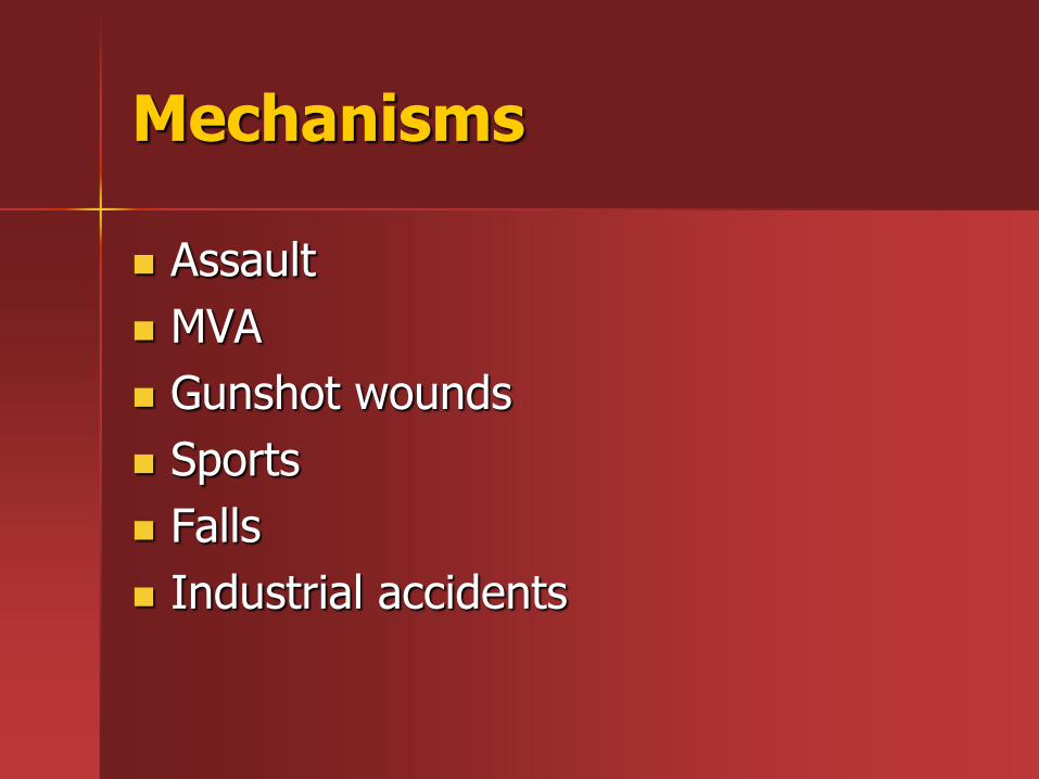
Mechanisms
Assault
MVA
Gunshot wounds
Sports
Falls
Industrial accidents

Associated Injuries
Brandt et al 1991
59% caused by MVA had intracranial injury
10% caused by fall/beating had intracranial injury
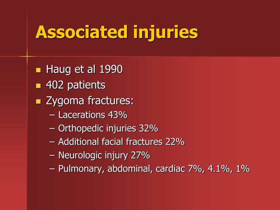
Associated injuries
Haug et al 1990
402 patients
Zygoma fractures:
– Lacerations 43%
– Orthopedic injuries 32%
– Additional facial fractures 22%
– Neurologic injury 27%
– Pulmonary, abdominal, cardiac 7%, 4.1%, 1%

Maxillary fractures:
– Lacerations and abrasions 75%
– Orthopedic injury 51%
– Other facial fractures 42%
– Neurologic injury 51%
– Pulmonary 13%, abdominal 5.7%, cardiac 3.8%

Ocular injury
Al-Qurainy et al 1991
363 patients with midface fractures
– 63% minor or transient ocular injury
– 16% moderately severe injury
– 12% severe ocular injury (angle recession, retinal or vitreous injury, optic nerve damage
– 90.6% of patients had some ocular injury
– 2.5% lost vision in the affected eye

Facial Skeleton
From: Netter FH. Atlas of Human Anatomy. Second Edition; East Hanover, Novartis,1997, plt. 1
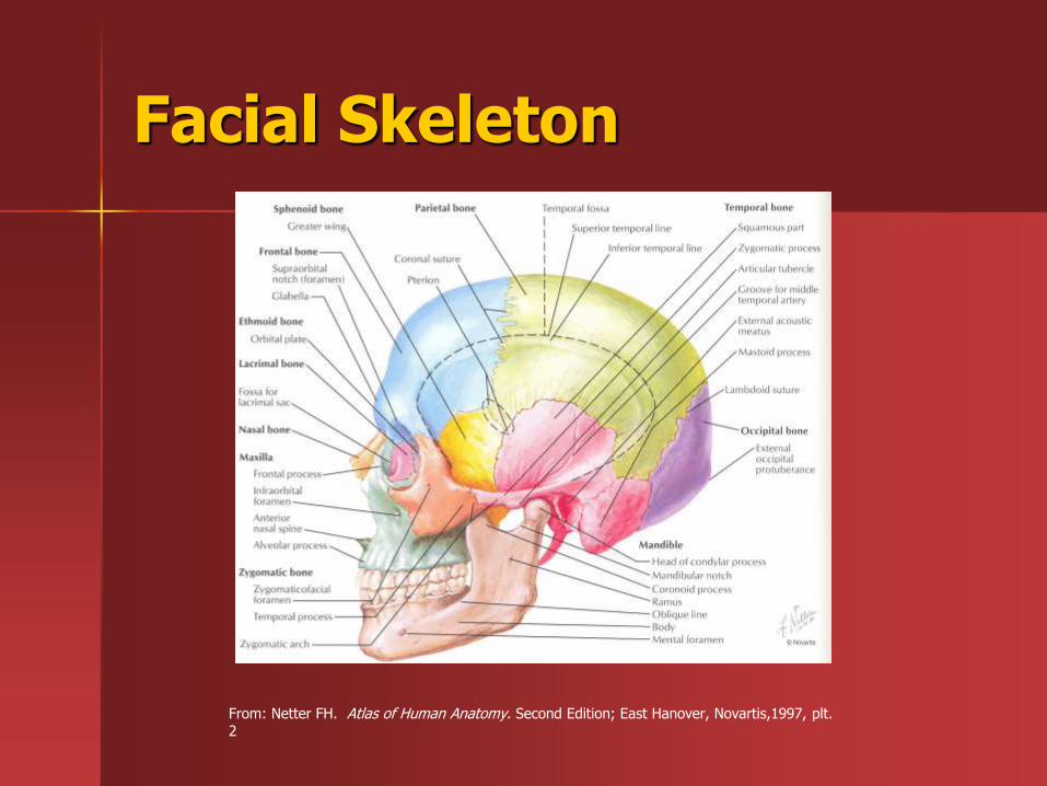
Facial Skeleton
From: Netter FH. Atlas of Human Anatomy. Second Edition; East Hanover, Novartis,1997, plt. 2

Orbit
7 bones composing the orbit: frontal, sphenoid, zygoma, maxilla, palatine, lacrimal, ethmoid
From: Netter FH. Atlas of Human Anatomy. Second Edition; East Hanover, Novartis,1997, plt. 1

Forces of mastication
From: Banks P, Brown A. Fractures of the Facial Skeleton, Oxford, Wright 2001 pg.6
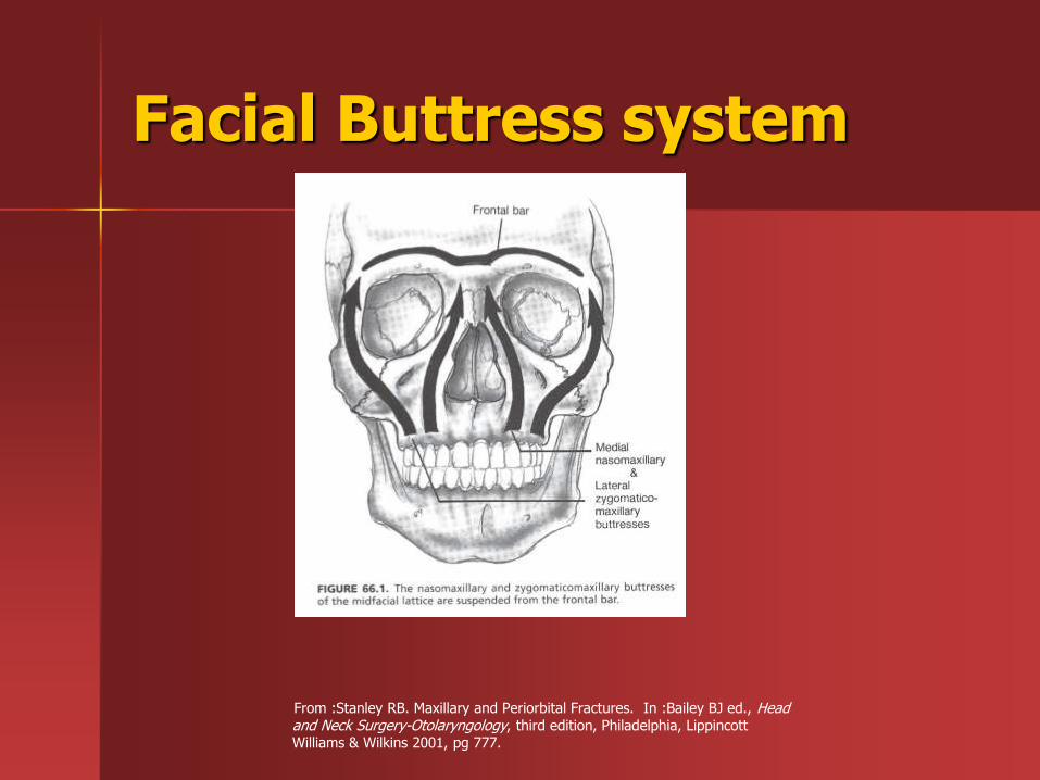
Facial Buttress system
From :Stanley RB. Maxillary and Periorbital Fractures. In :Bailey BJ ed., Head and Neck Surgery-Otolaryngology, third edition, Philadelphia, Lippincott Williams & Wilkins 2001, pg 777.

Facial Buttress system
From: Celin SE. Fractures of the Upper Facial and Midfacial Skeleton. In: Myers EN ed., Operative Otolaryngology Head and Neck Surgery, Philadelphia, WB Saunders Company 1997:1143-1192.

Facial buttress system
From: Rowe NL, Williams JL. Maxillofacial Injuries. Edinburgh, Churchill Livingstone,1985, pg 19.
LeFort fractures
Rene LeFort 1901 in cadaver skulls
Based on the most superior level
Frequently different levels on either side
LeFort I
LeFort II
LeFort III

From: Dolan KD, Jacoby CG, Smoker WR. Radiology of Facial Injury. New York, MacMillian Publishing Company 1988, pg76.
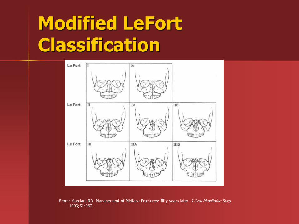
Modified LeFort Classification
From: Marciani RD. Management of Midface Fractures: fifty years later. J Oral Maxillofac Surg 1993;51:962.
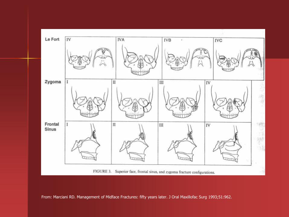
From: Marciani RD. Management of Midface Fractures: fifty years later. J Oral Maxillofac Surg 1993;51:962.
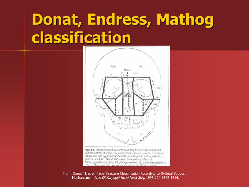
Donat, Endress, Mathog classification
From: Donat TL et al. Facial Fracture Classification According to Skeletal Support Mechanisms. Arch Otolaryngol Head Neck Surg 1998;124:1306-1314.

Evaluation
ABC’s
History
Palpation of entire facial skeleton
Occlusion
Ophthalmologic exam / consultation
C-spine
Imaging – CT

Imaging
CT has surpassed plain film xray
Allows precise diagnosis and surgical planning
Axial and coronal cuts
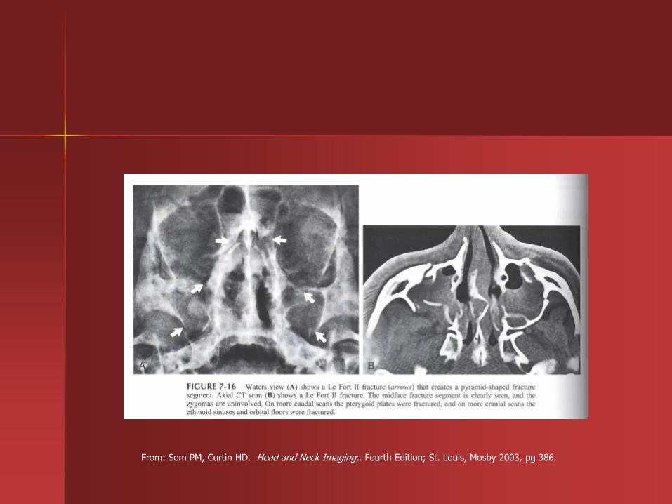
From: Som PM, Curtin HD. Head and Neck Imaging;. Fourth Edition; St. Louis, Mosby 2003, pg 386.
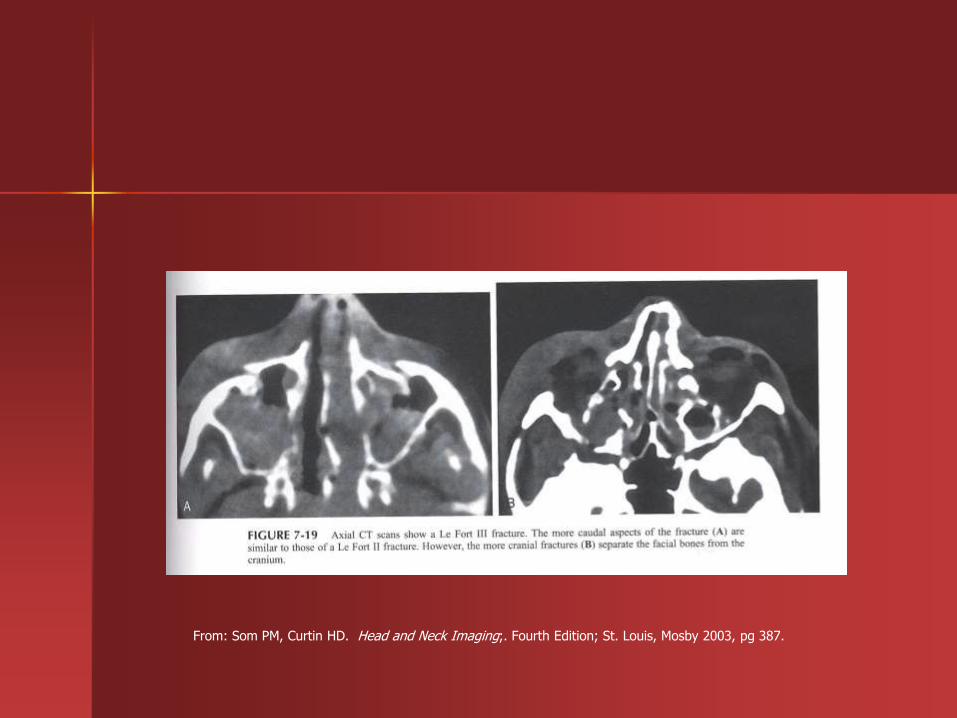
From: Som PM, Curtin HD. Head and Neck Imaging;. Fourth Edition; St. Louis, Mosby 2003, pg 387.

From: Som PM, Curtin HD. Head and Neck Imaging;. Fourth Edition; St. Louis, Mosby 2003, pg 393.


Treatment of maxillary fractures
Early repair
Single-stage
Extended access approaches
Rigid fixation
Immediate bone grafting
Re-suspension of soft tissues

Maxillary fractures
Steps of reconstruction-Rohrich and Shewmake
Reestablish facial height and width
IMF with ORIF of mandible
Zygomatic arch reconstruction restores facial width and projection
Reconstruction continues from stable bone to unstable and from lateral to medial

Internal fixation vs. traditional methods
Klotch et al 1987
43 patients
22 treated with ORIF using AO miniplates
21 treated with combination of intermaxillary fixation, and/or interosseous wiring, and/or primary bone grafting
Most severe injuries in rigid internal fixation group
Shorter IMF, early return to diet, lower percentage of tracheotomy
No plate infections
Haug et al 1995
134 patients treated by maxillomandibular fixation or rigid internal fixation
Postoperative problems in 60% vs 64%
Complication rates similar
Rigid fixation has benefits:
– Airway protection
– Enhanced nutrition
– More rapid return to pretraumatic function

Approaches
Circumvestibular
Facial degloving
Bicoronal
Transconjuctival
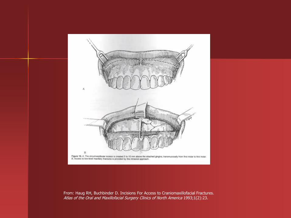
From: Haug RH, Buchbinder D. Incisions For Access to Craniomaxillofacial Fractures. Atlas of the Oral and Maxillofacial Surgery Clinics of North America 1993;1(2):23.
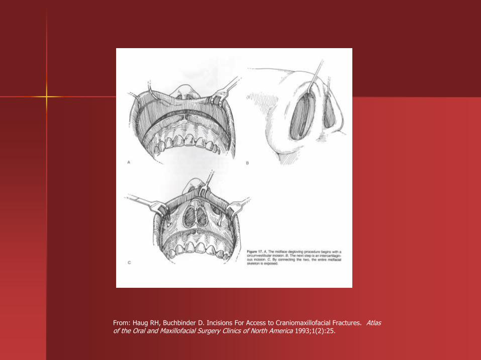
From: Haug RH, Buchbinder D. Incisions For Access to Craniomaxillofacial Fractures. Atlas of the Oral and Maxillofacial Surgery Clinics of North America 1993;1(2):25.

Bicoronal approach
From: Celin SE. Fractures of the Upper Facial and Midfacial Skeleton. In: Myers EN ed., Operative Otolaryngology Head and Neck Surgery, Philadelphia, WB Saunders Company 1997:1143-1192.

From: Cheney ML. Facial Surgery: Plastic and Reconstructive. Baltimore: Williams & Wilkins 1997.

From: Celin SE. Fractures of the Upper Facial and Midfacial Skeleton. In: Myers EN ed., Operative Otolaryngology Head and Neck Surgery, Philadelphia, WB Saunders Company 1997:1143-1192.

Treatment of Zygomaticomaxillary Complex fractures
Restore pre-injury facial configuration
Prevent cosmetic deformity
Prevent delayed visual disturbances
Repair within 5-7 days allows edema to decrease and avoids shortening of masseter with lateral and inferior rotation
Soft diet and malar protection
Closed reduction
ORIF with plating of one to four buttresses
Provide fixation as necessary for stable reduction
Ellis and Kittidumkerng 1996 48 patients Reduced fracture with Carroll-Girard screw
– 4.2% closed reductions – 31.2 % one point fixation – 27.1% two point fixation – 27.1% three point fixation – 10.4% four point fixation – Used exposure and fixation needed to provide
stable reduction

Approaches to FZ buttress
From: Strong EB, Sykes JM. Zygoma Complex Fractures. Facial Plastic Surgery 1990;14(1):108.
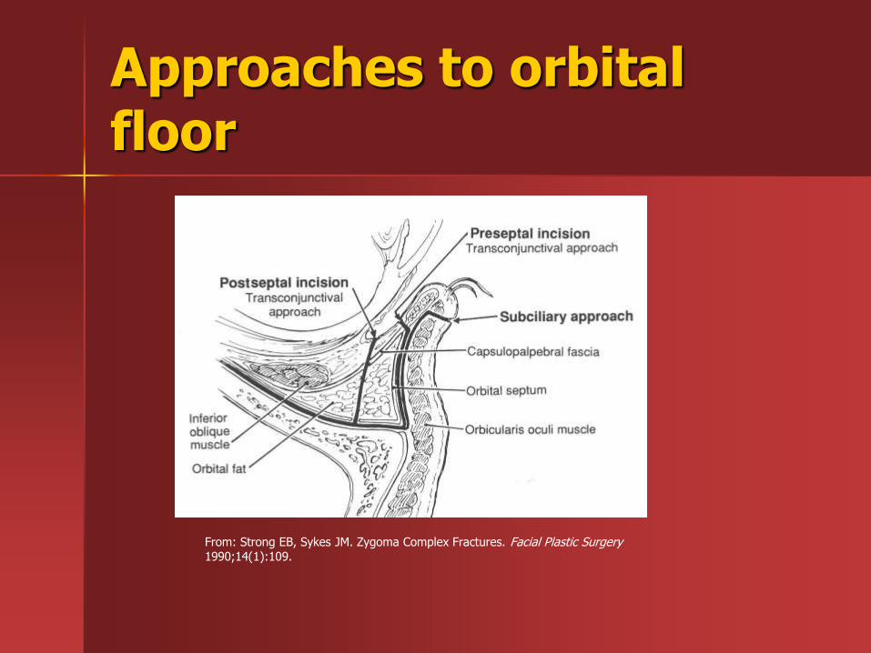
Approaches to orbital floor
From: Strong EB, Sykes JM. Zygoma Complex Fractures. Facial Plastic Surgery 1990;14(1):109.
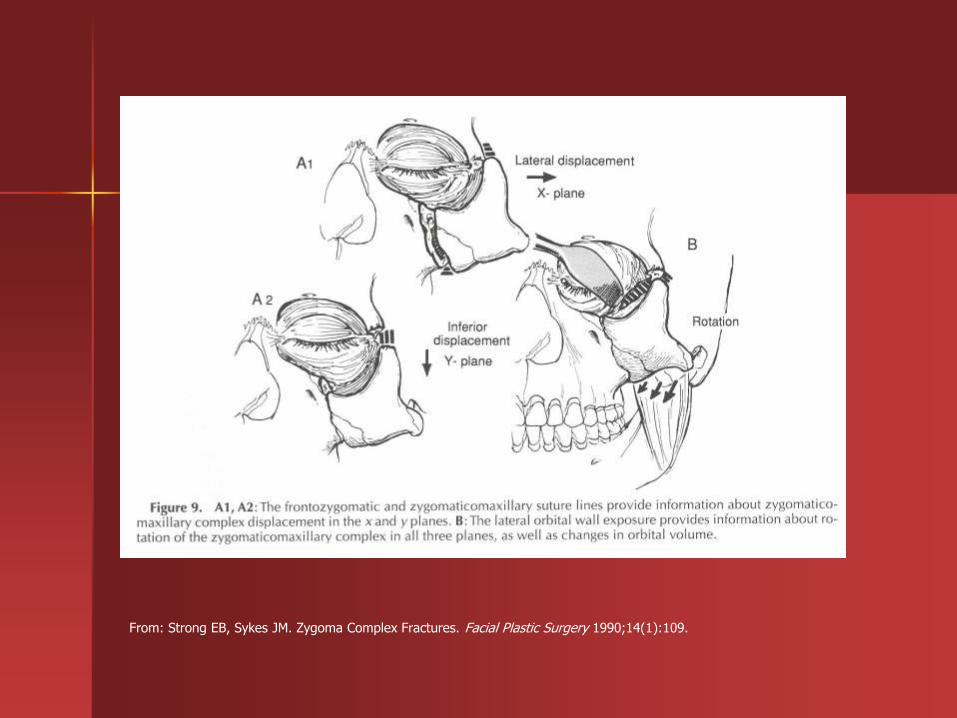
From: Strong EB, Sykes JM. Zygoma Complex Fractures. Facial Plastic Surgery 1990;14(1):109.

Orbital exploration
Shumrick et al 1997
97 patient with either ZMC or midface fractures
All explored had significant traumatic disruptions, no enopthalmos or diplopia in those not explored
Based on their 7 criteria 22 ZMC and 11 midface underwent orbital exploration

Persistent diplopia which failed to improve in 7 or more days, positive forced duction testing, radiologic evidence of perimuscular tissue entrapment
Cosmetically significant and clinically apparent enophthalmos associated with abnormal radiological findings

Radiological evidence of significant comminution and or displacment of the orbital rim
Radiological evidence of significant displacement or comminution of greater than 50% of the orbital floor with herniation of soft tissue into maxillary sinus

Combined orbital floor and medial wall defects with soft tissue displacement noted radiologically on CT scans
Radiological evidence of a fracture or comminution of the body of the zygoma itself as determined by CT
Physical or radiological evidence of exophthalmos or orbital content impingement caused by displaced periorbital fractures
Repair of the orbit
Approaches
– Transconjunctival with or without lateral canthotomy/cantholysis
– Subciliary
– Transconjunctival has lower incidence of ectropion/entropion
Materials for reconstruction
Autogenous tissues
– Avoid risk of infected implant
– Additional operative time, donor site morbidity , graft absorption
– Calvarial bone, iliac crest, rib, septal or auricular cartilage

Alloplastic implants
– Decreased operative time, easily available, no donor site morbidity, can provide stable support
– Risk of infection 0.4-7%
– Gelfilm, polygalactin film, silastic, marlex mesh, teflon, prolene, polyethylene, titanium

Ellis and Tan 2003
– 58 patients, compared titanium mesh with cranial bone graft
– Used postoperative CT to assess adequacy of reconstruction
– Titanium mesh group subjectively had more accurate reconstruction
Soft tissue resuspension
Wide exposure allows more accurate fracture reduction but may lead to problems in soft tissue covering of face
Need to close periosteum and provide suspension sutures to prevent descent of soft tissues

Conclusions
High index of suspicion for associated injuries- especially ocular
Assessment of buttress system
Wide exposure via cosmetically acceptable incisions
Rigid fixation
Soft tissue resuspension