Matrix Metalloproteinase Inhibition Attenuates Aortic...
17
Matrix Metalloproteinase Inhibition Attenuates Aortic Calcification Xiao Qin, Matthew A. Corriere, Lynn M. Matrisian, Raul J. Guzman Objective—Arterial calcification has been associated with matrix metalloproteinase (MMP)–mediated elastin degradation. In this study, we investigated whether inhibiting MMP activity could reduce calcium accumulation in rodent models of aortic calcification. Methods and Results—Aortic calcification was first induced in male Sprague-Dawley rats by administration of vitamin D 3 . Treatment with doxycycline decreased aortic calcium and phosphorus accumulation, and it reduced aortic gelatinase levels; however, it also prevented the bone resorption associated with high doses of vitamin D 3 . Using an in vivo model of localized aortic calcification, systemic doxycycline treatment reduced aortic calcium accumulation without affecting serum calcium levels, suggesting a more specific effect of doxycycline in the arterial wall. In organ culture, doxycycline limited aortic calcification caused by exposure to alkaline phosphatase and inorganic phosphate. When GM6001, a synthetic and specific inhibitor of MMPs, was used instead of doxycycline, it had a similar effect. In vivo, periadventitial delivery of GM6001 to calcifying arteries significantly reduced calcification compared with controls. Conclusions—These results suggest that MMPs are involved in aortic calcification, and inhibiting MMP activity could reduce calcium accumulation in the arterial wall. (Arterioscler Thromb Vasc Biol. 2006;26:1510-1516.) Key Words: artery calcification MMPs doxycycline GM6001 rat V ascular calcification occurs pathologically in diabetes and chronic renal disease as well as during the normal aging process. 1–3 When calcification is identified in the coronary arteries, it is associated with an increased risk of cardiac events. 4,5 In patients with renal failure, it is associated with significantly shorter survival, 6 and in patients with diabetes, it occurs at an accelerated rate and is a strong predictor of morbidity and mortality. 7 Although vascular calcification is associated with atherosclerosis, recent data suggest that it is independently regulated, and calcifying vessels share features with osteogenesis. 2,8 Consistent with this hypothesis is that diseased human arteries have been shown to express proteins usually seen in bone, including members of the bone morphogenetic protein family, noncol- lagenous bone matrix proteins, matrix Gla protein, decorin, the osteoblastic regulator osteoprotegrin, and the matrix metalloproteinases (MMPs). 9 –15 MMPs are involved in multiple processes in the vascular wall. 16 They are upregulated in human atherosclerotic and restenotic lesions, and they are involved in aneurysm forma- tion. 17–21 Inhibiting MMP activity prevents injury-induced arterial remodeling in rodent models. 22,23 Recent studies demonstrated a correlation between MMP-mediated elastin degradation and aortic calcification. Inhibiting elastin degra- dation with aluminum ions prevented calcification in the aortas of rats after calcium chloride–mediated injury; and mice deficient in MMP-2 and MMP-9 did not develop calcification in a similar model. 24 Additionally, Lee et al demonstrated increased MMP activity associated with in- creased levels of soluble elastin peptides in a rat subdermal model of elastin calcification. 25 In the present study, we sought to determine whether inhibiting MMP activity could prevent calcium accumulation in experimental models of aortic calcification. We demonstrate that doxycycline, an antibiotic that inhibits MMP activity and reduces MMP levels, and GM6001, a synthetic selective MMP inhibitor, can inhibit arterial calcification both in organ culture and in vivo. Materials and Methods A detailed Methods section is available in the online supplement, available at http://atvb.ahajournals.org. Experimental Procedures Arterial calcification was induced in male Sprague-Dawley rats by subcutaneous injection of 7.5 mg/mL vitamin D 3 for 3 consecutive days. Rats received daily doxycycline by subcutaneous injection beginning at 2 days before vitamin D 3 injections and ending at the time of harvest. In another group of rats, aortic calcification was induced by periadventitial application of 0.15 mol/L CaCl 2 as described recently. 24 Original received November 22, 2005; final version accepted April 24, 2006. From the Department of Surgery, Division of Vascular Surgery, Vanderbilt University Medical Center (X.Q., M.A.C., R.J.G.), Nashville, Tenn; Guangxi Medical University First Affiliated Hospital, China (X.Q.); and the Departments of Cell and Developmental Biology (R.J.G.) and Cancer Biology (L.M.M.), Vanderbilt University Medical Center, Nashville, Tenn. Correspondence to Raul J. Guzman, MD, Division of Vascular Surgery, Vanderbilt University Hospital, D-5237, MCN Building, 1161 21st Ave S, Nashville, TN 37232. E-mail [email protected]. © 2006 American Heart Association, Inc. Arterioscler Thromb Vasc Biol. is available at http://www.atvbaha.org DOI: 10.1161/01.ATV.0000225807.76419.a7 1510 by guest on May 31, 2018 http://atvb.ahajournals.org/ Downloaded from by guest on May 31, 2018 http://atvb.ahajournals.org/ Downloaded from by guest on May 31, 2018 http://atvb.ahajournals.org/ Downloaded from by guest on May 31, 2018 http://atvb.ahajournals.org/ Downloaded from by guest on May 31, 2018 http://atvb.ahajournals.org/ Downloaded from by guest on May 31, 2018 http://atvb.ahajournals.org/ Downloaded from by guest on May 31, 2018 http://atvb.ahajournals.org/ Downloaded from by guest on May 31, 2018 http://atvb.ahajournals.org/ Downloaded from by guest on May 31, 2018 http://atvb.ahajournals.org/ Downloaded from by guest on May 31, 2018 http://atvb.ahajournals.org/ Downloaded from by guest on May 31, 2018 http://atvb.ahajournals.org/ Downloaded from
Transcript of Matrix Metalloproteinase Inhibition Attenuates Aortic...

Matrix Metalloproteinase Inhibition AttenuatesAortic Calcification
Xiao Qin, Matthew A. Corriere, Lynn M. Matrisian, Raul J. Guzman
Objective—Arterial calcification has been associated with matrix metalloproteinase (MMP)–mediated elastin degradation.In this study, we investigated whether inhibiting MMP activity could reduce calcium accumulation in rodent models ofaortic calcification.
Methods and Results—Aortic calcification was first induced in male Sprague-Dawley rats by administration of vitamin D3.Treatment with doxycycline decreased aortic calcium and phosphorus accumulation, and it reduced aortic gelatinaselevels; however, it also prevented the bone resorption associated with high doses of vitamin D3. Using an in vivo modelof localized aortic calcification, systemic doxycycline treatment reduced aortic calcium accumulation without affectingserum calcium levels, suggesting a more specific effect of doxycycline in the arterial wall. In organ culture, doxycyclinelimited aortic calcification caused by exposure to alkaline phosphatase and inorganic phosphate. When GM6001, asynthetic and specific inhibitor of MMPs, was used instead of doxycycline, it had a similar effect. In vivo, periadventitialdelivery of GM6001 to calcifying arteries significantly reduced calcification compared with controls.
Conclusions—These results suggest that MMPs are involved in aortic calcification, and inhibiting MMP activity couldreduce calcium accumulation in the arterial wall. (Arterioscler Thromb Vasc Biol. 2006;26:1510-1516.)
Key Words: artery � calcification � MMPs � doxycycline � GM6001 � rat
Vascular calcification occurs pathologically in diabetesand chronic renal disease as well as during the normal
aging process.1–3 When calcification is identified in thecoronary arteries, it is associated with an increased risk ofcardiac events.4,5 In patients with renal failure, it is associatedwith significantly shorter survival,6 and in patients withdiabetes, it occurs at an accelerated rate and is a strongpredictor of morbidity and mortality.7 Although vascularcalcification is associated with atherosclerosis, recent datasuggest that it is independently regulated, and calcifyingvessels share features with osteogenesis.2,8 Consistent withthis hypothesis is that diseased human arteries have beenshown to express proteins usually seen in bone, includingmembers of the bone morphogenetic protein family, noncol-lagenous bone matrix proteins, matrix Gla protein, decorin,the osteoblastic regulator osteoprotegrin, and the matrixmetalloproteinases (MMPs).9–15
MMPs are involved in multiple processes in the vascularwall.16 They are upregulated in human atherosclerotic andrestenotic lesions, and they are involved in aneurysm forma-tion.17–21 Inhibiting MMP activity prevents injury-inducedarterial remodeling in rodent models.22,23 Recent studiesdemonstrated a correlation between MMP-mediated elastindegradation and aortic calcification. Inhibiting elastin degra-
dation with aluminum ions prevented calcification in theaortas of rats after calcium chloride–mediated injury; andmice deficient in MMP-2 and MMP-9 did not developcalcification in a similar model.24 Additionally, Lee et aldemonstrated increased MMP activity associated with in-creased levels of soluble elastin peptides in a rat subdermalmodel of elastin calcification.25 In the present study, wesought to determine whether inhibiting MMP activity couldprevent calcium accumulation in experimental models ofaortic calcification. We demonstrate that doxycycline, anantibiotic that inhibits MMP activity and reduces MMPlevels, and GM6001, a synthetic selective MMP inhibitor, caninhibit arterial calcification both in organ culture and in vivo.
Materials and MethodsA detailed Methods section is available in the online supplement,available at http://atvb.ahajournals.org.
Experimental ProceduresArterial calcification was induced in male Sprague-Dawley rats bysubcutaneous injection of 7.5 mg/mL vitamin D3 for 3 consecutivedays. Rats received daily doxycycline by subcutaneous injectionbeginning at 2 days before vitamin D3 injections and ending at thetime of harvest. In another group of rats, aortic calcification wasinduced by periadventitial application of 0.15 mol/L CaCl2 asdescribed recently.24
Original received November 22, 2005; final version accepted April 24, 2006.From the Department of Surgery, Division of Vascular Surgery, Vanderbilt University Medical Center (X.Q., M.A.C., R.J.G.), Nashville, Tenn;
Guangxi Medical University First Affiliated Hospital, China (X.Q.); and the Departments of Cell and Developmental Biology (R.J.G.) and Cancer Biology(L.M.M.), Vanderbilt University Medical Center, Nashville, Tenn.
Correspondence to Raul J. Guzman, MD, Division of Vascular Surgery, Vanderbilt University Hospital, D-5237, MCN Building, 1161 21st Ave S,Nashville, TN 37232. E-mail [email protected].
© 2006 American Heart Association, Inc.
Arterioscler Thromb Vasc Biol. is available at http://www.atvbaha.org DOI: 10.1161/01.ATV.0000225807.76419.a7
1510
by guest on May 31, 2018
http://atvb.ahajournals.org/D
ownloaded from
by guest on M
ay 31, 2018http://atvb.ahajournals.org/
Dow
nloaded from
by guest on May 31, 2018
http://atvb.ahajournals.org/D
ownloaded from
by guest on M
ay 31, 2018http://atvb.ahajournals.org/
Dow
nloaded from
by guest on May 31, 2018
http://atvb.ahajournals.org/D
ownloaded from
by guest on M
ay 31, 2018http://atvb.ahajournals.org/
Dow
nloaded from
by guest on May 31, 2018
http://atvb.ahajournals.org/D
ownloaded from
by guest on M
ay 31, 2018http://atvb.ahajournals.org/
Dow
nloaded from
by guest on May 31, 2018
http://atvb.ahajournals.org/D
ownloaded from
by guest on M
ay 31, 2018http://atvb.ahajournals.org/
Dow
nloaded from
by guest on May 31, 2018
http://atvb.ahajournals.org/D
ownloaded from
Calcium, Phosphorus, BoneDensitometry MeasurementsDetailed methods for calcium, phosphorus, and bone densitometrymeasurements are available in the online supplement.
Organ CultureSegments of rat aorta measuring 1 cm underwent calcification asdescribed previously.26 Doxycycline (100 �g/mL) was pre-equilibrated in medium and then added to the culture medium. In aseparate series of experiments, the synthetic MMP inhibitor GM6001(12.5 or 25 �mol/L) or GM6001-negative control was used insteadof doxycycline.
In Vivo Delivery of GM6001For in vivo evaluation of GM6001, rats underwent periadventitialapplication of CaCl2 solution or normal saline. The synthetic MMPinhibitor GM6001 or GM6001-negative control was then adminis-tered via the catheter on a daily basis until the time of aortic harvest.
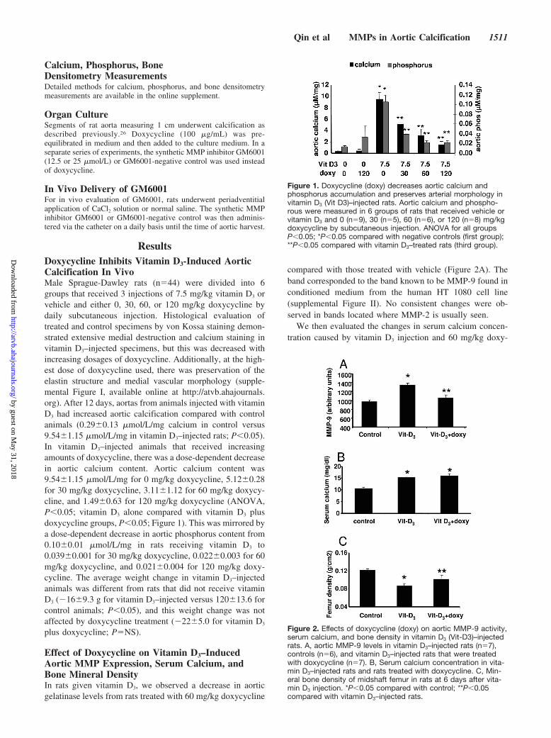
ResultsDoxycycline Inhibits Vitamin D3-Induced AorticCalcification In VivoMale Sprague-Dawley rats (n�44) were divided into 6groups that received 3 injections of 7.5 mg/kg vitamin D3 orvehicle and either 0, 30, 60, or 120 mg/kg doxycycline bydaily subcutaneous injection. Histological evaluation oftreated and control specimens by von Kossa staining demon-strated extensive medial destruction and calcium staining invitamin D3–injected specimens, but this was decreased withincreasing dosages of doxycycline. Additionally, at the high-est dose of doxycycline used, there was preservation of theelastin structure and medial vascular morphology (supple-mental Figure I, available online at http://atvb.ahajournals.org). After 12 days, aortas from animals injected with vitaminD3 had increased aortic calcification compared with controlanimals (0.29�0.13 �mol/L/mg calcium in control versus9.54�1.15 �mol/L/mg in vitamin D3–injected rats; P�0.05).In vitamin D3–injected animals that received increasingamounts of doxycycline, there was a dose-dependent decreasein aortic calcium content. Aortic calcium content was9.54�1.15 �mol/L/mg for 0 mg/kg doxycycline, 5.12�0.28for 30 mg/kg doxycycline, 3.11�1.12 for 60 mg/kg doxycy-cline, and 1.49�0.63 for 120 mg/kg doxycycline (ANOVA,P�0.05; vitamin D3 alone compared with vitamin D3 plusdoxycycline groups, P�0.05; Figure 1). This was mirrored bya dose-dependent decrease in aortic phosphorus content from0.10�0.01 �mol/L/mg in rats receiving vitamin D3 to0.039�0.001 for 30 mg/kg doxycycline, 0.022�0.003 for 60mg/kg doxycycline, and 0.021�0.004 for 120 mg/kg doxy-cycline. The average weight change in vitamin D3–injectedanimals was different from rats that did not receive vitaminD3 (�16�9.3 g for vitamin D3–injected versus 120�13.6 forcontrol animals; P�0.05), and this weight change was notaffected by doxycycline treatment (�22�5.0 for vitamin D3
plus doxycycline; P�NS).
Effect of Doxycycline on Vitamin D3–InducedAortic MMP Expression, Serum Calcium, andBone Mineral DensityIn rats given vitamin D3, we observed a decrease in aorticgelatinase levels from rats treated with 60 mg/kg doxycycline
compared with those treated with vehicle (Figure 2A). Theband corresponded to the band known to be MMP-9 found inconditioned medium from the human HT 1080 cell line(supplemental Figure II). No consistent changes were ob-served in bands located where MMP-2 is usually seen.
We then evaluated the changes in serum calcium concen-tration caused by vitamin D3 injection and 60 mg/kg doxy-
Figure 1. Doxycycline (doxy) decreases aortic calcium andphosphorus accumulation and preserves arterial morphology invitamin D3 (Vit D3)–injected rats. Aortic calcium and phospho-rous were measured in 6 groups of rats that received vehicle orvitamin D3 and 0 (n�9), 30 (n�5), 60 (n�6), or 120 (n�8) mg/kgdoxycycline by subcutaneous injection. ANOVA for all groupsP�0.05; *P�0.05 compared with negative controls (first group);**P�0.05 compared with vitamin D3–treated rats (third group).
Figure 2. Effects of doxycycline (doxy) on aortic MMP-9 activity,serum calcium, and bone density in vitamin D3 (Vit-D3)–injectedrats. A, aortic MMP-9 levels in vitamin D3–injected rats (n�7),controls (n�6), and vitamin D3–injected rats that were treatedwith doxycycline (n�7). B, Serum calcium concentration in vita-min D3–injected rats and rats treated with doxycycline. C, Min-eral bone density of midshaft femur in rats at 6 days after vita-min D3 injection. *P�0.05 compared with control; **P�0.05compared with vitamin D3–injected rats.
Qin et al MMPs in Aortic Calcification 1511
by guest on May 31, 2018
http://atvb.ahajournals.org/D
ownloaded from

cycline treatment. Serum calcium was significantly increasedin vitamin D3–injected rats compared with controls, and thiswas not affected by treatment with doxycycline. The calciumconcentration was 10.58�0.54 mg/dL in control rats versus15.23�0.44 in vitamin D3–injected rats (P�0.05 versuscontrols) and 15.95�0.84 in vitamin D3–injected plus doxy-cycline (P�NS compared with vitamin D3 alone; Figure 2B).
Bone density was significantly lower in vitamin D3–injected rats than in controls, but doxycycline treatmentpartially reversed this decrease. Bone density was0.120�0.002 g/cm2 in controls compared with 0.087�0.002in vitamin D3–injected rats (P�0.05 versus control) and0.101�0.005 in vitamin D3–injected plus doxycycline rats(P�0.05 versus controls; P�0.05 versus vitamin D3–injected;Figure 2C). This suggests that some effects of doxycycline onaortic calcification could occur through altering the increasedbone resorption caused by high doses of vitamin D3.
Doxycycline Inhibits Aortic Calcification Causedby Periadventitial CaCl2 AdministrationWe next evaluated whether the effects of doxycycline couldbe reproduced in a model of arterial calcification that is notcharacterized by alterations in bone resorption. We studiedthe effects of doxycycline treatment in rats that underwentperiadvential administration of 0.15 mol/L CaCl2. Animalsnot treated with doxycycline demonstrated medial and adven-titial accumulation of calcium by von Kossa staining. How-ever, animals treated with doxycycline had nearly completeinhibition of calcification as assessed by the von Kossa stainand by measurement of aortic calcium in harvested speci-mens. (Figure 3A and 3B). The aortic calcium content was0.06�0.015 �mol/L/mg for NaCl control rats (n�3),0.33�0.06 for CaCl2 rats not given doxycycline (n�4), and0.13�0.11 �moles/mg for rats treated with 60 mg/kg doxy-cycline (n�5; P�0.05; Figure 3C). Additionally, immuno-histochemical staining of these specimens using the macro-phage-specific antibody ED-1 demonstrated macrophageinfiltration in the adventitia of untreated rats, but this was notsee in doxycycline-treated rats (supplemental Figure III).Serum calcium levels in doxycycline-treated rats were notdifferent from control rats (12.0�0.13 for controls versus11.27�0.35 for doxycycline-treated rats; P�NS).
In Vitro Effect of Doxycycline and GM6001 onArterial CalcificationWe next investigated whether the inhibitory effects of doxy-cycline on in vivo calcification could be reproduced in anorgan culture system. This has the benefit of excluding theeffects of doxycycline on systemic, circulating factors. Aortaswere calcified in an organ culture system by addition of calfintestinal alkaline phosphatase and sodium phosphate toDMEM without serum (calcification medium).26 von Kossa–stained sections demonstrated calcium accumulation in theadventitia and outer medial layers of vessels cultured incalcification medium but little calcification in those alsotreated with doxycycline (supplemental Figure IV). Thecalcium content of vessels cultured in DMEM alone was0.284�0.090 �mol/L, and there was a 3-fold increase incalcium content to 0.953�0.280 �mol/L for those cultured in
calcification medium. Arteries cultured in calcification me-dium with 100 �g/mL doxycycline demonstrated a signifi-cant decrease in calcium accumulation (0.365�0.028;P�0.05 compared with calcification medium alone; Figure4A). Gelatinase levels in the medium of doxycycline-treatedsamples were also reduced (Figure 4B).
Although doxycycline has been useful for evaluating adrug with MMP-inhibiting properties, its experimental use islimited because it has several biological effects that are notrelated to MMPs. To determine whether the effects ofdoxycycline could be reproduced using a compound withmore specific anti-MMP activity, we used the syntheticMMP inhibitor GM6001. Histological evaluation of thesesegments by von Kossa’s stain failed to demonstratecalcium accumulation in GM6001-treated arteries (Figure 5Aand 5B). Aortas exposed to calcification medium showed anincrease in calcium content compared with control vessels(0.211�0.016 �mol/L for control compared with2.525�0.670 for calcification medium). When GM6001 wasadded to the calcification medium, there was a dose-dependent reduction in aortic calcium accumulation. Aorticcalcium was 2.331�0.456 �mol/L for 12.5 �mol/L (P�NS)and 0.434�0.088 for 25 �mol/L GM6001 (P�0.05 versus noGM6001; Figure 5C). Viability of cultured segments at 7
Figure 3. Doxycycline reduces aortic calcium accumulation andadventitial macrophage infiltration in rats undergoing periadven-titial administration of CaCl2. A and B, von Kossa–stained aorticsections from rat that underwent periadventitial application ofcalcium chloride and either no doxycycline (�doxy; A) or doxy-cycline (�doxy; B). Arrows indicate medial calcium; m, medium;a, adventitia. C, Calcium in aortas from rats that underwentapplication of NaCl (n�3), CaCl2 (n�4), and CaCl2 rats treatedwith 60 mg/kg doxycycline (n�5). *P�0.05 compared with NaClcontrol rats; **P�0.05 compared with CaCl2 rats not givendoxycycline.
1512 Arterioscler Thromb Vasc Biol. July 2006
by guest on May 31, 2018
http://atvb.ahajournals.org/D
ownloaded from

days was not affected by calcification medium or GM6001, asevidenced by measurement of lactic dehydrogenase secretioninto the medium, which was not different from controlsegments (data not shown).
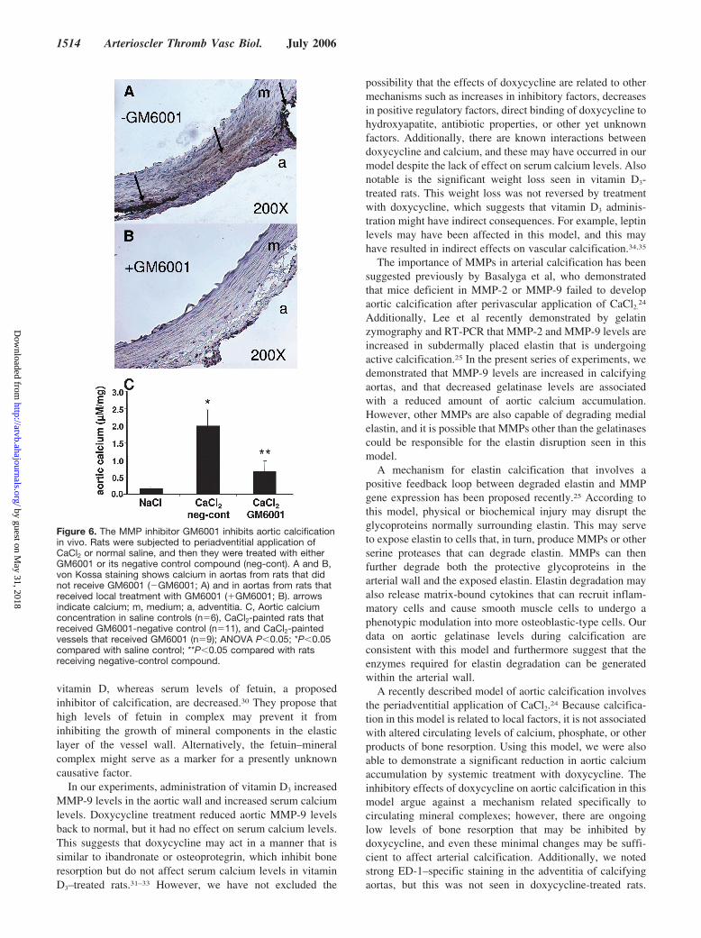
GM6001 Inhibits Aortic Calcification In VivoBased on our findings in organ culture, we next evaluatedwhether GM6001 could be used to inhibit aortic calcificationin vivo. Daily injections of GM6001 or its negative control(250 �g per day) were administered by catheter into theperiaortic space after periadventitial application of calciumchloride. Aortas from control rats that underwent paintingwith NaCl had minimal calcification after 7 days. However,CaCl2-painted arteries treated with GM6001-negative controlhad a �10-fold increase in calcium accumulation, and thiswas significantly decreased by local delivery of GM6001(0.166�0.026 �mol/L for normal saline controls;1.983�0.477 for CaCl2 with GM6001-negative control; and0.656�0.331 for CaCl2 painted vessels treated with GM6001;P�0.05). Aortic histology from rats that received the nega-tive control compound demonstrated extensive calcificationin the adventitia and in the outer medial layers, whereas therewas only occasional calcium staining in aortas from ratstreated with GM6001 (Figure 6).
DiscussionIn this study, we found that inhibiting MMP activity isassociated with decreased calcium accumulation in the arte-rial wall. We began our studies by using doxycycline, anantibiotic with known MMP-inhibiting properties, to demon-strate an association between decreased aortic gelatinaselevels and reduced aortic calcification. To explore the impor-tance of MMPs in arterial calcification more directly, we used
the synthetic MMP-specific inhibitor GM6001 and were ableto demonstrate that it decreased aortic calcification both invitro and in vivo. These findings suggest that MMPs play animportant role in arterial calcification, and further, thatinhibiting MMP activity may prevent arterial calcification inthe clinical setting.
The changes in arterial histology associated with hypervi-taminosis D have been well characterized.27–29 Rats exposedto sublethal doses of vitamin D initially develop calciumphosphate crystals along the elastic laminae followed byspreading of calcification into the surrounding medial smoothmuscle cells.27 The model is characterized by increased boneresorption and increased intestinal uptake of ingested cal-cium, leading to elevated serum calcium levels.28 Althoughthe mechanisms related to arterial calcification in this modelhave not been fully elucidated, it is thought to occur throughan interaction between degraded medial elastin and circulat-ing factors from resorbed bone. Price et al demonstratedrecently that a fetuin-matrix Gla protein–mineral complex isincreased in the circulation of rats exposed to toxic doses of
Figure 4. Addition of doxycycline (doxy) to calcifying aortas inorgan culture inhibits calcification and reduces MMP levels. A,Aortic calcium content measured in HCl extracts from 1-cm aor-tic specimens taken after 9 days in organ culture with the speci-fied treatments. B, MMP levels in conditioned medium deter-mined by gelatin zymography from cultured aortas. D0 indicatesDMEM without serum; CM, calcification medium. n�5 for eachgroup; *P�0.05 compared with control; **P�0.05 comparedwith CM groups. This experiment was performed twice withsimilar results.
Figure 5. The MMP inhibitor GM6001 inhibits in vitro aortic cal-cification. A and B, von Kossa–stained section of artery incu-bated in calcification medium (A) or in an artery incubated incalcification medium with 25 �mol/L GM6001 (B). C, Graph ofcalcium accumulation in artery wall of vessels cultured in calcifi-cation medium (CM) with 12.5 or 25 �mol/L GM6001. m indi-cates medium; arrows, calcium; a, adventitia (n�6 for eachgroup; *P�0.05 compared with control; **P�0.05 comparedwith CM-treated groups). This experiment was performed 3times with similar results.
Qin et al MMPs in Aortic Calcification 1513
by guest on May 31, 2018
http://atvb.ahajournals.org/D
ownloaded from
vitamin D, whereas serum levels of fetuin, a proposedinhibitor of calcification, are decreased.30 They propose thathigh levels of fetuin in complex may prevent it frominhibiting the growth of mineral components in the elasticlayer of the vessel wall. Alternatively, the fetuin–mineralcomplex might serve as a marker for a presently unknowncausative factor.
In our experiments, administration of vitamin D3 increasedMMP-9 levels in the aortic wall and increased serum calciumlevels. Doxycycline treatment reduced aortic MMP-9 levelsback to normal, but it had no effect on serum calcium levels.This suggests that doxycycline may act in a manner that issimilar to ibandronate or osteoprotegrin, which inhibit boneresorption but do not affect serum calcium levels in vitaminD3–treated rats.31–33 However, we have not excluded the
possibility that the effects of doxycycline are related to othermechanisms such as increases in inhibitory factors, decreasesin positive regulatory factors, direct binding of doxycycline tohydroxyapatite, antibiotic properties, or other yet unknownfactors. Additionally, there are known interactions betweendoxycycline and calcium, and these may have occurred in ourmodel despite the lack of effect on serum calcium levels. Alsonotable is the significant weight loss seen in vitamin D3-treated rats. This weight loss was not reversed by treatmentwith doxycycline, which suggests that vitamin D3 adminis-tration might have indirect consequences. For example, leptinlevels may have been affected in this model, and this mayhave resulted in indirect effects on vascular calcification.34,35
The importance of MMPs in arterial calcification has beensuggested previously by Basalyga et al, who demonstratedthat mice deficient in MMP-2 or MMP-9 failed to developaortic calcification after perivascular application of CaCl2.
24
Additionally, Lee et al recently demonstrated by gelatinzymography and RT-PCR that MMP-2 and MMP-9 levels areincreased in subdermally placed elastin that is undergoingactive calcification.25 In the present series of experiments, wedemonstrated that MMP-9 levels are increased in calcifyingaortas, and that decreased gelatinase levels are associatedwith a reduced amount of aortic calcium accumulation.However, other MMPs are also capable of degrading medialelastin, and it is possible that MMPs other than the gelatinasescould be responsible for the elastin disruption seen in thismodel.
A mechanism for elastin calcification that involves apositive feedback loop between degraded elastin and MMPgene expression has been proposed recently.25 According tothis model, physical or biochemical injury may disrupt theglycoproteins normally surrounding elastin. This may serveto expose elastin to cells that, in turn, produce MMPs or otherserine proteases that can degrade elastin. MMPs can thenfurther degrade both the protective glycoproteins in thearterial wall and the exposed elastin. Elastin degradation mayalso release matrix-bound cytokines that can recruit inflam-matory cells and cause smooth muscle cells to undergo aphenotypic modulation into more osteoblastic-type cells. Ourdata on aortic gelatinase levels during calcification areconsistent with this model and furthermore suggest that theenzymes required for elastin degradation can be generatedwithin the arterial wall.
A recently described model of aortic calcification involvesthe periadventitial application of CaCl2.24 Because calcifica-tion in this model is related to local factors, it is not associatedwith altered circulating levels of calcium, phosphate, or otherproducts of bone resorption. Using this model, we were alsoable to demonstrate a significant reduction in aortic calciumaccumulation by systemic treatment with doxycycline. Theinhibitory effects of doxycycline on aortic calcification in thismodel argue against a mechanism related specifically tocirculating mineral complexes; however, there are ongoinglow levels of bone resorption that may be inhibited bydoxycycline, and even these minimal changes may be suffi-cient to affect arterial calcification. Additionally, we notedstrong ED-1–specific staining in the adventitia of calcifyingaortas, but this was not seen in doxycycline-treated rats.
Figure 6. The MMP inhibitor GM6001 inhibits aortic calcificationin vivo. Rats were subjected to periadventitial application ofCaCl2 or normal saline, and then they were treated with eitherGM6001 or its negative control compound (neg-cont). A and B,von Kossa staining shows calcium in aortas from rats that didnot receive GM6001 (�GM6001; A) and in aortas from rats thatreceived local treatment with GM6001 (�GM6001; B). arrowsindicate calcium; m, medium; a, adventitia. C, Aortic calciumconcentration in saline controls (n�6), CaCl2-painted rats thatreceived GM6001-negative control (n�11), and CaCl2-paintedvessels that received GM6001 (n�9); ANOVA P�0.05; *P�0.05compared with saline control; **P�0.05 compared with ratsreceiving negative-control compound.
1514 Arterioscler Thromb Vasc Biol. July 2006
by guest on May 31, 2018
http://atvb.ahajournals.org/D
ownloaded from

Although the role of macrophages in this model has not beendefined, it is possible that they are another source of matrix-degrading enzymes necessary for the calcification to proceed.The potential role of inflammatory cells and their secretedmediators on arterial calcification has been reviewed recent-ly,36 and further studies in this regard are needed.
Our experiments in organ culture allowed us to focus on thedirect effects of MMP inhibition on the aortic wall. In thissystem, calcification is thought to occur because of inhibition ofpyrophosphate production and increased calcium�phosphorusproduct in the medium.26 Doxycycline reduced aortic calciumlevels, and this effect was associated with decreased MMP levelsin the culture medium. This likely occurred through the previ-ously described inhibitory effects of doxycycline on MMPsynthesis.37 However, the use of doxycycline in mechanisticstudies is limited because of its diverse actions, many of whichare not related to altering MMP levels. For this reason, weextended our experiments to include the synthetic compoundGM6001 that specifically inhibits the MMPs. We found thataddition of GM6001 to the calcification medium resulted in adose-dependent decrease in arterial calcium accumulation. Onhistology, GM6001-treated vessels had less calcium depositionin the adventitia and outer medial layers. GM6001 is a potent,hydroxamic acid–based MMP inhibitor with activity againstseveral MMPs, including collagenase, the gelatinases, andstromelysin. Both the gelatinases and stromelysin have elasto-lytic activity, and a possible role of MMPs in medial calcifica-tion is in initiating the process of elastin degradation that mayserve to provide a nidus for hydroxyapatite crystals to develop.These in vitro results suggested that synthetic MMP inhibitorsmight prevent arterial calcification in vivo.
Our final series of experiments involved the use ofGM6001 in vivo. For these studies, GM6001 was deliveredlocally, near the aorta at a concentration that was similar tothat used in organ culture, and we found that daily adminis-tration reduced aortic calcium content compared with nega-tive control treated vessels. Although the source of MMPactivity in this model is presently unknown, the potentialsources include medial smooth muscle cells, adventitialfibroblasts, or infiltrating inflammatory cells, all of whichwould have been affected by local delivery of the inhibitor.GM6001 is a synthetic inhibitor with activity against severalMMPs, and thus, the identity of MMPs involved in arterialcalcification remains to be determined. Based on the currentmodel, it is likely to be an MMP with elastolytic propertiesthat can be produced either within the aortic wall or by cellsthat can reside in the adventitia. One possibility is thatGM6001 has direct effects on the CaCl2 solution used toinhibit calcification. However, the accumulation of aorticcalcium we observed with the negative control compoundargues against this point. Another limitation of this techniqueis that compound delivery is limited to 5 to 7 days because ofthe formation of a fibrous capsule around the catheter.However, the fact that catheter delivery was successfulsuggests that aortic calcification occurs early in this modeland that it can be inhibited by drug administration during thefirst several days. Alternatively, sufficient compound mayhave diffused through the capsule to have an effect at latertime points. Efforts to understand the temporal aspects of this
model and the relative contributions of MMPs during thevarious phases of arterial calcification are under way.
Together, our data suggest that MMPs are involved inarterial calcification and that inhibiting MMPs may be aclinically useful method for reducing calcification in thevessel wall. Arterial calcification is a complex phenomenonthat is likely regulated at several levels, and further efforts tounderstand the role of MMPs in this pathologic process areneeded.
AcknowledgmentWe thank Dr Richard Hoover for his support and assistance inproducing this manuscript.
Sources of FundingThis research was supported by mentored clinical scientist develop-ment award HL069926 from the National Heart, Lung, and BloodInstitute (R.G.) and grants from the Lifeline Foundation and theWilliam J. von Liebig Foundation.
DisclosuresNone.
References1. Burke SK. Arterial calcification in chronic kidney disease. Semin
Nephrol. 2004;24:403–407.2. Abedin M, Tintut Y, Demer LL. Vascular calcification: mechanisms and
clinical ramifications. Arterioscler Thromb Vasc Biol. 2004;24:1161–1170.
3. Speer MY, Giachelli CM. Regulation of cardiovascular calcification.Cardiovasc Pathol. 2004;13:63–70.
4. Arad Y, Spadaro LA, Goodman K, Newstein D, Guerci AD. Prediction ofcoronary events with electron beam computed tomography. J Am CollCardiol. 2000;36:1253–1260.
5. Taylor AJ, Bindeman J, Feuerstein I, Cao F, Brazaitis M, O’Malley PG.Coronary calcium independently predicts incident premature coronaryheart disease over measured cardiovascular risk factors: mean three-yearoutcomes in the Prospective Army Coronary Calcium (PACC) Project.J Am Coll Cardiol. 2005;46:807–814.
6. London GM, Guerin AP, Marchais SJ, Metivier F, Pannier B, Adda H.Arterial media calcification in end-stage renal disease: impact onall-cause and cardiovascular mortality. Nephrol Dial Transplant. 2003;18:1731–1740.
7. Wolfe ML, Iqbal N, Gefter W, Mohler ER III, Rader DJ, Reilly MP.Coronary artery calcification at electron beam computed tomography isincreased in asymptomatic type 2 diabetics independent of traditional riskfactors. J Cardiovasc Risk. 2002;9:369–376.
8. Vattikuti R, Towler DA. Osteogenic regulation of vascular calcification:an early perspective. Am J Physiol Endocrinol Metab. 2004;286:E686–E696.
9. Bostrom K, Watson KE, Horn S, Wortham C, Herman IM, Demer LL.Bone morphogenetic protein expression in human atherosclerotic lesions.J Clin Invest. 1993;91:1800–1809.
10. Shanahan CM, Cary NR, Metcalfe JC, Weissberg PL. High expression ofgenes for calcification-regulating proteins in human atheroscleroticplaques. J Clin Invest. 1994;93:2393–2402.
11. Bini A, Mann KG, Kudryk BJ, Schoen FJ. Noncollagenous bone matrixproteins, calcification, and thrombosis in carotid artery atherosclerosis.Arterioscler Thromb Vasc Biol. 1999;19:1852–1861.
12. Shanahan CM, Cary NR, Salisbury JR, Proudfoot D, Weissberg PL,Edmonds ME. Medial localization of mineralization-regulating proteinsin association with Monckeberg’s sclerosis: evidence for smooth musclecell-mediated vascular calcification. Circulation. 1999;100:2168–2176.
13. Dhore CR, Cleutjens JP, Lutgens E, Cleutjens KB, Geusens PP, KitslaarPJ, Tordoir JH, Spronk HM, Vermeer C, Daemen MJ. Differentialexpression of bone matrix regulatory proteins in human atheroscleroticplaques. Arterioscler Thromb Vasc Biol. 2001;21:1998–2003.
14. Fischer JW, Steitz SA, Johnson PY, Burke A, Kolodgie F, Virmani R,Giachelli C, Wight TN. Decorin promotes aortic smooth muscle cell
Qin et al MMPs in Aortic Calcification 1515
by guest on May 31, 2018
http://atvb.ahajournals.org/D
ownloaded from

calcification and colocalizes to calcified regions in human atheroscleroticlesions. Arterioscler Thromb Vasc Biol. 2004;24:2391–2396.
15. Schoppet M, Al-Fakhri N, Franke FE, Katz N, Barth PJ, Maisch B,Preissner KT, Hofbauer LC. Localization of osteoprotegerin, tumornecrosis factor-related apoptosis-inducing ligand, and receptor activatorof nuclear factor-kappaB ligand in Monckeberg’s sclerosis and athero-sclerosis. J Clin Endocrinol Metab. 2004;89:4104–4112.
16. Galis ZS, Khatri JJ. Matrix metalloproteinases in vascular remodeling andatherogenesis: the good, the bad, and the ugly. Circ Res. 2002;90:251–262.
17. Halpert I, Sires UI, Roby JD, Potter-Perigo S, Wight TN, Shapiro SD,Welgus HG, Wickline SA, Parks WC. Matrilysin is expressed bylipid-laden macrophages at sites of potential rupture in atheroscleroticlesions and localizes to areas of versican deposition, a proteoglycansubstrate for the enzyme. Proc Natl Acad Sci U S A. 1996;93:9748–9753.
18. Nikkari ST, Geary RL, Hatsukami T, Ferguson M, Forough R, Alpers CE,Clowes AW. Expression of collagen, interstitial collagenase, and tissueinhibitor of metalloproteinases-1 in restenosis after carotid endarterecto-my. Am J Pathol. 1996;148:777–783.
19. Galis ZS, Sukhova GK, Lark MW, Libby P. Increased expression ofmatrix metalloproteinases and matrix degrading activity in vulnerableregions of human atherosclerotic plaques. J Clin Invest. 1994;94:2493–2503.
20. Allaire E, Forough R, Clowes M, Starcher B, Clowes AW. Local over-expression of TIMP-1 prevents aortic aneurysm degeneration and rupturein a rat model. J Clin Invest. 1998;102:1413–1420.
21. Petrinec D, Liao S, Holmes DR, Reilly JM, Parks WC, Thompson RW.Doxycycline inhibition of aneurysmal degeneration in an elastase-induced rat model of abdominal aortic aneurysm: preservation of aorticelastin associated with suppressed production of 92 kD gelatinase. J VascSurg. 1996;23:336–346.
22. Mason DP, Kenagy RD, Hasenstab D, Bowen-Pope DF, Seifert RA,Coats S, Hawkins SM, Clowes AW. Matrix metalloproteinase-9 overex-pression enhances vascular smooth muscle cell migration and altersremodeling in the injured rat carotid artery. Circ Res. 1999;85:1179–1185.
23. Bendeck MP, Conte M, Zhang M, Nili N, Strauss BH, Farwell SM.Doxycycline modulates smooth muscle cell growth, migration, andmatrix remodeling after arterial injury. Am J Pathol. 2002;160:1089–1095.
24. Basalyga DM, Simionescu DT, Xiong W, Baxter BT, Starcher BC,Vyavahare NR. Elastin degradation and calcification in an abdominalaorta injury model: role of matrix metalloproteinases. Circulation. 2004;110:3480–3487.
25. Lee JS, Basalyga DM, Simionescu A, Isenburg JC, Simionescu DT,Vyavahare NR. Elastin calcification in the rat subdermal model is accom-panied by upregulation of degradative and osteogenic cellular responses.Am J Pathol. 2006;168:490–498.
26. Lomashvili KA, Cobbs S, Hennigar RA, Hardcastle KI, O’Neill WC.Phosphate-induced vascular calcification: role of pyrophosphate andosteopontin. J Am Soc Nephrol. 2004;15:1392–1401.
27. Bonucci E, Sadun R. Dihydrotachysterol-induced aortic calcification. Ahistochemical and ultrastructural investigation. Clin Orthop Relat Res.1975;283–294.
28. Tanimura A, Cho T, Tanaka S. Aortic changes induced by hypercholes-terolemia and hypercalcemia in rats. Exp Mol Pathol. 1986;44:297–306.
29. Cannon EP, Williams BJ. Raised vascular calcium in an animal model:effects on aortic function. Cardiovasc Res. 1990;24:47–52.
30. Price PA, Williamson MK, Nguyen TM, Than TN. Serum levels of thefetuin-mineral complex correlate with artery calcification in the rat. J BiolChem. 2004;279:1594–1600.
31. Price PA, Faus SA, Williamson MK. Bisphosphonates alendronate andibandronate inhibit artery calcification at doses comparable to those thatinhibit bone resorption. Arterioscler Thromb Vasc Biol. 2001;21:817–824.
32. Price PA, June HH, Buckley JR, Williamson MK. Osteoprotegerininhibits artery calcification induced by warfarin and by vitamin D. Arte-rioscler Thromb Vasc Biol. 2001;21:1610–1616.
33. Price PA, Omid N, Than TN, Williamson MK. The amino bisphos-phonate ibandronate prevents calciphylaxis in the rat at doses that inhibitbone resorption. Calcif Tissue Int. 2002;71:356–363.
34. Tartaglia LA. The leptin receptor. J Biol Chem. 1997;272:6093–6096.35. Parhami F, Tintut Y, Ballard A, Fogelman AM, Demer LL. Leptin
enhances the calcification of vascular cells: artery wall as a target ofleptin. Circ Res. 2001;88:954–960.
36. Doherty TM, Asotra K, Fitzpatrick LA, Qiao J-H, Wilkin DJ, DetranoRC, Dunstan CR, Shah PK, Rajavashisth TB. Calcification in atheroscle-rosis: Bone biology and chronic inflammation at the arterial crossroads.Proc Natl Acad Sci U S A. 2003;100:11201–11206.
37. Liu J, Xiong W, Baca-Regen L, Nagase H, Baxter BT. Mechanism ofinhibition of matrix metalloproteinase-2 expression by doxycycline inhuman aortic smooth muscle cells. J Vasc Surg. 2003;38:1376–1383.
1516 Arterioscler Thromb Vasc Biol. July 2006
by guest on May 31, 2018
http://atvb.ahajournals.org/D
ownloaded from
Xiao Qin, Matthew A. Corriere, Lynn M. Matrisian and Raul J. GuzmanMatrix Metalloproteinase Inhibition Attenuates Aortic Calcification
Print ISSN: 1079-5642. Online ISSN: 1524-4636 Copyright © 2006 American Heart Association, Inc. All rights reserved.
Greenville Avenue, Dallas, TX 75231is published by the American Heart Association, 7272Arteriosclerosis, Thrombosis, and Vascular Biology
doi: 10.1161/01.ATV.0000225807.76419.a72006;26:1510-1516; originally published online May 11, 2006;Arterioscler Thromb Vasc Biol.
http://atvb.ahajournals.org/content/26/7/1510World Wide Web at:
The online version of this article, along with updated information and services, is located on the
http://atvb.ahajournals.org/content/suppl/2006/05/21/01.ATV.0000225807.76419.a7.DC1Data Supplement (unedited) at:
http://atvb.ahajournals.org//subscriptions/
at: is onlineArteriosclerosis, Thrombosis, and Vascular Biology Information about subscribing to Subscriptions:
http://www.lww.com/reprints
Information about reprints can be found online at: Reprints:
document. Question and AnswerPermissions and Rightspage under Services. Further information about this process is available in the
which permission is being requested is located, click Request Permissions in the middle column of the WebCopyright Clearance Center, not the Editorial Office. Once the online version of the published article for
can be obtained via RightsLink, a service of theArteriosclerosis, Thrombosis, and Vascular Biologyin Requests for permissions to reproduce figures, tables, or portions of articles originally publishedPermissions:
by guest on May 31, 2018
http://atvb.ahajournals.org/D
ownloaded from

1
ONLINE SUPPLEMENT
MATERIALS AND METHODS
Materials
Vitamin D3 (cholecalciferol) and doxycycline hyclate were purchased from Sigma-
Aldrich Chemical Co., Inc. (St. Louis, MO). The synthetic MMP inhibitor GM6001 and
its negative control GM6001-negative-control were purchased from Calbiochem, EMD
Biosciences, Inc, an Affiliate of Merck KGaA, (Darmstadt, Germany). Vitamin D3
solution for injections was prepared as described by Price.1 A stock solution was made
by dissolving 0.033g vitamin D3 in 200µl ethanol and suspended in 1.4ml of Emulphor
(alkamuls EL-620) before diluting it to 1.65 mg/ml (66,000IU/ml) in 20 ml D5W.
Doxycycline hyclate was dissolved in water to give a concentration of 20 mg/ml. Male
Sprague-Dawley rats weighing 100 g were purchased from Charles River (Bedford, MA).
Experimental Procedures
All procedures were approved by the Vanderbilt Institutional Animal Care and
Use Committee. Animals were housed in accordance with institutional policies and fed,
ad libitum with Purina 5001 rodent chow containing 0.67% phosphorus and 0.95%
calcium by weight. In order to induce aortic calcification, rats were given subcutaneous
injections of 7.5 mg/ml Vitamin D3 for three consecutive days. For evaluation of its
effects on aortic calcification, rats received 0, 30, 60, or 120 mg/kg doxycycline daily by
subcutaneous injection beginning at 2 days prior to vitamin D3 injections and ending at
the time of harvest. Rats were weighed prior to the experiment and immediately before
vessel harvest. Animals that appeared to be in distress were euthanized and not used for
analysis. Mortality was less than 5% in each group. On the day of aortic harvest,

2
animals were overdosed with pentobarbital and the aorta was harvested from the arch to
the iliac bifurcation. Blood was collected from the inferior vena cava and the serum was
collected and stored at -70ºC until needed for further evaluation. In order to insure
consistency, the thoracic aorta was used routinely for histologic evaluation, the infra-
renal aorta for gelatin zymography, and a 1 cm portion of aorta at the level of the
mesenteric vessels for evaluation of calcium and phosphorus levels.
In another group of rats, aortic calcification was induced by periadventitial
application of 0.15 M CaCl2 as recently described.2 Anesthesia was induced using
Enflurane and rats were anesthetized using 1.5 mg/kg ketamine and 15 mg/kg xylazine.
Through a midline incision, the aorta was exposed and CaCl2 or 0.9% NaCl as a control
were applied with 15 paint strokes using a cotton tipped swab. Two days prior to CaCl2
application, animals began treatment with 60 mg/kg of doxycycline or water and this was
performed daily until aortic harvest. One week after periadventitial CaCl2 administration,
animals were anesthetized with ketamine and xylazine then euthanized by
exsanguination. A segment of infrarenal aorta was excised and processed for calcium
determination and histology.
Calcium and Phosphorus measurements
Samples of aorta measuring 1 cm were incubated in 150 mM HCl for 24 hours with
gentle agitation. Calcium content was measured using a modified o-cresolphthalein
complexone method; 3 and phosphorous content was measured using the ammonium
molybdate method.4 Specimens were then dried and weighed. Results are expressed as
µmoles calcium per mg of aortic tissue for in vivo experiments and µmoles calcium for
3
organ culture experiments. Serum calcium was measured by a commercial laboratory
(Antech Diagnostics, Southhaven, MS).
Bone Densitometry measurements
Bone density was measured 6 days after beginning vitamin D3 treatment. Rats were
anesthetized with ketamine and xylazine then imaged using the GE PIXImus™ bone
densitometry system (Wipro GE Healthcare) which uses DEXA (dual-energy x-ray
absorptiometry) to determine bone mineral density. Equal size regions of interest were
selected manually to include the left femoral shaft, and bone density, measured as g
mineral/cm2, was determined automatically using the system software.
Histology and Immunohistochemical staining
For histology, the thoracic aorta was harvested within 10 minutes of death and rinsed in
PBS prior to fixation in 4% paraformaldehyde for 30 minutes. Specimens were then
dehydrated and embedded in paraffin for sectioning. Sections were stained by
hematoxylin and eosin and serial sections were stained using the von Kossa method (5%
AgNO3 for 1 hour under light) and counterstained with hematoxylin. For
immunohistochemistry, sections were hydrated, incubated in blocking serum, and then
incubated with the macrophage specific antibody ED-1 (Research Diagnostics, Inc.,
Flanders, NJ) overnight at 4ºC. Vessels were then incubated with secondary anti-mouse
antibody followed by DAB peroxidase substrate (Vector Laboratories, Inc, Burlingame,
CA) and then counterstained with hematoxylin.
MMP Gel zymography
Sections from the abdominal aorta of vitamin D3-injected rats were harvested at various
times and then snap frozen in liquid nitrogen until needed. Protein from aortic specimens

4
was extracted in a sodium dodecyl sulfate (SDS) containing buffer and then equal
amounts of protein from each extract were electrophoresed on an 8% SDS–
polyacrylamide gel containing 0.1% type I gelatin (J.T. Baker, Phillipsburg, NJ). Gels
were washed with 2.5% Triton X-100, incubated overnight in 0.05 mol/L Tris with 2.5
mmol/L CaCl2 and 0.02% NaN3, stained with Coomassie blue, then destained in methanol
and acetic acid solution. Gelatin degradation was observed as white lytic bands. Gels
were digitally photographed and bands were quantified using the Bio-Rad Quantity One
software. Conditioned medium from the HT1080 human fibrosarcoma cell line was used
to mark MMPs in the Zymograms.
Organ Culture
Segments of rat aorta measuring 1 cm were harvested and gently cleared of surrounding
tissues. Aortic segments underwent calcification as previously described.5 Briefly, aortic
segments were incubated in DMEM (Gibco, Long Island, NY) containing 1X
penicillin/streptomycin and without serum at 37°C in a humidified 5% CO2 / 95% air
atmosphere. In order to induce calcification, 7.5 units/ml of calf intestinal alkaline
phosphatase was added to DMEM and the phosphate ion concentration was increased to
3.8 µM by addition of sodium phospate (calcification medium). Doxycycline (100
µg/ml) was pre-equilibrated in medium and then added to the culture medium. In a
separate series of experiments, the synthetic MMP inhibitor GM6001 (12.5 or 25 µM) or
GM6001 negative control were used instead of doxycycline. Culture medium with
additives was pre-incubated in order to allow it to equilibrate at 37ºC and pH=7.4 before
applying to specimens. After 9 days, aortic specimens were rinsed in normal saline prior

5
to decalcifying in 500 µl of 0.15 M HCl for 24 hours with gentle agitation. Calcium was
then measured in 50 µl samples of the HCl solution as described above.
In vivo delivery of GM6001
For in vivo evaluation of GM6001, rats initially underwent peri-adventitial application of
CaCl2 solution or normal saline to induce calcification as described above. Immediately
after CaCl2 administration, PE-50 tubing with an occluded end and multiple side
perforations was placed adjacent to the infrarenal aorta. The other end of the catheter
was the tunneled subcutaneously to the intrascapular region and exteriorized. The
synthetic MMP inhibitor GM6001 was prepared by dissolving 5 mg of powdered
compound in 1 ml DMSO. For each dose, 25 µl of compound in DMSO was diluted to
400 µl in 0.9% NaCl. The commercially supplied negative control, named GM6001-
negative-control, was prepared in an identical manner. Compounds were administered
via the catheter on a daily basis until the time of aortic harvest. After 7 days of treatment,
the aorta was harvested and processed for calcium accumulation and histology as
described above.
Cell viability assay
For organ culture studies, cellular viability of aortas was assessed at 3 days and at 7 days
using the CytoTox-ONE homogenous membrane integrity assay (Promega Corp,
Madison, WI) which measures the release of lactate dehydrogenase from cells. The assay
was conducted using 100 µl of conditioned medium and according to the supplied
protocol.

6
Statistics
Data are presented as mean ± standard error of the mean. For in vivo studies, data was
initially evaluated using analysis of variance (ANOVA) followed by post-hoc evaluation
using Bonferroni’s method with p<0.05 considered significant. For organ culture studies,
group comparisons were performed using the Kruskal-Wallis test followed by pair wise
comparisons using the Mann-Whitney U test.
REFERENCES
1. Price PA, Faus SA, Williamson MK. Warfarin-induced artery calcification is
accelerated by growth and vitamin D. Arterioscler Thromb Vasc Biol.
2000;20:317-327.
2. Basalyga DM, Simionescu DT, Xiong W, Baxter BT, Starcher BC, Vyavahare
NR. Elastin degradation and calcification in an abdominal aorta injury model: role
of matrix metalloproteinases. Circulation. 2004;110:3480-3487.
3. Connerty HV, Briggs AR. Determination of serum calcium by means of
orthocresolphthalein complexone. Am J Clin Pathol. 1966;45:290-296.
4. Chen PS, Toribara TY, Huber W. Microdectermination of Phophorus. Analytic
Chemistry. 1956;28:1756-1758.
5. Lomashvili KA, Cobbs S, Hennigar RA, Hardcastle KI, O'Neill WC. Phosphate-
induced vascular calcification: role of pyrophosphate and osteopontin. J Am Soc
Nephrol. 2004;15:1392-1401.

7
ONLINE SUPPLEMENTAL DATA
Figure I. Representative sections of von Kossa stained aortic specimens show a normal
vessel (A), followed by aorta from vitamin D3-injected rat that did not receive
doxycycline (B), aorta from Vitamin D3-injected rats that received 30 mg/kg (C), 60
mg/kg (D), and 120 mg/kg (E) doxycycline by injection. Arrows denote calcium
staining, m = media, a = adventitia.
m
a
200X
m
200X
m
a
200X
m
a
200X
B
C D
E
a
m
a
200X
A
8
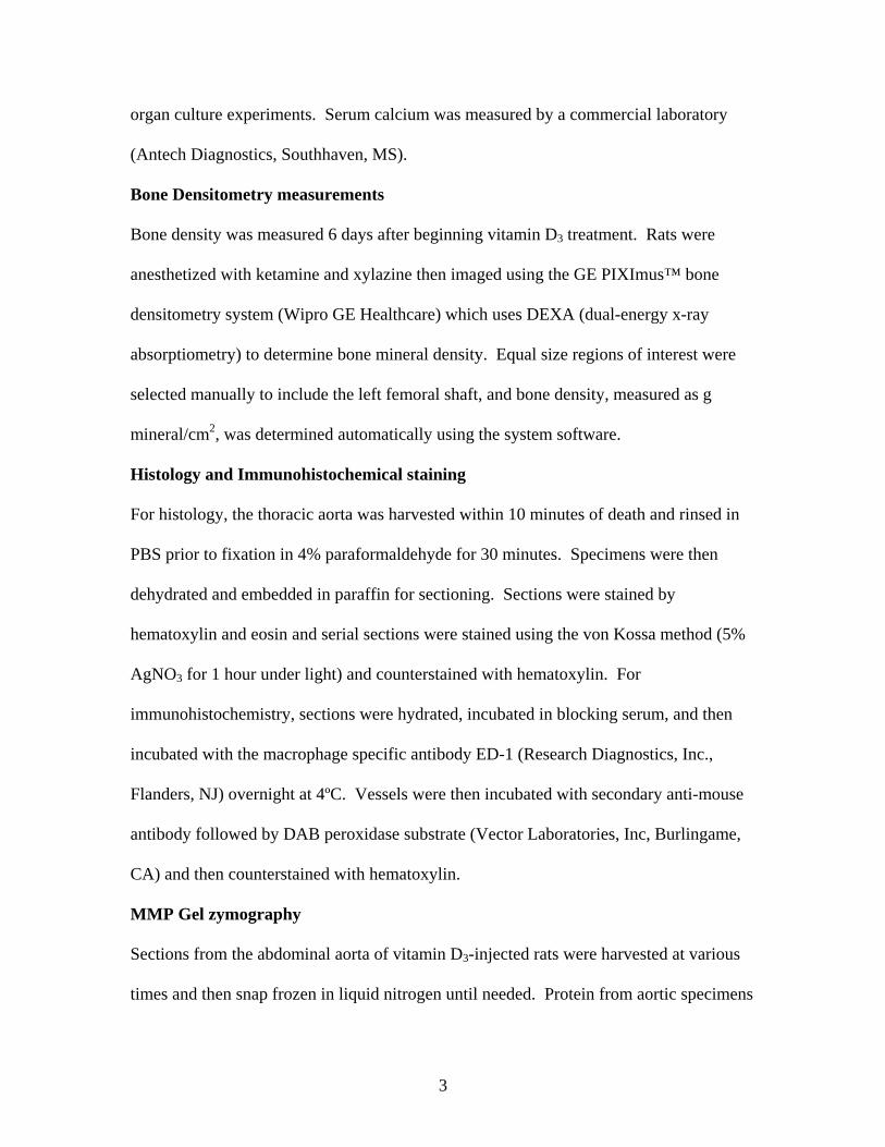
Figure II. A. Gelatin zymography of aortic lysates from vehicle treated controls,
vitamin D3-injected and vitamin D3-injected plus 60mg/kg doxycycline treated rats.
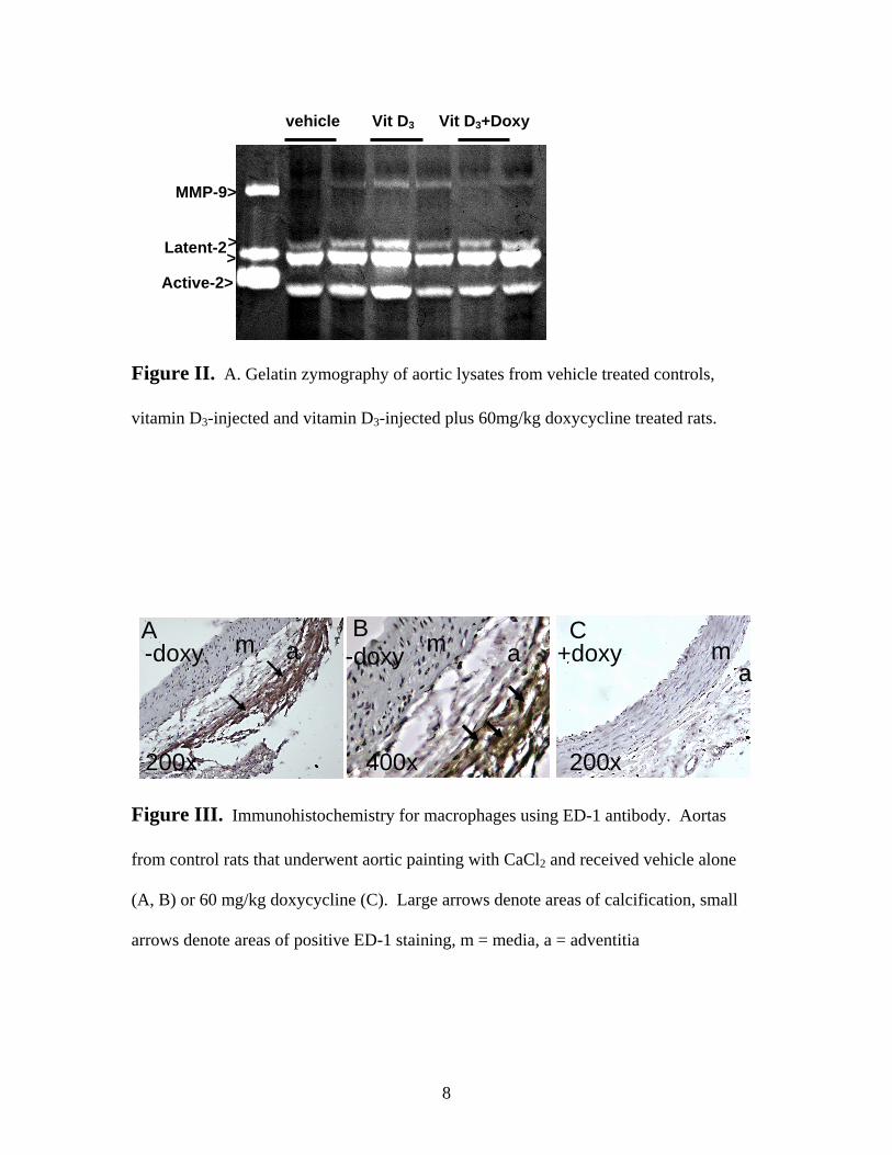
Figure III. Immunohistochemistry for macrophages using ED-1 antibody. Aortas
from control rats that underwent aortic painting with CaCl2 and received vehicle alone
(A, B) or 60 mg/kg doxycycline (C). Large arrows denote areas of calcification, small
arrows denote areas of positive ED-1 staining, m = media, a = adventitia
> >
Vit D3+Doxy
MMP-9>
vehicle Vit D3
Active-2>
Latent-2
m a
200x
A
am
200x
C B m a
400x
-doxy -doxy +doxy

9
Figure IV. Von Kossa staining of organ culture vessels incubated in calcification
medium (A), or in calcification medium with doxycycline (B). Arrows denote calcium in
arterial media.
A m
a
200X 100 µm
m a
200X
B -doxy +doxy