Matrix Mechanics Influence Fibroblast–Myofibroblast...
11
Matrix Mechanics Influence Fibroblast–Myofibroblast Transition by Directing the Localization of Histone Deacetylase 4 YANFEN LI, 1 CLAIRE B. TANG, 2 and KRISTOPHER A. KILIAN 1 1 Department of Bioengineering, University of Illinois at Urbana-Champaign, Urbana, IL 61801, USA; and 2 Department of Molecular and Cellular Biology, University of Illinois at Urbana-Champaign, Urbana, IL 61801, USA (Received 10 February 2017; accepted 7 July 2017; published online 18 July 2017) Associate Editor Alyssa Panitch oversaw the review of this article. Abstract Introduction—The propagation of mechanochemical signals from the extracellular matrix to the cell nucleus has emerged as a central feature in regulating cellular differentiation and de-differentiation. This process of outside-in signaling and the associated mechanotransduction pathways have been well described in numerous developmental and pathological contexts. However, it remains less clear how mechanotrans- duction influences the activity of chromatin modifying enzymes that direct gene expression programs. Objectives—The primary objective of this study was to explore how matrix mechanics and geometric confinement influence histone deacetylase (HDAC) activity in fibroblast culture. Methods—Polyacrylamide hydrogels were formed and func- tionalized with fibronectin patterns using soft lithography. Primary mouse embryonic fibroblasts (MEFs) were cultured on the islands until confluent, fixed, and immunolabeled for microscopy. Results—After 24 h MEFs cultured on soft hydrogels (0.5 kPa) show increased expression of class I HDACs relative to MEFs cultured on stiff hydrogels (100 kPa). A member of the class II family, HDAC4 shows a similar trend; however, there is a pronounced cytoplasmic localization on soft hydrogels suggesting a role in outside-in cytoplasmic signaling. Pharmacological inhibitor studies suggest that the opposing activities of extracellular related kinase 1/2 (ERK) and protein phosphatase 2a (PP2a) influence the localization of HDAC4. MEFs cultured on the soft hydrogels show enhanced expression of markers associated with a fibroblast– myofibroblast transition (Paxillin, aSMA). Conclusions—We show that the phosphorylation state and cellular localization of HDAC4 is influenced by matrix mechanics, with evidence for a role in mechanotransduction and the regulation of gene expression associated with fibroblast–myofibroblast transitions. This work establishes a link between outside-in signaling and epigenetic regulation, which will assist efforts aimed at controlling gene regulation in engineered extracellular matrices. Keywords—Myofibroblast differentiation, Soft lithography, Substrate stiffness, Histone deacetylase, Mechanotransduc- tion. Address correspondence to Kristopher A. Kilian, Department of Bioengineering, University of Illinois at Urbana-Champaign, Urbana, IL 61801, USA. Electronic mail: [email protected] Kristopher A. Kilian received B.S. and M.S. degrees in Chemistry from the University of Washington in 1999 and 2003 respectively. He worked for Merck Research Labs in the Methods Development group from 2000 to 2004 before travelling to Sydney, Australia to do his PhD with Justin Gooding at the University of New South Wales. In 2007, he joined the laboratory of Milan Mrksich at the University of Chicago as a postdoctoral fellow to investigate new methods for directing the differentiation of stem cells. Kris joined the faculty of the University of Illinois at Urbana-Champaign in 2011 with appointments in the Departments of Materials Science and Engi- neering, and Bioengineering. Kris is a 2008 recipient of the NIH Ruth L. Kirchstein National Research Service Award, a 2014 Kavli Fellow of the 19th German-American Frontiers of Science, and a 2015 recipient of the National Science Foundation’s CAREER award. His research interests include the design and development of model extracellular matrices for cell engineering and fundamental studies in cell biology. This article is part of the 2017 CMBE Young Innovators special issue. Cellular and Molecular Bioengineering, Vol. 10, No. 5, October 2017 (Ó 2017) pp. 405–415 DOI: 10.1007/s12195-017-0493-8 1865-5025/17/1000-0405/0 Ó 2017 Biomedical Engineering Society 405
Transcript of Matrix Mechanics Influence Fibroblast–Myofibroblast...

Matrix Mechanics Influence Fibroblast–Myofibroblast Transition
by Directing the Localization of Histone Deacetylase 4
YANFEN LI,1 CLAIRE B. TANG,2 and KRISTOPHER A. KILIAN1
1Department of Bioengineering, University of Illinois at Urbana-Champaign, Urbana, IL 61801, USA; and 2Department ofMolecular and Cellular Biology, University of Illinois at Urbana-Champaign, Urbana, IL 61801, USA
(Received 10 February 2017; accepted 7 July 2017; published online 18 July 2017)
Associate Editor Alyssa Panitch oversaw the review of this article.
AbstractIntroduction—The propagation of mechanochemical signalsfrom the extracellular matrix to the cell nucleus has emergedas a central feature in regulating cellular differentiation andde-differentiation. This process of outside-in signaling andthe associated mechanotransduction pathways have beenwell described in numerous developmental and pathologicalcontexts. However, it remains less clear how mechanotrans-duction influences the activity of chromatin modifyingenzymes that direct gene expression programs.Objectives—The primary objective of this study was toexplore how matrix mechanics and geometric confinementinfluence histone deacetylase (HDAC) activity in fibroblastculture.Methods—Polyacrylamide hydrogels were formed and func-tionalized with fibronectin patterns using soft lithography.Primary mouse embryonic fibroblasts (MEFs) were culturedon the islands until confluent, fixed, and immunolabeled formicroscopy.Results—After 24 h MEFs cultured on soft hydrogels(0.5 kPa) show increased expression of class I HDACsrelative to MEFs cultured on stiff hydrogels (100 kPa). A
member of the class II family, HDAC4 shows a similar trend;however, there is a pronounced cytoplasmic localization onsoft hydrogels suggesting a role in outside-in cytoplasmicsignaling. Pharmacological inhibitor studies suggest that theopposing activities of extracellular related kinase 1/2 (ERK)and protein phosphatase 2a (PP2a) influence the localizationof HDAC4. MEFs cultured on the soft hydrogels showenhanced expression of markers associated with a fibroblast–myofibroblast transition (Paxillin, aSMA).Conclusions—We show that the phosphorylation state andcellular localization of HDAC4 is influenced by matrixmechanics, with evidence for a role in mechanotransductionand the regulation of gene expression associated withfibroblast–myofibroblast transitions. This work establishesa link between outside-in signaling and epigenetic regulation,which will assist efforts aimed at controlling gene regulationin engineered extracellular matrices.
Keywords—Myofibroblast differentiation, Soft lithography,
Substrate stiffness, Histone deacetylase, Mechanotransduc-
tion.
Address correspondence to Kristopher A. Kilian, Department of
Bioengineering, University of Illinois at Urbana-Champaign,
Urbana, IL 61801, USA. Electronic mail: [email protected]
Kristopher A. Kilian received B.S. and M.S. degrees in Chemistryfrom the University of Washington in 1999 and 2003 respectively. Heworked for Merck Research Labs in the Methods Developmentgroup from 2000 to 2004 before travelling to Sydney, Australia to dohis PhD with Justin Gooding at the University of New South Wales.In 2007, he joined the laboratory of Milan Mrksich at the Universityof Chicago as a postdoctoral fellow to investigate new methods fordirecting the differentiation of stem cells. Kris joined the faculty ofthe University of Illinois at Urbana-Champaign in 2011 withappointments in the Departments of Materials Science and Engi-neering, and Bioengineering. Kris is a 2008 recipient of the NIHRuth L. Kirchstein National Research Service Award, a 2014 KavliFellow of the 19th German-American Frontiers of Science, and a2015 recipient of the National Science Foundation’s CAREERaward. His research interests include the design and development ofmodel extracellular matrices for cell engineering and fundamentalstudies in cell biology.
This article is part of the 2017 CMBE Young Innovators specialissue.
Cellular and Molecular Bioengineering, Vol. 10, No. 5, October 2017 (� 2017) pp. 405–415
DOI: 10.1007/s12195-017-0493-8
1865-5025/17/1000-0405/0 � 2017 Biomedical Engineering Society
405
INTRODUCTION
The mechanical properties of the cell and tissuemicroenvironment have been shown to influence cel-lular functions including proliferation,34,42,46,55 migra-tion,7 and differentiation.1,15,41,43,48,53,59 Cells sense theextracellular matrix (ECM) through cell surfacereceptors, and the formation of intracellular plaquescalled focal adhesions.25,60 Recruitment of signalingproteins, including paxilin, vinculin, and talin amongstothers, serves to stabilize adhesion to facilitate cellulartraction, and the subsequent propagation of forcethrough the cellular cytoskeleton. The cytoskeletonthen responds to mechanical stimuli such as changes insubstrate stiffness by rearranging actin microfilaments,microtubules, and intermediate filaments47,60 which inturn influences nuclear shape and structure,40,54 chro-matin condensation,23,29 and gene expression.16,26,28,45
The nucleus is generally the stiffest point within aeukaryotic cell12 and plays an essential role inmechanical strain transduction from the extracellularenvironment.22 The nucleus senses these changes insubstrate stiffness via several proteins including theLINC (linker of nucleoskeleton and cytoskeleton)complex which is a molecular linker of the nucleus andthe cytoskeleton5 and Nesprin-1, a protein which linksthe nucleus to the actomyosin cytoskeleton.11,30 Theseproteins act in a mechanosensory network with activ-ities that convey mechanical information outside of thecell to biochemical activities within the cell. Cells re-spond to changes in substrate stiffness by altering nu-clear morphology which then influences geneexpression by controlling gene accessibility to tran-scription factors. On softer substrates, the nucleus ismore rounded as opposed to a more flattened mor-phology on stiffer substrates,38 partially mediated byNesprin-1 and the actomyosin network.11 Other pro-teins are also involved in mechanosensor networks,such as YAP (Yes-associated protein) and TAZ (PDZ-binding motif) which respond to differences in sub-strate rigidity by shuttling between the nucleus and thecytoplasm to guide the activity of transcription fac-tors.39 YAP/TAZ sensing is dependent on changes inthe actin cytoskeletal tension and requires Rho activ-ity. On stiffer matrices, YAP/TAZ has been shown tolocalize to the nucleus, thus influencing the regulationof gene expression.14 While the mechanism betweennuclear morphology and stiffness have been studied,direct relationships between mechanochemical signals,and the epigenetic modulators that establish thechromatin state have yet to be elucidated.
Histones play a major role in gene expression byinfluencing chromatin structure and by controlling theaccessibility of regulatory factors to genes along theDNA.20 The affinity between DNA and histones can
be regulated via a variety of reversible posttransla-tional modifications of the amino-acid residues withinhistone tails. One key modification is acetylation oflysine residues which neutralize positive charge anddecrease affinity for the negative charged DNA back-bone, thus relaxing the chromatin structure. In turn,deacetylation is important for transcriptional repres-sion by allowing chromatin compaction and impedingbinding of transcription factors. The regulation ofhistone acetylation and deacetylation effectively setsthe chromatin state for gene expression regulation50
and is controlled by two primary families of enzymes:histone acetyltransferases (HATs) and histonedeacetylases (HDACs). In general, HDAC activity isassociated with gene silencing and transcriptionalrepression, though there is some evidence in promotinggene activation.62 Mammalian HDACs are furthercategorized into four families, class I, IIa, IIb, and IV,based on differences in structure, localization, func-tion, and expression patterns. Class I HDACs, con-sisting of HDAC1, 2, 3, and 8, have high enzymaticactivity and are ubiquitously expressed in mammaliancells. Class II HDACs, consisting of HDAC4, 5, 7, and9, have differential expression patterns dependent ontissue type, and in some cases will shuttle between thenucleus and cytoplasm.63 The presence of a nuclearlocalization signal (NLS) located at the N-terminus,and a nuclear export sequence (NES) present at the C-terminus, serves as binding sites for chaperone protein14-3-3, which facilitates translocation from the cyto-plasm to the nucleus.64
Of the Class II HDACs, HDAC4 in particular hasbeen implicated in controlling gene expression for avariety of cellular functions63 including chondrocytehypertrophy during skeletogenesis,57 neuronal home-ostasis,35,66 myoblast differentiation,35 and fibroblast–myofibroblast transition.17,19 When phosphorylated byprotein kinases such as calcium/calmodulin-dependentprotein kinase (CaMK),6 extracellular signal-regulatedkinases 1 and 2 (ERK1/2),67 and glycogen synthasekinase 3 (GSK3),9 phospho-HDAC4 binds to chaper-one protein 14-3-3 and is shuttled from the nucleus tothe cytoplasm. In contrast, when dephosphorylated byprotein phosphatases such as protein phosphatase 2A(PP2A), HDAC4 is shuttled from the cytoplasm to thenucleus.44
In this paper, we explore how the properties of theextracellular matrix influence the expression andlocalization of HDACs. Using model protein-conju-gated hydrogels we explore nuclear morphologicalcharacteristics, acetylation state, and the expression ofHDAC enzymes. We reveal a role for HDAC4 inmechanotransduction, where phosphostate—and sub-sequent cellular localization—are dynamically regu-lated in response to matrix mechanics.
LI et al.406

MATERIALS AND METHODS
Materials
Unless otherwise noted, all materials were pur-chased from Sigma-Aldrich Inc. Tissue culture plasticware was purchased from VWR. Glass coverslips werepurchased from Fisher Scientific. Cell culture mediaand reagents were purchased from Gibco. Mouse anti-HDAC1 (5356), mouse anti-HDAC2 (5113), mouseanti-HDAC3 (3949), rabbit anti-acetylated lysine(9441), rabbit anti-beta-actin (8457s), anti-rabbit IgGHRP-linked antibody (7074 s), and anti-mouse IgGHRP-linked antibody (7076P2) antibodies were pur-chased from Cell Signaling. Rabbit anti-HDAC4 (sc-11418) antibody is purchased from Santa CruzBiotechnology. Rabbit anti-PP2A (ab32141), mouseanti-aSMA (ab7817), and rabbit anti-paxillin(ab32084) antibody is purchased from Abcam. Mouseanti-vinculin (V9131) and rabbit anti-pHDAC4(SAB4504422) was purchased from Sigma-Aldrich Inc.Secondary antibodies Goat 488-anti-rabbit (ab150077)is purchased from Abcam and Goat 647-anti-mouse(A21236) along with Hoechst 33342 is purchased fromInvitrogen. FR180204 (ERK inhibitor) and SP600125(JNK inhibitor) is purchased from Calbiochem.
Cell Culture
Mouse embryonic fibroblasts (MEFs) were a gen-erous donation from Dr. Quanxi Li in the Departmentof Comparative Biosciences at the University of Illi-nois at Urbana-Champaign. MEFs were isolated fromuteri of 13.5 day pregnant CD-1 mice following anapproved protocol by the Illinois Institutional AnimalUse and Care Committee. MEFs were cultured in highglucose (5 g/mL) DMEM with 15% fetal bovine serum(FBS, Invitrogen) and 1% penicillin/streptomycin.Media was changed every 3–4 days and passaged at~80% confluency with 0.5% Trypsin:EDTA. MEFswere seeded on patterned polyacrylamide hydrogels at~200,000 cells/cm2 before fixation. For inhibitionstudies, FR180204 (ERK inhibitor) or SP600125 (JNKinhibitor) was supplemented to the media at 6 lM atinitial seeding and each media change.
Immunocytochemistry
MEFs on surfaces were fixed with 4%paraformaldehyde (Alfa Aesar) for 20 min at roomtemperature. 0.1% Triton X-100 in PBS was added for30 min to permeabilize cells which were then blockedwith 1% bovine serum albumin (BSA) for 15 min.Cells were then labeled with primary antibody in 1%BSA in PBS in 4 �C overnight with mouse anti-
HDAC1(1:200 dilution), mouse anti-HDAC2 (1:200dilution), mouse anti-HDAC3 (1:200 dilution), rabbitanti-HDAC4 (1:50 dilution), rabbit anti-pHDAC4(1:50 dilution), rabbit anti-PP2A (1:200 dilution),mouse anti-aSMA (1:200 dilution), rabbit anti-AcK(1:200 dilution), mouse anti-vinculin (1:200 dilution) orrabbit anti-paxillin (1:200 dilution). Secondary anti-body labeling was performed with Goat 488-anti-rab-bit (1:200 dilution) and Goat 647-anti-mouse (1:200dilution) along with Hoechst 33342 (1:3000 dilution)for 20 min in a humid chamber (37 �C). Immunoflu-orescence microscopy was conducted with an INCellAnalyzer 2000 (GE) or Leica Microsystems DMi8confocal.
Western Blot
For determination of protein levels, MEFs were lysedwith RIPA buffer with protease inhibitor (Santa Cruz)and protein concentration determined with Pierce BCAProtein Assay Kit (Thermo Fisher). 20 lg of proteinwas then mixed with 4 9 Laemmli sample loadingbuffer (BioRad), boiled for 5 min, and separated by10% Tris-Tricine SDS-PAGE (Bio-Rad) at 175 V for40 min. Proteins were then electrophoretically trans-ferred onto a nitrocellulose membrane at 100 V for45 min. The membrane was blocked with 5% BSA inTBST for 30 min and incubated in primary antibody(1:1000) for 1 h in room temperature and detected withhorseradish peroxidase-conjugated anti-mouse or anti-rabbit antibody (1:10,000) followed by Clarity WesternECL Substrate detection system (Bio-Rad).
Image Analysis and Statistical Analysis
Immunofluorescent images were analyzed usingImageJ (NIH). Average intensity of over 20 cells perpattern was measured for at least 15 patterns percondition and background intensity was subtracted.For HDAC4 and pHDAC4, the channel staining ofnuclei was used to create a selection overlay for theHDAC4 and pHDAC4 channel in order to locatenuclear boundary. Western blot images were analyzedusing ImageJ (NIH). Background was subtracted fromthe average intensity of each band and data was nor-malized to b-actin control.
One-way ANOVA was then employed for comparingtwo groups or multiple groups for statistical analysis andvalues of p<0.05 were considered statistically significant.Error bars represent standard error of the mean (SEM).
Gel Preparation and Micropatterning
0.5 kPa and 100 kPA polyacryamide hydrogels gelswere fabricated as previously described.56 Briefly, a
Matrix Mechanics Influence Fibroblast–Myofibroblast Transition 407

mixture of 5% polyacrylamide and 0.15% bis-acy-lamide were created for each desired stiffness whichwas then conjugated with 0.1% Ammonium Persulfate(APS) and 0.1% Tetramethylenediamine (TEMED)and pipetted onto a hydrophobically treated glassslide. An amino-silanized glass coverslip was thenplaced on top of the mixture to create a sandwich.After polymerization, gels were lifted off and immersedin 55% hydrazine hydrate (Fisher) for 2 h to convertamide groups to reactive hydrazide groups and washedin 5% glacial acetic acid for 1 h then stored in DIwater for later use.
Gel Patterning
Polydimethysiloxane (PDMS, Polysciences, Inc) waspolymerized on top of SU-8 patterned silion mastersfabricated via conventional photolithography to createPDMS stamps of 0.001 cm2 circles. 25 lg/ml fibronectinwas incubated with Sodium Periodate for 20 min tooxidize sugar groups into aldehydes and pooled on topof the patterned PDMS stamps for 30 min. Stamps werethen dried under air and applied to the surface ofhydrogels for 30 s to form desired patterns.
RESULTS AND DISCUSSION
Matrix Mechanics Influence the Expression of HistoneDeacetylase Enzymes
Gene expression in cells is regulated through com-plex networks of activity with chromatin modifyingenzymes. One particular class of enzymes that influ-ences the accessibility of DNA are the histonedeacetylases (HDACs). To explore the influence ofsubstrate stiffness on HDAC activity, we used the well-established material polyacrylamide that can be for-mulated to span all physiologically relevant mod-uli.13,65 Cells cultured on matrix coated polyacrylamidewill freely migrate and self-organize in a heterogeneousfashion. In contrast, cells in tissue are spatially orga-nized in well-defined architectures that are related tofunctional activity. In order to control the spatialcoordination of cell populations on hydrogel sub-strates, we used an approach based on soft lithographyto micropattern islands of covalently conjugated ma-trix proteins across our polyacrylamide sub-strates.2–4,31–33 PDMS stamps fabricated throughphotolithography were ‘inked’ with oxidized fi-bronectin and used to pattern 100,000 lm2 circlesacross polyacrylamide hydrogels pre-treated with hy-drazine hydrate to enable covalent hydrazone linkages(Fig. 1a). Primary mouse embryonic fibroblasts(MEFs) were seeded onto the patterned substrates at
high cell density and allowed to become confluent. Toexplore how stiffness may influence nuclear signaling,we first explored differences in nuclear area of cellsadherent to our hydrogels. Both cell and nuclear areawere significantly higher on the stiff (100 kPa) hydro-gels compared to those cultured on soft (0.5 kPa)hydrogels (Fig. 1b). This confirms previous studieswhich have shown that cells cultured on softer sub-strates have smaller nuclear areas due to transmittanceof forces via the cytoskeleton38 and differences in cellspreading.37 To ask whether differences in nuclear sizewere also related to changes in the chromatin state, wefixed and immunostained our cultures with an anti-body against global acetylated lysine residues (Ac-K).Figure 1b shows on average there is higher Ac-K incells cultured on the stiff hydrogels as opposed to thosecultured on soft hydrogels. Higher histone acetylationmay correspond to decondensed chromatin in a moreopen form with more active gene transcriptions21 andchanges in histone modifications such as acetylationhave been linked to changes in nuclear size andshape.10,27,36,49 Micropattern and mechanical strainhas also been shown to modulate nuclear shape andHDAC activity36 and HDAC1 and HDAC2 has beenshown to bind to nuclear actin.52 Thus, we immunos-tained our cultures for the class I HDACs that areknown to be involved in a wide variety of cellularprocesses. HDAC 1 shows similar expression in cellson soft and stiff hydrogels, while HDAC 2 and 3 aremore highly expressed in MEFs cultured on the softhydrogels (Fig. 1c). Western blot analysis revealsincreased HDAC1 and HDAC 2 expression on softhydrogels; however, only the difference in HDAC 1 isstatistically significant (Fig. S1). These results areconsistent with the pattern in Ac-K where we wouldexpect high HDAC activity would correspond with lowAc-K levels. Correlation plots of nuclear area andHDAC2 reveals no significant correlation (Fig. S2).
Histone Deacetylase 4 Expression and Localization isMechanosensitive
A class 2 deacetylase, HDAC4, has been implicatedin regulating gene expression associated with a varietyof cellular functions including fibroblast–myofibrob-last differentiation.17,19,63 Immunofluorescence imag-ing of HDAC4 in our MEF cultures demonstrates asimilar trend to our observations with the class 1HDACs—higher expression on soft substrates com-pared to stiff substrates after 4 h of culture (Fig. S3)and at 24 h of culture (Fig. 2b). However, whileHDAC1, 2, and 3 only show nuclear localization,HDAC4 will localize to both the nucleus and cyto-plasm.63 In our patterned cultures after reaching con-fluence (24 h), we see a striking trend in localization on
LI et al.408
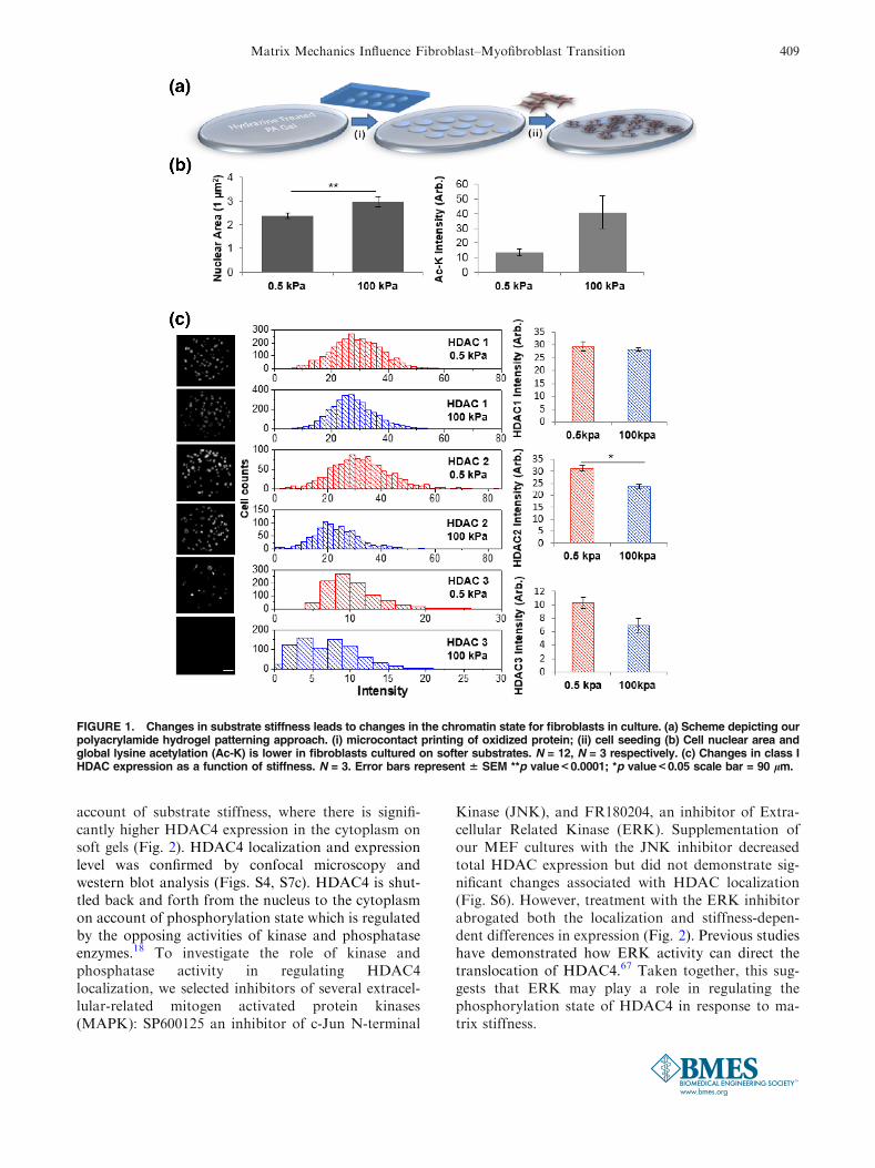
account of substrate stiffness, where there is signifi-cantly higher HDAC4 expression in the cytoplasm onsoft gels (Fig. 2). HDAC4 localization and expressionlevel was confirmed by confocal microscopy andwestern blot analysis (Figs. S4, S7c). HDAC4 is shut-tled back and forth from the nucleus to the cytoplasmon account of phosphorylation state which is regulatedby the opposing activities of kinase and phosphataseenzymes.18 To investigate the role of kinase andphosphatase activity in regulating HDAC4localization, we selected inhibitors of several extracel-lular-related mitogen activated protein kinases(MAPK): SP600125 an inhibitor of c-Jun N-terminal
Kinase (JNK), and FR180204, an inhibitor of Extra-cellular Related Kinase (ERK). Supplementation ofour MEF cultures with the JNK inhibitor decreasedtotal HDAC expression but did not demonstrate sig-nificant changes associated with HDAC localization(Fig. S6). However, treatment with the ERK inhibitorabrogated both the localization and stiffness-depen-dent differences in expression (Fig. 2). Previous studieshave demonstrated how ERK activity can direct thetranslocation of HDAC4.67 Taken together, this sug-gests that ERK may play a role in regulating thephosphorylation state of HDAC4 in response to ma-trix stiffness.
FIGURE 1. Changes in substrate stiffness leads to changes in the chromatin state for fibroblasts in culture. (a) Scheme depicting ourpolyacrylamide hydrogel patterning approach. (i) microcontact printing of oxidized protein; (ii) cell seeding (b) Cell nuclear area andglobal lysine acetylation (Ac-K) is lower in fibroblasts cultured on softer substrates. N = 12, N = 3 respectively. (c) Changes in class IHDAC expression as a function of stiffness. N = 3. Error bars represent 6 SEM **p value<0.0001; *p value<0.05 scale bar = 90 lm.
Matrix Mechanics Influence Fibroblast–Myofibroblast Transition 409

FIGURE 3. The phosphostate and nuclear localization of HDAC4 on soft hydrogels is guided by phosphatase PP2a. (a) Immunoflu-orescence images and quantitation of pHDAC4 and PP2a 4 h after fibroblast seeding. N = 3; Error bars represent 6 SEM. *p value<0.001; scale bar = 90 lm. (b) Immunofluorescence images and quantitation of pHDAC4 and PP2a 24 h after fibroblast seeding. N = 3;Error bars represent 6 SEM. *p value<0.001; scale bar = 90 lm. (c) Timecourse analysis of nuclear HDAC4 and PP2a.N = 3; Error barsrepresent 6 SEM. *0.5 kPa cytoplasmic HDAC4 is higher than nuclear, p<0.05. §0.5 kPa cytoplasmic HDAC4 is higher than 100 kPacytoplasmic HDAC4, p<0.05. ¥0.5 kPa nuclear HDAC4 is significantly higher than 100 kPa nuclear HDAC4, p<0.05. #0.5 kPa is sig-nificantly higher than 100 kPa, p value <0.05.
FIGURE 2. HDAC4 localization is influenced by substrate stiffness. (a) Immunofluorescence images of HDAC4 with and withoutERK inhibition in fibroblasts cultured on soft (0.5 kPa) and stiff (100 kPa) polyacrylamide hydrogels. (b) Quantitation of fluores-cence intensity. N = 3. Error bars represent 6 SEM. **p value <0.01; scale bar = 90 lm.
LI et al.410

Next we investigated a phosphatase that has beenpreviously associated with HDAC4 in regulatingtranslocation from the cytoplasm to the nucleus,protein phosphatase 2a (PP2a).44 PP2a has beenshown to dephosphorylate HDAC4 at multiple 14-3-3binding sites, including S246, S467, S632, to regulatenuclear/cytoplasmic shuttling of HDAC4.44,61 Ini-tially there is significantly higher PP2a and phos-phorylated HDAC4 at S632 (pHDAC4) on soft gelscompared to stiff gels (Fig. 3a). After 24 h we see thesame trend in higher PP2a expression; however theresults are not statistically significant. Interestingly,after 24 h there is a statistically significant increase inpHDAC4 on the stiff gels relative to the soft gels,which corresponds to the opposite trend in PP2a. Thisresult suggest that PP2a activity may regulate thephosphostate and subsequent localization ofHDAC4, which is consistent with previous reports ofthis phosphatase regulating nuclear/cytoplasmicshuttling (Figs. 3b, S7). To follow the dynamics ofHDAC4 activity we performed a timecourse study
and observe enhanced total HDAC4 levels in fibrob-lasts cultured on soft hydrogels over 5 days (Fig. 3c).On average we see higher expression of PP2a on softhydrogels relative to stiff hydrogels over time. How-ever, we do see a reproducible increase in nuclearPP2a over the first 2 days on stiff hydrogels; we sus-pect this is related to another bioactivity that is notassociated with HDAC4 localization.
Myofibroblast Differentiation is Enhanced WithinMicrotissues on Soft Matrices
A key bioactivity where gene expression has beenshown to be influenced by HDAC4 is the fibroblast–myofibroblast transition involved in wound heal-ing17,19 where HDAC4 has been shown to be requiredby TFGb1 induced myofibroblastic differentiation andsilencing of HDAC4 blocks TFGb1 stimulated aSMAexpression. However, the relationship betweenHDAC4 localization and fibroblast–myofibroblastdifferentiation, and a potential role for matrix
FIGURE 4. Fibroblast-myofibroblast transition is influenced by matrix mechanics. (a) Immunofluorescence images of fibroblastscultured on soft and stiff hydrogels stained for focal adhesion proteins and markers of smooth muscle. (b) Quantitation ofimmunofluorescence images. N = 3; Error bars represent 6 SEM. (c) Western blot of focal adhesion proteins and aSMA infibroblasts. N = 4, Error bars represent 6 SEM.
Matrix Mechanics Influence Fibroblast–Myofibroblast Transition 411
mechanics, remains to be explored. To investigatewhether our MEFs are undergoing myofibroblast dif-ferentiation due to differences in HDAC4 localizationon our engineered substrates, we immunostained ourcultures for proteins involved in fibroblast adhesion tofibronectin. After 24 h in culture we see a trend ofhigher paxillin staining for MEFs cultured on softhydrogels relative to stiff hydrogels (Figs. 4, S8).Paxillin is a focal adhesion protein which has beenshown to have a regulatory role in pulmonary arterialsmooth muscle cells58 and is an early marker for car-diac fibroblast to myofibroblast differentiation.8,51
Vinculin, another focal adhesion protein indicated asan early marker for cardiac fibroblast to myofibroblastdifferentiation,8 showed a trend of increased expres-sion as substrate stiffness increased. When our culturesare stained for smooth muscle actin (aSMA), the actinisoform employed by myofibroblasts, we see increasedexpression in cells cultured on soft hydrogels, howeverthese differences were not statistically significant.Western blot analysis corroborates the immunofluo-rescence results (Fig. 4c, S8). Taken together, theseresults suggest that matrix mechanics may influence thefibroblast to myofibroblast transition in culturedMEFs. This is interesting considering previousresearch with collagen coated hydrogels which indi-cated increased myofibroblast differentiation on stifferenvironments due to TFGb activation,24 though it isworth noting that our system has a wider range ofstiffness than those previously utilized (0.5–100 kPa vs.0.5–20 kPa). Our results suggest that HDAC4localization plays a role in fibroblast to myofibroblastdifferentiation in response to stiffness. While we didnot explore TGFb signaling in our system, it is rea-sonable to expect that matrix mechanics will influencethe secretory profile from MEFs. For instance, previ-ous research has shown that matrix mechanics caninfluence secretory profiles of mesenchymal stem cells.4
Future research will involve profiling the secretome todiscern relationships between matrix mechanics andcytokine secretion during fibroblast–myofibroblasttransitions on micropatterned hydrogels.
CONCLUSIONS
Morphogenetic processes in vivo are coordinated byinterplay between the microenvironment and intracel-lular pathways that regulate changes in gene expres-sion. In this paper we report the discovery of arelationship between matrix stiffness, mechanotrans-duction, the regulation of HDAC localization, andfibroblast–myofibroblast differentiation. HDAC4expression increases when fibroblasts are cultured onsoft hydrogels (0.5 kPa) relative to stiff hydrogels
(100 kPa), with preferential localization to the cyto-plasm through phosphorylation. The opposing activi-ties of HDAC4 and PP2a influences the phosphostateof HDAC4 which in turn determines the enzymes’localization. We propose that fibroblasts regulate thisbalance in response to extracellular mechanical cues toinfluence HDAC4 localization, and the acetylationstate during fibroblast–myofibroblast transition. Thisresult provides an example of how class II histonedeacetylase enzymes may be involved inmechanosensing as direct players in regulating cellularepigenetics and gene expression associated with mor-phogenesis. Since myofibroblast differentiation isinvolved in several physiological processes, includingangiogenesis and wound healing, we believe this workwill be of broad fundamental interest, and help guidethe design of materials for regenerative engineering.
ELECTRONIC SUPPLEMENTARY MATERIAL
The online version of this article (doi:10.1007/s12195-017-0493-8) contains supplementary material,which is available to authorized users.
ACKNOWLEDGMENTS
The authors would like to thank Dr. Quanxi Li inthe Department of Comparative Biosciences at theUniversity of Illinois for the generous isolation anddonation of MEFs and Dr. Karin Jensen in theDepartment of Bioengineering at the University ofIllinois for guidance with western blots.
CONFLICT OF INTEREST
Yanfen Li, Claire B. Tang, and Kristopher A. Kil-ian declare that they have no conflicts of interest.
FUNDING
Funding was provided by the National ScienceFoundation Grant No. 1454616 CAR. Y.L. was sup-ported by the National Science Foundation GraduateResearch Fellowship Program under Grant No. DGE– 1144245.
RESEARCH INVOLVED IN HUMAN AND
ANIMAL RIGHTS
No humans or animals were employed in thisresearch by the authors.
LI et al.412
REFERENCES
1Abdeen, A. A., J. Lee, N. A. Bharadwaj, R. H. Ewoldt,and K. A. Kilian. Temporal modulation of stem cellactivity using magnetoactive hydrogels. Adv. Healthc.Mater. 5(19):2536–2544, 2016.2Abdeen, A. A., J. Lee, Y. Li, and K. A. Kilian. Cytoskeletalpriming of mesenchymal stem cells to a medicinal pheno-type. Regen. Eng. Transl. Med. 3:5–14, 2017.3Abdeen, A. A., J. Lee, S. H. Mo, and K. A. Kilian. Spa-tially defined stem cell-laden hydrogel islands for directingendothelial tubulogenesis. J. Mater. Chem. B 3(40):7896–7898, 2015.4Abdeen, A. A., J. B. Weiss, J. Lee, and K. A. Kilian.Matrix composition and mechanics direct proangiogenicsignaling from mesenchymal stem cells. Tissue Eng. Part A20(19–20):2737–2745, 2014.5Alam, S. G., Q. Zhang, N. Prasad, Y. Li, S. Chamala, R.Kuchibhotla, B. Kc, V. Aggarwal, S. Shrestha, A. L. Jones,S. E. Levy, K. J. Roux, J. A. Nickerson, and T. P. Lele.The mammalian LINC complex regulates genome tran-scriptional responses to substrate rigidity. Sci. Rep.6:38063, 2016.6Backs, J., K. Song, S. Bezprozvannaya, S. Chang, and E.N. Olson. CaM kinase II selectively signals to histonedeacetylase 4 during cardiomyocyte hypertrophy. J. Clin.Invest. 116(7):1853–1864, 2006.7Banerjee, S., and M. C. Marchetti. Controlling cell-matrixtraction forces by extracellular geometry. New J. Phys.15(3):35015, 2013.8Baum, J., and H. S. Duffy. Fibroblasts and myofibroblasts:what are we talking about? J. Cardiovasc. Pharmacol.57(4):376–379, 2011.9Cernotta, N., A. Clocchiatti, C. Florean, and C. Brancol-ini. Ubiquitin-dependent degradation of HDAC4, a newregulator of random cell motility. Mol. Biol. Cell22(2):278–289, 2011.
10Chambliss, A. B., P.-H. Wu, W.-C. Chen, S. X. Sun, andD. Wirtz. Simultaneously defining cell phenotypes, cellcycle, and chromatin modifications at single-cell resolution.FASEB J. 27(7):2667–2676, 2013.
11Chancellor, T. J., J. Lee, C. K. Thodeti, and T. Lele.Actomyosin tension exerted on the nucleus through ne-sprin-1 connections influences endothelial cell adhesion,migration, and cyclic strain-induced reorientation. Biophys.J. 99(1):115–123, 2010.
12Dahl, K. N., A. J. S. Ribeiro, and J. Lammerding. Nuclearshape, mechanics, and mechanotransduction. Circ. Res.102(11):1307–1318, 2008.
13Discher, D. E., D. J. Mooney, and P. W. Zandstra. Growthfactors, matrices, and forces combine and control stemcells. Science 324(5935):1673–1677, 2009.
14Dupont, S., L. Morsut, M. Aragona, E. Enzo, S. Giulitti,M. Cordenonsi, F. Zanconato, J. Le Digabel, M. Forcato,S. Bicciato, N. Elvassore, and S. Piccolo. Role of YAP/TAZ in mechanotransduction. Nature 474(7350):179–183,2011.
15Evans, N. D., C. Minelli, E. Gentleman, V. LaPointe, S. N.Patankar, M. Kallivretaki, X. Chen, C. J. Roberts, and M.M. Stevens. Substrate stiffness affects early differentiationevents in embryonic stem cells. Eur. Cells Mater. 18:1–13,2009.
16Farge, E. Mechanical induction of Twist in the Drosophilaforegut/stomodeal primordium. Curr. Biol. 13(16):1365–1377, 2003.
17Glenisson, W., V. Castronovo, and D. Waltregny. Histonedeacetylase 4 is required for TGF beta 1-induced myofi-broblastic differentiation. Biochim. Biophys. Acta1773(10):1572–1582, 2007.
18Grozinger, C. M., and S. L. Schreiber. Regulation of his-tone deacetylase 4 and 5 and transcriptional activity by 14-3- 3-dependent cellular localization. Proc. Natl. Acad. Sci.USA. 97(14):7835–7840, 2000.
19Guo, W., B. Shan, R. C. Klingsberg, X. Qin, and J. A.Lasky. Abrogation of TGF-beta1-induced fibroblast-my-ofibroblast differentiation by histone deacetylase inhibi-tion. Am. J. Physiol. Lung Cell. Mol. Physiol. 297(5):L864–L870, 2009.
20Haberland, M., R. L. Montgomery, and E. N. Olson. Themany roles of histone deacetylases in development andphysiology: implications for disease and therapy. Nat. Rev.Genet. 10(1):32–42, 2009.
21Hebbes, T. R., A. W. Thorne, and C. Crane-Robinson. Adirect link between core histone acetylation and transcrip-tionally active chromatin. EMBO J. 7(5):1395–1402, 1988.
22Heo, S.-J., T. P. Driscoll, S. D. Thorpe, N. L. Nerurkar, B.M. Baker, M. T. Yang, C. S. Chen, D. A. Lee, and R. L.Mauck. Differentiation alters stem cell nuclear architec-ture, mechanics, and mechano-sensitivity. Elife, 5:e18207,2016.
23Heo, S.-J., S. D. Thorpe, T. P. Driscoll, R. L. Duncan, D.A. Lee, and R. L. Mauck. Biophysical regulation of chro-matin architecture instills a mechanical memory in mes-enchymal stem cells. Sci. Rep. 5(1):16895, 2015.
24Huang, X., N. Yang, V. F. Fiore, T. H. Barker, Y. Sun, S.W. Morris, Q. Ding, V. J. Thannickal, and Y. Zhou. Ma-trix stiffness-induced myofibroblast differentiation ismediated by intrinsic mechanotransduction. Am. J. Respir.Cell Mol. Biol. 47(3):340–348, 2012.
25Ingber, D. E. Cellular mechanotransduction: putting all thepieces together again. FASEB J. 20(7):27, 2006.
26Jagielska, A., A. L. Lowe, E. Makhija, L. Wroblewska, J.Guck, R. J. M. Franklin, G. V. Shivashankar, and K. J.Van Vliet. Mechanical strain promotes oligodendrocytedifferentiation by global changes of gene expression. Front.Cell. Neurosci. 11:93, 2017.
27Jain, N., K. V. Iyer, A. Kumar, and G. V. Shivashankar.Cell geometric constraints induce modular gene-expressionpatterns via redistribution of HDAC3 regulated by acto-myosin contractility. Proc. Natl. Acad. Sci. USA110(28):11349–11354, 2013.
28Karamichos, D., R. A. Brown, and V. Mudera. Collagenstiffness regulates cellular contraction and matrix remod-eling gene expression. J. Biomed. Mater. Res. A 83(3):887–894, 2007.
29Kocgozlu, L., P. Lavalle, G. Koenig, B. Senger, Y. Haikel,P. Schaaf, J.-C. Voegel, H. Tenenbaum, and D. Vautier.Selective and uncoupled role of substrate elasticity in theregulation of replication and transcription in epithelialcells. J. Cell Sci. 123(Pt 1):29–39, 2010.
30Kumar, A., and G. V. Shivashankar. Dynamic interactionbetween actin and nesprin2 maintain the cell nucleus in aprestressed state. Methods Appl. Fluoresc. 4(4):44008, 2016.
31Lee, J., A. A. Abdeen, T. H. Huang, and K. A. Kilian.Controlling cell geometry on substrates of variable stiffnesscan tune the degree of osteogenesis in human mesenchymalstem cells. J. Mech. Behav. Biomed. Mater. 38:209–218,2014.
32Lee, J., A. A. Abdeen, A. S. Kim, and K. A. Kilian.Influence of biophysical parameters on maintaining the
Matrix Mechanics Influence Fibroblast–Myofibroblast Transition 413
mesenchymal stem cell phenotype. ACS Biomater. Sci. Eng.1(4):218–226, 2015.
33Lee, J., A. A. Abdeen, K. L. Wycislo, T. M. Fan, and K. A.Kilian. Interfacial geometry dictates cancer celltumorigenicity. Nat Mater. 15(8):856–862, 2016.
34Lee, C., A. Grodzinsky, and M. Spector. The effects ofcross-linking of collagen-glycosaminoglycan scaffolds oncompressive stiffness, chondrocyte-mediated contraction,proliferation and biosynthesis. Biomaterials 22(23):3145–3154, 2001.
35Li, J., J. Chen, C. L. Ricupero, R. P. Hart, M. S. Schwartz,A. Kusnecov, and K. Herrup. Nuclear accumulation ofHDAC4 in ATM deficiency promotes neurodegenerationin ataxia telangiectasia. Nat. Med. 18(5):783–790, 2012.
36Li, Y., J. S. Chu, K. Kurpinski, X. Li, D. M. Bautista, L.Yang, K.-L. P. Sung, and S. Li. Biophysical regulation ofhistone acetylation in mesenchymal stem cells. Biophys. J.100(8):1902–1909, 2011.
37Li, Y., D. Lovett, Q. Zhang, S. Neelam, R. A. Kuchib-hotla, R. Zhu, G. G. Gundersen, T. P. Lele, and R. B.Dickinson. Moving cell boundaries drive nuclear shapingduring cell spreading. Biophys. J. 109(4):670–686, 2015.
38Lovett, D. B., N. Shekhar, J. A. Nickerson, K. J. Roux,and T. P. Lele. Modulation of nuclear shape by substraterigidity. Cell. Mol. Bioeng. 6(2):230–238, 2013.
39Low, B. C., C. Q. Pan, G. V. Shivashankar, A. Bershadsky,M. Sudol, and M. Sheetz. YAP/TAZ as mechanosensorsand mechanotransducers in regulating organ size andtumor growth. FEBS Lett. 588(16):2663–2670, 2014.
40Maniotis, A. J., C. S. Chen, and D. E. Ingber. Demon-stration of mechanical connections between integrins,cytoskeletal filaments, and nucleoplasm that stabilize nu-clear structure. Proc. Natl. Acad. Sci. 94(3):849–854, 1997.
41Mao, A. S., J.-W. Shin, and D. J. Mooney. Effects ofsubstrate stiffness and cell-cell contact on mesenchymalstem cell differentiation. Biomaterials 98:184–191, 2016.
42Mih, J. D., A. Marinkovic, F. Liu, A. S. Sharif, and D. J.Tschumperlin. Matrix stiffness reverses the effect of acto-myosin tension on cell proliferation. J. Cell Sci. 125(Pt24):5974–5983, 2012.
43Park, J. S., J. S. Chu, A. D. Tsou, R. Diop, Z. Tang, A.Wang, and S. Li. The effect of matrix stiffness on the dif-ferentiation of mesenchymal stem cells in response to TGF-b. Biomaterials 32:3921–3930, 2011.
44Paroni, G., N. Cernotta, C. Dello Russo, P. Gallinari, M.Pallaoro, C. Foti, F. Talamo, L. Orsatti, C. Steinkuhler,and C. Brancolini. PP2A regulates HDAC4 nuclear import.Mol. Biol. Cell 19(2):655–667, 2007.
45Provenzano, P. P., D. R. Inman, K. W. Eliceiri, and P. J.Keely. Matrix density-induced mechanoregulation of breastcell phenotype, signaling and gene expression through aFAK-ERK linkage. Oncogene 28(49):4326–4343, 2009.
46Provenzano, P. P., and P. J. Keely. Mechanical signalingthrough the cytoskeleton regulates cell proliferation bycoordinated focal adhesion and Rho GTPase signaling. J.Cell Sci. 124(Pt 8):1195–1205, 2011.
47Ralphs, J. R., A. D. Waggett, and M. Benjamin. Actinstress fibres and cell-cell adhesion molecules in tendons:organisation in vivo and response to mechanical loading oftendon cells in vitro. Matrix Biol. 21(1):67–74, 2002.
48Ren, K., T. Crouzier, C. Roy, and C. Picart. Polyelectrolytemultilayer films of controlled stiffness modulate myoblast cellsdifferentiation. Adv. Funct. Mater. 18(9):1378–1389, 2008.
49Rozwadowska, N., T. Kolanowski, E. Wiland, M. Siat-kowski, P. Pawlak, A. Malcher, T. Mietkiewski, M. Ols-
zewska, and M. Kurpisz. Characterisation of nucleararchitectural alterations during in vitro differentiation ofhuman stem cells of myogenic origin. PLoS ONE8(9):e73231, 2013.
50Ruthenburg, A. J., H. Li, D. J. Patel, and C. D. Allis.Multivalent engagement of chromatin modifications bylinked binding modules. Nat. Rev. Mol. Cell Biol.8(12):983–994, 2007.
51Santiago, J.-J., A. L. Dangerfield, S. G. Rattan, K. L.Bathe, R. H. Cunnington, J. E. Raizman, K. M. Bedosky,D. H. Freed, E. Kardami, and I. M. C. Dixon. Cardiacfibroblast to myofibroblast differentiation in vivo andin vitro: expression of focal adhesion components inneonatal and adult rat ventricular myofibroblasts. Dev.Dyn. 239(6):1573–1584, 2010.
52Serebryannyy, L. A., C. M. Cruz, P. de Lanerolle, R. D.Mullins, and P. D. McCrea. A role for nuclear actin inHDAC 1 and 2 regulation. Sci. Rep. 6(1):28460, 2016.
53Shih, Y.-R. V., K.-F. Tseng, H.-Y. Lai, C.-H. Lin, and O.K. Lee. Matrix stiffness regulation of integrin-mediatedmechanotransduction during osteogenic differentiation ofhuman mesenchymal stem cells. J. Bone Miner. Res.26(4):730–738, 2011.
54Swift, J., I. L. Ivanovska, A. Buxboim, T. Harada, P. C. D.P. Dingal, J. Pinter, J. D. Pajerowski, K. R. Spinler, J.-W.Shin, M. Tewari, F. Rehfeldt, D. W. Speicher, and D. E.Discher. Nuclear lamin-A scales with tissue stiffness andenhances matrix-directed differentiation. Science341(6149):1240104, 2013.
55Tan, P. S., and S. H. Teoh. Effect of stiffness of poly-caprolactone (PCL) membrane on cell proliferation. Mater.Sci. Eng. C 27(2):304–308, 2007.
56Tse, J. R., and A. J. Engler. Preparation of hydrogel sub-strates with tunable mechanical properties. Curr. Protoc.Cell Biol. 47:10.16:10.16.1–10.16.16, 2010.
57Vega, R. B., K. Matsuda, J. Oh, A. C. Barbosa, X. Yang,E. Meadows, J. McAnally, C. Pomajzl, J. M. Shelton, J. A.Richardson, G. Karsenty, and E. N. Olson. Histonedeacetylase 4 controls chondrocyte hypertrophy duringskeletogenesis. Cell 119(4):555–566, 2004.
58Veith, C., L. M. Marsh, M. Wygrecka, K. Rutschmann, W.Seeger, N. Weissmann, and G. Kwapiszewska. Paxillin regu-lates pulmonary arterial smooth muscle cell function in pul-monary hypertension. Am. J. Pathol. 181(5):1621–1633, 2012.
59Wang, L.-S., J. Boulaire, P. P. Y. Chan, J. E. Chung, and M.Kurisawa. The role of stiffness of gelatin-hydroxyphenylpro-pionic acid hydrogels formed by enzyme-mediated crosslink-ing on the differentiation of human mesenchymal stem cell.Biomaterials 31(33):8608–8616, 2010.
60Wang, N., J. P. Butler, and D. E. Ingber. Mechanotrans-duction across the cell surface and through the cytoskele-ton. Science 260(5111):1124–1127, 1993.
61Wang, A. H., M. J. Kruhlak, J. Wu, N. R. Bertos, M.Vezmar, B. I. Posner, D. P. Bazett-Jones, and X. J. Yang.Regulation of histone deacetylase 4 by binding of 14-3-3proteins. Mol. Cell. Biol. 20(18):6904–6912, 2000.
62Wang, A., S. K. Kurdistani, and M. Grunstein. Require-ment of Hos2 histone deacetylase for gene activity in yeast.Science 298(5597):1412–1414, 2002.
63Wang, Z., G. Qin, and T. C. Zhao. HDAC4: mechanism ofregulation and biological functions. Epigenomics 6(1):139–150, 2014.
64Wang, A. H., and X. J. Yang. Histone deacetylase 4 pos-sesses intrinsic nuclear import and export signals. Mol.Cell. Biol. 21(17):5992–6005, 2001.
LI et al.414

65Yeung, T., P. C. Georges, L. A. Flanagan, B. Marg, M.Ortiz, M. Funaki, N. Zahir, W. Ming, V. Weaver, and P.A. Janmey. Effects of substrate stiffness on cell morphol-ogy, cytoskeletal structure, and adhesion. Cell Motil. Cy-toskelet. 60(1):24–34, 2005.
66Yuan, H., K. Denton, L. Liu, X. J. Li, S. Benashski, L.McCullough, and J. Li. Nuclear translocation of histone
deacetylase 4 induces neuronal death in stroke. Neurobiol.Dis. 91:182–193, 2016.
67Zhou, X., V. M. Richon, A. H. Wang, X.-J. Yang, R. A.Rifkind, and P. A. Marks. Histone deacetylase 4 associateswith extracellular signal-regulated kinases 1 and 2, and itscellular localization is regulated by oncogenic Ras. Proc.Natl. Acad. Sci. 97(26):14329–14333, 2000.
Matrix Mechanics Influence Fibroblast–Myofibroblast Transition 415