Mathematical Model of Oxygen Distribution in Engineered Cardiac Tissue With Parallel Channel Array...
12
Mathematical model of oxygen distribution in engineered cardiac tissue with parallel channel array perfused with culture medium containing oxygen carriers Milica Radisic, 1 William Deen, 1 Robert Langer, 1,2 and Gordana Vunjak-Novakovic 2 1 Department of Chemical Engineering and 2 Harvard-Massachusetts Institute of Technology Division of Health Sciences and Technology, Massachusetts Institute of Technology, Cambridge, Massachusetts Submitted 3 August 2004; accepted in final form 6 November 2004 Radisic, Milica, William Deen, Robert Langer, and Gordana Vunjak-Novakovic. Mathematical model of oxygen distribution in engineered cardiac tissue with parallel channel array perfused with culture medium containing oxygen carriers. Am J Physiol Heart Circ Physiol 288: H1278 –H1289, 2005. First published November 11, 2004; doi:10.1152/ajpheart.00787.2004.—A steady-state model of oxygen distribution in a cardiac tissue construct with a parallel channel array was developed and solved for a set of parameters using the finite element method and commercial software (FEMLAB). The effects of an oxygen carrier [Oxygent; 32% volume perfluorocarbon (PFC) emulsion] were evaluated. The parallel channel array mimics the in vivo capillary tissue bed, and the PFC emulsion has a similar role as the natural oxygen carrier hemoglobin in increasing total oxygen content. The construct was divided into an array of cylindrical domains with a channel in the center and tissue space surrounding the channel. In the channel, the main modes of mass transfer were axial convection and radial diffusion. In the tissue region, mass transfer was by axial and radial diffusion, and the consumption of oxygen was by Michaelis-Menten kinetics. Neumann boundary conditions were im- posed at the channel centerline and the half distance between the domains. Supplementation of culture medium by PFC emulsion im- proved mass transport by increasing convective term and effective diffusivity of culture medium. The model was first implemented for the following set of experimentally obtained parameters: construct thickness of 0.2 cm, channel diameter of 330 m, channel center-to- center spacingof 700 m, and average linear velocity per channel of 0.049 cm/s, in conjunction with PFC supplemented and unsupple- mented culture medium. Subsequently, the model was used to define favorable scaffold geometry and flow conditions necessary to culti- vate cardiac constructs of high cell density (10 8 cells/ml) and clini- cally relevant thickness (0.5 cm). In future work, the model can be utilized as a tool for optimization of scaffold geometry and flow conditions. tissue engineering; cardiac myocyte; scaffold mass transport; perfluo- rocarbons THE KEY PARAMETER IN ENGINEERING functional three-dimensional tissues in vitro is oxygen supply (4, 11, 21). In native rat heart, oxygen is supplied by diffusion from capillaries that are spaced 20 m apart (27). Because the solubility of oxygen in plasma is low, the oxygen carrier hemoglobin increases total oxygen content of blood and, therefore, increases the mass of tissue that can be supported in a single pass through the capillary network. The average oxygen concentration in arterial and venous blood is 130 and 54 M, respectively (11). In conventional tissue-engineering approaches, cells are sup- ported on polymer scaffolds to form tissue constructs im- mersed in culture medium. Oxygen dissolved in the medium is transported by diffusion from the surface of the tissue construct into its interior. The medium is reoxygenated via a gas ex- changer (8), by aeration, or by membrane exchange (21). Diffusion alone is capable of providing enough oxygen for an 100-m-thick outer layer of a tissue construct, whereas the interior remains relatively acellular or becomes necrotic due to hypoxia (2). To solve this problem, a perfusion bioreactor system has been developed (5). In such a system, the entire construct is directly perfused with culture medium, and the transport of oxygen from the medium to the cells occurs via both diffusion and convection. However, the solubility of oxygen in water at 37°C is not sufficient to satisfy the metabolic demand of a large number of cells (e.g., 5 10 8 cells/ml construct) at low flow rates (e.g., 0.5 ml min 1 cm 2 ). Increasing the flow rate increases the shear stress exerted on the cells, which in turn can decrease cell number and viability. To provide enough oxygen for thick constructs at low flow rates, it is necessary to have a pool of oxygen in the medium that is available to quickly meet the changing local oxygen demand. By utilizing synthetic oxygen carriers [perfluorocarbons (PFCs)], the length of the tissue construct that can be supported per single pass through a perfusion bioreactor at a given flow rate will be increased. Our main hypothesis is that the functional assembly of engineered cardiac tissue will be enhanced by increasing local oxygen concentration in the tissue phase within the physiolog- ical range (up to 220 M). This hypothesis is consistent with the observation that cardiac constructs cultivated in perfusion at oxygen concentrations of 80 M exhibit weaker presence of cardiac markers and poorer organization of contractile apparatus compared with the constructs cultivated at oxygen concentrations of 220 m (3). In addition, it was demon- strated that isolated cardiomyocytes begin to downregulate energy-using processes (as assessed by changes in oxygen consumption rate, lactate output, and concentrations of intra- cellular ATP and phosphocreatine) at oxygen concentrations as high as 70 M (7). To test this hypothesis, we recently developed a biomimetic in vitro tissue culture system in which neonatal rat heart cells were cultured on an elastic, highly porous scaffold with a parallel array of channels perfused with culture medium supplemented with a synthetic oxygen carrier (Oxygent, PFC emulsion). In this system, the parallel-channel array mimics the role of the capillary network and the PFC emulsion mimics the role of hemoglobin. Constructs perfused with unsupplemented culture medium served as controls. Address for reprint requests and other correspondence: G. Vunjak-Nova- kovic, Massachusetts Institute of Technology, Harvard-MIT Division of Health Science and Technology, 77 Massachusetts Ave., E25-330, Cambridge, MA 02139 (E-mail: [email protected]). The costs of publication of this article were defrayed in part by the payment of page charges. The article must therefore be hereby marked “advertisement” in accordance with 18 U.S.C. Section 1734 solely to indicate this fact. Am J Physiol Heart Circ Physiol 288: H1278 –H1289, 2005. First published November 11, 2004; doi:10.1152/ajpheart.00787.2004. 0363-6135/05 $8.00 Copyright © 2005 the American Physiological Society http://www.ajpheart.org H1278
-
Upload
goh-kek-boon -
Category
Documents
-
view
214 -
download
0
Transcript of Mathematical Model of Oxygen Distribution in Engineered Cardiac Tissue With Parallel Channel Array...

Mathematical model of oxygen distribution in engineered cardiac tissue withparallel channel array perfused with culture medium containing oxygen carriers
Milica Radisic,1 William Deen,1 Robert Langer,1,2 and Gordana Vunjak-Novakovic2
1Department of Chemical Engineering and 2Harvard-Massachusetts Institute of Technology Division ofHealth Sciences and Technology, Massachusetts Institute of Technology, Cambridge, Massachusetts
Submitted 3 August 2004; accepted in final form 6 November 2004
Radisic, Milica, William Deen, Robert Langer, and GordanaVunjak-Novakovic. Mathematical model of oxygen distribution inengineered cardiac tissue with parallel channel array perfused withculture medium containing oxygen carriers. Am J Physiol Heart CircPhysiol 288: H1278–H1289, 2005. First published November 11,2004; doi:10.1152/ajpheart.00787.2004.—A steady-state model ofoxygen distribution in a cardiac tissue construct with a parallelchannel array was developed and solved for a set of parameters usingthe finite element method and commercial software (FEMLAB). Theeffects of an oxygen carrier [Oxygent; 32% volume perfluorocarbon(PFC) emulsion] were evaluated. The parallel channel array mimicsthe in vivo capillary tissue bed, and the PFC emulsion has a similarrole as the natural oxygen carrier hemoglobin in increasing totaloxygen content. The construct was divided into an array of cylindricaldomains with a channel in the center and tissue space surrounding thechannel. In the channel, the main modes of mass transfer were axialconvection and radial diffusion. In the tissue region, mass transfer wasby axial and radial diffusion, and the consumption of oxygen was byMichaelis-Menten kinetics. Neumann boundary conditions were im-posed at the channel centerline and the half distance between thedomains. Supplementation of culture medium by PFC emulsion im-proved mass transport by increasing convective term and effectivediffusivity of culture medium. The model was first implemented forthe following set of experimentally obtained parameters: constructthickness of 0.2 cm, channel diameter of 330 �m, channel center-to-center spacingof 700 �m, and average linear velocity per channel of0.049 cm/s, in conjunction with PFC supplemented and unsupple-mented culture medium. Subsequently, the model was used to definefavorable scaffold geometry and flow conditions necessary to culti-vate cardiac constructs of high cell density (108 cells/ml) and clini-cally relevant thickness (0.5 cm). In future work, the model can beutilized as a tool for optimization of scaffold geometry and flowconditions.
tissue engineering; cardiac myocyte; scaffold mass transport; perfluo-rocarbons
THE KEY PARAMETER IN ENGINEERING functional three-dimensionaltissues in vitro is oxygen supply (4, 11, 21). In native rat heart,oxygen is supplied by diffusion from capillaries that are spaced�20 �m apart (27). Because the solubility of oxygen in plasmais low, the oxygen carrier hemoglobin increases total oxygencontent of blood and, therefore, increases the mass of tissuethat can be supported in a single pass through the capillarynetwork. The average oxygen concentration in arterial andvenous blood is 130 and 54 �M, respectively (11).
In conventional tissue-engineering approaches, cells are sup-ported on polymer scaffolds to form tissue constructs im-
mersed in culture medium. Oxygen dissolved in the medium istransported by diffusion from the surface of the tissue constructinto its interior. The medium is reoxygenated via a gas ex-changer (8), by aeration, or by membrane exchange (21).Diffusion alone is capable of providing enough oxygen for an�100-�m-thick outer layer of a tissue construct, whereas theinterior remains relatively acellular or becomes necrotic due tohypoxia (2).
To solve this problem, a perfusion bioreactor system hasbeen developed (5). In such a system, the entire construct isdirectly perfused with culture medium, and the transport ofoxygen from the medium to the cells occurs via both diffusionand convection. However, the solubility of oxygen in water at37°C is not sufficient to satisfy the metabolic demand of a largenumber of cells (e.g., 5 � 108 cells/ml construct) at low flowrates (e.g., 0.5 ml �min�1 �cm�2). Increasing the flow rateincreases the shear stress exerted on the cells, which in turn candecrease cell number and viability. To provide enough oxygenfor thick constructs at low flow rates, it is necessary to have apool of oxygen in the medium that is available to quickly meetthe changing local oxygen demand. By utilizing syntheticoxygen carriers [perfluorocarbons (PFCs)], the length of thetissue construct that can be supported per single pass througha perfusion bioreactor at a given flow rate will be increased.
Our main hypothesis is that the functional assembly ofengineered cardiac tissue will be enhanced by increasing localoxygen concentration in the tissue phase within the physiolog-ical range (up to 220 �M). This hypothesis is consistent withthe observation that cardiac constructs cultivated in perfusionat oxygen concentrations of �80 �M exhibit weaker presenceof cardiac markers and poorer organization of contractileapparatus compared with the constructs cultivated at oxygenconcentrations of �220 �m (3). In addition, it was demon-strated that isolated cardiomyocytes begin to downregulateenergy-using processes (as assessed by changes in oxygenconsumption rate, lactate output, and concentrations of intra-cellular ATP and phosphocreatine) at oxygen concentrations ashigh as 70 �M (7). To test this hypothesis, we recentlydeveloped a biomimetic in vitro tissue culture system in whichneonatal rat heart cells were cultured on an elastic, highlyporous scaffold with a parallel array of channels perfused withculture medium supplemented with a synthetic oxygen carrier(Oxygent, PFC emulsion). In this system, the parallel-channelarray mimics the role of the capillary network and the PFCemulsion mimics the role of hemoglobin. Constructs perfusedwith unsupplemented culture medium served as controls.
Address for reprint requests and other correspondence: G. Vunjak-Nova-kovic, Massachusetts Institute of Technology, Harvard-MIT Division ofHealth Science and Technology, 77 Massachusetts Ave., E25-330, Cambridge,MA 02139 (E-mail: [email protected]).
The costs of publication of this article were defrayed in part by the paymentof page charges. The article must therefore be hereby marked “advertisement”in accordance with 18 U.S.C. Section 1734 solely to indicate this fact.
Am J Physiol Heart Circ Physiol 288: H1278–H1289, 2005.First published November 11, 2004; doi:10.1152/ajpheart.00787.2004.
0363-6135/05 $8.00 Copyright © 2005 the American Physiological Society http://www.ajpheart.orgH1278

In this study, a steady-state mathematical model that relatesdistribution of oxygen within the engineered tissue construct tothe medium flow rate inlet, PO2, and mass fraction of oxygencarrier is derived and solved using the finite element method.The model was used to compare oxygen distribution in theexperimentally obtained channeled cardiac tissue constructsperfused with PFC-supplemented and -unsupplemented culturemedium. Addition of PFC emulsion improved oxygen trans-port by improving apparent average velocity and by increasingeffective diffusivity. As a part of the assumption validation,resistances to the transport of oxygen were compared in eachphase, indicating that the transfer of oxygen from the PFCphase into the aqueous phase was not rate limiting. Subse-quently, the model was used to investigate scaffold geometryand flow conditions necessary to cultivate cardiac constructs ofphysiologically high cell density (108 cells/ml) and clinicallyrelevant thickness (0.5 cm).
METHODS
Cultivation of the cardiac tissue constructs. Porous poly(glycerol-sebacate) scaffolds were fabricated by means of a salt-leaching tech-nique, as previously described (32). Briefly, PGS solution in tetrahy-drofuran was poured into a Teflon mold filled with NaCl particles ofdesired sizes. The mold was transferred to a vacuum oven and curedat 120°C and 100 mTorr. The resulting material was soaked indeionized water to remove the NaCl particles. Scaffolds with porosityup to 91% were obtained after removal of water. Parallel channels ina square array were bored using a 120 W CO2 laser cutting/engravingsystem (model X-660, Universal Laser Systems, Scottsdale, AZ).Discs 5–6 mm in diameter and 2 mm thick were sterilized by 70%ethanol overnight followed by 4 h in 95% ethanol and 1 h in 100%ethanol. Ethanol was removed by vacuum filtration, and the scaf-folds were rinsed in PBS (GIBCO) for 1– 4 h followed by 1 h inFBS (GIBCO). Channel diameter and spacing were determinedfrom the light micrographs using image analysis software (ScionImage).
Cells were isolated from neonatal rat ventricles according to theprocedures approved by the Institute’s Committee on Animal Care asdescribed previously (3). For scaffold preconditioning with fibro-blasts, 0.5 � 106 to 1 � 106 fibroblasts were resuspended in 10 �l ofMatrigel (BD) and applied to the scaffold as described previously(24). The constructs were pretreated in six-well plates (1 construct/well) for 4 days in 5 ml of culture medium at 25 rpm. At the end ofpreconditioning, 2.3 � 106 cardiomyocytes in 15 �l of Matrigel wereadded to the construct and allowed to gel for 30 min at 37°C. Theconstructs were cultivated in the perfusion loops for an additional 3days as described previously (25). Briefly, the constructs were tightlyfitted inside the 5-mm inner diameter, 10-mm outer diameter siliconetubing rings, placed between two stainless steel screens, and posi-tioned in 1.5-ml polycarbonate perfusion cartridges (kindly donatedby the Advanced Tissue Sciences, La Jolla, CA; 1 scaffold/cartridge).The screens (85% open area) provided mechanical support duringperfusion, and the silicone ring routed the culture medium through thecentral area of the construct. Constructs were subjected to unidirec-tional medium flow at 0.1 ml/min provided by the IsmaTec (Cole-Parmer) multichannel peristaltic pump. The total volume of culturemedium in the gas-exchanger tubing, medium bag, and cartridge was30 ml. To investigate the effect of presence of oxygen carriers, the 27ml of culture medium was mixed with 3 ml of PFC emulsion(Oxygent, kindly donated by Alliance Pharmaceuticals, San Diego,CA). Because the extraphysiological levels of PO2 may be toxic to thecells, the medium in the gas exchanger was equilibrated with theincubator air, ensuring that the PO2 at the inlet to the perfusioncartridge does not exceed 160 Torr [or 222.47 �M (13)]. Unsupple-
mented culture medium served as a control. A total of seven rat litterswere used in seven independent experiments, with n � 4–6 con-structs/experiment.
Gas composition and pH at the inlet to perfusion cartridge weremeasured from medium samples using a gas blood analyzer (model1610, Lexington, MA). Oxygen concentration at the outlet of theperfusion cartridges was measured by inline ruthenium-based oxygensensors (kindly donated by Payload Systems). The fraction of circu-lating PFC emulsion was determined by sectioning off the lowerportions of the loop, collecting the culture medium with PFC emul-sion, and determining the absorbance of dilute emulsion at 970 nm.
At the end of cultivation, constructs were collected and evaluatedfor protein, DNA content, contractile response, cell distribution, andexpression of cardiac markers. Fluorescent micrographs of the wetconstructs were used in conjunction with an image analysis program(Scion Image) to determine channel diameter and spacing after cul-ture. Construct diameter and height were determined from histologicalsections.
Evaluation of hydraulic permeability of Biorubber scaffolds. Forevaluation of hydraulic permeability, a 5-mm-thick piece of Biorubberscaffold without the channels was fitted into a perfusion cartridge witha 5-mm-diameter open area and connected to the water reservoir viasilicone tubing and three-way stop-cock. The water reservoir wasplaced at the height of 1.195 m to provide a constant pressure head. Todetermine flow rate, the water draining from the perfusion cartridge onthe opening of three-way stop cock was collected at timed intervals.Flow around the scaffold was prevented by fixing the scaffold be-tween silicone gaskets, as described previously (25). The measure-ment was performed at room temperature (22°C). Hydraulic perme-ability was determined based on Darcy’s law.
Model parameters. A mathematical model (APPENDIX) was imple-mented for the set of experimental conditions summarized in Table 1.Average number of channels per construct was calculated based onthe measurements of channel radius, spacing, and construct diameter.The flow rate and average velocity per channel was calculated basedon the imposed flow rate (0.1 ml/min) and the number of channels.Maximum oxygen consumption (V̇O2 max) rate was calculated basedon the protein content, scaffold volume (without channels), andmaximum consumption rate per unit protein reported for nonbeatingmonolayers of neonatal rat cardiomyocytes (33). The nonbeatingrather than beating values were used due to the observation thatconstructs usually do not exhibit spontaneous contractions within thefirst three days of cultivation. The volume fraction of circulating PFCemulsion droplets (�) was higher than the nominal fraction (5.4%volume vs. 3.2%) due to some settling in the lower portions of theloop. The circulating PFC fraction was determined spectrophotometri-cally after the medium was collected from the lower portions of theloop.
In addition, mathematical modeling was used to predict oxygenconcentration profiles in tissue constructs of clinically relevant thick-ness (0.5 cm) with various channel geometry (diameter and spacing),flow rates, cell density, and concentration of circulating PFC emul-sion. The main goal was to determine channel geometry and cultureconditions (flow rate and concentration of PFC emulsion) that wouldyield high oxygen concentrations in the tissue space at physiologicallyhigh cell densities (108 cell/ml).
In one case, oxygen concentration profiles were compared in a0.5-cm-thick tissue construct with a 330-�m channel diameter and370-�m wall-to-wall spacing perfused at the average linear velocityper channel of 0.049 cm/s (or 0.1 ml/min bulk flow), with culturemedium supplemented with 0, 3.2, or 6.4% volume PFC emulsion (0,10, or 20% volume of Oxygent). The channel geometry and flowconditions used for these simulations were the same as those utilizedin experimental studies. The simulations were performed at two celldensities: 0.27 � 108 cells/ml, which is a low value measuredexperimentally, and 1 � 108 cells/ml, which is a physiologicallyrelevant density. The cells were assumed to be uniformly distributed
H1279OXYGEN DISTRIBUTION IN ENGINEERED CARDIAC TISSUE
AJP-Heart Circ Physiol • VOL 288 • MARCH 2005 • www.ajpheart.org

around the channel. The corresponding V̇O2 max was calculated to be8.8 and 33 �M/s, respectively, based on measured 60 mg protein/�gDNA and 27.6 nmol oxygen �mg protein�1 �min�1. Culture medium atthe inlet to the construct was assumed to be fully saturated withatmospheric oxygen with an oxygen concentration of 222.47 �M (or160 Torr).
In another case, oxygen concentration profiles were compared in a0.5-cm-thick tissue construct with a 100-�m channel diameter and100-�m wall-to-wall spacing perfused at the average linear velocityper channel of 0.049 or 0.135 cm/s (corresponding to 0.11 and 0.31ml/min of bulk flow, respectively), with culture medium supple-mented with 0, 3.2, or 6.4% volume PFC emulsion (0, 10, or 20%Oxygent). Such a fine channel array was not produced experimentallydue to the limitations associated with the laser cutting/engravingsystem but represent the target value in scaffold design necessary forhigh oxygen concentration in the tissue space. Cell density in thetissue space was set to be physiological (108 cells/ml; V̇O2 max � 33�M/s; Km � 6.875 �M). At the low � (0.033–0.064) used in thesimulation, the viscosity of PFC-supplemented culture medium iscomparable to that of water [�1 cp (20)]. The average velocity ofculture medium was chosen so that the shear stress at the walls was�1 dyn/cm2, which is considered physiological (12) and is below therange (1.6–3.3 dyn/cm2) reported to induce decreased viability inhybridoma and human embryonic kidney cells (16, 31).
For the Poiseuille flow in tubes, the magnitude of wall shear stress(�w) can be calculated as:
�w � �4U
Rc
(1)
where � is the viscosity of culture medium, U is the average fluidvelocity of the culture medium in the channel, and Rc is the channelradius.
The viscosity of culture medium was assumed to be the same asviscosity of water at 37°C, 0.69 cp (13). To estimate the viscosity ofthe PFC-supplemented culture medium, it was assumed that the
emulsion behaves as a suspension of rigid spheres, and the viscositywas calculated according to (10)
�PFCemulsion
�water
� 1 �5
2� (2)
where �PFCemission and �water are the viscocities of PFC emission andwater, respectively. Accordingly, for the most concentrated emulsionconsidered here (� � 0.064), the viscosity at 37°C is estimated to be0.80 cp. The estimated shear stresses for the geometries consideredhere are summarized in Table 2.
To simplify the mathematical model, Reynold’s numbers (Re) andPe numbers were calculated for all investigated channel geometriesand average velocities. The low values of Re and velocity entrancelengths (Lv) (Table 3) indicate that the flow was laminar and fullydeveloped over most of the length of the channel for 0.2- and0.5-cm-thick constructs. Therefore, the velocity profile Vz(r) in con-servation equations is given by:
Vzr � 2U1 � r2/Rc2 (3)
Relatively high values of Pe numbers imply that axial diffusionwithin the channel can be neglected. For the conditions investigatedexperimentally (Table 3), approximately one-half of the 0.2-cm-thickconstruct was within the mass transfer entrance length. For this
Table 1. Model parameters used to predict oxygen concentration profiles in a cardiac tissue construct based on channeledbiorubber scaffolds and perfused with pure culture medium or culture medium supplemented with PFC emulsion
Parameter Value Source
Rc, �m 165 MeasuredRt, �m 350 MeasuredL, mm 2 MeasuredNumber of channels 40 for cubic packing N � R2�/(2rt)2
Rc � 0.5cm, construct radiusU 0.049 cm/s (0.1 ml/min) SetOxygen consumption, nmol�mg protein�1�min�1 27.6 Ref. 33Km, �m 6.875 Ref. 7Protein amount, mg 0.743 (for PFC supplemented) Measured
0.624 (for pure culture medium)V̇O2max, �M/s 10.5 (for PFC supplemented) Calculated
8.8 (for pure culture medium)PFC in Oxygent, %vol/vol 32 Alliance Pharmaceuticalsd, �m 0.2 Alliance PharmaceuticalsOxygent in culture medium, % 17.1 Measured� PFC emulsion droplets in culture medium, %vol 5.4Solubility of oxygen in neat PFC, m 0.02 Ref. 11Da, cm2/s 2.4 � 10�5 Ref. 13Dt, cm2/s 2.0 � 10�5 Ref. 5Dp, cm2/s 5.6 � 10�5 Ref. 17Cin, �M 213.0 (153 mmHg for PFC supplemented) Measured
222.5 (160 mmHg -for pure culture medium)Cout, �M 177.0 (127 mmHg for PFC supplemented) Measured
152.6 (110 mmHg for pure culture medium)
Rc, channel radius; Rt, tissue radius; L, construct length; U, average velocity per channel; V̇O2max, maximal oxygen consumption; PFC, perfluorocarbon; d, PFCdroplet diameter; Da, aqueous oxygen diffusion coefficient; Dt, tissue oxygen diffusion coefficient; Dp, oxygen diffusion coefficient in neat PFC; Cin, inlet oxygenconcentration in aqueous phase; Cout, outlet oxygen concentration.
Table 2. Wall shear stresses in channels of constructsperfused with culture medium and PFC emulsion
2Rc, �M U, cm/s
�w, dyn/cm2
0% PFC emulsion 6.4%PFC emulsion
330 0.049 0.08 0.09100 0.049 0.27 0.31100 0.135 0.75 0.87
�w, Magnitude of wall shear stress.
H1280 OXYGEN DISTRIBUTION IN ENGINEERED CARDIAC TISSUE
AJP-Heart Circ Physiol • VOL 288 • MARCH 2005 • www.ajpheart.org

reason, it was impractical to describe the transport within the channelwith a single mass transfer coefficient.
Numerical method. The finite element method was used to solve themodel with a commercial software package FEMLAB 2.2 (Comsol,Burlington, MA) using built in two-dimensional variable general formpartial differential equations. This partial differential equations tem-plate takes into account diffusion in both axial and radial direction inthe channel.
Neumann boundary conditions were imposed at the boundariesbetween the domains and at the channel centerline (Fig. 1B). Dirichletboundary condition was imposed at the entrance and at the exit of thetissue construct. Because Pe number for this problem was high, thetransport was convection dominated in the channel subdomain. Tostabilize the discrete scheme, streamline diffusion was added.
Because the source term in the convection-diffusion equationfollows Michaelis-Menten kinetics and is nonlinear, the equations hadto be solved using a stationary nonlinear solver. The algorithm solvesthe equation by an affine invariant form of the damped Newton
method. The Jacobian that is needed for the nonlinear iterations is setto be calculated exactly using symbolic math toolbox. The maximumnumber of iterations was set to 25, minimum step size to 10�4, and thetolerance for convergence to 10 �6. The convergence was tested byrefining the mesh.
To validate the model, oxygen profile in the center (length/2) of thetissue space with cells respiring at V̇O2 max was compared with theone-dimensional analytical solution with zero-order kinetics. Theresults of simulations were expressed using oxygen concentration inthe aqueous phase of the culture medium (not the total oxygenconcentration), with 222.47 �M corresponding to 160 Torr.
RESULTS
Model solution and convergence. Oxygen distribution inone-half of the tissue construct perfused with pure culturemedium and culture medium supplemented with PFC emul-sion, as described in METHODS (Table 1), is shown in Fig. 2, A
Table 3. Important dimensionless numbers, mass transport, and velocity entrance length for all of the channel dimensionsand average velocities investigated
Re � 2�URc/� Lv � Rc(1.18 0.112Re)(Ref. 10) Pe � 2URc/Da Lm � rc (1 0.1Pe)(Ref. 10)
Rc � 0.0165 cm 0.23 0.020 cm 67.4 0.128 cmU � 0.049 cm/sRc � 0.005 cm 0.07 0.006 cm 20.4 0.015 cmU � 0.049 cm/sRc � 0.005 cm 0.19 0.006 cm 56.2 0.033 cmU � 0.135 cm/s
Re, Reynold’s number; Lm, mass transport length; Lv, velocity entrance length. Pe, channel Pe number.
Fig. 1. Model system. A: schematics of the construct with a parallel channel array. B: cross section of 1 channel. C: main transport processes in a differentialchannel layer. D: concentration differences (�C1, �C2, �C3) considered for the analysis of resistances. Rc, channel radius; Rt, half distance between channelcenters; L, length; PFC, perfluorocarbon.
H1281OXYGEN DISTRIBUTION IN ENGINEERED CARDIAC TISSUE
AJP-Heart Circ Physiol • VOL 288 • MARCH 2005 • www.ajpheart.org
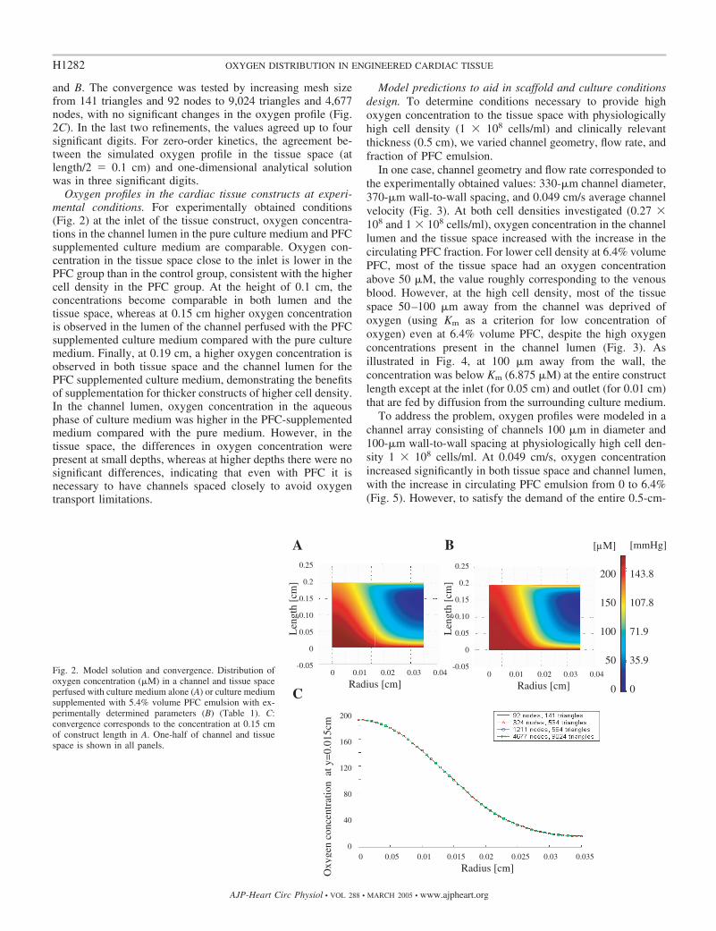
and B. The convergence was tested by increasing mesh sizefrom 141 triangles and 92 nodes to 9,024 triangles and 4,677nodes, with no significant changes in the oxygen profile (Fig.2C). In the last two refinements, the values agreed up to foursignificant digits. For zero-order kinetics, the agreement be-tween the simulated oxygen profile in the tissue space (atlength/2 � 0.1 cm) and one-dimensional analytical solutionwas in three significant digits.
Oxygen profiles in the cardiac tissue constructs at experi-mental conditions. For experimentally obtained conditions(Fig. 2) at the inlet of the tissue construct, oxygen concentra-tions in the channel lumen in the pure culture medium and PFCsupplemented culture medium are comparable. Oxygen con-centration in the tissue space close to the inlet is lower in thePFC group than in the control group, consistent with the highercell density in the PFC group. At the height of 0.1 cm, theconcentrations become comparable in both lumen and thetissue space, whereas at 0.15 cm higher oxygen concentrationis observed in the lumen of the channel perfused with the PFCsupplemented culture medium compared with the pure culturemedium. Finally, at 0.19 cm, a higher oxygen concentration isobserved in both tissue space and the channel lumen for thePFC supplemented culture medium, demonstrating the benefitsof supplementation for thicker constructs of higher cell density.In the channel lumen, oxygen concentration in the aqueousphase of culture medium was higher in the PFC-supplementedmedium compared with the pure medium. However, in thetissue space, the differences in oxygen concentration werepresent at small depths, whereas at higher depths there were nosignificant differences, indicating that even with PFC it isnecessary to have channels spaced closely to avoid oxygentransport limitations.
Model predictions to aid in scaffold and culture conditionsdesign. To determine conditions necessary to provide highoxygen concentration to the tissue space with physiologicallyhigh cell density (1 � 108 cells/ml) and clinically relevantthickness (0.5 cm), we varied channel geometry, flow rate, andfraction of PFC emulsion.
In one case, channel geometry and flow rate corresponded tothe experimentally obtained values: 330-�m channel diameter,370-�m wall-to-wall spacing, and 0.049 cm/s average channelvelocity (Fig. 3). At both cell densities investigated (0.27 �108 and 1 � 108 cells/ml), oxygen concentration in the channellumen and the tissue space increased with the increase in thecirculating PFC fraction. For lower cell density at 6.4% volumePFC, most of the tissue space had an oxygen concentrationabove 50 �M, the value roughly corresponding to the venousblood. However, at the high cell density, most of the tissuespace 50–100 �m away from the channel was deprived ofoxygen (using Km as a criterion for low concentration ofoxygen) even at 6.4% volume PFC, despite the high oxygenconcentrations present in the channel lumen (Fig. 3). Asillustrated in Fig. 4, at 100 �m away from the wall, theconcentration was below Km (6.875 �M) at the entire constructlength except at the inlet (for 0.05 cm) and outlet (for 0.01 cm)that are fed by diffusion from the surrounding culture medium.
To address the problem, oxygen profiles were modeled in achannel array consisting of channels 100 �m in diameter and100-�m wall-to-wall spacing at physiologically high cell den-sity 1 � 108 cells/ml. At 0.049 cm/s, oxygen concentrationincreased significantly in both tissue space and channel lumen,with the increase in circulating PFC emulsion from 0 to 6.4%(Fig. 5). However, to satisfy the demand of the entire 0.5-cm-
Fig. 2. Model solution and convergence. Distribution ofoxygen concentration (�M) in a channel and tissue spaceperfused with culture medium alone (A) or culture mediumsupplemented with 5.4% volume PFC emulsion with ex-perimentally determined parameters (B) (Table 1). C:convergence corresponds to the concentration at 0.15 cmof construct length in A. One-half of channel and tissuespace is shown in all panels.
H1282 OXYGEN DISTRIBUTION IN ENGINEERED CARDIAC TISSUE
AJP-Heart Circ Physiol • VOL 288 • MARCH 2005 • www.ajpheart.org

thick piece of tissue, average fluid velocity had to be increasedto 0.135 cm/s.
Oxygen transport improvements with the presence of PFCemulsion. The presence of 10–20% volume Oxygent (3.2–6.4% volume PFC emulsion droplets) increased the effectivediffusivity of the culture medium by 9–18%. In addition, theconvective term was increased by 62–123%, which is equiva-lent to the comparable increase in average fluid velocity. There-fore, both the transport in axial and radial direction is improvedcompared with the presence of the culture medium alone.
As demonstrated in governing equations (Eq. A6) and Table4, PFC emulsion contributes to the enhancement of masstransfer by increasing effective diffusivity and the convectiveterm (apparent average velocity). To illustrate this effect,simulations were performed in a densely packed channel array(100-�m channel diameter, 100-�m wall-to-wall spacing) per-fused at 0.135 cm/s, with culture medium supplemented with 0,3.2, and 6.4% of PFC emulsion.
For simplicity, the oxygen consumption in the tissue regionwas assumed to follow zero-order kinetics at 18 �M/s (corre-sponding to a cell density of �0.5 � 106 cells/ml). In eachcase, contribution of effective diffusivity alone, convectiveterm alone, and the combined effect of both terms was inves-tigated. Volume averaged and minimum oxygen concentration
as well as the mixing-cup culture medium concentration at theoutlet were compared.
The presence of PFC emulsion increased volume-averagedand minimum oxygen concentration in the tissue space as wellas the bulk oxygen concentration at the outlet from channellumen. Volume-averaged concentration in the tissue spaceincreased roughly linearly between 0 and 6.4% of PFC emul-sion (R2 � 0.9586). Minimum oxygen concentration in thetissue space was increased by 19.5 times with the addition of3.2% PFC and 28 times by the addition of 6.4% PFC. Con-currently with the increase in the concentration in the tissuespace, the concentration in the culture medium increased aswell. Addition of 3.2 and 6.4% of PFC emulsion increased theoutlet bulk medium aqueous concentration by 3.8 and 5 times,respectively. As illustrated in Fig. 6, �96.7–98.7% of theincrease in the minimum and volume-averaged oxygen con-centration can be contributed to the increase in the convectiveterm, with the reminder of the increase associated with theincrease in effective diffusivity.
DISCUSSION
We recently developed a novel biomimetic in vitro tissueculture system in which neonatal rat heart cells were cultured
Fig. 3. Comparison of predicted oxygen concentration profiles(�M) in a channel array supplemented with 0, 3.2, or 6.4% volumePFC emulsion. Cell density in the tissue space was set to the lowvalue measured experimentally (0.27 � 108 cells/ml) and thephysiological cell density (1 � 108 cells/ml). Channel array dimen-sions (330-�m channel diameter, 370-�m wall-to-wall spacing)and the average velocity of culture medium (0.049 cm/s) corre-spond to the experimentally obtained values. One-half of thechannel and surrounding tissue space are shown: array length (cm)vs. radius (cm).
H1283OXYGEN DISTRIBUTION IN ENGINEERED CARDIAC TISSUE
AJP-Heart Circ Physiol • VOL 288 • MARCH 2005 • www.ajpheart.org

on an elastic, highly porous scaffold with a parallel array ofchannels perfused with culture medium supplemented withsynthetic oxygen carrier (Oxygent PFC emulsion) (Radisic M,unpublished observation). In this system, parallel channel arraymimics the role of capillary network and the PFC emulsionmimics the role of hemoglobin. Constructs perfused withunsupplemented culture medium served as controls.
The main goal of this paper was to develop a steady-statemathematical model that can be used to predict oxygen con-centration profiles in the channel lumen and tissue spacesurrounding each channel as a function of channel geometry,cell density, PFC emulsion content, and flow rate. The derivedmathematical model helped rationalize experimental findingsin this system, and, compared with the previously derivedmodels of oxygen transport to PFC emulsions flowing in tubes(28), the model correctly included the effective diffusivity ofthe emulsion.
In future work, the model can be utilized to optimize channelgeometry and flow conditions for specific tissue-engineeringapplications. In optimizing flow conditions and scaffold geom-etry, a range of shear stresses tolerated by the cells of interestand channel diameter/spacing should be considered. For exam-ple, when optimizing for cardiac tissue constructs, the upperlevel of the shear stress considered should be 1.6 dyn/cm2. Forthe upper bound of channel diameter and spacing, the currentlyobtained experimental configuration (330-�m channel diame-ter and 370-�m wall-to-wall spacing) should be considered. Asa lower bound, the geometry found in the natural capillary bedsin the heart can be considered [�10-�m channel diameter and�20-�m wall-to-wall spacing (15, 27)]. The fraction of PFCemulsion can be increased from 0 to 0.064 with the upper limitset by the ability to keep the emulsion droplets suspended atrelatively low flow rates. The optimal geometry and flow ratewould then be determined as a set of conditions that maximizethe volume-averaged and the minimum oxygen concentrationin the tissue space.
To rationalize our decision to maintain low levels of hydro-dynamic shear in our tissue-engineering system, we specifythat, in the native heart, cardiomyocytes are shielded fromdirect contact with blood by endothelial cells. When exposed to
shear stress, cardiac myocytes round up and show signs ofdedifferentiation. (4, 5, 16, 30, 31), as documented in ourlaboratory’s previous work involving perfusion of cardiomyo-cytes on porous collagen sponges (25). In channeled tissueconstructs, endothelial cells are not present on the channelwalls to shield the myocytes from the effects of shear stress.Therefore, in the densely packed channel array (100-�m chan-nel diameter, 100-�m wall-to-wall spacing), where at most 10myocytes can span the distance between two channels, �20%of the myocytes are adjacent to the channel walls and can beexposed to excessive shear if the flow rate is too high. Inaddition, the fraction of cardiomyocytes exposed to the hydro-dynamic shear would increase with an increase in medium flowrate due to the penetration of culture medium to the scaffoldpores.
Perfusion of the macroporous scaffolds at low shear stressesand low average velocities had beneficial effects in varioustissue-engineering systems. Shear stress of up to 1 dyn/cm2
(average velocity of up to 640 �m/s) increased deposition ofmineralized matrix by marrow stromal osteoblasts of a tissue-engineered bone in a dose-dependant manner (1, 29). Simi-larly, perfusion in the range from 1 to 170 �m/s increased thecontent of DNA, glycosaminoglycans, and hydroxyproline of atissue-engineered cartilage compared with the static controls(9, 22, 26). In contrast, a 1-ml bolus of fluid at up to 2,500–25,000 �m/s led to the washout of 57% of chondrocytes froma porous PLA scaffold 1 h after seeding (14).
The appropriate average velocities also depend on the celltype being cultivated (16, 31). Although perfusion at 5–110�m/s had a beneficial effect on the constructs based onMC3T3-E1 immature osteoblasts-like cells, average velocityof 560 �m/s significantly reduced the viability in the samesystem (6). For cardiomyocyte/polyglycolic acid constructs,perfusion at 140–710 �m/s increased uniformity of cell distri-bution and expression of cardiac markers compared with thestatic controls (4, 5). In our laboratory’s previous work (24),perfusion in the range of 425–1,275 �m/s through the cardio-myocyte/collagen constructs improved cell viability comparedwith the static controls while maintaining high cell yield(�90%).
In the biomimetic system described here, the flow conditionsin the channel array (�100 �m in diameter) were set to mimicthe low Re number (�1) and physiological shear stress nor-mally found in the microvasculature and capillary networks invivo [�1.5 dyn/cm2 (12)]. In our best case, the culture mediumis perused through the dense channel array (100-�m diameter,100-�m wall-to-wall spacing) at the upper level of the exper-imentally feasible velocity (1,350 cm/s) (24), yielding a wallshear stress of �1 dyn/cm2.
In addition, substituting culture medium with oxygen carri-ers uncouples oxygen supply from the flow rate, thus enablingadequate supply without negative effects such as myocyterounding and cell washout. In addition, it provides the meansof studying the effects of flow rate, shear stress (as in Ref. 1),and oxygen supply independently.
When cultivated in the presence of PFC emulsion (5.4%volume circulating), the constructs had higher DNA and pro-tein content, as well as the higher cell density compared withthe unsupplemented culture medium (0.27 � 108 vs. 0.42 �108 cells/ml). As a result, V̇O2 max in the tissue space washigher for the PFC-supplemented vs. -unsupplemented culture
Fig. 4. Log plot of oxygen concentration along the length of a tissue constructcontaining physiological cell density (108 cells/ml) 100 �m away from thechannel wall. The channels are 330 �m in diameter, spaced 370 �m apart, andperfused with culture medium containing 6.4% volume of PFC emulsion at anaverage velocity of 0.049 cm/s (other conditions as specified in Fig. 3).
H1284 OXYGEN DISTRIBUTION IN ENGINEERED CARDIAC TISSUE
AJP-Heart Circ Physiol • VOL 288 • MARCH 2005 • www.ajpheart.org

medium (10.5 vs. 8.8 �M/s, respectively). The comparison ofmodeled oxygen profiles in the tissue constructs (Fig. 2)indicated that the differences in oxygen concentration betweenPFC supplemented and pure culture medium were more evi-dent at higher construct thickness than closer to the entrance.Although oxygen supply to the construct was increased by theaddition of PFC emulsion, so was the number of survivingcells, resulting in the increased oxygen consumption and ap-proximately comparable oxygen concentration profiles overmost of the construct volume. The main finding that more cellssurvive as more oxygen becomes available could be predictedby the model if the relationship between oxygen availabilityand cell survival rate was known in this tissue-engineering
system. As such, this model can serve as a tool for us and otherresearchers to establish a correct relationship between oxygenavailability and cell survival rate.
The channel array used in our experiments (330-�m channeldiameter and 370-�m wall-to-wall spacing) is the finest ob-tainable in the 0.2-cm-thick scaffolds using the existing laser/cutting engraving system (Fig. 2). To make a thick scaffold(0.5 cm) with a fine channel array (100 �m; Fig. 5), themachining will have to be modified. Although the oxygenconcentration in the tissue space with physiological cell density(Fig. 5) increased considerably with the increase of circulatingPFC concentration from 0 to 6.4%, we had to increase the flowrate, keeping the shear stress in the physiological range at �1dyn/cm2, to provide enough oxygen for the entire 0.5-cm-thickconstruct. At our best conditions (0.135 cm/s and 6.4% PFC),the oxygen is not depleted at any point in the scaffold and theminimum concentration of 33 �M is approximately five timesabove Km.
Finally, for in vivo implantation, it is essential that thegeometry of the engineered graft is compatible with the trans-port properties of blood. A physiological drop inPO2 acrosstissues is from 95 mmHg in the arterial blood to 40 mmHg invenous blood (Ref. 11, p. 91). If the patch is grafted as an
Fig. 5. Comparison of predicted oxygen concentration profiles(�M) in a channel array supplemented with 0, 3.2, or 6.4% volumePFC emulsion at physiological cell density (1 � 108 cells/ml) at thelow (0.049 cm/s) and high (0.135 cm/s) average velocities of culturemedium. Channel array dimensions are 100-�m channel diameterand 100-�m wall-to-wall spacing. One-half of the channel andsurrounding tissue space are shown: array length (cm) vs. radius(cm).
Table 4. Oxygen transport improvements in thepresence of PFC emulsion
Circulating PFC Fraction(Fraction of Oxygent)
Convective Term[1 (K � 1)�]
Effective Diffusivity(Eq. 11)
0.000 (0.00) 1.00 Da
0.032 (0.10) 1.62 1.09Da
0.054 (0.17) 2.05 1.15Da
0.064 (0.20) 2.23 1.18Da
H1285OXYGEN DISTRIBUTION IN ENGINEERED CARDIAC TISSUE
AJP-Heart Circ Physiol • VOL 288 • MARCH 2005 • www.ajpheart.org

atrioventricular shunt, such that it connects to arterial blood atthe inlet and venous blood at the outlet, the physiologicaldifference in total oxygen concentration at the inlet and outletwill be 8,630–5,874 � 2,756 �M. Assuming that the patch hasa physiological density of cells metabolizing at V̇O2 max, themetabolic demand for oxygen for the whole patch is 33 �M/s.The calculated blood perfusion rate necessary to keep the cellswell oxygenated under these conditions is 0.63 ml blood �mltissue�1 �min�1, a value comparable to the reported baselineflow rates of blood in the heart (0.7 blood �ml tissue�1 �min�1;Ref. 11). For an engineered construct that is 0.5 cm in diameterand 0.5 cm thick, and contains an array of channels that are 100�m in diameter and spaced 100 �m apart, the model predicts
the volumetric flow rate of blood of 1.9 � 10�6 ml/s perchannel. The wall shear stress in the channels of the tissue graftperfused with blood can be estimated from the reduced averagevelocity [4Q/� (2Rc)3] of the Casson fluid (Ref. 11, Eqs. 3.21and Ref. 18) where Q is volumetric flow rate per channel. Theobtained wall shear stress of 0.99 dyn/cm2 is high enough toavoid anomalous flow properties of blood that may occur atvery low shear stresses, below the shear stress causing celldamage (1.6 dyn/cm2) and sufficient to prevent spreading ofleukocytes and pseudopode formation that may increase flowresistance at low shear rates and mediate inflammatory re-sponse (19). Also, the reduced average velocity of blood flowin the channels (2.5 s�1) is �1 s�1, indicating that aggregationof red blood cells should not be expected. Finally, the pressuredrop across the construct under these conditions (0.14 mmHg)is considerably lower than that in the capillary bed [17 mmHg(Ref. 11)], and the total required blood flow is only 0.05ml/min, indicating that the graft would not significantly in-crease the peripheral resistance for blood flow.
In summary, a steady-state mathematical model for oxygenconcentration profile in a tissue construct with a parallelchannel array was developed as a function of concentration ofPFC emulsion channel geometry, flow rate, and cell density.The model was solved for a set of experimentally obtainedconditions and used to predict oxygen concentration profiles inconstructs of clinically relevant thickness (0.5 cm) and physi-ologically high cell densities. For the experimentally relevantparameters, the obtained cell density was higher when con-structs were perfused with the PFC-supplemented culture me-dium, leading to the increased oxygen consumption rate.Therefore, despite the improved delivery, the oxygen concen-tration profiles were comparable in the tissue space of theconstructs cultivated with pure and PFC-supplemented culturemedium. The concentration in the channel lumen was higher inthe PFC-supplemented medium. At an identical cell density,the concentration in the tissue space increased with the increasein PFC concentration and flow rate and for finer channel arrays.The presence of PFC emulsion increased axial transport byincreasing apparent convective term [by (K � 1)�, where K isthe partition coefficient] and radial transport by increasingeffective diffusivity, although the majority of the increase inoxygen concentration in the tissue space (�98%) could beattributed to the increase in convective term.
APPENDIX
Let Cp and Ca be oxygen concentrations in the PFC and aqueousphase, respectively, in a system depicted in Fig. 1 and � be the volumefraction of PFC in the emulsion. Assuming that the drops are 1) smallenough that the concentration is nearly uniform within a given dropand 2) each drop is very nearly in equilibrium with the adjacent media,the total oxygen concentration at any point in the emulsion is:
Ctotal � 1 � �Ca � �Cp (A1)
where Ctotal is the total concentration and � is fraction of circulatingPFC emulsion droplets. Because by definition the partition coefficientis K � Cp/Ca:
Ctotal � �1 � K � 1��Ca (A2)
Treating the emulsion as a homogenous fluid of constant density,the conservation equation for oxygen at steady state can be expressedas:
Fig. 6. Effect of PFC emulsion on the oxygen concentration in the engineeredcardiac tissue. A: volume average oxygen concentration in the tissue space. B:minimum oxygen concentration in the tissue space. C: mixing-cup outletoxygen concentration in the aqueous phase of the culture medium. Oxygenconcentration was calculated for a densely packed channel array (100-�mchannel diameter, 100-�m wall-to-wall spacing). Oxygen consumption rate inthe tissue space was set at 18 �M/s and was assumed to follow zero-orderkinetics. The velocity of culture medium was 0.135 cm/s. For 3.2 and 6.4% ofPFC, contribution of effective diffusivity alone (white bars) was comparedwith the contribution of convective term (black bars) and the combined effectof both terms (gray bars).
H1286 OXYGEN DISTRIBUTION IN ENGINEERED CARDIAC TISSUE
AJP-Heart Circ Physiol • VOL 288 • MARCH 2005 • www.ajpheart.org

Va � �Ctotal � ��J (A3)
where Va is velocity and J is the flux of oxygen relative to Va (thediffusive flux). The diffusive flux can be expressed in terms of eitherCtotal or Ca; the latter is preferable because Ca is more convenient formatching concentrations at the emulsion-tissue interface. Choosing Ca
gives:
J � �Deff�2Ca (A4)
where Deff is effective diffusivity, defined by Ref. 10 (p. 191):
Deff � Da�1 � 3�� � 1
� � 2��� (A5)
where Da is diffusivity of aqueous phase and Dp is diffusivity ofoxygen in PFC phase of culture medium and � � K Dp/Da. Combin-ing Eqs. A1–A5 and taking into account low hydraulic permeability ofthe tissue space, the conservation equation for oxygen in the channellumen becomes:
�1 � K � 1�� � Vzr�Ca
�z� Deff �1
r
�
�r�r
�Ca
�r� �
�2Ca
�z2 � (A6)
where r, z are radial and axial position, respectively, and Vz is axialvelocity component of the culture medium in the channel.
This governing equation is of the same form as Eq. 15 given inShah and Mehra (28) except that they used Da instead of the Deff ofthe PFC emulsion. Although axial diffusion can be neglected in Eq.A6 due to the relatively high value of Pe, the software used fornumerical solution considered axial diffusion automatically.
Because the hydraulic permeability of the tissue space is small, itis reasonable to assume that radial velocity component can be ne-glected in the channel and that there is no convection in the tissuespace (23). Governing equation for oxygen distribution in the tissueregion where oxygen consumption occurs according to the Michaelis-Menten kinetics can be expressed as:
0 � Dt�1
r
�
�r�r
�Ct
�r� �
�2Ct
�z2 � �V̇O2 maxCt
Km � Ct
(A7)
For inlet boundary conditions, the measured bulk oxygen concen-tration (Cin) of the culture medium was used for predictions atexperimental conditions. Therefore, the oxygen concentration at thechannel lumen and the tissue space (Ct) can be expressed as:
Car, 0 � Cin (A8)
Ctr, 0 � Cin (A9)
Similarly, the bulk oxygen concentration at the outlet (Cout) fromthe tissue constructs was measured at experimental conditions yield-ing the following outlet boundary conditions for the aqueous phaseand tissue space, respectively:
Car, L � Cout (A10)
Ctr, L � Cout (A11)
where L is the length of tissue construct (equivalent to the channellength).
For predictions of oxygen concentration profiles at conditions thatwere not obtained experimentally, Cin and Cout were not measured.Therefore, an alternative set of boundary conditions was used. It wasassumed that the culture medium entering the perfusion cartridge wasfully saturated with oxygen and Ca(r,0) � Ct(r,0) � 222.47 �M. Theaxial variations in the oxygen concentration cease to exist at the veryshort distance from the outlet of the channel array. Therefore,
�Ca/�zr, L � 0 (A12)
in the culture medium at the channel outlet. It was also assumed thatthe culture medium at the outlet was well mixed, with no variations in
the radial direction. Therefore, mixing cup concentration of theculture medium at the channel outlet was set to be the boundarycondition for the concentration at the tissue space outlet.
Ctr,L �
�0
RC
Car,LVzrdr
�0
RC
Vzrdr
(A13)
The validity of the boundary conditions used for predictions wasconfirmed by comparing the oxygen concentration measured at theoutlet of the experimentally obtained channeled tissue to the outletvalues obtained using the described boundary conditions in the chan-nel array of identical geometry, cell density, and flow conditions.
Symmetry boundary condition was applied at the centerline of thechannel lumen:
�Ca/�r0,z � 0 (A14)
The region supplied by each channel is approximated by a cylinder.Because these cylindrical regions are equally spaced, a no-flux bound-ary condition is applied at the half distance between channel centers(Rt), which is a common assumption in the well-known Kroghcylinder model.
�Ct/�rRt,z � 0 (A15)
Finally, the fluxes of oxygen and the concentrations have to matchat the interface between channel lumen and tissue space, yielding theremaining two boundary conditions:
Da
�Ca
�rRc,z � Dt
�Ct
�rRc,z (A16)
CaRc,z � CtRc,z (A17)
Assumption validation. To justify the assumption of uniform oxy-gen concentration within PFC droplets used in the model derivationand the local equilibrium between PFC and the aqueous phase at everypoint along the channel length, the following processes have to beconsidered (Fig. 1C): 1) diffusion of oxygen inside the PFC droplet;2) diffusion of oxygen from the PFC droplet to the surroundingaqueous phase of culture medium; and 3) transport of oxygen from thebulk fluid to the tissue wall.
There is a gradient in oxygen concentration from the center of PFCdroplet to the drop/medium interface (�C1), followed by the decreaseat the interface and a gradient through the boundary layer (�C2) (Fig.1D). The ratio of these two concentration differences is proportionalto the Biot number (Bi; Ref. 10, pg. 83):
Bi �k2d/2
KDp
��C1
�C2
(A18)
where k2 is the mass transfer coefficient for the absorption of oxygenfrom the PFC droplet into the aqueous phase of culture medium, d isa diameter of PFC droplet, K is the partition coefficient, and Dp isdiffusion coefficient of oxygen in neat PFC. Droplet mass transportcoefficient can be expressed in terms of droplet Sherwood number(Shd) as:
Shd �k2 � d
Da
(A19)
where Da is a diffusion coefficient for oxygen in the aqueous phase ofculture medium. By combining Eqs. 1 and 2, Biot number can beexpressed as:
H1287OXYGEN DISTRIBUTION IN ENGINEERED CARDIAC TISSUE
AJP-Heart Circ Physiol • VOL 288 • MARCH 2005 • www.ajpheart.org

Bi �ShdDa
2KDp
(A20)
Internal diffusion can be neglected for Bi �� 1. Because PFCparticles are of small diameter (0.2 �m), it can be assumed that theymove with the flow and Shd � 2. For Da � 2.4 � 10�5 cm2/s (5),Dp � 5.6 � 10�5 cm2/s (17) and K � 20.3 (the ratio of oxygensolubilities at 760 Torr and 37°C in neat perfluorooctyl bromide andwater); the Biot number in Eq. A20 has value of 0.02, implying thatthe resistance to internal diffusion can be neglected.
To determine �C2/�C3, we will compare the rate of transport fromthe boundary layer around the PFC droplet to the bulk fluid and fromthe bulk fluid to the channel wall (Fig. 1D) in the differential channellayer of length �z. Mass flow of oxygen from the PFC particles intothe aqueous phase of culture medium can be described by thefollowing equation:
r2 � k2ap�C2 (A21)
where k2 is the drop mass transport coefficient as defined in Eq. A18and ap is surface area of all PFC droplets in the control volumeconsidered.
ap � Sp Np � 6�Rc�z�
d(A22)
where Sp is the surface area of one droplet, Np is the number of all ofthe PFC droplets in the control volume considered, Rc is channelradius, and � is volume fraction of PFC emulsion. By combining Eqs.A21, A22, and A19 with Shd � 2, mass flow of oxygen from the PFCto aqueous phase can be expressed as:
r2 � 12��C2
DaRc�z�
d(A23)
Mass flow of oxygen (r3) from the bulk phase of culture medium tothe channel wall can be described as:
r3 � k3�C32�Rc�z (A24)
where k3 is a mass transport coefficient that is related to the channelSherwood number (Shc)
Shc �2k3Rc
Deff
(A25)
Deff is given by Eq. A5. Eq. A5 neglects interparticle interactions,a reasonable assumption for the dilute emulsion investigated (� �0.1). For the conditions investigated in this paper, culture medium issupplemented by 10–20% of Oxygent, a 32% volume PFC emulsion,resulting in � � 0.032–0.064. For the given conditions, with � �47.3, Deff � 1.09–1.18 Da. At steady state, r2 � r3:
12��C2
DaRc�z�
d� k3�C32�Rc�z (A26)
As a result, the ratio of �C2 to �C3 can be expressed as:
�C2
�C3
�Shc
12��Deff
Da� � d
Rc�2
(A27)
Accordingly for the range of � � 0.032–0.064 and Deff in therange of 1.09–1.18 Da, the ratio of �C2/�C3 will be in the range of4.17 to 2.26 � 10�6 Shc. Thus �C2 will not exceed 10% of �C3 ifShc � 2 � 104.
Shc declines rapidly from high values at the entrance to the channelto 3.576 or 4.360 for constant concentration and constant flux bound-ary conditions, respectively (Ref. 10, p. 392). At the experimentallyinvestigated conditions, Shc will drop below 2 � 104 very close to theentrance at z � 3.5 � 10�13 cm for constant concentration and z �6.5 � 10�13 cm for constant flux boundary conditions. Because there
is consumption of oxygen at the channel wall, the real boundarycondition is mixed or Robin boundary condition, and the real valuesof z will fall in between the values obtained for constant flux andconstant concentration boundary condition (Ref. 10, p. 392).
Consequently, because Shc is many orders of magnitude less than106 over most of the channel length at the conditions investigated,�C2 �� �C3 over most of the channel length, except very near to theentrance where Shc 3 �. Because �C1 �� �C2, then �C1 �� �C3.Therefore, absorbance from the PFC droplets into the aqueous phaseis not the rate-limiting step, and local equilibrium can be assumed atevery point in the channel.
To confirm the assumption of no convection in the tissue space andno radial velocity component in the channel lumen, we experimentallyestimated the hydraulic permeability of channel-free Biorubber scaf-fold. The estimated value was 8.1 � 0.4 10�12 m2 [(m3 �s �m2) (Pa s)m/Pa] (please refer to experimental section).
Based on Darcy permeability, the average axial velocity in thescaffold space was estimated to be 1.7 � 10�4 cm/s. The scaffoldaxial Pe number was calculated as:
Pe2 �US(Rt � Rr)
Da
(A28)
where Us is the average axial velocity of culture medium through thescaffold.
For the experimentally obtained channel geometry, Pes � 0.09. Forthe proposed thick scaffold (0.5 cm) with dense packing of narrowchannels (100-�m channel diameter and 100-�m channel spacing),the estimated scaffold axial velocity was 4.7 � 10�4 cm/s (at thehighest flow rate investigated), and Pes was 0.095. As Matrigel/cellsuspension is added to the scaffold, it is expected that the permeabilityof the tissue space and axial tissue Pe would decrease even further.The value of 0.095 is the upper bound of the tissue Pe numberobtainable in this system, thus justifying the assumption of no con-vection in the tissue space.
ACKNOWLEDGMENTS
We thank Alliance Pharmaceutical (San Diego, CA) for generous donationof the PFC emulsion (Oxygent), Drs. Yadong Wang and Robert Dennis forhelp with scaffold design and fabrication, Drs. Keipert and Faithfull forinsightful reviews of the manuscript, and Sue Kangiser for technical helpduring the preparation of the manuscript.
GRANTS
The work was supported by the National Aeronautics and Space Adminis-tration (grant no. NCC8-174) and Poitras fellowship (to M. Radisic).
REFERENCES
1. Bancroft GN, Sikavitsas VI, van den Dolder J, Sheffield TL, AmbroseCG, Jansen JA, and Mikos AG. Fluid flow increases mineralized matrixdeposition in 3D perfusion culture of marrow stromal osteoblasts in adose-dependent manner. Proc Natl Acad Sci USA 99: 12600–12605, 2002.
2. Bursac N, Papadaki M, Cohen RJ, Schoen FJ, Eisenberg SR, CarrierR, Vunjak-Novakovic G, and Freed LE. Cardiac muscle tissue engi-neering: toward an in vitro model for electrophysiological studies. Am JPhysiol Heart Circ Physiol 277: H433–H444, 1999.
3. Carrier RL, Papadaki M, Rupnick M, Schoen FJ, Bursac N, LangerR, Freed LE, and Vunjak-Novakovic G. Cardiac tissue engineering: cellseeding, cultivation parameters and tissue construct characterization. Bio-technol Bioeng 64: 580–589, 1999.
4. Carrier RL, Rupnick M, Langer R, Schoen FJ, Freed LE, andVunjak-Novakovic G. Effects of oxygen on engineered cardiac muscle.Biotechnol Bioeng 78: 617–625, 2002.
5. Carrier RL, Rupnick M, Langer R, Schoen FJ, Freed LE, andVunjak-Novakovic G. Perfusion improves tissue architecture of engi-neered cardiac muscle. Tissue Eng 8: 175–188, 2002.
6. Cartmell SH, Porter BD, Garcia AJ, and Guldberg RE. Effects ofmedium perfusion rate on cell-seeded three-dimensional bone constructsin vitro. Tissue Eng 9: 1197–1203, 2003.
H1288 OXYGEN DISTRIBUTION IN ENGINEERED CARDIAC TISSUE
AJP-Heart Circ Physiol • VOL 288 • MARCH 2005 • www.ajpheart.org

7. Casey TM and Arthur PG. Hibernation in noncontracting mammaliancardiomyocytes. Circulation 102: 3124–3129, 2000.
8. Chromiak JA, Shansky J, Perrone C, and Vandenburgh HH. Biore-actor perfusion system for the long-term maintenance of tissue engineeredskeletal muscle organoids. In Vitro Cell Dev Biol Anim 34: 694–703,1998.
9. Davisson TH, Sah RL, and Ratcliffe AR. Perfusion increases cellcontent and matrix synthesis in chondrocyte three-dimensional cultures.Tissue Eng 8: 807–816, 2002.
10. Deen WM. Analysis of Transport Phenomena. New York: Oxford Univ.Press, 1998, p. 316.
11. Fournier RL. Basic Transport Phenomena in Biomedical Engineering.Philadelphia, PA: Taylor & Francis, 1998.
12. Fukuda S, Yasu T, Predescu DN, and Schmid-Schonbein GW. Mech-anisms for regulation of fluid shear stress response in circulating leuko-cytes. Circ Res 86: 13–18, 2000.
13. Geankoplis CJ. Transport Processed and Unit Operations (3rd ed.).Englewood Cliffs, NJ: Prentice-Hall, 1993.
14. Giurea A, Klein TJ, Chen AC, Goomer RS, Coutts RD, Akeson WH,Amiel D, and Sah RL. Adhesion of perichondrial cells to a polylactic acidscaffold. J Orthop Res 21: 584–589, 2003.
15. Korecky B, Hai CM, and Rakusan K. Functional capillary density innormal and transplanted rat hearts. Can J Physiol Pharmacol 60: 23–32,1982.
16. Kretzmer G and Schugerl K. Response of mammalian cells to shearstress. Appl Microbiol Biotechnol 34: 613–616, 1991.
17. Mates vanLobensels E, Anderson JC, Hildebrandt J, and HlastalaMP. Modeling diffusion limitation of gas exchange in lungs containingperfluorocarbon. J Appl Physiol 86: 273–284, 1999.
18. Merril EW, Benis AM, Gilliand ER, Sherwood TK, and Salzman EW.Pressure flow relations of human in hollow fibers at low flow rates. J ApplPhysiol 20: 954–967, 1965.
19. Moazzam F, DeLano FA, Zweifach BW, and Schmid-Schonbein GW.The leukocyte response to fluid stress. Proc Natl Acad Sci USA 94:5338–5343, 1997.
20. Ni Y, Klein DH, and Pelura TJ. Rheology of concentrated perfluoro-carbon emulsions. Biomater Artif Cells Immobilization Biotechnol 20:869–871, 1992.
21. Obradovic B, Carrier RL, Vunjak-Novakovic G, and Freed LE. Gasexchange is essential for bioreactor cultivation of tissue engineered carti-lage. Biotechnol Bioeng 63: 197–205, 1999.
22. Pazzano D, Mercier KA, Moran JM, Fong SS, DiBiasio DD, Rulfs JX,Kohles SS, and Bonassar LJ. Comparison of chondrogensis in static andperfused bioreactor culture. Biotechnol Prog 16: 893–896, 2000.
23. Piret JM and Cooney CL. Model of oxygen transport limitations inhollow fiber bioreactors. Biotechnol Bioeng 37: 80–92, 1991.
24. Radisic M, Euloth M, Yang L, Langer R, Freed LE, and Vunjak-Novakovic G. High density seeding of myocyte cells for tissue engineer-ing. Biotechnol Bioeng 82: 403–414, 2003.
25. Radisic M, Yang L, Boublik J, Cohen RJ, Langer R, Freed LE, andVunjak-Novakovic G. Medium perfusion enables engineering of compactand contractile cardiac tissue. Am J Physiol Heart Circ Physiol 286:H507–H516, 2004.
26. Raimondi MT, Boschetti F, Falcone L, Migliavacca F, Remuzzi A, andDubini G. The effect of media perfusion on three-dimensional cultures ofhuman chondrocytes: integration of experimental and computational ap-proaches. Biorheology 41: 401–410, 2004.
27. Rakusan K and Korecky B. The effect of growth and aging on functionalcapillary supply of the rat heart. Growth 46: 275–281, 1982.
28. Shah N and Mehra A. Modeling of oxygen uptake in perfluorocarbonemulsions. Some comparisons with uptake by blood. ASAIO J 42: 181–189, 1996.
29. Sikavitsas VI, Bancroft GN, Holtorf HL, Jansen JA, and Mikos AG.Mineralized matrix deposition by marrow stromal osteoblasts in 3Dperfusion culture increases with increasing fluid shear forces. Proc NatlAcad Sci USA 100: 14683–14688, 2003.
30. Smith CG, Greenfield PF, and Randerson D. Shear sensitivity of threehybridoma cell lines in suspension culture. In: Modern Approaches toAnimal Cell Technology, edited by Spier RE and Griffith JB. Kent, UK:Butterworth, 1987.
31. Stathopoulos NA and Hellums JD. Shear stress effects on humanembryonic kidney cells in vitro. Biotechnol Bioeng 27: 1021–1026, 1985.
32. Wang Y, Ameer GA, Sheppard BJ, and Langer R. A tough biodegrad-able elastomer. Nat Biotechnol 20: 602–606, 2002.
33. Yamada T, Yang JJ, Ricchiuti NV, and Seraydarian MW. Oxygenconsumption of mammalian myocardial-cells in culture—measurementsin beating cells attached to the substrate of the culture dish. Anal Biochem145: 302–307, 1985.
H1289OXYGEN DISTRIBUTION IN ENGINEERED CARDIAC TISSUE
AJP-Heart Circ Physiol • VOL 288 • MARCH 2005 • www.ajpheart.org