Masterproef - Universiteit Hasselt · 1 Preface This thesis is the result of the studies conducted...
63
De transnationale Universiteit Limburg is een uniek samenwerkingsverband van twee universiteiten in twee landen: de Universiteit Hasselt en Maastricht University Universiteit Hasselt | Campus Diepenbeek | Agoralaan Gebouw D | BE-3590 Diepenbeek Universiteit Hasselt | Campus Hasselt | Martelarenlaan 42 | BE-3500 Hasselt 2010 2011 GENEESKUNDE master in de biomedische wetenschappen: milieu en gezondheid Masterproef The role of CD28null T cells in autoimmune diseases Promotor : Prof. dr. Niels HELLINGS Marijke Brulmans Masterproef voorgedragen tot het bekomen van de graad van master in de biomedische wetenschappen , afstudeerrichting milieu en gezondheid
Transcript of Masterproef - Universiteit Hasselt · 1 Preface This thesis is the result of the studies conducted...
De transnationale Universiteit Limburg is een uniek samenwerkingsverband van twee universiteiten in twee landen: de Universiteit Hasselt en Maastricht University
Universiteit Hasselt | Campus Diepenbeek | Agoralaan Gebouw D | BE-3590 DiepenbeekUniversiteit Hasselt | Campus Hasselt | Martelarenlaan 42 | BE-3500 Hasselt
2 0 1 02 0 1 1GENEESKUNDE
master in de biomedische wetenschappen: milieu engezondheid
MasterproefThe role of CD28null T cells in autoimmune diseases
Promotor :Prof. dr. Niels HELLINGS
Marijke Brulmans Masterproef voorgedragen tot het bekomen van de graad van master in de biomedischewetenschappen , afstudeerrichting milieu en gezondheid
De transnationale Universiteit Limburg is een uniek samenwerkingsverband van twee universiteiten in twee landen: de Universiteit Hasselt en Maastricht University
Universiteit Hasselt | Campus Diepenbeek | Agoralaan Gebouw D | BE-3590 DiepenbeekUniversiteit Hasselt | Campus Hasselt | Martelarenlaan 42 | BE-3500 Hasselt
2 0 1 02 0 1 1
GENEESKUNDEmaster in de biomedische wetenschappen: milieu engezondheid
MasterproefThe role of CD28null T cells in autoimmune diseases
Promotor :Prof. dr. Niels HELLINGS
Marijke Brulmans Masterproef voorgedragen tot het bekomen van de graad van master in de biomedischewetenschappen , afstudeerrichting milieu en gezondheid


1
Preface
This thesis is the result of the studies conducted during my 8 month internship at the
Biomedical research institute at Diepenbeek. It has been a very fulfilling experience, but all
good things must come to an end.
I would sincerely like to thank my supervisor, Bieke Broux, for the excellent guidance she has
given me during my stay at Biomed, for the corrections and improvements she has provided
me for my thesis, and for her pleasant company throughout my internship.
I would also like to thank my promoter Prof. Dr. Niels Hellings, and second examiner Prof.
Dr. Piet Stinissen for making the time to listen to my progress and for providing me with tips
and ideas for further experiments and for my thesis.
Special thanks go to Christel Boken, my fellow students and everybody at Biomed who has
helped me with my questions and experiments.
Last but definitely not least, I would like to thank my parents. They made it possible for me to
go to university, and they have supported me throughout my entire studies, especially these
last few months. I would never have gotten this far without them.

2
3
Content
Preface ........................................................................................................................................ 1
List of Abbreviations .................................................................................................................. 5
Abstract ...................................................................................................................................... 7
Abstract (Dutch) ......................................................................................................................... 9
Introduction .............................................................................................................................. 11
Introduction to the immune system ....................................................................................... 11
Immunosenescence: Aging of the immune system ................................................................ 12
Immunosenescence: CD28null
T cells .................................................................................... 13
CD4+CD28
null T cells and autoimmunity .............................................................................. 15
CD8+CD28
null T cells ............................................................................................................ 16
CD28null
T cells as a possible target for therapy .................................................................. 17
Materials & Methods ................................................................................................................ 19
Study Subjects ....................................................................................................................... 19
Cell preparation and culture ................................................................................................ 19
ELISA .................................................................................................................................... 21
Cytokine Array ...................................................................................................................... 21
Chemotaxis assay ................................................................................................................. 23
Flow cytometry ..................................................................................................................... 23
Immunohistochemistry .......................................................................................................... 24
Degranulation assay ............................................................................................................. 25
Statistical analysis ................................................................................................................ 25
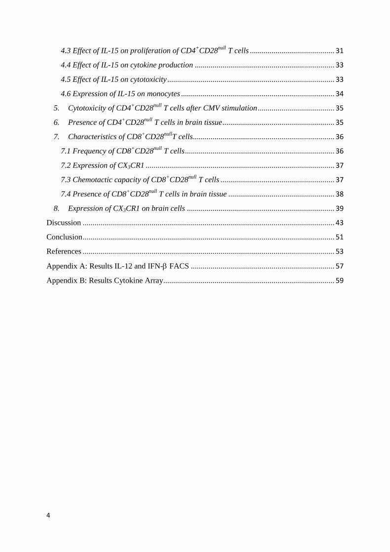
Results ...................................................................................................................................... 27
1. Frequency of CD4+CD28
null T cells and making of Cell lines ...................................... 27
2. Cytokine production by CD4+CD28
null T cells .............................................................. 27
2.1 Optimization of stimulation conditions ....................................................................... 27
2.2 Cytokine array ............................................................................................................ 28
3. Effect of IL-12/IFN- on the expression of CD28 ......................................................... 29
4. Effect of IL-15 on the proliferation and phenotype of CD4+CD28
null T cells ............... 30
4.1 Expression of the IL-15 receptor by CD4+CD28
null T cells ........................................ 30
4.2 Optimization of IL-15 concentration........................................................................... 31
4
4.3 Effect of IL-15 on proliferation of CD4+CD28
null T cells ........................................... 31
4.4 Effect of IL-15 on cytokine production ....................................................................... 33
4.5 Effect of IL-15 on cytotoxicity ..................................................................................... 33
4.6 Expression of IL-15 on monocytes .............................................................................. 34
5. Cytotoxicity of CD4+CD28
null T cells after CMV stimulation ....................................... 35
6. Presence of CD4+CD28
null T cells in brain tissue ......................................................... 35
7. Characteristics of CD8+CD28
nullT cells........................................................................ 36
7.1 Frequency of CD8+CD28
null T cells ............................................................................ 36
7.2 Expression of CX3CR1 ................................................................................................ 37
7.3 Chemotactic capacity of CD8+CD28
null T cells .......................................................... 37
7.4 Presence of CD8+CD28
null T cells in brain tissue ...................................................... 38
8. Expression of CX3CR1 on brain cells ........................................................................... 39
Discussion ................................................................................................................................ 43
Conclusion ................................................................................................................................ 51
References ................................................................................................................................ 53
Appendix A: Results IL-12 and IFN-FACS ......................................................................... 57
Appendix B: Results Cytokine Array ....................................................................................... 59
5
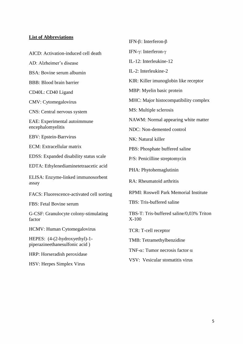
List of Abbreviations
AICD: Activation-induced cell death
AD: Alzheimer’s disease
BSA: Bovine serum albumin
BBB: Blood brain barrier
CD40L: CD40 Ligand
CMV: Cytomegalovirus
CNS: Central nervous system
EAE: Experimental autoimmune
encephalomyelitis
EBV: Epstein-Barrvirus
ECM: Extracellular matrix
EDSS: Expanded disability status scale
EDTA: Ethylenediaminetetraacetic acid
ELISA: Enzyme-linked immunosorbent
assay
FACS: Fluorescence-activated cell sorting
FBS: Fetal Bovine serum
G-CSF: Granulocyte colony-stimulating
factor
HCMV: Human Cytomegalovirus
HEPES: (4-(2-hydroxyethyl)-1-
piperazineethanesulfonic acid )
HRP: Horseradish peroxidase
HSV: Herpes Simplex Virus
IFN-: Interferon-
IFN-: Interferon-
IL-12: Interleukine-12
IL-2: Interleukine-2
KIR: Killer imunoglobin like receptor
MBP: Myelin basic protein
MHC: Major histocompatibility complex
MS: Multiple sclerosis
NAWM: Normal appearing white matter
NDC: Non-demented control
NK: Natural killer
PBS: Phosphate buffered saline
P/S: Penicilline streptomycin
PHA: Phytohemaglutinin
RA: Rheumatoïd arthritis
RPMI: Roswell Park Memorial Institute
TBS: Tris-buffered saline
TBS-T: Tris-buffered saline/0,03% Triton
X-100
TCR: T-cell receptor
TMB: Tetramethylbenzidine
TNF-: Tumor necrosis factor
VSV: Vesicular stomatitis virus

6
7
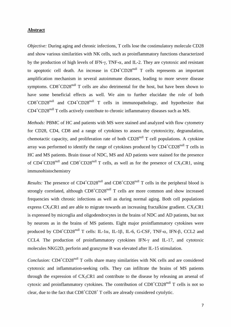
Abstract
Objective: During aging and chronic infections, T cells lose the costimulatory molecule CD28
and show various similarities with NK cells, such as proinflammatory functions characterized
by the production of high levels of IFN-, TNF-, and IL-2. They are cytotoxic and resistant
to apoptotic cell death. An increase in CD4+CD28
null T cells represents an important
amplification mechanism in several autoimmune diseases, leading to more severe disease
symptoms. CD8+CD28
null T cells are also detrimental for the host, but have been shown to
have some beneficial effects as well. We aim to further elucidate the role of both
CD8+CD28
null and CD4
+CD28
null T cells in immunopathology, and hypothesize that
CD4+CD28
null T cells actively contribute to chronic inflammatory diseases such as MS.
Methods: PBMC of HC and patients with MS were stained and analyzed with flow cytometry
for CD28, CD4, CD8 and a range of cytokines to assess the cytotoxicity, degranulation,
chemotactic capacity, and proliferation rate of both CD28null
T cell populations. A cytokine
array was performed to identify the range of cytokines produced by CD4+CD28
null T cells in
HC and MS patients. Brain tissue of NDC, MS and AD patients were stained for the presence
of CD4+CD28
null and CD8
+CD28
null T cells, as well as for the presence of CX3CR1, using
immunohistochemistry
Results: The presence of CD4+CD28
null and CD8
+CD28
null T cells in the peripheral blood is
strongly correlated, although CD8+CD28
null T cells are more common and show increased
frequencies with chronic infections as well as during normal aging. Both cell populations
express CX3CR1 and are able to migrate towards an increasing fractalkine gradient. CX3CR1
is expressed by microglia and oligodendrocytes in the brains of NDC and AD patients, but not
by neurons as in the brains of MS patients. Eight major proinflammatory cytokines were
produced by CD4+CD28
null T cells: IL-1, IL-1, IL-6, G-CSF, TNF-, IFN-, CCL2 and
CCL4. The production of proinflammatory cytokines IFN- and IL-17, and cytotoxic
molecules NKG2D, perforin and granzyme B was elevated after IL-15 stimulation.
Conclusion: CD4+CD28
null T cells share many similarities with NK cells and are considered
cytotoxic and inflammation-seeking cells. They can infiltrate the brains of MS patients
through the expression of CX3CR1 and contribute to the disease by releasing an arsenal of
cytoxic and proinflammatory cytokines. The contribution of CD8+CD28
null T cells is not so
clear, due to the fact that CD8+CD28
+ T cells are already considered cytolytic.

8
9
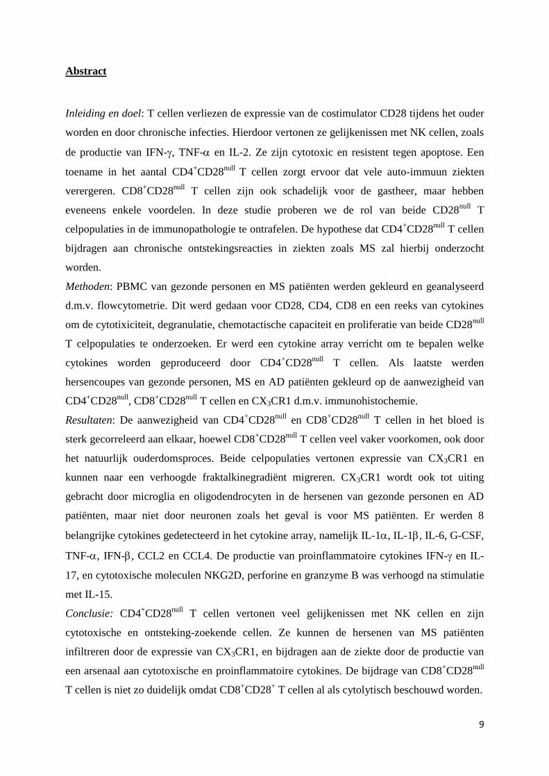
Abstract
Inleiding en doel: T cellen verliezen de expressie van de costimulator CD28 tijdens het ouder
worden en door chronische infecties. Hierdoor vertonen ze gelijkenissen met NK cellen, zoals
de productie van IFN-, TNF- en IL-2. Ze zijn cytotoxic en resistent tegen apoptose. Een
toename in het aantal CD4+CD28
null T cellen zorgt ervoor dat vele auto-immuun ziekten
verergeren. CD8+CD28
null T cellen zijn ook schadelijk voor de gastheer, maar hebben
eveneens enkele voordelen. In deze studie proberen we de rol van beide CD28null
T
celpopulaties in de immunopathologie te ontrafelen. De hypothese dat CD4+CD28
null T cellen
bijdragen aan chronische ontstekingsreacties in ziekten zoals MS zal hierbij onderzocht
worden.
Methoden: PBMC van gezonde personen en MS patiënten werden gekleurd en geanalyseerd
d.m.v. flowcytometrie. Dit werd gedaan voor CD28, CD4, CD8 en een reeks van cytokines
om de cytotixiciteit, degranulatie, chemotactische capaciteit en proliferatie van beide CD28null
T celpopulaties te onderzoeken. Er werd een cytokine array verricht om te bepalen welke
cytokines worden geproduceerd door CD4+CD28
null T cellen. Als laatste werden
hersencoupes van gezonde personen, MS en AD patiënten gekleurd op de aanwezigheid van
CD4+CD28
null, CD8
+CD28
null T cellen en CX3CR1 d.m.v. immunohistochemie.
Resultaten: De aanwezigheid van CD4+CD28
null en CD8
+CD28
null T cellen in het bloed is
sterk gecorreleerd aan elkaar, hoewel CD8+CD28
null T cellen veel vaker voorkomen, ook door
het natuurlijk ouderdomsproces. Beide celpopulaties vertonen expressie van CX3CR1 en
kunnen naar een verhoogde fraktalkinegradiënt migreren. CX3CR1 wordt ook tot uiting
gebracht door microglia en oligodendrocyten in de hersenen van gezonde personen en AD
patiënten, maar niet door neuronen zoals het geval is voor MS patiënten. Er werden 8
belangrijke cytokines gedetecteerd in het cytokine array, namelijk IL-1, IL-1, IL-6, G-CSF,
TNF-, IFN-, CCL2 en CCL4. De productie van proinflammatoire cytokines IFN- en IL-
17, en cytotoxische moleculen NKG2D, perforine en granzyme B was verhoogd na stimulatie
met IL-15.
Conclusie: CD4+CD28
null T cellen vertonen veel gelijkenissen met NK cellen en zijn
cytotoxische en ontsteking-zoekende cellen. Ze kunnen de hersenen van MS patiënten
infiltreren door de expressie van CX3CR1, en bijdragen aan de ziekte door de productie van
een arsenaal aan cytotoxische en proinflammatoire cytokines. De bijdrage van CD8+CD28
null
T cellen is niet zo duidelijk omdat CD8+CD28
+ T cellen al als cytolytisch beschouwd worden.

10
11
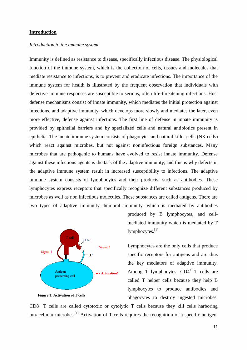
Figure 1: Activation of T cells
(www.pei.de)
Introduction
Introduction to the immune system
Immunity is defined as resistance to disease, specifically infectious disease. The physiological
function of the immune system, which is the collection of cells, tissues and molecules that
mediate resistance to infections, is to prevent and eradicate infections. The importance of the
immune system for health is illustrated by the frequent observation that individuals with
defective immune responses are susceptible to serious, often life-threatening infections. Host
defense mechanisms consist of innate immunity, which mediates the initial protection against
infections, and adaptive immunity, which develops more slowly and mediates the later, even
more effective, defense against infections. The first line of defense in innate immunity is
provided by epithelial barriers and by specialized cells and natural antibiotics present in
epithelia. The innate immune system consists of phagocytes and natural killer cells (NK cells)
which react against microbes, but not against noninfectious foreign substances. Many
microbes that are pathogenic to humans have evolved to resist innate immunity. Defense
against these infectious agents is the task of the adaptive immunity, and this is why defects in
the adaptive immune system result in increased susceptibility to infections. The adaptive
immune system consists of lymphocytes and their products, such as antibodies. These
lymphocytes express receptors that specifically recognize different substances produced by
microbes as well as non infectious molecules. These substances are called antigens. There are
two types of adaptive immunity, humoral immunity, which is mediated by antibodies
produced by B lymphocytes, and cell-
mediated immunity which is mediated by T
lymphocytes.[1]
Lymphocytes are the only cells that produce
specific receptors for antigens and are thus
the key mediators of adaptive immunity.
Among T lymphocytes, CD4+ T cells are
called T helper cells because they help B
lymphocytes to produce antibodies and
phagocytes to destroy ingested microbes.
CD8+ T cells are called cytotoxic or cytolytic T cells because they kill cells harboring
intracellular microbes.[1]
Activation of T cells requires the recognition of a specific antigen,
12
on an antigen-presenting cell, by the T cell receptor (TCR).[2]
Triggering of the TCR by the
appropriate major histocompatibiliy-antigen complex (MHC) is insufficient to induce T cell
proliferation unless it is accompanied by costimulatory signals derived from accessory cells.
(Figure 1) The costimulatory signal determines the activation threshold and the functional
outcome of the antigen-specific activation. Dysregulation of this activation threshold may
play a critical role in the activation of autoreactive T cells.
Immunosenescence: Aging of the immune system
The efficiency of the immune system of the elderly declines with age, resulting in an
increased susceptibility to infectious diseases and pathological conditions relating to
inflammation or autoreactivity. This overall change in immunity is termed
'immunosenescence'. The individual factors contributing to immunosenescence are diverse,
due to the multi-factorial complexity of the immune system. The interplay between B cells
and T cells is crucial for an effective response, so if one subset is affected, this will change the
function of the other. Immunosenescence is a descriptive term covering the deleterious age-
associated changes to immunity observed in all mammal studies so far. Immunity evolved to
protect against infectious diseases, and appropriate immunity is essential for 'normal'
longevity. While all components of innate and adaptive immunity change with age, the
clinical impact of these changes is not clear, and mechanisms and markers for
immunosenescence are controversial.[3]
For example, Cytomegalovirus (CMV) is a -
herpesvirus that continuously activates the immune system and is present in approximately
50% of the adult population and 90% of the elderly. In healthy hosts, CMV infection is
asymptomatic. Maintaining protective immunity against CMV is essential, but may have a
significant impact on the overall adaptive immunity due to the repeated stimulation of T
cells.[4,5]
There is epidemiological evidence for excess mortality in CMV-positive populations,
which is further increased in those co-infected with hepatitis A en B as well.[3]
The marked
influence of CMV on immune signatures is illustrated in the finding that cross-sectional
studies on several different European populations clearly indicate that the consensus view of
T cell immunosenescence is indeed true. This entails that the fraction of naive CD8 cells
decreases in the elderly and the fraction of late-differentiated memory cells increases, but only
for people who are infected with CMV. Such individuals also have higher levels of C-reactive
protein, indicating that they are more likely to suffer from inflammatory diseases, as well as
general frailty and increased mortality.[3]
13
Infection with other persistent herpes viruses, at least Epstein-Barr virus (EBV), Herpes
simplex virus (HSV) and vesicular stomatitis virus (VSV), does not appear to have any
similar effect. CMV is unique in the context that there is some advantage in early life to being
CMV-positive, due to the enhanced pro-inflammatory status in infected people, which might
have a protective effect against infection with other pathogens. Although immunosenescence
is not caused by CMV, because not all elderly people are CMV-positive, this infectious agent
seems to have a large impact on immune parameters in later life and contributes to increased
morbidity and eventual mortality.[3]
Immunosenescence: CD28null
T cells
The CD28 molecule is currently considered the primary costimulatory receptor functional in
transducing signals that augment T cell proliferation and lymphokine production.[6]
CD28 is
expressed on CD4+
and CD8+ T cells, and receives signals from its ligands CD80/CD86.
[7,8]
This signal is important in T cell activation, proliferation and survival, together with
interleukin-2 (IL-2) production. During aging and chronic infections, T cells lose this receptor
and become CD28null
T cells.[9,10]
The loss of CD28 expression in T cells has been attributed
to repeated antigenic stimulation which can also be observed in CD28+ T cells after repeated
antigen stimulation in vitro.[11,12]
This is particularly relevant for CD8+ T cells, which show
increased frequencies of CD28null
T cells in patients with chronic infections as well as during
normal aging. In contrast, the emergence of CD28null
T cells with the CD4+ compartment
appears to be the exception rather than the rule.[13]
The presence of CD4+CD28
null T cells has
been shown in patients with diseases such as multiple sclerosis (MS), reumathoid arthritis
(RA), Wegener’s granulomatosis, unstable angina, dermatomyositis and polymyositis.[14,15,16]
CD28null
T cells show specificity for human cytomegalovirus (HCMV) antigens, and their
presence was clearly associated with HCMV seropositivity.[17] These cells are also involved in
mechanisms increasing the risk of acute ischemic stroke.[18]
Lack of CD28 expression on T
cells has also been reported to increase with age, particularly within the CD8 subset.[19,20]
T cells that lose the expression of CD28 undergo in vivo clonal expansion[21]
and gain several
properties that are
similar to those of NK cells, such as pro-inflammatory functions
characterized by the production of high levels of interferon- (IFN-) and tumor necrosis
factor- (TNF-). (Tabel 1) These T cells are cytotoxic and resistant to apoptotic cell death.
The primary form of apoptosis of clonally expanded T cells is activation-induced cell death
(AICD), which is mainly controlled by the Fas (CD95) system. In the absence of appropriate
14
costimulation, TCR signaling induces Fas and Fas ligand expression. Ligation of Fas initiates
the recruitment of Fas-associated death domain protein and caspase-8, which then triggers the
proteolytic caspase cascade, resulting in the cleavage of various proteins and finally apoptotic
cell death. Several mechanisms exist to counterregulate death processes either at the receptor,
mitochondrial, or caspase level. The longevity of CD28null
T cells is not due to defective Fas
ligand expression, but is attributed to resistance against apoptosis which correlates with
enhanced expression of the anti-apoptotic molecule Bcl-2.[22,23]
The gain of cytolytic function
by CD4+CD28
null T cells is supported by the elevated production of the key cytolytic
molecules including perforin, granzyme B and granzyme A.[10,24]
They also express the
chemokine receptor CCR5, and upregulate CD161, a molecule that facilitates tissue invasion.
Expression of activating killer immunoglobin-like receptors (KIR) on CD4+CD28
null T cells
has been associated with severe disease manifestations in RA patients.[15,25,26]
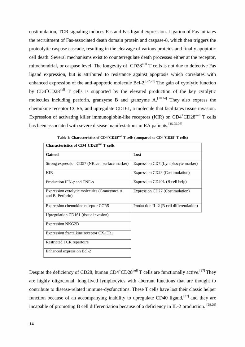
Table 1: Characteristics of CD4+CD28null T cells (compared to CD4+CD28+ T cells)
Despite the deficiency of CD28, human CD4+CD28
null T cells are functionally active.
[27] They
are highly oligoclonal, long-lived lymphocytes with aberrant functions that are thought to
contribute to disease-related immune-dysfunctions. These T cells have lost their classic helper
function because of an accompanying inability to upregulate CD40 ligand,[27]
and they are
incapable of promoting B cell differentiation because of a deficiency in IL-2 production. [28,29]
Characteristics of CD4+CD28
null T cells
Gained Lost
Strong expression CD57 (NK cell surface marker) Expression CD7 (Lymphocyte marker)
KIR Expression CD28 (Costimulation)
Production IFN- and TNF- Expression CD40L (B cell help)
Expression cytolytic molecules (Granzymes A
and B, Perforin)
Expression CD27 (Costimulation)
Expression chemokine receptor CCR5 Production IL-2 (B cell differentiation)
Upregulation CD161 (tissue invasion)
Expression NKG2D
Expression fractalkine receptor CX3CR1
Restricted TCR repertoire
Enhanced expression Bcl-2
15
Numerous receptors that control the function of CD4+CD28
null T cells are primarily expressed
on NK cells. These receptors include the KIR, NKG2D, the fractalkine receptor CX3CR1, and
Ig-like transcripts. NKG2D is an activating receptor that is expressed on most NK cells and
CD8+ T cells, but not on CD4
+ T cells.
[30] When expressed by CD4
+CD28
null T cells, NKG2D
can provide a costimulatory signal as identified in patients with RA, as well as in patients with
Wegener’s granulomatosis.[9]
The expression of NKG2D on these T cells is most likely as a
result of their exposure to TNF- and IL-15, which is structurally similar to IL-2 and is
secreted by mononuclear phagocytes following infection by viruses.[30]
The fractalkine
receptor CX3CR1 is a highly relevant marker to identify CD4+CD28
null T cells because they
strongly express this receptor while it is almost absent on CD4+CD28
+ T cells. This is less
clear in CD8+ T cells, where the expression of CX3CR1 is elevated on CD8
+CD28
null T cells,
but is also present on CD8+CD28
+ T cells. Loss of CD28 expression is also accompanied with
profound changes in T cell functions within the CD8 compartment, such as a lack of antibody
production following immunization in old age, an inability to mount a robust proliferative
response to stimulation, defects in cytokine production including IL-2, IL-4, IL-10 and IFN-
following stimulation, and a loss of antigen repertoire diversity.[31]
Accumulating evidence
shows that NK cell receptors are also expressed in CD8+CD28
null T cells. Both CD8
+CD28
null
and CD4+CD28
null T cells have shorter telomeres than their CD28
+ counterparts, indicative for
their repeated clonal expansions.[2,32]
In general, CD28null
T cells are an inflammation-seeking
effector-memory T cell population with cytotoxic properties, and have features that are
hallmarks of an aged immune system.[2]
CD4+CD28
null T cells and autoimmunity
CD4+CD28
null T cells do not depend on the CD28 pathway for activation, they are incapable
of activating B cells, have significant cytolytic activity, and express high levels of IFN-.
Thus, the presence of significant numbers of CD4+CD28
null T cells could shift immune
response from B cell activation and production of immunoglobulins toward production of
IFN- and involvement of macrophages releasing matrix-degrading proteases.[33]
These
deviating characteristics of CD4+CD28
null T cells make it likely that they are not just a marker
of an aging immune system, but contribute actively or passively to autoimmune disease
pathology. The functionality of the immune system is highly dependent on the diversity of the
naive T cell pool. If the T cell pool is composed for a considerable part of CD4+CD28
null T
cells, this could have serious implications for immune function. The accumulation of
16
CD4+CD28
null T cells also limits the peripheral space which is otherwise available for
functional naive T cells, competitor T cells or regulatory T cells. This alteration in T cell
composition may cause an imbalanced immune system with a changed threshold for T cell
activation.[19]
An increase in CD4+CD28
null T cells represents an important amplification
mechanism in MS and other immune diseases, leading to more severe disease symptoms
rather than being involved in the initial break of tolerance. These cells are readily triggered by
inflammation and subsequently perpetuate and re-enforce the inflammatory process. The
presence of these T cells has been shown in several autoimmune diseases, such as MS and
RA, were they actively contribute to the inflammation when present. Fractalkine is
upregulated in the cerebrospinal fluid of clinically isolated syndrome patients, and in the brain
of MS patients which enables the CD4+CD28
null T cells to migrate to the sites of
inflammation, through the expression of CX3CR1 on their surface. [34]
Chemokine receptors
and effector molecules such as perforin and granzyme B uniquely expressed on CD4+CD28
null
T cells may enable these cells to infiltrate tissue and to cause tissue damage. The expression
of stimulatory receptors lowers the activation threshold for antigen-specific stimulation.[9]
Mechanisms that restore CD28 expression and reduce the pool of CD4+CD28
null T cells are
therefore potential therapeutic targets in these auto-immune diseases.
A wide variety of inhibitory T cell populations exist within the CD8+ T cell compartment.
Means of induction (innate/adaptive), phenotypes (CD28+/CD28
null), and suppressive
mechanisms are highly diverse. A barrier in investigating CD8+ T cells is the lack of a
specific cell surface marker. Lack of CD28 cannot be used as a marker because the
CD8+CD28
null T cell population contain both cytotoxic and suppressor cells. A series of CD4
+
and CD8+ T cells have been described which can disrupt immune responses, including the
pathogenic immune responses causing autoimmune diseases, but it is currently unknown
whether these two classes of T cells have similar or distinct roles in regulating immune
responses. Recent evidence shows that they are functionally connected, but their roles in
immune diseases need further elucidation.[35]
CD8+CD28
null T cells
CD8+ T cells are responsible for inducing apoptosis of intracellular pathogen-infected or
transformed cells. The presence of senescent CD8+ T cells may have a variety of effects on
the immune system. They influence the quality and composition of the memory T cell pool,
and due to their resistance to apoptosis, they accumulate progressively over time. CD28null
T
17
cells are usually part of oligoclonal expansions, which leads to a reduction in the overall
spectrum of antigenic specificities within the T cell pool. A more direct effect of
CD8+CD28
null T cells lies in the area of suppressor cell activity. A population of
CD8+CD28
null T cells functions to suppress immune reactivity by inducing antigen-presenting
cells to become tolerogenic to helper T cells with the cognate antigen specificity. This same
subset of T cells has been implicated in the tolerance to allogeneic organ transplants. This
suggests a possible suppressive function which is beneficial in reducing the reactivity against
the allograft. Other immune suppressive functions might not be so beneficial, such as the
mediation of liver damage in hepatitis C infection, and stimulation of head and neck tumors
by a population of TNF- producing CD8+CD28
null T cells. CD8
+ T cells also seem to be
involved in the pathogenic mechanisms of disease. Failure to generate non-antigen-specific
CD8+ T cells is associated with the development of relapses in patients with MS.
[36] Expanded
populations of CD8+CD28
null T cells have also been reported in ankylosing spondylitis
patients, and seem to correlate with a more severe course of this autoimmune disease. Many
of the effects of CD8+ T cell replicative senescence seem to be deleterious, however, it is
possible that these cells may have some beneficial effects in some contexts such as in organ
transplantation.[37]
CD28null
T cells as a possible target for therapy
The pro-inflammatory cytokine IL-15, is essential for the development and activation of CD8+
T cells and NK cells. IL-15 is known as a regulator of haematopoiesis, cell survival and
proliferation of different cell types, and can be presented in two ways. Via the classic way, IL-
15 is released from one cell and captured by another, but it can also be presented in trans,
when it is still bound to the membrane of the first cell. Monocytes/macrophages have been
reported as the main source of this cytokine.[38]
Increased local expression of IL-15 has been
suggested to contribute to the immunopathology of several human inflammatory diseases
including rheumatoid arthritis. Previous studies have demonstrated that IL-15 is upregulated
on PBMC of MS patients, but the precise contribution of IL-15 to MS immunopathogenesis
has not been elucidated.[39]
A critical cytokine in the regulation of NK cells is interleukin-12
(IL-12), a proinflammatory cytokine highly expressed in an inflammatory environment.
Previous experiments found that IL-12 has an indirect effect on the de novo transcription and
translation of the CD28 gene.[40]
Previous studies also demonstrated that IFN-a cytokine
18
that both activates and induces proliferation in NK cells, decreases the inflammation[41,42]
and
suppresses the proliferation of CD4+CD28
null T cell populations in MS patients.
[8]
In this study, we will investigate the role of both CD4+CD28
null and CD8
+CD28
null T cells by
performing a series of experiments on both cell populations. Through these experiments, we
can gain a better understanding of how these cells work in the immune system, and how they
are involved in the course and development of autoimmune diseases such as MS. By looking
at the direct effects of previously described substances on CD4+CD28
null T cells, we can gain
a better understanding of how these work and can eventually be implemented in therapeutic
treatments. This can result in a more personalized therapy for patients with chronic diseases
such as MS and RA. We will work with a subgroup of healthy subjects and MS patients in
whom a CD4+CD28
null T cell population is present. Understanding the role of these cells in
immunity will also lead to better therapeutic interventions in several autoimmune diseases.
19
Materials & Methods
Study Subjects
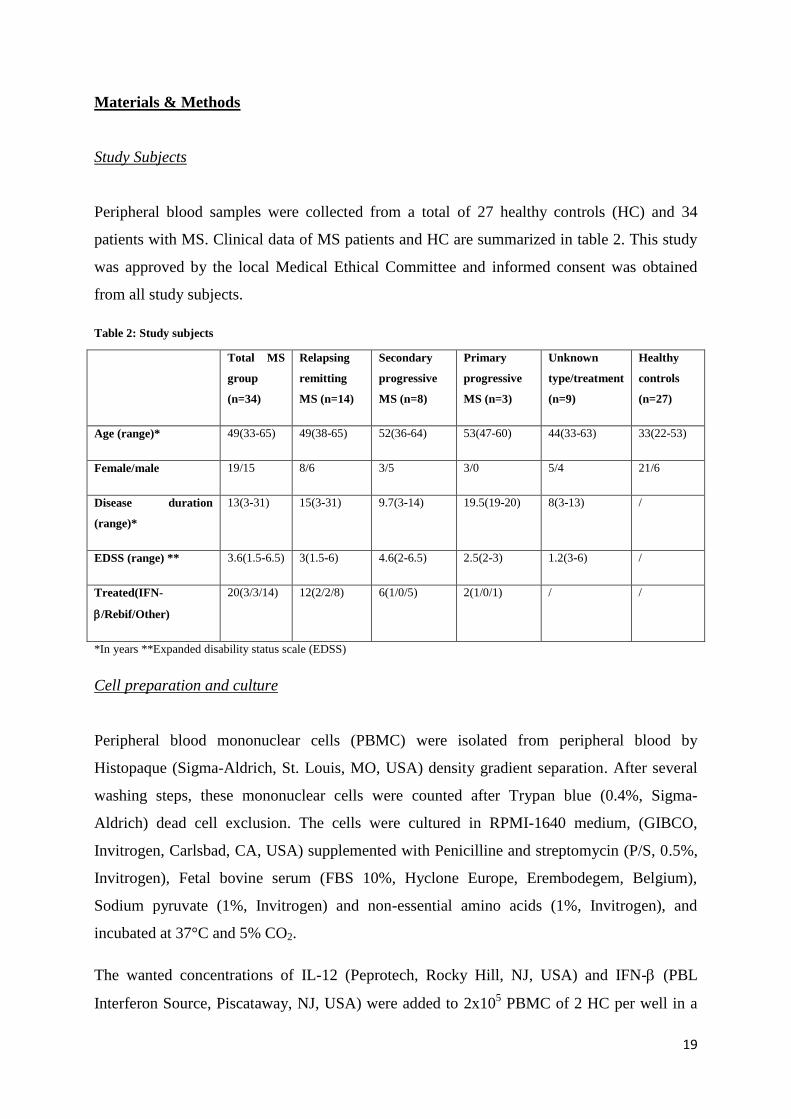
Peripheral blood samples were collected from a total of 27 healthy controls (HC) and 34
patients with MS. Clinical data of MS patients and HC are summarized in table 2. This study
was approved by the local Medical Ethical Committee and informed consent was obtained
from all study subjects.
Table 2: Study subjects
Total MS
group
(n=34)
Relapsing
remitting
MS (n=14)
Secondary
progressive
MS (n=8)
Primary
progressive
MS (n=3)
Unknown
type/treatment
(n=9)
Healthy
controls
(n=27)
Age (range)* 49(33-65) 49(38-65) 52(36-64) 53(47-60) 44(33-63) 33(22-53)
Female/male 19/15 8/6 3/5 3/0 5/4 21/6
Disease duration
(range)*
13(3-31) 15(3-31) 9.7(3-14) 19.5(19-20) 8(3-13) /
EDSS (range) ** 3.6(1.5-6.5) 3(1.5-6) 4.6(2-6.5) 2.5(2-3) 1.2(3-6) /
Treated(IFN-
/Rebif/Other)
20(3/3/14) 12(2/2/8) 6(1/0/5) 2(1/0/1) / /
*In years **Expanded disability status scale (EDSS)
Cell preparation and culture
Peripheral blood mononuclear cells (PBMC) were isolated from peripheral blood by
Histopaque (Sigma-Aldrich, St. Louis, MO, USA) density gradient separation. After several
washing steps, these mononuclear cells were counted after Trypan blue (0.4%, Sigma-
Aldrich) dead cell exclusion. The cells were cultured in RPMI-1640 medium, (GIBCO,
Invitrogen, Carlsbad, CA, USA) supplemented with Penicilline and streptomycin (P/S, 0.5%,
Invitrogen), Fetal bovine serum (FBS 10%, Hyclone Europe, Erembodegem, Belgium),
Sodium pyruvate (1%, Invitrogen) and non-essential amino acids (1%, Invitrogen), and
incubated at 37°C and 5% CO2.
The wanted concentrations of IL-12 (Peprotech, Rocky Hill, NJ, USA) and IFN- (PBL
Interferon Source, Piscataway, NJ, USA) were added to 2x10⁵ PBMC of 2 HC per well in a
20
96 well plate with U-shaped wells (NUNC, Roskilde, Denmark), and expression of CD4,
CD28 and 7-AAD was monitored at different time points by using flow cytometry.
For the IL-15 assay, PBMC of 5 HC were placed in two 96 well plates at a concentration of
1.5x10⁵ cells per well in culture medium. Culture medium with 15 ng/ml IL-15 (R&D
systems, Minneapolis, MN, USA) was added to two-thirds of the cells, of which one third
was supplemented with 5 µg/well anti-IL-15, and one third with 5 µg/well isotype control.
Plain culture medium was added to the remaining cells. Cells were stained at day 0,1,3 and 5
for the NKG2D receptor, CD69, perforin, granzym B, IFN-, IL-17, CD4 and CD28. In two
other plates, the same concentrations were used, but the cells were activated with 25 µl/ml
PMA (Sigma), 10 µl/ml CaI (Sigma) and 1 µl/ml Golgi-stop (BD) before staining.
To assess the production of IL-15 by monocytes, PBMC of 2 HC and 1 MS patient were
placed in a 24 well culture plate (Cellstar, Greiner Bio-One) at a concentration of 1.5x10⁶
cells per well. After two hours of incubation, monocytes were fixed to the bottom and top
cells were removed. These monocytes were subsequently activated with 100 ng/ml LPS
(Calbiochem VWR international, Leuven, Belgium) and 10 ng/ml IFN-(BD) (105 monocytes
per well in a 96 well plate). Expression of IL-15 and IL-15 receptor, together with the
expression of 3 markers for activation: CD80, CD86 and MHCII was monitored before and
after stimulation.
A CFSE assay was performed by adding an equal volume of 4 µM CFSE (Invitrogen) to
PBMC dissolved at 20x106 cells per ml in PBS/0.1% BSA. After an incubation period of 7
minutes at 37°C, cells were dissolved in 300 µl culture medium. After a second incubation
period of 15 minutes at 37°C, cells were dissolved in culture medium at 2x10⁶ cells per ml
and seeded in a 96 well plate at a concentration of 2x10⁵ cells per well. 100 µl of culture
medium containing following substances was added to every well: plain culture medium,
culture medium supplemented with 4 µg/ml aCD3, culture medium supplemented with 4
U/ml IL-2, culture medium supplemented with 100 ng/ml IL-15, culture medium
supplemented with 4 µg/ml aCD3 and 4 U/ml IL-2 or culture medium supplemented with 4
µg/ml aCD3 and 100 ng/ml IL-15. After 5 days of incubation at 37°C, cells were stained for
CD4-PERC and CD28-PE. This was done for 5 HC.
For some experiments, cell lines were made. Here, PBMC were labeled with CD4-FITC and
CD28-PE (both BD Biosciences, Franklin Lakes, NJ, USA) and FACS sorted with the
FACSAriaII high speed cell sorter (BD Biosciences) in a 96 well plate into CD4+CD28
null and
21
CD4+CD28
+ T cells (5 cells per well). 1x10
5 irradiated feeder cells were added to the sorted
cells. Cells were stimulates using Phytohemaglutinin (5 µg/ml, PHA, Sigma-Aldrich) and IL-
2 (5 U/ml, Roche) Medium was refreshed twice a week with culture medium containing 5
U/ml IL-2. When pellets became too big, they were split up into 2 new wells on a new 96 well
plate.
For the chemotaxis assay, an enrichment for CD8+ T cells was performed on PBMC of 6 HC
by following the protocol enclosed in the EasySep Human CD8 positive selection kit
(StemCell Technologies, Grenoble, France).
ELISA
For optimization of the cytokine array conditions, supernatant from 1 HC was used in an
enzyme-linked immunosorbent assay (ELISA) for IFN- production, following the protocol
enclosed in the human IFN gamma ELISA Ready-SET-Go! Kit (eBioscience Inc, San Diego,
CA, USA). The ELISA plate was coated with capture antibody in coating buffer and washed
with wash buffer (1xPBS/0.05% Tween-20), which was repeated after blocking with Assay
diluent. The supernatant of the sample was added to the wells (100 µl/well), and a standard
was made by serial dilution of a 500 pg/ml standard solution in assay diluent. After an
incubation period of 2 hours, detection antibody in assay diluent was used for detection, and
avidin-horseradish peroxidase (HRP) in assay diluent for conjugation. Finally, a
Tetramethylbenzidine (TMB) solution caused a color reaction and the plate was read at 450
nm on the BIO-RAD Benchmark Microplate Reader (Bio-Rad Laboratories N.V., Nazareth,
Ghent)
Cytokine Array
Enriched CD4+ T cells from 4 HC and 4 MS patients
were obtained using the EasySep Human CD4 positive
selection kit (StemCell Technologies, Grenoble, France)
following the manufacturer’s protocol. Briefly, PBMC
were dissolved in EasySep buffer (2% FBS/1mM EDTA,
Invitrogen, in phosphate buffered saline (PBS,
BioWittaker, Verviers, Belgium)) and labeled for CD4+ T cells with 100 µl/ml EasySep
Human CD4 Positive Selection Ab cocktail. After incubation, 50 µl/ml EasySep Magnetic
Figure 2: Schematic Drawing of EasySep®
TAC Magnetic Labeling of Human Cells
(Stemcell Technologies)
22
Nanoparticles were added, which attached to the labeled cells, making them magnetic. (figure
2) By Using the EasySep magnet, the labeled CD4+ T cells were pulled towards the sides of
the tube, while the other cells could be poured out. (figure 3) The CD4+ T cells were
subsequently labeled with CD28-PE (BD Biosciences) and a CD4 antibody clone that binds to
a different CD4 epitope on CD4+ T cells, CD4v4-FITC. (StemCell Technologies) In a 96 well
plate, 1x10⁵ sorted cells were seeded, together with 1x10
5 irradiated feeder cells per well. Half
of the wells contained feeder cells pulsed with Myelin basic protein (MBP, Biomed,
Diepenbeek, Belgium, made according to Deibler et al. 1972[42]
). The other half contained
unpulsed feeder cells and 4 µg/ml anti-CD3 (Clone 2G3,
Biomed) was added to both conditions. Supernatant was
removed after 5 days incubation for the cytokine array and
cytrometric bead assay, and stored at -20°C.
The cytokine array was performed following the protocol
enclosed in the Human Cytokine Array kit-Panel A (R&D
Systems, Minneapolis, MN, USA). Briefly, the cytokine
membranes were blocked for one hour in array buffer 4 while
samples of CD4+CD28
null T cells were incubated with
Cytokine Array Panel A Detection Antibody Cocktail for one
hour. The samples were subsequently added to the membranes
and incubated overnight. After washing, Streptavidin-HRP
was added to the membranes. After an incubation period of 30 minutes, membranes were
washed and exposed to chemiluminescent reagents with the ECL Plus Western Blotting
Detection System (GE Healthcare). The membranes were subsequentlyexposed to an X-ray
film in an X-ray film cassette for 30 seconds up to 10 minutes. Finally, the films and dots
were semi-quantitavely analyzed using ImageJ and GraphPad Prism 4 software.
Results from the cytokine array were validated using a cytometric bead assay, according to
the protocol enclosed in the Human Soluble Protein Master Buffer Kit (BD). This allows to
calculate the percentage of a given cytokine produced by the cells. Flex set standards were
prepared by pooling standard spheres of all cytokines of interest in one tube, and lyophilizing
them with assay diluent. A series dilution was made by transferring 500 µl of the standard
into tubes containing 500 µl assay diluent. Solutions of capture beads with capture bead
diluent and detection reagents with detection reagent diluent were made according to
calculations given in the protocol. Subsequently, 50 µl of the mixed capture beads were added
Figure 3: Schematic representation
of the EasySep procedure.
(StemCell Technologies)
23
to every tube, with 50 µl standard or sample. After a one hour incubation period, 50 µl of the
detection reagent mix was added to every assay tube. 1 ml of wash buffer was added after two
hours of incubation, and after centrifugation, the supernatant was removed. Pellets were
resuspended in wash buffer and samples were analyzed on the FACSAriaII high speed cell
sorter.
Chemotaxis assay
CD8+ T cells were isolated from PBMC of 6 HC with the EasySep procedure. A transwell
system with a pore size of 5 µm (Corning, Lowell, MA, USA) was used for the migration
assay. Fractalkine (Peprotech, Rocky Hill, NJ, USA) was added in the bottom compartment to
chemotaxis buffer (0.5% BSA (US Biological, Swamscott, MA, USA) /RPMI-1640) in
concentrations of 0; 0.1; 0.5; 1 and 10 ng/ml. In each insert, 5x10⁵
CD8+ T cells were added.
The total number of cells which had migrated to the bottom compartment was counted after 4
hours of incubation at 37°C and 5% CO2. The percentages of CD8+CD28
+ and CD8
+CD28
null
T cells were then determined by using flow cytometry on the FACSCalibur. The chemotactic
index of both cell populations was calculated by dividing the number of migrated cells with
fractalkine by the number of migrated cells without fractalkine.
Flow cytometry
For monitoring the expression of CD4, CD28 and 7-AAD, cells were dissolved in FACS
buffer (2% FBS/0.1% Sodium Azide in PBS) and labeled in a 96 well plate with V-shaped
wells (Greiner BIO-One GmbH, Frickenhausen, Germany) with CD4-FITC, CD28-PE and 7-
AAD (all BD Biosciences). After an incubating period in the dark for 15 minutes, they were
scanned with the FACSCalibur flow cytometer (BD Biosciences). Subsequent analysis was
done by using the BD CellQuest pro software. For monitoring the expression of CD4 or CD8
together with CD28 and CX3CR1, CD4-PerCP or CD8-PerCP was combined with CD28-
FITC (All BD) and CX3CR1-PE (MBL International, Woburn, MA, USA). To assess the
production of IL-15 on monocytes upon stimulation, cells were stained with HLA-
DR,DP,DQ-FITC, CD14-PerCP, CD80-PE, CD86-FITC (all BD), IL-15-PE and IL-
15receptor-PE (both R&D systems) before and after activation. For the IL-15 assay, cells
were stained with combinations of CD314-PE, CD69-PE, Perforin-PE, GranzymeB-FITC, IL-
17A-FITC, CD28-FITC, CD4-PerCP, CD28-APC, 2acontrol (all BD) and IFN--PE
(Immunotools, Friesoythe, Germany). For the degranulation assay, cells were stained with
24
combinations of CD107a-PerCP, CD4-PerCP, CD28-FITC, CD8-PerCP, CD28-PE, CD4-
FITC, CD8-FITC and CD69-PE. (all BD) To assess the presence of the IL-15 receptor, cells
were stained with IL15R-PE (R&D systems), CD4-PerCP and CD28-FITC. (All BD)
Immunohistochemistry
Frozen brain material from 6 MS patients, 1 Alzheimers Disease patient and 1 non-demented
control (NDC) was cut with a Leica CM1900UV cryostat (Leica Microsystems, Wetzlar,
Germany) into 10 µm sections. Slides were fixed in acetone for 10 minutes and dried. After
washing in Tris-buffered saline/0.03% Triton X-100 (TBS-T, Sigma-Aldrich), they were
blocked for 20 minutes with Protein Block Dakocytomation (Dako, Glostrup, Denmark) to
prevent aspecific binding.
To identify CD8+CX3CR1
+ T cells in these tissues, slides were incubated overnight with a rat
anti-human CX3CR1 antibody (MBL International, dilution factors and incubation periods are
presented in table 3) and washed again. Slides were subsequently incubated with an Alexa-
555 labeled goat anti-rat IgG antibody (Invitrogen). After washing, slides were incubated
overnight with a mouse anti-human CD8 antibody (AbCam, Cambridge, UK), followed by a
donkey anti-mouse Alexa-488 antibody (Invitrogen).
To assess the presence of CD4+CD28
null T cells in the brains of healthy people or patients
with Alzheimer’s disease (AD), slides were stained with a rat anti-human CX3CR1 antibody,
an Alexa-555 labeled goat anti-rat IgG antibody and subsequently with an Alexa-488 labeled
mouse anti CD4 antibody (Santa Cruz Biotechnology, Santa Cruz, CA, USA).
Brain tissue of patients with AD and NDC patients were stained with CX3CR1 in combination
with a mouse anti GFAP antibody (Sigma-Aldrich), a mouse anti APC (CC-1) antibody
(AbCam), a mouse anti NeuN antibody (Millipore, Billerica, MA, USA) or a rabit anti Iba-1
antibody (Wako Chemicals, Osaka, Japan) to determine the presence of fractalkine on
astrocytes, oligodendrocytes, neurons and microglia, respectively, in the brains of these
patients.
25
Table 3: Dilution factors and incubation periods of antibodies used for staining.
Primary Secundary Streptavidin
Rat anti-human CX3CR1:
1/500 ON
Goat anti-rat Alexa 555: 1/400 1.5h /
Mouse anti-human CD8:
1/200 ON
Donkey anti-mouse Alexa 488: 1/200 1.5h /
CD4-Alexa 488: 1/200 1.5h / /
GFAP: 1/500 ON Rabit anti-mouse biotin: 1/400 1h (Dako) Strep Alexa 488: 1/2000 1.5h
NeuN: 1/100 ON Rabit anti-mouse biotin: 1/400 1h Strep Alexa 488: 1/2000 1.5h
CC-1: 1/500 ON Rabit anti-mouse biotin: 1/400 1h Strep Alexa 488: 1/2000 1.5h
Iba-1: 1/300 (4°C) ON Swine anti-rabit biotin: 1/400 1h (Dako) Strep Alexa 488: 1/2000 1.5h
Control staining was performed by using only the secondary antibodies. After incubation,
slides were washed and a nuclear staining was performed using 4,6’-diamino-2-phenylindole
(DAPI, Molecular Probes, Invitrogen) for 10 minutes. After blocking for 10 minutes with
0.1% Sudan Black in 70% ethanol, slides were dipwashed in 70% ethanol and TBS. A final
washing step was performed with TBS and water and cover slides were subsequently applied
with fluorescent mounting medium (Dako). After drying, slides were analyzed on a Nikon
eclipse 80i microscope using NIS Elements BR 3.10 software (Both Nikon, Tokyo, Japan).
Degranulation assay
To assess the capacity of CD4+CD28
null and CD8
+CD28null T cells to expel cytotoxic
granules in response to stimulation with CMV, a degranulation assay was set up. PBMC of 3
HC were seeded in a 96 well plate at a concentration of 2x10⁵ cells per well. . Cells were
either stimulated with 500 ng/ml CMV pp65 (Peptivator, Miltenyi Biotec), or left
unstimulated. 2 µl anti-CD107a-PerCP and 1 µl Golgi-Plug (diluted 1/5 in culture medium,
BD) were added during the last four hours of culture before analyzing samples on a
FACSCalibur (BD). Cells were analyzed after 4 and 24 hours of stimulation.
Statistical analysis
Statistical analyses were performed using GraphPad Prism version 4.03. Two-tailed unpaired
Student’s T test (with Welch’s correction if necessary) was performed to compare two groups.
A P value of less than 0.05 was considered significant. A P value between 0.1 and 0.05 was
considered a trend.

26
27
Results
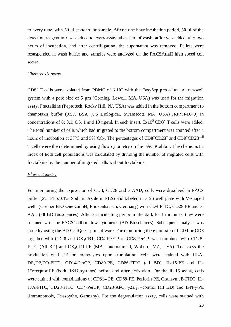
1. Frequency of CD4+CD28
null T cells and making of Cell lines
The frequency of CD4+CD28
null T cells in
peripheral blood of 34 MS patients and 20 HC
was determined using flow cytometry, and
ranged between 0 and 2.5%. (figure 4)
Because CD4+CD28
null T cells are only present
in the blood in small percentages, we
attempted to make cell lines to reach a greater
amount of cells to work with. Cells did not
grow as expected, and after several failed
attempts to make a cell line of CD4+CD28
null T
cells, these experiments were replaced by others. The reason why cells did not grow, is
unclear. According to protocol, cells should grow after two weeks while replacing the
medium twice a week. After over four weeks of incubation, cells still did not multiply
enough, and started to die. After three attempts, this experiment was abandoned and replaced
by alternative experiments on CD28null
T cells.
2. Cytokine production by CD4+CD28
null T cells
CD4+CD28
null T cells are known to produce IFN-
[7], but production of other cytokines and
chemokines has not yet been fully examined. To assess the range of cytokines that are
produced by CD4+CD28
null T cells, a cytokine array was performed. This array allowed the
simultaneous detection of 36 different cytokines, chemokines, and acute phase proteins in a
single sample.
2.1 Optimization of stimulation conditions
To determine the optimal conditions for the cytokine array, an ELISA for the production of
IFN- was performed. IFN- was chosen because it is known to be produced by CD4+CD28
null
T cells [7]
. First, different culture plates and cell concentrations were tested, and the amount of
IFN- in the supernatant of the culture was analyzed after stimulation with anti-CD3. In a 48
well plate, 2x10⁵ CD4
+CD28
null T cells were combined with the same amount of
feeder cells,
Figure 4: The frequency of senescent CD4+ T cells in
the peripheral blood of 20 HC and 34 MS patients was
determined by flow cytometry. There is no significant
difference in frequency between HC and MS patients.
28
and, in a 96 well plate, the same was done for 1x10⁵
CD4+CD28
null T cells and feeder cells.
Compared to the IFN- concentration in the supernatant of the 48 well plate (15.74 pg/ml), the
concentration in the 96 well plate was higher (32.35 pg/ml), but still not optimal for the
cytokine array. To increase the IFN-γ production, IL-2 was added to the culture. This
increased the concentration of IFN- in the supernatant of the 96 well plate to 234.4 pg/ml.
Therefore, this setup was chosen for the cytokine array.
2.2 Cytokine array
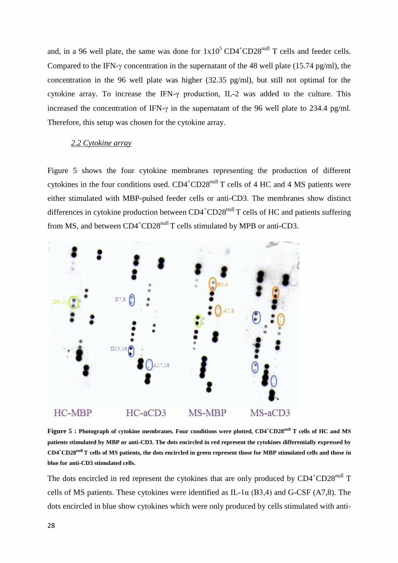
Figure 5 shows the four cytokine membranes representing the production of different
cytokines in the four conditions used. CD4+CD28
null T cells of 4 HC and 4 MS patients were
either stimulated with MBP-pulsed feeder cells or anti-CD3. The membranes show distinct
differences in cytokine production between CD4+CD28
null T cells of HC and patients suffering
from MS, and between CD4+CD28
null T cells stimulated by MPB or anti-CD3.
Figure 5 : Photograph of cytokine membranes. Four conditions were plotted, CD4+CD28null T cells of HC and MS
patients stimulated by MBP or anti-CD3. The dots encircled in red represent the cytokines differentially expressed by
CD4+CD28null T cells of MS patients, the dots encircled in green represent those for MBP stimulated cells and those in
blue for anti-CD3 stimulated cells.
The dots encircled in red represent the cytokines that are only produced by CD4+CD28
null T
cells of MS patients. These cytokines were identified as IL-1α (B3,4) and G-CSF (A7,8). The
dots encircled in blue show cytokines which were only produced by cells stimulated with anti-
29
CD3. CD4+CD28
null T cells produce TNF-CCL4DandIFN-γ (A17,18) after
stimulation with anti-CD3. Dots D9 and D10, encircled in green, represent CCL2, which was
only produced by cells stimulated with MBP. Appendix B shows the full results of the
differentially produced cytokines. Only the cytokines visible on the membranes were plotted.
By comparing the coordinates of the dots with those of the cytokines listed in the
manufactures guide, a cytokine could by placed with every dot on the membrane. Relative
production levels were calculated by comparing the dots of the cytokines with those of the
positive controls.
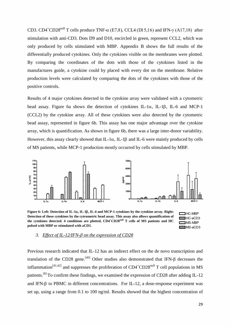
Results of 4 major cytokines detected in the cytokine array were validated with a cytometric
bead assay. Figure 6a shows the detection of cytokines IL-1, IL-1, IL-6 and MCP-1
(CCL2) by the cytokine array. All of these cytokines were also detected by the cytometric
bead assay, represented in figure 6b. This assay has one major advantage over the cytokine
array, which is quantification. As shown in figure 6b, there was a large inter-donor variability.
However, this assay clearly showed that IL-1, IL-1 and IL-6 were mainly produced by cells
of MS patients, while MCP-1 production mostly occurred by cells stimulated by MBP.
Figure 6: Left: Detection of IL-1, IL-1, IL-4 and MCP-1 cytokines by the cytokine array. Right:
Detection of these cytokines by the cytrometric bead assay. This assay also allows quantification of
the cytokines detected. 4 conditions are plotted, CD4+CD28null T cells of MS patients and HC
pulsed with MBP or stimulated with aCD3.
3. Effect of IL-12/IFN- on the expression of CD28
Previous research indicated that IL-12 has an indirect effect on the de novo transcription and
translation of the CD28 gene.[40]
Other studies also demonstrated that IFN-β decreases the
inflammation[41,42]
and suppresses the proliferation of CD4+CD28
null T cell populations in MS
patients.[8]
To confirm these findings, we examined the expression of CD28 after adding IL-12
and IFN- to PBMC in different concentrations. For IL-12, a dose-response experiment was
set up, using a range from 0.1 to 100 ng/ml. Results showed that the highest concentration of
30
IL-12 resulted in a decline in total expression of CD28 within the CD4 population,
represented by the mean fluorescence intensity (MFI). For the lowest concentrations (0.1 and
1 ng/ml), CD28 MFI increased with a peak at day 5. When gated on CD4+CD28
null T cells,
CD28 expression did not change significantly.
For IFN- a similar experiment was conducted, using a concentration range from 1 to 1000
U/ml. The total CD28 MFI within the CD4+ population decreased in all concentrations when
IFN- was added, the MFI of CD4+CD28
null T cells did not change. (Full results, see appendix
A)
4. Effect of IL-15 on the proliferation and phenotype of CD4+CD28
null T cells
Increased expression of IL-15 has been suggested to contribute to the immunopathology of
several inflammatory diseases, although the precise role of IL-15 has not yet been
elucidated.[39]
NK cells express the IL-15 receptor complex, and IL-15 secretion by
macrophages during inflammation results in an increased proliferation, survival, cytolytic
activity and cytokine secretion by these cells[43]
. In vitro studies show that IL-15 may be an
important regulator of IFN- production by NK cells[44]
. CD4+CD28
null T cells show several
similarities with NK cells, but the effects of IL-15 on this cell population have not been
studied so far. It is also not known whether soluble or membrane bound IL-15 have different
effects on the function of CD4+CD28
null T cells.
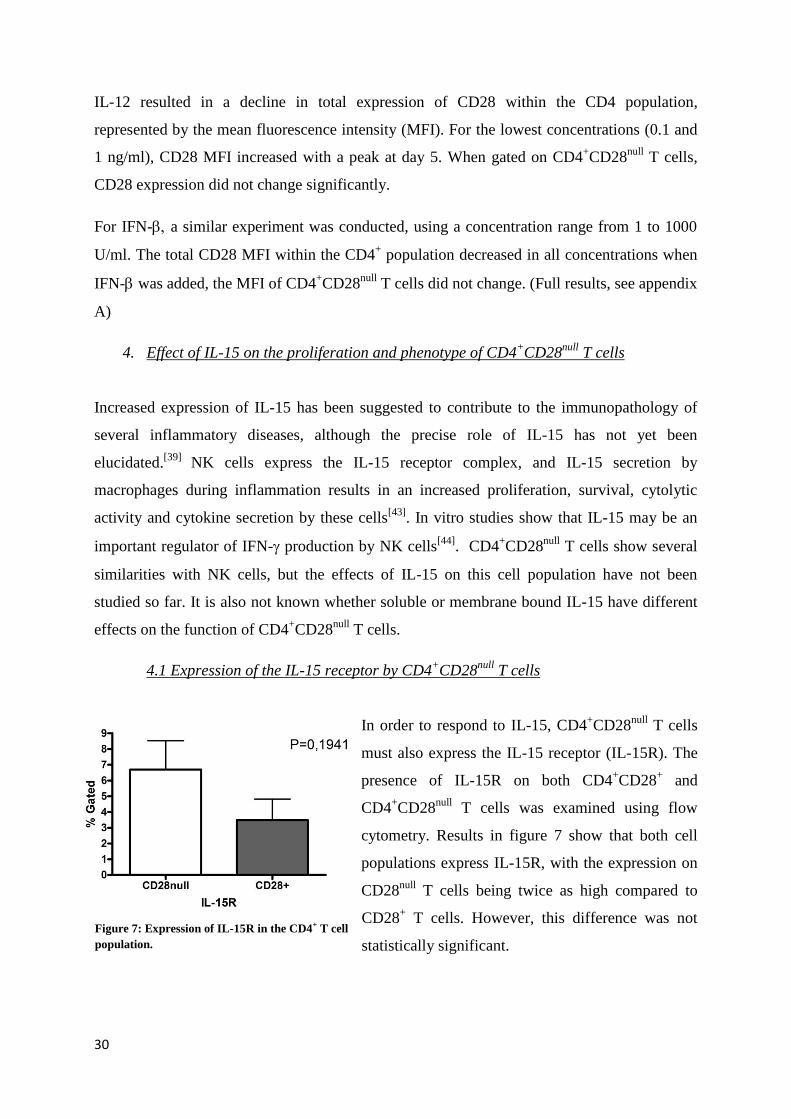
4.1 Expression of the IL-15 receptor by CD4+CD28
null T cells
In order to respond to IL-15, CD4+CD28
null T cells
must also express the IL-15 receptor (IL-15R). The
presence of IL-15R on both CD4+CD28
+ and
CD4+CD28
null T cells was examined using flow
cytometry. Results in figure 7 show that both cell
populations express IL-15R, with the expression on
CD28null
T cells being twice as high compared to
CD28+ T cells. However, this difference was not
statistically significant.
Figure 7: Expression of IL-15R in the CD4+ T cell
population.
31
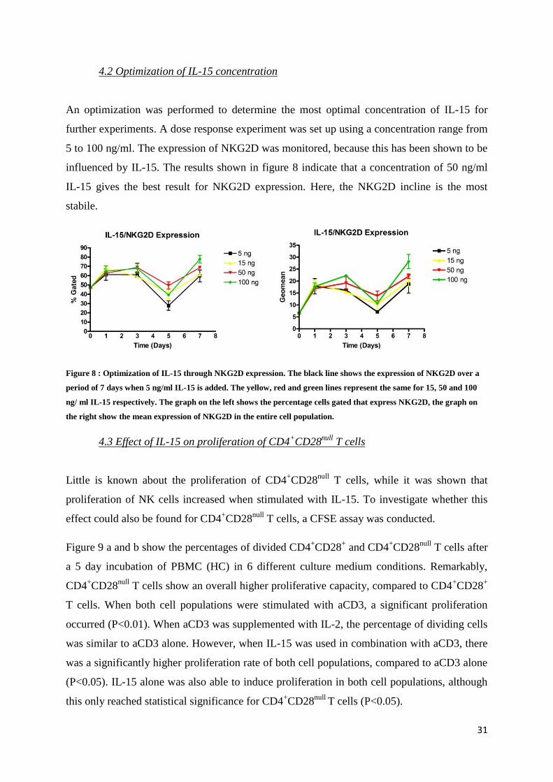
4.2 Optimization of IL-15 concentration
An optimization was performed to determine the most optimal concentration of IL-15 for
further experiments. A dose response experiment was set up using a concentration range from
5 to 100 ng/ml. The expression of NKG2D was monitored, because this has been shown to be
influenced by IL-15. The results shown in figure 8 indicate that a concentration of 50 ng/ml
IL-15 gives the best result for NKG2D expression. Here, the NKG2D incline is the most
stabile.
Figure 8 : Optimization of IL-15 through NKG2D expression. The black line shows the expression of NKG2D over a
period of 7 days when 5 ng/ml IL-15 is added. The yellow, red and green lines represent the same for 15, 50 and 100
ng/ ml IL-15 respectively. The graph on the left shows the percentage cells gated that express NKG2D, the graph on
the right show the mean expression of NKG2D in the entire cell population.
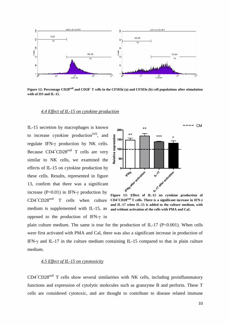
4.3 Effect of IL-15 on proliferation of CD4+CD28
null T cells
Little is known about the proliferation of CD4+CD28
null T cells, while it was shown that
proliferation of NK cells increased when stimulated with IL-15. To investigate whether this
effect could also be found for CD4+CD28
null T cells, a CFSE assay was conducted.
Figure 9 a and b show the percentages of divided CD4+CD28
+ and CD4
+CD28
null T cells after
a 5 day incubation of PBMC (HC) in 6 different culture medium conditions. Remarkably,
CD4+CD28
null T cells show an overall higher proliferative capacity, compared to CD4
+CD28
+
T cells. When both cell populations were stimulated with aCD3, a significant proliferation
occurred (P<0.01). When aCD3 was supplemented with IL-2, the percentage of dividing cells
was similar to aCD3 alone. However, when IL-15 was used in combination with aCD3, there
was a significantly higher proliferation rate of both cell populations, compared to aCD3 alone
(P<0.05). IL-15 alone was also able to induce proliferation in both cell populations, although
this only reached statistical significance for CD4+CD28
null T cells (P<0.05).
32
Figure 9: Percentage of divided CD4+CD28+ T cells (a) and CD4+CD28null T cells (b) after incubation with CFSE in
different culture medium conditions: plain culture medium, culture medium supplemented with aCD3, IL-2 or IL-15
and culture medium supplemented with aCD3 and IL-2 or aCD3 and IL-15. Significant differences between the
percentage gated cells in plain culture medium and other culture medium conditions are represented with * (P-value
<0.05), ** (P<0.01), *** (P<0.001) and trends with ~ (P-value between 0.1 and 0.05).
In the control condition (figure 10), only a peak of undivided cells can be seen, however in the
stimulated conditions, (figure 11) smaller peaks of cell division are apparent. When we gated
on the CFSEhi
cell population in the aCD3 + IL-15 condition, we see that they exist of 95.39%
CD28+ T cells, and 4.61% CD28
null T cells, which is comparable to the percentages seen ex
vivo in the peripheral blood (figure 12a). However, when gated on the CFSElo
population, this
balance has shifted to 70.64% CD28+ T cells and 26.36% CD28
null T cells, indicating a
specific effect on the proliferation of CD28null T cells (figure 12b)
Figure 10: Percentage of CFSE positive cells in CM. Figure 11: Percentage of CFSE positive cells after stimulation.
R3
R4
R3
R4
CD28+ CD28
null
33
Figure 12: Percentage CD28null and CD28+ T cells in the CFSEhi (a) and CFSElo (b) cell populations after stimulation
with aCD3 and IL-15.
4.4 Effect of IL-15 on cytokine production
IL-15 secretion by macrophages is known
to increase cytokine production[43]
, and
regulate IFN- production by NK cells.
Because CD4+CD28
null T cells are very
similar to NK cells, we examined the
effects of IL-15 on cytokine production by
these cells. Results, represented in figure
13, confirm that there was a significant
increase (P<0.01) in IFN- production by
CD4+CD28
null T cells when culture
medium is supplemented with IL-15, as
opposed to the production of IFN- in
plain culture medium. The same is true for the production of IL-17 (P<0.001). When cells
were first activated with PMA and CaI, there was also a significant increase in production of
IFN- and IL-17 in the culture medium containing IL-15 compared to that in plain culture
medium.
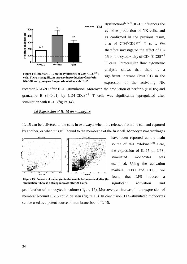
4.5 Effect of IL-15 on cytotoxicity
CD4+CD28
null T cells show several similarities with NK cells, including proinflammatory
functions and expression of cytolytic molecules such as granzyme B and perforin. These T
cells are considered cytotoxic, and are thought to contribute to disease related immune
M1
M2
4.61
95.39
M1
M2
70.64
29.36
Figure 13: Effect of IL-15 on cytokine production of
CD4+CD28null T cells. There is a significant increase in IFN-
and IL-17 when IL-15 is added to the culture medium, with
and without activation of the cells with PMA and CaI.
34
dysfunctions[24,27]
. IL-15 influences the
cytokine production of NK cells, and
as confirmed in the previous result,
also of CD4+CD28
null T cells. We
therefore investigated the effect of IL-
15 on the cytotoxicity of CD4+CD28
null
T cells. Intracellular flow cytometric
analysis shows that there is a
significant increase (P<0.001) in the
expression of the activating NK
receptor NKG2D after IL-15 stimulation. Moreover, the production of perforin (P<0.05) and
granzyme B (P<0.01) by CD4+CD28
null T cells was significantly upregulated after
stimulation with IL-15 (figure 14).
4.6 Expression of IL-15 on monocytes
IL-15 can be delivered to the cells in two ways: when it is released from one cell and captured
by another, or when it is still bound to the membrane of the first cell. Monocytes/macrophages
have been reported as the main
source of this cytokine.[38]
Here,
the expression of IL-15 on LPS-
stimulated monocytes was
examined. Using the activation
markers CD80 and CD86, we
found that LPS induced a
significant activation and
proliferation of monocytes in culture (figure 15). Moreover, an increase in the expression of
membrane-bound IL-15 could be seen (figure 16). In conclusion, LPS-stimulated monocytes
can be used as a potent source of membrane-bound IL-15.
Figure 14: Effect of IL-15 on the cytotoxicity of CD4+CD28null T
cells. There is a significant increase in production of perforin,
NKG2D and granzyme B upon stimulation with IL-15.
R1
R1
Figure 15: Presence of monocytes in the sample before (a) and after (b)
stimulation. There is a strong increase after 24 hours.
35
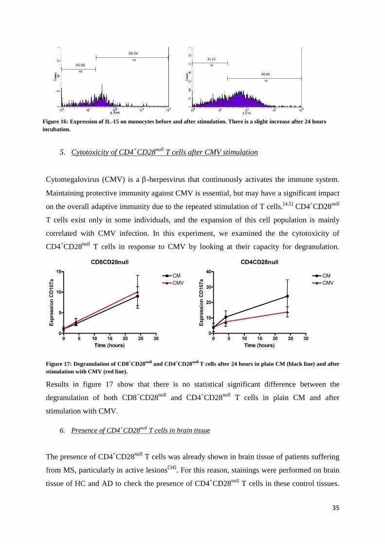
5. Cytotoxicity of CD4+CD28
null T cells after CMV stimulation
Cytomegalovirus (CMV) is a -herpesvirus that continuously activates the immune system.
Maintaining protective immunity against CMV is essential, but may have a significant impact
on the overall adaptive immunity due to the repeated stimulation of T cells.[4,5]
CD4+CD28
null
T cells exist only in some individuals, and the expansion of this cell population is mainly
correlated with CMV infection. In this experiment, we examined the the cytotoxicity of
CD4+CD28
null T cells in response to CMV by looking at their capacity for degranulation.
Figure 17: Degranulation of CD8+CD28null and CD4+CD28null T cells after 24 hours in plain CM (black line) and after
stimulation with CMV (red line).
Results in figure 17 show that there is no statistical significant difference between the
degranulation of both CD8+CD28
null and CD4
+CD28
null T cells in plain CM and after
stimulation with CMV.
6. Presence of CD4+CD28
null T cells in brain tissue
The presence of CD4+CD28
null T cells was already shown in brain tissue of patients suffering
from MS, particularly in active lesions[34]
. For this reason, stainings were performed on brain
tissue of HC and AD to check the presence of CD4+CD28
null T cells in these control tissues.
Figure 16: Expression of IL-15 on monocytes before and after stimulation. There is a slight increase after 24 hours
incubation.
M1
M2
59.04
40.96
M1
M2
68.58
31.21
36
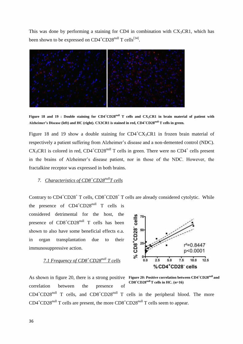
This was done by performing a staining for CD4 in combination with CX3CR1, which has
been shown to be expressed on CD4+CD28
null T cells
[34].
Figure 18 and 19 : Double staining for CD4+CD28null T cells and CX3CR1 in brain material of patient with
Alzheimer’s Disease (left) and HC (right). CX3CR1 is stained in red, CD4+CD28null T cells in green.
Figure 18 and 19 show a double staining for CD4+CX3CR1 in frozen brain material of
respectively a patient suffering from Alzheimer’s disease and a non-demented control (NDC).
CX3CR1 is colored in red, CD4+CD28
null T cells in green. There were no CD4
+ cells present
in the brains of Alzheimer’s disease patient, nor in those of the NDC. However, the
fractalkine receptor was expressed in both brains.
7. Characteristics of CD8+CD28
nullT cells
Contrary to CD4+CD28
+ T cells, CD8
+CD28
+ T cells are already considered cytolytic. While
the presence of CD4+CD28
null T cells is
considered detrimental for the host, the
presence of CD8+CD28
null T cells has been
shown to also have some beneficial effects e.a.
in organ transplantation due to their
immunosuppressive action.
7.1 Frequency of CD8+CD28
null T cells
As shown in figure 20, there is a strong positive
correlation between the presence of
CD4+CD28
null T cells, and CD8
+CD28
null T cells in the peripheral blood. The more
CD4+CD28
null T cells are present, the more CD8
+CD28
null T cells seem to appear.
Figure 20: Positive correlation between CD4+CD28null and
CD8+CD28null T cells in HC. (n=16)
37
7.2 Expression of CX3CR1
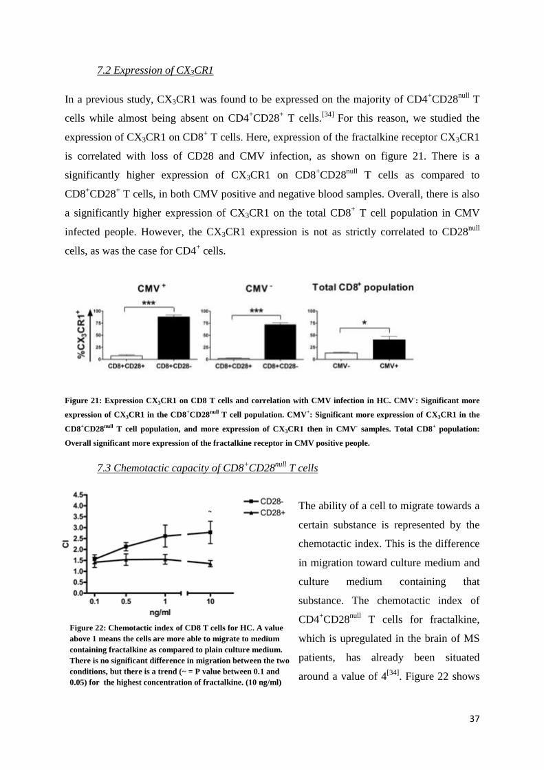
In a previous study, CX3CR1 was found to be expressed on the majority of CD4+CD28
null T
cells while almost being absent on CD4+CD28
+ T cells.
[34] For this reason, we studied the
expression of CX3CR1 on CD8+ T cells. Here, expression of the fractalkine receptor CX3CR1
is correlated with loss of CD28 and CMV infection, as shown on figure 21. There is a
significantly higher expression of CX3CR1 on CD8+CD28
null T cells as compared to
CD8+CD28
+ T cells, in both CMV positive and negative blood samples. Overall, there is also
a significantly higher expression of CX3CR1 on the total CD8+ T cell population in CMV
infected people. However, the CX3CR1 expression is not as strictly correlated to CD28null
cells, as was the case for CD4+ cells.
Figure 21: Expression CX3CR1 on CD8 T cells and correlation with CMV infection in HC. CMV-: Significant more
expression of CX3CR1 in the CD8+CD28null T cell population. CMV+: Significant more expression of CX3CR1 in the
CD8+CD28null T cell population, and more expression of CX3CR1 then in CMV- samples. Total CD8+ population:
Overall significant more expression of the fractalkine receptor in CMV positive people.
7.3 Chemotactic capacity of CD8+CD28
null T cells
The ability of a cell to migrate towards a
certain substance is represented by the
chemotactic index. This is the difference
in migration toward culture medium and
culture medium containing that
substance. The chemotactic index of
CD4+CD28
null T cells for fractalkine,
which is upregulated in the brain of MS
patients, has already been situated
around a value of 4[34]
. Figure 22 shows
Figure 22: Chemotactic index of CD8 T cells for HC. A value
above 1 means the cells are more able to migrate to medium
containing fractalkine as compared to plain culture medium.
There is no significant difference in migration between the two
conditions, but there is a trend (~ = P value between 0.1 and
0.05) for the highest concentration of fractalkine. (10 ng/ml)
38
the chemotactic index for CD8+CD28
+ and CD8
+CD28
null T cells towards fractalkine. When
the chemotactic index is higher than one, there is a greater migration toward fractalkine.
Experiments showed a difference in migration between CD8+CD28
+, situated around 1.5; and
CD8+CD28
null T cells, situated around 2.5. This difference was not significant. There is,
however, a trend noticeable for the highest concentration of fractalkine.
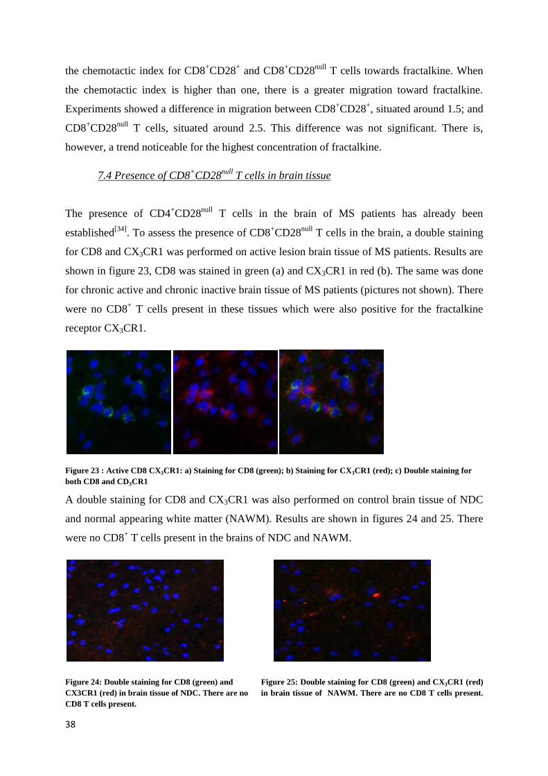
7.4 Presence of CD8+CD28
null T cells in brain tissue
The presence of CD4+CD28
null T cells in the brain of MS patients has already been
established[34]
. To assess the presence of CD8+CD28
null T cells in the brain, a double staining
for CD8 and CX3CR1 was performed on active lesion brain tissue of MS patients. Results are
shown in figure 23, CD8 was stained in green (a) and CX3CR1 in red (b). The same was done
for chronic active and chronic inactive brain tissue of MS patients (pictures not shown). There
were no CD8+ T cells present in these tissues which were also positive for the fractalkine
receptor CX3CR1.
Figure 23 : Active CD8 CX3CR1: a) Staining for CD8 (green); b) Staining for CX3CR1 (red); c) Double staining for
both CD8 and CD3CR1
A double staining for CD8 and CX3CR1 was also performed on control brain tissue of NDC
and normal appearing white matter (NAWM). Results are shown in figures 24 and 25. There
were no CD8+ T cells present in the brains of NDC and NAWM.
Figure 24: Double staining for CD8 (green) and Figure 25: Double staining for CD8 (green) and CX3CR1 (red)
CX3CR1 (red) in brain tissue of NDC. There are no in brain tissue of NAWM. There are no CD8 T cells present.
CD8 T cells present.
39
Control staining was performed by omitting the primary antibodies. As shown in pictures 26
and 27, there is little to no background staining for these secondary antibodies.
Figure 26: Control staining for DaM Alexa 488. Little Figure 27: Control staining for GaR Alexa 555. Little to no
to no background staining. background staining.
8. Expression of CX3CR1 on brain cells
In vitro analyses on brain cell cultures have indicated that fractalkine is mainly expressed on
neurons and astrocytes, while its receptor is present on neurons and microglia.[48]
It has also
been demonstrated that neurons, microglia and oligodendrocytes express CX3CR1 in MS
lesions.[34]
We studied the expression of fractalkine on neurons, microglia, oligodendrocytes
and astrocytes in the brains of AD patients and NDC.
For the identification of CX3CR1+ cells in brain tissue of an AD patient, double stainings
were performed for CX3CR1 in combination with Iba1 (microglia), CC-1 (oligodendrocytes),
GFAP (astrocytes) and NeuN (neurons), represented by figure 28. There is no overlap in
stainings for astrocytes and neurons, meaning that only microglia and oligodendrocytes
express the fractalkine receptor in the brains of AD patients.
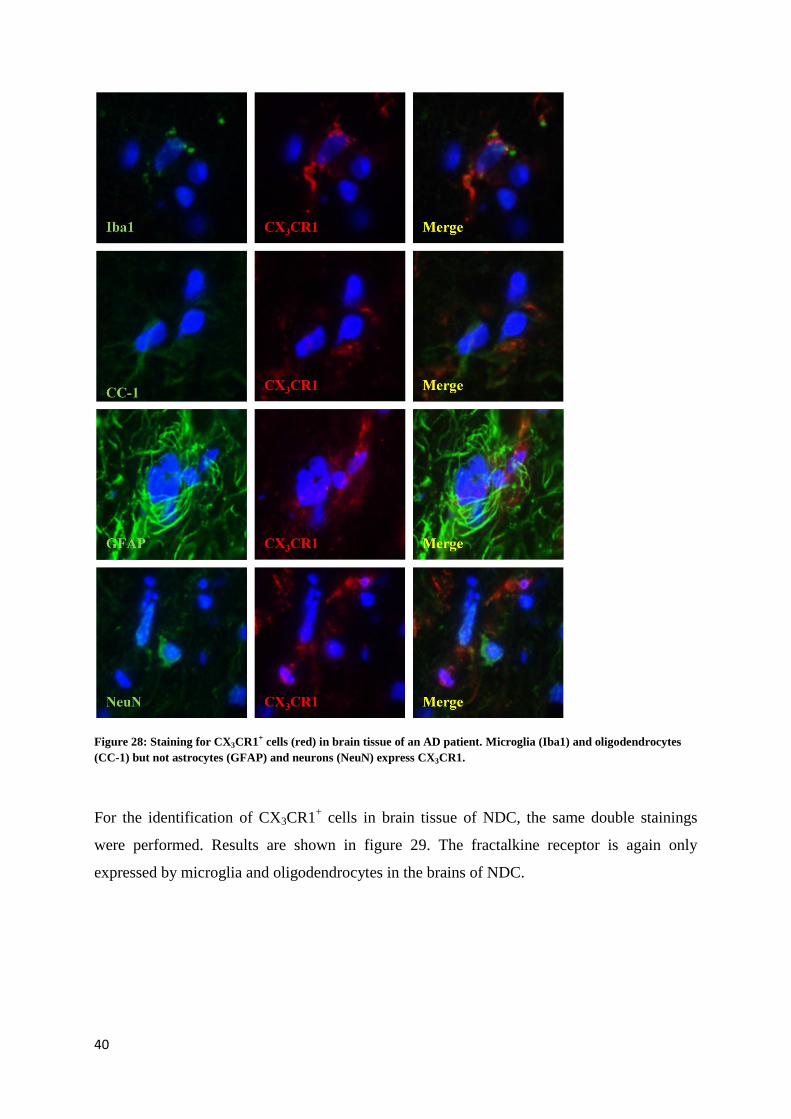
40
Figure 28: Staining for CX3CR1+ cells (red) in brain tissue of an AD patient. Microglia (Iba1) and oligodendrocytes
(CC-1) but not astrocytes (GFAP) and neurons (NeuN) express CX3CR1.
For the identification of CX3CR1+ cells in brain tissue of NDC, the same double stainings
were performed. Results are shown in figure 29. The fractalkine receptor is again only
expressed by microglia and oligodendrocytes in the brains of NDC.
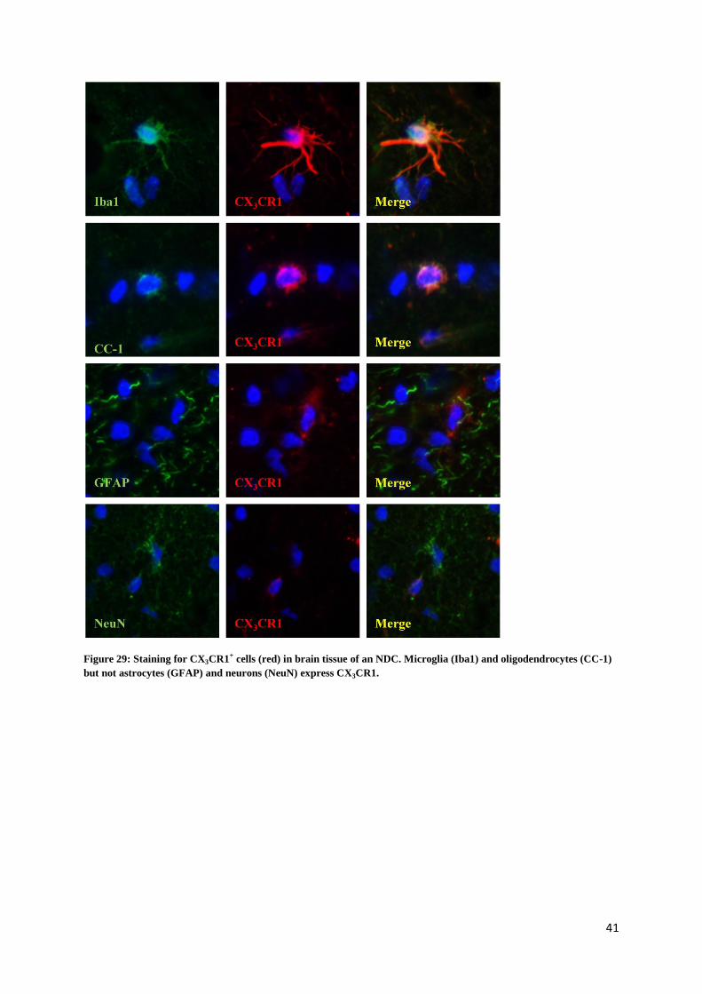
41
Figure 29: Staining for CX3CR1+ cells (red) in brain tissue of an NDC. Microglia (Iba1) and oligodendrocytes (CC-1)
but not astrocytes (GFAP) and neurons (NeuN) express CX3CR1.

42
43
Discussion
The vast majority of peripheral blood CD4+ T cells express the CD28 molecule, while a
subset of CD4+CD28
null T cells exists only in some individuals, mainly correlated with CMV
infection. On the other hand, CD8+CD28
null T cells are more common and show increased
frequencies with chronic infections as well as during normal aging[12]
. The presence of
CD4+CD28
null T cells has a negative effect on the host. These cells perpetuate after infection,
thereby causing a more severe inflammation. The distinction between CD8+CD28
+ T cells and
CD8+CD28
null T cells is not so clear. CD8
+CD28
+ T cells are already considered cytolytic,
and the presence of CD8+CD28
null T cells may also have some beneficial effects on the host
e.a. in organ transplantation due to their immunosuppressive action.
CD4+CD28
null T cells are enriched in a subset of patients suffering from a multitude of
diseases. Many studies have been performed to associate these cells with diseases such as
MS[7,8,34]
, coronary artery disease[10,24]
, rheumatoid arthritis[15,16,17,21,33,40]
, Wegener’s
granulomatosis[20]
, dermatomyositis and polymyositis[14]
, and several others. The
differentiation and functional properties of CD28null
T cells, both CD4 and CD8 T cells, as
well as their characterization and stimulatory pathways have been extensively
studied.[4,6,9,11,12]
Their true functional role in disease pathogenesis and immunosenescence
has, however, remained unclear. We aimed to further elucidate the role of both CD4+CD28
null
T cells and CD8+CD28
null T cells in MS through a series of experiments on HC and patients.
In this study, we found that there is a strong positive correlation between the presence of
CD4+CD28
null and CD8
+CD28
null T cells in HC. The pool of CD4
+CD28
null T cells seems to
increase with an expansion in CD8+CD28
null T cells and vice versa.
Previous studies already demonstrated that the fractalkine receptor CX3CR1 is expressed on
the vast majority of senescent CD4+CD28
null T cells, while it is almost absent on CD4
+CD28
+
T cells.[34]
We therefore studied the expression of CX3CR1 on CD8+ T cells in HC, and found
that it was correlated with loss of CD28 and CMV infection in both the CD8+CD28
null and
CD8+CD28
+ T cell populations. Expression of CX3CR1 was significantly elevated in both
CD8+CD28
+ and CD8
+CD28
null T cell populations upon CMV infection. We also found a
significantly higher expression of CX3CR1 in the total CD8+ T cell population after CMV
infection. CMV continuously activates the immune system, leading to repeated stimulation of
T cells and eventually the loss of CD28 through replicative senescence.[4,5]
These results show
that expression of CX3CR1 on CD8+ T cells is both correlated to CMV infection and thus loss
44
of CD28. The most striking difference with the CD4+ T cell population, is that CX3CR1 is
also expressed on CD8+CD28
+ T cells, while this is not the case for CD4
+CD28
+ T cells. The
distinction in characterization and function between CD8+CD28
+ T cells and CD8
+CD28
null T
cells is therefore not as clear as that between CD4+CD28
+ T cells and CD4
+CD28
null T cells.
This leads to the hypothesis that CD8+CD28
+ T cells, as well as CD8
+CD28
null T cells, can
migrate towards an increased fractalkine gradient. This has already been shown for
CD4+CD28
null T cells, which are able to migrate towards a fractalkine gradient in vitro.
[34] In
this study, we found that both populations showed an increased migratory capacity towards
the medium containing fractalkine, with a slightly higher chemotactic index for the
CD8+CD28
null T cells population in comparison to the CD8
+CD28
+ T cells. The difference in
chemotactic index between both cell populations was not significant, because the CD8+CD28
+
T cell population is also capable of migration to an increased fractalkine gradient through the
expression of CX3CR1.
In MS patients, fractalkine has been shown to be upregulated in the CSF and serum, and
several groups reported an abundant expression of fractalkine and its receptor in the brain.[45-
47] In vitro analyses on brain cell cultures have indicated that fractalkine is mainly expressed
on neurons and astrocytes, while its receptor is present on neurons and microglia.[48]
It has
also been demonstrated that neurons, microglia and oligodendrocytes express CX3CR1 in MS
lesions.[34]
We examined the expression of CX3CR1 on these brain cells in the brains of NDC
and AD patients. Results showed that the fractalkine receptor is strongly expressed by
microglia in both NDC and AD patients, while there is only a limited expression of CX3CR1
by oligodendrocytes in both conditions. In contrast with MS patients, neurons do not express
CX3CR1 in the brains of AD patients and NDC. The expression of CX3CR1 in the brains of
MS patients is presumably induced by inflammation of the brain. The brains of AD patients
and NDC are not inflamed, explaining why CX3CR1 is not expressed on these neurons.
Because of the identification of CX3CR1 on CD8+ T cells, we were able to perform double
stainings on brain material from MS patients. We found that there are CD8+ T cells present in
the MS brain, mainly in active lesions, but they do not express CX3CR1. This is contradictory
to the chemotaxis assay, which indicated that CD8+ T cells do express CX3CR1 and that they
are able to migrate to an increasing fractalkine gradient[34]
. However, when we compare
previous research on CD4+CD28
null T cells with this study, CD8
+ T cells are found to have a
lower chemotactic capacity. This, and the fact that the in vitro assay can never fully mimic the
in vivo situation, might explain why we did not find CD8+CX3CR1
+ T cells in the brains of
45
MS patients. Under healthy conditions, the blood brain barrier is closed, and does not allow
migration of T cells to the parenchyma of the brain. Also, fractalkine is only upregulated in
the brains of MS patients, compared to HC. This explains why no CD8+ T cells and
CD4+CD28
null T cells were found in the brains of NDC and AD patients.
We subsequently analyzed whether CD4+CD28
null T cells can contribute to the disease
process of MS, once they have migrated towards the inflamed tissue. We therefore
investigated what types of cytokines and chemokines they produce after being activated. The
inflammatory response is characterized by the production of various molecules that initiate
the recruitment of immune cells to the lesion sites, including the brain. Prolonged and
sustained inflammation may have cytotoxic effects, aggravating the incidence and severity of
the disease. Chemokines which are involved in the chemoattraction of cells are very important
inflammatory factors. They are involved in the recruitment of the main resident immune cell
types of the brain and of infiltrating monocytes from the bloodstream across the blood brain
barrier (BBB) into the CNS. They can be produced by glial and neuronal cells, which both
also express chemokine receptors. [48]
.
By performing a cytokine array on CD4+CD28
null T cells, our study found that these cells
produce a large amount of cytokines and chemokines after activation. Among these, G-CSF
and IL-1 were only produced by cells of MS patients, and CCL2 was specifically produced
by MBP-stimulated cells. This difference in expression might reveal a disease-specific
mechanism. Previous studies already showed that chemokines and chemokine receptors are
involved in the pathogenesis of MS. Different pairs of chemokines seem to play a pathogenic
role in MS, such as CCL1, CCL2, CCL3, CCL4, CCL5, CXCL9 and CXCL10. The influx of
autoreactive T cells and macrophages across the BBB into the CNS is a crucial step in the
development of MS. The migration of inflammatory cells is facilitated by the upregulation of
adhesion molecules and matrix metalloproteinases which degrade components of the
basement membrane. Chemokines can then attract leukocytes by forming concentration
gradients over the BBB, especially CCL4 is known mainly for its activity as a chemotactic
cytokine[49]
. CCL2, previously known as monocyte chemoattractant protein-1, acts on
monocytes, activated T cells, NK cells and microglia. CCL2 is known to be produced by
astrocytes, microglia, endothelial cells and macrophages. A critical role for CCL2 has been
established in animal models for MS. Several studies have demonstrated that CCL2 is not
only expressed in neuroinflammatory conditions, but are also constitutively present in the
brain in glial cells and neurons. This indicates that CCL2 may act as a modulator of neuronal
46
functions. It is established that CCL2 plays an important role as mediator in many
neuroinflammatory diseases characterized by neuronal degeneration, and that the expression
of CCL2 in the brain is increased in autoimmune encephalomyelitis (EAE), an animal model
for MS. [48]
. Previous research found that CCL2 was expressed by astrocytes and macrophages
within acute and chronic MS lesions in humans, and its receptor was detected on macrophages
and activated microglia in active MS lesions[49]
. [48]
. Other studies demonstrated that CCL2
deficient mice were resistant to the induction of EAE and showed a significant reduction in
macrophage recruitment to the CNS. Other studies indicated that induction of leukocyte
adhesion to the brain microvasculature is an important mechanism by which mainly CCL2
and CCL5 participate in the pathophysiology of MS[50]
.
Two other chemokines which were found to be produced by CD4+CD28
null T cells are CCL3
and CCL5. Significantly higher levels of these chemokines were found in the CSF of patients
with MS in relapses. Treatment with anti-CCL3 antibodies inhibits the onset of EAE, and
reduces the accumulation of mononuclear cells in the CNS. Patients with MS also have
elevated percentages of CCL5 receptor expressing T cells compared to HC, indicating that the
CCL5 receptor plays a crucial role in the recruitment of T cells in MS[49]
. Previous research
found that the receptors for CCL3 and CCL5 are also expressed on macrophages and
activated microglia in the active MS lesions, and their ligands were detected in MS
plaques[49]
.
Our study demonstrated that CD4+CD28
null T cells produce various cytokines, including IL-
1, G-CSF, TNF- and IFN-. Previously, several studies evaluated the involvement of
various cytokines in the pathogenesis of multiple sclerosis. Many cytokines participate in
nervous tissue damage by promoting demyelization and oligodendrocyte injury or by
enhancing local immune response. In addition, several authors reported increased levels of
some cytokines in serum and cerebrospinal fluid of patients with multiple sclerosis. These
findings suggest that cytokines can play a significant role in the immunopathogenesis of the
disease[51]
. Different research groups have also extensively studied the associations of
cytokine gene polymorphisms in different diseases. The role of cytokine gene polymorphisms
in MS has been previously reported in various populations. Several cytokine single gene
nucleotide polymorphisms were determined, including IL-1, IL-1, IL-1R and TNF-[52]
.
IL-1 is a pro-inflammatory cytokine and possesses a wide spectrum of metabolic,
physiological and haematopoietic activities[53]
. Previous research found that IL-1 plays a
major role in autoimmune and inflammatory diseases, and is expressed at elevated levels in
47
tissue and fluid samples isolated from patients with many chronic inflammatory diseases, such
as RA and MS[54]
.
Another cytokine produced by CD4+CD28
null T cells in MS patients, is granulocyte colony-
stimulating factor (G-CSF). G-CSF is a potent hematopoietic factor produced by mesothelial
cells, fibroblasts, and endothelial cells. Receptors for G-CSF are present on precursors and
mature neutrophilic granulocytes, monocytes, platelets, and endothelial cells. Expression of
G-CSF is induced by ischemia, where after G-CSF can pass the intact BBB. G-CSF is an
endogenous ligand in the CNS that has a dual activity; beneficial both in counteracting acute
neuronal degeneration and contributing to long-term plasticity after cerebral ischemia. At the
myeloid progenitor cell level, G-CSF stimulates the growth of neutrophil granulocyte
precursors and crucially regulates survival of mature neutrophils by inhibition of apoptosis.
G-CSF signaling appears to be a novel protective system in the brain that is involved in
counteracting acute neurodegeneration and regulating the formation of new neurons[55]
. In
other studies, however, G-CSF administration resulted in cases of worsened clinical MS
status. G-CSF enhanced MS autoreactive T cell line adhesion to the extracellular matrix
(ECM) proteins as effectively as the proinflammatory IFN- and TNF-, known to exacerbate
MS symptoms. This indicated a link between clinical worsening of MS symptoms induced by
G-CSF and the hyper-stimulation of T cell adhesion to ECM elicited by G-CSF[56]
. The
production of G-CSF by CD4+CD28
null T cells in MS patients may thus play a dual role in the
neuroprotection on the one hand, and in the increase of the clinical syndromes of MS on the
other hand.
Other proinflammatory cytokines such as TNF- and IFN-, trigger a chain of events that
result in the formation of demyelinated plaques and damage to axons[49]
. The primary role of
TNF- is the regulation of immune cells. TNF- shows a wide spectrum of biological
activities. It causes cytolysis of many tumor cell lines in vitro. The factor also enhances
phagocytosis and cytotoxicity in neutrophilic granulocytes and modulates the expression of
many other proteins, including IL-1 and IL-6. IL-1 and IL-6 in turn are important factors in
Th17 differentiation. In the presence of IL-2, TNF- promotes the proliferation and
differentiation of B cells. Although TNF- is required for normal immune responses, its
overexpression has severe pathological consequences. TNF- is able to induce apoptotic cell
death and inflammation, and inhibit tumorigenesis and viral replication. Dysregulation of
TNF- production has been implicated in a variety of human diseases, including major
depression, Alzheimer's disease and cancer. TNF- is found predominantly on monocytes and
48
T cells after cell activation. In vivo TNF- in combination with IL-1 is responsible for many
alterations of the endothelium, and is also a potent chemoattractant for neutrophils. TNF-
promotes the proliferation of astroglial cells and microglial cells and therefore may be
involved in pathological processes such as astrogliosis and demyelination[57]
.
IFN- is a remarkable cytokine that orchestrates many distinct cellular programs through
transcriptional control over large numbers of genes. In most situations, macrophages are
activated to acquire microbiocidal effector functions and secrete proinflammatory cytokines,
resulting in inflammation and recruitment of immune cells and subsequent elimination of the
microbe by phagocytosis or release of toxic metabolites. The most important activating factor
for macrophages is IFN-, which induces direct antimicrobial and antitumor mechanisms as
well as upregulating antigen processing and presentation pathways. IFN- directs growth,
maturation and differentiation of many cell types. It also enhances NK cell activity and
regulates B functions such as immunoglobulin production. IFN- initiates cell-mediated
immunity via the activation of NK cell effector functions, specific cytotoxic immunity trough
T cells, and macrophage activation. IFN- also orchestrates the trafficking of immune cells to
sites of inflammation trough upregulating expression of adhesion molecules and
chemokines[58]
.
The chemokines and cytokines detected in our study are all known to be produced in response
to inflammation. They are involved in the chemoattraction of specific immune cells into the
brains of MS patients, display several proinflammatory functions, and thus play a crucial role
in the immunopathogenesis of MS.
Another important part of this study focused on the effect of IL-15 on the phenotype and
function of CD4+CD28
null T cells. IL-15 secretion by macrophages is known to increase
cytokine secretion[43]
, and regulate IFN- production by NK cells. Because CD4+CD28
null T
cells are very similar to NK cells, we first examined the effects of IL-15 on cytokine
production by these cells. We established that CD4+CD28
null T cells express the IL-15
receptor. When cells were stimulated with soluble IL-15, there was a significant increase in
IFN-and IL-17 production by CD4+CD28
null T. IL-17 is a potent inflammatory cytokine
which induces the expression of pro-inflammatory genes and chemokines, neutrophil
recruitment and inflammation. The IL-17 receptor is expressed in different organs such as
lung, liver and spleen. Cells capable of responding to IL-17 include dendritic cells,
macrophages, lymphocytes, epithelial cells and fibroblasts[59]
. We did not detect production of
49
IL-17 using the cytokine array, for which we used different stimulatory agents to activate the
cells. This difference in activation might explain why IL-17 was only detected in the IL-15
assay, and not in the cytokine array. After adding IL-15 to the CD4+CD28
null T cells, there
was also a significant increase in the production of cytotoxic molecules NKG2D, perforin and
granzyme B. Both CD4+ and CD8
+ T cells had already been found to be perforin positive in
previous research[14]
. Our results thus prove that senescent CD4+ T cells become cytotoxic
after the loss of CD28. Moreover, the effect of IL-15 on NK cells was found to be present in
CD4+CD28
null T cells as well, implying a common mechanism or pathway.
IL-15 is also known to increase the proliferation of NK cells. We evaluated whether IL-15 has
the same effect on CD4+CD28
null T cells by performing a CFSE assay. Remarkably,
CD4+CD28
null T cells show an overall higher proliferative capacity, compared to CD4
+CD28
+
T cells. When IL-15 was used in combination with aCD3, there was a significantly higher
proliferation rate of both cell populations, compared to aCD3 alone. IL-15 alone was also able
to induce proliferation in both cell populations, although this only reached statistical
significance for CD4+CD28
null T cells. These findings confirm that IL-15 is also able to induce
proliferation in CD4+CD28
null T cells, as it does for NK cells. This again shows the range of
similarities between NK cells and CD4+CD28
null T cells, proving these senescent T cells are
also cytotoxic.
To evaluate whether CD4+CD28
null and CD8
+CD28
null T cells are responsive to CMV, we
determined their capacity to degranulate after stimulation with CMV viral peptides. We found
that there is no significant difference in degranulation of both CD4+CD28
null and
CD8+CD28
null T cells after activation with CMV, as compared to their capacity to degranulate
in plain CM. This might be due to the fact there was a great deal of background visible for the
CM conditions, and the experimental setup has yet to be optimized. Previous research did find
that most CD4+CD28
null T cells degranulated significantly when activated with aCD3. This
response could already be seen after 4 hours of activation in both HC and MS[34]
. The
cytotoxic capacities of senescent CD4+ T cells have been investigated by several studies.
These cells were found to express several NK cell receptors, such as NKG2D. Large
intracellular stores of IFN-, perforin and granzymes A and B were found to be present in
CD4+CD28
null T cells, and the majority of these cells were found to be CMV specific
[19,60,61].
Our study confirms the cytotoxic potential of CD4+CD28
null T cells, through the expression of
cytotoxic molecules and their ability to respond to IL-15 stimulation.

50
51
Conclusion
In this study, we found that there is a strong positive correlation between the presence of
CD4+CD28
null and CD8
+CD28
null T cells in HC. Both cell types express the chemokine
receptor CX3CR1, and are able to migrate towards an increasing fractalkine gradient. The
expression of CX3CR1 in the CD8+ T cell population is correlated with loss of CD28 and
CMV infection in both the CD8+CD28
null and CD8
+CD28
+ T cell subsets. The most striking
difference with the CD4+ T cell population, is CD8
+CD28
+ T cells also express a functional
fractalkine receptor CX3CR1, and also show an increased migration capacity towards medium
containing fractalkine while this is not the case for CD4+CD28
+ T cells. The distinction in
characterization and function between CD8+CD28
+ T cells and CD8
+CD28
null T cells is
therefore not as clear as that between CD4+CD28
+ T cells and CD4
+CD28
null T cells.
CD8+CD28
+ T cells are already considered cytolytic, and the presence of CD8
+CD28
null T
cells may also have some beneficial effects on the host e.a. in organ transplantation due to
their immunosuppressive action. CD8+CD28
null T cells are more common than CD4
+CD28
null
T cells and show increased frequencies with chronic infections as well as during normal
aging[12]
.
CX3CR1 is expressed by microglia and oligodendrocytes in the brains of NDC and AD
patients, but not by neurons as in the brains of MS patients. This is due to the lack of
inflammation in the brains of non-MS patients, that induces the expression of fractalkine in
the brains of MS patients. CD8+ and CD4
+ T cells are also exclusively present in the brains of
MS patients, mainly in active lesions. However, the CD8+ T cells do not express CX3CR1.
Six major cytokines were found to be produced by CD4+CD28
null T cells: IL-1, G-CSF,
TNF-, IFN-, CCL2 and CCL4, and four cytokines were validated in another experiment:
IL-1, IL-1, IL-6 and CCL2. These cytokines all show proinflammatory properties, although
G-CSF is also believed to have some neuroprotective abilities. TNF- and IFN- induce
capillary endothelial cells to express adhesion molecules and chemokines to facilitate the
trafficking of immune cells to sites of inflammation. One of those chemotactic cytokines is
CCL4. TNF- also contributes to the pathological process of demyelination by promoting the
proliferation of astroglial and microglial cells[57]
, while IFN- enhances NK cell activity and
activates macrophages[58]
. IL-1and CCL2 both play a major role in autoimmune and
inflammatory diseases, and are expressed at elevated levels in tissue and fluid samples

52
isolated from patients with many chronic inflammatory diseases[54]
and the presence of CCL2
in neurons throughout the brain indicates that it might participate in the neurodegenerative
processes that take place in these pathologies[48]
. More recently, transcriptional profiling with
gene microarrays and large-scale sequencing of transcripts from MS lesions have identified a
number of genes that are involved in the pathogenesis of acute disease. These include IL-6
which plays a role in the transition from relapsing-remitting to chronic MS[62]
. After IL-15
stimulation, CD4+CD28
null T cells are also found to upregulate the production of
proinflammatory cytokines IFN- and IL-17, and of cytotoxic molecules NKG2D, perforin
and granzym B. Through production of these cytokines, CD4+CD28
null T cells may actively
contribute to the immunopatholgy of MS.
In this study, we confirmed the role of CD4+CD28
null T cells in disease manifestation of MS.
In general, these cells share many similarities with NK cells and are considered cytotoxic and
inflammation-seeking cells. They can infiltrate the brains of MS patients through the
expression of CX3CR1 and release an arsenal of cytoxic and proinflammatory cytokines.
CD8+CD28
null T cells also have in vitro capacity to infiltrate into the brains of MS patients,
through the expression of CX3CR1, although this has not been proven. Their presence is
strongly correlated to that of CD4+CD28
null T cells and they are considered cytotoxic as well.
Their contribution to the immunopathology of inflammatory diseases such as MS is, however,
not as clear as that of CD4+CD28
null T cells. This is mainly due to the fact that there is no
clear distinction between CD8+CD28
null and CD8
+CD28
+ T cells, which are already
considered cytolytic.
53
References
1) ABBES, A.K. AND LICHTMAN, A.H. 2009. “Basic Immunology, Functions and Disorders of the Immune System.”
Saunders Elsevier. Third edition: pg 8-9
2) WARRINGTON, K.J., VALLEJO, A.N., WEYAND, C.M., GORONZY, J.J. 2003. “CD28 Loss in Senescent CD4+ T Cells:
Reversal by Interleukin-12 Stimulation.” Blood. 101:3543-3549.
3) CARUSO, C., BUFFA, S., CANDORE, G., COLONNA-ROMANO, G., DUNN-WALTERS, D., KIPLING, D., PAWELEC, G. 2009.
“Mechanisms of immunosenescence” Immunity and aging 6:10
4) THEWISSEN, M., SOMERS, V., HELLINGS, N., FRAUSSEN, J., DAMOISEAUX, J., STINISSEN, P. 2007. CD4+CD28null T Cells
in Autoimmune Disease: Pathogenic Features and Decreased Susceptibility to Immunoregulation. The journal of
Immunology. 179;6514-6523
5) FLETCHER, J.M., VUKMANOVIC-STEJIC, M., DUNNÉ, P.J., BIRCH, K.E., COOK, J.E., JACKSON, S.E., SALMON, M., RUSTIN,
M.H., AKBAR, A.N. 2005. Cytomegalovirus-specific CD4+ T cells in healthy carriers are continuously driven to
replicative exhaustion. Journal of Immunology. 175:8218–8225
6) PARK, W., WEYAND, C.M., SCHMIDT, D., GORONZY, J. J. 1997. "Costimulatory pathways controlling activation and
peripheral tolerance of human CD4+CD28- T cells." Journal of Immunology 27(5): 1082-1090.
7) MARKOVIC-PLESE, S., CORTESE, I., WANDINGER, K., MCFARLAND, H.F., MARTIN, R. 2001. “CD4+CD28– costimulation-
independent T cells in multiple sclerosis.” The journal of Clinical investigation. 108:1185-1193
8) MIYAZAKI, Y., IWABUCHI, K., KIKUCHI, S., FUKAZAWA, T., NIINO, M., HIROTANI, M., SASAKI, H., ONOÉ, K. 2008.
Expansion of CD4+CD28null T cells producing high levels of interferon- in peripheral blood of patients with multiple
sclerosis. Multiple sclerosis. 00:1-12
9) WENG, N., AKBAR, A.N., GORONZY, J. 2009. CD28− T cells: Their Role in the Age-associated Decline of Immune
Function. Trends in Immunology. 30: 306-312.
10) DUMITRIU, I.E., TRALLERO ARAGUA´S, E., BABOONIAN, C., CARLOS KASKI, J. 2008. CD4+CD28null T cells in Coronary
Artery Disease: When Helpers Become Killers. Cardiovascular Research. 81:11-19.
11) VALENZUELA , H. F., EFFROS, R.B. 2002. Divergent telomerase and CD28 expression patterns in human CD4 and CD8 T
cells following repeated encounters with the same antigenic stimulus. Clinical Immunology. 105:117–125.
12) EFFROS, R.B. 1997.” Loss of CD28 expression on T lymphocytes: A marker of replicative senescence” Developmental
and comparative immunology 21(6): 471-478
13) VALLEJO, A.N., BRANDES, J.C., WEYAND, C.M., GORONZY, J.J. 1999. “Modulation of CD28 expression: Distinct
regulatory pathways during activation and replicative senescence.” The journal of immunology; March
1999,(162)p6572-6579
14) FASTH, A. E., DASTMALCHI, M., RAHBAR, A., SALOMONSSON, S., PANDYA, J. M., LINDROOS, E., NENNESMO, I.,
MALMBERG, K. J., SODERBERG-NAUCLER, C., TROLLMO, C., LUNDBERG, I. E., MALMSTROM, V. 2009. "T cell infiltrates in
the muscles of patients with dermatomyositis and polymyositis are dominated by CD28null T cells." Journal of
Immunology 183(7): 4792-4799.
15) FASTH, A. E., BJORKSTROM, N.K., ANTHONI, M., MALMBERG, K. J., MALMSTROM, V. 2010. "Activating NK-cell receptors
costimulate CD4(+)CD28(-) T cells in patients with rheumatoid arthritis." Eur J Immunol 40(2): 378-387.
16) SCHMIDT, D., GORONZY, J.J., WEYAND, C. M. 1996. "CD4+ CD7- CD28- T cells are expanded in rheumatoid arthritis
and are characterized by autoreactivity." J Clin Invest 97(9): 2027-2037.
17) FASTH, A. E., SNIR, O., JOHANSSON, A. A., NORDMARK, B. ,RAHBAR, A. ,AF KLINT, E., BJORKSTROM, N. K., ULFGREN, A.
K., VAN VOLLENHOVEN, R. F., MALMSTROM, V., TROLLMO, C. 2007. “Skewed distribution of proinflammatory
CD4+CD28null T cells in rheumatoid arthritis.” Arthritis Res Ther. 9(5): R87.
18) NOWIK, M., NOWACKI, P. 2006. CD4+CD28null T cells in pathogenesis of acute ischemic stroke. Poster session
international Symposium on Atherosclerosis, Rome, Italy P. M. U. Department of neurology. Szezecin.
19) THEWISSEN, M., SOMERS, V., VENKEN, K., LINSEN, L., VAN PAASSEN, P., GEUSENS, P., DAMOISEAUX, J., STINISSEN, P.
2007. "Analyses of immunosenescent markers in patients with autoimmune disease." Clin Immunol 123(2): 209-218.
20) LAMPRECHT, P., MOOSIG, F., CSERNOK, E., SEITZER, U., SCHNABEL, A., MUELLER, A., GROSS, W. L. 2001. "CD28
negative T cells are enriched in granulomatous lesions of the respiratory tract in Wegener's granulomatosis." Thorax
56(10): 751-757.
21) MARTENS, P. B., GORONZY, J.J., SCHAID, D., WEYAND, C. M.1997. "Expansion of unusual CD4+ T cells in severe
rheumatoid arthritis." Arthritis Rheum 40(6): 1106-1114.
22) HOFF, H., KNIEKE, K., CABAIL, Z., HIRSELAND, H., VRATSANOS, G., BURMESTER, G. R., JORCH, G., NADLER, S. G.,
BROKER, B., HEBEL, K., BRUNNER-WEINZIERL, M. C. 2009. "Surface CD152 (CTLA-4) expression and signaling dictates
longevity of CD28null T cells." J Immunol 182(9): 5342-5351.
23) VALLEJO, A. N., SCHIRMER, M., WEYAND, C. M., GORONZY, J. J. 2000. "Clonality and longevity of CD4+CD28null T
cells are associated with defects in apoptotic pathways." J Immunol 165(11): 6301-6307.
24) TEO, F. H., OLIVEIRA, R.T.D., MAMONI, R.L., COELHO, O.R., BLOTTA, M.H.S.L. 2009. "Quantification and
Characterisation of CD4+CD28null and CD4+CD28+ T Cells in Patients with Chronic Coronary Artery Disease and
Healthy Subjects." Poster session Brazilian congress of Atherosclerosis. U. E. d. Campinas. Campinas.
25) SNYDER, M. R., MUEGGE, L.O., OFFORD, C., O'FALLON, W. M., BAJZER, Z., WEYAND, C. M., GORONZY, J. J. 2002.
"Formation of the killer Ig-like receptor repertoire on CD4+CD28null T cells." J Immunol 168(8): 3839-3846.
26) VAN BERGEN, J., THOMPSON, A., THOMPSON, A., VAN DER SLIK, A., OTTENHOFF, T. H., GUSSEKLOO, J., KONING, F.2004.
"Phenotypic and functional characterization of CD4 T cells expressing killer Ig-like receptors." J Immunol 173(11):
6719-6726.
54
27) WEYAND, C. M., BRANDES, J.C., SCHMIDT, D., FULBRIGHT, J. W., GORONZY, J. J.1998. "Functional properties of CD4+
CD28- T cells in the aging immune system." Mech Ageing Dev 102(2-3): 131-147.
28) APPAY, V., ZAUNDERS, J.J., PAPAGNO, L., SUTTON, J., JARAMILLO, A., WATERS, A., EASTERBROOK, P., GREY, P., SMITH,
D., MCMICHAEL, A.J., COOPER, D.A., ROWLAND-JONES, S.L., KELLEHER, A.D. 2002. “Characterization of CD4+ CTLs
Ex Vivo.” The Journal of Immunology, 2002, 168: 5954-5958.
29) PARK, W., WEYAND, C.M., SCHMIDT, D., GORONZY, J.J. 1997. “Co-stimulatory pathways controlling activation and
peripheral tolerance of human CD4+CD28- T cells.” Eur J Immunol. 1997 May;27(5):1082-90.
30) GROH, V., BRUHL, A., EL-GABALAWY, H., NELSON, J. L., SPIES, T.2003. "Stimulation of T cell autoreactivity by
anomalous expression of NKG2D and its MIC ligands in rheumatoid arthritis." Proc Natl Acad Sci U S A 100(16):
9452-9457.
31) FANN, M., CHIU, W.K., WOOD, W.H., LEVINE, B.L., BECKER, K.G., WENG, N. 2005. “Gene expression characteristics of
CD28null memory phenotype CD8+ T cells and its implications in T cell aging.” Immunological reviews; June
2005,(205)p190-206
32) WEYAND, C.M., FULBRIGHT, J.W., GORONZY, J.J. 2003. “Immunosenescence, autoimmunity, and rheumatoid arthritis.”
Experimental Gerontology. 38:833-841
33) PAWLIK, A., OSTANEK, L., BRZOSKO, I., BRZOSKO, M., MASIUK, M., MACHALINSKI, B., GAWRONSKA-SZKLARZ, B. 2003.
"The expansion of CD4+CD28- T cells in patients with rheumatoid arthritis." Arthritis Res Ther 5(4): R210-213.
34) BROUX, B., PANNEMANS, K., ZHANG, X., MARKOVIC-PLESE, S., BROEKMANS, T., OP’T EINDE, B., VAN WIJMEERSCH, B.,
SOMERS, V., GEUSENS, P., STINISSEN, P., HELLINGS, N. 2010. “CX3CR1 Drives Senescent CD4+ T Cells With Cytotoxic
Properties into the Multiple Sclerosis Brain.” Not yet published.
35) SUZUKI, M., KONYA, C., GORONZY, J.J., WEYAND, C.M. 2008. “Inhibitory CD8+ T cells in autoimmune disease.” Human
immunology; September 2008,(69)p781-789
36) FILACI, G., FRAVEGA, M., FENOGLIO, D., RIZZI, M., NEGRINI, S., VIGGIANI, R., INDIVERI, F. 2004. “Non-antigen specific
CD8+ T suppressor lymphocytes.” Clinical and experimental medicine; March 2004,(4)p86-92
37) EFFROS, R.B., DAGARAG, M., SPAULDING, C., MAN, J. 2005. “The role of CD8+ T cell replicative senescence in human
aging.” Immunological reviews; June 2005,(205)p147-157
38) SAIKALI, P., ANTEL, J.P., PITTET, C.L., NEWCOMBE, J., ARBOUR, N. 2010. “Contribution of Astrocyte-derived IL-15 to
CD8 T Cell Effector Functions in Multiple Sclerosis.” Journal of Immunology; September 2010,(185):000-000
39) UYTTEBROEK, L. 2006. “Assessing the role of central nervous system IL-15 in exacerbating the immune response in
multiple sclerosis.” University of Hasselt; Thesis for master in biomedical sciences, clinical and molecular sciences
40) GERLI, R., SCHILLACI, G., GIORDANO, A., BOCCI, E. B., BISTONI, O., VAUDO, G., MARCHESI, S., PIRRO, M., RAGNI, F.,
SHOENFELD, Y. MANNARINO, E. 2004. "CD4+CD28- T lymphocytes contribute to early atherosclerotic damage in
rheumatoid arthritis patients." Circulation 109(22): 2744-2748
41) BRUGALETTA, S., BIASUCCI, L.M., PINNELLI, M., BIONDI-ZOCCAI, G., DI GIANNUARIO, G., TROTTA, G., LIUZZO, G., CREA,
F. 2006. “Novel Anti-inflammatory Effect of Statins: Reduction of CD4+CD28null T Lymphocyte Frequency in Patients
With Unstable Angina.” Heart. 92:249-250.
42) RIZELLO, V., LIUZZO, G., BRUGALETTA, S., REBUZZI, A., BIASUCCI, L.M., CREA, F. 2006. “Modulation of CD4+CD28null T
Lymphocytes by Tumor Necrosis Factor Blockade in Patients With Unstable Angina.” Circulation. 113:2272-2277.
43) ALPDOGAN, O., V.D.B.M. 2005. “IL-7 and IL-15 therapeutic cytokines for immunodeficiency.” Trends Immunol. 26:56-
64.
44) CARSON, W.E., R.M., BAIOCCHI, R.A., MARIEN, M.J., BAOINI, N., GRABSTEIN, K. 1995. “Endogenous production of
interleukin 15 by activated human monocytes is critical for optimal production of interferon-gamma by natural killer
cells in vitro.” J Clin Invest. 96:2578-2582
45) NISHIYORI, A., MINAMI, M., OHTANI, Y., TAKAMI, S., YAMAMOTO, J., KAWAGUCHI, N., KUME, T., AKAIKE, A., SATOH, M.
1998. “Localization of fractalkine and CX3CR1 mRNAs in rat brain: does fractalkine play a role in signaling from
neuron to microglia?” FEBS Lett. 429:167-172
46) HARRISON, J.K., JIANG, Y., CHEN, S., XIA, Y., MACIEJEWSKI, D., MCNAMARA, R.K., STREIT, W.J., SALAFRANCA, M.N.,
ADHIKARI, S., THOMPSON, D.A. 1998. “Role for neuronally derived fractalkine in mediating interactions between
neurons and CX3CR1-expressing microglia.” Proc. Natl. Acad. Sci. U.S.A. 95:10896-10901
47) HULSHOF, S., VAN HAASTERT, E.S., KUIPERS, H.F., VAN DEN ELSEN, P.J., DE GROOT, C.J., VAN DER VALK, P., RAVID, R.,
BIBER, K. 2003. “ CX3CL1 and CX3CR1 expression in human brain tissue: noninflammatory control versus multiple
sclerosis.” J. Neuropathol. Exp. Neurol. 62:899-907
48) CONDUCTIER, G., BLONDEAU, N., GUYON, A., NAHON, J.L., ROVÈRE, C. 2010. “The role of monocyte chemoattractant
protein MCP1/CCL2 in neuroinflammatory diseases” Journal of Neuroimmunology 224(1-2);93-100
49) SZCZUCIŃSKI, A., LOSY, J. 2007. “ Chemokines and chemokine receptors in multiple sclerosis. Potential targets for new
therapies.” Acta neurol Scand 115:137-146
50) DOS SANTOS, A.C., BARSANTE, M.M., ARANTES, R.M., BERNARD, C.C., TEIXEIRA, M.M., CARVALHO-TAVARES, J. 2005.
“CCL2 and CCL5 mediate leukocyte adhesion in experimental autoimmune encephalomyelitis--an intravital microscopy
study.” J Neuroimmunol 162(1-2):122-9.
51) CARRIERI, P.B., MAIORINO, A., PROVITERA, V., SOCIA, E., PERRELLA, O. 1992. “Cytokines in the pathogenesis of multiple
sclerosis.” Acta Neurol (Napoli) Aug-Dec;14(4-6):333-41
52) BUJAK, M., FRANGOGIANNIS, N.G. 2009. “The role of IL-1 in the pathogenesis of heart disease.” Arch Immunol Ther Exp
(Warsz) 57(3):165-76
53) SARIAL, S., SHOKRGOZAR, M.A., AMIRZARGAR, A., SHOKRI, F., RADFAR, J., ZOHREVAND, P., ARJANG, Z., SAHRAIAN,
M.A., LOTFI, J. 2008. “IL-1, IL-1R and TNFalpha gene polymorphisms in Iranian patients with multiple sclerosis.” Iran
J Allergy Asthma Immunol 7(1):37-40.
55
54) BRADDOCK, M., QUINN, A., CANVIN, J. 2004. “Therapeutic potential of targeting IL-1 and IL-18 in inflammation.”
Expert Opin Biol Ther. 4(6):847-860
55) SCHNEIDER, A., KRÜGER, C., STEIGLEDER, T., WEBER, D., PITZER, C., LAAGE, R., ARONOWSKI, J. MAURER, M.H.,
GASSLER, N., MIER, W., HASSELBLATT, M., KOLLMAR, R.,SCHWAB, S., SOMMER, C., BACH, A., KUHN, H., SCHÄBITZ, W.
2005. “The hematopoietic factor G-CSF is a neuronal ligand that counteracts programmed cell death and drives
neurogenesis” Clin Invest. August 1; 115(8): 2083–2098.
56) SNIR , O., LAVIE, G., ACHIRON, A., BANK, I., BEN-AHARON, .T, SREDNI, B., COHEN, I.R., MANDEL, M. 2006. “G-CSF
enhances the adhesion of encephalitogenic T cells to extracellular matrix components: a possible mechanism for
exacerbation of multiple sclerosis.” J Neuroimmunol. Mar;172(1-2):145-55
57) Ibelgaufts, H. 2011 “COPE: Cytokines and Cells online pathfinder ecyclopedi: TNF alpha.” copewithcytokines.de
cope.cgi version 26.7.01;revision Feb 4, 2011
58) Schroder, K., Ravasi, T. 2004. “Interferon-gamma: an overview of signals, mechanisms and functions.”J Leukoc
Biol. 75(2):163-89.
59) TORRADO, E., COOPER, A.M. 2010. “IL-17 and Th17 cells in tuberculosis.” Cytokine Growth Factor Rev. 21(6):455-62.
60) NAKAJIMA, T., SCHULTE, S., WARRINGTON, K.J., KOPECKY, S.L., FRYE, R.L., GORONZY, J.J., WEYAND, C.M. 2002. “T-cell
mediated lysis of endothelial cells in acute coronary syndroms.” Circulation 105:570-575
61) VAN LEEUWEN, E.M., REMMERSWAAL, E.B., VOSSEN, M.T., ROWSHANI, A.T., WERTHEIM-VAN-DILLEN, P.M., VAN LIER,
R.A., TEN BERGE, I.J. 2004. “Emergence of a CD4+CD28-granzymeB+, cytomegalovirus-specific T cell subset after
recovery of primary cytomegalovirus infection.” J. Immunol. 173:1834-1841
62) STEINMAN, L. 2001.” Multiple sclerosis: a two-stage disease” Nature Immunology 2, 762 - 764

56
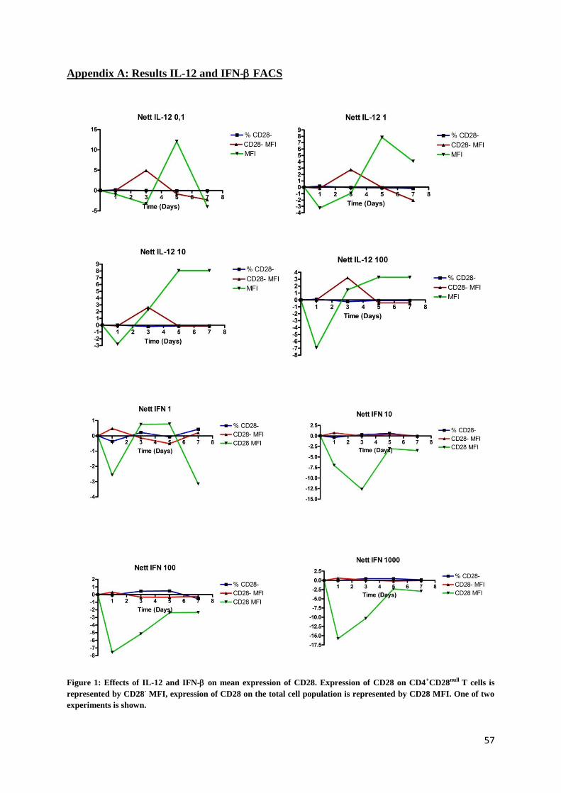
57
Appendix A: Results IL-12 and IFN-FACS
Figure 1: Effects of IL-12 and IFN- on mean expression of CD28. Expression of CD28 on CD4+CD28null T cells is
represented by CD28- MFI, expression of CD28 on the total cell population is represented by CD28 MFI. One of two
experiments is shown.

58

59
Appendix B: Results Cytokine Array
Figure 2: Cytokines produced by CD4+CD28null T cells in MS patients and HC, and detected with the a cytokine array
when pulsed with MBP or stimulated with aCD3.
Auteursrechtelijke overeenkomst
Ik/wij verlenen het wereldwijde auteursrecht voor de ingediende eindverhandeling:
The role of CD28null T cells in autoimmune diseases
Richting: master in de biomedische wetenschappen-milieu en gezondheid
Jaar: 2011
in alle mogelijke mediaformaten, - bestaande en in de toekomst te ontwikkelen - , aan de
Universiteit Hasselt.
Niet tegenstaand deze toekenning van het auteursrecht aan de Universiteit Hasselt
behoud ik als auteur het recht om de eindverhandeling, - in zijn geheel of gedeeltelijk -,
vrij te reproduceren, (her)publiceren of distribueren zonder de toelating te moeten
verkrijgen van de Universiteit Hasselt.
Ik bevestig dat de eindverhandeling mijn origineel werk is, en dat ik het recht heb om de
rechten te verlenen die in deze overeenkomst worden beschreven. Ik verklaar tevens dat
de eindverhandeling, naar mijn weten, het auteursrecht van anderen niet overtreedt.
Ik verklaar tevens dat ik voor het materiaal in de eindverhandeling dat beschermd wordt
door het auteursrecht, de nodige toelatingen heb verkregen zodat ik deze ook aan de
Universiteit Hasselt kan overdragen en dat dit duidelijk in de tekst en inhoud van de
eindverhandeling werd genotificeerd.
Universiteit Hasselt zal mij als auteur(s) van de eindverhandeling identificeren en zal geen
wijzigingen aanbrengen aan de eindverhandeling, uitgezonderd deze toegelaten door deze
overeenkomst.
Voor akkoord,
Brulmans, Marijke
Datum: 14/06/2011