
What do you remember about mass spectrometry? Mass Spectrometry.

1
Mass Spectrometry Worksheet CHEM 212
1. What is separated and detected by mass spectrometers? 2. Below is a generalized schematic of the components of a mass spectrometer.
Circle and label each of the above phases in the figure below.
Ionization Methods 3. Define the following terms: Base peak Molecular ion
ionization ion acceleration
selection of ions detection
2
There are several (many) ways to generate ions that can be detected for mass spectrometry. These methods vary in aggressiveness and effectiveness. 4. The GC-‐MS available in the department (and used in the CHEM 212 lab) uses electron
ionization (aka electron impact) to ionize molecules after separation by gas chromatography and prior to detection.
a. Write the reaction of a molecule with an electron to form the molecular ion.
b. What is a typical ionization energy for a valence electron? If the electron gives up 12-‐15eV of kinetic energy in colliding with the molecule, is that enough to result in loss of a valance electron?
c. Diagram an electron ionization setup. Describe the function of each component.
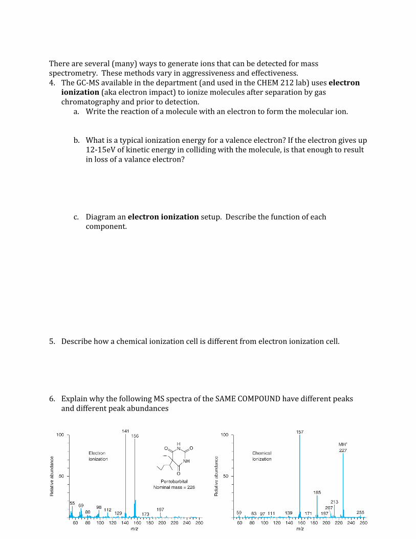
5. Describe how a chemical ionization cell is different from electron ionization cell. 6. Explain why the following MS spectra of the SAME COMPOUND have different peaks
and different peak abundances
3
7. You collect a mass spectrum of a known compound using electron ionization but do not observe a M+ peak. What’s likely happened? Suggest two methods of increasing the detected M+ ion abundance?
8. Is chemical or electron ionization the more aggressive method? What does it mean if an
ionization method is more aggressive?
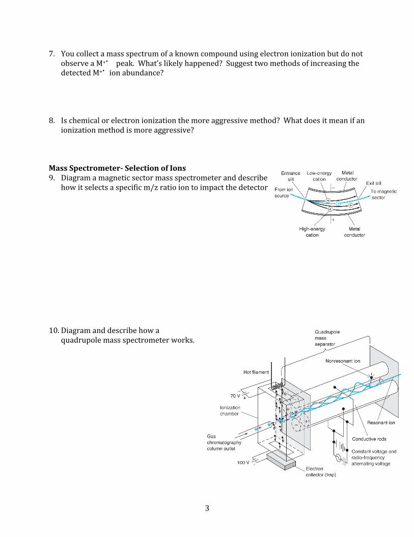
Mass Spectrometer-‐ Selection of Ions 9. Diagram a magnetic sector mass spectrometer and describe
how it selects a specific m/z ratio ion to impact the detector 10. Diagram and describe how a
quadrupole mass spectrometer works.

4
11. Diagram and describe how a TOF works. Mass Spectrometer-‐ Detecting Ions 12. Diagram an electron multiplier. What is the typical amplification? 13. Diagram and describe how a Channeltron converts ions into a
measureable current.
5
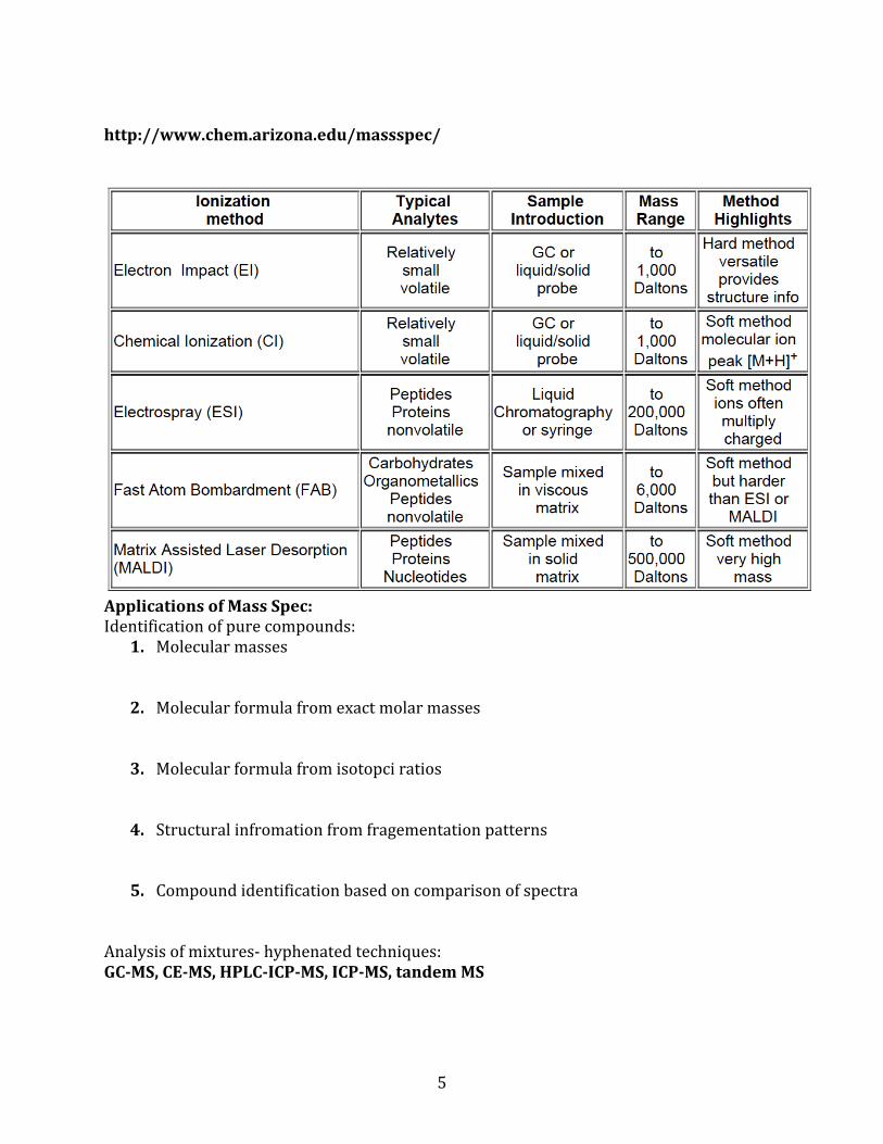
http://www.chem.arizona.edu/massspec/
Applications of Mass Spec: Identification of pure compounds:
1. Molecular masses
2. Molecular formula from exact molar masses
3. Molecular formula from isotopci ratios
4. Structural infromation from fragementation patterns
5. Compound identification based on comparison of spectra
Analysis of mixtures-‐ hyphenated techniques: GC-‐MS, CE-‐MS, HPLC-‐ICP-‐MS, ICP-‐MS, tandem MS

6
Gas Chromatography (GC) 14. State the function of each of the
components of a GC-‐MS. Carrier gas Sample injection-‐ Headspace injection-‐ Liquid injection-‐ Injector chamber-‐ Column Detector options: Gas Chromatography-‐Mass Spectrometry (GC-‐MS)
7
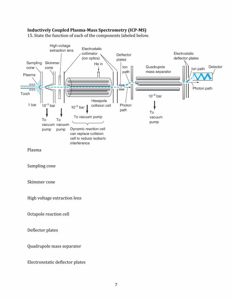
Inductively Coupled Plasma-‐Mass Spectrometry (ICP-‐MS) 15. State the function of each of the components labeled below.
Plasma Sampling cone Skimmer cone High voltage extraction lens Octapole reaction cell Deflector plates Quadrupole mass separator Electronstatic deflector plates
8
16. Draw a nebulizer and describe how the plasma is generated and how sample is introduced.
What did you learn today? What remains unclear?


