Marine Molecular Biotechnology - the-eye.eu [Marine molecular... · Volumes published in the Series...
296
Transcript of Marine Molecular Biotechnology - the-eye.eu [Marine molecular... · Volumes published in the Series...

Marine Molecular BiotechnologySubseries of Progress in Molecular and Subcellular BiologySeries Editor: Werner E. G. Müller
Progress in Molecular and Subcellular BiologySeries Editors: W.E.G. Müller (Managing Editor),Ph. Jeanteur, Y. Kuchino, A. Macieira-Coelho, R. E. Rhoads
39

Volumes published in the Series
Progress in Molecular Subseriesand Subcellular Biology Marine Molecular Biotechnology
Volume 25Signaling Through the Cell Matrix A. Macieira-Coelho (Ed.)
Volume 26Signaling Pathways for Translation:Insulin and NutrientsR.E. Rhoads (Ed.)
Volume 27Signaling Pathways for Translation:Stress, Calcium, and RapamycinR.E. Rhoads (Ed.)
Volume 28Small Stress Proteins A.-P. Arrigo and W.E.G. Müller (Eds.)
Volume 29Protein Degradation in Health and DiseaseM. Reboud-Ravaux (Ed.)
Volume 30Biology of AgingA. Macieira-Coelho
Volume 31Regulation of Alternative SplicingPh. Jeanteur (Ed.)
Volume 32Guidance Cues in the Developing BrainI. Kostovic (Ed.)
Volume 33Silicon BiomineralizationW.E.G. Müller (Ed.)
Volume 34Invertebrate Cytokines and the Phylogeny of ImmunityA. Beschin and W.E.G. Müller (Eds.)
Volume 35RNA Trafficking and Nuclear Structure DynamicsPh. Jeanteur (Ed.)
Volume 36Viruses and ApoptosisC. Alonso (Ed.)
Volume 38Epigenetics and ChromatinPh. Jeanteur (Ed.)
Volume 37Sponges (Porifera) W.E.G. Müller (Ed.)
Volume 39EchinodermataV. Matranga (Ed.)

Valeria Matranga (Ed.)
Echinodermata
With 75 Figures, 11 in Color
1 23

Dr. Valeria MatrangaIstituto die Biomedicina eImmunologia Molecolare „Alberto Monroy“Via U. La Malfa 15390146 PalermoItaly
ISSN 1611-6119ISBN 3-540-24402-6 Springer-Verlag Berlin Heidelberg New York
Library of Congress Control Number: 2004117278
This work is subject to copyright. All rights are reserved, whether the whole or part of the material is concerned,specifically the rights of translation, reprinting, reuse of illustrations, recitation, broadcasting, reproduction onmicrofilm or in any other way, and storage in data banks. Duplication of this publication or parts thereof is permit-ted only under the provisions of the German Copyright Law of September 9, 1965, in its current version, and per-missions for use must always be obtained from Springer-Verlag. Violations are liable for prosecution under theGerman Copyright Law.
Springer-Verlag is a part of Springer Science+Business Mediaspringeronline.com
© Springer Berlin Heidelberg 2005Printed in Germany
The use of general descriptive names, registered names, trademarks, etc. in this publication does not imply, even inthe absence of a specific statement, that such names are exempt from the relevant protective laws and regulationsand therefore free for general use.
Product liability: The publishers cannot guarantee the accuracy of any information about dosage and applicationcontained in this book. In every individual case the user must check such information by consulting the relevantliterature.
Production and typesetting: Friedmut Kröner, Heidelberg, GermanyCover design: design & production GmbH, Heidelberg, Germany
Printed on acid free paper 39/3150 YK 5 4 3 2 1 0
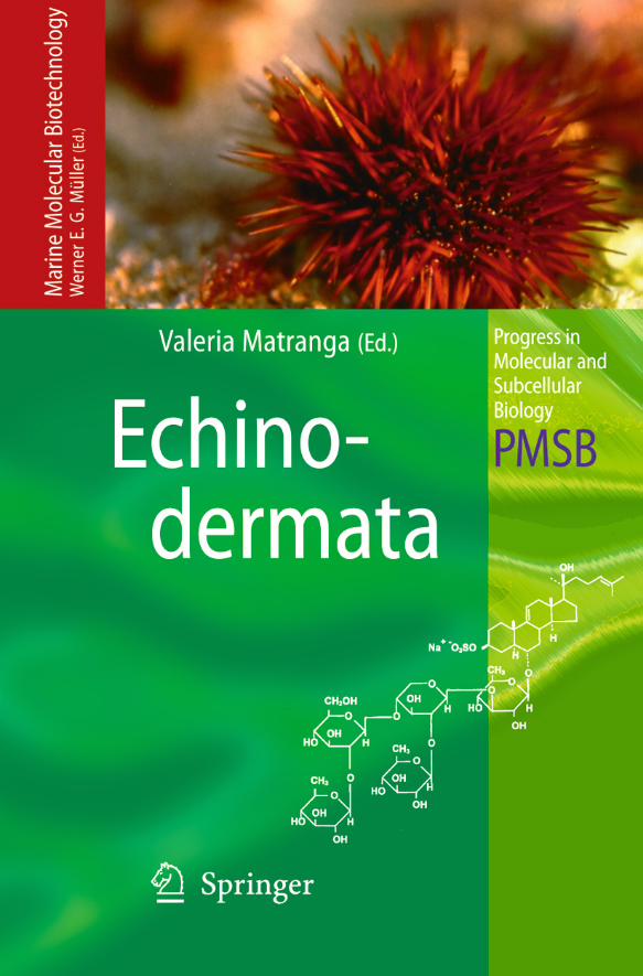
Preface to the Series
Recent developments in the applied field of natural products are impressive,and the speed of progress appears to be almost self-accelerating. The resultsemerging make it obvious that nature provides chemicals, secondary metabo-lites, of astonishing complexity. It is generally accepted that these naturalproducts offer new potential for human therapy and biopolymer science. Themajor disciplines which have contributed, and increasingly contribute, toprogress in the successful exploitation of this natural richness include molec-ular biology and cell biology, flanked by chemistry. The organisms of choice,useful for such exploitation, live in the marine environment. They have thelongest evolutionary history during which they could develop strategies tofight successfully against invading organisms and to form large multicellularplants and animals in aqueous medium. The first multicellular organisms, theplants, appeared already 1000 million years ago (MYA), then the fungiemerged and, finally, animals developed (800 MYA).
Focusing on marine animals, the evolutionary oldest phyla, the Porifera,the Cnidaria and the Bryozoa, as sessile filter feeders, are exposed not only toa huge variety of commensal, but also toxic microorganisms, bacteria andfungi. In order to overcome these threats, they developed a panel of defensesystems, for example, their immune system, which is closely related to thoseexisting in higher metazoans, the Protostomia and Deuterostomia. In addi-tion, due to this characteristic, they became outstandingly successful duringevolution: they developed a chemical defense system which enabled them tofight in a specific manner against invaders. These chemicals are of low mole-cular weight and of non-proteinaceous nature. Due to the chemical complex-ity and the presence of asymmetrical atom centers in these compounds, a highdiversity of compounds became theoretically possible. In a natural selectiveprocess, during evolution, only those compounds could survive which causedthe most potent bioactivity and provided the most powerful protection forthe host in which they were synthesized. This means that during evolutionnature continuously modified the basic structures and their derivatives foroptimal function. In principle, the approach used in combinatorial chemistryis the same, but turned out to be painful and only in few cases successful. Inconsequence, it is advisable to copy and exploit nature for these strategies toselect for bioactive drugs. Besides the mentioned metazoan phyla, other ani-

mal phyla, such as the higher evolved animals, the mollusks or tunicates, orcertain algal groups, also produce compounds for their chemical defensewhich are of interest scientifically and for potential application.
There is, however, one drawback. Usually, the amount of starting materialused as a source for the extraction of most bioactive compounds found inmarine organisms is minute and, hence, not sufficient for their further appli-cation in biomedicine. Furthermore, the constraints of the conventions for theprotection of nature limit the commercial exploitation of novel compounds,since only a small number of organisms can be collected from the biotope.Consequently, exploitation must be sustainable, i.e., it should not endangerthe equilibrium of the biota in a given ecosystem. However, the protection ofbiodiversity in nature, in general, and those organisms living in the marineenvironment, in particular, holds an inherent opportunity if this activity isbased on genetic approaches. From the research on molecular biodiversity,benefits for human society emerge which are of obvious commercial value;the transfer of basic scientific achievements to applicable products is the taskand the subject of Marine Molecular Biotechnology. This discipline uses mod-ern molecular and cell biological techniques for the sustainable production ofbioactive compounds and for the improvement of fermentation technologiesin bioreactors.
Hence, marine molecular biotechnology is the discipline which strives todefine and solve the problems regarding the sustainable exploitation ofnature for human health and welfare, through the cooperation between scien-tists working in marine biology/molecular biology/microbiology and chem-istry. Such collaboration is now going on successfully in several laboratories.It is the aim of this new subset of thematically connected volumes within ourseries “Progress in Molecular and Subcellular Biology” to provide an actualforum for the exchange of ideas and expertise between colleagues working inthis exciting field of “Marine Molecular Biotechnology”. It also aims to dis-seminate the results to those researchers who are interested in the recentachievements in this area or are just curious to learn how science can help toexploit nature in a sustainable manner for human prosperity.
Werner E.G. Müller
Preface to the SeriesVI

Preface
When, nearly 20 years ago, I was participating in the organization of a work-shop held in Palermo entitled “Regulation of Transcription in Sea UrchinEmbryos” (see Fig. 1), under the expert direction of Prof. G. Giudice, at thetime Director of both the University and CNR Institutes for DevelopmentalBiology, I was not aware of the extent to which echinoderms (sea urchins)contribute to the advancement of science. It was spring 1986 and, amongother well-known and those who would one day be well-known in the field,Tim Hunt (R. Timothy Hunt) presented his lecture on the “Role of maternalmRNA in the regulation of cell division in early cell cycles of the sea urchinembryos”. I am sure that at the moment most, if not all, of the audiencefound the way in which a messenger RNA could promote the synthesis of aprotein which was going up and down during cell cycles very bizarre. Manyyears later, his efforts, together with those of Paul M. Nurse and Leland H.Hartwell, were appreciated worldwide. They were awarded the Nobel Prize inPhysiology and Medicine 2001 for “their discoveries of key regulators of thecell cycle”. In fact, their work led to the discovery of cyclins, proteins wide-spread in the animal kingdom, that oscillate throughout the cell cycle, bind-ing and activating cyclin-dependent kinases. Needless to say that nowadayseverybody knows what cyclins are and how studies on their regulation areinspiring new therapies against human cancer. However, very few peopleremember that this all stemmed from a lesson from the sea urchin, just oneexample of how precious simple marine organisms can be in teaching scien-tists how to improve human health.
With this in mind, it was with great pleasure that I accepted the kind invi-tation of Prof. Müller to assemble a book that, by examining the recent pro-ductive research in support of marine biotechnology, would encourage fur-ther studies on the sustainable exploitation of biologically active compoundsfrom echinoderms. Echinodermata, a phylum that appeared back in the Pre-cambrian age, accounting for more than 7,000 living species, belongs to thebranch of the animal kingdom known as the deuterostomes, a group that alsoincludes man. Since they are phylogenetically more related to chordates thanto other invertebrate groups, it is not surprising that echinoderms possessregulatory mechanisms rather similar to those of vertebrates. This explains

the flourishing interest of scientists in the fields of developmental biology,molecular genomics and molecular evolution.
However, as many of the contributors to this book have pointed out, despitethe incredible amount of research accomplished during the past centuriesusing echinoderms as a model organism in the fields of zoology, ecology,embryology, cell biology, molecular biology, etc., modest efforts of the echin-oderm scientific community have been directed towards studies on the devel-opment of the sustainable production of bioactive compounds from echino-derms and their application in biomedicine.
This book illustrates the progress made in the exploitation of natural prod-ucts from marine invertebrates (echinoderms) at a specialized high level.Studies describe: (1) the discovery of new potential therapeutic tools forhuman health; (2) the introduction of new biomolecular and biocellular sen-sors for the detection of environmental contamination; (3) the developmentof new composite materials for biomedical applications; and (4) the improve-ments and limitations of aquacultural techniques and farming. Past andrecent findings are reported in the format of reviews on general topics ordetailed explanations where the description of a recently defined method isneeded.
Environmental and health issues are addressed by the 2004–2010 ActionPlan of the European Community which focuses on: (1) an integrated envi-ronmental and health monitoring system; (2) the standardization of methodsof analysis; and (3) common sampling and sample preparation procedures.Taking these points into consideration, I decided to invite only European sci-entists, with only one exception, to contribute their work and thoughts, mostof them being involved in EU R&D projects which encourage and supportmultidisciplinary research with the aim of ensuring the rapid transfer of tech-nology.
For all the above-mentioned reasons, I am particularly grateful to theMarine Molecular Biotechnology Series Editor, Prof. Werner Müller, who gaveme the opportunity to select those studies which I think will be able to pro-mote discussion in this rapidly growing field and open new routes forresearch on innovative bioactive compounds to be used in environmental andmedical research.
Many thanks are extended to all the contributors to this book; I am confi-dent that the high scientific value of their reviews on past and current find-ings will serve as a forum of ideas for the exploitation of echinoderm as abioresource and will promote the development of studies in the new excitingfield of marine molecular biotechnology.
I am also very grateful to the invaluable and friendly collaboration andassistance of all the actual members of the group (see Fig. 2) whose daily hardwork and professional skills were part of the success of this enterprise. Specialwarm thanks go to my colleagues Francesca Zito and Rosa Bonaventura fortheir continuous and generous collaboration, support and assistancethroughout all the difficulties related to the editorial work.
PrefaceVIII

Finally, I express my gratitude to my husband Benedetto and my daughterLaura for their continuous intellectual and practical support. In spite of thetime it took me away from them, their intelligent and open-minded attitudewas invaluable in this scientific endeavour.
In closing, I would like to recall the words of Prof. Monroy, who used to say:“Science is one of the best ways for mutual understanding. Its language is uni-versal and pays no attention to national barriers. This is to say that science isone of the best approaches to peace”.
Valeria Matranga
Preface IX
Fig. 1. Original sketch of a sea urchin,engaged in “transcribing”, drawn by G.Giudice and used for the cover of thescientific program of the workshop on“Regulation of Transcription in SeaUrchin Embryos”, held in Palermo in1986

Foreword 1
As Director of the Institute of Biomedicine and Molecular Immunology“Alberto Monroy”, I am pleased to introduce this book on the marine molec-ular biotechnology of echinoderms, edited by Dr. Valeria Matranga, with theprecious help of her team, who dedicated much time and effort. I know andappreciate the dedication of Valeria Matranga to her work on cell biology,especially her research on cell–environment interactions and on the valida-tion of the role played by cellular and molecular biology in the detection andassessment of environmental pollution. I am confident that this book willhave a great impact on our understanding of biological markers and their dif-ferent functions in the cell. In addition, the advancements described here willbe extremely useful for the evaluation of environmental risk. Moreover, thisresearch field ultimately addresses issues on the protection of human health,since we can now monitor potentially dangerous changes in the sea environ-ment along the coast where degradation products from the earth accumulate.
The topics addressed in the book, Echinodermata, will be of interest to sci-entists in various fields:∑ Cell biologists will find it useful to study cell responses to stress factors;∑ Marine biologists will be able to evaluate the damage to cells and organ-
isms caused by coastal water pollution, as well as the role of monitoringsuch phenomena;
∑ Researchers dedicated to Nutritional Sciences will appreciate the “warningmessages” provided by the environmental monitoring of seawater;
∑ Environmental scientists will take advantage of an additional way to fightseawater pollution, which could possibly be useful in the prevention andadequate correction of problems originating from ground pollution.
Finally, I wish Valeria Matranga and her team continuing success in theirengrossing and promising research field.
Istituto di Biomedicina e Immunologia Professor Giovanni BonsignoreMolecolare (IBIM) “Alberto Monroy” (Director)Palermo, Italy

Foreword 2
Biology owes a great deal to Echinodermata, especially in the field of develop-ment. Sea urchins have actually represented one of the main and one of thefirst biological materials on which the history of developmental biology wasbuilt.
Fertilization and parthenogenesis were, in fact, first clearly and correctlydescribed in sea urchins. Oskar Hertwig, working at the Zoological Stationof Naples, wrote in 1875: “I was lucky enough, by studying the egg of Toxop-neustes lividus, to find an object in which phases and intimate phenomenaof fertilization were clearly visible, that is: following fertilization, few min-utes after semen addition, around the sperm head, lodged in the cortex, aseries of rays was formed”. The description of a series of important obser-vations followed, which allowed him to conclude: “And therefore I was ableto formulate the low: the fertilization rests on the union of two sexually dif-ferent cells”.
Again, with regard to sea urchins, Edmund B. Wilson (1906) wrote: “Fertil-ization accordingly consists of two distinct phenomena: first the introductioninto the egg of the paternal hereditary characteristics potentially contained insome unknown manner in the substance of the sperm nucleus or of the chro-mosomes in which resolves itself. Second in the introduction into the egg of acentrosome which gives rise to the mechanisms by means of which the eggdivides and the hereditary substance is distributed to the resulting cells”.Thus, the concept of the centrosome is also due to sea urchins.
In 1895 it was again Hertwig who described parthenogenesis in seaurchins, while Jacque Loeb in 1909 showed that it is possible to induceparthenogenesis in sea urchins also by chemical treatments such as hyper-tonic seawater. This caused public surprise and also some concern, such thatsome people even advised women to avoid bathing in seawater owing to thedanger of parthenogenesis!
Furthermore, genetics, in spite of a long-lasting metamorphosis, owessome crucial initial experiments to sea urchins. It suffices here to recall theexperiments of Theodor Bovery, who in 1889 studied sea urchin hybrids,again at the Zoological Station of Naples, and concluded that chromosomesare qualitatively responsible for genetic character transmission, after observ-

ing the fate of hybrid merogones in which some chromosomes were selec-tively lacking.
Although not being the elective material for genetic studies, sea urchinsrepresented and still represent a very important model for studies of molecu-lar biology and molecular genomics. The story started with the demonstra-tion by Alberto Monroy that, following fertilization, there is an activation ofprotein synthesis in the sea urchin egg. This demonstration was achieved byEizo Nakano and Alberto Monroy in Palermo by preloading the amino acidpool of the unfertilized sea urchin egg with radioactive amino acids (whichwas carried out by injecting the labeled amino acids into the coelomic cavityof the adult female), and showing that immediately following fertilization, butnot before, there was a quick transfer of the amino acids from the pool to theproteins. This injection of amino acids into the adult sea urchin was depictedby a cartoonist (myself, at that time one of Monroy’s students; Fig. 1).
The story of molecular genetics has continued in sea urchins, especially inthe USA, where many research groups flourished, especially that of EricDavidson at Caltech, of which I will only recall here the regulatory gene net-works described in sea urchins. It has continued also in Europe, e.g. inPalermo with Monroy’s former students, including myself and youngercoworkers, both at the University and at the National Research Council ofPalermo. It has also continued at the Zoological Station of Naples and else-where, e.g. in Villefranche sur Mer, just to quote an example. Other groups arestudying molecular genomics of sea urchins in Japan.
It should be noted here that the hypothesis of a role for DNA methylation indifferentiation was first proposed in sea urchin embryos in 1958 by EdoardoScarano and by colleagues still working in Naples.
It is also worth recalling the contribution of sea urchins to the field of cellinteraction. Curt Herbst, in 1891, working at the Zoological Station of Naples,found that lowering the calcium concentration of seawater brought about aloosening of contacts between sea urchin blastomeres, which remained insidethe fertilization envelope and continued to develop, although with a “krank-like” aspect. This observation allowed Hans Driesch to easily separate the firsttwo blastomeres and to show that each gave rise to an entire, albeit smaller,
Foreword 2XIV
Fig. 1. Original sketch by G. Giudice representing a furious sea urchin chasing Prof. EizoNakano, armed with a syringe, and Prof. Alberto Monroy, first in the row, at the time oftheir joint experiments in the mid-1950s.

pluteus. Many years later, in 1961, I succeeded in dissociating sea urchin blas-tulae into single cells, essentially by removing calcium. These cells were ableto reaggregate and to develop into pluteus-like structures, which representedthe first example of entire larvae of any kind of embryos reconstituted fromdissociated cells. These studies were followed by those of some colleagues ofmine, e.g. Letizia Vittorelli and Valeria Matranga, and by others, includingYukio Yokota, Hans Noll and David McClay.
The theory of morphogen gradients, so popular in developmental biology,also originated in the study of sea urchins, following the beautiful micro-transplantation experiments done by Sven Hörstadius in 1928 and by theintelligent speculations of John Runnström.
Finally, the beginning of so-called chemical embryology can be attributedto studies on sea urchins: it was in fact Otto Waburg who discovered in 1908that following fertilization of sea urchin eggs there was a sudden increase inoxygen consumption.
I am aware that many important results obtained not only using seaurchins, but using echinoderms in general have not been included here. Ihope, however, to have succeeded in giving an idea of the contribution thatstudies on sea urchins have provided to developmental biology in the past inplaces like Naples Zoological Station, Woods Hole MBL, Stockholm Carolin-ska Institute, Sugashima Marine Station and Villefranche sur Mer Marine Sta-tion, and continue to provide especially in places like the USA (i.e. the MBL inWoods Hole, Caltech, Pennsylvania and so on), Italy (Naples and Palermo),France and Japan.
Dipartimento di Biologia Cellulare e dello Professor G. GiudiceSviluppo “Alberto Monroy”, Università di PalermoPalermo, Italy
Foreword 2 XV

Contents
Are Echinoderms of Interest to Biotechnology? . . . . . . . . . . . . 1C. Petzelt
References . . . . . . . . . . . . . . . . . . . . . . . . . . . . . . . . . 5
Cell Adhesion and Communication: A Lesson from Echinoderm Embryos for the Exploitation of New Therapeutic Tools 7F. Zito, C. Costa, S. Sciarrino, C. Cavalcante,V. Poma,V. Matranga
1 Introduction . . . . . . . . . . . . . . . . . . . . . . . . . . . 72 Why Study Echinoderms? . . . . . . . . . . . . . . . . . . . 102.1 Echinoderms for Biomedical Research: A Simple Model
to Study Biological Events Occurring in Higher Organisms . 112.1.1 Screening for and Testing of Toxic Substances . . . . . . . . 112.1.2 From Echinoderm Molecules to Mammalian Diseases:
How Fundamental Research Points to Clinical Trials . . . . . 122.1.3 Highly Conserved Proteins Associated
with Important Biological Functions . . . . . . . . . . . . . 133 Use of Echinoderm Embryos to Study the Basic
Mechanisms of Communication Among Cells . . . . . . . . 143.1 Sea Urchin Embryo Transparency: A Living Laboratory
for Studying Development and Morphogenesis . . . . . . . 173.1.1 ECM Patterning in Echinoids During Embryo Development 194 Apical Cell Surface . . . . . . . . . . . . . . . . . . . . . . . 204.1 The Apical Lamina . . . . . . . . . . . . . . . . . . . . . . . 214.2 The Hyaline Layer . . . . . . . . . . . . . . . . . . . . . . . . 214.2.1 Pl-Nectin: An Example of Signal(s) from Outside
to Inside the Embryo . . . . . . . . . . . . . . . . . . . . . . 224.2.2 Other Apical ECM Components . . . . . . . . . . . . . . . . 235 Basal Cell Surface . . . . . . . . . . . . . . . . . . . . . . . . 246 The Blastocoel Matrix . . . . . . . . . . . . . . . . . . . . . . 267 Receptors to ECM Molecules . . . . . . . . . . . . . . . . . . 268 Lateral Cell Surface . . . . . . . . . . . . . . . . . . . . . . . 27

8.1 Cell–Cell Adhesion and Communication:The Discovery of Toposome . . . . . . . . . . . . . . . . . . 27
8.2 Other Cell–Cell Adhesion Molecules . . . . . . . . . . . . . . 318.3 Cell Junctions . . . . . . . . . . . . . . . . . . . . . . . . . . 319 Signalling Pathways . . . . . . . . . . . . . . . . . . . . . . . 3210 Growth Factors . . . . . . . . . . . . . . . . . . . . . . . . . 3211 Concluding Remarks . . . . . . . . . . . . . . . . . . . . . . 35References . . . . . . . . . . . . . . . . . . . . . . . . . . . . . . . . . 36
Cell Signalling During Sea Urchin Development:A Model for Assessing Toxicity of Environmental Contaminants . . . 45C. Angelini, M.G. Aluigi, M. Sgro, S. Trombino, H. Thielecke, C. Falugi
1 Basic Research . . . . . . . . . . . . . . . . . . . . . . . . . . 461.1 Cell-to-Cell Signalling During Sea Urchin Development . . . 461.2 Fertilisation . . . . . . . . . . . . . . . . . . . . . . . . . . . 481.2.1 First Phase: Sperm Activation . . . . . . . . . . . . . . . . . 491.2.2 Second Phase: Egg Activation . . . . . . . . . . . . . . . . . 521.3 First Cell Cycle . . . . . . . . . . . . . . . . . . . . . . . . . . 551.4 Cleavages . . . . . . . . . . . . . . . . . . . . . . . . . . . . . 591.5 Ongoing Research . . . . . . . . . . . . . . . . . . . . . . . . 602 Applied Research . . . . . . . . . . . . . . . . . . . . . . . . 612.1 Neurotoxic Contaminants . . . . . . . . . . . . . . . . . . . 612.1.1 Health Risks . . . . . . . . . . . . . . . . . . . . . . . . . . . 612.1.2 Mode of Function of Organophosphates and Carbamates . . 622.1.3 Persistence in the Environment and Crops . . . . . . . . . . 622.2 Biosensors . . . . . . . . . . . . . . . . . . . . . . . . . . . . 632.3 Effects of Neurotoxic Compounds
on P. lividus Ion Dynamics . . . . . . . . . . . . . . . . . . . 63References . . . . . . . . . . . . . . . . . . . . . . . . . . . . . . . . . 65
Echinoderm Reactive Oxygen Species (ROS) Production Measured by Peroxidase, Luminol-Enhanced Chemiluminescence (PLCL) as an Immunotoxicological Tool . . . . . . . . . . . . . . . . . . . . . 71G. Coteur, B. Danis, P. Dubois
1 Introduction . . . . . . . . . . . . . . . . . . . . . . . . . . . 722 Measurement of Reactive Oxygen Species (ROS) Production 733 Modulation of ROS Production by Environmental Factors . 744 Impact of Metal Contaminations . . . . . . . . . . . . . . . . 755 Impact of PCB Contaminations . . . . . . . . . . . . . . . . 786 Impact of Complex Contaminations . . . . . . . . . . . . . . 797 Discussion . . . . . . . . . . . . . . . . . . . . . . . . . . . . 80References . . . . . . . . . . . . . . . . . . . . . . . . . . . . . . . . . 82
ContentsXVIII

Monitoring Chemical and Physical Stress Using Sea Urchin Immune Cells . . . . . . . . . . . . . . . . . . . . . . . . . . . . . . . 85V. Matranga, A. Pinsino, M. Celi, A. Natoli, R. Bonaventura,H.C. Schröder, W.E.G. Müller
1 Introduction . . . . . . . . . . . . . . . . . . . . . . . . . . . 852 Echinoderm Coelomocytes as Immune Effector Cells:
Morphological Features and Recognized Functions . . . . . 873 The Origin of Coelomocytes: Terminally Differentiated
or Circulating Stem Cells? . . . . . . . . . . . . . . . . . . . 904 Molecules Expressed by Echinoderm Coelomocytes . . . . . 914.1 Innate Immune Molecules: The Complement System . . . . 914.2 Agglutinins, Lectins and Adhesion Molecules . . . . . . . . 924.3 Cytoskeleton Proteins . . . . . . . . . . . . . . . . . . . . . . 944.4 NK Antigens and Cytokines . . . . . . . . . . . . . . . . . . 954.5 Stress Response Proteins . . . . . . . . . . . . . . . . . . . . 965 Cell Cultures of Coelomocytes: New Tools
to Detect Marine Pollution . . . . . . . . . . . . . . . . . . . 966 Laboratory Experiments . . . . . . . . . . . . . . . . . . . . 997 Field Studies . . . . . . . . . . . . . . . . . . . . . . . . . . . 1037.1 The Northern Adriatic Sea: First Example
of Monitoring Metal Pollution . . . . . . . . . . . . . . . . . 1037.2 The Southern Adriatic Sea: A Case Study
for the Assessment of TNT Exposure . . . . . . . . . . . . . 1047.3 The North Sea: A Norwegian Fiord as a Natural Gradient
of Metal Contamination . . . . . . . . . . . . . . . . . . . . 1058 Concluding Remarks . . . . . . . . . . . . . . . . . . . . . . 105References . . . . . . . . . . . . . . . . . . . . . . . . . . . . . . . . . 107
DNA Damage and Developmental Defects After Exposure to UV and Heavy Metals in Sea Urchin Cells and Embryos Compared to Other Invertebrates . . . . . . . . . . . . . . . . . . . . . . . . . . . 111H.C. Schröder, G. Di Bella, N. Janipour, R. Bonaventura, R. Russo,W.E.G. Müller,V. Matranga
1 Introduction . . . . . . . . . . . . . . . . . . . . . . . . . . . 1121.1 Atmospheric Ozone . . . . . . . . . . . . . . . . . . . . . . . 1121.2 Penetration of Solar UV Radiation in Marine Waters . . . . 1131.3 Effects of Solar UV Radiation on Aquatic Organisms . . . . 1131.4 DNA Damage . . . . . . . . . . . . . . . . . . . . . . . . . . 1141.5 DNA Repair . . . . . . . . . . . . . . . . . . . . . . . . . . . 1141.6 Solar UV Radiation and Evolution . . . . . . . . . . . . . . . 1141.7 Heavy Metals . . . . . . . . . . . . . . . . . . . . . . . . . . . 1152 Marine Invertebrates as Bioindicators . . . . . . . . . . . . . 115
Contents XIX

2.1 Sea Urchins . . . . . . . . . . . . . . . . . . . . . . . . . . . 1162.2 Sea Urchin Coelomocytes . . . . . . . . . . . . . . . . . . . 1162.3 Assay for DNA Integrity . . . . . . . . . . . . . . . . . . . . 1172.4 Radiation Source . . . . . . . . . . . . . . . . . . . . . . . . 1172.4.1 Full Solar Spectrum Lamp . . . . . . . . . . . . . . . . . . . 1172.4.2 UV-B Lamp . . . . . . . . . . . . . . . . . . . . . . . . . . . 1182.5 DNA Damage in Coelomocytes . . . . . . . . . . . . . . . . 1182.5.1 UV Radiation . . . . . . . . . . . . . . . . . . . . . . . . . . 1182.5.2 Cadmium . . . . . . . . . . . . . . . . . . . . . . . . . . . . . 1192.5.3 Combined Effects . . . . . . . . . . . . . . . . . . . . . . . . 1192.5.4 Expression of HSP70 . . . . . . . . . . . . . . . . . . . . . . 1212.6 Experimental Approach . . . . . . . . . . . . . . . . . . . . . 1212.6.1 Effect of UV-B Radiation on P. lividus Embryo Development 1212.6.2 Effect of Cadmium on P. lividus Embryo Development . . . 1232.7 Porifera . . . . . . . . . . . . . . . . . . . . . . . . . . . . . . 1252.8 Sponge Primmorphs . . . . . . . . . . . . . . . . . . . . . . 1252.8.1 UV Radiation . . . . . . . . . . . . . . . . . . . . . . . . . . 1262.8.2 Cadmium . . . . . . . . . . . . . . . . . . . . . . . . . . . . . 1282.8.3 Combined Effects . . . . . . . . . . . . . . . . . . . . . . . . 1282.9 DNA Repair . . . . . . . . . . . . . . . . . . . . . . . . . . . 1292.9.1 (6–4) Photolyase . . . . . . . . . . . . . . . . . . . . . . . . . 1292.10 Corals . . . . . . . . . . . . . . . . . . . . . . . . . . . . . . . 1303 Concluding Remarks . . . . . . . . . . . . . . . . . . . . . . 131References . . . . . . . . . . . . . . . . . . . . . . . . . . . . . . . . . 131
Echinoderms: Their Culture and Bioactive Compounds . . . . . . . . 139M.S. Kelly
1 Why Cultivate? . . . . . . . . . . . . . . . . . . . . . . . . . . 1402 Sea Urchin Aquaculture . . . . . . . . . . . . . . . . . . . . . 1422.1 Life History and State of the Art . . . . . . . . . . . . . . . . 1422.2 Sea Urchin Larviculture . . . . . . . . . . . . . . . . . . . . . 1432.3 Metamorphosis and the Post-Larval Stage . . . . . . . . . . 1462.4 Sea Urchin Grow-Out Systems . . . . . . . . . . . . . . . . . 1472.5 Juvenile and Adult Somatic Versus Gonadal Growth . . . . . 1472.6 Artificial Diets . . . . . . . . . . . . . . . . . . . . . . . . . . 1482.7 Carotenoids in Sea Urchin Diets . . . . . . . . . . . . . . . . 1492.8 Sea Urchin Harvest Protocol, Spoilage and Shelf-Life . . . . 1492.9 Disease in Cultured Sea Urchins . . . . . . . . . . . . . . . . 1503 Sea Cucumber Aquaculture . . . . . . . . . . . . . . . . . . . 1503.1 Life History and State of the Art . . . . . . . . . . . . . . . . 1503.2 Sea Cucumber Larviculture . . . . . . . . . . . . . . . . . . 1513.3 Metamorphosis and the Post-Larval Stage . . . . . . . . . . 1523.4 Sea Cucumber Growth to Maturity . . . . . . . . . . . . . . 153
ContentsXX

3.5 Disease in Cultured Holothurians . . . . . . . . . . . . . . . 1534 Bioactive Compounds from Echinoderms . . . . . . . . . . 1534.1 Triterpene Glycosides . . . . . . . . . . . . . . . . . . . . . . 1544.2 Glycosaminoglycans: Chondroitin Sulphate . . . . . . . . . 1544.3 Neuritogenic Gangliosides . . . . . . . . . . . . . . . . . . . 1554.4 Antimicrobial Activity . . . . . . . . . . . . . . . . . . . . . 1554.5 Other Types of Bioactive Compounds: Branched-Chain
Fatty Acids, Lectins, Opsonins, Analgesics and Anti-ulcer Compounds . . . . . . . . . . . . . . . . . . . 156
4.6 Regeneration of Nerve Tissue and Arm Regrowth in Crinoids . . . . . . . . . . . . . . . . . . . . . . . . . . . . 156
5 Sustainable Development . . . . . . . . . . . . . . . . . . . . 1575.1 The Research Requirement . . . . . . . . . . . . . . . . . . . 1575.2 Environmental Considerations . . . . . . . . . . . . . . . . . 1575.3 Economic Considerations . . . . . . . . . . . . . . . . . . . 158References . . . . . . . . . . . . . . . . . . . . . . . . . . . . . . . . . 159
Regenerative Response and Endocrine Disruptersin Crinoid Echinoderms: An Old Experimental Model,a New Ecotoxicological Test . . . . . . . . . . . . . . . . . . . . . . . . 167M.D. Candia Carnevali
1 Regeneration and its Biological Implications . . . . . . . . . 1681.1 Regeneration in Echinoderms . . . . . . . . . . . . . . . . . 1691.2 The Regenerative Potential of Crinoids . . . . . . . . . . . . 1712 Endocrine Disrupters and Echinoderms . . . . . . . . . . . 1743 Crinoid Regeneration and Endocrine Disrupters . . . . . . . 1783.1 Experimental Approach . . . . . . . . . . . . . . . . . . . . . 1803.1.1 Exposure Experiments . . . . . . . . . . . . . . . . . . . . . 1803.1.2 PCB Exposure . . . . . . . . . . . . . . . . . . . . . . . . . . 1813.1.3 4-NP Exposure . . . . . . . . . . . . . . . . . . . . . . . . . . 1813.1.4 TPT-Cl Exposure . . . . . . . . . . . . . . . . . . . . . . . . 1813.2 Biological Analysis . . . . . . . . . . . . . . . . . . . . . . . 1823.3 Chemical Analysis: Summary of Analytical Procedures
and Results . . . . . . . . . . . . . . . . . . . . . . . . . . . . 1823.3.1 PCBs . . . . . . . . . . . . . . . . . . . . . . . . . . . . . . . 1833.3.2 4-NP . . . . . . . . . . . . . . . . . . . . . . . . . . . . . . . 1833.3.3 TPT-Cl . . . . . . . . . . . . . . . . . . . . . . . . . . . . . . 1844 Exposure Effects of EDs and Biological Implications
on Regeneration . . . . . . . . . . . . . . . . . . . . . . . . . 1844.1 Mortality . . . . . . . . . . . . . . . . . . . . . . . . . . . . . 1854.2 Growth . . . . . . . . . . . . . . . . . . . . . . . . . . . . . . 1864.3 Malformations . . . . . . . . . . . . . . . . . . . . . . . . . . 188
Contents XXI

4.4 Histological Pattern . . . . . . . . . . . . . . . . . . . . . . . 1885 Conclusions and Future Prospects . . . . . . . . . . . . . . . 193References . . . . . . . . . . . . . . . . . . . . . . . . . . . . . . . . . 196
Echinoderm Adhesive Secretions: From Experimental Characterization to Biotechnological Applications . . . . . . . . . . 201P. Flammang, R. Santos, D. Haesaerts
1 Introduction . . . . . . . . . . . . . . . . . . . . . . . . . . . 2022 Tube Feet . . . . . . . . . . . . . . . . . . . . . . . . . . . . . 2033 Larval Adhesive Organs . . . . . . . . . . . . . . . . . . . . . 2074 Cuvierian Tubules . . . . . . . . . . . . . . . . . . . . . . . . 2115 Applications of Marine Bioadhesives . . . . . . . . . . . . . 2145.1 Design of Water-Resistant Adhesives . . . . . . . . . . . . . 2155.2 Development of New Antifouling Strategies . . . . . . . . . 2165.3 The Potential of Echinoderm Adhesives . . . . . . . . . . . . 217References . . . . . . . . . . . . . . . . . . . . . . . . . . . . . . . . . 218
Mutable Collagenous Tissue:Overview and Biotechnological Perspective . . . . . . . . . . . . . . 221I.C. Wilkie
1 Introduction . . . . . . . . . . . . . . . . . . . . . . . . . . . 2211.1 Collagenous Tissue . . . . . . . . . . . . . . . . . . . . . . . 2211.2 Mechanical Adaptability of Collagenous Tissue . . . . . . . 2222 Mutable Collagenous Tissue: Physiology and Organisation . 2232.1 Overview . . . . . . . . . . . . . . . . . . . . . . . . . . . . . 2232.2 Mechanical Adaptability of MCT . . . . . . . . . . . . . . . . 2242.2.1 Passive Mechanical Properties . . . . . . . . . . . . . . . . . 2242.2.2 Active Contractility . . . . . . . . . . . . . . . . . . . . . . . 2272.3 Organisation of MCT . . . . . . . . . . . . . . . . . . . . . . 2292.3.1 Cells . . . . . . . . . . . . . . . . . . . . . . . . . . . . . . . 2292.3.2 Extracellular Matrix . . . . . . . . . . . . . . . . . . . . . . . 2302.4 Molecular Mechanism of MCT Mutability . . . . . . . . . . 2352.4.1 Are Collagen Fibrils Involved? . . . . . . . . . . . . . . . . . 2352.4.2 Are Calcium Ions Involved? . . . . . . . . . . . . . . . . . . . 2362.4.3 Tensilin–Tensilin Protease Hypothesis . . . . . . . . . . . . 2362.4.4 Active Force Generation . . . . . . . . . . . . . . . . . . . . 2383 Mutable Collagenous Tissue: Biotechnological Perspective . 2383.1 Current Commercial Uses of MCT . . . . . . . . . . . . . . . 2383.2 Biotechnological Potential of MCT . . . . . . . . . . . . . . 2393.2.1 Pharmacological Agents or Strategies . . . . . . . . . . . . . 2393.2.2 New Composite Materials . . . . . . . . . . . . . . . . . . . . 2404 Concluding Remarks . . . . . . . . . . . . . . . . . . . . . . 243References . . . . . . . . . . . . . . . . . . . . . . . . . . . . . . . . . 245
ContentsXXII

Bioresources from Echinoderms . . . . . . . . . . . . . . . . . . . . . 251Y. Yokota
1 Introduction . . . . . . . . . . . . . . . . . . . . . . . . . . . 2512 Oriental Medicine and Historical Background . . . . . . . . 2533 Saponins . . . . . . . . . . . . . . . . . . . . . . . . . . . . . 2534 Glycolipids . . . . . . . . . . . . . . . . . . . . . . . . . . . . 2565 Pigments . . . . . . . . . . . . . . . . . . . . . . . . . . . . . 2566 Venoms . . . . . . . . . . . . . . . . . . . . . . . . . . . . . . 2577 Hemagglutinins . . . . . . . . . . . . . . . . . . . . . . . . . 2588 Prospects . . . . . . . . . . . . . . . . . . . . . . . . . . . . . 2589 Appendix: Antique Illustrations of Echinoderms in Japan . 259References . . . . . . . . . . . . . . . . . . . . . . . . . . . . . . . . . 263
Subject Index . . . . . . . . . . . . . . . . . . . . . . . . . . . . . . . . 267
Taxonomic Index . . . . . . . . . . . . . . . . . . . . . . . . . . . . . 273
Authors Index . . . . . . . . . . . . . . . . . . . . . . . . . . . . . . . 277
Contents XXIII

Are Echinoderms of Interest to Biotechnology?
C. Petzelt
Abstract. The huge potential of echinoderms as a so far fairly untappedsource of bioactive molecules is described. Examples are presented that showthe usefulness of echinoderm-derived molecules for therapeutic applicationin selected fields of cancer research, in the control of bacterial growth as sub-stances with new antibiotic properties, and finally in the context of technicalapplications such as antifouling substances. The molecules described here arebut the mere beginning of a commercial exploitation of echinoderms andmay incite a deeper involvement of biotechnology-oriented research in thismaterial.
Echinoderms have been used as embryos since the dawn of cell biology asmodel systems to study basic phenomena such as mitosis, cell division, differ-entiation, and organ formation, and are linked to the great cell biologists ofthe 19th and 20th centuries; among many others, Boveri, Heilbrunn, Mazia,Monroy, Wilson, Hertwig, and Brachet may be cited. Today, echinodermembryos continue to be the model of choice for many cell and molecular biol-ogists, offering exciting overtures on the way from molecular to cell biology(e.g. Arnone et al. 1997). At the same time they serve as a sensitive test systemfor toxicological and environmental studies (Matranga et al. 2000).
This chapter is not intended to cover the many applications of the peculiar-ities of the echinoderm embryonic system in cell and molecular biology;rather it will give examples of echinoderm-derived substances that may havebiotechnological value.
Without any doubt the marine environment has huge potential as a sourceof new compounds to be used in so far unknown strategies in the combat ofmany pathological situations. In the course of millions of years of evolution,many trials and errors have been made to protect the individual, either the cell
Progress in Molecular and Subcellular BiologySubseries Marine Molecular BiotechnologyV. Matranga (Ed.), Echinodermata© Springer-Verlag Berlin Heidelberg 2005
C. Petzelt ( e-mail: [email protected])Laboratoire International de Biologie Marine (LIBM), 85350 Ile d’Yeu, France. Presentaddress: Experimental Anaesthesiology, University Hospital Charité, Spandauer Damm 130,FH 31, 14050 Berlin, Germany

or the entire organism, and to gain advantage in the face of possible competi-tors. Substances have evolved that interfere with signalling or are simply poi-sonous to other cells. Such products, to be discussed later, could be used in thehuman environment to attack key problems such as cancer, bacterial infec-tions, or, more practically, biofouling. Surprisingly, echinoderms appear to bea rather untapped source in the pursuit of the identification of new and usefulproducts.
Successful identification depends to a large extent on the test system usedand on subsequent purification techniques. Probably one of the most directapproaches is the use of whole animals or pieces of an animal to test whetherthere is any antibacterial or cytotoxic activity.
Sasaki et al. (1985) identified in the macromolecular fractions of aqueousextracts from the sea urchin Strongylocentrotus nudus antitumor activitiesthat inhibited transplanted sarcoma 180 solid form in ICR mice. Unfortu-nately, this interesting work was apparently not followed up. Earlier on, it wasshown in an elegant study that triterpene glycoside isolated from 19holothurian species of the Pacific tropical zone exhibited cytotoxic activityagainst yeast and tumour cells, whereas bacteria were not affected. Out ofthese glycosides the stichoposides, theoturins, and oligosides of Holothuria ofthe genus Bohadschia were found to be the most active versus fungal andyeast microflora and tumour cells (Kuznetsova et al. 1982).
Haug et al. (2002) isolated from the sea urchin, Strongylocentrotus droe-bachiensis, the sea cucumber Cucumaria frondosa, and the starfish Asteriasrubens antibacterial activities that were detected in extracts from several tis-sues in all species tested, but mainly in the coelomocyte and body wallextracts. High antibacterial activity was also found in gastrointestinal organsand eggs from A. rubens and in eggs from C. frondosa. If differences inhydrophobicity and sensitivity to heat and proteinase K treatment were com-pared between active extracts, it was observed that several different com-pounds were responsible for the antibacterial activities detected. Lysozyme-like activity was identified in several tissues from A. rubens. Haemolyticactivity could be detected in all species tested, especially in the body wallextracts. These results indicate that echinoderms may serve as a useful sourcewhen searching for novel antibiotics.
Carballo et al. (2002) used two brine shrimp assays to identify potentialcytotoxic substances useful in cancer therapy. They incubated whole bodyextracts from three echinoderms (Holothuria impatiens, Pseudoconus califor-nica, and Pharia pyramidata) that showed a strong cytostatic (growth inhibi-tion) and cytotoxic effect against two human cell lines, lung carcinoma A-549and colon carcinoma HT-29. Palagiano et al. (1996) isolated up to 20 steroidglycosides from the starfish Henricia downeyae that caused growth inhibitionin bacteria and fungi. In this work, it is remarkable that the biological activityoriginally identified in ethanolic extracts was related to single compoundswhose molecular structures were even identified. Aminin et al. (1995) firstidentified in the Pacific brittle star Ophiopholis aculeata disulfated polyhy-
C. Petzelt2

droxysteroids that turned out to be potent Ca2+ agonists in mammalian cellsystems (Aminin et al. 1995; Agafonova et al. 2002).
Even in the main constituents of the immune systems of echinoderms,cytotoxic substances are found. Coelomocytes are intriguing entities express-ing variable effector mechanisms that are elicited specifically and are repeat-able after a variety of non-self challenges (Glinski and Jarosz 2000; Lin et al.2001). Stabili et al. (1996) were able to isolate from coelomocytes of the seaurchin Paracentrotus lividus a bactericidal protein and purified it to a singlepolypeptide chain with a molecular weight of 60 kDa.
Epibiosis, the colonization of biogenic surfaces by epibiotic organisms suchas bacteria, filamentous algae, and sessile invertebrates, poses a major threatto the fitness and survival of macroorganisms which could potentially befouled. Fouling of artificially submerged structures (e.g. ship hulls) can alsocause severe economic problems, establishing the need for refined bioassaysto determine the efficacy of potential antifouling compounds. Palagiano et al.(1996) identified steroid glycosides from the starfish H. downeyae with pro-found antifouling activities, compounds that were also found in severalspecies of the family Echinasteridae. Iken et al. (2003) monitored the brownalgal spore swimming behaviour in the presence of echinoderm extracts inorder to identify possible antifouling activities. They tested different concen-trations of aqueous and organic extracts from body walls of sympatric echin-oderms (starfish Luidia and Astropecten and the brittle star Astrocyclus).They found significant effects of those extracts on spore swimming behaviourat concentrations three orders of magnitude lower than that present naturallyin the echinoderm body walls.
Sulfated fucans are among the most prominent of all the sulfated polysac-charides of non-mammalian origin that exhibit biological activities in mam-malian systems. Pereira et al. (1999) investigated the anticoagulant activity ofechinoderm fucans in comparison with that of several species of brown algaeand found that the linear sulfated fucans from echinoderms had an anticoag-ulant action resembling that of mammalian dermatan sulfate, whereas thebranched fucans from brown algae were direct inhibitors of thrombin. Suchdifferences have also been described for the linear sulfated fucans derivedfrom sea cucumbers compared to algal fucans (Mulloy et al. 2000).
Glycosphingolipids and glycopeptides are normally occurring consti-tuents of various cell membranes in a wide variety of organisms. Surpris-ingly, these compounds derived from echinoderms exhibit biologicalfunction that might render them useful in medical applications. Glycosphin-golipids have been isolated from sea cucumbers that had neuritogenic activ-ity in the rat pheochromocytoma cell line PC-12, i.e. they were able to induceneurite differentiation in the same way as can be achieved by the addition ofnerve growth factor (Yamada 2002). It remains to be seen whether thesecompounds find medical applications, but even today they are of economicinterest in view of the high price of commercially available nerve growth fac-tor.
Are Echinoderms of Interest to Biotechnology? 3
Several unique lectins are found in echinoderms. In marine invertebrateslectins may be considered as humoral factors in the defence mechanism, asare immunoglobulins in vertebrates, resulting in activation of phagocytes. Onthe other hand, direct haemolytic activity has recently been found in a galac-tose-specific lectin from the sea cucumber Cucumaria echinata (Hatakeyamaet al. 1999). After binding to the specific carbohydrate chains on the erythro-cyte surface, these lectins damage the cell membrane, leading to cell lysis. Alectin with biological activities such as mitogenic and chemotactic character-istics was also described in the venom of the pedicellariae of the sea urchinToxopneustes pileolus (Nakagawa and Kimura 1982) as had been describedwith other bioactive substances many years earlier by Alender (1967) occur-ring in the spines of Diadema sea urchins.
A rich source of useful venoms has been found in the crown-of-thornsstarfish Acanthaster planci. One of its deadly venoms has been identified as amyotoxic phospholipase A (Mebs 1991), and several other candidates for sucheffects have been identified (Shiomi et al. 1985, 1988; Mebs 1989).
A not yet commercially exploited area is the phenomenon of biolumines-cence in the brittle star Amphipolis squamata. The photocytes of this animalproduce light dependent on cyclic nucleotide and IP3, and the system maybecome as useful and widespread as the luciferin–luciferase system is today(De Bremaeker et al. 2000; Deheyn et al. 2000a–e).
Finally, an example may be cited of how echinoderms may become usefuleven in bionics. It has been recently discovered by Aizenberg et al. (2001) thatin the brittle star Ophiocoma wendtii single calcite crystals are arranged insuch a way that they function as lenses. These lenses focus light onto nervebundles that run behind them, which presumably receive the signal to be fur-ther processed. Thus, these thousands of lenses form a compound eye thatcovers the upper surface of the animal, resulting in a function similar to a dig-ital camera that builds up the picture pixel by pixel. At present, engineers inthe photonic industry try to imitate the perfect calcite lenses and their use insignal reception.
This short overview was intended to stimulate the search for bioactivecompounds in echinoderms. It is surprising how little work has been done toidentify promising candidates for applied research, especially in view of thewidespread availability of the animals. It is to be hoped that the urgent needfor new strategies in cancer research or for new candidates for antibiotics inview of the fast development in resistance of harmful bacteria will lead to arenaissance in the field. After all, the potential rewards are of considerablemagnitude.
C. Petzelt4

References
Agafonova IG, Aminin DL, Shubina LK, Fedorov SN (2002) Influence of polyhydroxys-teroids on [Ca(2+)](i). Steroids 67:695–701
Aizenberg J, Tkachenko A, Weiner S, Addadi L, Hendler G (2001) Calcitic microlenses aspart of the photoreceptor system in brittle stars. Nature 412:819–822
Alender CB (1967) A biologically active substance from spines of two diadematid seaurchins. In: Russell FE, Saunders PR (eds) Animal toxins. Pergamon Press, Oxford, p 145
Aminin DL, Agafonova IG, Fedorov SN (1995) Biological activity of disulfated polyhydrox-ysteroids from the Pacific brittle star Ophiopholis aculeata. Comp Biochem Physiol CPharmacol Toxicol Endocrinol 112:201–204
Arnone MI, Bogarad LD, Collazo A, Kirchhamer CV, Cameron RA, Rast JP, Gregorians A,Davidson EH (1997) Green fluorescent protein in the sea urchin embryo: new experi-mental approaches to transcriptional regulatory analysis in embryos and larvae. Devel-opment 124:4649–4659
Carballo JL, Hernandez-Inda ZL, Perez P, Garcia-Gravalos MD (2002) A comparisonbetween two brine shrimp assays to detect in vitro cytotoxicity in marine natural prod-ucts. BMC Biotechnol 2:17–23
De Bremaeker N, Dewael Y, Baguet F, Mallefet J (2000) Involvement of cyclic nucleotides andIP3 in the regulation of luminescence of the brittlestar Amphipholis squamata (Echino-dermata). Luminescence 15:159–163
Deheyn D, Mallefet J, Jangoux M (2000) Cytological changes during light productionprocess in dissociated photocytes from the ophiuroid Amphipholis squamata (Echino-dermata). Cell Tissue Res 299:115–128
Deheyn D, Mallefet J, Jangoux M (2000) Evidence of seasonal variation in luminescence per-formance of Amphipholis squamata (Ophiuroidea, Echinodermata): effect of environ-mental factors. J Exp Mar Biol Ecol 245:245–264
Deheyn D, Mallefet J, Jangoux M (2000) Evidence from polychromatism and biolumines-cence that the cosmopolitan ophiuroid Amphipholis squamata (Echinodermata) mightnot represent a unique taxon. C R Acad Sci Paris 323:499–509
Deheyn D, Mallefet J, Jangoux M (2000) Expression of bioluminescence in Amphipholissquamata (Ophiuroidea: Echinodermata) in presence of various organisms: a laboratorystudy. J Mar Biol Assoc UK 80:179–180
Deheyn D, Jangoux M, Warnau M (2000) Alteration of bioluminescence in Amphipholissquamata (Ophiuroidea: Echinodermata) by heavy metal contamination: a field study.Sci Tot Environ 247:41–49
Glinski Z, Jarosz J (2000) Immune phenomena in echinoderms. Arch Immunol Ther Exp48:189–193
Hatakeyama T, Ohuchi K, Kuroki M, Yamasaki N (1995) Amino acid sequence of a C-typelectin CEL-IV from the marine invertebrate Cucumaria echinata. Biosci BiotechBiochem 59:1314–1317
Haug T, Kjuul AK, Styrvold OB, Sandsdalen E, Olsen OM, Stensvag K (2002) Antibacterialactivity in Strongylocentrotus droebachiensis (Echinoidea), Cucumaria frondosa(Holothuroidea), and Asterias rubens (Asteroidea). J Invertebr Pathol 81:94–102
Iken K, Greer SP, Amsler CD, McClintock JB (2003) A new antifouling bioassay monitoringbrown algal spore swimming behaviour in the presence of echinoderm extracts. Biofoul-ing 19:327–334
Kuznetsova TA, Anisimov MM, Popov AM, Baranova SI, Afiyatullov SS, Kapustina II,Antonov AS, Elyakov GB (1982) A comparative study in vitro of physiological activity oftriterpene glycosides of marine invertebrates of echinoderm type. Comp Biochem Phys-iol 73:41–43
Are Echinoderms of Interest to Biotechnology? 5

Lin W, Zhank H, Beck G (2001) Phylogeny of natural cytotoxicity: cytotoxic activity ofcoelomocytes of the purple sea urchin, Arbacia punctulata. J Exp Zool 290:741–750
Matranga V, Toia G, Bonaventura R, Müller WEG (2000) Cellular and biochemical responsesto environmental and experimentally induced stress in sea urchin coelomocytes. CellStress Chaper 5:113–120
Mebs D (1989) Der Dornenkronenseestern im Korallenriff. Eine oekologische Katatrophe.Naturwiss Rdschau 42:480–482
Mebs D (1991) A myotoxic phospholipase A2 from the crown-of-thorn starfish Acanthasterplanci. Toxicon 29:289–293
Mulloy B, Mourao PA, Gray E (2000) Structure/function studies of anticoagulant sulphatedpolysaccharides using NMR. J Biotechnol 77:123–135
Nakagawa J, Kimura A (1982) Partial purification of a toxic substance from pedicellariae ofthe sea urchin Toxopneustes pileolus. Jpn J Pharmacol 32:966–971
Palagiano E, Zollo F, Minale L, Iorizzi M, Bryan P, McClintock J, Hopkins T (1996) Isolationof 20 glycosides from the starfish Henricia downeyae, collected in the Gulf of Mexico. JNat Prod 59:348–354
Pereira MS, Mulloy B, Mourao PA (1999) Structure and anticoagulant activity of sulphatedfucans. Comparison between the regular, repetitive, and linear fucans from echinodermswith the more heterogeneous and branched polymers from brown algae. J Biol Chem274:7656–7667
Sasaki T, Uchida NA, Uchida H, Takasuka N, Kamiya H, Endo Y, Tanaka M, Hayashi T,Shimizu Y (1985) Antitumor activity of aqueous extracts of marine animals. J Pharma-cobiodyn 8:969–974
Shiomi K, Itoh K, Yamanaka H, Kikuchi T (1985) Biological activity of crude venom fromthe crown-of-thorns starfish Acanthaster planci. Bull Jpn Soc Fish 51:1151–1155
Shiomi K, Yamamoto S, Yamanaja H, Kikuchi T (1988) Purification and characterization ofa lethal factor in venom of the crown-of-thorns starfish (Acanthaster planci). Toxicon26:1077–1081
Stabili L, Pagliara P, Roch P (1996) Antibacterial activity in the coelomocytes of the seaurchin Paracentrotus lividus. Comp Biochem Physiol Biochem Mol Biol 113:639–644
Yamada K (2002) Chemo-pharmaceutical studies on the glycospingolipid constituents fromechinoderm, sea cucumbers, as the medicinal materials. J Pharm Soc Jpn 122:1133–1143
C. Petzelt6

Cell Adhesion and Communication: A Lesson fromEchinoderm Embryos for the Exploitation of NewTherapeutic Tools
F. Zito, C. Costa, S. Sciarrino, C. Cavalcante, V. Poma, V. Matranga
Abstract. In this chapter, we summarise fundamental findings concerningechinoderms as well as research interests on this phylum for biomedical andevolutionary studies. We discuss how current knowledge of echinoderm biol-ogy, in particular of the sea urchin system, can shed light on the understand-ing of important biological phenomena and in dissecting them at the molec-ular level. The general principles of sea urchin embryo development aresummarised, mainly focusing on cell communication and interactions, withparticular attention to the cell–extracellular matrix and cell–cell adhesionmolecules and related proteins. Our purpose is not to review all the work doneover the years in the field of cellular interaction in echinoderms. On the con-trary, we will rather focus on a few arguments in an effort to re-examine someideas and concepts, with the aim of promoting discussion in this rapidlygrowing field and opening new routes for research on innovative therapeutictools.
1Introduction
Echinodermata are among the most familiar marine invertebrates. The phy-lum, characterised by the great morphological variety of its members,belongs to a branch of the animal kingdom known as deuterostomes. TheEchinodermata have been extensively studied, particularly because of someaspects such as the ample fossil record extending back to the Precambrian,their ecological importance in the marine environment, the interesting mor-
Progress in Molecular and Subcellular BiologySubseries Marine Molecular BiotechnologyV. Matranga (Ed.), Echinodermata© Springer-Verlag Berlin Heidelberg 2005
F. Zito (e-mail: [email protected]), C. Costa, S. Sciarrino, C. Cavalcante,V. Poma,V. MatrangaIstituto di Biomedicina e Immunologia Molecolare (IBIM) “Alberto Monroy”, ConsiglioNazionale delle Ricerche,Via U. La Malfa 153, 90146 Palermo, Italy

phology of adult organisms and the advantage of experimentally manipula-ble embryos. Approximately 13,000 echinoderm fossil species are known(Fig. 1), while living species number about 7,000 and fall into five well-defined classes:∑ Crinoidea (sea lilies and feather stars), with about 600 living species. The
body is oriented with the mouth facing up and they may or may not have astalk.
∑ Asteroidea (starfishes or sea stars), the most familiar forms to man, withabout 1,500 living species. Typically, they have their oral surface on the ven-tral side and usually have multiple “arms” surrounding a central disc.
∑ Ophiuroidea (basket stars and brittle stars), with five arms, typically flexi-ble, radiating from a central disc, which are used for locomotion. The classincludes about 2,000 living species.
∑ Echinoidea (sea urchins, sand dollars and sea biscuits), with a usually glob-ular body, formed by the fusion of skeletal plates, without distinct arms.Some of them have bilateral symmetry, which occurred secondarily duringevolution. About 1,700 living species are known.
∑ Holothuroidea (sea cucumbers), characterised by a cylindrical shape and arelatively soft body. About 1,100 living species are known.The Concentricycloidea (sea daisies), with only two species, is an enigmatic
group of echinoderms whose phylogenetic position remains elusive, althoughevidence suggests a relationship with asteroids (Pearse and Pearse 1994). Thephylogenetic relationships among the five classes have been extensively con-troversial, but it seems generally accepted that the class of Crinoideabranched first and that the Echinoidea and Holothuroidea are sister clades(Fig. 2).
Although the main character of the body plan of adult echinoderms, thepentamerous symmetry, led the father of palaeontology, George Cuvier(1769–1832), to place them in the Radiata phylum, it is now widely accepted
F. Zito et al.8
Fig. 1. A Living fossil, the pencil urchin Eucidaris tribuloides. B Presumptive crinoid fossilfound in 1981, in the Black Forest, Germany

that the phylogenetic proximity of echinoderms is to chordates. Amongmarine invertebrates, in fact, the echinoderm clade has a unique position inthe evolutionary tree: during evolution it first appeared in the Metazoa phy-lum with features of deuterostomes, in direct evolutionary line with chordatesand vertebrates, thus implying that there is less divergence between echino-derms and vertebrates than between echinoderms and other invertebrates.This is not surprising since, although the two phyla appear so dissimilar, mol-ecular data and fossil records strongly support this grouping, even if theattempts to identify a common ancestor are highly speculative.As the eminentanthropologist Konrad Lorenz observed,“It is not a theory, but an irrefutable
Cell Adhesion and Communication: A Lesson from Echinoderm Embryos 9
Fig. 2. Hypothetical phylogenetictree of the five living classes of Echin-odermata. This hypothesis, predict-ing that the Crinoidea branched ear-lier than the other classes andsuggesting that the Echinoidea andHolothuroidea are sister clades, wasfirst proposed by Bather (1900) and issupported by morphological andmolecular evidence. (Littlewood et al.1997)

historical fact, that the living world – since its origin – has evolved from‘below’ to ‘above’”. Actually, it is becoming clearer that the genes encodingdevelopmental regulatory proteins already identified in other phyla, the mod-ified interactions among them and changes in their expression patterns are atthe basis of the evolution of animal morphology (Raff 1996; Wray and Lowe2000).
The name echinoderm is derived from the Greek word meaning spiny skinand it has been the original denomination for sea urchins, since spines aretheir main external characteristic. Echinoderms are exclusively marine inver-tebrates and, with a few exceptions, are all benthic organisms (bottom-dwellers). A number of lifestyles are typical among different classes, e.g. seastar and crinoids are predators and filter-feeders respectively, while seaurchins scrape algae from rocks, and holothurians, sand dollars and ophi-uroids often feed on remains.
Echinoderms usually have separate sexes with no evident sexual dimor-phism. Reproduction is typically achieved by external fertilisation, with eggsand sperm freely released into the seawater. The life cycle is usually complex.In most echinoderms the embryo develops into a planktonic larva with abilateral symmetry, that in some species goes through metamorphosis, typi-cally radical, before reaching the final adult morphology.
A number of morphological features are unique to the Echinodermata phy-lum, including:∑ a pentaradial symmetry in adults (higher-order radial symmetry can be
observed in several cases, but it is clearly a secondary modification);∑ a water vascular system, consisting of a set of water-filled canals branching
from a ring canal and leading to tube feet, which has many important func-tions, including locomotion, cellular respiration and feeding;
∑ a mesodermal endoskeleton composed of calcareous plates assembled in ameshwork;
∑ a complex subepithelial nervous system with radial nerves running undereach of the ambulacra, the row of tube feet, but without a recognisable cen-tral part;
∑ sensory neurons located primarily within the ectoderm of podia, withoutspecialised sense organs;
∑ a circulatory system, if present, consisting of a haemal system derived fromcoelomic sinuses.
2Why Study Echinoderms?
There are some features that make echinoderms so interesting to study. First,their sensitivity to environmental changes in seawater ecosystems. It is wellknown that the disappearance of fragile species from certain geographicareas is in direct relationship with high contamination of seawater and sedi-
F. Zito et al.10

ments, and echinoderms are one such fragile species. In particular, embryos,juveniles and adults of the classes Echinoidea and Asteroidea are well utilisedin studies on marine pollution, since they highly resent it. Second, their abil-ity to regenerate parts of the body (particularly Ophiuroidea and Asteroidea),based on stem cell recruitment. This phenomenon makes a fundamental con-tribution to the adaptive capacities of the whole species. Third, their ability tocause remarkable transformations in submarine substrates. Since the echino-derms are one of the most important marine invertebrates that do not feed onfiltration, as they graze on substrate (except for holothurians and crinoids),they can induce changes in the ratios and distributions of other marinespecies (fishes and others), and eventually cause segregation as well as speci-ation. These three features of echinoderms are just some examples of the hugenumber of ways in which echinoderms can be of help in the understanding ofimportant biological phenomena, and in dissecting them at the molecularlevel.
2.1Echinoderms for Biomedical Research: A Simple Model to StudyBiological Events Occurring in Higher Organisms
Recently, a number of studies have offered new notions that non-mammalianmodels may represent important future directions for studies on human dis-eases, since the results obtained from these models are in many cases applica-ble to mammals including humans. Few people realise that research on simplemarine organisms has led to some of our greatest medical advances, as well asto new insights into environmental pollution. One of the advantages of usingechinoderms is that they produce thousands of virtually identical embryos,and that the morphological abnormalities are readily visualised in the liveorganism under the light microscope. In the following text, some aspects ofthe medical advances achieved from such studies will be briefly outlined.
2.1.1Screening and Testing of Toxic Substances
Sea urchin gametes, embryos and larvae can be used for fast, low-cost andreliable screening and testing of toxic substances and for detailed studies ontheir mechanism of action. One example is the screening for the toxicitycaused by retinoids utilised in dermatological practice, since it has beenshown that foetal malformation is a major form of toxicity associated withsome of them (Kahn et al. 1988; Sciarrino and Matranga 1995). The sea urchinembryo has also been used as a model for screening for suspected mam-malian developmental neurotoxicants and for anticancer drug testing (Nish-ioka et al. 2003; Qiao et al. 2003). This test system is applicable also for exami-
Cell Adhesion and Communication: A Lesson from Echinoderm Embryos 11

nation of new and known pharmacologically active substances, includingtheir adverse effects and potential antidotes. Interestingly, a method has beendeveloped to study the invasive properties of metastatic cells and to test thedifferential effects of anti-tumoural substances on their invasive capacity,which makes use of the sea urchin embryo basement membrane (Livant et al.1995; Dyer et al. 2002). This structure is selectively permeable and can beobtained intact with the associated extracellular matrix (ECM) from seaurchin embryos (Livant et al. 1995). It has been demonstrated that all metasta-tic tumour cells placed in contact with these basement membranes were ableto invade them and the invasion was rapid and efficient; on the other hand, asexpected, non-metastatic cells failed in the invasion. These results suggestthat molecules participating in basement membrane recognition and inva-sion have been functionally conserved during evolution and that their consti-tutive activity may allow metastatic cells to escape their tissues of origin(Livant et al. 1995).
2.1.2From Echinoderm Molecules to Mammalian Diseases: How FundamentalResearch Points to Clinical Trials
The analysis of genome sequences led to the finding of novel non-traditionaltargets involved in disease pathogenesis, whose usage has the advantage ofremoving infection without inducing resistance. For example, it has beenshown that novel polysaccharides present on echinoderm surfaces seem ableto stimulate early host defence and microbial clearance, but not the laterphases of inflammatory tissue injury associated with sepsis. These are themost promising alternative or integrative treatments for pneumonia that areunder development (Cazzola et al. 2004).
In the last 10 years, a number of molecules with different effects on mam-malian cells have been purified from echinoderms. These include compoundswith anti-coagulant activity on human blood cells, such as the peptide“plancinin” isolated from the sea star (Koyama et al. 1998), a promising drugfor anti-thrombotic therapy. Other compounds display considerable cytotox-icity against a small panel of human solid tumour cell lines, such as polyhy-droxysterols and saponins isolated from the sea star (Wang et al. 2004) andthe glycolipid A-5 extracted from sea urchin intestine (Sahara et al. 1997,2002). The latter compounds have been suggested as useful drugs for cancerchemotherapy.
Recently, Meijer and Raymond (2003) have reviewed the steps that lead tothe identification of new drugs; that are now under evaluation for therapeuticuse against cancer, neurodegenerative diseases and cardiovascular disorders.This is an example of how results obtained from basic research, i.e. studies onthe cell cycle in the starfish oocyte model, can be utilised in applied medicalresearch and treatment. The starfish cyclin-dependent kinase CDK1/cyclin B
F. Zito et al.12

was initially identified as a universal M-phase-promoting factor and thenused as a screening target to identify pharmacological inhibitors. From thefirst inhibitors discovered, a more selective one was optimised, which is nowentering phase II clinical trials against cancers and phase I clinical testsagainst glomerulonephritis (Meijer and Raymond 2003).
2.1.3Highly Conserved Proteins Associated with Important Biological Functions
The study of echinoderms also led to the identification of proteins with highlevels of homology to vertebrate proteins expressed in particular syndromesor tumour cells. A sea urchin gene showing very strong sequence and struc-tural homology with the gene coding for dystrophin, which is defective inDuchenne muscular dystrophy, has been identified. The partial characterisa-tion of this gene helped in the construction of an evolutionary tree connect-ing the vertebrate dystrophin gene family with related genes in invertebrates(Wang et al. 1998). A novel protein homologue to the sea urchin fascin (anactin-bundling protein) has been found to be over-expressed in pancreaticductal adenocarcinoma, suggesting its use as a tumour marker with potentialdiagnostic and therapeutic implications for pancreatic carcinoma (Maitra etal. 2002).
Sea urchin sperm homologues of polycystin-1 and polycystin-2, the pro-teins mutated in autosomal-dominant polycystic kidney disease, have beensequenced (Mengerink et al. 2002; Neill et al. 2004). Both proteins have beenshown to co-localise exclusively to the plasma membrane over the spermacrosomal vesicle, where they may function as a cation channel mediating thesperm acrosome reaction. These data provide the first suggestion for the roleof a polycystin-1 protein in a specific cellular process (Mengerink et al. 2002).
Recently, a fasciclin-I-like protein has been purified from sea urchinovaries and, by in vitro assays, it has been shown to be active in promotingHT1080 human fibrosarcoma cell attachment (Sato et al. 2004). Fasciclin-I is aneuronal cell adhesion molecule and up to now various proteins belonging tothe family have been identified in different species, including bacteria, plantsand vertebrates, and in the sea urchin embryo and eggs (Brennan and Robin-son 1994; Wessel et al. 2000), although their biological function had not beencharacterised. However, latest findings indicate that the protein is highly con-served in evolution and suggest important biological roles (Sato et al. 2004).
There are also examples of the isolation of new human genes whose func-tion has been hypothesised on the basis of their high homology to alreadyknown and characterised echinoderm genes. A novel human homologue ofthe gene coding for echinoderm microtubule-associated protein (EMAP) hasbeen isolated from a locus of Usher syndrome type 1, an autosomal recessivegenetically heterogeneous disorder. The finding of its high level of homologyto the echinoderm cytoskeletal component EMAP, especially at the micro-
Cell Adhesion and Communication: A Lesson from Echinoderm Embryos 13

tubule-binding domain, and the proposed cytoskeletal nature of Usher dis-ease, define the human EMAP as a good candidate for the USH1a syndromeorigin (Eudy et al. 1997).
3Use of Echinoderm Embryos to Study the Basic Mechanismsof Communication Among Cells
Developmental biology is a discipline studying the mechanisms regulatingembryogenesis and it is one of the most attractive fields in rapid expansionamong the biological sciences. Its study is becoming essential for the compre-hension of any other fields in biology, since it combines molecular and cellu-lar biology, physiology, anatomy, immunology, research on cancer and alsoevolution and ecology. Development is mainly devoted to the production andorganisation of all the different cellular types constituting the adult organism.The generation of different types of cells is a process known as differentiation,while the organisation of differentiated cells in tissues and organs is per-formed during morphogenesis.
It is well known that cells do not behave as single entities, but rather theirassociation in multicellular structures requires precise co-ordinationbetween release and uptake of signals. Communication and interactionamong living cells are, in fact, fundamental events required for the properdevelopment of tissues and organs. Living cells continuously receive inputsfrom the environment and modify their behaviour throughout a complex net-work of signalling pathways. This communication occurs at various levelsand, to obtain co-ordinated responses, the integration of exchanged informa-tion is essential. Malfunctioning of this network of signalling pathways isoften connected with pathological conditions ranging from abnormal prolif-eration to cell death. Understanding the molecular basis of communicationamong living cells is a fundamental challenge for biologists, since, besidesproviding better understanding of the processes controlling growth, differen-tiation and death, it may increase the number of discoveries of new therapiesagainst diseases caused by inappropriate signalling.
Historically, within the phylum of Echinodermata, the sea urchin embryohas been an excellent experimental system for investigating the cellular basisof development, principally because of its relatively simple organisation andbecause of its optical transparency that makes the observation of morpho-genesis in vivo possible. Pioneering studies on development date back to 1892,when Driesch, following the experimental approach to embryology proposedby Roux about 10 years before, utilised the sea urchin embryo in his studieswith important outcomes for embryology (Driesch 1892). On the basis of hisresults, Driesch proposed the very modern concept of nucleus–cytoplasminteraction as an essential event for development (Driesch 1894). Later, from1928 to 1935, Hörstadius performed some of the most remarkable experi-
F. Zito et al.14

ments in the history of embryology using the sea urchin embryo (Figs. 3, 4).First, he separated each blastomere of the early embryo and followed theirfates; then he was able to recombine different series of blastomeres and, fromthe results obtained, to propose the well-known theory on the existence ofgraded properties within the unfertilised egg and the early embryo (Hörsta-dius 1939).
The sea urchin system was also one of the first in which time-lapsemicroscopy was exploited extensively. For example, the classic studies ofGustafson and Wolpert (1967) led to the identification of many of the basicbehaviours exhibited by cells in the embryo during morphogenetic move-ments. At the time, these authors remarked, “we are, however, still ignorantabout the final steps in the casual chain between the genes and the shapes theycontrol” (Gustafson and Wolpert 1967). In the past 35 years, our knowledge ofthe molecular basis of developmental processes and the relationship betweenmolecules and cell behaviour has advanced considerably. The classic studies
Cell Adhesion and Communication: A Lesson from Echinoderm Embryos 15
Fig. 3. Cover of the originalreview by Hörstadius (1939)on development of the Para-centrotus lividus sea urchinembryo (Kindly provided byM. Delarue, Laboratoire Biolo-gie et Multimédia, UPMC-P6,www.snv.jussieu.fr/bmedia/sommaires/dvpt.htm)
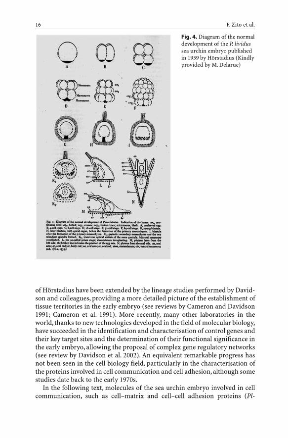
of Hörstadius have been extended by the lineage studies performed by David-son and colleagues, providing a more detailed picture of the establishment oftissue territories in the early embryo (see reviews by Cameron and Davidson1991; Cameron et al. 1991). More recently, many other laboratories in theworld, thanks to new technologies developed in the field of molecular biology,have succeeded in the identification and characterisation of control genes andtheir key target sites and the determination of their functional significance inthe early embryo, allowing the proposal of complex gene regulatory networks(see review by Davidson et al. 2002). An equivalent remarkable progress hasnot been seen in the cell biology field, particularly in the characterisation ofthe proteins involved in cell communication and cell adhesion, although somestudies date back to the early 1970s.
In the following text, molecules of the sea urchin embryo involved in cellcommunication, such as cell–matrix and cell–cell adhesion proteins (Pl-
F. Zito et al.16
Fig. 4. Diagram of the normaldevelopment of the P. lividussea urchin embryo publishedin 1939 by Hörstadius (Kindlyprovided by M. Delarue)

nectin, fibronectin, laminin, collagen, integrin, toposome, cadherin), growthfactors (BMP2/4, univin), signal transduction molecules (kinases), and theirgenes (when characterised) will be described. Our intention is not to reviewall the work done over the years in the field of cellular interaction in echino-derms, but rather we will focus on a few arguments on the assumption of re-examining some ideas and concepts.
3.1Sea Urchin Embryo Transparency: A Living Laboratory for StudyingDevelopment and Morphogenesis
To facilitate the reader in the following discussion on the relationshipbetween adhesion/signalling molecules and cell differentiation occurringduring embryogenesis, the development of the Mediterranean sea urchinspecies Paracentrotus lividus will be briefly described. The time scale andembryo morphology apply, with very little modifications, to many other seaurchin species.
Fertilisation is the first event that gives rise to development and triggersinstantaneously several changes within the egg. A transient decrease in themembrane potential, a slight increase in the intracellular pH and free calciumions activate several metabolic processes, causing, among other things, theexocytosis of cortical granules and the consequent elevation of the fertilisa-tion membrane (Fig. 5A). Cleavage stages begin within 1 h after fertilisationand are characterised by a number of cell divisions occurring about every30 min (Fig. 5B,C). While the first three divisions give rise to eight equivalentblastomeres, the fourth one produces 16 cells of different sizes: eightmesomeres at the animal pole, four macromeres and four micromeres at thevegetal pole (Fig. 5D). Cameron and colleagues were able to describe exten-sively the cell lineage for each of the 16 blastomeres; in addition, they showedthat most of the lineage founder cells for many tissues of the later embryo areestablished at the 64-cell stage (Cameron and Davidson 1991; Davidson et al.1998). It is noteworthy to underline that the lineage boundaries are detectedwell before the appearance of any morphological difference in the embryoand the spatially restricted patterns of gene expression already describedagree with these lineage boundaries. As cell divisions proceed, the embryodevelops into a blastula, which consists of one layer of epithelial cells, with atypical apical–basal polarity, surrounding a filled cavity, the blastocoel(Fig. 5E). Later, the embryo hatches from the fertilisation envelope to becomea free-swimming blastula. From this stage another phase of developmentstarts, which is characterised by a number of morphogenetic movements thatwill completely rearrange the embryo. The first cells to move are the primarymesenchyme cells (PMCs) that detach from the vegetal plate and ingress intothe blastocoel, where they migrate using short filopodia (mesenchyme blas-tula stage; Fig. 5F). Some PMCs migrate towards two ventro-lateral sites and
Cell Adhesion and Communication: A Lesson from Echinoderm Embryos 17

form clusters that are connected by other PMCs in a form of a subequatorialring. The morphology of PMCs during migration was described for the firsttime by Gustafson and Wolpert using time-lapse microscopy (1961, 1967).Shortly after PMC ingression, the vegetal plate epithelium invaginates to formthe archenteron, which is the future intestine. It is possible to distinguish atleast three gastrula stages during the elongation of the archenteron towardsthe animal pole: early, middle and late gastrula (Fig. 5G–I). PMCs produce theskeleton, first in the form of triradiate spicules, which elongate and pattern ina complex species-specific manner. Skeleton is formed by calcium and mag-nesium carbonate, which deposit on an organic matrix constituted by a num-ber of well-known spicule-specific proteins. In the laboratory, the larval stageof pluteus is obtained after 48 h post-fertilisation: it shows four arms, a com-plex patterned skeleton and a tripartite intestine, encircled by muscles under-going peristaltic contractions (Fig. 5J,K). At this point, the larva, if correctlyfed, is ready to continue development and form the juvenile sea urchinthrough metamorphosis.
From the developmental processes outlined above it is evident that cell–celland cell–ECM contacts play a fundamental role in morphogenetic movementsoccurring during embryogenesis. Knowledge of the key actors involved aswell as understanding of their complex interactions would eventually lead tothe unravelling of the developmental machinery. Furthermore, it should benoted that cells forming the blastula stage monolayer exhibit a structuralasymmetry of the cytoplasm and the plasma membrane is compartmen-
F. Zito et al.18
Fig. 5A–K. Development of the sea urchin embryo Paracentrotus lividus. A Fertilised egg;B two blastomeres; C four blastomeres; D 16 blastomeres; E early blastula; F mesenchymeblastula; G early gastrula; H middle gastrula; I late gastrula; J early pluteus; K pluteus

talised into distinct apical, basal and lateral domains, with characteristic lipidand protein compositions. Embryogenesis and morphogenesis are charac-terised by cell movements and complex cell rearrangements, which requireappropriate interactions of cells with the underlying ECM by means of spe-cific membrane receptors. For this reason, interest in the identification, purifi-cation and functional studies of ECM components, along with their ligandsand other molecules involved in cell–matrix adhesion, has increased in recentyears. In the following text, after a brief description of the appearance of theECM upon embryo development, protein molecules found in different plasmamembrane compartments of blastula cells will be described.
3.1.1ECM Patterning in Echinoids During Embryo Development
In recent years, it has been shown that the ECM of the sea urchin embryo is avery complex structure, consisting of a number of layers organising duringdifferent developmental steps. Using specific antibodies, different storagecompartments of the ECM components, like granules and vesicles, have beenidentified in the unfertilised egg cytoplasm. The protein contents of thesecompartments are exocytosed and assembled in a highly regulated fashion atdifferent moments after fertilisation (for a review see McClay et al. 1990). Theearly event after sperm entry is the elevation of the fertilisation envelopeabove a water-filled perivitelline space (Fig. 5A; for more detailed descriptionon ionic events occurring at this stage, see the chapter by Angelini et al., thisVol.). Then, a new ECM is secreted on the outer surface of the zygote, whichgives rise to different layers, the most acknowledged of them being the apicallamina and the hyaline layer. During cleavage, other ECM molecules are alsoreleased into the newly forming basal lamina, which will underlie the blasto-coelic cavity from the stage of blastula (Fig. 5E). A great number of in vitroand in vivo studies suggest that ECM has an important role during morpho-genesis, serving as a mechanical support to the embryo as well as a substratefor cell movements, and providing both spatial and temporal information toadherent cells. Thus the ECM is not a fixed structure but rather is able torespond to and effect changes in its local microenvironment.
Changes in ECM composition and regulated matrix remodelling are com-mon events associated with embryogenesis and are carried on by matrix met-alloproteases (MMPs). These molecules are a growing family of metalloen-dopeptidases that act by cleaving the protein components of the ECM andthereby regulating its composition (for a review see Stamenkovic 2003). Formany years MMPs were believed to function in facilitating cell migration sim-ply by removing barriers such as collagen. However, recent discoveries haveshed new light on the role of MMPs in embryology, physiology and disease. Itis becoming increasingly clear, in fact, that MMPs are also involved in thefunctional regulation of non-ECM molecules, including growth factors and
Cell Adhesion and Communication: A Lesson from Echinoderm Embryos 19

their receptors, cytokines and chemokines, adhesion receptors and cell-sur-face proteoglycans, and a variety of enzymes. MMPs therefore play an impor-tant role in the control of cellular interactions with their environment, pro-moting tissue turnover, both physiological, such as normal development, andpathological, such as inflammation and cancer. The sea urchin embryo wasfound to express a dynamic pattern of gelatinase activities associated with theexternal ECM, some expressed only in later stages of development, othersexpressed in the unfertilised egg and persisting throughout the course ofembryonic development (Mayne and Robinson 1996; Robinson 1997; Flood etal. 2000). Their substrate specificity and metal ion requirements suggest thatthese molecules are members of the MMP class of ECM remodellingenzymes.
In recent years, a new family of molecules in the animal kingdom has beendescribed, called ADAMs (A Disintegrin And Metalloprotease). These aremultidomain transmembrane proteins containing a metalloproteinase and adisintegrin domain and all or some of the following structures: a signal pep-tide, a propeptide, a cysteine-rich and an epidermal growth factor (EGF)-likedomain, a transmembrane region, and a cytoplasmic tail. As a consequence,ADAMs could have four potential functions: proteolysis, adhesion, fusion andintracellular signalling (Stone et al. 1999). Although the number of ADAMgenes has grown rapidly, the biological functions of most members are stillunclear. However, they seem to be key regulators of the cell–cell and cell–ECMinteractions (see review by White 2003). Recently, a sea urchin SpADAM genehas been sequenced and the deduced protein sequence includes all the char-acteristic domains of vertebrate ADAMs. The structure and the types of cellsin which SpADAM orthologues are expressed are then apparently conservedin deuterostomes (Rise and Burke 2002).
4Apical Cell Surface
It has been reported that the first ECM structure developing soon after fertil-isation of the egg is the external one. As viewed by electron microscopy afterruthenium staining (Lundgren 1973), the presence of at least two distinct lay-ers is evident: an inner apical lamina more tightly associated with the apicalplasma membrane, which is the thickest, and an outer layer, the hyaline layer,associated with the tips of microvilli (see Fig. 6).
F. Zito et al.20

4.1The Apical Lamina
The apical lamina is a fibrous layer surrounding the embryo, which remainson the apical surface after removal of hyalin from the hyaline layer (Hall andVacquier 1982; Burke et al. 1998), and is composed principally of fibropellins(Bisgrove et al. 1991; Burke et al. 1991). The gene coding for one of the threeknown fibropellins has been sequenced and, like other extracellular mole-cules, it contains a series of up to 20 epidermal growth-factor-like repeats(Delgadillo-Reynoso et al. 1989; Grimwade et al. 1991; Bisgrove and Raff1993). In vitro experiments showed that dissociated cells adhere to affinity-purified fibropellins in a temperature-, time- and dose-dependent manner,suggesting the role as a substrate for cell adhesion (Burke et al. 1998). Their invivo functional role has been investigated using monoclonal antibodies,which were shown to interfere with the initial phase of gastrulation (Naka-jima and Burke 1996). At this time no further proteins have been described asbelonging to this layer.
4.2The Hyaline Layer
Historically, the first function assigned to the hyaline layer has been to holdblastomeres together (Osanai 1960; Vacquier and Mazia 1968). In agreementwith this hypothesis, Citkowicz (1971) showed that the hyaline layer forms a
Cell Adhesion and Communication: A Lesson from Echinoderm Embryos 21
Fig. 6. Schematic diagram of the ultrastructure of the epithelium of the blastula embryo.(Adapted from Hardin 1996)

shell that remains structurally intact after removal of epithelial cells usinghyperosmotic solutions, demonstrating that it is a structure independentfrom cells that lie underneath. A number of proteins constituting the hyalinelayer have been identified and characterised. The major component is hyalin,a high molecular weight glycoprotein secreted by the cortical granules, whichcan be isolated by successive steps of solubilisation and precipitation throughremoval and re-addition of Ca2+ to seawater or other osmotically balancedmedia (Kane 1970). Hyalin has been extensively studied both structurally andfunctionally in sea urchin and sea star embryos (Vacquier 1969; Kane andStephens 1969; Spiegel and Spiegel 1979; Adelson et al. 1992) and partialcDNAs have been isolated by screening expression libraries with monoclonalantibodies to hyalin (Wessel et al. 1998). Its sequence contains consensus cal-cium-binding motifs, in agreement with its capacity to interact with calciumbiochemically (Robinson et al. 1992), and modular repeats similar to thatknown in other large ECM molecules (McClay 1991). Hyalin has been shownto support cell adhesion in vitro (McClay and Fink 1982) and to be involved insea urchin morphogenesis in vivo (Adelson and Humphreys 1988). Interest-ingly, a recent study has demonstrated that two mammalian cortical granuleenvelope proteins share common antigenic epitope(s) with echinodermhyalin and, like hyalin, play a role in early embryogenesis (Hoodbhoy et al.2000).
4.2.1Pl-Nectin: An Example of Signal(s) from Outside to Inside the Embryo
About 10 years ago, with the aim of purifying fibronectin from the sea urchinembryo, we isolated and characterised another ECM protein localised in thehyaline layer, namely Pl-nectin (Fig. 6). Although the protocol used for itspurification was that originally developed for fibronectin, the molecularweight and other features indicated that this protein was a new molecularspecies. Pl-nectin has been isolated from the species Paracentrotus lividus(Matranga et al. 1992) and Temnopleurus hardwickii (Th-nectin; Yokota et al.1994) as a collagen-binding molecule and, like other ECM components, it hasbeen found stored in specific cytoplasmic vesicles, recently named necto-somes, in the unfertilised egg (Kato et al. 2004). The genesis and distributionof the nectosome during oogenesis, its translocation to the cortex and gradualsecretion into the hyaline layer after fertilisation have recently been fully doc-umented by using immunoelectron microscopy in the Japanese species T.hardwickii (Kato et al. 2004). At later stages, Pl-nectin has been found to belocalised on the apical surface of ectoderm and endoderm cells. By in vitroassays, the protein has been shown to mediate cell-substrate adhesion in adose-dependent fashion, suggesting a functional role during development(Matranga et al. 1992). Thus it was crucial to investigate the in vivo Pl-nectinfunction by morphogenetic assays in which embryos were cultured in the
F. Zito et al.22

presence of monoclonal antibodies to Pl-nectin. To our surprise, after thistreatment, we observed the presence of a high number of embryos with dra-matic skeleton defects, but with normally developed ectoderm and endodermstructures (Fig. 7; Zito et al. 1998, 2000). In addition, depending on the amountof antibody used, it was possible to obtain embryos with different degrees ofskeleton defects, which were classified on an arbitrary scale (Zito et al. 2003).These data suggested that outer ectoderm–Pl-nectin interaction is indirectlyinvolved in inner skeleton formation. For more detailed description on thisissue, see Section 10. The partial sequence of the coding region of the Pl-nectin gene (accession no. AJ578435) has revealed its similarity with anothercomponent of the hyaline layer, namely echinonectin, purified from thespecies Lytechinus variegatus. This protein has a lectin-binding activity thatallowed its chromatographic purification and, like Pl-nectin, it is an adhesivesubstratum for cells (Alliegro et al. 1988; 1990). Although in a previous reportno cross-reaction between the two molecules was found, it is very likely thatPl-nectin and echinonectin belong to the same family of ECM components.
4.2.2Other Apical ECM Components
Concerning the presence of fibronectin-like molecules in the sea urchinembryo, first evidence was obtained by immunofluorescence experimentsusing antibodies against human fibronectin, even if the cross-reactive mater-ial was described as being differently distributed in the embryo, i.e. on theouter cell surface of the epithelial layer (Spiegel et al. 1983), on the basementmembrane (Spiegel et al. 1980; DeSimone et al. 1985), and also on the surfaceof migrating PMCs (Katow et al. 1982). A sea urchin fibronectin-like moleculehas been purified for the first time from ovaries of Pseudocentrotus depressusby Iwata and Nakano (1981, 1983). The same authors have also demonstratedthat this protein is involved in migration, adhesion and spicule synthesis of invitro-cultured micromeres (Miyachi et al. 1984). Later, fibronectin-like pro-teins were purified from five different species living in Mediterranean and
Cell Adhesion and Communication: A Lesson from Echinoderm Embryos 23
Fig. 7A, B. Skeletondefects obtained afterculturing embryos in thepresence of high con-centrations of mono-clonal antibody to Pl-nectin. A Controlembryo cultured in thepresence of unrelatedIgGs; B treated embryoshowing strong skeletondefects
Pacific seawaters and their biochemical and immunological relationshipshave been analysed for the purpose of comparative phylogenetic studies(Matranga et al. 1995). These proteins differed in their binding affinity togelatin but shared different epitopes, suggesting that they are members of asea urchin fibronectin superfamily. Furthermore, analysis of the different fea-tures of fibronectin-like proteins and sea urchin nectins, i.e. Pl-nectin and Th-nectin, has been carried out, since essentially the same method was utilised topurify both groups of molecules (Yokota et al. 1994). It has been shown thatthese two groups of proteins belong to different ECM protein families sincethey differ in their affinity to collagen as well as in their localisation inside theembryo.
A gene coding for a new ECM protein has been sequenced in the directdeveloper Heliocidaris erythrogramma. By the use of polyclonal antibodiesraised against the protein, it has been demonstrated that it is localised on theapical surface of ectoderm, in tight association with the plasma membrane.The protein has been named apextrin and it has been proposed to be involvedin apical cell adhesion (Haag et al. 1999).
Although a reasonable amount of information is currently available on thehyaline layer and apical lamina components, the spatial relationship betweenthem is still unclear. An approach to such studies could come from experi-ments of co-immunoprecipitation after transient cross-linking of surfacemolecules in live embryos.Alternatively, electron microscopic observations ofrotary shadowed purified molecules in diluted solutions, or in combinationwith other potential partners, would serve to solve the intricate meshworkthat surrounds the embryo.
5Basal Cell Surface
As previously reported, the basal lamina develops when new ECM moleculesare released, from the blastula stage, at the basal surfaces of epithelial cells,into the constituting blastocoelic cavity (Fig. 6). A discrete number of reportshave described the formation of this layer and its function in sea urchin devel-opment (Okazaki and Nijima 1964; Kawabe et al. 1981; Galileo and Morrill1985; Amemiya 1989).
Different experimental approaches have shown the presence of few types ofcollagens in the sea urchin embryo, in contrast to about 20 different types ofthe protein found in higher metazoan phyla. By immunofluorescence, Wesselet al. (1984) showed collagen localisation on the basal lamina of the embryo,using antibodies against known vertebrate ECM components, i.e. collagentypes I, III and IV. Partial biochemical characterisation of collagen-like mole-cules has been carried out from in vitro-cultured micromeres (Pucci-Minafraet al. 1975; Benson et al. 1990; Shimizu et al. 1990). Collagen genes have beencharacterised in several species of sea urchin. Using mouse type IV cDNAs,
F. Zito et al.24

genomic clones have been isolated from S. purpuratus and characterised(Venkatesan et al. 1986). A putative collagen gene has also been cloned from aP. lividus genomic library using a C. elegans collagen gene as a probe (D’A-lessio et al. 1989, 1990; Exposito et al. 1992). It has also been demonstrated thatfibrillar collagen types I and II and non-fibrillar collagen type IV areexpressed only by PMCs and secondary mesenchyme cells (SMC) (Angerer etal. 1988; D’Alessio et al. 1989; Wessel et al. 1991; Lethias et al. 1995).
The first evidence of the presence of a vertebrate laminin-like molecule as acomponent of the embryonic sea urchin basal lamina came from studies thatutilised an monoclonal antibody specific to laminin (McCarthy and Burger1987). Later, the isolation and characterisation of a cDNA clone encoding aregion of the carboxy terminal globular domain (G domain) of the alpha-1 chainof laminin from the sea urchin S. purpuratus was reported (Benson et al.1999).
Among echinoderm-specific ECM components, pamlin has been isolatedfrom H. pulcherrimus embryos (Katow 1995). The author showed that PMCsisolated from mesenchyme blastulae bound exclusively to pamlin, whichstimulates their migration in in vitro functional assays. Analysis of the mole-cular image of pamlin by transmission electron microscopy showed that theprotein is composed of three subunits, which can aggregate in a complex fash-ion, giving rise to a large supramolecular network (Omoto and Katow 1998).
Other ECM molecules have been identified as part of the basal lamina sur-rounding the blastocoel (Fig. 6). A novel protein has been purified from thesea urchin species P. lividus and H. pulcherrimus, and its biochemical charac-terisation and functional features have been described (Tesoro et al. 1998).The protein has been named Pl-200K or Hp-200K, respectively, because of thespecies from which it was isolated, and, as assessed by affinity chromatogra-phy columns, it shows different binding affinities to type I collagen andheparin. Pl-200 K has been shown to function as an adhesive substrate, and tobe involved in the regulation of sea urchin embryo skeletogenesis (Tesoro etal. 1998).
By means of specific monoclonal antibodies, a molecule with multiple cal-cium-binding domains, ECM3, has been shown to be arranged in fibres on thebasal surface of the ectoderm. These studies established ECM3 as a strongcandidate for a PMC substrate molecule and point to several possible mecha-nisms by which interactions between PMC filopodia and ECM3-containingfibers could provide guidance information to migrating PMCs (Hodor et al.2000).
As in the case of external ECM, it would be interesting to know the relation-ship among all the ECM proteins described so far and their spatial organisa-tion.
Cell Adhesion and Communication: A Lesson from Echinoderm Embryos 25

6The Blastocoel Matrix
It is useful to remember that the blastocoel is not an empty cavity, but itbecomes filled with a matrix whose composition is still not well known(Fig. 6). Several methods have been used to visualise and study the structureof the blastocoel matrix that forms at the blastula stage. Earlier electronmicroscope studies described this matrix as a “meshwork of fibrous and gran-ular material” which is an extension of the basal lamina (Katow and Solursh1979). More recent studies have revealed an oriented meshwork, formed byfibrillar sheets interconnected by finer filaments, that completely fill the blas-tocoelic cavity, in contact with the basal lamina (Cherr et al. 1992). Althoughdetailed information about the structure of the blastocoel matrix is available,in this case little is known about the protein molecules constituting it (forreview see Solursh 1986).
7Receptors to ECM Molecules
ECM molecules are clearly important, but equally significant are the receptorsby which cells interact with it, the major class being the integrin superfamily(for a review see Berman et al. 2003; Danen and Sonnenberg 2003). More than20 of these receptors have been identified so far in different vertebrates: theyare heterodimers of two transmembrane chains, a and b, and are widelyexpressed among the animal kingdom (Bokel and Brown 2002). The differentcombination of a and b subunits generates integrins with differing ligandspecificity (Hynes 1992). Due to their structure, integrins can interact withECM by their extracellular domains and transduce external signals to theinside by their cytoplasmic tails, which interact with the cytoskeleton, sig-nalling molecules and other cellular proteins, resulting in regulation of manybiological functions. These include adhesion, motility, shape, polarity, growthand differentiation (Yamada 1997). Although a reasonable number of ECMproteins have been described in the sea urchin embryo, little is known abouttheir putative receptors. In recent years, however, it has been shown that thissystem expresses integrins structurally similar to those characterised in otheranimals (Burke 1999). Marsden and Burke (1997) reported evidence of threenovel b integrin subunits that are expressed during early development of S.purpuratus. The full-length cDNA sequence for one of them, bG, shows 58 %similarity to vertebrate integrins, particularly to the cytoplasmic domain inwhich amino acids of the human b1 subunit involved in cell adhesion and sig-nalling are conserved (Marsden and Burke 1997). Functional assays usingantibodies and Fab fragments against another sea urchin b integrin, bL,demonstrated its involvement in the initial phase of gastrulation and in theorganisation of actin filaments (Marsden and Burke 1998). Our preliminary
F. Zito et al.26

results, in collaboration with R.D. Burke, indicate that a b integrin serves as areceptor for Pl-nectin. In fact, by immunodetection using confocalmicroscopy, we found that both molecules are localised on the apical cell sur-face of gastrula embryos. In addition, we were able to show, by immunopre-cipitation and affinity chromatography, a calcium-dependent bindingbetween the two molecules, in agreement with what is known for vertebrateintegrins (unpubl. results).
An a integrin subunit has been recently cloned from S. purpuratus and itwas found to exhibit 74–77 % sequence similarity to mammalian a5, a8, aIIband av integrins, but its function or its putative ligand(s) have not been inves-tigated (Susan et al. 2000). In contrast, an a integrin similar to the a5 subgroupof vertebrate integrins, namely alphaSU2, has been described from L. variega-tus, whose ligand has been identified (Hertzler and McClay 1999). In fact, thisintegrin appears to be involved in the binding of epithelial cells to laminin, asit has been demonstrated by its localisation on the basal cell surface of epithe-lia at the mid-blastula stage and by transfection experiments using an a-inte-grin-deficient CHO cell line. Transfected alphaSU2-expressing CHO cells havebeen shown to bind to isolated sea urchin basal lamina and to purifiedlaminin, while they bind weakly or not at all to fibronectin, type I collagen andtype IV collagen (Hertzler and McClay 1999).
A hypothetical integrin b5 subunit has been recognised in an RGDS pep-tide-binding receptor, FR-1R. The molecule localises to the basal side of theectoderm and to PMCs in sand dollar embryos and it is involved in PMCsmigration and gastrulation (Katow and Sofuku 2001).
8Lateral Cell Surface
From as early as 1900, it was documented that sea urchin embryos can be dis-sociated into single cells after a simple treatment with Ca2+-free seawater(Fig. 8A; Herbst 1900). Later, based on Herbst’s observation, Giudice (1962)observed that dissociated cells were able to spontaneously reaggregate anddifferentiate into structures closely resembling normal larvae if Ca2+ andMg2+ were restored in the seawater (Fig. 8B). The possibility of obtaining nor-mal bipinnaria larvae from dissociated cells has been also demonstrated inthe starfish Asterina pectinifera (Dan-Sohkawa et al. 1986).
8.1Cell–Cell Adhesion and Communication: The Discovery of Toposome
The ease of dissociating and reaggregating cells of the sea urchin embryooffered an exceptional model for studies on molecules involved in cell–celladhesion and for the development of methods for their isolation. In the late
Cell Adhesion and Communication: A Lesson from Echinoderm Embryos 27

1970s, it was first shown that Fab fragments (Fabs) from antibodies to plasmamembranes purified from blastula embryos were able to prevent reaggrega-tion of dissociated cells (Fig. 8C). The inhibition was reversed if soluble pro-teins extracted with n-butanol from purified membranes were added(Fig. 8C). In addition, these n-butanol extracts were able to strongly stimulatethe rate of reaggregation of dissociated cells, suggesting the presence of someaggregating-factor(s) (Fig. 8B; Noll et al. 1979). In agreement with this
F. Zito et al.28
Fig. 8A–D. Schematic drawing illustrat-ing dissociation and reaggregation exper-iments leading to identification of theactive component mediating cell adhesionin the sea urchin embryo. BuOH n-Butanol; PM plasma membrane
hypothesis, it was observed that the exposure of dissociated cells to n-butanolcompletely removed the protein(s) responsible for reaggregation, since cellswere not able to reaggregate even in the presence of Ca2+ and Mg2+ (Fig. 8D).However, the treatment with n-butanol did not affect the viability of the cells,since both reaggregation and embryonic development were completelyrestored by readdition of extracted proteins to the butanol-treated cells(Fig. 8D; Noll et al. 1979). As a consequence, an easy and low-cost procedure,i.e. the non-cytolytic treatment of live dissociated cells with diluted n-butanol, allowed the preparation of large quantities of crude extracts, fromwhich the isolation, purification and characterisation of the active componentcould be attempted. In fact, the biochemical identity and biological activity ofa large and oligomeric glycoprotein complex, called toposome, was laterachieved (Noll et al. 1985; Matranga et al. 1986). Toposome is a 22S complexconsisting of six 160-kDa subunits that are processed proteolytically as devel-opment proceeds. Cuts are revealed only after analysis by SDS-PAGE, sincenicks introduced by specific enzymes, probably a cathepsin B-like protease(Yokota and Kato 1988), do not cause fragmentation of the native protein, thussecuring the embryonic integrity. The need for toposome processing has beenexplained by postulating that a limited number of subunits could be gener-ated by this strategy. Their differential association could then give rise to var-ious molecular populations, each specifying a positional code guiding the cellin the embryo; from this comes the name toposome (Noll et al. 1985). Sup-porting this interesting hypothesis, toposome-specific monoclonal antibodieshave been shown to stain cell surface structures in a pattern consistent with apositional code. The biological activity of the whole toposome complex, orparts of it, in mediating cell adhesion of dissociated cells has been tested andit was found that the oligomeric integrity of toposome is essential for its func-tion (Matranga et al. 1986; Scaturro et al. 1998). The ultrastructural localisa-tion of toposome has been investigated by electron microscopy of immuno-gold-labeled eggs and hatched blastulae (Gratwohl et al. 1991). Toposomeswere seen on the surface of the egg, as well as stored in yolk granules and inthe electron-dense lamellar compartment of the cortical granules. In thehatched blastula, toposomes modified by limited proteolysis in the yolk gran-ules have been found associated with the plasma membranes, while unmodi-fied 160-kDa toposomes, originating from the cortical granules, have beenfound on the outside of the hyaline layer. The latter distribution suggests thatthese toposomes function by attaching the apical lamina to the surface of themicrovilli and thereby to the cytoskeleton of the growing embryo. Therefore,the authors proposed a different function for the two differently localisedpopulations of toposomes (Gratwohl et al. 1991).
An important aspect of dissociation and reaggregation experiments is theeffect on the transduction of signals from the exterior of the cell to thenucleus, ultimately involving DNA synthesis. An intriguing paradox is that inthe sea urchin embryos, a “no-contact inhibition” was found. In fact, dissoci-ated cells stop DNA synthesis until cell contacts are re-established, i.e. in the
Cell Adhesion and Communication: A Lesson from Echinoderm Embryos 29

formation of the reaggregates (Sconzo et al. 1970; De Petrocellis and Vittorelli1975). Later, we found that toposomes were responsible for the required signaltransduction since reaggregation-inhibiting Fab restored DNA synthesis indissociated cells (Vittorelli et al. 1980). Apparently, the binding of Fabs to thecontact sites mimics cell–cell adhesion and thus stimulates DNA synthesis, inthe same way that binding of Fabs to the receptors of epidermal growth factoror insulin mimics the action of those hormones (Kahn et al. 1978; Schreiber etal. 1981).
Further analysis on toposome molecules led to the characterisation of itsprecursor from sea urchin coelomic fluids of both male and female adults(Cervello and Matranga 1989). The authors produced, for the first time, evi-dence that the so-called vitellogenin (Vg), found in the coelomic fluid of bothmale and female sea urchin adults, and its intermediate form, the so-calledmajor yolk protein (MYP) present in granules of unfertilised eggs, are bothunprocessed precursor forms of toposome. Both proteins promote cell reag-gregation of dissociated blastula cells, suggesting that processing is notrequired for the cell-adhesion function, but rather directs their localisationduring development (Cervello and Matranga 1989). Historical work on the22S particle assigned to the protein a nutritional role due to its accidentaloccurrence in granules of the egg and to the need for the non-feeding embryoto develop soon. Similarly, the so-called vitellogenin, although found in bothmale and female sea urchins, has been recognised as the bona fide precursorto yolk protein, asking for genetic and functional analogies with the verte-brate homologue. It was then important to find the coding sequence for theprotein. Previous efforts to gain decisive evidence of a yolk-related nutritionalrole by cloning the gene for the 22S glycoprotein particle from S. purpuratusfailed because they resulted in the isolation of only a short cDNA and genomicDNA fragments (Shyu et al. 1986, 1987).
Recently, full-length cDNAs from four different sea urchin species havebeen reported. They include the Hawaiian Tripneustus gratilla toposomemRNA complete coding sequence (cds), submitted back in 2001 and releasedin 2003 (accession no.AY026514) and the Mediterranean P. lividus cds submit-ted in 2003 and released in 2004 (accession no. AY274929). Strikingly, it hasbeen discovered that the protein, also ironless, is a member of the transferrinfamily (H. Noll et al., pers. comm., work in prep.), in agreement with sequencedata from reports on Pseudocentrotus depressus (Unuma et al. 2001), S. purpu-ratus (Brooks and Wessel 2002) and H. pulcherrimus (Yokota et al. 2003)cDNAs. Other ironless members of the transferrin family continue to be dis-covered, two of which are also membrane-associated (Morabito and Moczyd-lowski 1994; McNagny et al. 1996). Their functions, however, remainunknown.
F. Zito et al.30

8.2Other Cell–Cell Adhesion Molecules
Among cell–cell adhesion molecules, cadherins have been fully characterisedin vertebrate organisms (see review by Koch et al. 2004). These proteins aretransmembrane glycoproteins that mediate homophilic calcium-dependentcell–cell adhesion in a number of cellular junctions, and their function hasbeen shown to be critical both in normal development and in the develop-ment of the invasive and metastatic phenotype (see reviews by Wheelock andJohnsony 2003a; Hazan et al. 2004). Several pathways are activated by cad-herin-mediated cell–cell interactions and numerous studies are in progress toelucidate the complex relationships among them (Wheelock and Johnsony2003b). Although recent work on sequence analysis has shed new light on themolecular basis of cadherin adhesion, understanding the specificity of theseinteractions remains a major challenge (Patel et al. 2003). The first immuno-logical evidence for the presence of a cell adhesion protein similar to themouse E-cadherin in the sea urchin embryo has been shown in P. lividus(Ghersi and Vittorelli 1990; Ghersi et al. 1993). Furthermore, the use of poly-clonal antibodies raised against a cloned sea urchin cadherin, which recog-nises at least three major polypeptides, and the cloning of a novel sea-urchin-specific cadherin molecule support the hypothesis for the presence of severalcadherins in this system (Miller and McClay 1997).
8.3Cell Junctions
The presence of cell junctions in the sea urchin embryo has been describedsince the 1960s. The blastular wall has the structure of a simple epithelium,similar to that of vertebrates. At their apical surfaces, the cells are joined bytypical junctional complexes, including zonulae adherens or belt desmo-somes, septate junctions and spot desmosomes, while hemidesmosome-likestructures appear localised at their basal surface (Fig. 6; Wolpert and Mercer1963; Spiegel and Howard 1983). Septate junctions have also been observed atthe four-blastomere stage (Chang and Afzelius 1973), although they constitutea continuous layer only later in development (Gilula 1973).At least three typesof desmosome, two types of septate junction and a tricellular junction havebeen described (Spiegel and Howard 1983). Studies on the osmotic and struc-tural properties of the blastular wall date back to 1940, when a decrease in thepermeability of the blastula wall to small molecules such as sucrose (Moore1940) was observed, coinciding with the formation of junctional structuresand with an apparent increase in the adhesion between cells.
Cell Adhesion and Communication: A Lesson from Echinoderm Embryos 31

9Signalling Pathways
A key feature of both cell–ECM and cell–cell adhesion is the linkage of recep-tors or other transmembrane molecules to the cytoskeleton, which activatesspecific intracellular pathways. In particular, cadherins and integrins havebeen described to modulate and to be modulated by multiple signalling trans-duction pathways (Thiery 2003). These are complex networks involving anumber of steps, including the production of second messengers, the activa-tion of protein kinases and their subcellular distribution that brings theminto contact with the appropriate targets. Although each adhesion proteinactivates a particular pathway, i.e. the tyrosine kinases and mitogen-activatedprotein (MAP) kinase cascade components activated by integrins and the Wntpathway influenced by cadherins, it is becoming increasingly clear that celladhesion proteins may share much of the same basic components of signaltransduction machinery. Cell adhesion proteins, in addition to their well-established role in providing positional information, have been demonstratedto regulate also the cellular response to other extracellular stimuli, such as sol-uble growth factors. However, much remains to be learned concerning themechanisms that co-ordinate positional and biochemical signals (for a reviewsee Aplin et al. 1998). Some of the components of such signalling pathwayshave been described in echinoderm embryos and some pathways are going tobe elucidated, but this issue will not be further extended in this chapter.
10Growth Factors
Growth factors, like the ECM, have been shown to fulfil vital developmentalroles in many embryonic systems. Among them, transforming growth factor-beta (TGF-b) superfamily members appear to have diversified greatly withthe evolution of vertebrates, but, although they are believed to be widely dis-tributed among the animal kingdom, only a few invertebrate deuterostomeTGF-b molecules have been identified so far. Sequence comparisons suggestan early origin and an evolutionary conservation of the molecular conforma-tion of the members of TGF-b superfamily (see reviews by Hogan et al. 1994;Kingsley 1994; Chin et al. 2004). Among echinoderms, the first gene encodinga member of the TGF-b superfamily to be reported was identified in the seaurchin embryo, and it was named univin (Stenzel et al. 1994). Sequence com-parisons placed univin in the bone morphogenetic protein (BMP) group ofthe TGF-b superfamily along with the vertebrate BMPs, decapentaplegic pro-tein from Drosophila and Vg-1 from Xenopus. Recently, we have shown theinvolvement of this growth factor in the ectoderm inductive signals neededfor the skeleton morphogenesis in the sea urchin embryo (Zito et al. 2003). Inthis system, skeletogenesis is a fully documented example of ecto-mesoderm
F. Zito et al.32

induction, since PMCs, although already determined to only synthesise theskeleton (Okazaki 1975), need signals from the surrounding ectoderm todirect skeleton growth and patterning (Armstrong et al. 1993; Ettensohn andMalinda 1993; Guss and Ettensohn 1997). As previously mentioned, we foundthat the inhibition of Pl-nectin interaction with ectoderm cells produced per-turbed embryos, in which only skeleton elongation and patterning werespecifically affected. Interestingly, we observed that univin expression wasstrongly inhibited in skeleton-defective embryos, while its misexpression,obtained by univin mRNA injection, rescued skeletogenesis. These findingsare in agreement with the hypothesis of the involvement of diffusible mole-cules in the ecto-mesoderm induction suggested by other researchers (Kiy-omoto and Tsukahara 1991; Page and Benson 1992; Ettensohn and Malinda1993; Guss and Ettensohn 1997). Our data, in particular, support a model inwhich some ectodermal cells secrete processed univin or a related growth fac-tor into the blastocoel, where it signals PMCs to synthesise the spicule matrixproteins required for spicule growth (Fig. 9). The ability of ectodermal cells toproduce the signal depends on their association with Pl-nectin on the apicalECM. Since by our preliminary results the interaction between ectodermalcells and Pl-nectin seems mediated by an integrin receptor, it is reasonable tohypothesise the involvement of one of the signalling pathways already knownfor integrins in other systems. Studies aimed at the identification of such apathway are in progress in our laboratory. Furthermore, this model predicts
Cell Adhesion and Communication: A Lesson from Echinoderm Embryos 33
Fig. 9. Model to explain ecto-mesoderminduction in the sea urchin embryo. Ecto-derm cells properly interacting with theouter Pl-nectin secrete into the blastocoelthe growth factor univin, which signalsPMCs to synthesise the spicule. The inter-action of ectoderm cells with Pl-nectin,possibly mediated by an integrin recep-tor, activates a yet unknown signallingpathway. The model predicts also theexpression of a putative TGF-b receptoron PMCs and thus a signalling pathway

that PMCs should express a TGF-b receptor. Recently, one similar to verte-brate type I receptor, Alk2, has been found to be expressed in ingressed PMCsof S. purpuratus embryos, which can mediate both activin and BMP signals (L.Angerer, unpubl. observ.). However, whether this receptor mediates the skele-ton-promoting signal is not yet known.
Two very recent papers have shown the involvement of a MAP kinase sig-nalling pathway in the development of the micromere lineage and mes-enchyme differentiation (Fernandez-Serra et al. 2004; Rottinger et al. 2004).Whether a MAP kinase signalling is required for the ecto-mesoderm induc-tion guiding skeletogenesis remains to be demonstrated.
Other BMPs homologues have been recently cloned in the sea urchinembryo, specifically SpBMP5–7 (Ponce et al. 1999), TgBMP2/4 (Hwang et al.1999) and LvBMP2/4 (Angerer et al. 2000), and AtBMP2/4 in the starfish (Shihet al. 2002). Their developmental expression patterns have been described byNorthern blotting and in situ hybridisation experiments. Functional studiesaltering LvBMP2/4 mRNA levels showed the involvement of this growth fac-tor in regulating cell fate allocation along the sea urchin animal–vegetalembryonic axis (Angerer et al. 2000). Evidence of the presence of growth-fac-tor-like molecules belonging to other families than TGF-bs has also beenreported. In 1987, a cDNA clone whose protein product displayed strikinghomology to the EGF family of proteins was identified and characterised inthe sea urchin embryo (Hursh et al. 1987). The presence of other growth fac-tors in echinoderms has been shown by indirect methods. Platelet-derivedgrowth factor-BB (PDGF-BB) and TGF-a have been reported to be involved indevelopment by using human recombinant proteins, which rescued seaurchin embryo abnormalities induced by ECM disrupters (Ramachandran etal. 1993), or using anti-human PDGF-B and TGF-a antibodies which affectedembryo development (Govindarajan et al. 1995). Antibodies against mam-malian receptors for PDGF and EGF, or a dominant/negative RNA for PDGFreceptor, have been reported to be involved in early differentiation and mor-phogenesis of the embryo (Ramachandran et al. 1995, 1997). A new memberof the fibroblast growth factor receptor (FGFR) family has been cloned in S.purpuratus and it has been shown to contain a series of domains characteris-tic of the family, as three immunoglobulin-like motifs, an acid box, a trans-membrane domain, a relatively long juxtamembrane sequence, a split tyro-sine kinase domain and two conserved intracellular tyrosine residues(McCoon et al. 1996). These authors detected SpFGFR protein only in musclecells of the embryo, suggesting that its function may be required to supportthe proliferation, migration and/or differentiation of myoblasts, rather thanbeing involved in commitment to a muscle fate (McCoon et al. 1998).
F. Zito et al.34

11Concluding Remarks
In the last 30 years, a number of adhesion molecules have been identified inthe echinoderm model system, some homologous to already known verte-brate adhesion molecules, while others are specific to the phylum. However, itis noteworthy to remark here that some of the echinoderm-specific cell-adhe-sion proteins share epitope(s) with mammalian proteins. These include, forexample, hyalin, whose homology has not been found in vertebrates, but someof its antigenic epitopes are shared with two mammalian cortical granuleenvelope proteins (Hoodbhoy et al. 2000). Taken all together, the findingsdescribed up to now reveal all the possible scenarios, such as a great conser-vation of some basic processes as well as the development of new genes andprocesses during evolution. We can find cell-adhesion proteins developedearly in metazoan evolution that have been conserved and have conservedtheir developmental roles. There are other cases in which only some of thefunctional epitopes have been conserved and new proteins have been assem-bled from new arrangements of old domains. Eventually, entirely new proteinsevolved, which do not show any close equivalent in invertebrates, and exam-ples can be found in some uniquely vertebrate functions such as vascular biol-ogy, neurobiology and neural crest migration (Hynes and Zhao 2000). Never-theless, one message still remains clear: that is, the phylogenetic closeness ofechinoderms to chordates. Support for this relationship, other than by molec-ular data, is provided by fossil records. A new group of small fossils have justbeen discovered in China (Shu et al. 2004). These fossils, dating back to theLower Cambrian (about 520 million years ago), have been interpreted to bethe most primitive echinoderms yet known, and have been supposed to be thebridge linking echinoderms and other deuterostomes.
All the topics discussed up to now imply as a consequence that the knowl-edge of the functional genome of echinoderms might lead to the definition ofbasic pathways involved in development as well as in diseases. Further, impor-tant outcomes for the understanding of echinoderm and chordate evolutionwill be obtained from the comparative analysis of the expression patterns ofregulatory genes in Echinodermata (Wray and Lowe 2000). Currently, a sys-tematic analysis of the echinoderm genome, namely that of the sea urchin S.purpuratus, is in progress (http://sugp.caltech.edu/), which is expected to pro-vide new understanding on the organisation of the genome, as well as newinsights into the possible application of echinoderm genes in biotechnologyand human therapy. An analogous analysis of the genome of the Mediter-ranean species P. lividus has recently started within the NoE MarineGenomics Europe, joining together the European scientific laboratories(www.marine-genomics-europe.org).
It is hoped that studies on the Echinodermata will result not only in impor-tant cell biological insights, but also in development of further therapeutictools.
Cell Adhesion and Communication: A Lesson from Echinoderm Embryos 35

Acknowledgements. C.C. and V.P. were supported by the Italian Ministry for Education, Uni-versity and Research, Master MAMBO, prot. MIUR no. 1473/663. We wish to thank Drs. A.Bonomolo and R. Bonaventura for help in preparation of the figures.
References
Adelson DL, Humphreys T (1988) Sea urchin morphogenesis and cell-hyalin adhesion areperturbed by a monoclonal antibody specific for hyalin. Development 104:391–402
Adelson DL,Alliegro MC, McClay DR (1992) On the ultrastructure of hyalin, a cell adhesionprotein of the sea urchin embryo extracellular matrix. J Cell Biol 116:1283–1289
Alliegro MC, Ettensohn CA, Burdsal CA, Erickson HP, McClay DR (1988) Echinonectin: anew embryonic substrate adhesion protein. J Cell Biol 107:2319–2327
Alliegro MC, Burdsal CA, McClay DR (1990) In vitro biological activities of echinonectin.Biochemistry 29:2135–2141
Amemiya S (1989) Development of the basal lamina and its role in migration and patternformation of primary mesenchyme cells in sea urchin embryos. Dev Growth Differ31:131–145
Angerer LM, Chambers SA,Yang Q,Venkatesan M, Angerer RC, Simpson RT (1988) Expres-sion of a collagen gene in mesenchyme lineages of the Strongylocentrotus purpuratusembryo. Genes Dev 2:239–246
Angerer LM, Oleksyn DW, Logan CY, McClay DR, Dale L,Angerer RC (2000) A BMP pathwayregulates cell fate allocation along the sea urchin animal–vegetal embryonic axis. Devel-opment 127:1105–1114
Aplin AE, Howe A,Alahari SK, Juliano RL (1998) Signal transduction and signal modulationby cell adhesion receptors: the role of integrins, cadherins, immunoglobulin-cell adhe-sion molecules, and selectins. Pharmacol Rev 50(2):197–263
Armstrong N, Hardin J, McClay DR (1993) Cell–cell interactions regulate skeleton forma-tion in the sea urchin embryo. Development 119:833–840
Bather FA (1900) The Echinodermata. In: Lankester RR (ed) A treatise on zoology, part III,A and C. Black, London
Benson S, Smith L,Wilt F, Shaw R (1990) The synthesis and secretion of collagen by culturedsea urchin micromeres. Exp Cell Res 188:141–146
Benson S, Page L, Ingersoll E, Rosenthal E, Dungca K, Signor D (1999) Developmental char-acterization of the gene for laminin alpha-chain in sea urchin embryos. Mech Dev81:37–49
Berman AE, Kozlova NI, Morozevich GE (2003) Integrins: structure and signaling. Bio-chemistry (Mosc) 68:1284–1299
Bisgrove BW, Raff RA (1993) The SpEGF III gene encodes a member of the fibropellins: EGFrepeat-containing proteins that form the apical lamina of the sea urchin embryo. DevBiol 157:526–538
Bisgrove BW,Andrews ME, Raff RA (1991) Fibropellins, products of an EGF repeat-contain-ing gene, form a unique extracellular matrix structure that surrounds the sea urchinembryo. Dev Biol 146:89–99
Bokel C, Brown NH (2002) Integrins in development: moving on, responding to, and stick-ing to the extracellular matrix. Dev Cell 3:311–321
Brennan C, Robinson JJ (1994) Cloning and characterization of HLC-32, a 32-kDa proteincomponent of the sea urchin extraembryonic matrix, the hyaline layer. Dev Biol165:556–565
Brooks JM, Wessel GM (2002) The major yolk protein in sea urchins is a transferrin-like,iron binding protein. Dev Biol 245:1–12
F. Zito et al.36

Burke RD (1999) Invertebrate integrins: structure, function and evolution. Int Rev Cytol191:257–284
Burke RD, Myers RL, Sexton TL, Jackson C (1991) Cell movements during the initial phaseof gastrulation in the sea urchin embryo. Dev Biol 146:542–557
Burke RD, Lail M, Nakajima Y (1998) The apical lamina and its role in cell adhesion in seaurchin embryos. Cell Adhes Commun 5:97–108
Cameron RA, Davidson EH (1991) Cell type specification during sea urchin development.Trends Gen 7:212–218
Cameron RA, Fraser SE, Britten RJ, Davidson EH (1991) Macromere cell fates during seaurchin development. Development 113:1085–1091
Cazzola M, Page CP, Matera MG (2004) Alternative and/or integrative therapies for pneumo-nia under development. Curr Opin Pulm Med 10:204–210
Cervello M, Matranga V (1989) Evidence of a precursor–product relationship between vitel-logenin and toposome, a glycoprotein complex mediating cell adhesion. Cell Differ Dev26:67–76
Chang DC, Afzelius BA (1973) Electron microscopic study of membrane junctions of Arba-cia punctulata blastomeres. Biol Bull 145:428
Cherr GN, Summers RG, Baldwin JD, Morrill JB (1992) Preservation and visualization of thesea urchin embryo blastocoelic extracellular matrix. Microsc Res Tech 22:11–22
Chin D, Boyle GM, Parsons PG, Coman WB (2004) What is transforming growth factor-beta(TGF-beta)? Br J Plast Surg 57:215–221
Citkowicz E (1971) The hyaline layer: its isolation and role in echinoderm development. DevBiol 24:348–362
D’Alessio M, Ramirez F, Suzuki HR, Solursh M, Gambino R (1989) Structure and develop-mental expression of a sea urchin fibrillar collagen gene. Proc Natl Acad Sci USA86:9303–9307
D’Alessio M, Ramirez F, Suzuki HR, Solursh M, Gambino R (1990) Cloning of a fibrillar col-lagen gene expressed in the mesenchymal cells of the developing sea urchin embryo. JBiol Chem 265:7050–7054
Danen EH, Sonnenberg A (2003) Integrins in regulation of tissue development and func-tion. J Pathol 201:632–641
Dan-Sohkawa M, Yamanaka H, Watanabe K (1986) Reconstruction of bipinnaria larvaefrom dissociated embryonic cells of the starfish, Asterina pectinifera. J Embryol ExpMorphol 94:47–60
Davidson EH, Cameron RA, Ransick A (1998) Specification of cell fate in the sea urchinembryos: summary and some proposed mechanisms. Development 125:3269–3290
Davidson EH, Rast JP, Oliveri P, Ransick A, Calestani C,Yuh CH, Minokawa T,Amore G, Hin-man V, Arenas-Mena C, Otim O, Brown CT, Livi CB, Lee PY, Revilla R, Rust AG, Pan Z,Schilstra MJ, Clarke PJ, Arnone MI, Rowen L, Cameron RA, McClay DR, Hood L, BolouriH (2002) A genomic regulatory network for development. Science 295:1669–1678
Delgadillo-Reynoso MG, Rollo DR, Hursh DA, Raff RA (1989) Structural analysis of theuEGF gene in the sea urchin Strongylocentrotus purpuratus reveals more similarity tovertebrate than to invertebrate genes with EGF-like repeats. J Mol Evol 29:314–327
De Petrocellis B, Vittorelli ML (1975) Role of cell interactions in development and differen-tiation of the sea urchin Paracentrotus lividus. Changes in the activity of some enzymesof DNA biosynthesis after cell dissociation. Exp Cell Res 94:392–400
DeSimone DW, Spiegel E, Spiegel M (1985) The biochemical identification of fibronectin inthe sea urchin embryo. Biochem Biophys Res Commun 133:183–188
Driesch H (1892) The potency of the first two cleavage cells in echinoderm development.Experimental production of partial and double formations. In: Willier BH, OppenheimerJM (eds) Foundations of experimental embryology . Hafner, New York
Driesch H (1894) Analytische Theorie de organischen Entwicklung. W Engelmann, Leipzig
Cell Adhesion and Communication: A Lesson from Echinoderm Embryos 37

Dyer ES, Paulsen MT, Markwart SM, Goh M, Livant DL, Ljungman M (2002) Phenylbutyrateinhibits the invasive properties of prostate and breast cancer cell lines in the sea urchinembryo basement membrane invasion assay. Int J Cancer 101:496–499
Ettensohn CA, Malinda KM (1993) Size regulation and morphogenesis: a cellular analysis ofskeletogenesis in the sea urchin embryo. Development 119:155–167
Eudy JD, Ma-Edmonds M, Yao SF, Talmadge CB, Kelley PM, Weston MD, Kimberling WJ,Sumegi J (1997) Isolation of a novel human homologue of the gene coding for echino-derm microtubule-associated protein (EMAP) from the Usher syndrome type 1a locus at14q32. Genomics 43:104–106
Exposito JY, D’Alessio M, Solursh M, Ramirez F (1992) Sea urchin collagen evolutionarilyhomologous to vertebrate pro-alpha 2(I) collagen. J Biol Chem 267:15559–15562
Fernandez-Serra M, Consales C, Livigni A,Arnone MI (2004) Role of the ERK-mediated sig-naling pathway in mesenchyme formation and differentiation in the sea urchin embryo.Dev Biol 268:384–402
Flood J, Mayne J, Robinson JJ (2000) Identification and characterization of gelatin-cleavageactivities in the apically located extracellular matrix of the sea urchin embryo. BiochemCell Biol 78:455–462
Galileo DS, Morrill JB (1985) Patterns of cells and extracellular material of the sea urchinLytechinus variegatus (Echinodermata; Echinoidea) embryo, from hatched blastula tolate gastrula. J Morphol 185:387–402
Ghersi G, Vittorelli ML (1990) Immunological evidence for the presence in sea urchinembryos of a cell adhesion protein similar to mouse uvomorulin (E-cadherin). Cell Dif-fer Dev 31:67–75
Ghersi G, Salamone M, Dolo V, Levi G,Vittorelli ML (1993) Differential expression and func-tion of cadherin-like proteins in the sea urchin embryo. Mech Dev 41:47–55
Gilula NB (1973) Septate junction development in sea urchin embryos. J Cell Biol 55:172Giudice G (1962) Reconstitution of whole larvae from disaggregated cells of sea urchin
embryos. Dev Biol 5:402–411Govindarajan V, Ramachandran RK, George JM, Shakes DC, Tomlinson CR (1995) An ECM-
bound, PDGF-like growth factor and a TGF-alpha-like growth factor are required forgastrulation and spiculogenesis in the Lytechinus embryo. Dev Biol 172:541–551
Gratwohl EK, Kellenberger E, Lorand L, Noll H (1991) Storage, ultrastructural targeting andfunction of toposomes and hyalin in sea urchin embryogenesis. Mech Dev 33:127–138
Grimwade JE, Gagnon ML, Yang Q, Angerer RC, Angerer LM (1991) Expression of twomRNAs encoding EGF-related proteins identifies subregions of sea urchin embryonicectoderm. Dev Biol 143:44–57
Guss KA, Ettensohn CA (1997) Skeletal morphogenesis in the sea urchin embryo: regulationof primary mesenchyme gene expression and skeletal rod growth by ectoderm-derivedcues. Development 124:1899–1908
Gustafson T, Wolpert L (1961) Studies on the cellular basis of morphogenesis in sea urchinembryos: directed movements of primary mesenchyme cells in normal and vegetalizedlarvae. Exp Cell Res 24:64–79
Gustafson T, Wolpert L (1967) Cellular movement and contact in sea urchin morphogene-sis. Biol Rev 42:442–498
Haag ES, Sly BJ, Andrews ME, Raff RA (1999) Apextrin, a novel extracellular protein associ-ated with larval ectoderm evolution in Heliocidaris erythrogramma. Dev Biol 211:77–87
Hall HG, Vacquier VD (1982) The apical lamina of the sea urchin embryo: major glycopro-teins associated with the hyaline layer. Dev Biol 89:168–178
Hardin J (1996) The cellular basis of sea urchin gastrulation. Curr Topics Dev Biol33:159–262
Hazan RB, Qiao R, Keren R, Badano I, Suyama K (2004) Cadherin switch in tumour progres-sion. Ann N Y Acad Sci 1014:155–163
F. Zito et al.38

Herbst C (1900) Über das auseinandergehen wilhelm in furchungs- und gewebezellen inkalkfreiem medium. Arch Entwicklungsmech Org 9:424–463
Hertzler PL, McClay DR (1999) alphaSU2, an epithelial integrin that binds laminin in the seaurchin embryo. Dev Biol 207:1–13
Hodor PG, Illies MR, Broadley S, Ettensohn CA (2000) Cell–substrate interactions duringsea urchin gastrulation: migrating primary mesenchyme cells interact with and alignextracellular matrix fibers that contain ECM3, a molecule with NG2-like and multiplecalcium-binding domains. Dev Biol 222:181–194
Hogan BL, Blessing M, Winnier GE, Suzuki N, Jones CM (1994) Growth factors in develop-ment: the role of TGF-beta related polypeptide signalling molecules in embryogenesis.Dev (Suppl):53–60
Hoodbhoy T, Carroll EJ Jr, Talbot P (2000) Relationship between p62 and p56, two proteinsof the mammalian cortical granule envelope, and hyalin, the major component of theechinoderm hyaline layer, in hamsters. Biol Reprod 62:979–987
Hörstadius S (1939). The mechanics of sea urchin development, studied by operative meth-ods. Biol Rev 14:132–179
Hursh DA, Andrews ME, Raff RA (1987) A sea urchin gene encodes a polypeptide homolo-gous to epidermal growth factor. Science 237:1487–1490
Hwang SL, Chen CA, Chen C (1999) Sea urchin TgBMP2/4 gene encoding a bone morpho-genetic protein closely related to vertebrate BMP2 and BMP4 with maximal expressionat the later stages of embryonic development. Biochem Biophys Res Commun258:457–463
Hynes RO (1992) Integrins: versatility, modulation, and signaling in cell adhesion. Cell69:11–25
Hynes RO, Zhao Q (2000) The evolution of cell adhesion. J Cell Biol 150:F89–F95Iwata M, Nakano E (1981) Fibronectin from the ovary of the sea urchin, Pseudocentrotus
depressus. Wilhelm Roux Arch Dev Biol 194:377–384Iwata M, Nakano E (1983).Characterization of sea-urchin fibronectin. Biochem J
215:205–208Kahn CR, Baird KL, Jarrett DB, Flier JS (1978) Direct demonstration that receptor crosslink-
ing or aggregation is important in insulin action. Proc Natl Acad Sci USA 75:4209–4213Kahn TA, Blumer J, Silverman RA, Bickers DR (1988) Screening for the developmental toxi-
city of retinoids: use of the sea urchin model. Fund Appl Toxicol 11:511–518Kane RE (1970) Direct isolation of the hyaline layer protein released from the cortical gran-
ules of the sea urchin egg at fertilization. J Cell Biol 45:615–622Kane RE, Stephens RE (1969) A comparative study of the isolation of the cortex and the role
of the calcium-insoluble protein in several species of sea urchin egg. J Cell Biol41:133–144
Kato KH,Abe T, Nakashima S, Matranga V, Zito F,Yokota Y (2004) ‘Nectosome’: a novel cyto-plasmic vesicle containing nectin in the egg of the sea urchin, Temnopleurus hardwickii.Dev Growth Differ 46:239–447
Katow H (1995) Pamlin, a primary mesenchyme cell adhesion protein, in the basal laminaof the sea urchin embryo. Exp Cell Res 218:469–278
Katow H, Sofuku S (2001) An RGDS peptide-binding receptor, FR-1R, localizes to the basalside of the ectoderm and to primary mesenchyme cells in sand dollar embryos. DevGrowth Differ 43:601–610
Katow H, Solursh M (1979) Ultrastructure of blastocoel material in blastulae and gastrulaeof the sea urchin Lytechinus pictus. J Exp Zool 210:561–567
Katow H, Yamada KM, Solursh M (1982) Occurrence of fibronectin on the primary mes-enchyme cell surface during migration in the sea urchin embryo. Differentiation22:120–124
Kawabe R, Armstrong P, Pollock E (1981) An extracellular fibrillar matrix in gastrulatingsea urchin embryos. Dev Biol 85:509–515
Cell Adhesion and Communication: A Lesson from Echinoderm Embryos 39

Kingsley DM (1994) The TGF-beta superfamily: new members, new receptors, and newgenetic tests of function in different organisms. Genes Dev 8:133–146
Kiyomoto M, Tsukahara J (1991) Spicule formation-inducing substance in sea urchinembryo. Dev Growth Differ 33:443–450
Koch AW, Manzur KL, Shan W (2004) Structure-based models of cadherin-mediated celladhesion: the evolution continues. Cell Mol Life Sci 61:1884–1895
Koyama T, Noguchi K,Aniya Y, Sakanashi M (1998) Analysis for sites of anticoagulant actionof plancinin, a new anticoagulant peptide isolated from the starfish Acanthaster planci,in the blood coagulation cascade. Gen Pharmacol 31:277–282
Lethias C, Exposito JY, Descollonges Y, Penin F, Garrone R (1995) Expression of a new fibril-lar collagen class in the sea urchin embryo during spiculogenesis. Biol Cell 84:98
Littlewood DTJ, Smith AB, Clough KA, Emson RH (1997) The interrelationships of theechinoderm classes: morphological and molecular evidence. Biol J Linn Soc 61:409–438
Livant DL, Linn S, Markwart S, Shuster J (1995) Invasion of selectively permeable sea urchinembryo basement membranes by metastatic tumor cells, but not by their normal coun-terparts. Cancer Res 55:5085–5093
Lundgren B (1973) Surface coating of the sea urchin larva as revealed by ruthenium red. JSubmicrosc Cytol 5:61–70
Maitra A, Iacobuzio-Donahue C, Rahman A, Sohn TA, Argani P, Meyer R, Yeo CJ, CameronJL, Goggins M, Kern SE,Ashfaq R, Hruban RH,Wilentz RE (2002) Immunohistochemicalvalidation of a novel epithelial and a novel stromal marker of pancreatic ductal adeno-carcinoma identified by global expression microarrays: sea urchin fascin homolog andheat shock protein 47. Am J Clin Pathol 118:52–59
Marsden M, Burke RD (1997) Cloning and characterization of novel beta integrin subunitsfrom a sea urchin. Dev Biol 181:234–245
Marsden M, Burke RD (1998) The beta L integrin subunit is necessary for gastrulation in seaurchin embryos. Dev Biol 203:134–148
Matranga V, Kuwasaki B, Noll H (1986) Functional characterization of toposomes from seaurchin blastula embryos by a morphogenetic cell aggregation assay. EMBO J5:3125–3132
Matranga V, Di Ferro D, Zito F, Cervello M, Nakano E (1992) A new extracellular matrix pro-tein of the sea urchin embryo with properties of a substrate adhesion molecule.WilhelmRoux Arch Dev Biol 201:173–178
Matranga V, Yokota Y, Zito F, Tesoro V, Nakano E (1995) Biochemical and immunologicalrelationships among fibronectin-like proteins from different sea urchin species.WilhelmRoux Arch Dev Biol 204:413–417
Mayne J, Robinson JJ (1996) Purification and metal ion requirements of a candidate matrixmetalloproteinase: a 41 kDa gelatinase activity in the sea urchin embryo. Biochem CellBiol 74:211–218
McCarthy RA, Burger MM (1987) In vivo embryonic expression of laminin and its involve-ment in cell shape change in the sea urchin Sphaerechinus granularis. Development101:659–671
McClay DR (1991) The role of cell adhesion during gastrulation in the sea urchin. In: KellerR, Clark WH Jr, Griffin F (eds) Gastrulation: movements, patterns, and molecules.Plenum Press, New York, pp 313–327
McClay DR, Fink RD (1982) Sea urchin hyalin: appearance and function in development.Dev Biol 92:285–293
McClay DR, Alliegro MC, Black SD (1990) The ontogenetic appearance of extracellularmatrix during sea urchin development. In: Adair WS, Mecham R (eds) Organization andassembly of plant and animal extracellular matrix. Academic Press, New York, pp 1–15
McCoon PE, Angerer RC, Angerer LM (1996) SpFGFR, a new member of the fibroblastgrowth factor receptor family, is developmentally regulated during early sea urchindevelopment. J Biol Chem 271:20119–20125
F. Zito et al.40

McCoon PE, Blackstone E,Angerer RC,Angerer LM (1998) Sea urchin FGFR muscle-specificexpression: posttranscriptional regulation in embryos and adults. Dev Biol 200:171–181
McNagny KM, Rossi F, Smith G, Graf T (1996) The eosinophil-specific cell surface antigen,EOS47, is a chicken homologue of the oncofetal antigen melanotransferrin. Blood87:1343–1352
Meijer L, Raymond E (2003) Roscovitine and other purines as kinase inhibitors. Fromstarfish oocytes to clinical trials. Acc Chem Res 36:417–425
Mengerink KJ, Moy GW,Vacquier VD (2002) suREJ3, a polycystin-1 protein, is cleaved at theGPS domain and localizes to the acrosomal region of sea urchin sperm. J Biol Chem.277:943–948
Miller JR, McClay DR (1997) Characterization of the role of cadherin in regulating celladhesion during sea urchin development. Dev Biol 192:323–339
Miyachi Y, Iwata M, Sato H, Nakano E (1984) Effect of fibronectin on cultured cells derivedfrom isolated micromeres of the sea urchin, Hemicentrotus pulcherrimus. Zool Sci1:265–271
Moore AR (1940) Osmotic and structural properties of the blastular wall in Dendrasterexcentricus. J Exp Zool 84:73–79
Morabito MA, Moczydlowski E (1994) Molecular cloning of bullfrog saxiphilin: a uniquerelative of the transferrin family that binds saxitoxin. Proc Natl Acad Sci USA91:2478–2482
Nakajima Y, Burke RD (1996) The initial phase of gastrulation in sea urchins is accompa-nied by the formation of bottle cells. Dev Biol 179:436–446
Neill AT, Moy GW, Vacquier VD (2004) Polycystin-2 associates with the polycystin-1homolog, suREJ3, and localizes to the acrosomal region of sea urchin spermatozoa. MolReprod Dev 67:472–477
Nishioka D, Marcell V, Cunningham M, Khan M, Von Hoff DD, Izbicka E (2003) The use ofearly sea urchin embryos in anticancer drug testing. Method Mol Med 85:265–276
Noll H, Matranga V, Cascino D,Vittorelli L (1979) Reconstitution of membranes and embry-onic development in dissociated blastula cells of the sea urchin by reinsertion of aggre-gation-promoting membrane proteins extracted with butanol. Proc Natl Acad Sci USA76:288–292
Noll H, Matranga V, Cervello M, Humphreys T, Kuwasaki B, Adelson D (1985) Characteriza-tion of toposomes from sea urchin blastula cells: a cell organelle mediating cell adhesionand expressing positional information. Proc Natl Acad Sci USA 82:8062–8066
Okazaki K (1975) Spicule formation by isolated micromeres of the sea urchin embryo. AmZool 15:567–581
Okazaki K, Nijima L (1964) Basement membrane in sea urchin larvae. Embryologia8:89–100
Omoto T, Katow H (1998) Initial analysis of the molecular image of pamlin, a sea urchin celladhesion protein, by transmission electron microscopy. Dev Growth Differ 40:287–295
Osanai K (1960) Development of the sea urchin egg with the inhibited breakdown of thecortical granules. Sci Rep Tohoku Univ 36:77–87
Page L, Benson S (1992) Analysis of competence in cultured sea urchin micromeres. ExpCell Res 203:305–311
Patel SD, Chen CP, Bahna F, Honig B, Shapiro L (2003) Cadherin-mediated cell–cell adhe-sion: sticking together as a family. Curr Opin Struct Biol 13:690–698
Pearse VB, Pearse JS (1994) Echinoderm phylogeny and the place of the concentricycloids.In: David B, Guille A, Feral JP, Roux M (eds) Echinoderms through time. Balkema, Rot-terdam, pp 121–126
Ponce MR, Micol JL, Peterson KJ, Davidson EH (1999) Molecular characterization and phy-logenetic analysis of SpBMP5–7, a new member of the TGF-beta superfamily expressedin sea urchin embryos. Mol Biol Evol 16:634–645
Cell Adhesion and Communication: A Lesson from Echinoderm Embryos 41
Pucci-Minafra I, Minafra S, Gianguzza F, Casano C (1975) Amino acid composition of colla-gen extracted from the spicules of sea urchin embryos (Paracentrotus lividus). Boll Zool42:201–204
Qiao D, Nikitina LA, Buznikov GA, Lauder JM, Seidler FJ, Slotkin TA (2003) The sea urchinembryo as a model for mammalian developmental neurotoxicity: ontogenesis of thehigh-affinity choline transporter and its role in cholinergic trophic activity. EnvironHealth Perspect 111:1703–1705
Raff RA (1996) The shape of life. Genes, development, and the evolution of animal form.University of Chicago Press, Chicago
Ramachandran RK, Seid CA, Lee H, Tomlinson CR (1993) PDGF-BB and TGF-alpha rescuegastrulation, spiculogenesis, and LpS1 expression in collagen-disrupted embryos of thesea urchin genus Lytechinus. Mech Dev 44:33–40
Ramachandran RK, Govindarajan V, Seid CA, Patil S, Tomlinson CR (1995) Role for platelet-derived growth factor-like and epidermal growth factor-like signaling pathways in gas-trulation and spiculogenesis in the Lytechinus sea urchin embryo. Dev Dyn 204:77–88
Ramachandran RK, Wikramanayake AH, Uzman JA, Govindarajan V, Tomlinson CR (1997)Disruption of gastrulation and oral–aboral ectoderm differentiation in the Lytechinuspictus embryo by a dominant/negative PDGF receptor. Development 124:2355–6364
Rise M, Burke RD (2002) SpADAM, a sea urchin ADAM, has conserved structure andexpression. Mech Dev 117:275–281
Robinson JJ (1997) Characterization of a metalloproteinase: a late stage specific gelatinaseactivity in the sea urchin embryo. J Cell Biochem 66:337–345
Robinson JJ, Hall D, Brennan C, Kean P (1992) Hyalin, a sea urchin extraembryonic matrixprotein: relationship between calcium binding and hyalin gelation. Arch Biochem Bio-phys 298:129–134
Rottinger E, Besnardeau L, Lepage T (2004) A Raf/MEK/ERK signaling pathway is requiredfor development of the sea urchin embryo micromere lineage through phosphorylationof the transcription factor Ets. Development 131:1075–1087. Erratum in: Development131:2233
Sahara H, Ishikawa M, Takahashi N, Ohtani S, Sato N, Gasa S, Akino T, Kikuchi K (1997) Invivo anti-tumor effect of 3¢-sulphonoquinovosyl 1¢-monoacylglyceride isolated from seaurchin (Strongylocentrotus intermedius) intestine. Br J Cancer 75:324–332
Sahara H, Hanashima S, Yamazaki T, Takahashi S, Sugawara F, Ohtani S, Ishikawa M,Mizushina Y, Ohta K, Shimozawa K, Gasa S, Jimbow K, Sakaguchi K, Sato N, Takahashi N(2002) Anti-tumor effect of chemically synthesized sulfolipids based on sea urchin’s nat-ural sulphonoquinovosylmonoacylglycerols. Jpn J Cancer Res 93:85–92
Sato K, Nishi N, Nomizu M (2004) Characterization of a fasciclin I-like protein with cellattachment activity from sea urchin (Strongylocentrotus intermedius) ovaries. ArchBiochem Biophys 424:1–10
Scaturro G, Zito F, Matranga V (1998) The oligomeric integrity of toposome is essential forits morphogenetic function. Cell Biol Int 22:321–326
Schreiber AB, Lax I,Yarden Y, Eshhar Z, Schlessinger J (1981) Monoclonal antibodies againstreceptor for epidermal growth factor induce early and delayed effects of epidermalgrowth factor. Proc Natl Acad Sci USA 78:7535–7539
Sciarrino S, Matranga V (1995) Effects of retinoic acid and dimethylsulfoxide on the mor-phogenesis of the sea urchin embryo. Cell Biol Int 19:675–680
Sconzo G, Pirrone AM, Mutolo V, Giudice G (1970) Synthesis of ribosomal RNA in disaggre-gated cells of sea urchin embryos. Biochim Biophys Acta 199:441–446
Shih LJ, Chen CA, Chen CP, Hwang SP (2002) Identification and characterization of bonemorphogenetic protein 2/4 gene from the starfish Archaster typicus. Comp BiochemPhysiol B Biochem Mol Biol 131:143–151
F. Zito et al.42

Shimizu K, Amemiya S, Yoshizato K (1990) Biochemical and immunological characteriza-tion of collagen molecules from echinothurioid sea urchin Asthenosoma ijimai. BiochimBiophys Acta 1038:1039–1046
Shu DG, Conway Morris S, Han J, Zhang ZF, Liu JN (2004) Ancestral echinoderms from theChengjiang deposits of China. Nature 430:422–428
Shyu AB, Raff RA, Blumenthal T (1986) Expression of the vitellogenin gene in female andmale sea urchin. Proc Natl Acad Sci USA 83:3865–3869
Shyu AB, Blumenthal T, Raff RA (1987) A single gene encoding vitellogenin in the seaurchin Strongylocentrotus purpuratus: sequence at the 5¢ end. Nucleic Acids Res15:10405–10417
Solursh M (1986) Migration of sea urchin primary mesenchyme cells. In: Browder L (eds)The cellular basis of morphogenesis. Plenum, New York, pp 391–431
Spiegel E, Howard L (1983) Development of cell junctions in sea-urchin embryos. J Cell Sci62:27–48
Spiegel E, Spiegel M (1979) The hyaline layer is a collagen-containing extracellular matrix insea urchin embryos and reaggregating cells. Exp Cell Res 123:434–441
Spiegel E, Burger MM, Spiegel M (1980). Fibronectin in the developing sea urchin. J Cell Biol87:309–313
Spiegel E, Burger MM, Spiegel M (1983) Fibronectin and laminin in the extracellular matrixand basement membrane of sea urchin embryos. Exp Cell Res 144:47–55
Stamenkovic I (2003) Extracellular matrix remodelling: the role of matrix metallopro-teinases. J Pathol 200:448–464
Stenzel P, Angerer LM, Smith BJ, Angerer RC, Vale WW (1994) The univin gene encodes amember of the transforming growth factor-beta superfamily with restricted expressionin the sea urchin embryo. Dev Biol 166:149–158
Stone AL, Kroeger M, Sang QX (1999) Structure–function analysis of the ADAM family ofdisintegrin-like and metalloproteinase-containing proteins. J Protein Chem 18:447–465
Susan JM, Just ML, Lennarz WJ (2000) Cloning and characterization of alphaP integrin inembryos of the sea urchin Strongylocentrotus purpuratus. Biochem Biophys Res Com-mun 272:929–935
Tesoro V, Zito F, Yokota Y, Nakano E, Sciarrino S, Matranga V (1998) A protein of the basallamina of the sea urchin embryo. Dev Growth Differ 40:527–535
Thiery JP (2003) Cell adhesion in development: a complex signaling network. Curr OpinGenet Dev 13:365–371
Unuma T, Okamoto H, Konishi K, Ohta H, Mori K (2001) Cloning of cDNA encoding vitel-logenin and its expression in red sea urchin, Pseudocentrotus depressus. Zool Sci18:559–565
Vacquier VD (1969) The isolation and preliminary analysis of the hyaline layer of sea urchineggs. Exp Cell Res 54:140–142
Vacquier VD, Mazia D (1968) Twinning of sea urchin embryos by treatment with dithiothre-itol. Roles of cell surface interactions and of the hyaline layer. Exp Cell Res 52:459–468
Venkatesan M, de Pablo F,Vogeli G, Simpson RT (1986) Structure and developmentally reg-ulated expression of a Strongylocentrotus purpuratus collagen gene. Proc Natl Acad SciUSA 83:3351–3355
Vittorelli ML, Matranga V, Feo S, Giudice G, Noll H (1980) Inverse effects of thymidineincorporation in dissociated blastula cells of the sea urchin Paracentrotus lividusinduced by butanol treatment and Fab addition. Cell Differ 9:63–70
Wang J, Pansky A, Venuti JM, Yaffe D, Nudel U (1998) A sea urchin gene encoding dys-trophin-related proteins. Hum Mol Genet 7:581–588
Wang W, Hong J, Lee CO, Im KS, Choi JS, Jung JH (2004) Cytotoxic sterols and saponins fromthe starfish Certonardoa semiregularis. J Nat Prod 67:584–591
Cell Adhesion and Communication: A Lesson from Echinoderm Embryos 43

Wessel GM, Marchase RB, McClay DR (1984) Ontogeny of the basal lamina in the sea urchinembryo. Dev Biol 103:235–245
Wessel G, Etkin M, Benson S (1991) Primary mesenchyme cells of the sea urchin embryorequire autonomously produced nonfibrillar collagen for spiculogenesis. Dev Biol148:261–272
Wessel GM, Berg L, Adelson DL, Cannon G, McClay DR (1998) A molecular analysis ofhyalin-a substrate for cell adhesion in the hyaline layer of the sea urchin embryo. DevBiol 193:115–126
Wessel GM, Zaydfudim V, Hsu YJ, Laidlaw M, Brooks JM (2000) Direct molecular interactionof a conserved yolk granule protein in sea urchins. Dev Growth Differ 42:507–517
Wheelock MJ, Johnsony KR (2003a) Cadherins as modulators of cellular phenotype. AnnuRev Cell Dev Biol 19:207–235
Wheelock MJ, Johnsony KR (2003b) Cadherin-mediated cellular signaling. Curr Opin CellBiol 15:509–514
White JM (2003) ADAMs: modulators of cell–cell and cell–matrix interactions. Curr OpinCell Biol 15:598–606
Wolpert L, Mercer EH (1963) An electron microscope study of the development of the blas-tula of the sea urchin embryo and its radial polarity. Exp Cell Res 30:280–300
Wray GA, Lowe CJ (2000) Developmental regulatory genes and echinoderm evolution. SystBiol 49:28–51
Yamada KM (1997) Integrin signaling. Matrix Biol 16:137–141Yokota Y, Kato KH (1988) Degradation of yolk granules in sea urchin eggs and embryos. Cell
Differ 23:191–199Yokota Y, Matranga V, Zito F, Cervello M, Nakano E (1994) Nectins in sea urchin eggs and
embryos. J Mar Biol Assoc UK 74:27–34Yokota Y, Unuma T, Moriyama A, Yamano K (2003) Cleavage site of a major yolk protein
(MYP) determined by cDNA isolation and amino acid sequencing in sea urchin, Hemi-centrotus pulcherrimus. Comp Biochem Physiol B Biochem Mol Biol 135:71–81
Zito F, Tesoro V, McClay DR, Nakano E, Matranga V (1998) Ectoderm cell–ECM interactionis essential for sea urchin embryo skeletogenesis. Dev Biol 196:184–192
Zito F, Nakano E, Sciarrino S, Matranga V (2000) Regulative specification of ectoderm inskeleton disrupted sea urchin embryos treated with monoclonal antibody to Pl-nectin.Dev Growth Differ 42:499–506
Zito F, Costa C, Sciarrino S, Poma V, Russo R, Angerer LM, Matranga V (2003) Expression ofunivin, a TGF-beta growth factor, requires ectoderm–ECM interaction and promotesskeletal growth in the sea urchin embryo. Dev Biol 264:217–227
F. Zito et al.44
Cell Signalling During Sea Urchin Development: A Model for Assessing Toxicity of EnvironmentalContaminants
C. Angelini, M.G. Aluigi, M. Sgro, S. Trombino, H. Thielecke,C. Falugi
Abstract. The early development of sea urchins has been thoroughly studiedsince the beginning of the 20th century thanks to the particular features of themodel involving cell signalling, making it easy to follow the complex cell-to-cell interactions that lead to development. In this chapter, the prominent roleof cell-to-cell communication in developmental events is discussed, as well asthe role of intracellular ion changes that are in turn regulated by signalmolecules belonging to the cholinergic system. The results seem to indicatethat the zygote stage is the most suitable to study the role of the cholinergicsystem, as at this stage, a calcium spike can be evoked by exposure toacetylcholine (ACh) or to muscarinic drugs, at any time before the nuclearbreakdown. The described outcomes also open a path to a new way ofconsidering biomarkers. In fact, most environmental factors have the capacityto interfere with the cholinergic system: stress, wounds, inflammation andpollution in general. In particular, this offers a way to investigate the presencein the environment and the degree of aggressiveness of neurotoxiccontaminants, such as organophosphate and carbamate pesticides, largelyused in European countries for many purposes, including agricultural pestcontrol and medical treatment. These drugs exert their function byinterfering with the regulation of the cholinergic system and the consequentelectrical events. Thus, the sea urchin zygote could represent a reliable modelto be used in biosensors with the capacity to translate the effect of neurotoxicpesticides, and generally of stress-inducing contaminants, in living cellresponses, such as electrical responses.
Progress in Molecular and Subcellular BiologySubseries Marine Molecular BiotechnologyV. Matranga (Ed.), Echinodermata© Springer-Verlag Berlin Heidelberg 2005
C. Angelini, M.G. Aluigi, M. Sgro, S. Trombino, C. Falugi (e-mail: [email protected])Dipartimento di Biologia Sperimentale, Ambientale ed Applicata (DIBISAA), University ofGenoa,Viale Benedetto XV, 16132 Genova, ItalyH. ThieleckeDepartment of Biohybrid Systems, Fraunhofer Institute for Biomedical Engineering, St.Ingbert, Germany

1Basic Research
1.1Cell-to-Cell Signalling During Sea Urchin Development
The early development of sea urchins has been thoroughly studied since thebeginning of the 20th century. The great challenge of understanding develop-ment has been achieved thanks to the particular characteristics of the modelinvolving cell signalling: echinoderm eggs are about 80–100 µm in diameter,and may be observed with the aid of a simple stereomicroscope. Each stage ofthe development, including fertilisation, occurs outside the maternal body,the eggs and embryos are transparent and all the cell dynamics can be fol-lowed in vivo. During fertilisation and egg activation and the first develop-mental events, the cortical reaction responsible for the block to polyspermy isrevealed by a dramatic elevation of the envelope, called the fertilisation layer(Fig. 1; see Giudice 1973, 1986 for reviews). Moreover, a large number of eggsare released for each spawning, and development proceeds synchronously upto metamorphosis.
For this reason, sea urchin development represents one of the few modelsselected by developmental biologists for both basic and applied research. Theadvantage of this model is its simple organisation, making it easy to follow thecomplex cell-to-cell interactions that lead to development with non-invasivemethods which in other animal models are possible only in cultured cell lines.
The development of echinoderms, and in particular of sea urchins, hasbeen studied at each stage, including fertilisation. The most significant dis-
C. Angelini et al.46
Fig. 1. Paracentrotus lividus unfertilised and fertilised eggs. Fertilised eggs show the ele-vated fertilisation layer (arrows)

coveries on this subject have been possible thanks to this model: for instance,most of the electric events responsible for fertilisation were first studied dur-ing early sea urchin development (see Epel 1975, 1980 for reviews). In a recentpaper, Quiao et al. (2003) have shown that sea urchin developmental eventsare exportable to mammalian development.
In recent years, there has been increasing awareness concerning the promi-nent role of cell-to-cell communication in developmental events: in particu-lar, neurotransmitter system molecules were found to have a relevant role inmodulating cell-to-cell interaction mediated by ion fluxes or ion intracellularchanges. Buznikov and colleagues (since the 1970s: Buznikov et al. 1972, 1996,2001a; Buznikov 1980, 1990; Quiao et al. 2003) have detailed the history ofclassic neurotransmitter involvement (biogenic amines, acetylcholine andGABA) in early developmental events from segmentation to the formation ofhighly specialised structures, and have adapted the discoveries obtained fromsea urchin embryos to about every other animal model (Buznikov 1990;Buznikov et al. 1996; Quiao et al. 2003). Ryberg and colleagues (Gustafson etal. 1972; Ryberg 1973, 1974, 1979) used the localisation of acetylcholinesteraseactivity (AChE, E.C. 3.1.1.7., the lytic enzyme of the neurotransmitter acetyl-choline) as a marker to investigate cholinergic neurodevelopment, and founda diffuse net of neuro-epithelial cells, not organised in a real nervous system,while Nakajima (1986, 1988; Nakajima et al. 1993) identified a system oforganised catecholaminergic ganglia. The presence and role of a serotonergicsystem were studied by Thorndyke and colleagues (Thorndyke et al. 1992;Beer et al. 2001). Professor Drews’ group (Drews 1975) localised cholin-esterase (ChE) in sea urchin mesenchyme cells undergoing morphogeneticmovements, and compared this function with the one found in high-verte-brate developing structures. Following his discoveries, some authors inter-preted the presence of AChE in cells and tissues as a “symptom” of cell migra-tion (Weinberger et al. 1984).
Professor Minganti’s group focused attention on the role of cholinergicneurotransmitter system molecules in cell-to-cell communication mediatedby ion fluxes, and confirmed the exportability of the results to other animalmodels as well, including chordates and high vertebrates (Minganti et al.1981). We have followed Minganti’s line of research with studies on the devel-opment of the Mediterranean sea urchin, Paracentrotus lividus, from fertilisa-tion up to metamorphosis. Cholinergic signalling system molecules havebeen found at each developmental stage, playing different roles according tothe temporal windows and the degree of differentiation, including the neuro-muscular function in the rudiment and in the juvenile (Vidal et al. 1993;Falugi et al. 1999; 2002), localised in radial muscles, in the tube feet and at thebasis of the spines (Fig. 2).
Here, we present a summary of studies on fertilisation and early cell cycles,some conducted by our group, along with information from some of the out-standing papers available in the literature.
Cell Signaling During Sea Urchin Development 47
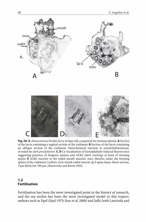
1.2Fertilisation
Fertilisation has been the most investigated point in the history of research,and the sea urchin has been the most investigated model in this respect:authors such as Epel (Epel 1975; Kuo et al. 2000) and Jaffe, both Laurinda and
C. Angelini et al.48
Fig. 2A–E. Paracentrotus lividus larva 18 days old, competent for metamorphosis. A Sectionof the larva containing a sagittal section of the rudiment. B Section of the larva containingan oblique section of the rudiment. Histochemical reaction to acetylcholinesterase,revealed by dark precipitation. C, D Co-localisation of formaldehyde-induced fluorescencesuggesting presence of biogenic amines and AChE (dark staining) at basis of formingspines. E AChE reaction in the radial mouth muscles. musc Muscles under the formingspines of the rudiment (rudim); mrm mouth radial muscle; Sp b spine bases. Resin section,5 µm thick; bar 100 µm. (Karnovsky and Roots 1964)

Lionel (Jaffe 1976, 1980, 1983; Jaffe and Robinson 1978; Turner et al. 1984),investigated the electrophysiological events; Vacquier (1969, 1981; Vacquierand Moy 1977) and Lennarz (Schmell et al. 1977; Ohlendieck et al. 1994;Ohlendieck and Lennarz 1996; Just and Lennarz 1997) investigatedsperm–egg reception and adhesion; Shen (Rakow and Shen 1990; Shen 1995)and Chambers (Chambers and De Armendi 1979; Longo et al. 1986; McCullohet al. 1987; Crossley et al. 1988; Ivonnet and Chambers 1997) investigated cal-cium dynamics in egg responses; Schuel (e.g. Schuel et al. 1991) investigatedsperm dynamics; and Whitaker (Whitaker and Irvine 1984; Crossley et al.1988; Wilding et al. 1996; Harrison et al. 2002) investigated the mechanisms ofcalcium release from inner stores. Each author’s approach included morpho-logical, biochemical and electrophysiological methods, sometimes with dif-ferent hypotheses and interpretations.
Essentially, fertilisation occurs in two phases: the first phase is charac-terised by sperm activation events; the second, by the egg activation events.
1.2.1First Phase: Sperm Activation
Sperms are immobilised in male gonads and genital ducts, as the membranein these sites is stabilised by cholesterol and glycoproteins (Gilbert 1994) andby the sperm fluid, which has a low pH and contains calmodulins (Gilbert1994).
During the first phase of fertilisation, the sperm motility is activated, thesperm are able to swim towards the egg, and are capacitated to the acrosomereaction, during which the acrosome vesicle releases the enzymes it containsinto the water surrounding the egg envelope. During these phases, electricalevents caused by inward–outward ion fluxes take place.
Actually, in each of these events, calcium is made available for the spermsince it is abundant in seawater as well as in the peri-ovular jelly coat; the lat-ter probably also plays a role in modifying the sperm membrane, and also inactivating the acrosome reaction (Darszon et al. 1987; Garcia-Soto et al. 1987;Liévano et al. 1990). Moreover, K+ and Ca2+ channels are activated by speract,the attractant molecule released by the egg, thus leading the sperm to find theegg of the same species (Babcock et al. 1992).
At the same time, Na+, abundant in the marine environment, causes anincrease in the intracellular pH level, which in turn activates flagellum dynein,maintained low by the low pH of the sperm fluid. This event seems to beguided by the activation of nicotinic receptors, present in the membrane ofthe flagellum, as shown by experiments with a-bungarotoxin, a type of snakevenom which is specific for these receptors (Nelson 1976; Falugi et al. 1993b),and by histochemical staining of agonist and antagonist drug binding (Bac-cetti et al. 1995). In each case, ionic changes play a role in the phase in whichswimming is activated as well as in the phase in which the egg membrane is
Cell Signaling During Sea Urchin Development 49

“attacked”; these events take place thanks to: (1) an enhanced swimmingactivity and (2) exocytosis of the acrosome granule which, releasing lyticenzymes, exposes the egg membrane, free from accessory envelopes (jellycoat), and perforates the primary envelope (vitelline membrane). Simple neu-rotransmitter signal molecules play a crucial role in these processes as well.
Cholinergic system molecules were discovered in sperm cells of differentanimal species (Bishop et al. 1977; Sastry et al. 1979; Baccetti et al. 1995). Thesefindings led researchers to investigate the possible role played by neurotrans-mitter system molecules in these cells. In particular, the presence in sperm ofcholinergic molecules has been reported and studied by Nelson (1973, 1978,1990) and Nelson et al. (1970), who supplied evidence for the implication ofthe cholinergic system in sperm propulsion. The activity of the enzymeacetylcholinesterase (AChE, E.C. 3.1.1.7.) was evaluated by biochemical meth-ods in the sperm flagellum of the sea urchin P. lividus (Cariello et al. 1986).AChE is the lytic enzyme of the cholinergic system, and its function is tocleave ACh into choline and acetate, thus restoring ACh receptor excitability.In sperm of the same sea urchin species, AChE was also localised by histo-chemical methods on the head membrane (Falugi et al. 1991). ACh receptorswere also found and localised in the sperm cell structures: muscarinic recep-tors mainly in the acrosome, nicotinic receptors both in the acrosome and inthe flagellum membrane (Baccetti et al. 1995). The two different families ofreceptors seem to play different roles in the different moments of fertilisation:the nicotinic ones (nAChRs, see Stroud et al. 1990), gating Na+ channels onactivation by their specific ligands (ACh and cholinomimetic agonist drugs),may result in a change of the Na+ influx and consequently H+ efflux, increas-ing the internal pH and activating the dynein of the flagellum. The muscarinicones, known in five different molecular forms, associated with G-protein inthe intracellular domain, trigger a second messenger transduction cascade(Birdsall et al. 1978; Watson and Arkinstall 1994), activating intracellulardynamics related to fertilisation (Falugi et al. 1993a).
In addition, the effects of cholinomimetic drugs on P. lividus sperm motil-ity and fertilisation ability suggested an involvement of the nAChRs andmAChRs in these phenomena (Falugi et al. 1993b). This was also clear fromexperiments with potentiometric dyes, which helped to evidence the patternof membrane depolarisation and fluidity during swimming and acrosomereaction caused by the presence of either eggs, calcium ionophore A 23187 ornicotine (Falugi et al. 1993a).
The localisation of the studied molecules in the different cell districts isuseful in order to understand their function. The presence of moleculesimmunologically related to AChRs was found to be constant in gametes ofdifferent organisms, and their localisation seems to be related to the differ-ent mode of interaction between sperm and egg in the different species. Inparticular, P. lividus sperm presented molecules immunologically correlatedto muscarinic receptors mainly at the acrosome and nicotinic receptors atthe acrosome and along the flagellum (Fig. 3), thus confirming previous
C. Angelini et al.50
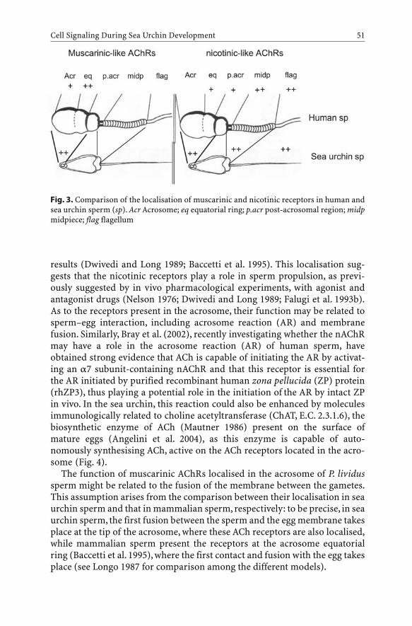
results (Dwivedi and Long 1989; Baccetti et al. 1995). This localisation sug-gests that the nicotinic receptors play a role in sperm propulsion, as previ-ously suggested by in vivo pharmacological experiments, with agonist andantagonist drugs (Nelson 1976; Dwivedi and Long 1989; Falugi et al. 1993b).As to the receptors present in the acrosome, their function may be related tosperm–egg interaction, including acrosome reaction (AR) and membranefusion. Similarly, Bray et al. (2002), recently investigating whether the nAChRmay have a role in the acrosome reaction (AR) of human sperm, haveobtained strong evidence that ACh is capable of initiating the AR by activat-ing an a7 subunit-containing nAChR and that this receptor is essential forthe AR initiated by purified recombinant human zona pellucida (ZP) protein(rhZP3), thus playing a potential role in the initiation of the AR by intact ZPin vivo. In the sea urchin, this reaction could also be enhanced by moleculesimmunologically related to choline acetyltransferase (ChAT, E.C. 2.3.1.6), thebiosynthetic enzyme of ACh (Mautner 1986) present on the surface ofmature eggs (Angelini et al. 2004), as this enzyme is capable of auto-nomously synthesising ACh, active on the ACh receptors located in the acro-some (Fig. 4).
The function of muscarinic AChRs localised in the acrosome of P. lividussperm might be related to the fusion of the membrane between the gametes.This assumption arises from the comparison between their localisation in seaurchin sperm and that in mammalian sperm, respectively: to be precise, in seaurchin sperm, the first fusion between the sperm and the egg membrane takesplace at the tip of the acrosome, where these ACh receptors are also localised,while mammalian sperm present the receptors at the acrosome equatorialring (Baccetti et al. 1995), where the first contact and fusion with the egg takesplace (see Longo 1987 for comparison among the different models).
Cell Signaling During Sea Urchin Development 51
Fig. 3. Comparison of the localisation of muscarinic and nicotinic receptors in human andsea urchin sperm (sp). Acr Acrosome; eq equatorial ring; p.acr post-acrosomal region; midpmidpiece; flag flagellum

A matter of debate over recent years has been the fact that the binding sitesfor pharmacological agents may differ between in vitro and in vivo experi-ments, as suggested from experiments on mouse sperm (Florman and Storey1982). However, experiments on sea urchin fertilisation are normally carriedout in seawater, the natural medium in which this event takes place, so as toeliminate any possible difference between in vivo and in vitro experiments.
1.2.2Second Phase: Egg Activation
It is well known that sea urchin egg activation and fertilisation are led and fol-lowed by electrical phenomena, especially as far as ion dynamics are con-cerned. Following the contact of the egg’s membrane with that of the sperm,an early depolarisation event of the egg membrane takes place, caused by aninflux of Na+ ions (see Epel 1975, 1980), which makes the membrane permis-sive to the fusion (McCulloh et al. 1987).
This spike is immediately followed by an explosive increase in intracellular[Ca2+], released from intracellular stores (Giudice 1973; Longo 1987). Thisevent evokes a strong membrane depolarisation (about 90 mV) and activatesthe egg, causing the so-called fast block to polyspermy as well as the corticalreaction. The egg, thus activated, begins to “breathe” and starts its metabolicactivities, including DNA synthesis and the trigger for the first cell cycles(Giudice 1973; Longo 1987).
As far as the mode of regulation and unfolding of these ion dynamics isconcerned, a great deal of research has been carried out since the beginningof the last century and, according to Ohlendieck and Lennarz (1996), the num-ber of investigations on the subject has greatly exceeded that of the worksconcerning the philosopher’s stone over the past centuries! However, recentlysome progress has been made towards the comprehension of these phenom-ena.
C. Angelini et al.52
Fig. 4A–C. ChAT ultrastructural immunogold reaction in Paracentrotus lividus sperm.A Control; B, C reacted sperm (¥12,000)

1.2.2.1First Step: Na+ Influx
This event was described by Epel (1980): as soon as the sperm touches the eggmembrane, a minor influx of Na+ takes place throughout the egg membrane;at the same time, sperm–egg membrane fusion takes place, immediately fol-lowed by calcium release from intracellular stores. The meaning of this firstNa+ influx was apparently forgotten by researchers in the following years, whorather chose to focus on the calcium wave responsible for the block topolyspermy.
Just recently, this event has been re-examined, by considering old papers onthe effects of nicotine exposure during sea urchin egg fertilisation and byobserving that the ionic dynamics (first Na+ influx, immediately followed byexplosive Ca2+ release) are very similar to what happens in the neuromuscu-lar synapses. Nicotine had been reported by Jaffe (1980) to cause polyspermy;thus it is very likely that nicotinic receptors may be present just as in the neu-romuscular post-synaptic membrane. To be precise, nicotinic receptors werehistochemically revealed by curare-prevented a-bungarotoxin binding(Falugi and Prestipino 1988; Falugi et al. 1989) at the egg surface. Morerecently, by exposing sea urchin unfertilised eggs to acetylcholine togetherwith eserine (Physostigmine, Sigma), we have observed, after the addition ofsperm, a membrane depolarisation of about 40 mV (starting from a restpotential of –70 mV, to the spike of –30 mV), while the normal final depolari-sation in control eggs is +0 mV, with a depolarisation ranging between 70 and90 mV. Moreover, the exposure to acetylcholine prior to fertilisation signifi-cantly increased the percentage of polyspermic eggs as compared to controls(Harrison et al. 2002; Angelini et al. 2004). The pharmacological properties ofthese receptors were verified with electrophysiological methods by Chambersand co-workers, who confirmed the presence of nicotinic receptors in theunfertilised sea urchin egg, and characterised them as being identical to theones present in the neuromuscular synapses (Ivonnet and Chambers 1997).According to these authors, the presence of nAChR channels at the surface ofunfertilised eggs can account for the capacity of nicotine to impair the blockto polyspermy, by lowering the positive shift of the egg’s membrane potentialcaused by sperm.
This could also explain our results described above (Angelini et al. 2004)Actually, the same effect of maintaining the fertilisation potential of the egg atlow levels could be exerted by an increased quantity of ACh, which in this casecould maintain the membrane fusibility for a longer period of time thanunder physiological conditions. This relationship between the first step ofdepolarisation due to Na+ influx and membrane fusibility seems to be reli-able. In fact, as previously demonstrated (Longo 1987; McCulloh et al. 1987;McCulloh and Chambers 1992), a low membrane depolarisation allows spermentry and membrane fusion in the sea urchin Lytechinus pictus and Strongy-locentrotus purpuratus.
Cell Signaling During Sea Urchin Development 53
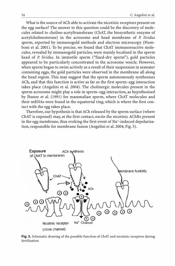
What is the source of ACh able to activate the nicotinic receptors present onthe egg surface? The answer to this question could be the discovery of mole-cules related to choline-acetyltransferase (ChAT, the biosynthetic enzyme ofacetylcholinesterase) in the acrosome and head membrane of P. lividussperm, reported by immunogold methods and electron microscopy (Piom-boni et al. 2001). To be precise, we found that ChAT immunoreactive mole-cules, revealed by immunogold particles, were mainly localised in the spermhead of P. lividus. In immotile sperm (“fixed-dry sperm”), gold particlesappeared to be particularly concentrated in the acrosome vesicle. However,when sperm began to swim actively as a result of their suspension in seawatercontaining eggs, the gold particles were observed in the membrane all alongthe head region. This may suggest that the sperm autonomously synthesisesACh, and that this function is active as far as the first sperm–egg interactiontakes place (Angelini et al. 2004). The cholinergic molecules present in thesperm acrosome might play a role in sperm–egg interaction, as hypothesisedby Ibanez et al. (1991) for mammalian sperm, where ChAT molecules andtheir mRNAs were found in the equatorial ring, which is where the first con-tact with the egg takes place.
Therefore, our hypothesis is that ACh released by the sperm surface (whereChAT is exposed) may, at the first contact, excite the nicotinic AChRs presentin the egg membrane, thus evoking the first event of Na+-induced depolarisa-tion, responsible for membrane fusion (Angelini et al. 2004; Fig. 5).
C. Angelini et al.54
Fig. 5. Schematic drawing of the possible function of ChAT and nicotinic receptors duringfertilisation
1.2.2.2Calcium Dynamics
Although the egg membrane is effectively an excitable membrane, its similar-ity with the synapses is only limited to the nicotinic receptors, because theinflux of Na+ ions caused by acetylcholine in the sea urchin egg is not suffi-cient to evoke the release of calcium ions and the rapid block to polyspermy,as we demonstrated in cooperation with the laboratory of Prof. Whitaker(Harrison et al. 2002), thanks to the use of cholinomimetic agonists andantagonists on P. lividus and Lytechinus pictus eggs and zygotes. We did notfind any evidence of an involvement of cholinergic molecules in this process,as nicotinic AChR agonists were not capable of evoking any [Ca2+] variationin unfertilised eggs by themselves, nor were the antagonists capable of pre-venting the calcium spike provoked by sperm. Also, muscarinic agonists andantagonists were completely inactive in the course of these events.
Thus, in the fast block to polyspermy a different kind of signal and recep-tor molecule must be active for the Ca2+ ion release. The latter was elicitedby a signal transduction cascade, since Whitaker and Irvine (1984) hadshown that an inositol-triphosphate (IP3) injection in the cytoplasm wassufficient to activate the sea urchin unfertilised egg and to cause the corticalreaction.
Actually, in August 2000, a paper by Kuo et al. was published, reporting thatIP3 release was due to an atypical neurotransmitter, nitric oxide (NO). Theseauthors showed that nitric oxide synthase (NOs) is present in the post-acroso-mal area of the sperm, and that it is activated by contact with the jelly coat, tosynthesise NO, which is subsequently carried inside the egg together with thesperm material. This would be the signal that triggers the transduction cas-cade, including the release of IP3; these events result in an explosive release ofcalcium from the inner stores, identified as cortical vesicles of the endoplas-mic reticulum (see Giudice 1986; Shen 1995 for extensive reviews) and,recently, also vacuolar-like structures (Churchill et al. 2002).
1.3First Cell Cycle
Therefore, cholinergic molecules do not seem to be involved in the block topolyspermy and cortical reaction. Nevertheless, several authors have found acomplete set of cholinergic system molecules in the eggs both before and afterfertilisation. Among these, our group has localised nicotinic AChR moleculesin the sea urchin P. lividus by use of curare-prevented FITC-a-bungarotoxinbinding at the surface of unfertilised and fertilised eggs of P. lividus (Falugi etal. 1989), alongside muscarinic AChR molecules (Piomboni et al. 2001), as wellas other cholinergic molecules, such as acetylcholinesterase (Piomboni et al.2001) and maternal choline acetyltransferase (ChAT) molecules, assembled
Cell Signaling During Sea Urchin Development 55

on the oolemma during egg maturation and fertilisation processes (Angeliniet al. 2004). Actually, muscarinic receptor molecules are present in the eggstarting from the first maturation events, but are assembled on the egg mem-brane only after fertilisation. This was demonstrated by both ultrastructurallocalisation of anti-mAChR immunoreactivity and Western blot of cytoplasmhomogenates compared with membrane extracts of unfertilised and fertilisedeggs (Piomboni et al. 2001). A similar distribution pattern was found forChAT (Angelini et al. 2004). Moreover, the cortical granules display a contentof active molecules of AChE, identified by the enzyme reaction product assuggested by Karnovsky and Roots (1964). These molecules are synthesisedwithin the endoplasmic reticulum, processed in the Golgi vesicles and fromthere exposed at the membrane surface (Piomboni et al. 2001). During thecortical reaction, these molecules are extruded into the perivitelline space(Fig. 6).
Such a coordinated distribution of signal molecules suggests a function forthe cholinergic system, displayed after fertilisation. The key to the under-standing of this possible function lies in the role played by muscarinic AChRs,which consists of triggering intracellular signal transduction (Watson andArkinstall 1994). This appears to be the case in all the systems in which theyhave been identified. It has been recently reported that mAChRs are func-tional only when assembled on the cell surface, while inactive subunits arecontained in ER-derived vesicles (Mei and Xiong 2003). This is true for ChATas well, as it is known that ChAT activity is regulated by its binding to thecholinergic membrane (Dobransky and Rylett 2003) and by interaction withother cellular proteins related to this membrane (Gabrielle et al. 2003). Inother words, the enzyme is not active when it is inside the cytoplasm, while itis active when it is exposed on the cholinergic membrane.
Furthermore, by FURA-2 experiments, it was shown that ACh and car-bamylcholine, the natural signal and the cholinomimetic agonist of themAChR, respectively, are capable of evoking a [Ca2+]i spike. This occurs whenthe exposure is performed at a time preceding the nuclear envelope break-down (NEB) occurring at the first cell cleavage, i.e. approximately 15 min forP. lividus and 40 min for L. pictus after fertilisation. In contrast, atropine(antagonist of mAChRs, non-selective among the different molecular forms ofthe receptors) prevented further Ca2+ spikes caused by agonist molecules. Theability to respond to them was acquired again after rinsing the atropine pre-sent in the egg-containing water (Harrison et al. 2002); for this reason, wehypothesised that mAChRs could be involved in the regulation of intracellu-lar ionic dynamics related to the cleavage of the first cell cycles (Harrison etal. 2002). All our findings seem to suggest a model of autocrine regulation ofthese dynamics. To be precise, those ChAT molecules that after fertilisationare exposed at the egg surface may autonomously synthesise ACh. The eggcannot receive ACh from elsewhere, because after fertilisation the zygote isenclosed in the fertilisation membrane, which is in turn completely sur-rounded by seawater, and transported by waves and currents. The ACh pro-
C. Angelini et al.56

Cell Signaling During Sea Urchin Development 57
Fig. 6. A Schematic drawings of the relative localisation of AChE activity, ChATimmunoreactivity and muscarinic receptors during maturation phases of sea urchin eggs.lc Lampbrush chromosomes; nu nucleus. B AChE histochemical localisation in the corticalgranules of Paracentrotus lividus mature egg. C Cortical granules extrusion (arrows) duringthe cortical reaction. D AChE activity in the elongated microvilli of the fertilisation cone.E Muscarinic receptors included in cortical vesicles in unfertilised eggs. F Muscarinic recep-tor immunoreactivity at the membrane surface of fertilised egg. G ChAT immunoreactivityin immature ovarian egg. H ChAT immunoreactivity in fertilised eggs. Bars 50 µm. The elec-tron microscopy figures were taken at instrument magnification ¥20,000

duced by the ChAT assembled on the egg membrane is released into the peri-vitelline space and reaches the mAChRs that at this stage are also exposed atthe surface (Piomboni et al. 2001). As a result, muscarinic activation evokes a[Ca2+] response, which further enhances development. The hypothesis thatcalcium may be involved in the regulation of the first cell cycle may be alsosupported by the findings of Steinhardt (1990a, 1990b), who proposed thatintracellular [Ca2+]i changes might be possible signals for the regulation ofdifferent mitosis stages, in both animal and plant cells. In particular, togetherwith coworkers, he forwarded the hypothesis that calcium might be res-ponsible for the regulation of the NEB at the moment of cleavage. The above-cited authors showed that a calcium transient immediately precedes the NEB,which may also be prevented by subtracting calcium from the cell (Baitingeret al. 1990).
According to these discoveries, we proposed a model that could help toexplain the function of inter- and intracellular signals mediated by choliner-gic molecules during fertilisation and the first cell cycles – both gametes pos-sess cholinergic molecules: ChAT, which is necessary in order to produce ACh,
C. Angelini et al.58
Fig. 7. Schematic drawing of the hypothesis about “autocrine” behaviour of acetylcholine infertilised eggs: ChAT synthesises ACh, which activates the muscarinic receptors exposed atthe surface after fertilisation, triggering the transduction cascade causing Ca2+ release frominner stores. Musc rec Muscarinic receptors; G-prot G-protein

AChE, which cleaves ACh bound to the receptors, and ACh nicotinic and mus-carinic receptors (Figs. 6, 7). Moreover, their activities are displayed at differ-ent times: during the first phase, the nicotinic receptors in the sperm tail areinvolved in promoting swimming [a-bungarotoxin blocks their movement(see Nelson 1976; Falugi et al. 1993b); then the receptors present in the acro-some (both nAChRs and mAChRs) may be excited by the ACh synthesised bythe ChAT present on the egg surface.
During the second phase, when sperm activate the eggs, ACh, auto-nomously synthesised by ChAT on the sperm head, activates the nicotinicreceptors present in the egg membrane (characterised by Ivonnet and Cham-bers 1997) and the first step of depolarisation caused by Na+ influx mediatedby the nicotine receptors takes place. This makes the egg membrane permis-sive to fusion with the sperm membrane.
Successively, the cholinergic molecules are not involved in the calciumwave, which is released by intracellular stores at fertilisation through IP3release by membrane phospholipases; this, in turn, is caused by the NO,inserted into the egg intracellular domain by the sperm ingression (Kuo et al.2000). However, the cortical reaction following the calcium wave causes theexposure of muscarinic receptors at the egg surface, and AChE is released intothe perivitelline space together with the other cortical granule components.After this exposure, mAChRs become active and excitable, evoking furthercalcium spikes (as demonstrated by Harrison et al. 2002) when bound by theACh, autonomously synthesised by ChAT exposed on the egg membrane. Thisevent might be related to the calcium spike that was reported to take placeduring the nuclear envelope breakdown (Steinhardt 1990a, b; Wilding et al.1996).
1.4Cleavages
From cleavage and until the blastula stage, AChE activity, which had disap-peared during fertilisation, starts to be present and active once more(Fig. 8A), starting from the perinuclear envelope; this, at least in vertebratemuscle cells (Miki and Mizoguti 1981), is the first site of synthesis. Also, atthese stages, the final destination of the molecules is at the blastomere surfacewhere presumably they play a role in cell-to-cell interaction (Fig. 8B). Exten-sive studies conducted by Buznikov and coworkers demonstrated that, duringthese events, the typical neurotransmitters mainly involved in regulating cell-to-cell interaction are represented by biogenic amines (Buznikov et al. 2001);these are responsible for inducing messages among blastomeres (Shmuklerand Buznikov 1998).
Cell Signaling During Sea Urchin Development 59

1.5Ongoing Research
At this point, our interest is towards the molecular forms of muscarinic recep-tors involved in the different stages of egg activation and cell cycling. We arenow investigating the changes that take place when passing from the “embry-onic” to the “adult” molecular forms of the m2 receptor. These changes arepossibly due to alternative splicing during developmental events (Angelini etal. 2004). Another aim is to specify the involvement of the ryanodine receptorpossibly by identifying its mRNA transcription and pharmacological charac-terisation during the events supported by calcium activation. From our pre-liminary results, the activation of egg and calcium spiking during the first cellcycle seems to be due to different release mechanisms: the first, brought aboutby sperm activation, seems to be mainly due to IP3 release, as demonstratedby Whitaker and Irvine (1984), while the second, susceptible to muscarinicreceptor activation, seems to be mainly due to calcium release mediated byryanodine receptor. Actually, from preliminary results, ryanodine receptorsseem to be mainly expressed during the first cell cycle, while the amount ofthese molecules found and characterised by both incorporation in artificialmembranes and Northern blot analysis indicates a very scarce presence dur-ing the other stages: unfertilised egg and up to the four-cell stage (Angelini etal. 2000; Prestipino, pers. comm.).
C. Angelini et al.60
Fig. 8A, B. AChE histochemical reaction in Paracentrotus lividus. A Zygote, showing darklabelling around the nucleus, in the cytoplasm and in the sperm pathway from the fertilisa-tion cone (arrow) to the point of contact with the female pronucleus. B Two-blastomerestage, showing the enzyme activity product around the nuclei and in the furrow betweenthe blastomeres. In toto mount, bar 80 µm

2Applied Research
The described results open a path to a new way of considering biomarkers. Infact, most environmental factors have the capacity to interfere with thecholinergic system: stress, wounds, inflammation and pollution generally(Kaufer et al. 1998), in a number of organisms from invertebrates (Dauber-schmidt et al. 1996) and fish (Fulton and Key 2000), to high vertebrates, suchas birds (Misawa et al. 1981), up to mammalians, including man (Wessler2003). In particular, some broadly employed pollutants and drugs specificallyaffect the cholinergic system, and consequently the other typical neurotrans-mitter systems, such as those depending on biogenic amines (Buznikov et al.2001b). These are the so-called neurotoxic compounds.
2.1Neurotoxic Contaminants
A class of contaminants diffused all over the world is represented by neuro-toxic substances, in particular organophosphate (OP) and carbamate (CB)pesticides. These are largely used in European countries for many purposes,for example in agriculture, gardening and even domestic pest control.Among these, OP compounds have been used for a great number of otherpurposes, e.g. as chemical weapons and medical compounds (anxiolytics,antispasmoics, regulators of eye pressure, and drugs for Alzheimer’s disease,etc.).
Neurotoxic pesticides inhibit cholinesterase activities (acetylcholin-esterase, AChE, E.C. 3.1.1.7 and pseudocholinesterase, BuChE, E.C. 3.1.1.8.)and, consequently, the status of the cholinergic neurotransmitter system.
2.1.1Health Risks
These insecticides are strongly suspected to be harmful, and clearly they arein the case of acute intoxication, when organisms come into contact with highdoses, generally as a result of accidents or occupational exposure. However,there is also the possibility of subtle chronic (low-dose, long-term) damagedue to aerosol diffusion, or to residuals in crops and vegetables, possibly rein-forced by the presence of traces of other pollutants, such as other neurotoxicsubstances, heavy metals and hydrocarbons, among others. On the otherhand, the no-effective concentration for humans (NOEC), indicated by thepharmaceutical companies and databases, is not surely ascertained, because itis obtained by experimental exposure of animals, generally rats or mice, andthen by estimating it as several-fold lower. Moreover, the doses that do not
Cell Signaling During Sea Urchin Development 61

affect adults may heavily strike embryonic differentiation, a very sensitivestage in the life of an organism (Romero et al. 1989)
2.1.2Mode of Function of Organophosphates and Carbamates
These neurotoxic compounds target the cholinergic neuromuscular system,where they inhibit the activity of acetylcholinesterase (AChE, E.C. 3.1.1.7.)and pseudocholinesterases (E.C. 3.1.1.8.). These are the lytic enzymes of sig-nal choline esters, including acetylcholine (ACh), and consequently their inhi-bition causes an overflow of ACh at receptor sites; this, in turn, affects intra-cellular responses. In this way, neurotoxic compounds may cause alteration ofall functions of the cholinergic neurotransmission system, and of other neu-rotransmitters, the release of which is influenced by the cholinergic system(Buznikov et al. 2001). The pharmacology of OPs has been extensively stud-ied, since they were initially conceived as chemical weapons during WorldWar II (Karczmar et al. 1970). Some differences between the two classes ofneurotoxic pesticides are known: (1) OPs irreversibly link the AChE moleculeby the phosphate group (Sultatos 1994), while carbamates compete for thesubstrate acetylcholine (ACh) (Mineau 1991); and (2) OPs can leave residualsin the environment, while carbamates only leave small inorganic molecules(e.g. carbon dioxide) (Hayes and Laws 1991).
2.1.3Persistence in the Environment and Crops
The persistence of these compounds in the environment is generally consid-ered fairly short, but there is evidence that they may persist in sediments overlong periods of time, such as in the River Rhine (Dauberschmidt et al. 1996),where lethality of fish, molluscs and aquatic birds lasted for months and kilo-metres downstream after the actual contamination had taken place. Accord-ing to Ragnarsdottir (2000), an OP pesticide presenting a short half-life in thelaboratory increases to 1 year under conditions of low pH and temperature.The same author reported that OPs are detected in soils years after applica-tion, probably due to sorption of the OPs onto soil particles, making themunavailable for microbial metabolism (Ragnarsdottir 2000). Insofar as livingorganisms are concerned, the effects of such compounds may last muchlonger, because the AChE of blood may be affected for up to several months(see the case report by Romero et al. 1989).
C. Angelini et al.62

2.2Biosensors
For the reasons explained above, at present, a great deal of effort is concen-trated on creating “biosensors”, capable of perceiving neurotoxic compoundsin the environment, as well as in food and water.
Most of the biosensors are represented by devices that have the capacity tomeasure, with high sensitivity, the activity of acetylcholinesterase in the pres-ence of suspected inhibitors and in particular OP or carbamate compounds.These high-technology instruments can measure the presence and amount ofneurotoxic compounds in environmental matrices, or in rough material andelaborated foods. The biosensors used for this purpose are generally based onhighly sensitive molecular forms of AChE, immobilised in devices capable ofrecording changes in activity in real time, and by transferring them to screensor other recording devices (Crew et al. 2004; Pritchard et al. 2004), or by use ofmutated bacteria or yeast (Wu et al. 2002).
In order to evaluate the effects on living organisms, and their health risks,another kind of biosensor is now being studied. Actually, the effects of neuro-toxic compounds, and particularly of OPs and carbamates, are not onlydirected towards AChE activity, but may also affect ACh receptors, directly(Abdallah et al. 1992; Sultatos 1994). Thus, the best biosensor in this caseshould be a biosensor with the capacity to translate the effect of neurotoxicpesticides in living cells to cell responses, such as the electrical ones. Theadvantages of this biosensor are represented by the fact that complex cellresponse is taken into account, and that the AChE molecules and the AChreceptors are in their natural environment, and follow their natural transduc-tion cascades up to the cell response. For this purpose, a model of this biosen-sor is being devised in the laboratory of the Fraunhofer Institute for Biomed-ical Engineering (IBMT), based on microtechnology.
2.3Effects of Neurotoxic Compounds on P. lividus Ion Dynamics
From this point of view, the early phases of sea urchin development offer verygood material for the preparation of biosensors based on living cells, becausethe effects on the cholinergic system are per se translated into changes inmembrane potential. Actually, by exposing P. lividus zygotes to diazinon, anOP compound, we found that the increase in [Ca2+]i, either caused by mus-carinic drug exposure or the physiological one preceding the NEB, was pre-vented. The K+ outward channel probably linked to excitation of the mus-carinic receptor m2 was also blocked by highly diluted phentoate, another OPcompound commonly used as an insecticide (Percivale 2003).
This offers the possibility of adapting this model to a biosensor capable ofrecording the impedance changes by using P. lividus zygotes (the moment
Cell Signaling During Sea Urchin Development 63

when the cholinergic system is mostly dependent on electrical variations)(Fig. 9).
The employment of these cells is at present innovative, as well as bioethi-cally compatible. In addition, it may solve some controversial points, such as:1. The problem of experiments on animals, which are more expensive,
besides causing pain, which is particularly evident in higher organisms.Gametes from sea urchins may be obtained easily by intra-oral 1/1,000 AChinjection: this treatment allows gamete spawning without sacrificing theadults, which survive in good health (as demonstrated by further naturalspawning in a few weeks: M. Franzoni and M. Sgro, preliminary results,pers. comm.).
C. Angelini et al.64
Fig. 9. A Schematic drawing of the living cell-based biosensor (IBMT). B The micro-devicefor impedance recording

2. The improved knowledge of developmental biology, within the emergingknowledge that neurotransmitter molecules are not limited to neuromus-cular structures, but are generally involved in cell-to-cell communication,leading to interaction between developing cells and tissues.
3. Exporting the results to other organisms, including man, by comparing theeffects on P. lividus zygotes with the effects on human stem cells (addressedto development).
4. Allowing us to establish conversion parameters among the different cellsources, in order to use the most suitable and available for each situation ofrisk assessment.
Acknowledgements. This work was supported by the EC within the project SENS-PESTI,QLK4-CT-2002-02264. We thank Dr. Mario Franzoni and Dr. Marco Sgro for allowing us touse their preliminary data on rearing sea urchin specimens; and Dr. Mario Mori for contin-uous supply of living sea urchins in optimal reproductive conditions.
References
Abdallah EAM, Jett DA, Eldefrawi ME, Eldefrawi AT (1992) Differential effects of paraoxonon the M3 muscarinic receptor and its effector system in rat submaxillary gland cells. JBiochem Toxicol 7:125–132
Angelini C, DelGaudio R, Falugi C, Pernigotti R, Geraci G, Prestipino G, Pesando L (2000)Calcium signaling in sea urchin zygotes. In: Proc 15th Meeting Sea Urchin Development,29 Sept–3 Oct, Woods Hole, Massachusetts
Angelini C, Baccetti B, Piomboni P, Trombino S, Aluigi MG, Stringara S, Gallus L, Falugi C(2004) Acetylcholine synthesis and possible functions during sea urchin development.Eur J Histochem 49(3):235–244
Babcock DF, Bosma MM, Battaglia DE, Darszon A (1992) Early persistent activation ofsperm K+ channels by the egg peptide speract. Proc Natl Acad Sci USA 89:6001–6005
Baccetti B, Burrini AG, Collodel G, Falugi C, Moretti E, Piomboni P (1995) Localisation oftwo classes of acetylcholine receptor-like molecules in sperms of different animalspecies. Zygote 3:207–217
Baitinger C, Alderton J, Poenie M, Schulman H, Steinhardt RA (1990) MultifunctionalCa2+/calmodulin-dependent protein kinase is necessary for nuclear envelope break-down. J Cell Biol 111(5):1763–1773
Beer AJ, Moss C, Thorndyke M (2001) Development of serotonin-like and SALMFamide-likeimmunoreactivity in the nervous system of the sea urchin Psammechinus miliaris. BiolBull 200(3):268–280
Birdsall NJM, Burgen ASV, Hulme EC (1978) The binding of agonists to brain muscarinicreceptors. Mol Pharmacol 14:723–736
Bishop MR, Rama Sastry BW, Stavinoha B (1977) Identification of acetylcholine and propi-onylcholine in bull spermatozoa by integrated pyrolysis, gas chromatography and massspectrometry. Biochim Biophys Acta 500:440–444
Bray C, Son J-H, Meizel S (2002) A nicotinic acetylcholine receptor is involved in the acro-some reaction of human sperm initiated by recombinant human sperm initiated byrecombinant human ZP3. Biol Reprod 67:782–788
Cell Signaling During Sea Urchin Development 65

Buznikov GA (1980) Biogenic monoamines and acetylcholine in protozoa and metazoanembryos. In: Salanki J, Turpaev M (eds) Neurotransmitters: comparative aspects.Akademial Kiado, Budapest, pp 7–28
Buznikov GA (1990) Neurotransmitters in embryogenesis. In: Turpaev TM (series ed) Phys-iology and general biology, section F of Soviet Scientific Reviews. Harwood, London
Buznikov GA, Sakharova AV, Manukhin BN, Markova LN (1972) The role of neurohumors inearly embryogenesis. IV. Fluorimetric and histochemical study of serotonin in cleavingeggs and larvae of sea urchins. J Embryol Exp Morphol 27(2):339–351
Buznikov GA, Shmukler YB, Lauder JM (1996) From oocyte to neuron: do neurotransmit-ters function in the same way throughout development? Cell Mol Neurobiol 16(5):533–559
Buznikov GA, Lambert HW, Lauder JM (2001a) Serotonin and serotonin-like substances asregulators of early embryogenesis and morphogenesis. Cell Tissue Res 305(2):177–186
Buznikov GA, Nikitina LA, Bezuglov VV, Lauder JM, Padilla S, Slotkin TA (2001b) An inver-tebrate model of the developmental neurotoxicity of insecticides: effects of chlorpyrifosand dieldrin in sea urchin embryos and larvae. Environ Health Perspect 109(7):651–661
Cariello L, Romano G, Nelson L (1986) Acetylcholinesterase in sea urchin spermatozoa.Gamete Res 14:323–332
Chambers EL, De Armendi J (1979) Membrane potential, action potential and activationpotential of eggs of the sea urchin, Lytechinus variegatus. Exp Cell Res 122:203–218
Churchill GC, Okada Y, Thomas JM, Genazzani AA, Patel S, Galione A (2002) NAADP mobi-lizes Ca(2+) from reserve granules, lysosome-related organelles, in sea urchin eggs. Cell111(5):703–708
Crew A, Wedge R, Hart JP, Marty JL, Fournier D (2004) A samperometric biosensor array tomeasure organophosphate concentration in raw products. In: Proc 8th World Congr onBiosensors, 24–26 May, Granada, Spain
Crossley I, Swann K, Chambers E, Whitaker M (1988) Activation of sea urchin eggs by inos-itol phosphates is independent of external calcium. Biochem J 252(1):257–262
Darszon A, Garcìa-Soto J, González M, Guerrero A, Sánchez JA, Liévano A (1987) Ion trans-port mechanism in sea urchin sperm. In: Mohri H (ed) New horizon in sperm cellresearch. Japanese Scientific Society Press/Gordon and Breach Scientific, Tokyo/NewYork, pp 169–183
Dauberschmidt C, Dietrich DR, Schattler C (1996) Toxicity of organophosphorous insecti-cides in the zebra mussel, Dreissena polymorpha. P Arch Environ Contam Toxicol30:373–378
Dobransky T, Rylett RJ (2003) Functional regulation of choline acetyltransferase by phos-phorylation. Neurochem Res 28(3–4):537–542
Drews U (1975) Cholinesterase in embryonic development. Prog Histochem Cytochem7:1–52
Dwivedi C, Long NJ (1989) Effect of cholinergic agents on human spermatozoan motility.Biochem Med Metabol Biol 42(1):66–70
Epel D (1975) The program and mechanism of fertilization in the echinoderm egg.Am Zool15:507–522
Epel D (1980) Ionic triggers in the fertilization of sea urchin eggs. Ann N Y Acad Sci339:74–85
Falugi C, Prestipino G (1988) Localization of putative nicotinic cholinoreceptors in the earlydevelopment of Paracentrotus lividus. Cell Mol Biol 35(2):147–161
Falugi C, Pera C, Prestipino G (1989) Localization of a-bungarotoxin receptors and AChEactivity in eggs and embryos of Paracentrotus lividus Lamk. Acta Embryol Morphol Exp10(2):113–119
Falugi C, Borgiani L, Faraldi G, Tagliafierro G, Toso F, Drews U (1991) Localization and pos-sible functions of neurotransmitter systems in sperms of different animal species. In:
C. Angelini et al.66

Baccetti B (ed) Comparative spermatology, 20 years later. vol 75. Serono Symposia Pub-lications, Raven Press, New York, pp 475–479
Falugi C, Moretti E, Lammerding-Köppel M, Drews U (1993a) A cholinergic system isinvolved in gamete interaction. In: Facchinetti F, Henderson HW, Pierantoni R, PolzonettiMagni A (eds) Cellular communication in reproduction. Society for Endocrinology,Bristol, pp 101–104
Falugi C, Pieroni M, Moretti E (1993b) Cholinergic molecules and sperm functions. J Sub-microsc Cytol Pathol 25(1):63–69
Falugi C, Facondini L, Angelini C, Diaspro A, Robello M, Pedrotti ML (1999) Onset of acholinergic-like system during neurogenesis of the sea urchin, Paracentrotus lividus. In:Candia Carnevali MD, Bonasoro G (eds) Echinoderm Research 1998. AA Balkema, Rot-terdam, pp 197–202
Falugi C, Diaspro A, Angelini C, Pedrotti L, Raimondo M, Robello M (2002) Three dimen-sional mapping of cholinergic molecules by confocal laser scanning microscopy in seaurchin larvae. Micron 33(3):233–239
Florman HM, Storey BT (1982) Characterization of cholinomimetic agents that inhibit invitro fertilization of the mouse. Evidence for a sperm-specific binding site. J Androl3:157–164
Fulton MH, Key PB (2000) Acetylcholinesterase inhibition in estuarine fish and inverte-brates as an indicator of organophosphorus insecticide exposure and effects. EnvironToxicol Chem 20(1):37–45
Gabrielle P, Jeana M, Lorenza EC (2003) Cytosolic choline acetyltransferase binds specifi-cally to cholinergic plasma membrane of rat brain synaptosomes to generate mem-brane-bound enzyme. Neurochem Res 28(3–4):543–549
Garcia-Soto J, Gonzalez-Martinez M, De la Torre L, Darszon A (1987) Internal pH can regu-late Ca2+ uptake and the acrosome reaction in sea urchin sperm. Dev Biol 120:112–120
Gilbert SF (ed) (1994) Developmental biology. Sinauer Associates, SunderlandGiudice G (ed) (1973) Developmental biology of the sea urchin embryo. Academic Press,
New YorkGiudice G (ed) (1986) The sea urchin embryo. Springer, Berlin Heidelberg New YorkGustafson T, Ryberg E, Treufeldt R (1972) Acetylcholine and contractile activity in the
echinopluteus. A study of the cellular basis of larval behaviour. Acta Embryol Exp 1972:199–223
Harrison PK, Falugi C, Angelini C, Whitaker MJ (2002) Muscarinic signalling affects intra-cellular calcium concentration during the first cell cycle of sea urchin embryos. Cell Cal-cium 31(6):289–297
Hayes WJ Jr, Laws ER Jr (1991) Organic phosphorous pesticides. In: Hayes WJ Jr, Laws ER Jr(eds) Handbook of pesticide toxicology. Academic Press, San Diego
Ibanez CF, Pelto-Huikko M, Soder O, Ritzen EM, Hersh LB, Hokfelt T, Persson H (1991)Expression of choline acetyltransferase mRNA in spermatogenic cells results in an accu-mulation of the enzyme in the postacrosomal region of mature spermatozoa. Proc NatlAcad Sci USA 88:3676–3680
Ivonnet PI, Chambers EL (1997) Nicotinic acetylcholine receptors of the neuronal typeoccur in the plasma membrane of sea urchin eggs. Zygote 5(3):277–287
Jaffe LA (1976) Fast block to polyspermy in sea urchins is electrically mediated. Nature261:68–71
Jaffe LA (1980) Electrical polyspermy block in sea urchins: nicotine and low-sodium exper-iments. Dev Grow Diff 22:503–507
Jaffe LA, Robinson KR (1978) Membrane potential of the unfertilized sea urchin egg. DevBiol 62:215–228
Jaffe LR (1983) Sources of calcium in egg activation: a review and hypothesis. Dev Biol99:265–276
Cell Signaling During Sea Urchin Development 67

Just ML, Lennarz WJ (1997) Reexamination of the sequence of the sea urchin egg receptorfor sperm: implications with respect to its properties. Dev Biol 184(1):25–31
Karczmar AG, Usdin E, Willis JH (1970) Anticholinesterase agents. In: Karczmar AG, UsdinE, Willis JH (eds) International encyclopedia of pharmacology and therapeutics, vol 13.Pergamon Press, Oxford
Karnovsky MJ, Roots L (1964) A “direct colouring” thiocholine method for cholinesterase. JHistochem Cytochem 12:219–221
Kaufer D, Friedman A, Seidman S, Soreq H (1998) Acute stress facilitates long-lastingchanges in cholinergic gene expression. Nature 393(6683):373–377
Kuo RK, Baxter GI, Thompson SH, Stricker SA, Patton C, Bonaventura J, Epel D (2000) NO isnecessary and sufficient for egg activation at fertilization. Nature 406:633–636
Liévano A, Vega-SaendzeMiera EC, Darszon A (1990) Ca++ channels from the sea urchinsperm plasma membrane. J Gen Physiol 95:273–296
Longo FJ (1987) Fertilization. Chapman and Hall, LondonLongo FJ, Lynn JW, McCulloh DH, Chambers EL (1986) Correlative ultrastructural and elec-
trophysiological studies of sperm–egg interactions of the sea urchin, Lytechynus varie-gatus. Dev Biol 118:155–166
Mautner HG (1986) 8-Choline acetyltransferase. In: Boulton AA, Baker GB, Yu PH (eds)Neuromethods. Vol 5. Neurotransmitter enzymes. Humana Press, Clifton, New Jersey,pp 273–304
McCulloh DH, Lynn JW, Chambers EL (1987) Membrane depolarization facilitates spermentry, large fertilization cone formation, and prolonged current responses in sea urchinoocytes. Dev Biol 124:171–190
Mei L, Xiong WC (2003) Two birds with one stone: a novel motif for ACh receptor assemblyquality control. Trends Neurosci 26(4):178–181
Miki A, Mizoguti H (1981) Acetylcholinesterase activity in the myotome of the early chickembryo. Cell Tissue Res 227:23–40
Mineau P (1991) Cholinesterase inhibiting insecticides. Their impact on wildlife and envi-ronment, vol II. Chemicals in agriculture . Elsevier, Amsterdam
Minganti A, Falugi C, Raineri M, Pestarino M (1981) Acetylcholinesterase in the embryonicdevelopment: an invitation to a hypothesis. Acta Embryol Morphol Exp 2:30–31
Misawa M, Doull J, Kitos PA, Uyeki EM (1981) Teratogenic effects of cholinergic insecticidesin chick embryos. I. Diazinon treatment on acetylcholinesterase and choline acetyltrans-ferase activities. Toxicol Appl Pharmacol 57(1):20–29
Nakajima Y (1986) Development of the nervous system of sea urchin embryos: formation ofthe ciliary bands and the appearance of two types of ectoneural cells in the pluteus. DevGrowth Differ 28:531–542
Nakajima Y (1988) Serotonergic nerve cells of starfish larvae. In: Burke RD, Mladenov PV,Lambert P, Parsley RL (eds) Echinoderm biology. AA Balkema, Rotterdam
Nakajima Y, Burke RD, Noda Y (1993) The structure and development of the apical ganglionin the sea urchin pluteus larvae of Strongylocentrotus droebachiensis and Mespilia glob-ulus. Dev Growth Differ 35:531–538
Nelson L (1973) Preliminary evidence for cholinoceptive sites in the excitability of sperma-tozoa. Nature 242:401–402
Nelson L (1976) a-Bungarotoxin binding by cell membranes. Blockage of sperm cell motil-ity. Exp Cell Res 101:221–224
Nelson L (1978) Chemistry and neurochemistry of sperm motility control. Fed Proc37(11):2543–2547
Nelson L (1990) Pesticide perturbation of sperm cell function. Bull Environ Contam Toxicol45(6):876–882
Nelson L, McGrady V, Fangboner ME (1970) Control of flagellar movement. In: Baccetti B(ed) Comparative spermatology. Academic Press, New York, pp 465–478
C. Angelini et al.68

Ohlendieck K, Lennarz W (1996) Molecular mechanisms of gamete recognition in seaurchin fertilization. Curr Top Dev Biol 32:39–110
Ohlendieck K, Partin JS, Lennarz WJ (1994) The biologically active form of the sea urchinegg receptor for sperm is a disulfide-binded homo-multimer. J Cell Biol 125(4):817–824
Percivale G (2003) Caratterizzazione funzionale in membrane modello del recettore dellarianodina nele fasi precoci dello sviluppo embrionale del ricco di mare Paracentrotuslividus. Thesis (Prestipino G, Tutor), University of Genova, Italy, 148 pp
Piomboni P, Baccetti B, Moretti E, Gambera L, Angelini C, Falugi C (2001) Localization ofmolecules related to cholinergic signaling in eggs and zygotes of the sea urchin, Paracen-trotus lividus. J Submicrosc Cytol Pathol 1–2:187–193
Pritchard JA, Piano M, Pittson RW, Embury L, Drago GA (2004) A disposable on-site detec-tion system for organophosphates using screen-printed biosensors. In: Proc 8th WorldCongr on Biosensors, 24–26 May, Granada, Spain
Quiao D, Nikitina LA, Buznikov GA, Lauder JM, Sadler FJ, Slotkin TA (2003) The sea urchinembryo as a model for mammalian developmental neurotoxicology: ontogenesis of thehigh-affinity choline transporter and its role in cholinergic trophic activity. EnvironHealth Perspect 111(14):1730–1735
Ragnarsdottir KV (2000) Environmental fate and toxicology of organophosphate pesti-cides. J Geol Soc 157(4):859–876
Rakow TL, Shen S (1990) Multiple stores of calcium are released in the sea urchin eggs dur-ing fertilization. Proc Natl Acad Sci USA 87:9285–9289
Romero P, Barnett PG, Midtling JE (1989) Congenital anomalies associated with maternalexposure to oxydemeton-methyl. Environ Res 50:256–261
Ryberg E (1973) The localization of cholinesterases and non-specific esterases in theechinopluteus. Zool Scripta 2:163–170
Ryberg E (1974) The localization of biogenic amines in the echinopluteus. Acta Zool325:171–189
Ryberg E (ed) (1979) Studies on the structural and chemical basis for regulation of muscu-lar and ciliary responses to environmental stimuli in echinoplutei. Östen Grankvist,Stockholm
Sultatos LG (1994) Mammalian toxicology of organophosphorous insecticides. J ToxicolEnviron Health 43(3):271–289
Sastry BVR, Bishop MR, Sen TK (1979) Distribution of a-bungarotoxin-binding proteins infractions from bull spermatozoa. Biochem Pharmacol 28:1271–1274
Schmell E, Earles BJ, Breaux C, Lennarz WJ (1977) Identification of a sperm receptor on thesurface of the eggs of the sea urchin Arbacia punctulata. J Cell Biol 72:35–46
Schuel H, Berkerey D, Schuel R, Chang MC, Zimmerman AM, Zimmerman S (1991) Reduc-tion of fertilizing capacity of sea urchin sperm by cannabinoids derived from mari-huana. I. Inhibition of the acrosome reaction induced by egg jelly. Mol Reprod Dev29:51–59
Shen SS (1995) Mechanisms of calcium regulation in sea urchin eggs and their activitiesduring fertilisation. In: Moscona AA, Monroy A (eds) Current topics in developmentalbiology, vol 30. (Series eds: Pedersen RA, Schatten GP). Academic Press, San Diego,pp 65–102
Shmukler YB, Buznikov GA (1998) Functional coupling of neurotransmitters with secondmessengers during cleavage divisions: facts and hypotheses. Perspec Dev Neurobiol5:469–480
Steinhardt A (1990a) Intracellular free calcium and the first cell cycle of the sea urchinembryo (Lytechinus pictus). J Reprod Fert Suppl 42:191–197
Steinhardt RA (1990b) Calcium regulation of the first cell cycle of the sea urchin embryo.Ann N Y Acad Sci 582:199–206
Stroud RM, McCarty MP, Shuster M (1990) Nicotinic acetylcholine receptors superfamily ofligand-gated ion channels. Biochemistry 29(50):11009–11023
Cell Signaling During Sea Urchin Development 69

Thorndyke MC, Crawford BD, Burke RD (1992) Localisation of a SALMFamide neuropep-tide in the larval nervous system of the sand dollar, Dendraster excentricus. Acta Zool73:207–212
Turner PR, Sheetz MP, Jaffe LA (1984) Fertilization increases the phosphoinositide contentof sea urchin eggs. Nature 310:414–415
Vacquier V (1981) Dynamic changes in egg cortex. Dev Biol 84:1–26Vacquier VD (1969) The isolation and preliminary analysis of the hyaline layer of sea urchin
eggs. Exp Cell Res 54:140–142Vacquier VD, Moy GW (1977) Isolation of bindin, the protein responsible for adhesion of
sperm to sea urchin eggs. Proc Natl Acad Sci USA 74:2456–2460Vidal AM, del Castillo J, Smith DS (1993) Contractile properties of the articular capsule or
ligament, in the primary spines of the sea urchin Eucidaris tribuloides. Comp BiochemPhysiol 106(3):643–647
Watson S,Arkinstall S (eds) (1994) The G-protein linked receptor. (Facts Book 6.) AcademicPress, London
Weinberger C, Penner PL, Brick IY (1984) Polyingression, an important morphogeneticmovement in chick gastrulation. Am Zool 24:545–554
Wessler I, Kilbinger H, Bittinger F, Unger R, Kirkpatrick CJ (2003) The non-neuronal cholin-ergic system in humans: expression, function and pathophysiology. Life Sci 72:2055–2061
Whitaker MJ, Irvine RF (1984) Inositol 1,4,5-triphosphate microinjection activates seaurchin eggs. Nature 312:636–639
Wilding M,Wright EM, Patel R, Ellis Davies G,Whitaker M (1996) Local perinuclear calciumsignals associated with mitosis-entry in sea urchin embryos. J Cell Biol 135(1):191–199
Wu CF, Valdes JJ, Rao G, Bentley WE (2002) Enhancement of organophosphorus hydrolaseyield in Escherichia coli using multiple gene fusions. Biotechnol Bioeng 75(1):100–103
C. Angelini et al.70

Echinoderm Reactive Oxygen Species (ROS)Production Measured by Peroxidase, Luminol-Enhanced Chemiluminescence (PLCL) as an Immunotoxicological Tool
G. Coteur, B. Danis, P. Dubois
Abstract. The importance of reactive oxygen species (ROS) production ininvertebrate immunity prompted the use of this response in immunotoxico-logical studies in several taxa including marine organisms. In this chapter, wereview the effects of environmental factors and contaminants such as heavymetals and polychlorinated biphenyls (PCBs) on the production of ROS bythe main immune effector cells of echinoderms, the so-called amoebocytes.ROS production was measured by the peroxidase, luminol-enhanced chemi-luminescence (PLCL) method. This method was found to predominantlyreflect the production of superoxide anions and peroxides, among whichhydrogen peroxide and peroxynitrite are the main species detected. Exoge-nous factors such as water temperature and salinity can influence thisimmune response in echinoderms. However, gender, handling stress and par-asitism by a castrating ciliate apparently did not affect it. The impact of met-als on ROS production differed greatly according to the duration and routes ofexposure; in vitro and short-term in vivo exposures to metals caused an inhi-bition of this immune response, while the opposite effect was observed in along-term in vivo exposure study. On the other hand, PCBs systematically hada stimulatory effect on ROS production independent of the echinodermspecies or exposure routes. From the study of complex field contaminations, itappeared that contaminants released in the environment, such as metals,modulate starfish amoebocyte ROS production. This impact potentially rep-resents a threat to the sustainability of natural populations of echinodermsand thereby to the stability of benthic ecosystems.
Progress in Molecular and Subcellular BiologySubseries Marine Molecular BiotechnologyV. Matranga (Ed.), Echinodermata© Springer-Verlag Berlin Heidelberg 2005
G. Coteur (e-mail: [email protected]), B. Danis, P. DuboisLaboratoire de Biologie Marine (CP 160/15), Université Libre de Bruxelles, 50, Av. F.D. Roo-sevelt, 1050 Bruxelles, Belgium

1Introduction
In contrast to vertebrates, invertebrates defend themselves against offendingmicroorganisms only by means of non-specific, innate mechanisms. The effi-ciency of this primitive immune system is witnessed by the fact that 95 % ofexisting animals are invertebrates. This system relies on both cellular andhumoral components. The immune cells are involved in phagocytosis, encap-sulation, hydrolytic enzyme secretion and the respiratory burst (Chia andXing 1996). The latter is a mechanism by which a phagocytic cell drasticallyincreases its oxygen consumption upon encounter with foreign material. Theoxygen is converted into the very reactive superoxide anion O2– by a mem-brane-bound enzyme, the NAD(P)H-oxidase (Babior 1984). This free radicalin turn yields several radical or non-radical oxidants such as the hydroxylradical (·OH–), hydrogen peroxide (H2O2) or singlet oxygen (1O2). The reduc-tion of hydrogen peroxide by the myeloperoxidase (MPO) enzyme may alsolead to the formation of hypochlorous acid (HClO).All these oxidants are col-lectively called reactive oxygen species (ROS). The transduction of the stimu-latory signal from the membrane to the NAD(P)H-oxidase involves phospho-lipase C (PLC) and protein kinase C (PKC) and requires the presence ofcalcium ions (Lennartz 1999; Torreilles et al. 1999). ROS are thought to destroyforeign particles by oxidising macromolecules such as lipids, proteins ornucleic acid, resulting in lipid peroxidation, enzymatic activity disruptionand DNA damage (Buechter 1988). They might also act by inducing anosmotic imbalance in the lysosomes, ending in the activation of hydrolyticenzymes (Reeves et al. 2002).
The importance of ROS production in invertebrate immunity promptedthe use of this response in immunotoxicological studies in several taxaincluding marine organisms. Most of these studies are dedicated to bivalvemolluscs or arthropods (Baier-Anderson and Anderson 2000). In thesegroups, metals generally inhibit ROS production, although, in some instances,an increase in this immune response was observed after both in vitro (Ander-son et al. 1997) and in vivo (Dyrynda et al. 1998) contaminations. DecreasedROS production was also observed in bivalves after experimental polycyclicaromatic hydrocarbons (PAH) exposure and in mussels collected in the fieldafter an oil spill (Larson et al. 1989; Dyrynda et al. 1997). Similarly, Pipe et al.(1995) determined the production of ROS by immune cells of mussels col-lected in contaminated sites and found a negative correlation between theimmune response and the level of organic contamination. The observedeffects appear to vary greatly according to the contaminant and the inverte-brate species studied and the exposure mode. The mechanism by which theproduction of ROS is affected remains unclear and probably depends on thecontaminant studied. Metals are known to take part in the formation of cer-tain reactive species (Kehrer 2000) but can also act by inactivating divalentcation-requiring enzymes involved in ROS production. Other contaminants
G. Coteur, B. Danis, P. Dubois72

such as pentachlorophenol are uncouplers of oxidative phosphorylation andmay act by interfering with the activity of the NAD(P)H-oxidase (Baier-Anderson and Anderson 1997).
Despite their ecological and economical importance, echinoderms havebeen largely ignored in the field of immunotoxicology. Numerous representa-tives of this phylum are key species of marine benthic ecosystems. By theirabundance or their trophic role, they often act as structuring species of theirecosystem. Moreover, since the marine sediments represent a reservoir formany contaminants, echinoderms that live within or upon the sediments areparticularly at risk concerning the impact of marine pollution. ROS produc-tion in this phylum has been reported by a few authors (Ito et al. 1992, Wheat-ley et al. 1998). Therefore, we investigated the effects of environmental factorsand contaminants such as heavy metals and polychlorinated biphenyls(PCBs) on the production of ROS by the main immune effector cells of echin-oderms, called amoebocytes. These results are reviewed and discussed in thepresent work along with a description of the method used to measure ROSproduction.
2Measurement of Reactive Oxygen Species (ROS) Production
Luminol-enhanced chemiluminescence (LCL) has been used for a long timefor the measurement of the respiratory burst of vertebrate phagocytic cells.This method is considered to be sensitive to both the formation of oxygenradicals and the activity of the endogenous peroxidase, myeloperoxidase(MPO). The signal obtained using invertebrate immune cells is often muchlower compared to their vertebrate counterparts (Torreilles et al. 1996). Thiscan be improved by adding an exogenous peroxidase (horseradish peroxi-dase, HRP) to the measuring medium (Coteur et al. 2002). Adding HRPincreases the signal intensity and makes it more specific to the presence ofperoxides. We used this system to measure ROS production by echinodermamoebocytes and determined the specificity of this method. It was found thatperoxidase, luminol-enhanced chemiluminescence (PLCL) is predominantlyaffected by superoxide anions and peroxides, among which hydrogen perox-ide and peroxynitrite appear to be the main species detected (Coteur et al.2002).
The detailed protocol of this method is as follows. Experimental animalsare wrapped in a towel to absorb external seawater and then bled by cuttingthe tip of one arm and draining 3 ml of coelomic fluid in an equal volume ofsterile anticoagulant buffer [Ca2+, Mg2+-free artificial seawater (CMFASW)(Noble 1970) with 1.2¥10–2 M EDTA] at 4 °C. The amoebocyte concentrationof this suspension is determined as follows: 200 µl of the cell suspension fromeach animal is distributed in wells of a UV-transparent microplate (96-wells,UVstar, Greiner), cells are mildly agitated for 5 s and the absorbance at 280 nm
Echinoderm Reactive Oxygen Species (ROS)Production 73

is determined using a microplate reader (Spectrafluor Plus, TECAN; threeflashes per well). This is related to the cell concentration by using the follow-ing predetermined relation (Coteur et al. 2002): A280=0.157 ¥ cell concentra-tion (¥106 cells ml–1)+0.067. The suspension is then centrifuged (400 g,10 min, 4 °C) and the supernatant replaced by CMFASW (without EDTA), thevolume of which is adjusted to obtain a concentration of 1±0.25 ¥ 106
cells ml–1. This concentration is used for normalising chemiluminescencemeasurements. For PLCL measurements, 100 µl of a luminol/HRP solution[freshly prepared by 100-fold dilution in artificial seawater (ASW) of a stocksolution of luminol 10 mg ml–1, HRP 5 mg ml–1, dimethylsulfoxide (DMSO)1 M in ASW] is added to 20 µl of a bacteria suspension (Micrococcus luteus,5¥109 bacteria ml–1 in ASW) or an equivalent volume of ASW (unstimulatedcontrols) in replicate wells of an opaque white microplate (96 wells, Lumitrac,Greiner). The chemical background (i.e. the chemiluminescence of the bacte-ria and the solutions alone) is then measured using a microplate reader (Spec-trafluor Plus, TECAN) with the following settings: luminescence mode, inte-gration time = 0.5 s, photomultiplier gain = 180. The reading time for 80 wellsis about 75 s. Subsequently, 80 µl of amoebocyte suspension in CMFASW isadded (resulting in a final amoebocyte concentration of 4±1¥105 cells ml–1).(Thus, the resulting final solution contains 60 % of the nominal Ca2+ and Mg2+
concentrations of seawater.) The microplate is stored at 13±1 °C (i.e. the aver-age field seawater temperature) and PLCL is measured every 10 min over a 2-h period. Measurements are normalised with the actual amoebocyte concen-tration in each sample and expressed as the sum of all 10-min intervalmeasurement [in relative light units (RLU), an internal scale of the instru-ment] for 106 cells ml–1 (total chemiluminescence) for resting or stimulatedamoebocytes.
3Modulation of ROS Production by Environmental Factors
In order to distinguish the influence of environmental pollution from naturalvariability of echinoderm immune responses, it is crucial to delineate theendogenous or exogenous factors that affect these responses. For instance,injury, acute temperature shock, acidic pH or heavy metal shock were shownto affect the sea urchin immune system (Matranga et al. 2000, 2002). More-over, a year-round study of a particular immune response is necessary inorder to identify periods that might be characterised by increased variabilitycompared to others. Therefore, we investigated the effects of environmentalfactors and the annual variability of ROS production by amoebocytes of thestarfish Asterias rubens (Coteur et al. 2004).
The ROS production by amoebocytes is efficiently stimulated by the pres-ence of bacteria or bacterial wall components such as lipopolysaccharide(LPS) but not by the soluble stimulant phorbol-myristate-acetate (PMA, a
G. Coteur, B. Danis, P. Dubois74

Echinoderm Reactive Oxygen Species (ROS)Production 75
protein kinase C activator) (Coteur et al. 2002). Other factors such as watertemperature affected the production of ROS both in experimental conditionsand in the field. The ROS production in starfish maintained in very coldwaters (£ 6 °C) was dramatically increased compared to starfish held at (evenslightly) higher temperatures and this effect was also observed in the fieldduring the coldest month of the year (Fig. 1).
It is thus a threshold-type effect that precludes the use of ROS productionin the field during this period since very high natural variations can occur inlimited time.Another factor that partially determines the immune response isthe water salinity as ROS production decreases with increasing salinity. How-ever, handling stress, gender and parasitism by a castrating ciliate did notinfluence amoebocyte ROS production in our experimental setup (Coteur etal. 2004).
4Impact of Metal Contaminations
In order to have a comprehensive picture of the effects of metals on ROS pro-duction, this immune response was measured after in vitro exposure of A.rubens amoebocytes or after in vivo experimental contamination ofstarfishes. Furthermore, natural populations exposed to these contaminantswere monitored in the field.
A first experiment was designed to screen the potential of several metals toaffect ROS production in vitro. Amoebocytes were exposed to different con-centrations of selected metals prior to measuring the unstimulated ROS pro-duction. Among all metals tested, only mercury and silver were shown tomodulate the resting amoebocyte ROS production (linear regressions:
Fig. 1. Bacteria-stimulatedreactive oxygen species (ROS)production (mean ± SE, n=10)in starfishes collected monthlyin Ambleteuse (France) in func-tion of the water temperaturemeasured at each samplingoccasion. RLU Relative lightunits

G. Coteur, B. Danis, P. Dubois76
p<0.001, R2=0.277 and p=0.004, R2=0.165, for Hg and Ag respectively) (Fig. 2).The lack of ROS production inhibition by other metals (Al, Cr, Cd, Zn, Pb, Feand Cu) is somewhat surprising since Larson et al. (1989) found that, amongseveral types of xenobiotics, Cu was the most immunosuppressive in vitro.However, these authors used a contaminating copper concentration range of200–2,500 µg l–1. In our hands, the maximal copper concentration tested was2 µg l–1 which is closer to environmentally relevant concentrations. In order tocompare the sensitivity of A. rubens amoebocytes with that of bivalve hemo-cytes, we calculated the IC50 values (the metal concentration inducing 50 % ofresponse suppression) for mercury and silver. These values (14.8 and23.0 µg l–1 for Hg and Ag respectively) are 60–600 times lower than thoseobtained using the phagocytic activity of hemocytes of the clams Spisulapolynyma and Mya arenaria (Brousseau et al. 2000; Fournier et al. 2000). Itthus seems that starfish amoebocyte ROS production is highly sensitive to invitro contamination by silver and mercury but not to other metals (at least ator close to environmentally relevant concentrations).
The impact of in vivo metal contaminations was studied along a metal con-tamination gradient occurring in a Norwegian fjord, the Sørfjord, wherewastes of three smelters, built in the innermost part of the fjord, were dis-charged in this area for more than 60 years. Concentrations of mercury in fishand of cadmium and lead in mussels from this fjord resulted in public advi-sories to regulate human consumption (North Sea Task Force 1993).
ROS production was measured both in starfishes sampled along the gradi-ent (long-term – life-long – contaminations) and in starfishes transferred upthe gradient (short-term experimental contamination) (Coteur et al. 2003a).The production of ROS in starfishes from field populations increased alongthe pollution gradient in direct relation with the contamination of thestarfishes by cadmium, lead and zinc (e.g. zinc, Fig. 3).
Fig. 2. Reactive oxygenspecies (ROS) production byunstimulated starfish amoe-bocytes exposed to differentmetal solution (percentages ofcontrol, means and SD areindicated for Hg and Ca as anexample; n=6)

In contrast, when starfishes were transferred from the control site to thecontaminated head of the fjord, the temporary accumulation of some metalssuch as zinc or cadmium in starfishes was accompanied by an inhibition ofROS production (Fig. 4).
Echinoderm Reactive Oxygen Species (ROS)Production 77
Fig. 3. Bacteria-stimulated reactive oxygen species (ROS) production (mean ± SE, n=10)and zinc concentration in the pyloric caeca (mean ± SE, n=5) of starfishes collected along ametal pollution gradient in a Norwegian fjord. RLU Relative light units
Fig. 4. Effects of short-term transfer experiments up the gradient of metal contaminationin a Norwegian fjord. Bacteria-stimulated reactive oxygen species (ROS) production (mean± SE, n=10) and zinc concentration in the pyloric caeca (mean ± SE, n=5) of starfishestransferred from the uncontaminated site (opening of the fjord) to the most contaminatedsite (head of the fjord) over time. RLU Relative light units
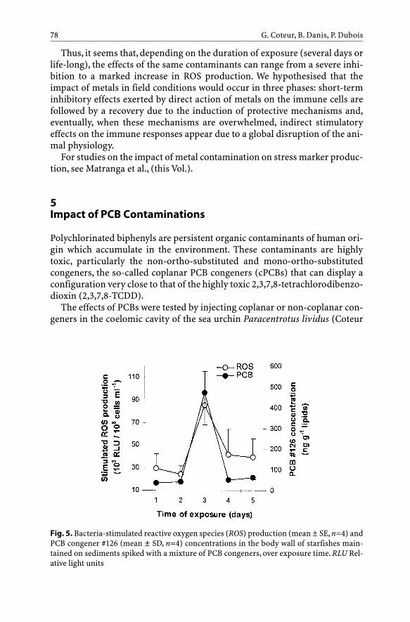
Thus, it seems that, depending on the duration of exposure (several days orlife-long), the effects of the same contaminants can range from a severe inhi-bition to a marked increase in ROS production. We hypothesised that theimpact of metals in field conditions would occur in three phases: short-terminhibitory effects exerted by direct action of metals on the immune cells arefollowed by a recovery due to the induction of protective mechanisms and,eventually, when these mechanisms are overwhelmed, indirect stimulatoryeffects on the immune responses appear due to a global disruption of the ani-mal physiology.
For studies on the impact of metal contamination on stress marker produc-tion, see Matranga et al., (this Vol.).
5Impact of PCB Contaminations
Polychlorinated biphenyls are persistent organic contaminants of human ori-gin which accumulate in the environment. These contaminants are highlytoxic, particularly the non-ortho-substituted and mono-ortho-substitutedcongeners, the so-called coplanar PCB congeners (cPCBs) that can display aconfiguration very close to that of the highly toxic 2,3,7,8-tetrachlorodibenzo-dioxin (2,3,7,8-TCDD).
The effects of PCBs were tested by injecting coplanar or non-coplanar con-geners in the coelomic cavity of the sea urchin Paracentrotus lividus (Coteur
G. Coteur, B. Danis, P. Dubois78
Fig. 5. Bacteria-stimulated reactive oxygen species (ROS) production (mean ± SE, n=4) andPCB congener #126 (mean ± SD, n=4) concentrations in the body wall of starfishes main-tained on sediments spiked with a mixture of PCB congeners, over exposure time. RLU Rel-ative light units

et al. 2001) and of the starfish A. rubens (Danis et al., pers. comm.). In bothstudies, PCBs were found to increase the production of ROS; coplanar con-geners being the most effective in this respect. At very high concentrations,however, a steep drop in ROS production was observed probably due to directcellular toxicity (Danis et al., pers. comm.). Similarly, when starfishes weremaintained on a sediment spiked with a mixture of PCB congeners, the amoe-bocyte ROS production followed closely the transient accumulation of copla-nar congeners, such as congener #126, in the starfish tissues (Fig. 5).
Likewise, the contamination by coplanar congener #77 was found toincrease the ROS production of starfishes exposed through water, sedimentsor food (Danis et al. 2003).
6Impact of Complex Contaminations
We investigated amoebocyte ROS production in natural populations ofstarfishes exposed to complex, multi-elemental contaminations in order toassess the general impact of contaminants on the immune function of
Echinoderm Reactive Oxygen Species (ROS)Production 79
Fig. 6. Bacteria-stimulated reactive oxygen species (ROS) production (103 RLU/106
cells ml–1; mean ± SE, n=10) in starfishes collected from different stations in the North Sea.RLU Relative light units

starfishes in the field. Starfishes were collected at different North Sea stationspresenting a large range of metal and PCB contamination levels (Coteur et al.2003b). Significant differences were found in ROS production of starfishescollected at the different stations. In particular, ROS production was signifi-cantly inhibited at the stations located off the Elbe/Weser estuaries (BW sta-tions) and off the North Sea Canal (LN3) (Fig. 6).
Moreover, ROS production was negatively linked with the levels of metalcontamination in the starfish tissues and in the sediments (Fig. 7). However,no link was found between the immune response and the non-coplanar PCBconcentrations in the samples.
From this study it can be concluded that the contamination levels observedin field conditions are sufficient to induce immunomodulation in echino-derms and that this modulation is particularly linked with metal contamina-tion of the environment.
7Discussion
The production of reactive oxygen species constitutes one of the main mech-anisms of destruction of foreign material in invertebrates. In echinoderms,it was first demonstrated by Ito et al. (1992) who found that sea urchinamoebocytes produce hydrogen peroxide when stimulated by erythrocytes.From our studies, bacteria or bacterial wall components also appear as effi-cient stimulators of ROS production. On the other hand, the parasitism ofstarfishes by a castrating ciliate or the challenge of sea urchins by theamoeba Paramoeba invadens did not modulate this oxidative activity(Wheatley et al. 1998; Coteur et al. 2004). Exogenous factors such as watertemperature and salinity can influence this immune response in echino-
G. Coteur, B. Danis, P. Dubois80
Fig. 7. Bacteria-stimulatedreactive oxygen species (ROS)production (mean ± SE, n=10)in starfishes collected from dif-ferent stations in the North Seain function of the cadmiumconcentration in the sedimentsof these stations. RLU Relativelight units

derms. It has been hypothesised that this increased ROS production is linkedto the elevated concentration of dissolved oxygen, a major source of ROS, atlow water temperature (Regoli et al. 2000). Sea urchin coelomocytes react byoverexpressing a heat shock protein (Hsp70-like protein) when subjectedeither to a rise or to a drop in temperature, the latter inducing a greaterstress response than the former (Matranga et al. 2000). The sharp rise inHsp70 expression in coelomocytes at low temperatures might represent aprotective response towards the ROS produced by these cells, since Hsp70 isan efficient antioxidant (Chong et al. 1998). From our studies, it is concludedthat if a particular period is to be selected for comparing ROS production inthe field, we would suggest either October to December, which is charac-terised by constant (but low) resting and stimulated ROS production (thelatter being around 50¥103 RLU/106 cells ml–1), or the period between Apriland August, during which stimulated ROS production is high and constant(around 100¥103 RLU/106 cells ml–1) but somewhat offset by an increasedvariation in resting ROS production.
When these precautions are taken, ROS production appears as a very effi-cient tool for immunotoxicological studies since it is modulated by the mostabundant and toxic contaminants. However, concerning metals, the effect dif-fers greatly according to the exposure type: the duration and routes of expo-sure appear crucial for determining the impact on ROS production. This vari-ability in the impact of metals makes it necessary to always includebackground-exposed starfishes whether in experimental conditions or in thefield in order to work on relative instead of absolute grounds.
In contrast to metals, PCBs have systematically the same effect on ROS pro-duction independent of the echinoderm species or exposure routes in exper-imental conditions. Either the stimulatory effect of PCB could be explained bya direct action of PCBs on the intrinsic mechanism of ROS production byamoebocytes as suggested by Coteur et al. (2001), or PCB detoxificationmechanisms could take place in amoebocytes; these processes lead to the pro-duction of a certain amount of oxygen free radicals, adding to the ROS pro-duced spontaneously by the immune cells.
A contaminant-induced increase or decrease in ROS production both rep-resent a hazard to the health of individuals. Decreased production will lead torecurrent infections since invading microorganisms will not be efficientlydestroyed. This was illustrated in molluscs in which contaminants thatreduced ROS production by oyster hemocytes exacerbated the severity ofinfections (see Baier-Anderson and Anderson 2000). It was also shown thatsome mollusc pathogens have the ability to inhibit ROS production and con-sequently to survive in their host (LaPeyre et al. 1995). On the other hand,increased ROS production can lead to damages to self tissues since ROS are,by their very nature, totally non-specific and are able to alter a wide range ofself-macromolecules such as DNA and enzymes (Buechter 1988).
From the study of complex field contaminations, it appears that contami-nants released in the environment, such as metals, modulate starfish amoebo-
Echinoderm Reactive Oxygen Species (ROS)Production 81

cyte ROS production. This impact potentially represents a threat to the sus-tainability of natural populations of echinoderms and thereby to the stabilityof benthic ecosystems.
Acknowledgements. G. Coteur and B. Danis were holders of doctoral grants from the“Fonds pour la formation à la recherche dans l’industrie et dans l’agriculture” (FRIA) andfrom the “Fondation David et Alice Van Buuren”. G. Coteur and Ph. Dubois are, respectively,Postdoctoral Researcher and Research Associate of the National Fund for ScientificResearch (NFSR, Belgium). Research was supported by Belgian federal research pro-grammes ICAS (SSTC contract MN/11/30) and ECOTOX2 (SSTC contract EV/11/23). This isa contribution of the Centre Interuniversitaire de Biologie Marine (CIBIM).
References
Anderson RS, Brubacher LL, Calvo LMR, Burreson EM, Unger MA (1997) Effect of in vitroexposure to tributyltin on generation of oxygen metabolites by oyster hemocytes. Envi-ron Res 74:84–90
Babior M (1984) The respiratory burst of phagocytes. J Clin Invest 73:599–601Baier-Anderson C, Anderson RS (1997) The effect of pentachlorophenol on pyridine
nucleotide production in oyster hemocytes: NADPH and immunomodulation. J Shell-fish Res 16:111–114
Baier-Anderson C, Anderson RS (2000) Immunotoxicity of environmental pollutants inmarine invertebrates. Rec Adv Mar Biotechnol 5:189–225
Brousseau P, Pellerin J, Morin Y, Cyr D, Blakley B, Boermans H, Fournier M (2000) Flowcytometry as a tool to monitor the disturbance of phagocytosis in the clam Mya arenariahemocytes following in vitro exposure to heavy metals. Toxicology 142:145–156
Buechter DD (1988) Free radicals and oxygen toxicity. Pharmaceut Res 5:253–260Chia FS, Xing J (1996) Echinoderm coelomocytes. Zool Stud 35:231–254Chong KY, Lai CC, Lille S, Chang CS, Su CY (1998) Stable overexpression of the constitutive
form of heat shock protein 70 confers oxidative protection. J Mol Cell Cardiol 30:599–608Coteur G, Danis B, Fowler SW, Teyssie J-L, Dubois Ph, Warnau M (2001) Effects of PCBs on
reactive oxygen species (ROS) production by the immune cells of Paracentrotus lividus(Echinodermata). Mar Pollut Bull 42:667–672
Coteur G, Warnau M, Jangoux M, Dubois Ph (2002) Reactive oxygen species (ROS) produc-tion by amoebocytes of Asterias rubens (Echinodermata). Fish Shellfish Immunol12:187–200
Coteur G, Gillan D, Joly G, Pernet Ph, Dubois Ph (2003a) Field contamination of the starfishAsterias rubens by metals. Part 2: effects on cellular immunity. Environ Toxicol Chem22:2145–2151
Coteur G, Gosselin P,Wantier P, Chambost-Manciet Y, Danis B, Pernet Ph,Warnau M, DuboisPh (2003b) Echinoderms as bioindicators, bioessays, and impact assessment tools ofsediment-associated metals and PCBs in the North Sea. Arch Environ Contam Toxicol45:190–202
Coteur G, Corriere N, Dubois Ph (2004) Environmental factors influencing the immuneresponses of the common European starfish (Asterias rubens). Fish Shellfish Immunol16:51–63
Danis B, Cotret O, Teyssié JL, Fowler SW, Bustamante P, Warnau M (2003) Delineation ofPCB uptake pathways in a benthic sea star using a radiolabelled congener. Mar Ecol ProgSer 253:155–163
G. Coteur, B. Danis, P. Dubois82

Dyrynda EA, Law RJ, Dyrynda PEJ, Kelly CA, Pipe RK, Graham KL, Ratcliffe NA (1997)Modulations in cell-mediated immunity of Mytilus edulis following the ‘Sea Empress’ oilspill. J Mar Biol Assoc UK 77:281–284
Dyrynda EA, Pipe RK, Burt GR, Ratcliffe NA (1998) Modulations in the immune defences ofmussels (Mytilus edulis) from contaminated sites in the UK. Aquat Toxicol 42:169–185
Fournier M, Cyr D, Blakley B, Boermans H, Brousseau P (2000) Phagocytosis as a biomarkerof immunotoxicity in wildlife species exposed to environmental xenobiotics. Am Zool40:412–420
Ito T, Matsutani T, Mori K, Nomura T (1992) Phagocytosis and hydrogen peroxide produc-tion by phagocytes of the sea urchin Strongylocentrotus nudus. Dev Comp Immunol16:287–294
Kehrer JP (2000) The Haber-Weiss reaction and mechanisms of toxicity. Toxicology149:43–50
LaPeyre JF, Chu FLE, Vogelbein WK (1995) In vitro interaction of Perkinsus marinus mero-zoites with eastern and Pacific oyster hemocytes. Dev Comp Immunol 19:291–304
Larson KG, Roberson BS, Hetrick FM (1989) Effect of environmental pollutants on thechemiluminescence of hemocytes from the American oyster Crassostrea virginica. DisAquat Org 6:131–136
Lennartz MR (1999) Phospholipases and phagocytosis: the role of phospholipid-derivedsecond messengers in phagocytosis. Int J Biochem Cell Biol 31:415–430
Matranga V, Toia G, Bonaventura R, Muller WEG (2000) Cellular and biochemical responsesto environmental and experimentally induced stress in sea urchin coelomocytes. CellStress Chaper 5:113–120
Matranga V, Bonaventura R, Di Bella G (2002) hsp70 as a stress marker of sea urchin coelo-mocytes in short term cultures. Cell Mol Biol 48:345–359
Noble PB (1970) Coelomocyte aggregation in Cucumaria frondosa: effect of ethylenedi-aminetetraacetate, adenosine, and adenosine nucleotides. Biol Bull 139:549–556
North Sea Task Force (1993) Assessment report 1993. Subregion 6 (Norway). State PollutionControl Authorities. HS-Trykk A/S Press, Oslo
Pipe RK, Coles JA, Thomas ME, Fossato VU, Pulsford AL (1995) Evidence for environmen-tally derived immunomodulation in mussels from the Venice Lagoon. Aquat Toxicol32:59–73
Reeves EP, Lu H, Jacobs HL, Messina CGM, Bolsover S, Gabella G, Potma EO, Warley A, RoesJ, Segal AW (2002) Killing activity of neutrophils is mediated through activation of pro-teases by K+ flux. Nature 416:291–297
Regoli F, Nigro M, Bompadre S, Winston GW (2000) Total oxidant scavenging capacity(TOSC) of microsomal and cytosolic fractions from Antarctic,Arctic and Mediterraneanscallops: differentiation between three potent oxidants. Aquat Toxicol 49:13–25
Torreilles J, Guerin MC, Roch P (1996) Reactive oxygen species and defense mechanisms inmarine bivalves. C R Acad Sci Paris Life Sci 319:209–218
Torreilles J, Guerin MC, Roch P (1999) Modified Alsever’s solution is not a good medium forreactive oxygen metabolite study in bivalves. Fish Shellfish Immunol 8:65–69
Wheatley K, Brown RG, Scheibling RE, Jellett JF (1998) Coelomocyte oxidative activity ofthe green sea urchin (Strongylocentrotus droebochiensis) following challenge by bacter-ial and amoebic pathogens. In: Mooi R, Telford M (eds) Echinoderms: San Francisco.AABalkema, Rotterdam, pp 881–886
Echinoderm Reactive Oxygen Species (ROS)Production 83


Monitoring Chemical and Physical Stress Using Sea Urchin Immune Cells
V. Matranga, A. Pinsino, M. Celi, A. Natoli, R. Bonaventura,H.C. Schröder, W.E.G. Müller
Abstract. Coelomocytes are the cells freely circulating in the body fluid con-tained in echinoderm coelom and constitute the defence system, which, inresponse to injuries, host invasion, and adverse conditions, is capable ofchemotaxis, phagocytosis, and production of cytotoxic metabolites. Red andcolourless amoebocytes, petaloid and philopodial phagocytes, and vibratilecells are the cell types that, in different proportions, constitute the mixedcoelomocyte cell population found in sea urchins. Advances in cellular andmolecular biology have made it possible to identify a number of specific pro-teins expressed in coelomocytes under resting conditions or when activatedby experimentally induced stress. Only recently, coelomocytes have been usedfor pollution studies with the aim of introducing a new biosensor for detec-tion of stress at both cellular and molecular levels, as sentinel of sea health. Inthis chapter, we briefly review the important features of these valuable cellsand describe studies on their use in the laboratory and in the field for theassessment of chemical and physical pollution of the sea.
1Introduction
Life on earth originated in the sea. Marine animals were the first animals thathad to evolve defence strategies for their survival. To cope with possibly dan-gerous events, marine organisms have developed over millions of years what
Progress in Molecular and Subcellular BiologySubseries Marine Molecular BiotechnologyV. Matranga (Ed.), Echinodermata© Springer-Verlag Berlin Heidelberg 2005
V. Matranga (e-mail: [email protected]), A. Pinsino, M. Celi, A. Natoli, R. BonaventuraIstituto di Biomedicina e Immunologia Molecolare (IBIM) “Alberto Monroy”, ConsiglioNazionale delle Ricerche,Via U. La Malfa 153, 90146 Palermo, ItalyH.C. Schröder, W.E.G. MüllerInstitut für Physiologische Chemie, Abteilung Angewandte Molekularbiologie, JohannesGutenberg-Universität, Duesbergweg 6, 55099 Mainz, Germany

we now call an immune system, as a strong defence mechanism able to resisthost attacks in an efficient way. The term “immunis” comes from the Latinmeaning “exempt”, referring to protection against foreign agents. It is stillbelieved that in all organisms of the animal kingdom the cells of self are vir-tually marked, so that they are not attacked by their own defence mechanism.Therefore, the immune system must have the capacity to discriminatebetween self and non-self, to transform itself to deal with future dangers, and,in addition, to change, since the self also evolves with time (e.g. duringembryo development). However, the self/non-self model of the immune sys-tem has recently been disproved because of many inconsistencies: (1) not allforeign cells need to be destroyed, some in fact must be assimilated for nour-ishment; (2) in mammals the growing embryo, in principle a host, is notdestroyed by the immune system of the mother. This is just to quote twoexamples. To solve these apparent paradoxes one very attractive theory hasbeen proposed by Polly Matzinger, the present head of the National Instituteof Allergy and Infectious Diseases at NIH, Bethesda, Maryland, USA. In herSeptember 2001 lecture entitled, “An Innate Sense of Danger”, she proposedthat cellular apoptosis signals and directs the immune mechanism (reviewedin Matzinger 2002). In other words, the triggering factor comes from the“sense of danger” originated during damage to body tissues, rather than fromthe classical recognition of foreign antigens. Her theory has given hints tostudies on tumour regression, graft rejection, pregnancy failure and autoim-mune diseases. Another puzzling question, which the self/non-self model canneither predict nor explain, is: why is the mere foreignness of a protein notenough to elicit immunity, and why are noxious substances like mineral oil,mycobacteria, and aluminum hydroxide needed in order to get a clearresponse? Charles Janeway, one of the most brilliant and imaginative immu-nologists, recently deceased (April 2003), proposed, together with his col-league Medzhitov, the so-called Pathogen-Associated Molecular Pattern the-ory (Medzhitov and Janeway 2002). They postulate that immune responsescannot occur unless antigen-presenting cells are first activated, and that thisactivation occurs via pattern recognition receptors. Their prediction of thepresence of evolutionarily conserved receptors recognizing molecules frominfectious non-self organisms was confirmed when, studying Drosophila Tollinnate immune sensor, they found the mammalian Toll-like receptors(Medzhitov et al. 1997), which are now studied by laboratories around theworld.
The above-mentioned new ways of interpreting the machinery of theimmune system bring to the concept that the innate immune system is an evo-lutionarily ancient form of host defence found in most multicellular organ-isms. The basic mechanisms operating in sensing and attacking foreignagents are already found in echinoderms. It is stimulating that these hypothe-ses can be tested in the echinoderm model and that, to a certain extent, stud-ies on their defence system might provide many useful insights into theunderstanding of the mechanisms involved. In the following text, we briefly
V. Matranga et al.86

review the state of the art on what is known about the echinoderm immunesystem, with particular emphasis on mechanisms and/or molecules involvedin sensing experimental or environmental stress.
2Echinoderm Coelomocytes as Immune Effector Cells:Morphological Features and Recognized Functions
In all organisms the first defence against infections involves a physical barrierto invasions by pathogens or foreign substances, provided by their body coatswhich physically prevent the interaction between the host and the invadingorganism. However, when pathogens accidentally or experimentally penetratethis barrier they first encounter generic or specific molecules that restrict theinfection. The second-line defence consists of cells that can engulf (phagocy-tose) foreign substances or pathogens or can aggregate under formation oflarge coagulates (clots) in the case of wounds. Other cells secrete in themedium specific anti-host molecules, which will be discussed below. Phago-cytosis is a complex process which involves several steps, including: (1)chemotaxis, whereby phagocytic cells are attracted towards chemotacticchemicals like microbial products, complements, or damaged cells; (2) adhe-sion, whereby phagocytes stick to each other and attach to foreign agents; (3)opsonization, a process that enhances adhesion, whereby specific proteins(opsonins) are coated on the microbial surface; and (4) ingestion, wherebyphagocytes extend their cellular projections, engulfing the foreign organism.Finally, the foreign organism can be digested by enzymes in the lysosome. Allthe steps described above have been studied using echinoderms as a modelsystem. In the past, each of the echinoderm classes has preferentially beenused for a certain type of study; in the case of classical immune/defence stud-ies, involving self/non-self recognition, which occurs when grafting cells fromone individual to another, scientists have mostly used the classes of Holothu-riodea and Echinoidea. For the purpose of our description and to take care ofthe amount and the heterogeneity of information present in the literature, wewill focus our interest on echinoids, specifically sea urchins. In the body ofadult sea urchins a cell population lives, which has been identified as the trueimmune effector because of many proven characteristics. Quite vast, althoughnot so recent, literature is available which describes the morphologies presentin this mixed cell population (see Smith 1981; Matranga 1996). However, pos-sibly due to the way in which cells have been collected or preserved beforetheir observation by optical or electron microscopy, a variety of misleadingclassifications are reported, which have generated confusion. The easiest wayto give a nomenclature that reflects the actual morphology of the cells is theimmediate observation of fresh and live cells just taken from the sea urchinwithout any addition of anti-coagulant solution (Fig. 1). Under these condi-tions it appears clear that some cells are capable of rapid movements, while
Monitoring Chemical and Physical Stress Using Sea Urchin Immune Cells 87

others show very slow motions. Within the first group, fast-moving cells arered and colourless, each constituting 5–7 % of the total cell population. Pecu-liar to this cell type is their characteristic locomotion (about 0.5 µm s–1)achieved by rapid changes in their body shape, where a leading edge of the cellis protruded towards their march direction (Fig. 2). Consequently, these cellsare referred to as amoebocytes, from the Greek amoeba, which means change,referring to the shape of the cell (Fig. 1a,b). The red pigment, calledechinochrome, is thought to be utilized by echinoderms as an anti-bacterici-dal agent. This notion comes from the single report present to our knowledgein the literature, describing the release of a red pigment by the sand dollarMellita quinquiesperforata coelomocytes in response to stress (Smith andSmith 1985). In the same study the authors showed the release of histamine onchallenge and suggested that the coelomocyte stress response may be an evo-lutionary precursor to the mammalian allergic response. Due to their fastmovement, it seems conceivable to suppose that this cell type is utilized as afirst defence mobilization, as in the case of wound healing or prompt attack byforeign agents. Indeed, red amoebocytes have been observed trapped in clot-ting and encapsulation (Smith 1981).
Another type of very fast cells, constituting 5–6 % of the total cell popula-tion, are the so-called vibratile cells (Fig. 1 c); these are round cells which,thanks to a long flagellum, can move in a straight direction along a helicoidalpattern. The function of this less studied cell type is at present unknown.
Finally, the most abundant morphology observed (80–85 %) is constitutedby cells with a dendritic-like phenotype, namely “petaloid” or “philopodial.”Their conversion from the first to the second morphology is easily observedunder the microscope within 5–10 min, and a detailed description, as well as
V. Matranga et al.88
Fig. 1. Paracentrotuslividus coelomocytes:a red amoebocyte;b colourless amoebo-cyte; c vibratile cell;d petaloid phagocyte;e philopodial phago-cyte. Bar 10 µm

different functional states of the same cell type, can be found in several papers(Edds 1985, 1993; Henson et al. 1999).
These four major cell types have been consistently seen and described forat least three different species, i.e. the Pacific Ocean Strongylocentrotus purpu-ratus (Hillier and Vacquier 2003), the North Sea Strongylocentrotus droe-bachiensis (Bertheussen and Seljelid 1978), and the Mediterranean Sea Para-centrotus lividus (Matranga and Bonaventura 2002). However, the proportionof each cell type can vary not only among species but also between individu-als of the same species, according to their size and physiological conditions, aswe will see below. It should be recalled that these cells, within a few minutes(5–10 min) after collection from the sea urchin body, tend to clump; therefore,for their use and/or observation, they need to be placed in an anti-coagulantsolution, which, on the other hand, might affect their physiological morphol-ogy. However, much work still needs to be done on the function of all thesecell types. Most of the immune responses that we expect to be carried out areperformed by phagocytes (reviewed by Smith 1981). It seems that the petaloidstage is involved in migration towards the sites of injuries, while the fillopo-dial stage is involved in clotting. In fact, because of their high numeric repre-sentation and the ability to change the shape of their cytoplasmic protrusions,phagocytes are able to form large clots in which other coelomocyte types aretrapped or possibly contribute to their formation. However, of course, phago-cytes deserve their name from their ability to engulf (phagocyte) foreign par-ticles, such as pathogens, as observed in the clearance of injected bacteria (Yui
Monitoring Chemical and Physical Stress Using Sea Urchin Immune Cells 89
Fig. 2. Red and colourless amoebocytes freshly collected and rapidly moving under themicroscope. Bright fields of cells viewed on a Zeiss Axioscope 2 plus using a 63¥ objectivelent. Pictures taken every 8 s. Bar 10 µm

and Bayne 1983) or even synthetic beads (Bertheussen 1981). In addition,encapsulation, a process by which the host is surrounded by a layer of cellsand eventually digested by lytic enzymes released into the cavity, is per-formed by phagocytes and so-called spherule cells (possibly colorless amoe-bocytes), the latter being claimed to release bactericidal substances (Johnson1969; Gerardi et al. 1990).
However, due to the small number of controversial reports present in theliterature, the real function of each of these cell types is still not acknowl-edged.
3The Origin of Coelomocytes: Terminally Differentiated orCirculating Stem Cells?
Nowadays, the origin of coelomocytes is becoming an extremely interestingfield of research for at least two reasons: (1) the access to a new, interestingand easy to use model system to work on and (2) the potential source of stemcells for the production of new therapeutic tools (see, for example, theirinvolvement in regeneration phenomena). The axial organ, a complex circula-tory system composed of closed vessels, is believed to be the source of coelo-mocytes which, in analogy to the vertebrate system, has been regarded as theancestral primary lymphoid gland. This notion comes from an old studywhich described the release of coelomocytes from the axial organ after echi-noid injury (Millott 1969). Indeed, 10 years later, another report confirmedthese results, describing the presence in the axial organ of many coelomocytesrich in pigment and granules, separated into compartments by elastic fibers.The authors, reporting what they called “transformational stages of coelomo-cytes”, suggested the axial organ as the site of their storage and maturation, aswell as excretory functions via the water vascular system (Bachmann andGoldschmid 1978).
More recently, two populations of cells, namely adherent (B-like) cellsresembling mammalian B lymphocytes and non-adherent (T-like) cellsresembling mammalian T lymphocytes, have been fractionated from the axialorgan of the sea star Asteria rubens (Leclerc and Bajelan 1992). The character-ization and identification of molecules produced and released into the coelomby these two cell types would be of extreme interest. An alternative hypothe-sis explaining the presence of the mixed cell population in the coelomic fluidpostulates that these cells represent different maturation stages of the samecell line (Smith 1981). Although attractive, no evidence has been providedconfirming this hypothesis. On the contrary, recent studies have shown arapid increase in the amount of the red amoebocytes (previously referred toas red spherulae cells because of lack of movement caused by the mediumused) in response to pollution or experimentally induced stresses (Matrangaet al. 2000). This evidence can be explained by either the rapid division of cir-
V. Matranga et al.90

culating stem cells or the recruitment from the axial organ or similar reser-voirs. Both hypotheses need experimental confirmation.
Unfortunately, these lines of investigation have not been followed verymuch over the years. The development of techniques enabling the culture ofcoelomocytes as mixed cell population and, more interestingly, as single celltypes constitutes a necessary step for studies on the origin and function ofcoelomocytes; efforts are awaited in this direction.
4Molecules Expressed by Echinoderm Coelomocytes
Many studies performed in the early 1950s and 1960s demonstrated that, inresponse to graft reactions or invading agents, sea urchin coelomocytes pro-duce and release into the so-called coelomic fluid a variety of different pro-teins. In addition, several other stimuli or even the puncture of a needle wereshown to produce the activation of a certain number of genes (Smith et al.1992). The advent of molecular biology techniques, combined with efforts inbiochemical purification of molecules, led to the discovery of an extendedcollection of proteins, expressed in coelomocytes under physiological oradverse/stress conditions (reviewed in Matranga 1996; Matranga andBonaventura 2002). The repertoire of identified and characterized proteinsspecifically expressed in sea urchin coelomocytes is growing fast. The listincludes most of the immune molecules already described for the vertebrateimmune system, such as cytokines, lectins, perforins, lysosomal enzymes,serum proteins, profilins, and others. Below we give a brief overview of theseproteins, arbitrarily grouped into five main categories, which we think are ofinterest for the exploitation of new biotechnological tools.
4.1Innate Immune Molecules: The Complement System
Aquatic organisms are under constant assault by pathogens, which must bekept in check by the fast-acting innate immune system. Indeed, innate immu-nity is fighting against pathogens using defence mechanisms that are quicklymobilized and triggered by receptors recognizing a broad spectrum ofpathogens. The lower deuterostomes, including echinoderms, possess aninnate immune system part of which shares similarities with the vertebratecomplement system. The latter is composed of a complex group of serum pro-teins which are activated in a cascade-like fashion. Only the alternative path-way and the lectin pathway are of interest here, since the classical pathwayconcerns the adaptive immune system which involves antigen–antibody com-plexes that, to our best knowledge, are not present in echinoderms. The alter-native complement system pathway activates C1, C4, C2, C3, C5 and finally C6
Monitoring Chemical and Physical Stress Using Sea Urchin Immune Cells 91
to C9, which form the membrane attack complex. The lectin pathway activatesC2, C3, C4 and some C1 homologue calcium-dependent lectin family proteins.There is growing evidence from several studies that describe the identifica-tion of genes coding for homologues of the vertebrate complement systemproteins. It has been shown, for example, that complement component C3 isspecifically expressed in sea urchin coelomocytes (Al-Sharif et al. 1998).While in earlier studies the whole mixed coelomocyte population was used, invery recent reports some authors have separated by gradients the four popu-lations of cells and were able to show that only phagocytes express the SpC3gene and contain the protein (Gross et al. 2000). Further studies have shownthat SpC3 acts as a humoral opsonin, tagging target cells for ingestion by thecell type called, by authors, polygonal phagocytes (Clow et al. 2004), most pre-sumably the petaloid form of phagocytes. Interestingly, the same authorsfound that SpC3 is already present in the developing sea urchin embryo, witha peak in transcript levels just prior to and during gastrulation (Shah et al.2003). In addition, continuous exposure to heat killed Vibrio diazatrophicus, amarine pathogen of sea urchins, from the hatching blastula to the pluteusstages, highly increased SpC3 expression. These results provided evidence fora complement-based immune system in defence against pathogens, which seaurchin embryos may use in their aquatic environment. In later studies, twocDNAs from coelomocytes, SpCRL (S. purpuratus complement related pro-tein, long form) and SpCRS (short form), were characterized. Their deducedprotein sequences have domains that are also found in regulatory proteins,such as factor H and factor I, and in the terminal pathway components C6 andC7 (Multerer and Smith 2004).
4.2Agglutinins, Lectins and Adhesion Molecules
One of the major events that are macroscopically observed in response topathogens or experimental manipulation is the aggregation of coelomocytesto form clots or to encapsulate hosts. Classically, studies on agglutination ofexogenously injected red blood cells into the sea urchin coelomic cavity led tothe partial characterization of many molecules. The aggregation of phago-cytes at inflammatory sites is a feature that invertebrates and vertebrates havein common. The recruitment of cells, mainly phagocytes and red amoebo-cytes, is due to chemotaxis and cellular proliferation is triggered by cytokines(Prendergast et al. 1983). In most cases the recognition and binding of non-self material is based on the presence of carbohydrate-specific molecules,which may therefore represent an evolutionary ancient binding principle.Candidates of the last functions are proteins isolated from the coelomic fluidand most probably produced by coelomocytes. Evidence has been reportedfor a so-called aggregation factor found in Holothuria polii (Canicattì andRizzo 1991) as well as in sea urchins (Canicattì et al. 1992). It is now generally
V. Matranga et al.92

accepted that the invertebrate host defence functions first by immobilizingand then by encapsulating offensive (attacking) microorganisms. During thisprocess, two mechanisms are operating, possibly in cooperation: some mole-cules are involved in the adhesion between cells surrounding and encapsulat-ing the host, while others, such as perforins (for a review see Glinski andJarosz 2000), attack the invading agent.
Some years ago we showed that colourless amoebocytes, at the timereferred to as colourless spherule cells (Cervello et al. 1994), serve as reser-voirs for the precursor of a cell adhesion protein (toposome) which is laterused by the embryo (Matranga et al. 1986) and whose synthesis has recentlybeen demonstrated (Unuma et al. 2001; see Zito et al., this Vol.). The protein isreleased into the medium in response to stress induced by centrifugation, sug-gesting its involvement in the clotting of coelomocytes after injuries.
Hillier and Vacquier (2003) have recently demonstrated that intercoelomo-cyte adhesion is mediated by a coelomic plasma protein with a relative mole-cular mass (Mr) of 75 kDa, named amassin. The protein forms large disulfide-bonded aggregates to which coelomocytes attach. What is interesting is that,by sequence comparison data, it has been shown that amassin shares a proteindomain with olfactomedin (OLF), a group of extracellular matrix vertebrateproteins specific to the olfactory neuroepithelium (Yokoe and Anholt 1993).The authors claim that this is the first report assigning a function to a proteinof the OLF family and suggest that other as yet undescribed proteins belong-ing to this family may function in intercellular adhesion.
Among cell–cell and cell–matrix adhesion molecules, integrins, a family ofproteins, play a fundamental role. Through the recent use of polymerase chainreaction, it has been possible to identify integrin beta subunits in coelomo-cytes. Comparative sequence analysis with vertebrate integrins will con-tribute to the understanding of the evolution and function of this importantgroup of receptors (for a review on the argument see Burke 1999).
Even if not related to the immune response, a few reports have describedthe presence of globin/hemoglobin in echinoderms. This is the case for fourdifferent globin molecules – two of them have been sequenced – found in thesea cucumber Caudina arenicola (McDonald et al. 1992). By partial aminoacid sequencing (about 40 amino acids) little homology with holothurian glo-bins has been found in recently discovered hemoglobins contained in coelo-mocytes circulating in the water vascular system of the ophiuroid Hemipholiselongata (Christensen et al. 2003). The authors were able to demonstrate ahigher affinity for oxygen in juveniles than in adults; the latter exhibiting aheterogeneous mixture composed of three major fractions. Taken together,these findings, confirming once again the role of the “hemal” system carriedout by coelomocytes in some of the echinoderm classes (holothurians andcrinoids), provide new insights into the understanding of the evolution andfunction of globin genes.
Monitoring Chemical and Physical Stress Using Sea Urchin Immune Cells 93

4.3Cytoskeleton Proteins
The cytoskeleton is a cellular “scaffolding” or “skeleton,” used to maintainand/or alter cellular shape. It appears to be evident that an organized set ofproteins must operate in order to accomplish the rapid movements that allcoelomocyte populations carry out. The expression of one such specific pro-tein, kinesin, was first shown by domain-specific monoclonal antibodies(McAbs); the protein is highly concentrated in restricted cellular domains,always in association with microtubules which are well spread in fast-movingcells and perinuclear in stationary cells (Henson et al. 1992).
The partial purification of a 110-kDa peptide, possessing K+EDTA-, Ca2+-,Mg2+-, and F-actin-activated Mg2+-ATPase activities characteristic of myosin-like motor proteins involved in particle/vesicle intracellular trafficking, hasbeen reported (D’Andrea et al. 1994). The authors claim that coelomocytesdisplay particle/vesicle movements even in cells devoid of microtubules(petaloid) and possess an unconventional myosin which may be the motorprotein driving particle/vesicle translocation.
The presence of other intermediate filament proteins has been shown inpetaloid coelomocytes from two species of echinoderms, the sea urchinStrongylocentrotus droebachiensis and the sea cucumber Cucumaria frondosa(Holy et al. 2000). Three isoforms have been characterized: a 68-kDa seaurchin lamin, as well as two putative lamin isoforms of approximately 70 and68 kDa in sea cucumber coelomocytes. In this study, McAbs were producedthat recognize lamin isoforms in immunofluorescence and Western blot andwere used for tracing studies on their intercellular localization and function.Other authors have studied the actin-based centripetal flow process in seaurchin coelomocytes, demonstrating that it is the result of a two-part mecha-nism: actin polymerization at the cell edge coupled with actomyosin contrac-tion at the cell centre (Henson et al. 1992). Extended studies suggested a sig-nificant cross-talk between the two underlying mechanisms and indicate thatchanges in actomyosin tension may be translated into alterations of the struc-tural organization of the actin cytoskeleton (Henson et al. 2003).
A molecular approach provided evidence for the increased expression of agene (SpCoel1) coding for a newly discovered sea urchin profilin, in responseto minor injuries, such as puncture by a needle of the peristomal membrane(Smith et al. 1992). Its sequence has striking similarity to the mammalian pro-filin, a small protein that has both actin and phosphatidyl-inositol-biphos-phate binding functions (Goldschmidt-Clermont et al. 1991). It is supposedthat sea urchins can utilize this mechanism which links the signal transduc-tion system to the cytoskeleton.
V. Matranga et al.94

4.4NK Antigens and Cytokines
Special consideration should be given to these classes of proteins because theycould constitute a potential source of new therapeutic drugs, to be derivedfrom coelomocyte exploitation.
The obvious analogies in the features and functions of coelomocytes tohuman lymphocytes prompted the search for natural killer (NK) cells andtheir related antigens. It was not surprising that CD56 and CD57 antigenswere shown to be expressed in a subpopulation, although not specified, ofLythechinus variegatus coelomocytes, by immunocytometry analysis (Koroset al. 2000). On the other hand, only phagocytes from Arbacia punctulata wereshown to have the strongest cytotoxic activity and the highest binding tohuman NK markers, namely CD14, CD56 and CD158b (Lin et al. 2001).
Interestingly, using a mouse thymocyte proliferation assay, Burke andWatkins (1991) showed the presence of a cytokine activity extractable fromthe coelomic fluid of the sea star Pisaster ochraceus, which can be blockedwith antibodies to mammalian interleukin (IL)-1 alpha. Based on the increasein bacteria clearance after addition of recombinant IL-1 alpha to coelomo-cytes cultured in vitro, the authors proposed that endogenous interleukinsstimulate recruitment of phagocytic cells as part of the non-specific cellulardefence mechanism operating in asteroids.
The same line of evidence came from studies reporting that IL-1 and 6 andtumour necrosis factor (TNF) are found in the coelomic fluid of the echino-derm Asterias forbesi (Beck and Habich 1986, 1996). However, attempts at iso-lation and characterization of the true echinoderm TNF proved elusive sinceits presence has been demonstrated only indirectly by the use of cross-react-ing antibodies to the human inflammatory cytokine.
Recently, in the framework of the UVTOX European project, the presence ofa stress-inducible TNF-alpha-like activity in P. lividus sea urchin coelomo-cytes has been demonstrated (Matranga and Madaratz, pers. comm.). Thisevidence came from experiments in which the culture medium obtained fromcoelomocytes exposed to physical stress (temperature and UV-B radiation)was added to a mouse macrophage cell line, displaying a dose-dependentapoptosis in response to TNF-alpha (Mosman 1983; Hansen et al. 1989). Fur-thermore, coelomocyte-secreted activity was, at least partly, neutralized byantibodies raised against human or mouse TNF-alpha (not shown). Moreimportantly, by RT-PCR with different oligonucleotide primer pairs designedon the basis of murine TNF-alpha sequence, these authors were able to suc-cessfully amplify TNF-alpha fragments from total RNA obtained from seaurchin coelomocytes (not shown). Products of the expected size ranges werecloned into bacterial vectors and amplified for sequencing (not shown). Thefull length sequence analysis of the putative TNF-alpha mRNA, which is inprogress, will show whether sea urchin coelomocytes can produce vertebrate-like TNF-alpha molecules.
Monitoring Chemical and Physical Stress Using Sea Urchin Immune Cells 95

4.5Stress Response Proteins
The “4-D” model postulated by Matzinger for the functioning of the verte-brate immune system, namely Danger, Death, Destruction, and Distress, con-stitutes an attractive working hypothesis that could be tested using sea urchincoelomocytes. However, very few, if any, systematic and consistent studiestesting this hypothesis are provided in the literature. We will give just a fewexamples of proteins, related to stress response, which have attracted theattention of investigators. It is known that, during an inflammatory insult,changes in the plasma concentration of certain divalent cations, involving, forexample, iron-specific binding and transporting proteins, provide a majorhost defence response in vertebrates. Strikingly, a ferritin molecule wascloned from an echinoderm coelomocyte cDNA library showing a conservediron-responsive element sequence and sharing a high analogy to the verte-brate homologue (Beck et al. 2002). These authors showed an increasedexpression of ferritin mRNA after stimulation of coelomocytes with drugssuch as LPS or PMA, with a parallel decrease in the amount of iron in “invitro” culture supernatants over time. These results suggest that echinodermferritin is a protein involved in acute phases of toxification and that seques-tration of iron is an ancient host defence response in animals.
Recently, acute effects, as opposed to chronic effects, of temperature, chem-ical, UV-B radiation and pollution stresses have been demonstrated in studieson the expression of a few stress proteins. Briefly, we have demonstrated theoverexpression of hsp70 (Matranga et al. 2000, 2002) and acetylcholineesterase (Angelini et al. 2003); these results will be discussed in the followingsections.
5Cell Cultures of Coelomocytes: New Tools to Detect Marine Pollution
As already mentioned, the development of in vitro cultures of coelomocyteshas been considered important and recommended in order to better analyzethe mechanisms involved in defence/stress response and to overcome thestress of manipulation. Media for keeping sea urchin coelomocytes outside ofthe animal, for a reasonably sufficient period of time, generally involve the useof anti-coagulant reagents, such as the chelating agents EDTA and EGTA. Inour studies, P. lividus coelomocytes were kept and separated into subpopula-tions by gradients in an EDTA-containing buffer (ISO-EDTA). This mediumwas good enough to show, for the first time, that coelomocytes respond tophysical and chemical stresses by elevating the hsp70 expression levels(Matranga et al. 2000). However, it was not preserving the morphological fea-tures of freshly collected cells and it was not usable at all for in vitro exposure
V. Matranga et al.96

to chemicals or other stresses. A medium involving EGTA, although utilizedwith success for the American species Lythechinus variegatus (Koros 1993),was, in our hands, not working.We then used successfully a medium modifiedfrom that originally described by Henson et al. (1992). We were able, underthese conditions, to keep cells for short-term cultures in vitro, but not longerthan 4–6 h (Matranga et al. 2002). In fact, by Hoechst staining, we observedthat the number of apoptotic cells increased after prolonged time culture:from 5–6 % for culture times up to 6 h, gradually increasing to 10–12 %between 8 and 12 h, and above 20 % after 14 h. The number of apoptotic cellsreached 25 and 44 % after 16 and 18 h, respectively (not shown). Therefore, forstudies on chronic effects of pollutants on sea urchin coelomocytes, it wasnecessary to define a culture medium for long-term cultures. For this purposewe took advantage of the technique described for primary cultures of epithe-lial cells and myocytes obtained from the heart of the mollusc Pecten max-imus (Le Marrec-Croq et al. 1999), which utilizes a simple medium based onsterile seawater. In fact, mollusc cultures were maintained viable in vitro for atleast 2 months and used as a sensitive test to study the effects of pollutants atthe cellular level (Pennec et al. 2002).
The medium has been adapted to the P. lividus coelomocytes osmolarity,thanks to collaboration with G. Dorange within the framework of theUVTOX European project. Total cells were collected by bleeding with a ster-ile syringe introduced through the peristomal membrane and plated on tis-sue culture-treated 24-well plates. Half of the medium was refreshed everyother day. Under these conditions, cells attached to the plastic-ware withoutthe need of any special adhesive factor. The number of attaching cells grad-
Monitoring Chemical and Physical Stress Using Sea Urchin Immune Cells 97
Fig. 3a–d. Surface antigenslocalized on sea urchin coelomo-cytes attached to the substrate.Bright field and immunofluores-cence images of whole mountsdecorated with WGA-FITC. Bar10 µm

ually increased with time, and within 1 h virtually all cells plated werebound to the substratum. The vitality of cells was confirmed by the expres-sion of surface antigens, as assessed by WGA-FITC fluorescence on wholemount specimen (Fig. 3).
When maintained in culture for longer periods of time, cells have the ten-dency to form bundles and fibres, although maintaining their individuality(Fig. 4); petaloid-shaped phagocytes were no longer visible and possiblyphilopodial phagocytes were converted into a fibroblastoid-like morphology.Interestingly, both red and colourless amoebocytes, as well as vibratile cells,continued to be observable, constituting an easy criterion for assessing theviability of the culture.
An attempt to keep cells in culture for more than 8 days, which in somecases lasted up to 20 days (Fig. 5), resulted in the formation of a meshwork ofcell clumps interconnected by macrofibres made of cellular protrusions(Fig. 5a). Even after such a long time it was possible to distinguish red amoe-bocytes. Possibly, colourless amoebocytes were passively trapped or adheredto the large phagocyte aggregates present in the culture (Fig. 5b,c), in agree-ment with earlier reports on hanging drops cultures (Johnson 1969).
The culture media tested, namely the Henson and the Le Marrec-Croqmodified coelomocyte media, respectively, referred to as HMCC and LMCCbelow, were used for the next experiments and constitute good starting pointsto define an appropriate coelomocyte culture medium for establishing con-tinuous cell cultures.
V. Matranga et al.98
Fig. 4. Sea urchin coelomocyte cultures. Cells, maintained in culture in vitro for 4 (a) and 8(b) days tend to form bundles and fibres. Bar 10 µm

6Laboratory Experiments
The growing concern over the health state of the sea has attracted the atten-tion of many laboratories to the use of aquatic invertebrates, e.g. echino-derms, as tools for monitoring environmental hazards. However, only fewinvestigations report the effects of pollution in terms of cellular and biochem-ical modifications on adults of higher marine invertebrates, such as the seaurchin. Most of the reports present in the literature describe in fact thetoxic/teratogenic effects of various chemical agents and drugs on the develop-ment of sea urchin embryos (Kobayashi 1980; Ozretic and Krajnovic-Ozretic1985; Sconzo et al. 1995; Morale et al. 1998; Russo et al. 2003; Geraci et al. 2004;Roccheri et al. 2004). The studies described below were aimed at finding a bio-logical indicator to be used as a stress marker and to characterize the
Monitoring Chemical and Physical Stress Using Sea Urchin Immune Cells 99
Fig. 5. Sea urchin coelomocyte long-term cultures. Cells were maintained for20 days in culture after plating. Bar:a 158 µm; b 100 µm; c 50 µm

response to induced or accidental stresses by the use of molecular markersusing sea urchin coelomocytes as biosensors.
It was already known that sea urchin embryos respond to temperaturestress by the activation of specific genes (Giudice 1989), whose sequence forthe P. lividus species has been identified and described (Sconzo et al. 1992).However, there were no studies on the presence of constitutive and/orinducible hsp70 protein in adult sea urchin cells at that time. In a firstattempt, we tested the hypothesis that coelomocytes play a role in defencemechanisms activated by adverse external conditions, challenging whole P.lividus sea urchin to low and high temperature stress. For our purpose, adultindividuals were kept at 16 °C, the control temperature (CT), with subse-quent 35 °C heat stress (HS) or 4 °C cold stress (CS) for 4 h. After a 1-h recov-ery at 16 °C, coelomocytes were gently collected at 4 °C to prevent handlingstress as much as possible, lysed, and analyzed by Western blotting. The anti-hsp70 McAb used for immunodetection, previously shown to cross-reactwith other invertebrate hsp70s (Koziol et al. 1997), recognized both the con-stitutive and the inducible forms. We found an overexpression of the hsp70protein in coelomocytes obtained from temperature-stressed urchins, with atwo- and a five-fold increase in the protein levels for heat and cold stressrespectively (Matranga et al. 2000). The inductive response was time-depen-dent, i.e. individuals kept for 60 min at the non permissive temperature hadtwice the hsp70 levels as those exposed to the stress for 30 min. No increasesin hsp70 expression were found for periods between 1 and 4 h, and periodslonger than 4 h under temperature stress led to death of the animals(Matranga et al. 2000).
The presence and function of acetylcholinesterase (AChE) in vertebrateblood cells and plasma have been elucidated, suggesting its pivotal role in ionexchange (for a review of AchE activity in sea urchin embryos see Angelini etal., this Vol.); in addition, its activity has been found to be associated with theperinuclear region of leucocytes (Falugi 1985). Bearing in mind the similaritybetween coelomocytes and vertebrate blood cells, we investigated the pres-ence of AChE activity in coelomocytes exposed to cold stress, with the aim ofassessing the possibility of using the AChE activity present in these cells as abiomarker for both field and laboratory studies on environmental stress. Ourresults showed that cholinesterase activity increased with the time of expo-sure to cold stress and it was detected in petaloid phagocytes and colourlessamoebocytes (Angelini et al. 2003). In both cases, the function might belinked to cytoskeletal dynamics, which follows changes in intracellular ionconcentrations. This has been observed in sea urchin zygotes, as a result of theaction of molecules related to the cholinergic system (Harrison et al. 2002).
In our experiments on whole sea urchins, we found a discrete variability inthe basal levels of hsp70 as well as a different inductive response when com-paring coelomocytes that were obtained from different individuals. This canbe explained by the fact that different sea urchins possess a different personalhistory, thus reflecting different physiological conditions. We cannot in fact
V. Matranga et al.100

avoid the possibility that individuals utilized for the experiment were alreadyunder stress pressure caused, for example, by pathogens or other foreignagents (drugs, pollutants).We now know that pollution can be one of such fac-tors increasing the hsp70 expression (Matranga et al. 2000; see following sec-tion).
It was then important to repeat these experiments using the same cell pop-ulation taken from one individual and confirm the results obtained withwhole adults as object of study. Then, using in vitro short-term cultures(1–4 h) of sea urchin coelomocytes, an identical number of cells, having anidentical cell type composition, could be exposed to the same stress condi-tions, giving us also the possibility of monitoring their morphology. Whentheir response to temperature, acidic pH, and heavy metals was tested, usingagain the hsp70 protein as a stress marker, results recapitulated thoseobtained with whole sea urchins (Matranga et al. 2002). This was an importantachievement since, for the first time, coelomocyte cultures were used to“sense” heavy metal pollution, induced in our case experimentally by theaddition of cadmium chloride to the medium. Moreover, since a dose-depen-dent increase in the hsp70 levels was shown, this could have been convenientlyused for studies applied to environmental monitoring.
However, a concern for the applicability of the measurements was given bythe fact that in order to elicit a response in a short period of time (4 h), highconcentrations of cadmium were used, in the micromolar range. We knowthat such high cadmium levels are never reached in the marine environment,even in highly polluted areas. Then it was important to keep cells in culturefor longer periods of time and to test lower cadmium concentrations, in thenanomolar range. This was possible thanks to the definition of a coelomocyteculture medium, LMCC medium, by which cells could be easily maintainedfor 3–8 days in culture (see previous section). The hsp70 expression was thenmonitored in total coelomocytes continuously exposed to CdCl2 for 3 days,after plating the cells for 5 days in LMCC medium in 24-well plates. Underthese conditions cells were responsive to the heavy metal and showed anincrease in hsp70 levels. However, higher expression levels were observed atthe lowest CdCl2 doses, probably due to damage of cells exposed to the heavymetal for long periods of time. Figure 6 shows the results of a representativeexperiment.
It is well known that UV radiation causes damage to DNA and/or protein ina variety of organisms (for more information see Schröder et al., this Vol.).Many invertebrates, including sea urchins, spawn and/or develop in the 1-mzone of the sea and thus are susceptible to genetic damage. The effects on seaurchin embryo development have already been recently described by a fewauthors (Epel et al. 1999; Lesser et al. 2003; Bonaventura et al. 2005).
To test whether sea urchin coelomocytes respond to UV-B radiation(312 nm) (1,000 J/m2) alone, or in combination with different concentrationsof CdCl2, by activating the expression of hsp70, in vitro cultures have beenused as follows. Sea urchins were bled through a cut in the peristomal mem-
Monitoring Chemical and Physical Stress Using Sea Urchin Immune Cells 101

brane. The fluid was poured onto ISO-EDTA and the cell suspension wasdivided into a certain number of Petri dishes. Cultures of coelomocytes werekept under different stress (cadmium) conditions for 2 h; then the cells wereor were not exposed to UV-B, collected, centrifuged and the pellet lysed aspreviously described (Matranga et al. 2002). Equal amounts of protein wereloaded onto SDS-PAGE and analysed by Western blotting for the expression ofhsp70 using a commercially available antiserum (Fig. 7). A two-fold increasein the expression of hsp70 is found in UV-B-stressed coelomocytes as com-pared to the control. Similarly, a three-fold increase in the hsp70 level isobserved in coelomocytes exposed to 10–4 M CdCl2. When coelomocytes wereexposed to both 10–4 M CdCl2 and UV-B radiation, we did not observe an addi-
V. Matranga et al.102
Fig. 6. Densitometric analysisof Western blot with anti-hsp70 antibody of lysates fromcoelomocytes exposed for3 days in culture to differentCdCl2 concentrations
Fig. 7. Western blot analysis ofcoelomocytes exposed to differentCdCl2 concentrations for 2 h, with(+) or without (–) UV-B irradia-tion (1,000 J/m2), probed withanti-hsp70 antibody. Histogramshows the densitometric scanningof the filter

tive effect, the increase in hsp70 level being about 2.5-fold. We observed thesame trend for coelomocytes exposed to both 10–5 M CdCl2 and UV-B. Thisapparent paradox could be explained by the experimental conditions used:i.e., cells were first exposed to cadmium for 2 h and then UV-B treated.We canhypothesize that, in agreement with what is known about the protective effecton apoptosis produced by hsp70 expression (Samali and Cotter 1996), underthese conditions the first stress is somehow protecting cells from the secondone (UV-B).
7Field Studies
Only recently, we have tried to establish the use of echinoderm coelomocytes,obtained from sea urchins and sea stars, as a cell laboratory wherein to testdifferent environmental hazards. Below we report a few examples of analysesof samples collected in oceanographic campaigns held in the northern andsouthern Adriatic Sea as well as in the North Sea.
7.1The Northern Adriatic Sea: First Example of Monitoring MetalPollution
Back in 1995, during an EU-sponsored summer course on “Monitoring ofenvironmental stress using modern techniques”, we assessed pollution bymeans of analysing cellular and molecular markers of stress, using coelomo-cytes from sea urchins collected in the seawaters around Croatia, during thewar. Some tracts of this coast had been previously designated as pollutedareas, as a result of urban runoff and/or industrial waste from a nearby fishcannery, and others as non-polluted (Müller et al. 1998; Schröder et al. 1999).Animals were collected from three sites: (1) along the coast near Rovinj(Istria), known as the “urban contaminated area”, (2) in front of RuderBoskovic Marine Station, the “industrial contaminated site” and (3) from theuncontaminated Limski Canal (north of Rovinj), the “unpolluted controls”.The first difference observed between polluted and non-polluted coelomo-cytes was found at the cellular level. In fact, in those samples obtained fromsea urchins collected from polluted areas, regardless of urban runoff or indus-trial waste contamination, a consistently greater number of red amoebocyteswas observed (Matranga et al. 2000). We do not have an explanation yet toaccount for this increase in cell number.A few hypotheses can be put forward:(1) conversion of pre-existing phenotypes, for example from colourless to redamoebocytes; (2) rapid cell duplication activated by a mitogenic signal, possi-bly linked to the production of trophic factors, such as inflammatorycytokines IL-1 and 6 and/or TNF; and (3) recruitment from the so-called
Monitoring Chemical and Physical Stress Using Sea Urchin Immune Cells 103

hematopoietic areas present in the adult urchin (axial organ).Although all areattractive and supported by a few reports present in the literature, thesehypotheses need experimental confirmation.
At the molecular level, the differential expression of stress markers in coelo-mocytes obtained from sea urchins collected from polluted seawaters wasdemonstrated by a two-fold increase in hsp70 levels in comparison with con-trols (Matranga et al.2000).In addition,we found that both phagocytes and redamoebocytes, isolated by gradients, expressed hsp70 at high levels.
7.2The Southern Adriatic Sea: A Case Study for the Assessment of TNT Exposure
The studies described above were the first example of the use of sea urchincoelomocytes for monitoring environmental stress and opened the way toother field studies. This was the case for a campaign that took place in June2003 around Pianosa Island (Tremiti Islands Marine Protected Area, AdriaticSea, Italy), sponsored by the Istituto Centrale per la Ricerca Scientifica e Tec-nologica Applicata al Mare (ICRAM). This site was chosen because of the largenumber of TNT-containing bombs dumped at sea at the end of World War II.Obviously, the disposal of the explosive 2,4,6-trinitrotoluene (TNT) repre-sents a serious hazard to marine ecosystems, and several agencies stronglyrecommend their monitoring. Analyses of P. lividus specimens, collectedaround Pianosa Island, and compared with an unimpacted site in the samearchipelago near Caprara Island (Tremiti Islands Marine Protected Area,Adriatic Sea, Italy), were aimed at detecting the hsp70 expression levels andthe differences in the subpopulation ratio of coelomocytes. In Western blot-ting experiments using total cell lysates, we found a significant increase in theexpression of hsp70 in samples from the TNT-impacted site around PianosaIsland with respect to the Tremiti Islands (not shown). In addition, when sep-arated into subpopulations, coelomocytes obtained from sea urchins col-lected around Pianosa Island showed a high proportion of red amoebocytes,which, in contrast, were scarcely visible in specimens from control sites (notshown). These results support the suitability of sea urchin coelomocytes asvalid tools for monitoring the effects of TNT exposure on aquatic fauna andemphasize their important role as stress bioindicators for future ecotoxico-logical studies in the marine environment.
V. Matranga et al.104

7.3The North Sea: A Norwegian Fiord as a Natural Gradient of Metal Contamination
An interesting confirmation of the results obtained in field studies came fromthe analysis of hsp70 levels present in coelomocytes from the sea star Asteriarubens collected in the Norwegian fiords during the campaign held inMay–June 2000, thanks to collaboration with Drs. Coteur and Dubois. Thefiord, located in southwestern Norway, is about 38 km long, 1–3 km wide, andat maximum 390 m deep (see for details Coteur et al. 2003). The study areaand sampling sites are extremely interesting because they constitute a sort ofnatural dose-dependent open-sea laboratory where the heavy metal concen-tration decreases going from the head of the Sørfjord, where first zinc thentitanium/iron smelters are located, to the open sea. In fact, studies on theaccumulation of Cd, Pb, Zn, and Cu in the sea star Asteria rubens, living alongthe natural gradient, demonstrated a direct relationship with the level of con-tamination of the environment, at least for Cd and Pb (Coteur et al. 2003). Wethen tested hsp70 expression in the same samples and found again an increasein levels of the protein, which also correlated with the levels of heavy metalsmeasured in animal tissues (not shown). This result, demonstrating hsp70 asa marker of stress for another class of echinoderms, reinforces its efficiency inpollution monitoring.
8Concluding Remarks
Our current understanding of inflammatory and immune responses inhumans probably has its roots in the comparative approach to immunology.For example, in the late 1900s, research on echinoderms provided initial evi-dence of the importance of phagocytic cells in reactions to foreign material.Over the last 40 years it has been widely documented that sea urchin coelo-mocytes, the echinoderm immune effector cells, mediate cellular responses todanger through chemotaxis, phagocytosis, encapsulation and cytotoxicity.More recently, the production of a variety of proteins, whose genes are acti-vated by an efficient responsive system, has been reported. However, despiterecent advances in molecular and cellular biology, a clear understanding ofthe molecular mechanisms operating in echinoderms’ response to damage/stress is still lacking. This is partially due to the small number of reports,often contradictory, which makes interpretation of results a very difficult task.The possibility of using the sea urchin coelomocyte as a stress model undercontrolled laboratory conditions was first proposed by Koros (1993) andrecently described by Matranga (Matranga et al. 2000; Matranga 2002). Theiruse has been extended to in vitro short-term cultures and field studies, whichmade coelomocytes from P. lividus, and possibly from other echinoderm
Monitoring Chemical and Physical Stress Using Sea Urchin Immune Cells 105

species, a good tool for the assessment of molecular and cellular alterations inresponse to chemical and physical stresses. The introduction of novel tech-niques for the assessment of contamination in the marine environment is oneof the top priorities within the European Community. Efforts to identify andapply “early” stress markers, long before pathological conditions such as dis-eases, mortality, or population changes occur, are greatly awaited. Moreover,recent evidence has suggested that the use of a battery of biomarkers, beingmore advantageous than the use of a single biomarker, offers an effective earlywarning system in biomonitoring aquatic environments (Ringwood et al.1999). To this end, it would be interesting to develop a panel of sensible mole-cular stress markers expressed by sea urchin coelomocytes maintained inlarge plants or large-scale facilities where different stress conditions might betested.
Marine invertebrates rely on innate immunity, the study of which hasrecently grown in higher vertebrates. It is now believed that the Toll-likereceptors, discovered by comparative immunological studies, are responsiblefor a large proportion of the innate immune recognition of pathogens, sens-ing the “pathogen-associated molecular patterns” and/or providing the “dan-ger signal”, as speculated by Janeway and Matzinger, respectively. It is stimu-lating that the most abundant class of sea urchin coelomocyte, namelyphagocytes, has a striking similarity in morphology, and probably in func-tion, to dendritic cells, the immune cells present in the mammalian system inthose tissues that are in contact with the environment (skin, nose, lungs, stom-ach and intestine). Recently described endogenous “danger signals”, releasedby tissues undergoing stress, damage or abnormal death, and necessary forthe activation of dendritic cells (a necessary step for the initiation of primaryand secondary immune responses in humans), are: heat-shock proteins,nucleotides, reactive oxygen intermediates, extracellular-matrix breakdownproducts, neuromediators and cytokines like the IFNs (Gallucci and Matzin-ger 2001). The fact that most of these molecules have been found in sea urchincoelomocytes in response to physical and chemical stress once again demon-strates the suitability of this model system to sense pollution and the interestin this field for the exploitation of new biotechnological tools.
Acknowledgements. The authors wish to thank Dr. F. Zito for stimulating discussions andcritical reading of the manuscript and Mr. Davide Comacchio for technical assistance andhelp in maintenance of sea urchins in aquaria. This work was fully supported by the UVTOXEU project, contract no. EVK3-CT-1999-00005. Partial support from ASI projectno. I/R/073/01 and ICRAM project REDCOD no. B4-3070/2003/368585/SUB/D3 is alsoacknowledged. A.N. and R.B. were supported by the Italian Ministry for Education, Univer-sity and Research.
V. Matranga et al.106

References
Al-Sharif WZ, Suyer JO, Lambris JD, Smith LC (1998) Sea urchin coelomocytes specificallyexpress a homologue of the complement component C3. J Immunol 160:2983–2997
Angelini C,Amaroli A, Falugi C, Di Bella G, Matranga V (2003) Acetylcholinesterase activityis affected by stress conditions in Paracentrotus lividus coelomocytes. Mar Biol143:623–628
Bachmann S, Goldschmid A (1978) Fine structure of the axial complex of Sphaerechinusgranularis (Lam.) (Echinodermata: Echinoidea). Cell Tissue Res 193:107–123
Beck G, Habich GS (1986) Isolation and characterization of a primitive interleukin-1-likeprotein from an invertebrate, Asterias forbesi. Proc Natl Acad Sci USA 83:7429–7433
Beck G, Habich GS (1996) Characterization of an IL-6-like molecule from an echinoderm(Asterias forbesi). Cytokine 8:507–512
Beck G, Ellis TW, Habich GS, Schluter SF, Marchalonis JJ (2002) Evolution of the acute phaseresponse: iron release by echinoderm (Asterias forbesi) coelomocytes, and cloning of anechinoderm ferritin molecule. Dev Comp Immunol 26:11–26
Bertheussen K (1981) Endocytosis by echinoid phagocytosis in vitro. I. Recognition of for-eign matter. Dev Comp Immunol 5:241–250
Bertheussen K, Seljelid R (1978) Echinoid phagocyte in vitro. Exp Cell Res 111:401–412Bonaventura R, Poma V, Costa C, Matranga V (2005) UVB prevents skeleton growth and
simulates the expression of stress markers in sea urchin embryo. Biophys Biochem ResComm 328:150–157
Burke RD (1999) Invertebrate integrins: structure, function, and evolution. Int Rev Cytol191:257–284
Burke RD, Watkins RF (1991) Stimulation of starfish coelomocytes by interleukin-1.Biochem Biophys Res Commun 180:579–584
Canicattì C, Rizzo A (1991) A 220 kDa coelomocyte aggregating factor involved inHolothuria polii cellular clotting. Eur J Cell Biol 56:79–83
Canicattì C, Pagliara P, Stabili L (1992) Sea urchin coelomic fluid agglutinin mediates coelo-mocyte adhesion. Eur J Cell Biol 58:291–295
Cervello M, Arizza V, Lattuca G, Parrinello N, Matranga V (1994) Detection of vitellogeninin a subpopulation of sea urchin coelomocytes. Eur J Cell Biol 64:314–319
Christensen AB, Colacino JM, Bonaventura C (2003) Functional and biochemical propertiesof the hemoglobins of the burrowing brittle star Hemipholis elongata say (Echinoder-mata, Ophiuroidea). Biol Bull 205:54–65
Clow LA, Raftos DA, Gross PS, Smith LC (2004) The sea urchin complement homologue,SpC3, functions as an opsonin. J Exp Biol 207:2147–2155
Coteur G, Pernet P, Gillan D, Joly G, Maage A, Dubois P (2003) Field contamination of thestarfish Asterias rubens by metals. Part 1: short- and long-term accumulation along apollution gradient. Environ Toxicol Chem 22:2136–2144
D’Andrea L, Danon MA, Sgourdas GP, Bonder EM (1994) Identification of coelomocyteunconventional myosin and its association with in vivo particle/vesicle motility. J CellSci 107:2081–2094
Edds KT (1985) Morphological and cytoskeletal transformation in sea urchin coelomo-cytes. In: Cohen WD (ed) Blood cells of marine invertebrates: experimental systems incell biology and comparative physiology. AR Liss, New York, pp 53–74
Edds KT (1993) Effects of cytochalasin and colcemid on cortical flow in coelomocytes. CellMotil Cytoskeleton 26:262–273
Epel D, Hemela K, Schick M, Patton C (1999) Development in the floating world: defenses ofeggs and embryos against damage from UV radiation. Am Zool 39:271–278
Falugi C (1985) Histochemical localization of acetylcholinesterase in blood cells. Basic ApplHistochem 29:105–113
Monitoring Chemical and Physical Stress Using Sea Urchin Immune Cells 107

Gallucci S, Matzinger P (2001) Danger signals: SOS to the immune system. Curr OpinImmunol 13:114–119
Geraci F, Pinsino A, Turturici G, Savona R, Giudice G, Sconzo G (2004) Nickel, lead, and cad-mium induce differential cellular responses in sea urchin embryos by activating the syn-thesis of different HSP70s. Biochem Biophys Res Commun 322:873–877
Gerardi P, Lassegues M, Canicattì C (1990) Cellular distribution of sea urchin antibacterialactivity. Biol Cell 70:153–157
Giudice G (1989) Heat shock proteins in sea urchin embryos. Dev Growth Differ 31:103–106Glinski Z, Jarosz J (2000) Immune phenomena in echinoderms. Arch Immunol Ther Exp
48:189–193Goldschmidt-Clermont P, Machesky L, Doberstein S, Pollard T (1991) Mechanism of the
interaction of human platelet profilin with actin. J Cell Biol 113:1081–1089Gross PS, Clow LA, Smith LC (2000) SpC3, the complement homologue from the purple sea
urchin, Strongylocentrotus purpuratus, is expressed in two subpopulations of the phago-cytic coelomocytes. Immunogenetics 51:1034–1044
Hansen MB, Nielsen SE, Berg K (1989) Re-examination and further development of a pre-cise and rapid dye method for measuring cell growth/cell kill. J Immunol Methods119:203–210
Harrison PK, Falugi C, Angelini C, Whitaker MJ (2002) Muscarinic signalling affects intra-cellular calcium concentration during the first cell cycle of sea urchin embryos. Cell Cal-cium 31:289–297
Henson JH, Nesbitt D, Wright BD, Scholey JS (1992) Immunolocalisation of kinesin in seaurchin coelomocytes: association of kinesis with intracellular organelles. J Cell Sci103:309–320
Henson JH, Svitkina TM, Burns AR, Hughes HE, MacPartland KJ, Nazarian R, Borisy GG(1999) Two components of actin-based retrograde flow in sea urchin coelomocytes. MolBiol Cell 10:4075–4090
Henson JH, Kolnik SE, Fried CA, Nazarian R, McGreevy J, Schulberg KL, Detweiler M, Tra-bosh VA (2003) Actin-based centripetal flow: phosphatase inhibition by calyculin-Aalters flow pattern, actin organization, and actomyosin distribution. Cell MotilCytoskeleton 56:252–266
Hillier BJ, Vacquier VD (2003) Amassin, an olfactomedin protein, mediates the massiveintercellular adhesion of sea urchin coelomocytes. J Cell Biol 160:597–604
Holy J (2000) Intermediate filament proteins in echinoderm coelomocytes. Comp BiochemPhysiol B Biochem Mol Biol 127:491–504
Johnson PT (1969) The coelomic elements of sea urchins (Strongylocentrotus). 3. In vitroreaction to bacteria. J Invertebr Pathol 13:42–62
Kobayashi N (1980) Comparative sensitivity of various developmental stages of sea urchinsto some chemicals. Mar Biol 58:163–171
Koros AMC (1993) Neuroendocrine marker expression on sea urchin coelomocytes andother immunoregulatory parameters may be used to monitor environmental changes.Mar Environ Res 35:137–140
Koros AMC, Goodwin DG, Siderits RH, Malavasi F (2000) “Natural Killer” (NK) cell anti-gens, CD 56, CD57, and others are expressed on breast and lung tumor cells as well as seaurchin coelomocytes. Tissue Antigens 55:77–78
Koziol C, Batel R,Arinc E, Schröder HC, Müller WEG (1997) Expression of the potential bio-marker heat shock protein 70 and its regulator, the metazoan DnaJ homolog, by temper-ature stress in the sponge Geodia cydonium. Mar Ecol Prog Ser 154:261–268
Leclerc M, Bajelan M (1992) Homologous antigen for T cell receptor in axial organ cellsfrom the asterid Asterias rubens. Cell Biol Int Rep 16:487–490
Le Marrec-Croq F, Glaise D, Guguen-Guillouzo C, Chesne C, Guillouzo A, Boulo V, DorangeG (1999) Primary cultures of heart cells from the scallop Pecten maximus (mollusca-bivalvia). In Vitro Cell Dev Biol Anim 35:289–295
V. Matranga et al.108

Lesser MP, Kruse VA, Barry TM (2003) Exposure to ultraviolet radiation causes apoptosis indeveloping sea urchin embryos. J Exp Biol 206:4097–4103
Lin W, Zhang H, Beck G (2001) Phylogeny of natural cytotoxicity: cytotoxic activity ofcoelomocytes of the purple sea urchin, Arbacia punctulata. J Exp Zool 290:741–750
Matranga V (1996) Molecular aspects of immune reactions in Echinodermata. In: MüllerWEG, Rinkevich B (eds) Invertebrate immunology, vol 15. PMSB Series. Springer, BerlinHeidelberg New York, pp 235–247
Matranga V, Bonaventura R (2002) Sea urchin coelomocytes, the progenitors of vertebrateimmune effectors, as bio-indicators of stress and pollution. In: Yokota Y, Matranga V,Smolenicka Z (eds) The sea urchin: from basic biology to aquaculture. Swets andZeitlinger, Lisse, The Netherlands, pp 161–176
Matranga V, Kuwasaki B, Noll H (1986) Functional characterization of toposomes from seaurchin blastula embryos by a morphogenetic cell aggregation assay. EMBO J 5:3125–3132
Matranga V, Toia G, Bonaventura R, Muller WEG (2000) Cellular and biochemical responsesto environmental and experimentally induced stress in sea urchin coelomocytes. CellStress Chaperones 5:158–165
Matranga V, Bonaventura R, Di Bella G (2002) hsp70 as a stress marker of sea urchin coelo-mocytes in short term cultures. Cell Mol Biol 48:345–359
Matzinger P (2002) The danger model: a renewed sense of self. Science 296:301–305McDonald GD, Davidson L, Kitto GB (1992) Amino acid sequence of the coelomic C globin
from the sea cucumber Caudina (Molpadia) arenicola. J Protein Chem 11:29–37Medzhitov R, Janeway CA Jr (2002) Decoding the patterns of self and nonself by the innate
immune system. Science 12:298–300Medzhitov R, Preston-Hurlburt P, Janeway CA Jr (1997) A human homologue of the
Drosophila Toll protein signals activation of adaptive immunity. Nature 388:394–397Millott N (1969) Injury and the axial organ of echinoids. Experientia 25:756Morale A, Coniglio L, Angelini C, Cimoli G, Bolla A, Alleteo D, Russo P, Falugi C (1998) Bio-
logical effects of a neurotoxic pesticide at low concentrations on sea urchin early devel-opment. A teratogenic assay. Chemosphere 37:3001–3010
Mosman T (1983) Rapid colorimetric assay for cellular growth and cytotoxicity assays. JImmunol Methods 65:55
Müller WEG, Batel R, Lacorn M, Steinhart H, Simat T, Lauenroth S, Hassanein HMA,Schröder HC (1998) Accumulation of cadmium and zinc in the marine sponge Suberitesdomuncula and its potential consequences on single-strand breaks and on expression ofheat shock protein: a natural field study. Mar Ecol Prog Ser 167:127–135
Multerer KA, Smith LC (2004) Two cDNAs from the purple sea urchin, Strongylocentrotuspurpuratus, encoding mosaic proteins with domains found in factor H, factor I, andcomplement components C6 and C7. Immunogenetics 56:89–106
Ozretic B, Krajnovic-Ozretic M (1985) Morphological and biochemical evidence of the toxiceffect of pentachlorophenol on the developing embryos of the sea urchin. Aquat Toxicol7:255–263
Pennec JP, Gallet M, Gioux M, Dorange G (2002) Cell culture of bivalves: tool for the study ofthe effects of environmental stressors. Cell Mol Biol (Noisy-le-grand) 48:351–358
Prendergast RA, Lutty GA, Scott AL (1983) Directed inflammation: the phylogeny of lym-phokines. Dev Comp Immunol 7:629–632
Ringwood AH, Hameedi MJ, Lee RF, Brouwer M, Peters EC, Scott GI, Luoma SN, Digiulio RT(1999) Bivalve biomarker workshop: overview and discussion summaries. Biomarkers4:391–399
Roccheri MC, Agnello M, Bonaventura R, Matranga V (2004) Cadmium induces the expres-sion of specific stress proteins in sea urchin embryos. Biophys Biochem Res Commun321:80–87
Monitoring Chemical and Physical Stress Using Sea Urchin Immune Cells 109

Russo R, Bonaventura R, Zito F, Schröder HC, Müller I, Müller WEG, Matranga V (2003)Stress to cadmium monitored by metallothionein gene induction in Paracentrotuslividus embryos. Cell Stress Chaperones 8:232–241
Samali A, Cotter TG (1996) Heat shock proteins increase resistance to apoptosis. Exp CellRes 223:163–170
Schröder HC, Hassanein HMA, Lauenroth S, Koziol C, Mohamed TAAA, Lacorn M, Stein-hart H, Batel R, Müller WEG (1999) Induction of DNA strand breaks and expression ofhsp70 and GRP78 homolog by cadmium in the marine sponge Suberites domuncula.Arch Environ Contam Toxicol 36:47–55
Sconzo G, Scardina G, Ferraro MG (1992) Characterization of a new member of the seaurchin Paracentrotus lividus hsp 70 gene family and its expression. Gene 121:353–358
Sconzo G, Romancino D, Fasulo G, Cascino D, Giudice G (1995) Effect of doxorubicin andphenytoin on sea urchin development. Pharmacie 50:616–619
Shah M, Brown KM, Smith LC (2003) The gene encoding the sea urchin complement pro-tein, SpC3, is expressed in embryos and can be upregulated by bacteria. Dev CompImmunol 27:529–538
Smith LC, Britten RJ, Davidson EH (1992) SpCoel1: a sea urchin profilin gene expressedspecifically in coelomocytes in response to injury. .Mol Biol Cell 3:403–414
Smith SL, Smith AC (1985) Sensitization and histamine release by cells of the sand dollar,Mellita quinquiesperforata. Dev Comp Immunol 9:597–603
Smith VJ (1981) The echinoderms. In: Ratcliffe NA, Rowley AT (eds) Invertebrate bloodcells, vol 2. Academic Press, London
Unuma T, Okamoto H, Konishi K, Ohta H, Mori K (2001) Cloning of cDNA encoding vitel-logenin and its expression in red sea urchin, Pseudocentrotus depressus. Zool Sci18:559–565
Yokoe H, Anholt RR (1993) Molecular cloning of olfactomedin, an extracellular matrix pro-tein specific to olfactory neuroepithelium. Proc Natl Acad Sci USA 90:4655–4659
Yui MA, Bayne CJ (1983) Echinoderm immunology: bacterial clearance by the sea urchinStrongylocentrotus purpuratus. Biol Bull 165:473–486
V. Matranga et al.110
DNA Damage and Developmental Defects AfterExposure to UV and Heavy Metals in Sea UrchinCells and Embryos Compared to OtherInvertebrates
H.C. Schröder, G. Di Bella, N. Janipour, R. Bonaventura, R. Russo,W.E.G. Müller, V. Matranga
Abstract. The depletion of the stratospheric ozone layer and the resultingincrease in hazardous ultraviolet-B (UV-B) radiation reaching the Earth areof major concern not only for terrestrial but also for aquatic organisms. UV-Bis able to penetrate clear water to ecologically significant depths. This chapterdeals with the effects of UV radiation on DNA integrity in marine benthicorganisms, in particular sea urchins in comparison to other marine inverte-brates (sponges and corals). These animals cannot escape the damagingeffects of UV-B radiation and may be additionally exposed to pollution fromnatural or anthropogenic sources. Besides eggs and larvae that lack a protec-tive epidermal layer and are particularly prone to the damaging effects of UVradiation, coelomocytes from the sea urchin Paracentrotus lividus were usedas a “cellular sensor” to analyse the effects on DNA caused by UV-B, heavymetals (cadmium), and their combined actions. From our data we concludethat sea urchin coelomocytes as well as cells from other marine invertebratesare useful bioindicators of UV-B and heavy metal stress, responding to thesestressors with different extents of DNA damage.
Progress in Molecular and Subcellular BiologySubseries Marine Molecular BiotechnologyV. Matranga (Ed.), Echinodermata© Springer-Verlag Berlin Heidelberg 2005
H.C. Schröder (e-mail: [email protected]), N. Janipour, W.E.G. MüllerInstitut für Physiologische Chemie, Abteilung Angewandte Molekularbiologie, JohannesGutenberg-Universität, Duesbergweg 6, 55099 Mainz, GermanyG. Di Bella, R. Bonaventura, R. Russo,V. MatrangaIstituto di Biomedicina e Immunologia Molecolare (IBIM) “Alberto Monroy”, ConsiglioNazionale delle Ricerche,Via U. La Malfa 153, 90146 Palermo, Italy

1Introduction
It is now well established that the thinning of the stratospheric ozone (O3)layer results in higher levels of mid-ultraviolet (UV-B) radiation reaching theEarth’s surface (Smith et al. 1992; El-Sayed et al. 1996). The biologically dam-aging effects of decreased UV-B absorption in the stratosphere are especiallypronounced in the Antarctic where the decline in ozone levels (the so-calledozone hole) amounts to more than 50 % (Gleason et al. 1993). However, reduc-tion of ozone concentration is also occurring at temperate and tropical lati-tudes (Stolarski et al. 1992).
1.1Atmospheric Ozone
The UV waveband of the electromagnetic spectrum is subdivided into threeregions: UV-A (320–400 nm), UV-B (280–320 nm), and UV-C (200–280 nm).The biological effects of UV radiation strongly depend on wavelength. In gen-eral, shorter-wavelength UV radiation (<320 nm) is more active than UVradiation at longer wavelength.
Atmospheric O3 is created in the stratosphere at altitudes between about 25and 100 km; it selectively absorbs UV radiation with different efficiencydepending on wavelength (Molina and Molina 1986). The most damagingUV-C radiation is completely absorbed before reaching the Earth’s surface,while UV-A and visible light (spectral range 400–700 nm) are not significantlyabsorbed by the ozone layer. The release of chlorofluorocarbons and otheranthropogenic chemicals, including nitrous oxide and bromine-containinghalons, is causing a degradation of the ozone layer, resulting in an increase inthe intensity, in particular of the short-wavelength UV-B radiation penetrat-ing the Earth’s atmosphere. In contrast to UV-B, the solar spectrum in the UV-A and visible range is only little or unchanged by ozone depletion (Cadwell etal. 1986).
The intensity of biologically damaging UV radiation at the Earth’s surfacedepends on a variety of factors including cloud cover, sun angle, atmosphericaerosols and seasonal and geographical factors (Frederick et al. 1989). In gen-eral, a strong seasonal variation is observed in temperate regions, while sea-sonal dependence is much less near the equator.
H.C. Schröder et al.112

1.2Penetration of Solar UV Radiation in Marine Waters
In seawater, sunlight is attenuated by absorption and scattering with increas-ing depth (Jerlov 1968; Dustan 1982). In parallel, a change in the spectral com-position of the light is observed. The attenuation of visible light is higher atthe wavelengths at either end of its spectral range. In the UV range, shorterwavelengths (UV-B and UV-C) are more attenuated than longer wavelengths(UV-A). Nevertheless, UV-B light still penetrates in clear seawater up to 30 m(Smith et al. 1992) or more (about 50 m in the Antarctica; Smith et al. 1992)and harmful effects can still be measured at depths up to 20–25 m (Smith andBaker 1979, 1981; Karentz and Lutze 1990; Regan et al. 1992; Smith et al. 1992;Prézelin et al. 1994). The penetration of UV-B is, however, influenced by sev-eral constituents of the seawater, in particular by dissolved organic carbon(Baker and Smith 1982; Scully and Lean 1994; Crump et al. 1999), the amountof which is higher in coastal waters than the open sea.
1.3Effects of Solar UV Radiation on Aquatic Organisms
In humans, increased UV-B exposure may cause skin cancer, eye damage, andchanges in the immune system (Griffiths et al. 1998). However, deleteriouseffects of UV-B radiation are also observed in aquatic organisms and ecosys-tems (Häder et al. 1995). There are many studies concerning the effects of UV-B irradiation on phytoplanktonic organisms (Helbling et al. 1992). However,zooplankton and benthic invertebrates are also prone to UV-B damage(Hunter et al. 1979; Damkaer et al. 1980; Karanas et al. 1981; Malloy et al. 1997).In particular, sessile species such as sponges and corals are sensitive to theadverse effects caused by UV-B radiation (reviewed in Acevedo and Nolan1993). These animals do not possess a UV-B-adsorbing epidermal layer likehigher plants and animals and cannot avoid the harmful effects of solar radi-ation.
The most serious effects of UV-B radiation are observed in those organ-isms that are exposed to sunlight during their developmental stages. Eggs andlarvae residing in surface waters may receive high levels of UV-B, causingimpaired development (Hunter et al. 1979; Damkaer et al. 1981; Damkaer andDey 1983; Ringelberg et al. 1984; El-Sayed 1988; Jeffery 1990; Shick et al. 1991;Bothwell et al. 1993; Blaustein et al. 1994; Little and Fabacher 1994; Häder et al.1995).
Protection from harmful UV-B radiation can be achieved by behavioural(e.g. the daily vertical migration of planktonic organisms) or physiological(e.g. development of shells, enhanced melanin production, or induction ofantioxidant defense and repair processes) adaptation mechanisms (El-Sayed1988; Vincent and Roy 1993; Hessen 1994).
DNA Damage and Developmental Defects After Exposure to UV and Heavy Metals 113

1.4DNA Damage
The deleterious effects of UV-B on marine organisms are caused by photo-chemical absorption of radiation by macromolecules. UV-B induces directdamage to DNA, proteins, and some other molecules that have absorbancemaxima in the UV-B region. The most important macromolecule affected byUV-B radiation is DNA (Friedberg et al. 1995). The absorbing centers withinthe DNA are the purine and pyrimidine bases, which have absorption maximaat 260–265 nm. The absorption maxima of proteins in the UV-B and UV-Cregions are around 280 nm. UV-A radiation causes indirect damage to DNAvia formation of oxygen and hydroxyl radicals (Fridovich 1986; Peak and Peak1989; Karentz et al. 1994). These reactive oxygen species (ROS) interact withDNA to form strand breaks, alkali-labile sites and DNA protein cross-links.The major lesions induced by UV-B are DNA photoproducts [cyclobutanepyrimidine dimers and pyrimidine(6–4)pyrimidone dimers]. The most com-mon lesion caused by UV-A is the formation of 8-hydroxydeoxyguanosine.
1.5DNA Repair
DNA lesions could be cytotoxic, by inhibiting DNA replication and transcrip-tion, or cause mutations, which are possibly harmful for living organisms.Therefore several mechanisms have evolved to repair damaged DNA. Thereare two major DNA repair mechanisms: photoreactivation and nucleotideexcision repair. Photoreactivation repair of UV-induced cyclobutane pyrimi-dine dimers is a photoenzymatic process consisting of two steps: binding of aphotoreactivating enzyme (photolyase) to the dimers and subsequent repairand dissociation of the complex after irradiation with visible light. Aquaticorganisms may be highly sensitive to exposure to UV-B in the absence of vis-ible light (Hirosawa and Miyachi 1983; Buhlmann et al. 1987). Nucleotide exci-sion repair involves a series of steps, including recognition of the defectivesite, incision of the damaged DNA strand at or near the lesion, subsequentexcision and synthesis of the DNA around the damaged site using the comple-mentary intact strand as a matrix, and, finally, ligation of the remaining nickin the repaired strand (Sancar 1996a). DNA excision repair does not dependon light.
1.6Solar UV Radiation and Evolution
The formation of the stratospheric ozone layer was a result of the oxygenationof the atmosphere due to evolution of photosynthesis. In the period before the
H.C. Schröder et al.114

“Cambrian explosion”, there were only low levels of oxygen in the atmosphere(Cloud 1968; Canfield and Teske 1996). Consequently, during this period, aless dense ozone shield protected living organisms from UV radiation thanexists at present (Loomis 1988; Towe 1994; Brasier 2000). This suggests thatliving organisms were exposed to high levels of UV radiation. Therefore, itcan be deduced that the oldest still extant Metazoa, the sponges (Porifera),which developed about 800 million years ago, must have acquired efficientprotection systems to resist those environmental effects (Müller and Müller1998); this assumption has been confirmed by experimental results (seeSect. 2.9.). The absence of a protective ozone layer is also considered to be onemajor reason for the confinement of life to the ocean during that time period(Fisher 1965).
1.7Heavy Metals
Cadmium and some other heavy metals are natural constituents of seawaterbut can also be considered as conservative pollutants. Increased concentra-tions of these metals may be present in marine organisms due to bioaccumu-lation. Therefore, marine contamination by even low concentrations of heavymetals can disturb marine ecosystems (Radenac et al. 2001). The toxic as wellas the potential mutagenic and teratogenic effects caused by cadmium havebeen described in many organisms (Flick et al. 1971), including marine inver-tebrates (Schröder et al. 1999b). They involve the production of ROS, DNAstrand breaks, and inhibition of DNA synthesis and repair enzymes (Hartwig1994; Schröder et al. 1999b) Studies on ROS production in response to envi-ronmental factors and contaminants, such as heavy metals and polychlori-nated biphenyls (PCBs), by echinoderm immune effector cells are reviewed byCoteur et al. (this Vol.). In particular, marine sponges are able to accumulatehigh amounts of cadmium (Müller et al. 1998). Also, organic derivatives ofheavy metals often found in the marine environment, e.g. tributyltin (TBT),may cause damage to marine invertebrates (Batel et al. 1993). Similarly, stud-ies on the accumulation of cadmium (Radenac et al. 2001; Coteur et al. 2003)and PCBs (Noblet et al. 2003) have been reported for asteroids and echinoids.
2Marine Invertebrates as Bioindicators
Until recently, research on biological effects of UV-B radiation in the marineenvironment primarily focused on phytoplankton. However, marine inverte-brates are highly sensitive to physical and/or chemical environmental stres-sors and, consequently, the analysis of their molecular responses to UV-B hasgained increasing attention. In addition, these animals developed various pro-
DNA Damage and Developmental Defects After Exposure to UV and Heavy Metals 115
tective mechanisms against such stressors; e.g. by induction of heat shockproteins (Koziol et al. 1996; Müller et al. 1998; Matranga et al. 2000). A fewstress proteins have been used as tools to detect marine environmental pollu-tion using marine invertebrates as bioindicators, including HSP70 and metal-lothioneins (Lyons-Alcantara et al. 1998; Müller et al. 1995, 1998; Schröder etal. 1999a,b; Cajaraville et al. 2000; Matranga et al. 2000, 2002; Matranga andBonaventura 2002; Roccheri et al. 2004).
2.1Sea Urchins
Sea urchins have been proposed to represent valuable bioindicator organismsto detect environmental perturbations (Dinnel et al. 1988; Sconzo et al. 1995;Morale et al. 1998; Matranga et al. 2000). These animals are exposed to solarUV radiation during all stages of their life cycle. In particular, their (transpar-ent) embryos are poorly protected against irradiance by UV light.
Although DNA repair enzymes (e.g. photolyases) protect sea urchins, UVradiation may cause direct damage to DNA by formation of pyrimidinedimers or cross-links between DNA and protein. As a consequence, delayedcell divisions, developmental delays, and abnormalities may occur. In a Euro-pean Union project (“UVTOX”), sea urchin coelomocytes have recently beenused as a novel biosensor of the effects caused by UV-B and heavy metals(cadmium) and their combinations (Schröder et al. 2000a).
2.2Sea Urchin Coelomocytes
The coelomocytes are immune effector cells of sea urchins, which are con-tained in the coelomic cavity of the animals. These cells were found tostrongly respond to a number of environmental and experimentally inducedstresses (Matranga et al. 2000, 2002; Matranga and Bonaventura 2002; see alsoMatranga et al., this Vol.).
In the experiments described below, the sea urchin P. lividus was investi-gated. The coelomocytes were obtained as a total cell population by bleedingthe animals through a cut in the peristomal membrane. The coelomic fluidwas poured into an ISO-EDTA anti-coagulant solution. The resulting cell sus-pension was divided into several Petri dishes. The coelomocytes were har-vested in CCM (coelomocytes culture medium) (Henson et al. 1992), kept inPetri dishes, and then exposed to UV or metal stress. Under long-term cultureconditions using a modified medium (Le Marrec et al. 1999), the cells tend toform bundles and fibres (see Matranga et al., this Vol., Figs. 4, 5).
H.C. Schröder et al.116

2.3Assay for DNA Integrity
One achievement of the European Union UVTOX project was the introduc-tion of novel techniques for the assessment of the effects of increased UV-Bexposure, due to ozone depletion, in marine invertebrates without or in com-bination with stress by pollution (Schröder et al. 2000a). Analysis of DNAdamage and repair was carried out using a quick and sensitive fluorometricmicroplate assay, Fast Micromethod (Batel et al. 1999; Schröder et al. 2004).This technique measures the rate of unwinding of cellular DNA upon expo-sure to alkaline conditions using a fluorescent dye, which preferentially bindsto double-stranded DNA. The sensitivity of this assay is similar or even supe-rior to Comet Assay (Bihari et al. 2002). The Fast Micromethod has majoradvantages compared to other tests for DNA damage: the assay is performedin 96-well microplates within 3 h or less; it requires only minimal amounts ofmaterial (30 ng of DNA per well; ~3¥103 cells or ~25 µg of tissue). The assaycan also be applied to frozen tissue samples.
The procedure applied in the experiments described below was as follows.The cells (coelomocytes cultures, sponge primmorphs, or other cells) or tis-sues under study were exposed to solar UV irradiation, UV-B light or heavymetal stress (or combinations of UV and heavy metals).After stress exposure,the cells were applied to a black microplate; then lysing solution containingPicoGreen was added. The lysing solution consisted of 4.5 M (for coelomo-cytes) or 9 M (for sponge cells) urea, 0.1 % SDS, 0.2 M EDTA, pH 10. Lysis ofcells was allowed for 1 h in the dark. DNA unwinding was started by additionof 250 µl of 0.025 M NaOH to each well. Measurements were performed for upto 1 h at 480 nm excitation and 520 nm emission using a Fluoroskan II fluo-rescence ELISA plate reader. Effects were expressed as strand scission factor(SSF), which was calculated after 20 min of denaturation, as follows: SSF=log(% dsDNA in sample/% dsDNA in control). Negative values for SSFs areindicative of increased frequencies of strand breaks.
2.4Radiation Source
2.4.1Full Solar Spectrum Lamp
A full solar spectrum lamp was used, which allows sunlight simulation (SOL500; Dr. Hönle AG, Planegg, Germany) with filters H1 (wavelengths � 320 nm;UV-A and visible light) or H2 (wavelengths � 295 nm; UV-B, UV-A, andvisible light). The cell concentration was 2¥104 cells/ml. The energy output ofthe lamp was monitored with a UVM-CP radiometer (with A and B sensor;UV-Consulting Peschl, Mainz, Germany).
DNA Damage and Developmental Defects After Exposure to UV and Heavy Metals 117

2.4.2UV-B Lamp
Irradiation was performed using a monochromatic UV-B lamp (VL215.Mfrom Vilber Lourmat with a peak emission at 312 nm, L+F-Labortechnik,Wasserburg). For elimination of UV-C (<290 nm) the light was filteredthrough cellulose diacetate (Kodacel; Eastman Kodak, Rochester).
2.5DNA Damage in Coelomocytes
Sea urchin coelomocytes were used as a bioindicator to study the effects ofexposure to (solar) UV-A and UV-B radiation, heavy metals (cadmium chlo-ride), and combinations on the occurrence of DNA strand breakage using theFast Micromethod.
2.5.1UV Radiation
Irradiation of sea urchin coelomocytes using the full solar spectrum lamp(SOL 500) at wavelengths ≥320 nm (filter H1) or ≥295 nm (filter H2) resultedin a dose-dependent increase in DNA damage, as determined by FastMicromethod (Fig. 1). The increase in frequency of DNA single-strand breaks(expressed as SSF) was much higher if a filter was used that excludes only UV-C but not UV-B (filter H2). In the experiment shown in Fig. 1, the cells wereirradiated for the same time periods (10 and 60 s) with filters H1 and H2, andthe UV-A (filter H1) and UV-B (filter H2) doses were determined (UV-radiometer with a UV-A and UV-B sensor respectively). Interestingly, the SSF
H.C. Schröder et al.118
Fig. 1. DNA damage induced by irradi-ation of sea urchin coelomocytes usinga SOL 500 lamp with filter H1 (UV-Aand visible light; wavelengths ≥320 nm)or filter H2 (UV-B, UV-A, and visiblelight; wavelengths ≥ 295 nm). DNA sin-gle-strand breaks were determinedusing Fast Micromethod and areexpressed as strand scission factor(SSF) value. Control (Co): non-irradi-ated cells

values were found to increase after incubation of the cells following irradia-tion due to the formation of transient breaks during repair of DNA damage(see Figs. 3 and 4).
Similar results were obtained after irradiation of coelomocytes using amonochromatic (312 nm) UV-B lamp (results not shown).
2.5.2Cadmium
The effects of mixed pollution on marine invertebrates have been intensivelystudied in the past (Müller et al. 2000), but less attention has been paid to thecombined effects of exposure to UV radiation and environmental pollutants.This problem has become increasingly important due to depletion of the UV-B protective ozone layer. Therefore, we determined the effects of cadmium (asa model pollutant) and its combined effects with UV-B (see Sect. 2.5.3) onDNA integrity in sea urchin coelomocytes.
Analysis of DNA damage in coelomocytes treated with various concentra-tions of cadmium chloride (10–6 to 10–12 M) for 2 h revealed a dose-dependenteffect of the heavy metal on frequency of single-strand breaks (Fig. 2). Even atthe low concentration of cadmium chloride of 10–12 M, an induction of DNAstrand breaks in coelomocytes was found (Fig. 2).
2.5.3Combined Effects
In the following, we determined the effects of different combinations of UVand cadmium exposure on DNA integrity in sea urchin coelomocytes. Unex-pectedly, we found that after exposure of the cells to radiation emitted by full
DNA Damage and Developmental Defects After Exposure to UV and Heavy Metals 119
Fig. 2. Effect of sublethal concentra-tions of cadmium chloride on DNAintegrity in sea urchin coelomocytes.The coelomocytes (4.8¥105 cells/ml)were exposed to various concentra-tions of cadmium chloride (10–6 to10–12 M) for 2 h at 16 °C. For furtherdetails, see Fig. 1

solar spectrum lamp at wavelengths ≥320 nm (UV-A and visible light; filterH1), lower levels of DNA single-strand breaks [decrease in SSF ¥ (–1) value]were present in coelomocytes if cadmium chloride was added during a post-irradiation repair period of 30 or 60 min at 16 °C (Fig. 3), even at a concentra-tion (10–6 M) that has a strong DNA damaging effect. The same result wasobserved after exposure of the cells to wavelengths ≥295 nm (UV-B, UV-A,and visible light; filter H2) and subsequent recovery in the presence of 10–6 Mcadmium chloride (Fig. 4). These effects are most likely caused by a cadmium-induced inhibition of the enzymes (endonucleases) involved in DNA excisionrepair occurring after UV damage of DNA (Hartwig 1994).
H.C. Schröder et al.120
Fig. 3. Effect of cadmiumon DNA repair in sea urchincoelomocytes after irradia-tion using a SOL 500 lampwith filter H1 (UV-A andvisible light; wavelengths≥320 nm). Coelomocytes(4.8¥105 cells/ml) were irra-diated with 90 or560 mJ/cm2 UV-A in theabsence or presence of10-6 M cadmium chloride;cadmium chloride was pre-sent throughout the repairperiod of 30 or 60 min at16 °C. For further details,see Fig. 1
Fig. 4. Effect of cadmiumon DNA repair in sea urchincoelomocytes after irradia-tion using a SOL 500 lampwith filter H2 (UV-B, UV-A,and visible light; wave-lengths ≥295 nm). Coelo-mocytes (4.8¥105 cells/ml)were irradiated with 6.5 or40 mJ/cm2 UV-B in theabsence or presence of10–6 M cadmium chloride;cadmium chloride was pre-sent throughout the repairperiods of 30 or 60 min at16 °C. For further details,see Fig. 1

2.5.4Expression of HSP70
Sea urchin coelomocytes also respond to UV-B radiation by an induction ofexpression of the heat shock protein HSP70 (see Matranga et al., this Vol.).Similarly, an increase in HSP70 level was observed in coelomocytes exposedto 10–4 M cadmium chloride (Matranga et al. 2002).
2.6Experimental Approach
2.6.1Effect of UV-B Radiation on P. lividus Embryo Development
Sea urchin embryos occurring in the superficial layers of seawater and lackinga protective epidermal layer are particularly prone to UV-B damage. Theeffects of UV radiation have been studied on larvae and early juveniles of thesea urchin Strongylocentrotus intermedius (Yabe et al. 1998). We usedembryos of the sea urchin P. lividus as a cellular biosensor for monitoring theeffects of UV-B and its combined effects with pollutants (cadmium).
The results showed that blastula and pluteus larvae are seriously damagedby UV-B when exposed to 500 and 1,000 J/m2.When embryos were exposed atthe mesenchyme blastula stage to different UV-B doses and observed at 68 hof development, we found an increase in the number of defective embryos,which paralleled the increase in the UV dose used. Table 1 summarizes theresults obtained, taking into consideration the two major perturbed cate-gories, i.e. gut- and skeleton-defective embryos. The typical morphologies ofaberrant embryos are shown in Fig. 5, which illustrates a representative exper-iment; while control embryos were well-developed plutei (Fig. 5a, b), UV-exposed embryos were partially inhibited in the differentiation of their endo-derm derivatives, as the three parts of the digestive apparatus failed to beproperly organized (Fig. 5 c, g and d, h). Similarly, a severe inhibition of skele-
DNA Damage and Developmental Defects After Exposure to UV and Heavy Metals 121
Table 1. Abnormalities in sea urchin embryos exposed to UV-B radiation.Numbers refer to the percentage of 100 embryos scored, having the follow-ing morphologies: N normal; G gut defects; S skeleton defects
Morphology UV-B radiation (J/m2)
0 50 300 500 1000
N 92.9 77.6 48.0 19.4 4.8G 7.1 22.4 33.3 66.7 72.6S – – 18.7 13.9 22.6

tal patterning was observed in UV-B-exposed embryos, which showed eithera failure in proper elongation or incorrect patterning of skeletal rods (Fig. 5e,i and f, j).As a consequence, UV-B-treated embryos had poorly or asymmetri-cally developed arms.
It has already been mentioned that UV radiation, causing DNA damage,induces the expression of genes involved in the DNA repair machinery. Thehuman XPB/ERCC-3 DNA excision repair gene, coding for a helicase thatunwinds the DNA in 3¢ to 5¢ direction, is highly conserved and related geneswere found in different organisms: Drosophila melanogaster (Mounkes et al.1992), Dictyostelium discoideum (Lee et al. 1997), Saccharomyces cerevisiae(Park et al. 1992), and Geodia cydonium (Batel et al. 1998).
Since it was known that XPB/ERCC-3 gene expression increases after UVtreatment in sponges (Batel et al. 1998), it was conceivable to hypothesize itsactivation after UV stress also in other marine invertebrate organisms, likethe sea urchin. Therefore, it was interesting to isolate the homologous cDNAin P. lividus embryos. To this end, we used as source total RNA from 32-cellstage embryos irradiated with UV-B, at a dose of 200 J/m2, and kept in thedark for 2 h. The degenerated oligonucleotide primers were designed on the
H.C. Schröder et al.122
Fig. 5a–j. Morphological observation of sea urchin embryos exposed to UV-B radiation atmesenchyme blastula stage. Embryos were continuously cultured for 66 h (48 h after irradi-ation). Control embryos (a, b), embryos with gut defects (c, d, g, h), and embryos withskeleton defects (e, f, i, j). Bar 50 mm

basis of a multiple sequence alignment of the three XPB/ERCC-3 nucleotidesequences from Drosophila (Mounkes et al. 1992), mouse (Weeda et al. 1991),and G. cydonium (Batel et al. 1998). The partial cDNA sequence (469 bp) andits deduced amino acid sequence [deposited in the National Center forBiotechnology Information (NCBI) databank; accession number AJ439717]are shown in Fig. 6. The comparative analysis revealed an 87 % amino acididentity with Ciona intestinalis (NCBI accession no. T31655), 83 % with Musmusculus and Homo sapiens (Weeda et al. 1990), 80 % with D. melanogaster,73 % with G. cydonium and Caenorhabditis elegans (Hartman et al. 1989),69 % with S. cerevisiae (Park et al. 1992), and 67 % with D. discoideum (Lee etal. 1997). The Pl-ercc3 probe is currently used in Northern blotting and RT-PCR experiments with embryos exposed to UV-B radiation (unpubl. results).
2.6.2Effect of Cadmium on P. lividus Embryo Development
A study of the effects of sublethal concentrations of cadmium chloride on seaurchin embryo development revealed a decrease in the percentage of normalembryos with increasing concentrations of cadmium chloride (10–6 to 10–3 M)24 and 48 h after fertilization (Russo et al. 2003). Embryos exposed to 10–5 to10–3 M cadmium chloride developed no significant differences compared tocontrols until the swimming blastula stage (about 12 h after fertilization).However, these embryos showed developmental defects when controlembryos reached the late gastrula stage (about 24 h of culture). The numberof delayed and abnormal embryos increased with increasing concentrationsof cadmium chloride. At 10–3 M cadmium chloride, 51–67 % of the embryos
DNA Damage and Developmental Defects After Exposure to UV and Heavy Metals 123
Fig. 6. Nucleotide sequence of Paracentrotus lividus cDNA encoding the DNA repair heli-case XPB/ERCC3 (Pl-ercc3), and its deduced amino acid sequence. Forward and reverseoligonucleotide primers used for RT-PCR amplification are underlined

were delayed at the early gastrula stage. Concentrations of cadmium chloridehigher than 10–3 M were lethal to the embryos. These results confirm previousstudies (Pagano et al. 1982; Fernandez and Beiras 2001; Radenac et al. 2001)and are in agreement with the results from studies on vertebrates (Chernoff1973; Layton and Layton 1979; Samarawickrama and Webb 1979). Our recentstudies on time-dependent continuous exposure of P. lividus sea urchinembryos reveal the synthesis of a specific set of stress proteins (90, 72–70, 56,28 and 25 kDa) which was dependent on the duration of the treatment (Roc-cheri et al. 2004).
Metallothioneins are possibly involved in detoxification processes inmarine organisms occurring after exposure to heavy metals such as cad-mium, zinc and copper (Bonham et al. 1987; Lyons-Alcantara et al. 1998;Cajaraville et al. 2000). Therefore, we also investigated the effects of exposureto sublethal concentrations of cadmium chloride on the expression of themetallothionein gene during the development of P. lividus sea urchin embryos(Russo et al. 2003). Northern blot analysis and RT-PCR experiments revealedthat the metallothionein gene is constitutively expressed at low levels in con-trol embryos at cleavage, swimming blastula, late gastrula and pluteus stages(6, 12, 24 and 48 h after fertilization; Russo et al. 2003). The levels of metalloth-ionein transcripts increase with the developmental stage, in agreement withresults reported by others (Wilkinson and Nemer 1987). However, whenembryos were cultured in the presence of sublethal concentrations of cad-mium chloride and harvested at cleavage, swimming blastula, late gastrulaand pluteus stages, a time- and dose-dependent increase in the levels of tran-scription of metallothionein gene was observed (Russo et al. 2003). Embryosexposed to 10–5 M cadmium chloride showed, if at all, only a very smallincrease in metallothionein mRNA, in agreement with the absence of mor-phological abnormalities; at 10–4 M cadmium chloride a modest increase inmetallothionein expression was observed, while embryos exposed to 10–3 Mcadmium chloride showed a strong increase in the levels of metallothioneintranscripts at the blastula (12 h; three-fold) and gastrula (24 h; two-fold)stages. Quantitative analysis of sea urchin embryos using a relative RT-PCRshowed a two-fold increase in the levels of metallothionein transcripts evenafter 6 h following cadmium exposure, and a three-fold (two-fold) increaseafter 12 h (24 h), confirming results obtained by Northern blotting. The levelsof metallothionein transcripts decreased in embryos treated for 48 h. Interest-ingly, morphological abnormalities were observed only after 24 h of exposureto the pollutant. An increase in expression of metallothionein gene after cad-mium exposure was also found in the marine sponge Suberites domuncula(Schröder et al. 2000b).
Besides development of malformations, DNA damage and disturbances ofmitotic spindle formation have been described in sea urchin gametes andembryos following cadmium exposure (Pagano et al. 1982).
H.C. Schröder et al.124
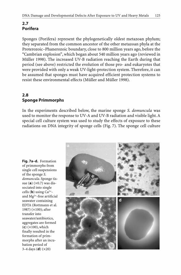
2.7Porifera
Sponges (Porifera) represent the phylogenetically oldest metazoan phylum;they separated from the common ancestor of the other metazoan phyla at theProterozoic–Phanerozoic boundary, close to 800 million years ago, before the“Cambrian explosion”, which began about 540 million years ago (reviewed inMüller 1998). The increased UV-B radiation reaching the Earth during thatperiod (see above) restricted the evolution of those pro- and eukaryotes thatwere provided with only a weak UV-light-protection system. Therefore, it canbe assumed that sponges must have acquired efficient protection systems toresist these environmental effects (Müller and Müller 1998).
2.8Sponge Primmorphs
In the experiments described below, the marine sponge S. domuncula wasused to monitor the response to UV-A and UV-B radiation and visible light. Aspecial cell culture system was used to study the effects of exposure to theseradiations on DNA integrity of sponge cells (Fig. 7). The sponge cell culture
DNA Damage and Developmental Defects After Exposure to UV and Heavy Metals 125
Fig. 7a–d. Formationof primmorphs fromsingle cell suspensionsof the sponge S.domuncula. Sponge tis-sue (a) (¥0.7) was dis-sociated into singlecells (b) using Ca2+-and Mg2+-free artificialseawater containingEDTA (Rottmann et al.1987) (¥100); aftertransfer intoseawater/antibiotics,aggregates are formed(c) (¥100), whichfinally resulted in theformation of prim-morphs after an incu-bation period of3–4 days (d) (¥20)

starts from dissociated cells of S. domuncula, which subsequently form aggre-gates; in those aggregates the cells start DNA synthesis and proliferate. Theaggregates, termed primmorphs, show a tissue-like appearance and can becultured for more than 5 months.
2.8.1UV Radiation
The primmorphs from S. domuncula cells were used as a model to analyze theeffects of UV-A and UV-B on DNA integrity in sponge cells in vitro. The FastMicromethod was adapted to work with low-integrity sponge DNA; unwind-ing was performed at pH 11.6. At this pH only strand breaks and cross-linkscould be measured. At higher pH (>12.0), alkali-labile sites (enzyme bindingsites, apurinic and apyrimidinic sites) are the predominant effects of DNAdenaturation.
Sponge cells (primmorphs) were irradiated with a SOL 500 lamp, whichcovered the whole solar spectrum using either H1 (wavelengths ≥320 nm;UV-A and visible light) or H2 (wavelengths ≥295 nm; UV-B, UV-A and visiblelight) filters. Subsequently, aliquots were analyzed for DNA single-strandbreaks using Fast Micromethod. As in sea urchin coelomocytes, a dose-dependent increase in DNA damage was observed following exposure ofsponge cells to 80–940 J/m2 of UV-A or 5–60 J/m2 of UV-B (Fig. 8). The UV-Aand UV-B doses were monitored using a UV radiometer with an A or B sensorrespectively. Again, the effects of UV-B (plus UV-A and visible light; filter H2)were much (several-fold) stronger than those observed with UV-A (plus visi-ble light; filter H1). The extent of UV-A-induced DNA single-strand breaks
H.C. Schröder et al.126
Fig. 8. UV-induced DNAdamage in S. domunculacells. Cells (1.5¥105 cells/ml)were exposed to the indi-cated doses of UV-A andUV-B using a SOL 500 lampwith filter H1 (UV-A andvisible light; wavelengths≥320 nm) or filter H2 (UV-B, UV-A, and visible light;wavelengths ≥295 nm). Con-trol (Co): non-irradiatedcells

increased during a post-irradiation repair period at 16 °C for 1.5 h (Fig. 9), dueto the formation of transient breaks by the action of repair enzymes. Theapparent decrease in strand breaks observed at higher UV doses (Fig. 9) maybe caused by UV-induced formation of DNA cross-links resulting in lower SSF¥ (–1) values.
Induction of DNA single-strand breaks was also measured following irra-diation of sponge cells (primmorphs) using a monochromatic UV-B lamp(peak at 312 nm; results not shown). DNA repair was accompanied by a time-dependent increase in the number of single-strand breaks; after a repairperiod of 2 h, the extent of DNA single-strand breaks decreased, reaching sim-ilar levels as in non-irradiated control cells after an incubation period of 18 h.The production of DNA single-strand breaks strongly depended on themedium used; the SSF ¥ (–1) values were lower (negative) in Ca2+-/Mg2+-freeseawater containing EDTA, but increased in Ca2+-/Mg2+-containing seawater.The decrease in SSF ¥ (–1) value in EDTA-containing, Ca2+-/Mg2+-free seawa-ter may be caused by a higher radiosensitivity (induction of formation ofDNA cross-links) of the sponge single cells present under these conditions,compared to sponge primmorphs formed in the presence of calcium (Ca-/Mg-containing seawater).
DNA Damage and Developmental Defects After Exposure to UV and Heavy Metals 127
Fig. 9. DNA damage and repair in S. domuncula cells after exposure of cells(1.5¥105 cells/ml) to the indicated doses of UV-A and UV-B using a full solar spectrum SOL500 lamp with filter H1 (UV-A and visible light; wavelengths ≥320 nm) or filter H2 (UV-B,UV-A, and visible light; wavelengths ≥295 nm). DNA repair was allowed to occur for 0, 10,20, 30 and 60 min at 16 °C. Control (Co): non-irradiated cells

The Fast Micromethod has also been applied for measuring the extent ofDNA single-strand breaks in whole tissue samples of the marine sponge Geo-dia cydonium, irradiated with UV light under controlled experimental condi-tions, or from specimens collected in the field (Batel et al. 1998). The tissuesamples were irradiated with 0–300 mJ/cm2 UV-B (monochromatic UV-Blamp). One hour later, samples were analyzed for DNA strand breaks. In theuntreated controls, which remained in the dark, the SSF was set to zero(0.00±0.04; n=5). Irradiation at a dose of 10 mJ/cm2 significantly increasedthe number of strand breaks [SSF ¥ (–1)] to 0.14±0.02. Higher doses of UV-Bfurther increased the extent of DNA damage; at 300 mJ/cm2 the SSF ¥ (–1)reached a value of 0.32±0.08.
2.8.2Cadmium
DNA damage in sponges is also induced by cadmium chloride. Exposure ofwhole specimen of marine sponges, e.g., S. domuncula, to cadmium resultedin a strong accumulation of the metal in the sponge (Schröder et al. 1999b).Asa consequence a strong increase in the extent of DNA single-strand breakswas found using Fast Micromethod. The maximal increase was observed afteran incubation period of 12 h in the presence of 1 mg/l of cadmium chlorideand after an incubation period of 1–3 days in the presence of 10 or 100 µg/l ofcadmium chloride; after a prolonged incubation period, the number of dam-ages decreased, most likely due to DNA repair (Schröder et al. 1996).
Field experiments using S. domuncula collected from five stations in thenorthern Adriatic Sea, characterized by a gradient of pollution, revealed sig-nificant differences in the cadmium levels between these stations (Müller etal. 1998). The frequency of DNA strand breaks roughly paralleled the gradientof pollution (cadmium levels) at these sites.
2.8.3Combined Effects
Results of the effect of cadmium on DNA repair in S. domuncula cells afterirradiation with full solar spectrum lamp with filter H1 (wavelengths≥320 nm) or filter H2 (wavelengths ≥295 nm) did not indicate an inhibition ofDNA repair processes as found in sea urchin coelomocytes (results notshown). Possibly some mechanism(s) preventing cadmium toxicity despite itshigh accumulation in sponge tissue (a 17,500 enrichment has been deter-mined under field conditions; Müller et al. 1998), such as expression of metal-lothionein-like proteins (Schröder et al. 2000b), are responsible for the findingthat an inhibitory effect of cadmium on DNA repair could not be detected inthe sponge system.
H.C. Schröder et al.128

2.9DNA Repair
Demosponges have an efficient DNA excision repair system. They are able torepair DNA damage caused by genotoxins, e.g. benzo[a]pyrene (Zahn et al.1983). The mechanisms of repair in sponges are similar to those found inhigher Metazoa.We found that in G. cydonium the expression of a homologueof the human XPB/ERCC-3 excision repair gene is induced after exposure toUV radiation (Batel et al. 1998). In humans, the product of the XPB/ERCC-3gene is involved in the early step of DNA excision repair (Sancar 1996a); itcorrects the repair defect in xeroderma pigmentosum and in Cockayne’s syn-drome. The human XPB/ERCC-3 gene product is a helicase unwinding theDNA in the 3¢Æ5¢ direction from the site of the lesion (Weeda et al. 1991). Lab-oratory studies revealed that after irradiation of G. cydonium with 30 or100 mJ/cm2 UV-B light, a dose-dependent increase in the steady-state level ofGCXPB occurs; values of up to 29-fold with respect to the controls which werekept in the dark have been determined. In parallel, the DNA integrity in thesponge samples was measured using Fast Micromethod. The data revealedthat the degree of DNA strand breaks paralleled the increase in expression ofthe GCXPB gene (Batel et al. 1998).
2.9.1(6–4) Photolyase
Sponges have not only an excision DNA repair system, but also a photolyase-based photoreactivating system. Two types of DNA photolyases have beenfound that cleave cyclobutane pyrimidine dimers (CPD): CPD photolyase and(6–4) photolyase (Sancar 1996b). The photolyases are part of the crypto-chrome family and associated with the blue light photoreceptors (Kanai et al.1997; Kobayashi et al. 2000). The photolyase of the hexactinellid spongeAphrocallistes vastus was studied (Schröder et al. 2003). A cDNA was isolatedfrom A. vastus, which comprises high sequence similarity to genes encodingthe protostomian and deuterostomian (6–4) photolyases. The A. vastussequence was assigned to the class I photolyases based on its high sequencesimilarity, especially within the N-terminal a/b-domain and C-terminal heli-cal domain. Using functional studies, we demonstrated that this gene codesfor a photolyase-related protein. After transfection into the Escherichia coliSY2 strain, which is deficient in photoreversal activity for pyrimidinecyclobutane dimers (Kim and Sundin 2001), the sponge gene caused resis-tance of the bacteria to UV light (Fig. 10). Irradiation was performed with aUV-B lamp (peak at 312 nm). The UV-induced damage in the bacteria wasalmost completely repaired during the light-repair phase; at a dose of 0.2 J/m2
only a small reduction in the survival rate was seen (Fig. 10). A strong UV-Bsensitivity was observed if the cells were transformed with the empty vector,
DNA Damage and Developmental Defects After Exposure to UV and Heavy Metals 129

irrespective of a post-treatment with light. The recombinant sponge proteinbound to UV-modified DNA that contained thymine dimers, while it failed tobind to non-treated DNA, suggesting that the sponge gene displays thymidinedimer-repairing enzyme activity (Schröder et al. 2003).
2.10Corals
The detrimental effects of increasing solar UV radiation on corals have beenrecognized for a long time (Jokiel 1980; Shick et al. 1996). Exposure to UVlight has been implicated in the process of coral bleaching. In addition, UV-induced DNA damage in coral-reef microbial communities has been observed(Lyons et al. 1998). As a result of the increased UV exposure, a decrease inskeletal growth and an increase in larval production have been described(Jokiel and York 1982). Most of these studies did not distinguish between theeffects of UV-A and UV-B, although the latter wavelengths are more affectedby stratospheric ozone depletion.
Primmorphs from Dendronephthya klunzingeri were used to determine theeffect of increasing exposure to UV-B and visible light (480 nm). The modi-fied procedure was used to dissociate the coral cells and form primmorphs(Custodio et al. 1998; Müller et al. 1999). The results revealed that prim-morphs irradiated with UV-B responded with an increased expression ofHSP90 only if exposed to low levels of UV-B (Wiens et al. 2000; Ammar et al.2001). After irradiation with 30 J/cm2 of UV-B, the primmorphs reacted witha 5.5-fold increase in HSP90 protein, compared with the controls, as revealedin Western blot experiments. Upregulation was observed following anincrease in UV-B irradiation to 100 J/cm2, while at levels above 100 J/cm2 no
H.C. Schröder et al.130
Fig. 10. Survival of E. coli strain SY2/pGEX(which does not have photolyase activity; opencircles), E. coli SY2/pMS969 [complementedwith the ph (6–4) photolyase gene from E. coli;solid squares), and E. coli SY2 carrying the A.vastus APHVAPH gene (solid triangles) afterUV-B irradiation at doses between 0.5 and2 J/m2. Following UV irradiation, photoreacti-vation was allowed to occur by irradiation ofthe bacteria for 1 h with a lamp emitting visi-ble light (Translux EC halogen photocuringunit, 400 and 520 nm with a maximum at480 nm; Heraeus Kulzer, Wehheim, Germany).Subsequently, the bacteria were incubatedovernight at 26 °C. Survival rate, which is givenin percent, was calculated on the basis of num-ber of colonies formed. Mean ± SE (n=5)
HSP90 was detected. In contrast to the studies with UV-B, visible light causesan up-regulation of the expression of the gene for HSP90 in a wider exposurerange, between 20 and 1,000 J/cm2. The increase in HSP90 expression follow-ing exposure to visible light was less pronounced and reached a maximum at300 J/cm2, with a 3.2-fold increase (Wiens et al. 2000).
Interestingly, in the absence of UV-B no transcripts of UVS-related proteincould be visualized, while after treatment of the primmorphs with 30–300 J/cm2 a strong up-regulation was seen in the expression of this gene (Wiens etal. 2000).
3Concluding Remarks
In summary, the results show that marine invertebrates are highly sensitive tosolar UV-B radiation without or in combination with heavy metal (cadmium)exposure. However, the degree of DNA damage and the course of subsequentrepair processes somewhat differ between different species (sea urchins com-pared to sponges) and different developmental stages (sea urchin embryos).The results show that marine invertebrates may be endangered by theincrease in UV-B radiation due to ozone depletion, especially during earlystages of development. In addition, the results show that cell cultures ofmarine invertebrates (sea urchin coelomocytes and sponge primmorphs)might be useful tools for monitoring stress caused by physical (UV-B radia-tion) and chemical (cadmium) factors in the aquatic environment.
Acknowledgements. This work was supported by grants from the European Commission(EVK3-CT-1999-00005 “UVTOX”) and the Bundesministerium für Bildung und Forschung(project Center of Competence BIOTECmarin).
References
Acevedo J, Nolan C (eds) (1993) Environmental UV radiation: causes – effects – conse-quences. Commission of the European Communities, Directorate-General XII for Sci-ence, Research and Development, Environment Programme, Brussels
Ammar MSA, Schröder HC, Wiens M, Müller WEG (2001) Induction of heat-shock geneexpression by selected natural and anthropogenic disturbances in the octocoral (Den-dronephthya klunzingeri). ACP-EU Fish Res Rep 10:85–95
Baker KS, Smith RC (1982) Spectral irradiance penetration in natural waters. In: Calkins J(ed) The role of solar ultraviolet radiation in marine ecosystems. Plenum, New York,pp 233–246
Batel R, Bihari N, Rinkevich B, Dapper J, Schäcke H, Schröder HC, Müller WEG (1993) Mod-ulation of organotin-induced apoptosis by the water pollutant methyl mercury in ahuman lymphoblastoid tumor cell line and a marine sponge. Mar Ecol Prog Ser93:245–251
DNA Damage and Developmental Defects After Exposure to UV and Heavy Metals 131

Batel R, Fafandjel M, Blumbach B, Schröder HC, Hassanein HM, Müller IM, Müller WEG(1998) Expression of the human XPB/ERCC-3 excision repair gene-homolog in thesponge Geodia cydonium after exposure to ultraviolet radiation. Mutat Res 409:123–133
Batel R, Jak¥iæ Z, Bihari N, Hamer B, Fafan¥jel M, Chauvin C, Schröder HC, Zahn RK,Müller WEG (1999) A microplate assay for DNA damage determination (FastMicromethod) in cell suspensions and solid tissues. Anal Biochem 270:195–200
Bihari N, Batel R, Jak¥iæ Z, Müller WEG, Waldmann P, Zahn RK (2002) Comparisonbetween the Comet assay and Fast Micromethod for measuring DNA damage in HeLacells. Croatia Chem Acta 75:793–804
Blaustein AR, Hoffman PD, Hokit DG, Kiesecker JM,Walls SC, Hays JB (1994) UV repair andresistance to solar UV-B in amphibian eggs: link to population declines? Proc Natl AcadSci USA 19:1791–1795
Bonham K, Zafarullah M, Gedamu L (1987) The rainbow trout metallothioneins: molecularcloning and characterization of two distinct cDNA sequences. DNA 6:519–528
Bothwell ML, Sherbot D, Roberge AC, Daley RJ (1993) Influence of natural ultraviolet radi-ation on lotic periphytic diatom community growth, biomass accrual, and species com-position: short-term versus long-term effects. J Phycol 29:24–35
Brasier MD (2000) The Cambrian explosion and the slow burning fuse. Sci Progr 83:77–92Buhlmann B, Bossard P, Uehlinger U (1987) The influence of longwave ultraviolet radiation
(UV-A) on the photosynthetic activity of phytoplankton. J Plankton Res 9:935–943Cadwell MM, Camp LB, Warner CW, Flint SD (1986) Action spectra and their key role in
assessing biological consequences of solar UV-B radiation change. In: Worrest RC, Cald-well MM (eds) Stratospheric ozone reduction, solar ultraviolet radiation and plant life.Springer, Berlin Heidelberg New York, pp 87–111
Cajaraville MP, Bebianno MJ, Blasco J, Porte C, Sarasquete C, Viarengo A (2000) The use ofbiomarkers to assess the impact of pollution in coastal environments of the IberianPeninsula: a practical approach. Sci Total Environ 257:295–311
Canfield DE, Teske A (1996) Late Proterozoic rise in atmospheric oxygen concentrationinferred from phylogenetic and sulphur-isotope studies. Nature 382:127–132
Chernoff N (1973) Teratogenic effects of cadmium in rats. Teratology 8:29–32Cloud PE (1968) Atmospheric and hydrospheric evolution of primitive earth. Science
160:729–736Coteur G, Pernet P, Gillan D, Joly G, Maage A, Dubois P (2003) Field contamination of the
starfish Asterias rubens by metals. Part 1: Short- and long-term accumulation along apollution gradient. Environ Toxicol Chem 22:2136–2144
Crump D, Lean D, Berrill M, Coulson D, Toy L (1999) Spectral irradiance in pond water:influence of water chemistry. Photochem Photobiol 70:893–901
Custodio MR, Prokic I, Steffen R, Koziol C, Borojevic R, Brümmer F, Nickel M, Müller WEG(1998) Primmorphs generated from dissociated cells of the sponge Suberites domuncula:a model system for studies of cell proliferation and cell death. Mech Ageing Dev105:45–59
Damkaer DM, Dey DB (1983) UV damage and photoreactivation potentials of larvalshrimp, Pandalus platyceros, and adult euphausiids, Thysanoessa raschii. Oecologia60:169–175
Damkaer DM, Dey DB, Heron GA, Prentice EF (1980) Effects of UV-B radiation on near-sur-face zooplankton of Puget Sound. Oecologia 44:149–158
Damkaer DM, Dey DB, Heron GA (1981) Dose/dose-rate responses of shimp larvae to UV-Bradiation. Oecologia 48:178–182
Dinnel PA, Pagano GG, Oshida PS (1988) A sea urchin test system for marine environmen-tal monitoring. In: Burke RD, Mladenov PV, Lambert P, Parsley RL (eds) Echinodermbiology. AA Balkema, Rotterdam, pp 611–619
Dustan P (1982) Depth-dependent photoadaptation by zooxanthellae of the reef coral Mon-tastrea annularis. Mar Biol 68:253–264
H.C. Schröder et al.132

El-Sayed SZ (1988) Fragile life under the ozone hole. Nat Hist 97:73–80El-Sayed SZ, Van Dijken G, Gonzales-Rhodas G (1996) Effects of ultraviolet radiation on
marine ecosystems. Int J Environ Stud 51:199–216Fernandez N, Beiras R (2001) Combined toxicity of dissolved mercury with copper, lead and
cadmium on embryogenesis and early larval growth of the Paracentrotus lividus seaurchin. Ecotoxicology 10:263–271
Fisher AG (1965) Fossils, early life, and atmospheric history. Proc Natl Acad Sci USA53:1205–1215
Flick DF, Krayville HF, Dimitroff JM (1971) Toxic effects of cadmium: a review. Environ Res4:71–85
Frederick JE, Snell HE, Haywood EK (1989) Solar ultraviolet radiation at the earth’s surface.Photochem Photobiol 50:443–450
Fridovich I (1986) Biological effects of the superoxide radical. Arch Biochem Biophys247:1–11
Friedberg EC, Walker GC, Siede W (eds) (1995) DNA repair and mutagenesis. ASM Press,Washington
Gleason JF, Bhartia PK, Herman JR, McPeters R, Newman P, Stolarski RS, Flynn L, Labow G,Larko D, Seftor C, Wellemeyer C, Komhyr WD, Miller AJ, Planet W (1993) Record lowglobal ozone in 1992. Science 260:523–526
Griffiths H, Mistry P, Herbert KE, Lunec J (1998) Molecular and cellular effects of ultravio-let light-induced genotoxicity. Crit Rev Clin Lab Sci 35:189–237
Häder DP, Worrest RC, Kumar HD, Smith RC (1995) Effects of increased solar ultravioletradiation on aquatic ecosystems. Ambio 24:174–180
Hartman PS, Hevelone J, Dwarakanath V, Mitchell DL (1989) Excision repair of UV radia-tion-induced DNA damage in Caenorhabditis elegans. Genetics 122:379–385
Hartwig A (1994) Role of DNA repair inhibition in lead- and cadmium-induced genotoxic-ity: a review. Environ Health Perspect 102 (Suppl 3):45–50
Helbling EW, Villafañe V, Ferrario M, Holm-Hansen O (1992) Impact of natural ultravioletradiation on rates of photosynthesis and on specific marine phytoplankton species. MarEcol Prog Ser 80:89–100
Henson JH, Nesbitt D, Wright BD, Sholey JM (1992) Immunolocalization of kinesin in seaurchin coelomocytes. J Cell Sci 103:309–320
Hessen DO (1994) Daphnia responses to UV-light. Arch Hydrobiol Beih 43:185–195Hirosawa T, Miyachi S (1983) Inactivation of Hill reaction by long-wavelength ultraviolet
radiation (UV-A) and its photoreactivation by visible light in the cyanobacterium, Ana-cystis nidulans. Arch Microbiol 135:98–102
Hunter JR, Taylor JH, Moser HG (1979) The effect of ultraviolet irradiation on eggs and lar-vae of the northern anchovy, Engraulis mordax and the Pacific mackerel, Scomber japon-icus during the embryonic stage. Photochem Photobiol 29:325–338
Jeffery WR (1990) Ultraviolet irradiation during ooplasmic segregation prevents gastrula-tion, sensory cell induction, and axis formation in the ascidian embryo. Dev Biol140:388–400
Jerlov NG (1968) Optical oceanography. Elsevier, AmsterdamJokiel PL (1980) Solar ultraviolet radiation and coral reef epifauna. Science 207:1069–1071Jokiel PL, York RH (1982) Solar ultraviolet photobiology of the reef coral Pocillopora dami-
cornis and symbiotic zooxanthellae. Bull Mar Sci 32:301–315Kanai S, Kikuno R, Toh H, Ryo H, Todo T (1997) Molecular evolution of the photolyase-blue-
light photoreceptor family. J Mol Evol 45:535–548Karanas JK, Worrest RC, Van Dyke H (1981) Impact of UV-B radiation on the fecundity of
the copepod Acartia clausii. Mar Biol 65:125–133Karentz D, Lutze LH (1990) Evaluation of biologically harmful ultraviolet radiation in
Antarctica with a biological dosimeter designed for aquatic environments. LimnolOceanogr 35:549–561
DNA Damage and Developmental Defects After Exposure to UV and Heavy Metals 133

Karentz D, Bothwell ML, Coffin RB, Hanson A, Herndl GJ, Kilham SS, Lesser MP, Lindell M,Moeller RE, Morris DP, Neale PJ, Sanders RW,Weiler CS,Wetzel RG (1994) Impact of UV-B radiation on pelagic freshwater ecosystems. Report of working group on bacteria andphytoplankton. Arch Hydrobiol Beih Ergebn Limnol 43:31–69
Kim JJ, Sundin GW (2001) Construction and analysis of photolyase mutants ofPseudomonas aeruginosa and Pseudomonas syringae: contribution of photoreactivation,nucleotide excision repair, and mutagenic DNA repair to cell survival and mutability fol-lowing exposure to UV-B radiation. Appl Environ Microbiol 67:1405–1411
Kobayashi T, Ishikawa T, Hirayama J, Daiyasu H, Kanai S, Toh H, Fukuda I, Tsujimura T, Ter-ada N, Kamei Y, Yuba S, Iwai S, Todo T (2000) Molecular analysis of zebrafish pho-tolyase/cryptochrome family: two types of cryptochromes present in zebrafish. GenesCells 5:725–738
Koziol C, Wagner-Hülsmann C, Cetkovic H, Gamulin V, Kruse M, Pancer Z, Schäcke H,Müller WEG (1996) Cloning of the heat-inducible biomarker, the cDNA encoding the 70-kDa heat shock protein, from the marine sponge Geodia cydonium: response to naturalstressors. Mar Ecol Prog Ser 136:156–161
Layton WM Jr, Layton WM (1979) Cadmium-induced limb defects in mice: strain associ-ated differences in sensitivity. Teratology 19:229–235
Lee SK, Yu SL, Garcia MX, Alexander H, Alexander S (1997) Differential developmentalexpression of the rep B and rep D xeroderma pigmentosum related DNA helicase genesfrom Dictyostelium discoideum. Nucleic Acids Res 25:2365–2374
Le Marrec F, Glaise D, Guguen-Guillouzo C, Chesnè C, Guillouzo A, Boulo V, Dorange G(1999) Primary cultures of heart cells from the scallop Pecten maximus (Mollusca-Bivalvia). In Vitro Cell Dev Biol 35:289–295
Little EE, Fabacher DL (1994) Comparative sensitivity of rainbow trout and two threatenedsalmonids,Appache Trout and Lahontan Cutthout Trout, to UV-B radiation.Arch Hydro-biol Beih 43:217–226
Loomis WF (1988) Four billion years. Sinauer Associates, SunderlandLyons MM, Aas P, Pakulski JD,Van Waasbergen L, Miller RV, Mitchell DL, Jeffrey WH (1998)
DNA damage induced by ultraviolet radiation in coral-reef microbial communities. MarBiol 130:537–543
Lyons-Alcantara M, Mooney R, Lyng F, Cottell D, Mothersill C (1998) The effects of cad-mium exposure on the cytology and function of primary cultures from rainbow trout.Cell Biochem Funct 16:1–13
Malloy KD, Holman MA, Mitchell D, Detrich HW (1997) Solar UV-B-induced DNA damageand photoenzymatic DNA repair in Antarctic zooplankton. Proc Natl Acad Sci USA94:1258–1263
Matranga V, Bonaventura R (2002) Sea urchin coelomocytes, the progenitors of vertebrateimmune effectors, as bio-indicators of stress and pollution. In: Yokota Y, Matranga V,Smolenicka Z (eds) The sea urchin: from basic biology to aquaculture. Swet andZeitlinger, Lisse, The Netherlands, pp 161–176
Matranga V, Toia G, Bonaventura R, Müller WEG (2000) Cellular and biochemical responsesto environmental and experimentally induced stress in sea urchin coelomocytes. CellStress Chaperones 5:113–120
Matranga V, Bonaventura R, Di Bella G (2002) Hsp70 as a stress marker of sea urchin coelo-mocytes in short term cultures. Cell Mol Biol 48:345–359
Molina LT, Molina MJ (1986) Absolute absorption cross sections of ozone in the 185- to 350-nm wavelength range. J Geophys Res 91:14501–14508
Morale A, Coniglio L, Angelini C, Cimoli G, Bolla A, Alleteo D, Russo P, Falugi C (1998) Bio-logical effects of a neurotoxic pesticide at low concentrations on sea urchin early devel-opment. A teratogenic assay. Chemosphere 37:3001–3010
H.C. Schröder et al.134

Mounkes LC, Jones RS, Liang BC, Gelbart W, Fuller MT (1992) A Drosophila model for xero-derma pigmentosum and Cockayne’s syndrome: haywire encodes the fly homolog ofERCC3, a human excision repair gene. Cell 71:925–937
Müller WEG (1998) Origin of Metazoa: sponges as living fossils. Naturwissenschaften85:11–25
Müller WEG, Müller I (1998) Sponge cells and tissue as biological monitors of aquatic pol-lution. In: Wells PG, Lee K, Blaise C (eds) Microscale testing in aquatic toxicology –advances, techniques and practice. CRC Press, Boca Raton, pp 97–112
Müller WEG, Koziol C, Kurelec B, Dapper J, Batel R, Rinkevich B (1995) Combinatory effectsof temperature stress and nonionic organic pollutants on stress protein (hsp70) geneexpression in the fresh water sponge Ephydatia fluviatilis. Environ Toxicol Chem14:1203-1208
Müller WEG, Batel R, Lacorn M, Steinhart H, Simat T, Lauenroth S, Hassanein H, SchröderHC (1998) Accumulation of cadmium and zinc in the marine sponge Suberitesdomuncula and its potential consequences on single-strand breaks and on expression ofheat-shock protein: a natural field study. Mar Ecol Prog Ser 167:127–135
Müller WEG, Wiens M, Batel R, Steffen R, Schröder HC, Borojevic R, Custodio MR (1999)Establishment of a primary cell culture from a sponge: primmorphs from Suberitesdomuncula. Mar Ecol Prog Ser 178:205–219
Müller WEG, Koziol C, Wiens M, Schröder HC (2000) Stress response in marine sponges:genes and molecules involved and their use as biomarkers. In: Storey KB, Storey J (eds)Environmental stressors and gene responses. Elsevier, Amsterdam, pp 193–208
Noblet JA, Zeng EY, Ritter KJ (2003) Evaluation of polychlorinated biphenyl bioaccumula-tion patterns in white sea urchins (Lytechinus pictus) using multiple approaches. Envi-ron Toxicol Chem 22:2719–2226
Pagano G, Esposito A, Giordano GG (1982) Fertilization and larval development in seaurchins following exposure of gametes and embryos to cadmium. Arch Environ ContamToxicol 11:47–55
Park E, Guzder SN, Koken MH, Jaspers-Dekker I, Weeda G, Hoeijmakers JH, Prakash S,Prakash L (1992) RAD25 (SSL2), the yeast homolog of the human xeroderma pigmento-sum group B DNA repair gene, is essential for viability. Proc Natl Acad Sci USA89:11416–11420
Prézelin BB, Boucher NP, Smith RC (1994) Marine primary production under the influenceof the Antarctic ozone hole: Icecolors ‘90. In: Weiler CS, Penhale PA (eds) Ultravioletradiation in Antarctica: measurements and biological effects. Antarctic Res Ser 62.American Geophysical Union, Washington, DC, pp 159–186
Radenac G, Fichet D, Miramand P (2001) Bioaccumulation and toxicity of four dissolvedmetals in Paracentrotus lividus sea-urchin embryo. Mar Environ Res 51:151–166
Regan JD, Carrier WL, Gucinski H, Olla BL, Yoshida H, Fujimura RK, Wicklund RI (1992)DNA as a solar dosimeter in the ocean. Photochem Photobiol 56:35–42
Ringelberg J, Keyser AL, Flik BJG (1984) The mortality effect of ultraviolet radiation in atranslucent and in a red morph of Acanthodiaptomus denticornis (Crustacea, Copepoda)and its possible ecological relevance. Hydrobiologia 112:217–222
Roccheri MC, Agnello M, Bonaventura R, Matranga V (2004) Cadmium induces the expres-sion of specific stress proteins in sea urchin embryos. Biophys Biochem Res Commun321:80–87
Russo R, Bonaventura R, Zito F, Schröder HC, Müller I, Müller WEG, Matranga V (2003)Stress to cadmium monitored by metallothionein gene induction in Paracentrotuslividus embryos. Cell Stress Chaperones 8:232–241
Samarawickrama GP, Webb M (1979) Acute effects of cadmium on the pregnant rat andembryo-fetal development. Environ Health Perspect 28:245–249
Sancar A (1996a) DNA excision repair. Annu Rev Biochem 65:43–81
DNA Damage and Developmental Defects After Exposure to UV and Heavy Metals 135

Sancar A (1996b) No “end of history” for photolyases. Science 272:48–49Schröder HC, Batel R, Lauenroth S, Hassanein HMA, Lacorn M, Steinhart H, Müller WEG
(1999a) Induction of DNA damage and expression of heat shock protein HSP70 by poly-chlorinated biphenyls in the marine sponge Suberites domuncula Olivi. J Exp Mar BiolEcol 233:285–300
Schröder HC, Hassanein HMA, Lauenroth S, Koziol C, Mohamed TA-AA, Lacorn M, Stein-hart H, Batel R, Müller WEG (1999b) Induction of DNA strand breaks and expression ofHSP70 and GRP78 homolog by cadmium in the marine sponge Suberites domuncula.Arch Environ Contam Toxicol 36:47–55
Schröder HC, Batel R, Müller WEG (2000a) Novel cellular and molecular tools to assess theeffects of pollution and solar UV radiation on marine invertebrates. In: Proc EurOCEAN2000, Eur Conf on Marine Science and Ocean Technology, Hamburg, 29 Aug–2 Sept. Pro-ject synopses, vol I: marine processes, ecosystems and interactions. Office for OfficialPublications of the European Communities, Luxembourg, pp 128–134
Schröder HC, Shostak K, Gamulin V, Lacorn M, Skorokhod A, Kavsan V, Müller WEG(2000b) Purification, cDNA cloning and expression of a cadmium-inducible cysteine-rich metallothionein-like protein from the marine sponge Suberites domuncula. MarEcol Prog Ser 200:149–157
Schröder HC, Krasko A, Gundacker D, Leys SP, Müller IM, Müller WEG (2003) Molecularand functional analysis of the (6–4) photolyase from the hexactinellid Aphocallistes vas-tus. Biochim Biophys Acta 1651:41–49
Schröder HC, Batel R, Schwertner H, Boreiko O, Müller WEG (2004) Fast Micromethod DNAsingle-strand break assay. In: Henderson DS (ed) DNA repair protocols: eukaryotic sys-tems. Humana Press, Totowa, New Jersey (in press)
Sconzo G, Romancino D, Fasulo G, Cascino D, Giudice G (1995) Effect of doxorubicin andphenytoin on sea urchin development. Pharmacie 50:616–619
Scully NM, Lean DRS (1994) The attenuation of ultraviolet radiation in temperate lakes.Arch Hydrobiol Beih Ergebn Limnol 43:135–144
Shick JM, Lesser MP, Stochaj WR (1991) Ultraviolet radiation and photooxidative stress inzooxanthellate Anthozoa: the sea anemone Phyllodiscus semoni and the octocoral Clavu-laria sp. Symbiosis 10:145–173
Shick JM, Lesser MP, Jokiel PL (1996) Effects of ultraviolet radiation on corals and othercoral reef organisms. Global Change Biol 2:527–545
Smith RC, Baker KS (1979) Penetration of UV-B and biologically effective dose-rates in nat-ural waters. Photochem Photobiol 29:311–323
Smith RC, Baker KS (1981) Optical properties of the clearest natural waters (200–800 nm).Appl Opt 20:177–184
Smith RC, Prézelin BB, Baker KS, Bidigare RR, Boucher NP, Coley T, Karentz D, Maclntyre S,Matlick HA, Menzies D, Ondrusek M, Wan Z, Waters KJ (1992) Ozone depletion: ultravi-olet radiation and phytoplankton biology in Antarctic waters. Science 255:952–959
Stolarski RS, Bojkov R, Bishop L, Zerefos C, Staehelin J, Zawodny J (1992) Measured trendsin stratospheric ozone. Science 256:342–349
Towe KM (1994) Earth’s early atmosphere: constraints and opportunities for early evolu-tion. In: Bengtson S (ed) Early life on earth. Columbia University Press, New York,pp 36–47
Vincent WF, Roy S (1993) Solar ultraviolet-B radiation and aquatic primary production:damage, protection, and recovery. Environ Rev 1:1–12
Weeda G, van Ham RC, Masurel R, Westerveld A, Odijk H, de Wit J, Bootsma D, van der EbAJ, Hoeijmakers JH (1990) Molecular cloning and biological characterization of thehuman excision repair gene ERCC-3. Mol Cell Biol 10:2570–2581
H.C. Schröder et al.136

Weeda G, Ma L, van Ham RC, Bootsma D, van der Eb AJ, Hoeijmakers JH (1991) Character-ization of the mouse homolog of the XPBC/ERCC-3 gene implicated in xeroderma pig-mentosum and Cockayne’s syndrome. Carcinogenesis 12:2361–2368
Wiens M, Shokry M, Nawar AH, Koziol C, Hassanein HMA, Eisinger M, Müller IM, MüllerWEG (2000) Induction of heat-shock (stress) protein gene expression by selected nat-ural and anthropogenic disturbances in the octocoral Dendronephthya klunzingeri.Aquat Toxicol 245:265–276
Wilkinson DG, Nemer M (1987) Metallothionein genes Mta and MTb expressed under dis-tinct quantitative tissue-specific regulation in sea urchin embryos. Mol Cell Biol 7:48–58
Yabe K, Onodera S, Makino M, Suzuki M, Kagoshima K (1998) The effects of ultraviolet irra-diation on larvae and early juveniles of the sea urchin, Strongylocentrotus intermedius.Nippon Suisan Gakkaishi 64:606–609
Zahn RK, Zahn G, Müller WEG, Michaelis ML, Kurelec B, Rijavec M, Batel R, Bihari N (1983)DNA damage by PAH and repair in a marine sponge. Sci Total Environ 26:137–156
DNA Damage and Developmental Defects After Exposure to UV and Heavy Metals 137

Echinoderms: Their Culture and BioactiveCompounds
M.S. Kelly
Abstract. Of the five extant classes of echinoderms, it is the sea urchins(Echinoidea) and the sea cucumbers (Holothuroidea) that are both commer-cially fished and heavily overexploited. In sea urchins, it is the gonad of bothmales and females, normally referred to as ‘roe’, that is a sought-after food. Inthe sea cucumber, the principal product is the boiled and dried body-wall or‘bêche-de-mer’ for which there is an increasing demand. Many sea urchin andsea cucumber fisheries still have no management system or restrictions inplace, and for those that do, the prognosis for catches to continue even at areduced level is poor. Cultivation of these species increasingly becomes anecessity, both for stock enhancement programs and as a means to meet mar-ket demand. Sea urchin culture has been practised on a large scale in Japan formany decades, and effective methods for the culture and reseeding of speciesin these waters have been long established. Juvenile urchins are produced intheir millions in state-sponsored hatcheries, for release to managed areas ofseafloor. Outside of Japan, sea urchin cultivation is still a fairly recent practice,less than 10 years old, and largely still at a research level, although a range ofspecies are now being produced in a variety of different culture systems. It isessential that the culture systems are adapted to be species-specific and meetwith local environmental constraints. Sea cucumber cultivation originated inJapan in the 1930s and is now well established there and in China. Methods formass cultivation of the tropical Holothuria scabra are now well establishedand practised in India, Australia, Indonesia, the Maldives and the SolomonIslands, with the focus of the research effort for both temperate and tropicalspecies being centred on the production of juveniles in hatcheries for therestoration and enhancement of wild stocks. Like many other marine organ-isms, echinoderms have been, and continue to be, examined as a source ofbiologically active compounds with biomedical applications. Sea cucumberhas been valued in Chinese medicine for hundreds of years as a cure for a
Progress in Molecular and Subcellular BiologySubseries Marine Molecular BiotechnologyV. Matranga (Ed.), Echinodermata© Springer-Verlag Berlin Heidelberg 2005
M.S. Kelly (e-mail: [email protected])Scottish Association for Marine Science, Oban, Argyll, Scotland, PA37 1QA, UK

wide variety of ailments. Some more recently isolated compounds, mainlyfrom sea cucumbers and starfish, and including those with antitumour,antiviral, anticoagulant and antimicrobial activity, are summarised below.When wild stocks decline, the demand created in the market place raises theprice of the product and, consequently, culturing is more likely to becomeviable economically. As this review shows, there have been dramatic advancesin the culture methods of sea urchins and sea cucumbers in the last10–15 years, to the extent that one can conclude that currently the majorobstacles to successful cultivation are indeed economic rather than biological.Hence the future of the echinoculture industry is closely linked to that of thefisheries, whose fate will ultimately determine the market forces that willshape this growing industry.
1Why Cultivate?
Of the five extant classes of echinoderms, it is the sea urchins (Echinoidea)and the sea cucumbers (Holothuroidea) that are both commercially fishedand heavily overexploited. In sea urchins, it is the gonad of both males andfemales, normally referred to as ‘roe’, that is a sought-after food. The largestmarket is Japan, but there are also undersupplied markets in Europe. In thesea cucumber, the principal product is the boiled and dried body-wall or‘bêche-de-mer’, for which there is an increasing demand from China, where itis valued as food but also for its curative properties. There is also a trade in seacucumbers for home aquaria and biomedical products (Bruckner et al. 2003),and at one time there was fishing activity for starfish (Asteroidea), which wereprocessed for fish or poultry meal or for fertiliser (Sloan 1984). The extent anddemise of the world’s sea urchin and sea cucumber fisheries have beenreviewed by Sloan (1984), Conand and Byrne (1993) and Keesing and Hall(1998), and more comprehensively for sea urchins by Andrew et al. (2002) andfor sea cucumbers by Bruckner et al. (2003).
Most, if not all, sea urchin fisheries have followed the same pattern of rapidexpansion to an unsustainable peak, followed by an equally rapid decline.World landings of sea urchin, having peaked at 120,000 t (metric tonnes) in1995, are now in the region of 90,000 t/year. However, over half this catchcomes from the recently expanded Chilean fishery for Loxechinus albus;which also appears to have peaked, the catch now being sustained by the dis-covery of new fishing grounds. The other major sea urchin fisheries, in termsof tonnage landed, are in Japan, Maine (USA), British Columbia (Canada) andCalifornia (USA) (Andrew et al. 2002). In Europe, the sea urchin stocks (Para-centrotus lividus) of first France and then Ireland were overfished in the 1980sto supply the French markets and those stocks have never recovered (Barnesand Crook 2001; Barnes et al. 2002). There are large populations of edible
M.S. Kelly140

urchins in Scotland (Echinus esculentus and Psammechinus miliaris) and Nor-way (Strongylocentrotus droebachiensis), but these stocks are unsuited tocommercial fishing as their roe content is either too low or too variable(Hagen 2000; Kelly 2000; Kelly et al. 2001; Sivertsen 2004).
The global landing of sea cucumbers was estimated as 13,000 t dried weight(130,000 t live weight) in 1995. The largest fisheries today are in Indonesia andthe Philippines; however, in the last decade, the number of countries andspecies involved in the trade have increased in both tropical and temperateregions and the fishery has spread to non-traditional areas such as Mexico,the Galapagos and North America. Bruckner et al. (2003) list 29 species from50 countries that are fished for import into Hong Kong, many of which will bere-exported to China and other trade centres in Singapore and ChineseTaipei.
Regulations are now imposed on some sea urchin and sea cucumber fish-eries, although few are based on formal stock assessments. For example, in thestate of Maine, on the east coast of the USA, the sea urchin fishery is ‘zoned’,in part based on the reproductive/spawning cycle along the coast. Regulationlimits fishing effort; there is a limited entry scheme to the fishery, restrictionson gear size, fishing times and minimum and maximum landing sizes. How-ever, researchers believe even this diminished catch is likely to prove unsus-tainable (L. Harris, University of New Hampshire, USA, pers. comm.).
In contrast, the Japanese sea urchin fishery has endured for more than50 years, yielding more than 13,000 t in 1998. Six species account for the bulkof the commercial landings, but it is important to note that there is govern-ment subsidy for urchin stock enhancement and this is used as a managementtool to conserve and rebuild stocks (Andrew et al. 2002).
Many sea cucumber fisheries show evidence of overexploitation or havealready collapsed and some still have no known management or restrictions,including the world’s largest fishery in Indonesia (Bruckner et al. 2003). Pos-sible management measures suggested for sea cucumber fisheries include theimposition of minimum landing sizes, closed seasons, no-take marine pro-tected areas, bag limits, prohibition of night fishing for nocturnal species andrestrictions on the use of SCUBA for harvesting. However, the artisanal natureof the fishery makes implementation of such measures difficult. Bruckner etal. (2003) propose that listing sea cucumbers in the Convention on Interna-tional Trade in Endangered Species (CITES) Appendix II might be an appro-priate way to ensure that harvest to international markets is conducted in asustainable manner, without detriment to the target species or their ecosys-tem. Compounding the impact of overexploitation of wild echinoderm popu-lations is the increasing conviction among researchers that recruitment ofjuvenile sea urchins is both sporadic and unpredictable (see Lawrence 2001for reviews; also Kelly 2000; Harris et al. 2001), and the fact that some speciesof sea urchin are extremely long-lived (Ebert 1998). Sea cucumbers are a valu-able source of income for many coastal communities, particularly among thedeveloping nations of the Indo-Pacific, but as with sea urchins, their high
Echinoderms: Their Culture and Bioactive Compounds 141

value and sedentary, shallow-water habit have left them vulnerable to overex-ploitation through unregulated and illegal fishing activity. Once their densityis reduced below a critical mass, populations may take as long as 50 years torecover (Dalzell et al. 1996; Battaglene 1999; Bruckner et al. 2003).
As the prognosis for catches to continue at the current level is poor, cultiva-tion of these species increasingly becomes a necessity, both for stockenhancement programs and as a means to meet market demand. In addition,as wild stocks decline, cultivation is more likely to become viable economi-cally. The need for effective culture methods is now reflected in increasedresearch effort. Culture of sea urchins for reseeding is practised on a largescale in Japan and to a lesser extent in South Korea and the Philippines(Andrew et al. 2002). Sea cucumbers are cultured for reseeding on a large scalein China. Outside these countries most echinoculture is at a research or semi-commercial scale. There is no commercial-scale cultivation of starfish, brittlestars (ophiuroids) or feather stars (crinoids).
2Sea Urchin Aquaculture
2.1Life History and State of the Art
Edible sea urchins are among the orders of regular Echinoidea (Lawrence2001), dioecious (separate sexes) and broadcast spawners; mature individualsshed gametes to seawater where fertilisation occurs. The eggs develop to formpluteus larvae, which, after a period of planktonic development, feeding onmicroalgae, settle to a substrate and undergo metamorphosis to form tinyjuvenile sea urchins. The estimated time for these individuals to reach marketsize (40–50 mm horizontal test diameter) is commonly in the order of1–3 years, again varying according to species.
Sea urchin culture has been practised on a large scale in Japan for manydecades, and effective methods for the culture and reseeding of species inthese waters have been long established. Juvenile urchins are produced intheir millions in state-sponsored hatcheries, for release to managed areas ofseafloor. The nationally co-ordinated reseeding program initiated in the1960s has now developed to the extent that over 66 million juveniles werereleased onto reefs in 2000; of these, over 80 % were Strongylocentrotus inter-medius (Agatsuma et al. 2004). The effectiveness of the reseeding program hasnot been easy to assess, in part because of the difficulty in discriminatingbetween reseeded and wild individuals. In Hokkaido prefecture, reseeding ofS. intermedius began in 1985 with the release of 1 million juveniles, increasingto over 60 million in 1996. However, for the first 8 years of this program, thetotal harvest of urchins from this area continued to decline and it is only since1992 that the catch has begun to stabilise. The contribution of released sea
M.S. Kelly142
urchins to the overall catch has been estimated to be between 62 and 80 %(Agatsuma et al. 2004). There are also much smaller-scale reseeding programsoperating in South Korea and on Luzon Island in the Philippines (Andrew etal. 2002).
Outside of Japan, sea urchin cultivation is still a fairly recent practice, lessthan 10 years old. There have been researchers and companies developingmethods for sea urchin (Paracentrotus lividus) cultivation in southern Irelandfor over 20 years (Leighton 1995), and until relatively recently a concentratedeffort into developing ‘closed-cycle’ land-based systems in France, also with P.lividus (Grosjean et al. 1998). Echinoculture (Psammechinus miliaris, E. escu-lentus, P. lividus) has been conducted in Scotland since 1995 and there are alsoestablished research teams on the east coast of North America – Florida,Alabama, Maine, New Hampshire, New Brunswick and Newfoundland –working on S. droebachiensis and Lytechinus variegatus; on the west coast ofNorth America, including California and British Columbia (S. droebachiensis,S. franciscanus, S. purpuratus); in Chile (Loxechinus albus); Norway (S. droe-bachiensis); Israel (P. lividus); and in New Zealand (Evechinus chloroticus).This list is not intended to be exclusive or exhaustive but to illustrate theextent of the research base. The biology and ecology of these and other edibleechinoids have recently been reviewed (Lawrence 2001).
While including discussion of the basic methodologies employed, both inand outside of Japan, this chapter will concentrate on examining some of themore recent advances and innovations in sea urchin culture, outside of Japan,with reference to the particular challenges remaining for the culturist at eachstage of the sea urchin life cycle.
2.2Sea Urchin Larviculture
Echinoids have been successfully raised in the laboratory for over 100 years(MacBride 1903). The reproductive periodicity for many echinoid species iswell described; temperate water species in culture tend to have one spawningperiod per year (Himmelman 1977; Byrne 1993; Kelly 2000; Fig. 1) and broodstock are collected locally. Gravid individuals are induced to spawn either bytemperature shock or, commonly, by injection of 0.5 M KCl to the coelom viathe peristomal membrane. The concentration of sperm allowed to mix withthe eggs must be controlled to optimise fertilisation and development successrates. The fertilised eggs hatch in approximately 10–15 h, depending on thespecies, to release a ciliated blastula which develops to the four-armed, thensix-armed, then eight-armed pluteus larvae (Fig. 2). To raise large numbers oflarvae in a commercial context the culture techniques must be refined interms of food quality and quantity, larval density and water quality; and thenshown to be effective once scaled up to large batches of larvae (>100,000). Sta-tic (no through-flow) aerated systems with a variable number of complete or
Echinoderms: Their Culture and Bioactive Compounds 143

partial water changes throughout the larval life have been widely used(Fenaux et al. 1988; Leighton 1995; Grosjean et al. 1998; Kelly et al. 2000). Inlarge-scale culture in Japan, partial exchange systems (Sakai et al. 2004) andcontinuous flow systems are used, the water flow being increased as the larvaedevelop (Hagen 1996). Upwelling silos, of the type used for fragile halibutyolk-sac larvae, have been tested on a small scale (M. Russell, Villanova Uni-versity, USA, pers. comm.) and may also prove suitable for the large-scale cul-ture of sea urchin larvae.
The planktonic diatom Chaetoceros gracilis is widely used as larval food insea urchin hatcheries in Japan (Sakai et al. 2004). Many studies have compareddifferent species or combinations of species of microalgae as larval foods forother species of sea urchin. Some species of microalgae used regularly andwith success include Isocrysis galbana (Gonzalez et al. 1987), Cricosphaera(Hymenomonas) elongata (Fenaux et al. 1988) and C. carterae (Leighton1995), the diatom Phaeodactylum tricornutum (Grosjean et al. 1998) andDunaliella tertiolecta (Kelly et al. 2000; Jimmy et al. 2003). One noteworthyobservation from these studies is there is no one optimal larval diet; differentsea urchin species are reported to respond best to different algae. Of course,the biochemical and therefore nutritional value of the same species ofmicroalgae, grown in different laboratories, may not be identical. However, itseems likely that there are true species-specific differences in echinoid larvaldietary requirements.
Echinoid larvae demonstrate considerable plasticity in their morphologyin response to varying food ration and quality. Growing larvae must increase
M.S. Kelly144
Fig. 1. Annual reproductive cycles (Nov 1995 to Oct 1997) of the sea urchin Psammechinusmiliaris in a Scottish sea loch as described by the gonad index (GI). GI was calculated as wetweight of gonad divided by drained wet weight of sea urchin expressed as a percentage(n=10) for two littoral (LA and LB) and two subtidal (SA and SB) populations. Seawatertemperature (°C) and day length (h) � are illustrated. Error bars represent 95 % confidencelimits. (Kelly 2000)

the ciliated band length in order to increase feeding capability (McEdward1984; Strathmann et al. 1992). Ciliated band length is increased by increasingarm length and by developing additional pairs of larval arms. The relativeproportions of the larval body, e.g. post-oral arm length to larval body length,can therefore be a useful indicator of the nutritional status of larvae in culture(Fig. 3). Underfeeding will increase arm length relative to body length andoverfed larvae may show a reduction in the length of the larval and in partic-ular post-oral arms (Kelly et al. 2000; Jimmy et al. 2003).
One labour-intensive aspect of larval culture is the need for the simultane-ous production of microalgae as live feed. However, sea urchin larvae mayprove suited to culture using artificial diets, as research on Lytechinus variega-tus (J.M. Lawrence, University of South Florida, USA, pers. comm.) has shown.It is the lipid or fatty acid component that is lost or destroyed in some formsof preserved algae. For example, their loss renders spray-dried microalgae arelatively poor food source for bivalve larvae which require poly- and highlyunsaturated fatty acids (PUFAs and HUFAs) (Caers et al. 1998). Some speciesof sea urchin larvae have been shown to grow well when fed the greenmicroalga Dunaliella tertiolecta (Kelly et al. 2000, Jimmy et al. 2003), which is
Echinoderms: Their Culture and Bioactive Compounds 145
Fig. 2. Life cycle of the regular echinoid Psammechinus miliaris in culture. Gravid adultsshed their gametes to seawater where fertilisation and first cleavage of the developingembryo occur within hours. Over the next 21 days, the planktonic larvae, maintained in aer-ated 250-l containers of seawater and fed microalgae, develop to a point where they arecompetent to settle. Newly metamorphosed juveniles are maintained on PVC wave platescoated with diatoms. At approximately 5-mm test diameter they are weaned to other foods(soft macroalgae or artificial diets) and transferred to a grow-out system where they matureto market size (40–50 mm test diameter)

known to be deficient in eicosapentaenoic acid (EPA) and docosahexaenoicacid (DHA). Therefore, sea urchins may prove suited to culture using spray-dried or other preserved algal preparations.
2.3Metamorphosis and the Post-Larval Stage
Sea urchin juveniles have been produced on a commercial or semi-commer-cial scale by hatcheries in Japan, South Korea, Ireland, Norway, Scotland andin British Columbia, Canada. When deemed competent to settle (judged bythe size and state of development of the echinorudiment; Fig. 3), sea urchinlarvae are presented with a substrate likely to induce metamorphosis, butwhich will subsequently serve as a food source for the early juvenile. Most cul-turists use a natural biofilm or a specially seeded diatom substrate createdfrom species isolated locally and grown on a PVC wave plate. Optimising dietsfor the early juveniles and/or the replacement of diatom biofilms with artifi-cial diets is probably one of the most challenging areas left to research. Thevariation in size and subsequent variation in growth rates of post-larvaeremain a bottleneck in the supply of hatchery-reared juveniles. Hatchery-reared juveniles are robust enough to survive transfer to sea cages or othergrow-out systems from a small size (5-mm test diameter) (Kelly 2002; Sakai etal. 2004). At this point they are weaned onto other diets, soft macroalgae orartificial diets, depending on the grow-out system.
M.S. Kelly146
Fig. 3. Relative propor-tions of the echinoidlarva that can be used asa measure of its nutri-tional status. a Larvallength; b larval bodylength; c larval width; dpost-oral arm length; erudiment length; R echi-norudiment; PO post-oralarm

2.4Sea Urchin Grow-Out Systems
In contrast to the Japanese systems where hatchery-reared juveniles aremainly released to managed areas of seafloor (Hagen 1996; Sakai et al. 2004),researchers in other countries newer to echinoculture have experimentedwith a wide range of grow-out systems for juvenile and adult urchins, rangingfrom relocation from poor to good feeding grounds (Moylan 1997) to theranching of urchins caged on the seafloor (Cuthbert et al. 1995). Wild-col-lected adults of many species have been held in a variety of tank and sea-cagesystems for roe conditioning (Lawrence et al. 1997; Cook et al. 1998; McBrideet al. 1998; Fernandez and Boudouresque 2000; Hammer et al. 2000; Kelly et al.2001; Kennedy 2002; Pearce et al. 2002a). Hatchery-reared juveniles have beengrown in suspended culture (Kelly 2002, and also at Instituto de Fomento, Pes-quero, Hueihue, Chile) in closed recirculation systems (Grosjean et al. 1998)and in dammed rock pools in southern Ireland (J. Chamberlain, DunmanusSeafoods Ltd., pers. comm.).A sea-cage cultivation system of stacking basketssuspended from a ladder-like structure over which a work barge or raft canoperate is being developed by Norwegian researchers (Aas 2004). The timetaken for juveniles of most species to reach market size is in the order of1–3 years.
Systems that accelerate growth to market size while producing a uniformsize class would give an economic advantage. One possible route to obtainingsustainable and environmentally friendly systems for urchin culture is to fur-ther examine their potential in integrated systems. They have already beenshown to thrive in polyculture with the Atlantic salmon (Kelly et al. 1998) andto have a role in land-based integrated systems (Shpigel et al. 2004). However,many species are true omnivores, so the potential for their integration intosystems where natural prey items, for example, mussels, are already producedshould be explored.
2.5Juvenile and Adult Somatic Versus Gonadal Growth
Sea urchins can produce gonads with viable gametes years before they reachthe typical ‘adult’ size for their species (Jensen 1969; Kelly 2001). Suchsmall/young urchins are frequently termed ‘juvenile’ despite the fact they mayhave gonads, and young Psammechinus miliaris can successfully reproduce intheir first year (Kelly 2001). However, it is likely once gametes start to formthat urchins begin to partition ingested energy differently (Hagen 1998; Guil-lou and Lumingas 1999; Otero-Villanueva et al. 2004), and ultimately dietsshould be designed that promote somatic growth in urchins that are belowmarket size, rather than encourage the sequestering of nutrients as gonad bio-mass.
Echinoderms: Their Culture and Bioactive Compounds 147

2.6Artificial Diets
The use of artificial diets has been widely adopted by researchers outside ofJapan as they generally produce better growth rates than seaweeds. Seaweeds,potentially a cheap source of feed, have the disadvantage of being of variablequality and in variable supply over a season. In some regions, the large-scaleharvesting of seaweeds would be regarded as an environmentally unsoundpractice.
Artificial diets are required to raise sea urchins in monoculture from juve-niles to market size, for use as a finishing diet to perfect roe quality in urchinsfrom polyculture systems and for use in enhancing the roe of fished urchinswith unmarketable roe content. There is a large body of literature on the for-mulation of artificial diets for juvenile and adult sea urchins, many of whichhave been tested in comparison to seaweed as a reference diet. Most artificialdiets contain a selection of soybean meal and cereals, either with or withoutanimal-origin proteins and lipids (Cook et al. 1998; Fernandez andBoudouresque 2000; Spirlet et al. 2001), and range from simple moist or agar-bound diets (Klinger et al. 1994; Goebel and Barker 1998) to pellets extrudedin commercial processing equipment (Lawrence et al. 1997; Pantazis et al.2000; Olave et al. 2001). The impact of differing protein levels (de Jong-West-man et al. 1995; McBride et al. 1998; Hammer et al. 2000), the relative value ofdifferent protein sources (Pearce et al. 2002a), the necessity of minerals(Kennedy 2002) and effect of binder type (Pearce et al. 2002b) have all beenexamined. There now appears to be a consensus emerging from the literaturethat there is little advantage to feeding protein levels in excess of 30 %, andthat lipid levels between 4 and 8 % are satisfactory. Although relatively little isknown of the lipid biosynthetic pathways in sea urchins, they appear to havesome capability for the elongation and desaturation of fatty acids (Bell et al.2001; Kennedy 2002), and the inclusion of more expensive animal-origin oilsis not essential for growth in some species (Pantazis et al. 2000; Kennedy2002), although Floreto et al. (1996) suggested they may benefit juvenilegrowth in Tripneustes gratilla. It is also very likely there are species-specificdifferences in dietary needs in the juvenile and adult stages.
An inherent outcome of feeding an artificial formulation is a change in thebiochemical composition of the urchin and its gonads (Fernandez 1997;Liyana-Pathirana et al. 2002), which will affect both flavour and colour. Freeamino acids are the major factors influencing taste (Murata et al. 2002), andfatty acids and carotenoids are important in the development of ‘off flavours’,post-mortem (Liyana-Pathirana and Shahidi 2003).
Further research is required to better elucidate how each dietary compo-nent influences gonad biochemistry and the relation to gonad flavour. Somediets have been tested on more than one species, for example the ‘Wenger’ diet(Watts et al. 1998; Olave et al. 2001); however, further trials of one pre-defineddiet on a range of sea urchin species would amplify species-specific differ-
M.S. Kelly148

ences in nutritional needs and assist researchers in optimising artificial dietsfor each species in culture.
2.7Carotenoids in Sea Urchin Diets
Roe colour is a critical factor in the commercial product; poor or variablegonad colour at point of sale has a detrimental effect on the value of allspecies. Therefore cost-effective diets that positively enhance roe colour inadult urchins are key to the success of the industry. Sea urchins do not synthe-sise carotenoid pigments de novo, so the coloration of their gonad is the resultof selective accumulation and modification of pigments from their diet. How-ever, relatively little information is available on the way the primary dietarysources of carotenoids, be they of vegetable or animal origin, influence roecolour in the echinoids of commercial importance. For a review of the occur-rence and distribution of carotenoids in echinoids, see Matsuno andTsushima (2004).
In addition to several studies on the efficiency of pigment transfer fromdiets to gonads (Havardsson and Imsland 1999; McLaughlin and Kelly 2001;Robinson et al. 2002), the effect of carotenoids from natural and artificial dietson gonad development has been researched (Plank et al. 2002). As well asinfluencing colour, carotenoids are thought to have a role in biologicaldefence (Kawakami et al. 1998) and reproduction (George et al. 2001). Studiesusing the same diet formulations and with the same pigment sources (b-carotene from a spray-dried microalgal preparation) (Robinson et al. 2002;Kelly 2004) have a different effect in different urchin species, an indication ofspecies-specific pathways of carotenoid metabolism and expression.
Artificial diets do alter gonad carotenoid composition, but as carotenoidcomposition changes with sex, season, nutritive state and reproductive stage(Griffiths and Perrott 1976; Borisovets et al. 2002; Young et al. 2004), furtherresearch is required to unravel the complexities of pigment metabolism andexpression in echinoids.
2.8Sea Urchin Harvest Protocol, Spoilage and Shelf Life
Japan remains the world’s largest consumer of sea urchin roe, and roeexported to Japan is usually delivered processed as chilled, frozen or cannedproduce. However, there is another undersupplied market in Europe; here,and in France particularly, the market demands a whole urchin. Therefore,harvest protocols should be developed that guarantee the shelf life and qual-ity of sea urchins that are marketed intact. In Europe, sea urchins must con-form to the EC Directive on Shellfish Hygiene (statutory instrument 994) and
Echinoderms: Their Culture and Bioactive Compounds 149

Food Safety (fishery products and live shellfish).Although there is no require-ment for classification of growing water, as with bivalves, other shellfish pro-duce for human consumption must meet the End Product Standard for shell-fish toxins and bacterial contamination by Escherichia coli. There have beencomparatively few published studies (Cook 1999) on the impact of handlingand packing protocols on the viability of whole, harvested sea urchins.Spoilage will begin as soon as the physical condition of the sea urchin beginsto deteriorate; bacteria are the most important cause of seafood spoilage andspoilage rates are temperature dependent (Dalgaard et al. 2002). For manyseafood species, increasing the temperature from 0 to 4 °C doubles the rate ofspoilage and cuts the shelf life in half. Sanitation in the handling process isalso important. Information on the spoilage rate of sea urchin gonads in situwould enable growers to guarantee the shelf life of urchins, when appropri-ately packed.
2.9Disease in Cultured Sea Urchins
There are reports of catastrophic sea urchin die-offs attributable to patho-genic water-borne microorganisms (Lessios et al. 1984; Scheibling and Henni-gar 1997), and of heavy infestations by a parasitic nematode in Norwegianpopulations of S. droebachiensis (Sivertsen 1996). The appearance of conta-gious disease typically accompanies the intensification of culture effort. InJapan, where sea urchins have been in culture the longest, there are reports ofbacterial diseases affecting juveniles maintained in tanks (Tajima andLawrence 2001), the outbreaks related to high summer and low spring seawa-ter temperatures. The symptoms include green or black lesions on the bodysurfaces, spine loss, discoloration of the peristomal membrane and tube feetthat are limp or unable to attach to surfaces. Several bacterial strains havebeen isolated as the causative agents and methods for their control reported.However, as yet, there is no substantial reporting of contagious sea urchin dis-eases in cultures in other countries.
3Sea Cucumber Aquaculture
3.1Life History and State of the Art
The sea cucumber species targeted for culture belong to two families, thedeposit-feeding Aspidochirotida, which includes the Holothuriidae and theStichopodidae, and the suspension-feeding Dendrochirotida, which includesthe genus Cucumaria. The sea cucumber species in cultivation are dioecious,
M.S. Kelly150

broadcast spawners, the fertilised eggs developing into planktonic larvaebefore settling and undergoing metamorphosis to the juvenile sea cucumber.The average life span of a sea cucumber is thought to be 5–10 years and mostspecies first reproduce at 2–6 years. A number of species are reported toreproduce asexually by fission, and this has been examined as a technique topropagate commercially important species (Reichenbach et al. 1996). Theyalso have the capability to eviscerate part or all of their internal organs as adefence against predation, the shed organs being rapidly regenerated.
Sea cucumber cultivation originated in Japan in the 1930s and juveniles ofthe temperate species Stichopus japonicus were first produced in 1950 (Batta-glene 1999). In the last 15 years, commercial production in Japan has acceler-ated, where annually an estimated 2.5 million juveniles are released. In China,cultured rather than fished S. japonicus now account for around 50 % of thecountry’s estimated annual production of 2375 t of dry sea cucumber. Meth-ods for mass cultivation of the tropical Holothuria scabra are now well estab-lished and practised in India, Australia, Indonesia, the Maldives and theSolomon Islands (Battaglene 1999). Other tropical species in culture includeActinopyga mauritania (Ramofafia et al. 1996) and H. fuscogilva (Ramofafia etal. 2000), with the focus of the research effort centred on the production ofjuveniles in hatcheries for the restoration and enhancement of wild stocks.
3.2Sea Cucumber Larviculture
Brood stock, collected from the wild, is most commonly induced to spawnthrough thermal stimulation, by raising the seawater temperature in holdingtanks by 3–5 °C for 1 h. S. japonicus broodstock is collected in spring, whenmature (Hagen 1996). In general, H. scabra has a bi-annual peak in gonadoso-matic index, indicating two spawning periods a year, but closer to the equatora proportion of the population spawns year-round (Battaglene 1999). Fertili-sation occurs spontaneously once the gametes are allowed to mix in seawater;the fertilised eggs are held in suspension by aeration and egg development israpid. In H. scabra the larval life cycle is around 14 days at 28 °C, including thefeeding or auricularia stage, the doliolaria or non-feeding stage and settlingpentacula stage (Fig. 4). As with larval sea urchins, holothurian larvae are feda mixture of microalgal species, with the number of algal cells provided grad-ually being increased over the larval life. H. scabra larvae grow well on a dietof the red microalgae Rhodomonas salina and the diatom Chaetoceros calci-trans (Battaglene 1999). Further refinement of larval culture techniques isrequired to improve larval survivorship in this and other species of seacucumbers.
Echinoderms: Their Culture and Bioactive Compounds 151

3.3Metamorphosis and the Post-Larval Stage
Metamorphosis and settlement are challenging stages in the culture of seacucumber juveniles. Competent pentacula larvae are provided with a sub-strate of bacteria and diatoms, which provide the appropriate settlement cues,and to which they adhere with their buccal podia. Typically, S. japonicus is set-tled on PVC plates coated with small periphytic diatoms such as Navicula,Amphora, Achnanthes and Nitzchia sp. The plates are coated in outdoor tanksin direct sunlight, although the light intensity, nutrient enrichment and cope-pod levels must be controlled to produce suitable plates (Ito and Kitamura1997). Leaves of the sea grass (Thallassia hemprichii) are the preferred settle-ment substrate of H. scabra and soluble extracts of the leaves have been shownto induce settlement onto clean plastic surfaces (Mercier et al. 2000).
M.S. Kelly152
Fig. 4. The 14-day larval cycle of cultured sandfish (Holothuria scabra) at a water tempera-ture of 28 °C. (Reproduced with permission from Battaglene 1999)

Post-settlement juvenile sea cucumbers are grown either on diatom-coatedplates, held in fine mesh bags in tanks (method used for S. japonicus in Japanand China) or on the bottom of tanks, where juveniles of 10–20 mm are trans-ferred to a fine sand substrate and fed a diet supplemented by algal extracts orpowdered algae (methods for H. scabra developed in India; Battaglene 1999).Throughout the juvenile stage it is necessary to periodically detach the juve-niles from the substrate for grading, transfer between tanks or to supply freshsubstrates. KCl (1 %) in seawater is an effective agent for detaching H. scabrafrom settlement surfaces (Battaglene and Seymour 1998).
3.4Sea Cucumber Growth to Maturity
After a 6-month, on-growing nursery phase, and at a length of 2–8 cm, juve-nile S. japonicus are released to managed areas of the seafloor. They are recov-ered after 1 year when they measure approximately 20 cm (Hagen 1996).There is a lack of information on growth rates and survivorship in tropicalspecies, and, as with all Holothuria, measurements of growth are complicatedby their ability to change shape, eviscerate and retain water and sediment inthe gut and coelomic cavity. However, Battaglene et al. (1999) suggest thereshould be no impediment to the large-scale production of juvenile H. scabrafor stock enhancement programs provided they can be released at a size of6 cm and with a weight of 20 g.
3.5Disease in Cultured Holothurians
There is little published information on parasites and diseases of culturedholothurians. Copepods and ciliates are the main predators of the auricu-laria, copepods also compete with juveniles for food and in some hatcherieshave been controlled by the use of pesticides (Battaglene 1999). Routine fil-tering of seawater and the regular transfer of juveniles to clean tanks canprevent copepod infestation (Battaglene 1999). Fungal infections of the skinare also a problem in the cage culture of wild-collected H. scabra in Indone-sia.
4Bioactive Compounds from Echinoderms
Like many other marine organisms, echinoderms have been, and continue tobe, examined as a source of biologically active compounds with biomedicalapplications. Sea cucumber has been valued in Chinese medicine for hun-
Echinoderms: Their Culture and Bioactive Compounds 153

dreds of years as a cure for a wide variety of ailments. More recently isolatedcompounds, mainly from sea cucumbers and starfish, are summarised below.
4.1Triterpene Glycosides
Triterpenes are mainly synthesised by higher plants, but in animals, choles-terol is an example of a triterpene-like structure. Triterpenes and a group ofplant steroids are broadly classified together as saponins (glycosidic surfac-tants) which affect the solubility of membrane proteins. A well-describedtoxic effect of plant saponins, when given in high doses, is the haemolysis ofred blood cells by the disruption of their membranes.
Novel triterpene glycosides, both sulphated and non-sulphated, have beenisolated from sea cucumbers from polar, temperate and tropical regions, someof which have been reported to have significant cytotoxicity against humantumour cell lines (Zou et al. 2003), virucidal activity (Maier et al. 2001), anti-tumour and antiviral activity (Rodriguez et al. 1991), antifungal activity(Murray et al. 2001; Chludil et al. 2002) and to cause haemolysis by membranedisruption (Kalinin et al. 1996).
4.2Glycosaminoglycans: Chondroitin Sulphate
Glycosaminoglycans (GAGs) (mucopolysaccharides) are polymers of acidicdisaccharides containing derivatives of the amino sugars glucosamine orgalactosamine. Sulphated polysaccharides abound in vertebrate tissues, andsome invertebrate species are a rich source of sulphated GAGs with novelstructures. The anticoagulant and antithrombotic characteristics are amongthe most widely studied properties of the sulphated polysaccharides, forexample the anticoagulant GAG heparin is an important therapeutic agent inthe prevention and treatment of thrombosis.A replacement agent for heparinis sought as there are problems with both allergy to heparin and heparinresistance. Recently isolated sulphated polysaccharides from the body wall ofsea cucumbers, fucosylated chondroitin sulphates (FucCS), have structuresanalogous to heparin and have been investigated for possible biological activ-ity in mammalian systems. Tapon-Bretaudiere et al. (2002) found that FucCSfrom a sea cucumber promoted the proliferation of blood vessels and had aconcomitant capacity to prevent venous and arterial thrombosis. Mourao etal. (1996), Mourao and Pereira (1999) and Li et al. (2000) have described novelFucCS from sea cucumbers that possess anticoagulant activity in vivo and assuch are promising drugs for antithrombotic therapy.
Sea cucumbers have long been used in traditional Chinese medicine forprevention of disease and as a longevity tonic. Products containing sea
M.S. Kelly154

cucumber chondroitin sulphate are now available through natural productoutlets for the treatment of arthritic pain and to promote healthy joints andmobility (Natural Products website, 2004, http://www.psoriasis.com/seacu-cumber.html).
4.3Neuritogenic Gangliosides
Sphingolipids are structural lipids where the parent structure is sphingosine(a long-chain amino alcohol) rather than glycerol. Glycosphingolipids (GSLs)contain at least one monosaccharide residue and they are found in the plasmamembranes of all animal and some plant cells. Where sialic acid (N-acetylneuraminic acid) is present, these compounds are termed gangliosides. Gan-gliosides are found in highest concentration in the nervous system where theycan constitute 5 % of the lipid. Many new and biologically active gangliosideshave been described from starfish. To cite but a few, by way of examples,Higuchi et al. (1991, 1993, 1995) describe compounds with neuritogenic andantitumour activity from Asterina pectinifera, Asterias amurensis andAstropecten latespinosus. New neuritogenic gangliosides have also beendescribed from sea cucumbers, for example Stichopus japonicus andHolothuria species (Yamada et al. 2001; Kaneko et al. 2003).
4.4Antimicrobial Activity
Results from some recent studies suggest that echinoderms are a potentialsource of novel antibiotics. Haug et al. (2002) found antibacterial activity indifferent body parts of the sea urchin Strongylocentrotus droebachiensis, thestarfish Asterias rubens and the sea cucumber Cucumaria frondosa. Antibac-terial and antifungal activity has been found in alcoholic extracts of a range ofholothurian species from the Tamil Nadu coast, India. The bacteriaAeromonas hydrophila, Escherichia coli, Enterococcus sp., Klebsiella pneumo-niae, Pseudomonas aeruginosa, Salmonella typhi, Staphylococcus aureus andVibrio harveyi and the fish-borne mould Aspergillus sp. were inhibited atvarying levels by extracts of the sea cucumbers Actinopyga miliaris,Holothuria atra and H. scabra (Abraham et al. 2002).Antibacterial activity hasalso been found in extracts of the body wall, coelomocytes and eggs in a vari-ety of species (Stabili and Pagliara 1994; Stabili et al. 1996; Haug et al. 2002).
Echinoderms: Their Culture and Bioactive Compounds 155

4.5Other Types of Bioactive Compounds: Branched-Chain Fatty Acids,Lectins, Opsonins, Analgesics and Anti-ulcer Compounds
Branched-chain fatty acids or fatty alcohols have been reported to possessantitumour activity in various tumour models.Yang et al. (2003) reported thata branched-chain fatty acid, 12-methyltetradecanoic acid (12-MTA), isolatedfrom a sea cucumber, inhibited proliferation of prostate cancer cell lines inculture via apoptosis. The authors suggest that this agent may be a noveladjunctive therapy for selected malignancies including prostate cancer.
Lectins are proteins that possess binding sites for specific mono- andoligosaccharides. A lectin has been isolated from sea cucumber which exhib-ited cytotoxicity against mouse cancer cells and human lung cancer cells(Gana and Merca 2002). Opsonins are proteins known to bind to the surfaceof many kinds of pathogens and tag them as targets for phagocytosis. A newopsonin-like molecule in the coelomic fluid of the sea cucumber H. leucospi-lota has been reported (Xing and Chia 2000). A crude water extract of aMalaysian sea cucumber has been shown to possess analgesic activity with arelatively long duration of action (Yaacob et al. 1994). Anti-ulcerative effectson rat stomach cells have been demonstrated in an aqueous/ethanolic extractof Stichopus japonicus body-wall muscle (Migas and Klemenchenko 1990).
4.6Regeneration of Nerve Tissue and Arm Regrowth in Crinoids
Sea cucumbers, starfish and crinoids (feather stars or sea lilies) are wellknown for their striking regenerative potential. Crinoids can rapidly andcompletely regenerate arms lost following self-induced or traumatic amputa-tion. Thus they provide a valuable experimental model for investigation of theregenerative process from the macroscopic to the molecular level (CandiaCarnevali and Bonasoro 2001; see Candia Carnevali, this Vol.) and for theidentification of the genes involved in the process of neural regeneration(Thorndyke et al. 2001). Echinoderm regeneration also provides a convenientmodel for examining the effects of persistent micropollutants on the develop-mental physiology (cell proliferation, morphogenesis, differentiation, tissuerenewal) of marine animals. The regeneration response of the crinoid Ante-don mediterranea is especially sensitive to endocrine disrupters such as poly-chlorinated biphenyls (PCBs), and exposure to these chemicals induces sig-nificant variations in the timing and mode of arm regeneration (CandiaCarnevali et al. 2001).
M.S. Kelly156

5Sustainable Development
5.1The Research Requirement
Although there has been a recent increase in research effort into echinocul-ture, the technology developed for many species outside of Japan has thus farlargely been at a research scale and it is likely to require some adaptation toallow commercial operations. Being able to guarantee a supply of seed (juve-niles) for on-growing underpins any successful aquaculture operation, andseed supply may prove to be a bottleneck initially as the growing industryscales up. Not all the technologies developed so far (and in particular diet for-mulations) may be totally transferable between species, and further refine-ments will be needed.
The aspects still requiring further research for commercial cultivation ofsea urchins can be summarised as the need to:∑ complete the life cycle in culture;∑ improve larval diets and shorten larval life;∑ provide suitable settlement substrates that maximise survival at metamor-
phosis and of the post-larval stages;∑ refine artificial diet formulations for juveniles and adults to maximise
growth rates and survivorship and produce gonads of the desired taste, tex-ture, flavour and colour;
∑ optimise grow-out facilities for juveniles and adults either at sea (in con-tainers or ‘ranched’) or land based;
∑ attend to packing, food hygiene, transport and marketing requirements.The first three points in the list are also relevant for developing sea cucum-
ber cultivation where there is a similar bottleneck in seed supplies. Countriesalready marketing fished produce will have experience in transport and mar-keting; others will have to establish trade outlets.
There is potentially the need to culture a range of echinoderm species forthe growing market in compounds for biomedical research, and culture couldplay a conservation role in meeting the needs of the home aquarium trade.
5.2Environmental Considerations
While major reseeding/sea ranching programs, such as that for sea urchins inJapan and for sea cucumbers in China, may be an appropriate way to enhancestocks in areas formerly depleted by fishing, releasing large numbers of cap-tive-bred animals into the wild will undoubtedly impact the genetic composi-tion of those populations. The FAO recommends a minimum breeding popu-lation size of 50 for short-term conservation and 500 for longer-term
Echinoderms: Their Culture and Bioactive Compounds 157

conservation. Relatively small numbers of brood stock are used in hatcheries(Agatsuma 2004; Sakai et al. 2004), and the release of their juveniles willdecrease the genetic diversity in local populations. Similarly, where culturedurchins are caged on the seafloor or in suspended culture, their gametes willstill be shed to the surrounding seawater. Therefore, consideration should begiven to both (1) the desire to genetically manipulate brood stock for bettergrowth characteristics of their progeny and (2) the preservation of geneticdiversity in sea urchin populations (Robinson 2004a).
Echinoculture is now poised to expand at a time when globally the aqua-culture industry is receiving bad publicity, accused of a range of negativeenvironmental impacts. Therefore, to succeed, echinoculture must developeffectively in a framework of increased legislation affecting businesses oper-ating in the marine environment. This may provide the incentive for polycul-ture of sea urchins or sea cucumbers with other species that feed at differenttrophic levels. In such systems, the echinoderms, feeding on uneaten feeds,detritus or seaweeds grown on waste nutrients from the polyculture systems,may serve to reduce the environmental impact of the aquaculture activity(Hagen 1996; Kelly et al. 1998; Shpigel et al. 2004).
5.3Economic Considerations
It is a general trend that aquaculture operations for marine species do notstart until the (wild) fished stock has been diminished to a point where earn-ings and lifestyle of the people involved are affected (Robinson 2004b). Whenwild stocks decline, the demand created in the market place raises the price ofthe product and consequently culturing is more likely to become viable eco-nomically. As this review of culture methods has shown, there have been dra-matic advances in the culture methods of sea urchins and sea cucumbers inthe last 10–15 years, to the extent that one can conclude that currently themajor obstacles to successful cultivation are indeed economic rather than bio-logical. For example, it is the cost of producing seed, infrastructure for grow-out systems and artificial diets for growing juveniles to market size ratherthan the technical difficulty of these operations that will constrain the growthof the industry. At present, it is the reseeding operations that are cost-effec-tive, the ‘cost’ of the grow-out period being borne by the environment. One ofthe few examples, outside of Japan, of farmed urchins reaching the marketplace comes from Southern Ireland, where hatchery-reared juveniles aretransplanted to rock pools where they can be fed drift algae, until they reachmarket size (J. Chamberlain, Dunmanus Seafoods Ltd., pers. comm.).
Hence the future of the echinoculture industry is closely linked to that ofthe fisheries, whose fate will ultimately determine the market forces that willshape this growing industry.
M.S. Kelly158

Acknowledgements. Biochemical definitions were in part drawn from Lackie and Dow(2000).
References
Aas K (2004) Technology for sea-based farming of sea urchins. In: Lawrence JM, Guzman O(eds) Sea urchins: fisheries and aquaculture. DEStech Publications, Lancaster, Pennsyl-vania, pp 366–373
Abraham TJ, Nagarajan J, Shanmugam SA (2002) Antimicrobial substances of potential bio-medical importance from holothurian species. Indian J Mar Sci 31:161–164
Agatsuma Y, Sakai Y, Andrew NL (2004) Enhancement of Japan’s sea urchin fisheries. In:Lawrence JM, Guzman O (eds) Sea urchins: fisheries and aquaculture. DEStech Publica-tions, Lancaster, Pennsylvania, pp 18–36
Andrew NL, Agatsuma Y, Ballesteros E, Bazhin AG, Creaser EP, Barnes DKA, Botsford LW,Bradbury A, Campbell A, Dixon J, Einarsson S, Gerring PK, Hebert K, Hunter M, Hur SB,Johnson CR, Juinio-Menez MA, Kalvass P, Miller RJ, Moreno CA, Palleiro JS, Rivas D,Robinson SML, Schroeter SC, Steneck RS, Vadas RL, Woodby DA, Xiaoqi Z (2002) Statusand management of world sea urchin fisheries. Oceanogr Mar Biol Annu Rev 40:343–425
Barnes DKA, Crook AC (2001) Implications of temporal and spatial variability in Paracen-trotus lividus populations to the associated commercial coastal fishery. Hydrobiologia465(1–3):95–102
Barnes DKA, Verling E, Crook A, Davidson I, O’Mahoney M (2002) Local population disap-pearance follows (20 yr after) cycle collapse in a pivotal ecological species. Mar Ecol ProgSer 226:311–313
Battaglene SC (1999) Culture of tropical sea cucumbers for stock enhancement. Naga,ICLARM Q 22(4):4–11
Battaglene SC, Seymour JE (1998) Detachment and grading of the tropical sea cucumbersandfish, Holothuria scabra, juveniles from settlement substrates. Aquaculture159:263–274
Battaglene SC, Seymour JE, Ramofafia C (1999) Survival and growth of cultured juvenile seacucumbers. Aquaculture 178:293–322
Bell MV, Dick JR, Kelly MS (2001). Biosynthesis of eicosapentaenoic acid in the sea urchinPsammechinus miliaris. Lipids 36(1):79–82
Borisovets EE, Zadorozhny PA, Kalinina MV, Lepskaya NV, Yakush EV (2002) Changes ofmajor carotenoids in gonads of sea urchins (Strongylocentrotus intermedius and S.nudus) at maturation. Comp Biochem Physiol B 132(4):779–790
Bruckner AW, Johnson KA, Field JD (2003) Conservation strategies for sea cucumbers: canCITES Appendix II listing promote sustainable international trade? SPC Beche-de-MerInfo Bull 18:24–36
Byrne M (1993) Annual reproductive cycles of the commercial sea urchin Paracentrotuslividus from an exposed intertidal and a sheltered subtidal habitat on the west coast ofIreland. Mar Biol 104:275–289
Caers M, Couteau P, Lombeida P, Sorgeloos P (1998) The effect of lipid supplementation ongrowth and fatty acid composition of Tapes philippinarum spat. Aquaculture 162(3–4):287–299
Candia Carnevali MD, Bonasoro F (2001) Microscopic overview of crinoid regeneration.Microscopy Res Tech 55:403–426
Candia Carnevali MD, Bonasoro F, Patruno M, Thorndyke MC, Galassi S (2001) PCB expo-sure and regeneration in crinoids (Echinodermata). Mar Ecol Prog Ser 215:155–167
Echinoderms: Their Culture and Bioactive Compounds 159

Chludil HD, Muniain CC, Seldes AM, Maier MS (2002) Cytotoxic and antifungal triterpeneglycosides from the Patagonian sea cucumber Hemoiedema spectabilis. J Nat Prod65:860–865
Conand C, Byrne M (1993) A review of recent developments in the world of sea cucumberfisheries. Mar Fish Rev 55:1–13
Cook EJ (1999) Psammechinus miliaris (Gmelin) (Echinodermata: Echinoidea): factorseffecting its somatic growth and gonadal growth and development. PhD Thesis, NapierUniversity, Edinburgh
Cook EJ, Kelly MS, Mckenzie JD (1998) Gonad development and somatic growth in thegreen sea urchin, Psammechinus miliaris fed artificial and natural diets. J Shellfish Res17(5):1549–1555
Cuthbert FM, Hooper RG, McKeever T (1995) Sea urchin aquaculture phase I: sea urchinfeeding and ranching experiments. Report of Project AUI-503. Canadian Centre for Fish-eries Innovation, Government of Newfoundland
Dalgaard P, Buch P, Silberg S (2002) Seafood spoilage predictor – development and distrib-ution of a product specific application software. Int J Food Microbiol 73(2–3):343–349
Dalzell P,Adams TJH, Polunin NVC (1996) Coastal fisheries in the Pacific Islands. OceanogrMar Biol Annu Rev 34:395–531
De Jong-Westman M, Qian P-Y, March BE, Carefoot TH (1995) Artificial diets in sea urchinculture: effects of dietary protein level and other additives on egg quality, larval morpho-metrics and larval survival in the green sea urchin Strongylocentrotus droebachiensis.Can J Zool 73:2080–2090
Ebert TA (1998) An analysis of the importance of allee effects in management of the red seaurchin Strongylocentrotus franciscanus. In: Mooi R, Telford T (eds) Echinoderms: SanFrancisco. AA Balkema, Rotterdam, pp 619–627
Fenaux L, Cellario C, Rassoulzadegan F (1988) Sensitivity of different morphological stagesof the larva of Paracentrotus lividus (Lamarck) to quantity and quality of food. In: BurkeR, Mladenov P, Lambert P, Parsley R (eds) Echinoderm biology.AA Balkema, Rotterdam,pp 259–266
Fernandez C (1997) Effect of diet on the biochemical composition of Paracentrotus lividus(Echinodermata: Echinoidea) under natural and rearing conditions (effect of diet onbiochemical composition of urchins). Comp Biochem Physiol A 118(4):1377–1384
Fernandez C, Boudouresque CF (2000) Nutrition of the sea urchin Paracentrotus lividus(Echinodermata: Echinoidea) fed different artificial food. Mar Ecol Prog Ser204:131–141
Floreto EAT, Techima S, Ishikawa M (1996) The effects of seaweed diets on the growth, lipidand fatty acids of juveniles of the white sea urchin Tripneustes gratilla. Fish Sci62(4):589–593
Gana AE, Merca FE (2002) Isolation and purification of a cytotoxic lectin from brown seacucumber by affinity chromatography. Philipp Agric Sci 85:236–247
George SB, Lawrence JM, Lawrence AL, Smiley J, Plank L (2001) Carotenoids in the adultdiet enhance egg and juvenile production in the sea urchin Lytechinus variegatus. Aqua-culture 199(3–4):353–369
Goebel N, Barker MF (1998) Artificial diets supplemented with carotenoid pigments asfeeds for sea urchins. In: Mooi R, Telford M (eds) Echinoderms: San Francisco. AABalkema, Rotterdam, pp 667–672
Gonzalez LP, Castilla JC, Guisado C (1987) Effect of larval diet and rearing temperature onmetamorphosis and juvenile survival of the edible sea urchin Loxechinus albus (Molina,1782) (Echinoidea, Echinidae). J Shellfish Res 6(2):109–115
Griffiths M, Perrott P (1976) Seasonal changes in the carotenoids of the sea urchin Strongy-locentrotus droebachiensis. Comp Biochem Physiol B 55:435–441
M.S. Kelly160
Grosjean P, Spirlet C, Gosselin P (1998) Land-based, closed-cycle echinoculture of Paracen-trotus lividus (Lamarck) (Echinoidea: Echinodermata): a long-term experiment at apilot scale. J Shellfish Res 17(5):1523–1531
Guillou M, Lumingas LJL (1999) Variation in the reproductive strategy of the sea urchinSphaerechinus granularis (Echinodermata: Echinoidea) related to food availability. J MarBiol Assoc UK 79:131–136
Hagen NT (1996) Echinoculture: from fishery enhancement to closed cycle cultivation.World Aquacult 27(4): 6–19
Hagen NT (1998) Effect of food availability and body size on out-of-season gonad yield inthe green sea urchin, Strongyocentrotus droebachiensis. J Shellfish Res 17(5):1533–1539
Hagen NT (2000) Echinoderm culture. In: Stickney RR (ed) Encyclopedia of aquaculture.Wiley-Interscience, New York, pp 247–253
Hammer HS, Watts SA, Lawrence JM, Lawrence AL, McClintock JB (2000) The effect ofdietary protein concentration on gonad composition and gametic condition in the seaurchin Lyechinus variegatus. Am Zool 40(6):1042–1043
Harris LG, Tyrell MC, Williams CT, Sisson CG, Chavanich S, Chester CM (2001) Decliningsea urchin recruitment in the Gulf of Maine: is overharvesting to blame? In: Barker M(ed) Echinoderms 2000. AA Balkema, Rotterdam, pp 439–444
Haug T, Kjuul AK, Styrvold OB, Sandsdalen E, Olsen OM, Stensvag K (2002) Antibacterialactivity in Strongylocentrotus droebachiensis (Echinoidea), Cucumaria frondosa(Holothuroidea), and Asterias rubens (Asteroidea) J Invertebr Pathol 81(2):94–102
Havardsson B, Imsland AK (1999) The effect of astaxanthin in feed and environment tem-perature on carotenoid concentration in the gonads of the green sea urchins Strongylo-centrotus droebachiensis Müller. J World Aqua Soc 30(2):208–218
Higuchi R, Inagaki K, Natori T, Komori T, Kawajiri S (1991) Biologically-active glycosidesfrom Asteroidea 25. Glycosphingolipids from the starfish Asterina pectinifera. 2. Struc-ture of three ganglioside molecular-species and a homogeneous ganglioside, and biolog-ical-activity of the ganglioside. Liebigs Ann 1:1–10
Higuchi R, Inukai K, Jhou JX, Honda M, Komori T, Tsuji S, Nagai Y (1993) Biologically-activeglycosides from Asteroidea 31. Glycosphingolipids from the starfish Asterias amurensisversicolor Sladen. 2. Structure and biological-activity of ganglioside molecular-speciesLiebigs Ann 4:359–366
Higuchi R, Matsumoto S, Fujita M, Komori T, Sasaki T (1995) Biologically-active glycosidesfrom Asteroidea 32. Glycosphingolipids from the starfish Astropecten latespinosus. 2.Structure of two new ganglioside molecular-species and biological-activity of the gan-glioside. Liebigs Ann 3:545–550
Himmelman JH (1977) Reproductive cycle of the green sea urchin Strongylocentrotus droe-bachiensis. Can J Zool 56:1828–1836
Ito S, Kitamura H (1997) Induction of larval metamorphosis in the sea cucumber Stichopusjaponicus by periphytic diatoms. Hydrobiologia 358:281–284
Jensen M (1969) Breeding and growth of Psammechinus miliaris. Ophelia 7:65–78Jimmy RA, Kelly MS, Beaumont AR (2003) The effect of diet type and quantity on the devel-
opment of common sea urchin larvae Echinus esculentus.Aquaculture 220(1–4):261–275Kalinin VI, Prokofieva NG, Likhatskaya GN, Schentsova EB, Agafonova IG, Avilov SA, Droz-
dova OA (1996) Hemolytic activities of triterpene glycosides from the holothurian orderdendrochirotida: some trends in the evolution of this group of toxins. Toxicon 34(4):475–483
Kaneko M, Kisa F, Yamada K, Miyamoto T, Higuchi R (2003) Structure of a new neurito-genic-active ganglioside from the sea cucumber Stichopus japonicus. Eur J Org Chem6:1004–1008
Kawakami T, Tsushima M, Katabami Y, Mine M, Ishida A, Matsuno T (1998) Effect of beta,beta-carotene, beta-echinenone, astaxanthin, fucoxanthin, vitamin A and vitamin E on
Echinoderms: Their Culture and Bioactive Compounds 161

the biological defence of the sea urchin Pseudocentrotus depressus. J Exp Mar Biol Ecol226(2):165–174
Keesing JK, Hall KC (1998) Review of the status of world sea urchin fisheries points toopportunities for aquaculture. J Shellfish Res (5):1597–1604
Kelly MS (2000) The reproductive cycle of the sea urchin Psammechinus miliaris (Gmelin)(Echinodermata: Echinoidea) in a Scottish sea loch. J Mar Biol Assoc UK 80:909–919
Kelly MS (2001) Environmental parameters controlling gametogenesis in the echinoidPsammechinus miliaris. J Exp Mar Biol Ecol 266(1):67–80
Kelly MS (2002) Survivorship and growth rates of hatchery-reared sea urchins.Aquacult Int10:309–316
Kelly MS (2004) Echinus esculentus, a fully farmed approach for a designer roe? In:Lawrence JM, Guzman O (eds) Sea urchins: fisheries and aquaculture. DEStech Publica-tions, Lancaster, Pennsylvania, pp 256–263
Kelly MS, Brodie CC, McKenzie JD (1998) Somatic and gonadal growth of the sea urchinPsammechinus miliaris (Gmelin) maintained in polyculture with the Atlantic salmon. JShellfish Res 17(5):1557–1562
Kelly MS, Hunter AJ, Scholfield C, McKenzie JD (2000) Morphology and survivorship of lar-val Psammechinus miliaris (Gmelin) (Echinoidea: Echinodermata) in response to vary-ing food quantity and quality. Aquaculture 183:223–240
Kelly MS, Owen PV, Pantazis P (2001) The commercial potential of the common sea urchinEchinus esculentus from the west coast of Scotland. Hydrobiologia 465(1–3):85–94
Kennedy E (2002) Effect of proteins, lipids, minerals, and pigment in prepared diets on thesomatic growth of juvenile green sea urchins, Strongylocentrotus droebachiensis. MScthesis, Memorial University of Newfoundland
Klinger TS, Lawrence JM, Lawrence AL (1994) Digestive characteristics of the sea urchinLytechinus variegatus (Lamarck) (Echinodermata: Echinoidea) fed prepared feeds. JWorld Aqua Soc 25(4):489–496
Lackie JM, Dow JAT (2000) The dictionary of cell and molecular biology, 3rd edn.AcademicPress, London
Lawrence JM (2001) Edible sea urchins: biology and ecology. Developments in aquacultureand fisheries science 32. Elsevier, Amsterdam, 419 pp
Lawrence JM, Olave S, Otaiza R, Lawrence AL, Bustos E (1997) Enhancement of gonad pro-duction in the sea urchin Loxechinus albus in Chile fed extruded feeds. J World Aqua Soc28(1):91–96
Leighton P (1995) Contributions towards the development of echinoculture in NorthAtlantic waters with particular reference to Paracentrotus lividus (Lamarck). PhD thesis,National University of Ireland, Galway
Lessios HA, Cubit JD, Robertson DR, Shulman MJ, Parker MR, Garrity SD, Levings SC (1984)Mass mortality of Diadema antillarum on the Caribbean coast of Panama. Coral Reefs3(4):173–182
Li ZG, Wang HL, Li JZ, Zhang GS, Gao CJ (2000) Basic and clinical study on the antithrom-botic mechanism of glycosaminoglycan extracted from sea cucumber Chinese Med JPeking 113(8):706–711
Liyana-Pathirana CM, Shahidi F (2003) Effect of an artificial diet on lipid, free amino acid,and carotenoid composition of green sea urchin gonads. Off-flavors in aquaculture. ACSSymp Ser 848:83–93
Liyana-Pathirana CM, Shahidi F, Whittick A (2002) Effect of an artificial diet on the bio-chemical composition of the gonads of the sea urchin Strongylocentrotus droebachiensis.Food Chem 79(4):461–472
MacBride EW (1903) The development of Echinus esculentus together with some points onthe development of E. miliaris and E. acutus. Philos Trans R Soc B 195:285–330
M.S. Kelly162

Maier MS, Roccatagliata AJ, Kuriss A, Chludil H, Seldes AM, Pujol CA, Damonte EB (2001)Two new cytotoxic and virucidal trisulfated triterpene glycosides from the Antarctic seacucumber Staurocucumis liouvillei. J Nat Prod 64(6):732–736
Matsuno T, Tsushima M (2004) Carotenoids in sea urchins. In: Lawrence JM (ed) (2001) Edi-ble sea urchins: biology and ecology. Developments in aquaculture and fisheries science32. Elsevier, Amsterdam, pp 115–138
McBride SC, Lawrence JM, Lawrence AL, Mulligan TJ (1998) The effect of protein concen-tration in prepared diets on growth, feeding rate, total organic absorption, and grossassimilation efficiency of the sea urchin Strongylocentrotus franciscanus. J Shellfish Res17(5):1562–1570
McEdward LR (1984) Morphometric and metabolic analysis of the growth and develop-ment of an echinopluteus. J Exp Mar Biol Ecol 82:259–287
McLaughlin G, Kelly MS (2001) Effect of artificial diets containing carotenoid-rich microal-gae on gonad growth and colour in the sea urchin Psammechinus miliaris (Gmelin). JShellfish Res 20(1):377–382
Mercier A, Battaglene SC, Hamel J-F (2000) Settlement preferences and early migration ofthe tropical sea cucumber Holothuria scabra. J Exp Biol Mar Ecol 249:89–110
Migas EA, Klemenchenko EG (1990) Pharmacological effects of the sea-cucumber Sticho-pus japonicus extract. Biol Morya-Mar Biol 5:72–76
Mourao PAS, Pereira MS (1999) Searching for alternatives to heparin – sulfated fucans frommarine invertebrates. Trends Cardiovas Med 9(8):225–232
Mourao PAS, Pereira MS, Pavao MSG, Mulloy B, Tollefsen DM, Mowinckel MC,Abildgaard U(1996) Structure and anticoagulant activity of a fucosylated chondroitin sulfate fromechinoderm – sulfated fucose branches on the polysaccharide account for its high anti-coagulant action. J Biol Chem 271(39):23973–23984
Moylan E (1997) Gonad conditioning and wild stock enhancement of the purple sea urchinParacentrotus lividus on the west coasts of Ireland. Bull Aquacult Assoc Can 97(1):38–45
Murata Y, Yokoyama M, Unuma T, Sata NU, Kuwahara R, Kaneniwa M (2002) Seasonalchanges of bitterness and pulcherrimine content in gonads of green sea urchin Hemicen-trotus pulcherrimus at Iwaka in Fukushima prefecture. Fish Sci 68(1):184–189
Murray AP, Muniain C, Seldes AM, Maier MS (2001) Patagonicoside A: a novel antifungaldisulfated triterpene glycoside from the sea cucumber Psolus patagonicus. Tetrahedron57(47):9563–9568
Olave S, Bustos E, Lawrence JM, Carcamo P (2001) The effect of size and diet on gonad pro-duction by the Chilean sea urchin Loxechinus albus. J World Aqua Soc 32(2):210–214
Otero-Villanueva M, Kelly MS, Burnell G (2004) How diet influences energy partitioning inthe regular echinoid Psammechinus miliaris; constructing an energy budget. J Exp MarBiol Ecol 304(2):159–181
Pantazis P, Kelly MS, Connolly JG, Black KD (2000) Effect of artificial diet on growth, lipidutilisation, and gonad biochemistry in the adult sea urchin Psammechinus miliaris. JShellfish Res 19(2):995–1001
Pearce CM, Daggett TL, Robinson SMC (2002a) Effect of protein source ratio and proteinconcentration in prepared diets on gonad yield and quality of the green sea urchin,Strongylocentrotus droebachiensis. Aquaculture 214(1–4):307–332
Pearce CM, Daggett TL, Robinson SMC (2002b) Effect of binder type and concentration onprepared feed stability and gonad yield and quality of the green sea urchin Strongylocen-trotus droebachiensis. Aquaculture 205:301–323
Plank LR, Lawrence JM, Lawrence AL, Olvera RM (2002) The effect of dietary carotenoidson gonad production and carotenoid profiles in the sea urchin Lytechinus variegatus. JWorld Aqua Soc 33(2):127–137
Ramofafia C, Foyle TP, Bell JD (1996) Growth of juvenile Actinopyga mauritania(Holothuroidea) in captivity. Aquaculture 152:119–128
Echinoderms: Their Culture and Bioactive Compounds 163

Ramofafia C, Battaglene SC, Bell JD, Byrne M (2000) Reproductive biology of the commer-cial sea cucumber Holothuria fuscogilva in the Solomon Islands. Mar Biol 136:1045–1056
Reichenbach N, Nishar Y, Saeed A (1996) Species and size-related trends in asexual propa-gation of commercially important species of tropical sea cucumbers (Holothuria). JWorld Aqua Soc 27(4):475–482
Robinson SM (2004a) A roundtable discussion of the future of aquaculture of sea urchins.In: Lawrence JM, Guzman O (eds) Sea urchins: fisheries and aquaculture. DEStech Pub-lications, Lancaster, Pennsylvania, pp 387–392
Robinson SM (2004b) The evolving role of aquaculture in the global production of seaurchins. In: Lawrence JM, Guzman O (eds) Sea urchins: fisheries and aquaculture.DEStech Publications, Pennsylvania, pp 343–357
Robinson SMC, Castell JD, Kennedy EJ (2002) Developing suitable colour in the gonads ofcultured green sea urchins (Strongylocentrotus droebachiensis). Aquaculture206:289–303
Rodriguez J, Castro R, Riguera R (1991) Holothurinosides – new antitumor nonsulfatedtriterpenoid glycosides from the sea-cucumber Holothuria forskalii. Tetrahedron47(26):4753–4762
Sakai Y, Tajima KI, Agatsuma Y (2004) Mass production of seed of the Japanese edible seaurchins Strongylocentrotus droebachiensis and Strongylocentrotus nudus. In: LawrenceJM, Guzman O (eds) Sea urchins: fisheries and aquaculture. DEStech Publications, Lan-caster, Pennsylvania, pp 287–298
Scheibling RE, Hennigar AW (1997) Recurrent outbreaks of disease in sea urchins Strongy-locentrotus droebachiensis in Nova Scotia: evidence for a link with large-scale meteoro-logic and oceanographic events. Mar Ecol Prog Ser 152(1–3):155–165
Sivertsen K (1996) Incidence, occurrence and distribution of the nematode Echinodermatamatsi in its echinoid host, Strongylocentrotus droebachiensis, in northern Norway. MarBiol 126:703–714
Sivertsen K (2004) Harvestable sea urchin Strongylocentrotus droebachiensis resourcesalong the Norwegian coast. In: Lawrence JM, Guzman O (eds) Sea urchins: fisheries andaquaculture. DEStech Publications, Lancaster, Pennsylvania, 85 pp
Shpigel M, McBride SC, Marciano S, Lupatsch I (2004) Propagation of the European seaurchin Paracentrotus lividus in Israel. In: Lawrence J, Guzman O (eds) Sea urchins: fish-eries and aquaculture. DEStech, Publications, Lancaster, Pennsylvania, 85 pp
Sloan NA (1984) Echinoderm fisheries of the world: a review. In Keegan BF, O’Connor BDS(eds) Proc 5th Int Echinoderm Conf, Galway. AA Balkema, Rotterdam, pp 109–124
Spirlet C, Grosjean P, Jangoux M (2001) Cultivation of Paracentrotus lividus (Echinoder-mata: Echinoidea) on extruded feeds: digestive efficiency, somatic and gonadal growth.Aquacult Nutr 7(2):91–99
Stabili L, Pagliara P (1994) Antibacterial protection in Marthasterias glacialis eggs – charac-terization of lysozyme-like activity. Comp Biochem Physiol B 109(4):709–713
Stabili L, Pagliara P, Roch P (1996) Antibacterial activity in the coelomocytes of the seaurchin Paracentrotus lividus. Comp Biochem Physiol B 113:639–644
Strathmann RR, Fenaux L, Strathmann MF (1992) Heterochronic developmental plasticityin larval sea urchins and its implications for evolution of non-feeding larvae. Evolution46:972–986
Tajima K, Lawrence M (2001) Disease in edible urchins. In: Lawrence JM (ed) Edible seaurchins: biology and ecology. Developments in aquaculture and fisheries science 32.Elsevier, Amsterdam, pp 139–148
Tapon-Bretaudiere J, Chabut D, Zierer M, Matou, S, Helley D, Bros A, Mourao PAS, FischerAM (2002) A fucosylated chondroitin sulfate from echinoderm modulates in vitrofibroblast growth factor 2-dependent angiogenesis. Mol Cancer Res 1(2):96–102
M.S. Kelly164

Thorndyke MC, Chen WC, Beesley PW, Patruno M (2001) Molecular approach to echino-derm regeneration. Microsc Res Tech 55(6):474–485
Watts SA, Boettger SA, McClintock JB, Lawrence JM (1998) Gonad production in the seaurchin Lytechinus variegatus (Lamark) fed prepared diets. J Shellfish Res 17(5):1591–1595
Xing J, Chia FS (2000) Opsonin-like molecule found in coelomic fluid of a sea cucumber,Holothuria leucospilota. Mar Biol 136(6):979–986
Yaacob HB, Kim KH, Mustapha MS (1994) Antinociceptive effect of the water extract ofMalaysian sea-cucumber. Asia Pac J Pharmacol 9(1):23–28
Yamada K, Matsubara R, Kaneko M, Miyamoto T, Higuchi R (2001) Constituents ofHolothuroidea. 10. Isolation and structure of a biologically active ganglioside molecularspecies from the sea cucumber Holothuria leucospilota. Chem Pharm Bull 49(4):447–452
Yang PY, Collin P, Madden T, Chan D, Sweeney-Gotsch B, McConkey D, Newman RA (2003)Inhibition of proliferation of PC3 cells by the branched-chain fatty acid, 12-methylte-tradecanoic acid, is associated with inhibition of 5-lipoxygenase. Prostate 55(4):281–291
Young A, Phillips D, Waring C, Kelly MS (2004) Carotenoid profiles of the edible echinoidParacentrotus lividus. In: Lawrence J, Guzman O (eds) Sea urchins: fisheries and aqua-culture. DEStech Publications, Lancaster, Pennsylvania, 172 pp
Zou ZR, Yi YH, Wu HM, Wu JH, Liaw CC, Lee KH (2003) Intercedensides A–C, three newcytotoxic triterpene glycosides from the sea cucumber Mensamaria intercedens Lam-pert. J Nat Prod 66(8):1055–1060
Echinoderms: Their Culture and Bioactive Compounds 165


Regenerative Response and Endocrine Disruptersin Crinoid Echinoderms: An Old ExperimentalModel, a New Ecotoxicological Test
M.D. Candia Carnevali
Abstract The regenerative phenomena that reproduce developmentalprocesses in adult organisms and are regulated by endocrine and neurohu-moral mechanisms can provide new sensitive tests for monitoring the effectsof exposure to anthropogenic chemicals such as endocrine disrupter (ED)contaminants. These pollutants in fact can be bioaccumulated by the organ-isms, causing dysfunctions in steroid hormone production/metabolism andactivities and inducing dramatic effects on reproductive competence, devel-opment and growth in many animals, man included. Current research isexploring the effects of exposure to different classes of compounds wellknown for their ED activity, such as polychlorinated biphenyls (PCBs),nonylphenols and organotins, on regenerative potential of echinoderms, a rel-atively unexplored and promising applied approach which offers the uniquechance to study physiological developmental processes in adult animals. Theselected test species is the crinoid Antedon mediterranea, which represents avaluable experimental model for investigation into the regenerative processfrom the macroscopic to the molecular level. The present study employs anintegrated approach which combines exposure experiments, chemical analy-sis and biological analysis utilizing classical methods of light (LM) and elec-tron (TEM and SEM) microscopy and immunocytochemistry. The experi-ments were carried out on experimentally induced arm regenerations incontrolled conditions with exposure concentrations comparable to those ofmoderately polluted coastal zones in order to reproduce common conditionsof exposure to environmental contaminants. The results of the exposure testswere analysed in terms of effects at the whole organism, at the tissue and cel-lular level, and possible sites of action of EDs. Our results show that prolonged
Progress in Molecular and Subcellular BiologySubseries Marine Molecular BiotechnologyV. Matranga (Ed.), Echinodermata© Springer-Verlag Berlin Heidelberg 2005
M.D. Candia Carnevali (e-mail: [email protected])Dipartimento di Biologia, Universita’ degli Studi di Milano, Via Celoria 26, 20133 Milano,Italy

exposure to these compounds significantly affects the regenerative mecha-nisms by inducing appreciable anomalies in terms of regeneration times,overall growth, general morphology and histological and cellular pattern. Aconcentration/effect relationship could be found for all the substances. Inter-estingly, contrasting results in terms of inhibition or acceleration of regener-ation phenomenon were obtained for the different chemicals.
1Regeneration and Its Biological Implications
Development in animals does not necessarily mean embryogenesis and doesnot always start from an egg, fertilized or not. Regeneration is in fact a distincttype of developmental process typically occurring in adult animals: it caninvolve limited processes of cell turnover and tissue repair, replacement oflost parts or organs, and even complete regrowth of whole individuals fromsmall body fragments. Due to its obvious close relation with fission phenom-ena and cloning processes, regeneration can be considered the specific devel-opmental strategy complementary to asexual reproduction, in the same wayas embryogenesis is complementary to sexual gametic reproduction. There-fore, although regeneration unavoidably involves analogous problems of cel-lular identity and positioning and often superficially resembles embryogene-sis in an accelerated form, this basic difference in its intrinsic asexualstarting-point makes regeneration a significantly different biological process.In embryogenesis, the structure is totally created ex novo, whereas in regener-ation an already existing structure is reformed after its loss or severe injuryand the new cells develop in an established context of mature tissues and cells.Therefore, in all animals, the regenerative processes related to differentorgans and structures should be regarded as fundamentally distinct develop-mental processes rather than as an accelerated recapitulation of ontogeneticprocesses (Candia Carnevali and Bonasoro 2001a).
Although a response to injury is evoked in all animals, the degree of mor-phological and functional recovery can show a remarkable variability notonly between unrelated groups, but also between closely related species. Incontrast to the old traditional views which regard the regenerative potentialas a prerogative of the simplest and most primitive animals, regeneration isactually a widespread phenomenon through phylogeny and its quite hetero-geneous distribution from the lowest to the highest phyla appears to be inde-pendent of their organization and complexity level (Ferretti and Géraudie1998; Thouveny and Tassava 1998; Candia Carnevali and Bonasoro 2001a). Infact, the regenerative capabilities appear to depend upon the individualpotential for histogenetic and morphogenetic plasticity expressed in terms ofrecruitment of stem cells and/or dedifferentiated cells, cell proliferation andmigration, supply of specific regulatory/trophic factors, and finally expres-sion or re-expression of the developmental programme in adult animals.
M.D. Candia Carnevali168

1.1Regeneration in Echinoderms
In spite of the wide choice of potential models for studying regeneration, thisphenomenon has been extensively explored only in a few animals, which tra-ditionally monopolized the attention of the developmental biologists. In con-trast, with regard to many animal groups well known for their regenerativecapabilities, there are surprising gaps in knowledge in terms of not only cellu-lar and molecular aspects of regeneration, but also basic mechanisms (CandiaCarnevali and Bonasoro 2001a).
Regarding general mechanisms, regeneration in all animals is traditionallyconsidered to involve one or other of two basic processes, epimorphosis andmorphallaxis. In epimorphosis, new tissues arise from undifferentiated cells(stem cells or dedifferentiated cells) which form a typical blastema. This is adiscrete centre of proliferative activity providing a pool of new cells which cangive rise to all the regenerated structures. In morphallaxis, extensive phenom-ena of rearrangement/recycling from existing tissues take place: no blastemais involved and there is only limited and localized proliferation of cellsderived from existing tissues by dedifferentiation, transdifferentiation and/ormigration. In spite of this apparently clear and well-established differencebetween epimorphosis and morphallaxis, recent results obtained in the samemodel in different experimental conditions (see crinoid arm regeneration,Candia Carnevali and Bonasoro 2001b) suggest that the traditional classifica-tion of these two processes is too reductive and that the mechanisms at thetissue/cellular level can be largely interchangeable and interpretable in a moreplastic and dynamic light.
Echinoderms, which thanks to their spectacular regenerative capabilitieswere the favourite models of the pioneer regenerationists of the 19th andearly 20th centuries, after a long period of unexplainable neglect, werereproposed to our attention by a series of recent papers (for review see Can-dia Carnevali and Bonasoro 2001b; Candia Carnevali et al. 2001 c; Thorndykeand Candia Carnevali 2001) exploring the basic mechanisms of the regener-ative phenomenon and its cellular and molecular aspects. Regenerativepotential finds in echinoderms its maximum expression (Hyman 1955). It isa common phenomenon in all the classes, extensively employed to recon-struct external parts (arms or other appendages) and internal organs(gonads, gut, visceral mass) often subjected to amputation, self-induced ortraumatic, rapidly followed by complete successful regrowth of the lostparts. Regeneration in echinoderms is largely a predicted phenomenon andin most cases follows autotomy, which can be considered the most importantproximate cause of structural loss and depends on the presence and proper-ties of “mutable collagenous tissues” (MCTs) (for a review see Wilkie 2001;Wilkie, this Vol.) at the level of the autotomy plane. Under physiological con-ditions regeneration is prompted by autotomy and proceeds from theretained side of a fractured autotomy plane.
Regenerative Response and Endocrine Disrupters in Crinoid Echinoderms 169

Reconstitutive regeneration of arms is particularly frequent in crinoidsand ophiuroids which have fragile arms often involved in self-induced ortraumatic mutilations. Interestingly, in many cases, the detached body frag-ments can survive in good health for a long time and undergo phenomena ofpartial or total regeneration independently of the donor animal (CandiaCarnevali et al. 1998). These phenomena, which are also quite common inasteroids, provide evidence of the wide exploitation and implications ofregenerative potential in echinoderms. In particular in asteroids, besides theextensive application in common repair mechanisms, arm regeneration offersin fact the most complete example of cloning strategies. As well known, in afew starfish species, individual autotomized arms can regenerate to producenew complete adults. This extreme case clearly shows that in echinodermsregeneration is an indispensable complement of the programme of asexualreproduction which leads to the development of new individuals through fis-sion mechanisms (Emson and Wilkie 1980; Mladenov and Burke 1994).Besides asteroids, also many ophiuroids and holothuroids undergo asexualpropagation involving the splitting of adults into two or three pieces, withsubsequent regenerative development of complete individuals from each iso-lated portion. This extensive and strategic employment of regenerative phe-nomena throughout the phylum indicates that in echinoderms regenerationactually represents an essential component of the life cycle, and this has awide range of biological implications. In fact, if a close correlation betweenthe regenerative potential of the individual and its possibility of survival canbe inferred easily, self-repair abilities not only appear to be an undoubtedadvantage for the individual, but also give a fundamental contribution to theadaptive capacities of the species and its fitness, since they increase the indi-vidual’s chances of reproducing, sexually or asexually, even when it is dramat-ically compromised in its body integrity.
Regeneration by both epimorphosis and morphallaxis is found in echin-oderms (Candia Carnevali and Bonasoro 2001a). Epimorphosis withblastema formation seems to typically occur whenever regeneration is awidely predictable, rapid and effective phenomenon, which takes placefollowing autotomy (for instance in crinoids and ophiuroids; CandiaCarnevali and Bonasoro 2001b; Thorndyke et al. 2001). These epimorphicmechanisms appear to reproduce rather closely what usually happens inembryonic developmental processes. In contrast, morphallaxis seems to be amore complicated and slower regenerative process which tends to followtraumatic mutilations, for instance in arm tip regeneration of asteroids(Mladenov et al. 1989; Moss et al. 1998). In this case, amputation is not a pre-dictable event and the morphogenetic mechanisms imply phenomena ofsubstantial rearrangement of the old structures, which appear to be uniqueto regeneration and not shared by embryonic development. As stated above,investigation of crinoid regeneration under different experimental condi-tions (see Candia Carnevali and Bonasoro 2001b) suggests that this dis-tinction is quite artificial, and that the borders between these two processes
M.D. Candia Carnevali170

are not so defined, especially in terms of mechanisms at the tissue/cellularlevel.
Although occurring in all echinoderm classes, regeneration has been stud-ied most thoroughly in crinoids, ophiuroids and asteroids (for review seeCandia Carnevali and Bonasoro 2001b; Candia Carnevali et al. 2001 c;Thorndyke and Candia Carnevali 2001). In holothuroids it is also ubiquitousbut has been explored in terms of mechanisms only in a few cases (Dolmatovand Ginanova 2001; Garcia-Arraras and Greenberg 2001). Regeneration alsooccurs in echinoids, but is limited in terms of extent and degree of capabilitiesand only a few examples have been investigated (Dubois and Ameye 2001;Bonasoro et al. 2004). In the species examined so far most results throw lighton aspects related to wound healing, growth, morphogenesis and differentia-tion, but in most cases many crucial questions remain largely unanswered,especially those related to specific cellular and molecular aspects, and muchwork is still required. In fact, also in echinoderm regeneration, a completeinterpretation of the real biological significance and evolutionary implica-tions of the phenomenon can be achieved only by a broad-spectrum inte-grated understanding which emerges from diverse perspectives and from dif-ferent experimental approaches applied to the most representative models.
1.2The Regenerative Potential of Crinoids
Crinoids are well known for their spectacular regenerative capabilities exten-sively and successfully employed to reconstruct both external parts, namelyarms, pinnules and cirri, and internal organs, such as digestive apparatus,gonads and even complete visceral mass, which can be frequently lost follow-ing traumatic injury,predation or spontaneous autotomy.Specimens collectedin nature always show regenerating arms at different stages of growth (Fig.1a).These regenerative phenomena can be easily reproduced in the laboratory bymimicking the autotomy conditions and amputating the arms at the level of theautotomy plane (sutures) (Fig. 1b–d).Arm regeneration in crinoids representsthe most thoroughly explored model in echinoderm regeneration studies(Candia Carnevali and Bonasoro 2001b). Recently, we have carried out a com-prehensive study of the overall process of arm regeneration in the comatulidAntedon mediterranea, a valuable and flexible experimental model previouslysuccessfully employed by old classical studies of developmental biology (Per-rier 1873; Minckert 1905; Reichensperger 1912),which was re-explored in all itsaspects from the macroscopic to the molecular level.
This phenomenon can be described on the whole as a typical blastemalregeneration in which new structures develop from migratory pluripotential,actively proliferating cells in the presence of presumptive regulatory factors.The overall process can be subdivided into three main phases (Fig. 2a): arepair phase, an early regenerative phase and an advanced regenerative phase,
Regenerative Response and Endocrine Disrupters in Crinoid Echinoderms 171

whose crucial aspects are related to common fundamental mechanisms suchas (1) intervention of stem cells and/or employment of dedifferentiated cells(Fig. 2 b–e), (2) cell migration and proliferation, (3) contribution of putativegrowth factors, particularly in terms of specific neurally derived factors and(4) mechanisms of pattern formation. The data obtained so far are derivedfrom an integrated approach which utilizes different methods (first of all clas-sical methods of microscopy – LM, confocal, TEM, SEM – and specific meth-ods of immunocytochemistry, but also basic methods of biochemistry andmolecular biology) on experimentally induced arm regenerations (standardor abnormal) obtained under significantly different experimental conditions,including extreme mutilations (explants) or exposure to specific types ofenvironmental contaminants. In particular, the normal mechanisms and pat-tern of the regenerative processes under standard conditions have been estab-lished in serial experiments of regeneration at different stages followingpseudo-autotomic amputations (Candia Carnevali et al. 1993, 1995, 1997). Aparallel analysis has been carried out on the regenerative processes of boththe normal regenerating arms and the respective amputated arm segments
M.D. Candia Carnevali172
Fig. 1. Schematic presentation of the experimental model. a Specimen of Antedon mediter-ranea with three regenerating arms at different stages (arrows). b Main anatomical featuresof the arm in sagittal section. ag Ambulacral groove; cc coelomic canal; li ligament; mu mus-cle; os ossicle; su suture. c Normal arm, experimental amputations and regenerating arm.d Stereomicroscopic view of regenerating arm (2 weeks post-amputation). Bar 1 mm

(explants, Fig. 1 c; Candia Carnevali et al. 1998), which can be maintainedunder good living conditions for about 3 weeks and represent excellent mod-els for testing the arm regenerative potential in terms of autonomy ofresources and control and for comparing regenerative mechanisms in thesame individual. Different types of isolated explants have been successfullyemployed: during the culture period they are able to undergo extensive repairand regenerative processes in parallel with their donor arms. Comparison
Regenerative Response and Endocrine Disrupters in Crinoid Echinoderms 173
Fig. 2. a Schematic reconstruction of the main phases of arm regeneration. Top downwardsRepair phase [0–24 h post-amputation (pa)], early regenerative phase (24–72 h pa),advanced regenerative phase (72 h to 3 weeks pa). b Regenerative blastema; cl cicatriciallayer; ra: regrowing arm. b–e TEM: migratory cells involved in regenerative processes. Pre-sumptive stem cells: amoebocyte (b), coelomocyte (c), phagocyte (d), granulocyte (e). Bars4 µm

between the regenerative processes of arm explants and normal regeneratingarms of corresponding stages highlights that beside general similarities in thebasic regenerative processes there are some meaningful differences in termsof mechanisms employed and cellular/tissue elements involved. The regener-ative potential, mechanisms and pattern have also been explored and com-pared under other experimental conditions, particularly with regard to aber-rant regenerations resulting from arms deliberately subjected to traumaticmutilations which do not reproduce autotomy (Candia Carnevali and Bona-soro 2001b). The bulk of the results obtained so far in crinoids not only throwlight on the most relevant aspects related to wound healing, morphogenesis,differentiation and growth in echinoderm regeneration, but also stronglysuggest employing this fascinating and promising experimental model for asuccessful applied approach.
2Endocrine Disrupters and Echinoderms
Some persistent and widely diffused contaminants of anthropogenic origincan be easily bioaccumulated by the organisms and exert their effects as“endocrine disrupters” (EDs). According to a recent definition (Holmes et al.1997; Stahl et al. 1999), an ED is “an exogenous substance that causes adversehealth effects in an intact organism, or its progeny, secondary to changes inendocrine function”.This means that these compounds can affect significantlythe exposed organisms, by modifying the homeostatic status of hormones,particularly steroids, mimicking the action of natural hormones, interferingsynergistically or antagonistically with their synthesis, activity and metabo-lism, interacting with their nuclear receptors and, finally, inducing dramaticeffects on gene expression (Colborn et al. 1993; Soto et al. 1995; Fairley et al.1996; Gray et al. 1996; Cooper and Kavlock 1997; Cadbury 1998).
There is a long list of compounds (including pesticides, fungicides, insecti-cides, commercial and industrial chemicals, medical drugs, contraceptives) insome cases well known for their adverse effects on organisms, which exerttheir action by modifying or modulating natural hormonal activity inhumans and wildlife populations: this list includes compounds with estab-lished estrogenic/anti-estrogenic activity, as well as chemicals with andro-genic/anti-androgenic activity. Detailed knowledge of routes of uptake, andpattern of bioactivation, biotransformation and excretion is far from beingavailable for most of these compounds. Another unresolved problem is theequivocal relationship between hormonal disturbances and toxicity.Endocrine dysfunctions, in fact, can be in some cases a secondary responserelated to toxicity rather than an independent primary response to the chem-ical (Depledge and Billinghurst 1999). Several wildlife species have demon-strated a significant susceptibility to endocrine-disrupting compounds. Insome extensively explored animal models, the exposure to EDs results in
M.D. Candia Carnevali174

appreciable disruption of steroid hormone production accompanied byrelated effects on reproductive and developmental mechanisms which canvary a great deal according to the species. Since there are many categories ofpollutants displaying these potential actions, it is particularly difficult to finda direct correlation between the exposure levels of the pollutant and itsobservable effects on organisms, and we are still far from a broad detailedunderstanding of their potential effects in terms of endocrine-regulatedprocesses. This is particularly true with invertebrates. In fact, in spite of theincreasing number of standardized laboratory tests with invertebrate species,there is still a great demand for bioassay systems and/or biomarkers specificfor evaluating the potential threats of EDs to invertebrates, which actuallyrepresent more than 95 % of the extant animal species in natural ecosystems.It is important to underline that, among invertebrates, there are relevant dif-ferences in terms of physiology and biochemistry and the same process canbe regulated by different mechanisms of endocrine controls. So, the use of thesame biomarker in one taxon has limited value in predicting similar effects inother taxa. Even when a common mechanism is involved in regulating a phys-iological process among taxa, sufficient differences among organisms withrespect to the mechanism itself may significantly alter the level of susceptibil-ity to EDs. Another crucial point in exploring the effects of EDs in inverte-brates is related to their mode of action which changes with the seasonal cycleand/or the life stage of the target animal. In invertebrates, in general, ecotoxi-cological testing has been mainly addressed to measuring adverse effects onearly developmental stages, with particular reference to larval stages(Schweitzer et al. 1997; Novelli et al. 2002), which are more sensitive thanadults, or to identifying specific biomarkers of a number of relevantendocrine systems in adult organisms.
In recent years, interest has grown in using aquatic invertebrates as tools formonitoring environmental hazards. Although acute and chronic toxicity testswith benthic freshwater organisms are being developed for many compounds,the list of representative organisms for the marine environment is still veryincomplete. In spite of the number and variety of potentially useful inverte-brate models, and the structural and functional key role of such animals in themarine ecosystem, the attention tends always to focus on vertebrates, eventhough several excellent examples of ED effects on marine invertebrates areactually available in the literature (for a review see Depledge and Billinghurst1999). It is enough to quote the phenomena of imposex and intersex describedin gastropod molluscs, which can be regarded as some of the most dramaticand well-documented examples of adverse effects of ED contaminants in themarine field (Bryan et al. 1986; Gibbs et al. 1988; Horiguchi et al. 1995). How-ever, with regard to important marine invertebrates such as echinoderms, theavailable information is still rather limited,particularly as far as adults are con-cerned (Kobahashi 1984; den Besten et al. 1989; 1990; 1991a,b; Anderson et al.1994; den Besten 1998; Coteur et al. 2001; Békri and Pelletier 2004). In fact, theteratogenic effects of various chemical agents and drugs on the development of
Regenerative Response and Endocrine Disrupters in Crinoid Echinoderms 175

embryos and larvae have been slightly more extensively studied, namely in seaurchins (Schweitzer et al.1997; Novelli et al.2002).For a review on the effects ofphysical and chemical pollutants on fertilization and embryogenesis see alsoAngelini et al., and Matranga et al., this book.
Echinoderms are prime candidates for being selected as marine targetmacroinvertebrates and utilized as test animals because of their ubiquitousdistribution, tractability and sensitivity. They offer a wide range of models forstudying the effect of exposure to ED contaminants. A series of importantaspects make echinoderms particularly relevant and amenable for this eco-toxicological approach. First, echinoderms are benthic animals and are par-ticularly susceptible to the presence of micropollutants stored in marine sed-iments. Primary uptake across external epithelia (respiratory surfaces,epidermis, etc.) or secondary uptake from food represent important routes ofentry for many dissolved aquatic pollutants which can be rapidly bioaccumu-lated by these organisms.
Second, regulatory factors and hormones, including peptides and steroids,similar to those of vertebrates have been recently detected and characterizedin echinoderms. In particular, considerable published evidence indicates thatvertebrate-type steroids can be synthesized (both androgens and estrogens)and used as terminal hormones along the neuro-endocrine cascades regulat-ing reproductive, growth and developmental processes (Schoenmakers 1979,1980; Schoenmakers and Voogt 1980; Voogt et al. 1984; 1990; 1991; Shirai andWalker 1988; den Besten et al. 1989; Aminin et al. 1995; Shubina et al. 1997;LeBlanc et al. 1999), and they have been demonstrated to control reproductiveactivities and growth at cellular and tissue level. Current research is actuallyfocusing on echinoderm endocrinology and the improved knowledge interms of comparative physiology and biochemistry and specific mechanismsinvolved, including steroid metabolism and possible modulations by ED com-pounds, is providing a rather good background of necessary information(Janer et al. 2004; Lutz et al. 2004). It is relevant to point out that although indi-vidual components of endocrine systems have undergone significant evolu-tionary divergence in response to specific adaptations, endocrine controlstrategies and basic hormonal regulatory mechanisms have been, on thewhole, rather conserved among closely related animal groups. In terms ofevolutionary relationships, echinoderms are deuterostome invertebrates andare phylogenetically more related to chordates than to other invertebrategroups. A short analysis of the phylogenetic distance among genes is given inthe chapter by Zito et al., in this book. This means that there is less divergencebetween echinoderms and vertebrates than between echinoderms and themajor protostome invertebrates (annelids, arthropods, molluscs): it is not sur-prising, therefore, that echinoderms possess control mechanisms of physio-logical processes rather similar, and maybe nearly homologous, to those ofvertebrates, in terms of molecules and actions. In this light, echinoderms maytherefore share with vertebrates similar targets in terms of EDs and be sus-ceptible to the same chemicals known for causing reproductive dysfunctions
M.D. Candia Carnevali176

in vertebrates. It is worth noting that in some echinoderm classes there is lim-ited but significant published evidence of the disruptive effects of contami-nants on steroid metabolism and steroid levels and on the effects on themono-oxygenase (MO) system (den Besten et al. 1989, 1990, 1991a,b;Schweitzer et al. 1997; den Besten 1998). For this reason, echinoderms can beconsidered key organisms in both basic and applied research in this field andcan be usefully employed for developing new successful experimentalapproaches and strategies.
A third noteworthy point is that many echinoderm species are ecologicallyrelevant and occupy important positions in the food chain of vertebratewildlife: their loss could have dramatic consequences on the marine ecosys-tem. In addition, some echinoderm species are edible and are highly valued byhumans, whereas other species are relevant for commercial and recreationalpurposes. So it is important to understand the factors that might influencelong-term population viability of these valued resources and to develop a test-ing programme, in the field and in the laboratory, in order to protect ecologi-cally and economically important species from the potential effects of thisdangerous class of environmental contaminants.
A last important characteristic is that echinoderms, as already seen,besides the normal processes of sexual reproduction, have spectacular andunique capacity for regeneration and offer a wide range of models for study-ing this phenomenon. Regenerating echinoderms can be regarded as particu-larly valuable experimental models to test the effects of exposure to differenttypes of EDs. In fact, the regenerative phenomena which, as explained above,have the peculiarity of representing developmental processes in the adultorganism, are characterized by enhanced and active phenomena of cell prolif-eration, morphogenesis, differentiation and tissue renewal. They are typicallymodulated by endocrine and neurohumoral mechanisms comparable to, ifnot the same as, those usually involved in reproductive and developmentalprocesses.Vertebrate-type regulatory factors, including peptides and steroids,which have been demonstrated to play a role in these processes, are very likelyto modulate cellular differentiation and tissue growth during regenerationprocesses (Candia Carnevali et al. 2001 c; Thorndyke and Candia Carnevali2001). For this reason, and on the basis of what has been suggested by a fewprevious data (Walsh et al. 1986; Fingerman 1997), it was assumed that regen-erative processes can be susceptible to EDs present in the environment. It isvery probable, in fact, that exposure to pseudo-hormonal contaminants caninduce variations, in terms of time, mechanisms and actions, in the physiol-ogy of regenerative development which is amongst the most sensitive phe-nomena with respect to environmental stress and consists of stages that canbe directly and conveniently monitored for cellular damage. This idea is inagreement with the general trend of ecotoxicological testing with EDs, whichhas been frequently addressed to detect early effects in developmental stages,embryonic or larval, more sensitive than adults from this point of view. Toxi-city tests on the sea urchin Paracentrotus lividus, for instance, highlighted that
Regenerative Response and Endocrine Disrupters in Crinoid Echinoderms 177

ED compounds such as organotins can cause critical and consistent damagein early life stages (Schweitzer et al. 1997; Novelli et al. 2002). On the otherhand, previous data obtained from the ophiuroid Ophioderma brevispina(Walsh et al. 1986) showed clearly that exposure to organotin compounds sig-nificantly affected arm regeneration processes, demonstrating successfullyhow useful the study of regenerative development in adult organisms can be.In the light of what has been seen above, regenerating echinoderms appear torepresent ideal bioindicators of ED-induced stress at the whole-organism, cel-lular and molecular level.
It is relevant to recall that an important goal in studying EDs is not onlydetermining the specific disruptive activity of each compound, but also estab-lishing the most sensitive test species and the most specific forms of response(endpoints) at which the hormonal dysfunction is unequivocally expressed.For this reason, unique endocrine-regulated processes, such as echinodermregeneration, can provide an important target for toxic action and an originaland easily quantifiable endpoint that makes the regenerating animal a verysensitive bioindicator: in fact, it can provide at the same time precious indica-tions of the specific effects of these persistent pollutants on biological mecha-nisms at the tissue and cellular level and an early assessment of possibledegenerative modifications of the marine ecosystem.
3Crinoid Regeneration and Endocrine Disrupters
On the basis of their spectacular regenerative capabilities, crinoid echino-derms are prime candidates for this ecotoxicological approach. In particular,as previously explained, in the common feather star, A. mediterranea, armregeneration is a common phenomenon which can be easily studied underlaboratory conditions thanks to the favourable possibility of inducing experi-mentally arm autotomy and subsequent regeneration (Fig. 1 c,d), and follow-ing the complete regrowth of the lost arm in a relatively short period of time(about 3 weeks). A. mediterranea is a widespread species in the Mediter-ranean Sea, representative of the benthic fauna and easily found. It is a micro-suspension-feeding organism on which persistent sediment-bound pollutantshave an immediate impact: it can, in fact, concentrate persistent andhydrophobic pollutants directly by primary uptake from water and from sus-pended matter and sediment, and secondarily from food, thus giving rise tosignificant biomagnification phenomena. These animals can be maintainedin the laboratory for many months and, due to their remarkable capacity forsurvival, are particularly amenable to the experimental approach. For allthese reasons, A. mediterranea should be considered as a suitable ecotoxico-logical model to test endocrine-disrupting activity of selected compounds ona typical marine species. This idea has been recently confirmed by a series ofpreliminary results obtained with specimens exposed to specific contami-
M.D. Candia Carnevali178

nants belonging to the ED class, which have clearly shown a wide range ofinteresting potential effects of the exposure to these pollutants on the regen-erative processes at all levels, macroscopic and microscopic (Candia Carnevaliet al. 2001a,b, 2003).
In this chapter, we analyse and compare the effects of exposure to differentclasses of ED compounds on the process of arm regeneration in A. mediter-ranea. The selected types of test compounds are: polychlorinated biphenyls(PCBs), nonylphenols (4-NP) and tripheniltin (TPT-Cl) (Fig. 3). PCBs arecommercial products widely used as heat transfer fluids, hydraulic fluids,flame retardants and dielectric fluids because of their unique properties,including resistance to biological and chemical oxidation and nonflammabil-ity. Nonylphenols are products of the slow and incomplete biodegradation ofnonylphenol polyethoxylates which are widely employed for domestic andindustrial uses. TPT-Cl is a typical organotin compound, extensively used inagriculture and in antifouling paints, which tends to accumulate in the soiland sediments.
The widespread use of these different compounds coupled with improperdisposal has led to significant global environmental contamination.Although acute and chronic toxicity tests with benthic freshwater animalshave being developed for these contaminants, they have to be still definitelyestablished for marine organisms. These three classes of chemicals havewidely documented endocrine-disrupting activities which can be expressedby contrasting effects on the exposed animal models. The first two com-
Regenerative Response and Endocrine Disrupters in Crinoid Echinoderms 179
Fig. 3. The tested molecules: typicalendocrine disrupter compounds

pounds (PCBs, 4-NP) are included in the category of contaminant withestrogenic effects (anti-estrogenic or estrogenic, respectively) (Granmo1991; Arnold et al. 1996; Dickey 1997; Depledge and Billinghurst 1999; ); thethird (TPT-Cl) is a typical compound with androgenic effect (Fait et al. 1994;Matthiessen and Gibbs 1998). The aim of this study is (1) to offer a compar-ative account of the possible insidious effects of exposure to ED contami-nants on developmental physiology of typical marine animals, with particu-lar reference to mechanism(s) of action of EDs on growth, differentiation,and repair/regeneration processes at the whole organism, tissue and cellularlevel in the model species, and to possible homeostatic mechanisms throughwhich the organism can interact and adapt to adverse environmental condi-tions; and (2) to assess the validity of the regenerative response for bioassay.A relevant point is that in our experiments, exposure to EDs was performedin seawater at concentration ranges reproducing those of moderately pol-luted coastal areas. The idea was to simulate realistic exposure conditions forwildlife marine fauna.
3.1Experimental Approach
Our experimental approach employed laboratory tests with individual com-pounds based on exposure experiments with regenerating crinoid samples.During the overall exposure period, lasting 2 weeks, the concentrations of theselected compounds were measured in the exposure water and in the animaltissues in order to follow bioconcentration kinetics.
3.1.1Exposure Experiments
Specimens of A. mediterranea, collected from along the Tyrrhenian coast ofItaly (Giglio Island), were maintained in closed glass aquaria containing 50 lof artificial seawater with an internal circulation system, at 14 °C, and fed withInverteMin (Tetra Marin). Exposure to the selected pollutants was performedunder controlled conditions, in terms of environmental parameters and con-tamination levels, under static or semistatic conditions (20 % water renewal in24 h) as explained below. The exposure concentration was selected on thebasis of available data on real contamination levels in the coastal areas.Groups of 30 specimens were employed in each aquarium (exposure, controland solvent-control aquaria). In each exposed or control specimen, experi-mental regeneration was induced in two to three arms, mimicking the auto-tomy conditions. Immediately after the amputation, the experimental animalswere put in the test aquaria and exposed to different concentrations of theselected compound for prefixed periods (72 h, 1 week and 2 weeks): in this
M.D. Candia Carnevali180

way, the exposure period corresponded to well-defined and establishedregenerative stages.At each stage, chemical analyses of water and echinodermtissues were performed in order to check the variability of exposure concen-tration and the bioaccumulation.
3.1.2PCB Exposure
The exposure to PCBs tests were performed under static conditions byemploying a commercial mixture of Aroclor 1260. Some replicates of theexperiments were performed at different exposure concentrations. The expo-sure medium was obtained by adding concentrated water solutions of PCBs(mother solutions) to the exposure aquaria (Candia Carnevali et al. 2001a,b),reaching initial concentrations of 23, 77 and 81 ng/l respectively.
3.1.34-NP Exposure
The exposure to 4-NP tests were performed under static conditions. Somereplicates of the experiments were performed at different exposure concen-trations (2, 5 and 10 µg/l), which were obtained by adding suitable smallaliquots (less than 2 ml) of 4-NP acetone solutions in the exposure aquariumuntil the pre-established final concentration was reached (Candia Carnevaliet al. 2003).
3.1.4TPT-Cl Exposure
The exposure tests to TPT-Cl (Merck) were performed under semistatic con-ditions (20 % water renewal in 24 h). The experimental animals were exposedto different concentrations of TPT-Cl (50, 100 and 225 ng/l). The exposuremedium was obtained by adding to the aquaria 1.25 ml ethanol-TPT-Cl solu-tion at the start of the experiment and 0.250 ml ethanol-TPT-Cl solutions dayby day for the 20 % renewal (Barbaglio et al. 2004). The final ethanol concen-tration in exposure aquaria was 0.025 ml/l (lower than that officially allowedin long-term ecotoxicity tests with aquatic invertebrates). As far as theselected TPT-Cl exposure concentrations are concerned, the maximum con-centration was close to LC50 experimental values quoted in the literature (Rip-pen 1990), whereas the minimum concentration was close to NOEC (noobserved effect concentration) experimental values known for echinoderms(Walsh et al. 1986).
Regenerative Response and Endocrine Disrupters in Crinoid Echinoderms 181

3.2Biological Analysis
At whole-organism level the analysis focused on the effects of ED pollutantson crinoid regenerative processes by taking into account abnormal morpho-logical aspects related to growth and development. This analysis was suitablyassociated with an accurate histopathological investigation focusing on spe-cific anomalies at tissue and cellular levels. This integrated analysis providedextensive information on the possible alterations, resulting in a highly sensi-tive condition index related to ecologically relevant, individual-level res-ponses. The histological examination, in particular, still provides one of themost rapid and sensitive methods of detecting adverse acute or chroniceffects of exposure which represent intermediate levels of response betweenthose detectable at the whole-organism level and those at the molecular level,and can reflect prior toxicant-induced molecular and biochemical aspects ofcell physiological alteration (Hinton 1997). Thus the biological aspects of theregenerative processes were investigated in regenerating crinoids at whole-organism, tissue and cellular levels by employing classical morphologicalmethods, both macroscopic and microscopic (stereo, light, electron – TEM,SEM – and confocal microscopy) as described in detail in previous papers(Candia Carnevali et al. 1993; Candia Carnevali and Bonasoro 2001b), associ-ated with specific immunocytochemical protocols and/or statistical analyseswhenever appropriate.
Exposed and control regenerating arms were prefixed with 2 % glutaralde-hyde in 0.1 M cacodylate buffer for 4–5 h, and then, after overnight washing inthe same buffer, post-fixed with 1 % osmium tetroxide in the same buffer.After standard dehydration in an ethanol series, the samples were embeddedin Epon-Araldite 812. The semithin and thin sections, cut with a ReichertUltracut E (diamond knife), were stained by conventional methods (crystalviolet-basic fuchsin for LM; uranyl acetate and lead citrate for TEM) and thenobserved in a Jenaval light microscope and Jeol 100 SX electron microscoperespectively. A range of specific immunocytochemistry (ICC) techniques uti-lizing commercial monoclonal antibodies was employed to monitor cell pro-liferation (BrdU method; see Candia Carnevali et al. 1995, 1997) or patterndistribution of common cellular biomarkers (cytochrome P450; see CandiaCarnevali et al. 2001a,b 2003). The results of the exposure tests were comparedwith those obtained by a parallel analysis of normal regenerating samplesunder standard conditions.
3.3Chemical Analysis: Summary of Analytical Procedures and Results
The chemical analysis performed in parallel with the biological analysis is animportant component of the integrated ecotoxicological approach presented
M.D. Candia Carnevali182

here and the results add great value to those derived from the biologicalanalysis. Nevertheless, the details of the analytical work are rather far fromthe aims of the present review and, for completeness, it seems to be sufficientto give only a schematic overview of the main points and to briefly summarizethe most relevant results obtained so far. Analytical techniques used in quan-tification include high performance liquid chromatography (HPLC), high res-olution capillary gas chromatography (GC) with flame ionization detector(FID) and selective detection, and mass spectrometric (MS) detection.Aquar-ium water samples were extracted by liquid–liquid extraction or solid-phaseextraction according to the polarity of the investigated pollutants.
3.3.1PCBs
Three exposure concentrations were tested: 23, 77 and 81 ng/l. The concentra-tion in the exposure aquaria was controlled daily during 2 weeks of experi-ments by extracting the PCB solution in seawater with n-hexane (pesticidegrade). The PCB congener concentration decreases with the exposure time: interms of total PCBs, at the end of the experiments, the analytical values ofexposure water were about 20 % of the initial nominal concentrations. ForPCB determination in the animal tissues, three animals were collected fromeach exposure and control aquarium at 72 h, 1 week and 2 weeks. The concen-tration of chemicals in water and animal tissue extracts was measured by GC-ECD for PCB (Candia Carnevali et al. 2001a,b) after clean-up on a Florisil col-umn (4¥0.7 cm i.d.). As far as bioconcentration is concerned, in the tests witha nominal exposure concentration of 77 ng/l the 14-day total PCB concentra-tion was 2,257 ng/g (on a lipid basis), not far from those measured in other fil-ter feeders collected along the Mediterranean coasts.
3.3.24-NP
Three exposure concentrations have been tested: 2, 5 and 10 µg/l. The waterconcentration was controlled at the end of the exposure period by extractingthe seawater 4-NP solution with an SPE (solid phase extraction) procedure. ALiChrolut EN cartridge was used as solid-phase column and 500 ml of samplewas passed through the column to extract 4-NP which was then recoveredwith 5 ml of methanol. The 4-NP concentration decreases in the exposuremedium with the exposure time: at the end of the experiments the analyticalvalues of exposure water were about 50 % of the initial nominal concentra-tions. For 4-NP determination in the animal tissues three animals were col-lected from each exposure and control aquarium at 72 h, 1 week and 2 weeks.HPLC-fluorescence was used to analyse both water and tissues extracts (Can-
Regenerative Response and Endocrine Disrupters in Crinoid Echinoderms 183

dia Carnevali et al. 2003). As far as bioconcentration is concerned, this wasquite rapid, the tissue concentration reaching high values (0.4–0.6 µg/g) aftera short exposure period (72 h) with the lowest exposure concentration(2 µg/l).
3.3.3TPT-Cl
Three exposure concentrations were tested: 50, 100 and 225 ng/l. TPT-Clanalyses were performed by gas-chromatographic separation and mass-spec-trometry detection after derivatization of the original compound (TPT-Cl) inthe extraction medium. In terms of chemical parameters, a detailed chemicalanalysis of water and tissue samples from our exposure experiments is still inprogress (Dagnac et al., unpubl.). Nevertheless, preliminary analytical resultsclearly indicate that (1) actual concentrations measured in exposure mediumare much lower than the nominal ones (i.e. 1.3 and 9.3 ng/l for nominal expo-sure concentrations of 50 and 225 ng/l respectively; Tremolada et al. 2004);and (2) TPT-Cl concentration in the animal tissues appears to be significantlyhigh (53 ng/g fresh wt.) with the lowest exposure concentration (50 ng/l), inspite of the low final concentrations in the exposure water.
4Exposure Effects of EDs and Biological Implications on Regeneration
Arm regeneration in Antedon consists of a typical epimorphic blastemalprocess which has been reconstructed in its main phases – repair phase, earlyregenerative phase and advanced phase – particularly with respect to its cellu-lar and molecular aspects (Candia Carnevali et al. 2001b, 2003). These phasesare schematically shown in Fig. 2a in relation to the basic anatomy of the arm,whose main components are detailed in Fig. 1b: a segmental series of brachialossicles,connected by muscles and ligaments,a central brachial nerve,a multi-ple system of coelomic canals, and an ambulacral epithelium. Our recent stud-ies have also shown that arm regeneration is a typical nerve-dependent phe-nomenon in which the nervous system acts as a primary source of regulatoryfactors involved not only in the regenerative processes of the neural tissueitself, but also to a large extent in development and regrowth of all other struc-tures.In addition,the nervous system and the coelomic canals act as importantsources/vehicles for the different types of migratory cells, which are responsi-ble for the regenerative processes, including presumptive stem elements(undifferentiated amoebocytes and coelomocytes), phagocytes and granulo-cytes (Fig. 2b–e). It should be noted here that coelomic fluids of echinodermscontain different types of cells, generically called coelomocytes, which corre-
M.D. Candia Carnevali184

spond to structurally and functionally distinct elements. Due to their capabil-ity to respond to injuries,host invasion and cytotoxic agents,coelomocytes areregarded as the immune effectors of echinoderms (Chia and Xing 1996; Grosset al. 1999). For a review on coelomocytes see Matranga et al., in this book.Coelomocytes are not yet well characterized functionally in crinoids, but aremorphologically distinguishable as undifferentiated (presumptive stem cells,Fig. 2b,c) and differentiated (phagocytes and granulocytes, Fig. 2d,e) cells.These latter cells are apparently typical of crinoids and are extensivelyemployed in repair processes (Fig. 2e). They are characterized by a massivecontent of dense granules and represent well-differentiated migratory ele-ments,present in the coelomic fluids or scattered in the connective tissues,par-ticularly around the brachial nerve.
In our experiments, the effects of exposure to ED compounds on the regen-eration process have been monitored at three regenerative stages, 72 h, 1 weekand 2 weeks post-amputation,which are considered significant stages in termsof developmental processes (growth, morphogenesis and differentiation).Here, we give an account of our recent results on the effects of the exposure todifferent contaminants representative of distinct classes of ED compounds oncrinoid regeneration, with particular reference to the following points:∑ effects at whole-organism level (mortality, growth, malformations);∑ effects at tissue and cellular level (histological pattern, morphogenesis, dif-
ferentiation);∑ possible sites of action, particularly in terms of steroid dysfunction.
4.1Mortality
No toxic effects were observed in PCB-exposed animals in the sets of experi-ments: all the experimental animals survived in good condition until the endof the prefixed exposure period (72 h, 1 week, 2 weeks). In contrast, some mor-tality was observed for 4-NP at the highest exposure concentration (5 and10 µg/l), particularly for the longest exposure periods (1 week and 2 weeks).In particular, a mean mortality of 30 % was recorded for 5 µg/l exposure con-centration and 60 % for the 10 µg/l concentration. As far as TPT-Cl is con-cerned, according to the data obtained so far (Barbaglio et al. 2004), no toxiceffects were observed with the concentration range from 50–225 ng/l. In allthese experiments, the mortality was less than 20 % in both exposed and con-trol samples, a value that is considered acceptable for the validity of the eco-toxicological results. The TPT-Cl concentration of 1,000 ng/l appeared to beclose to acute toxicity threshold for A. mediterranea; 50 % mortality was evi-dent in specimens exposed to this concentration within the first 72 h (LC50),and, in the surviving samples, growth did not progress further and regenera-tive development appeared to stop at a very early stage (24–72 h post-ampu-tation).
Regenerative Response and Endocrine Disrupters in Crinoid Echinoderms 185

4.2Growth
The overall growth of the regenerating arms of A. mediterranea was signifi-cantly affected by PCBs, 4-NP and TPT-Cl exposure in comparison with con-trol animals (Candia Carnevali et al. 2001a,b; 2003; Barbaglio et al. 2004).However, contrasting results were obtained for the three classes of chemicals:in fact, some compounds accelerated the regenerative growth, whereas othersappreciably inhibited the regrowth process. As far as PCB exposure is con-cerned, if during the early regenerative phase (72 h post-amputation) theexposed samples did not show significant anomalies in terms of growth anddevelopment, at the advanced regenerative phase (1–2 weeks post-amputa-tion) an unusual accelerated growth was clearly evident in all the exposedarms in comparison with the growth of the standard regenerating arms at thesame stages (Fig. 4a,b,e,f). This involved the overall size of the regeneratingarm, which appeared much more developed, and the differentiation of itsanatomical structures, external or internal, such as the lateral pinnules andthe brachial skeletal components (ossicles). In contrast, 4-NP appeared tocause an appreciable delay in the overall growth of the regenerating blastemaobservable with all the exposure concentration tested (Fig. 4a,d,e,g). Withregard to TPT-Cl-exposed samples, a clear effect in terms of enhancedgrowth, even though less marked than that of PCB samples, could be alsodetected (Fig. 4a,c,e,h). A quantitative analysis carried out in parallel with thequalitative morphological analysis on the measured lengths of all the experi-mental regenerates, in both exposed and control samples, showed that interms of average overall sizes of the regenerating arms, a clear correlationbetween growth and exposure could be observed for all the test compounds:the differences in the overall growth were particularly evident at 2-week post-amputation; at this stage, which corresponded to a long-term exposureperiod, the exposed samples also displayed a remarkable variability in termsof overall growth if compared with the uniform standard size of the controls.Quantitative data showed that in PCB-exposed samples the growth increasewas significant at both 1 week and 2 weeks post-amputation (CandiaCarnevali et al. 2001a,b), whereas in TPT-Cl-exposed samples a significanteffect in terms of enhanced growth could be detected only at the advancedstage of 2 weeks and with the higher concentration employed (225 ng/l)(Barbaglio et al. 2004).
M.D. Candia Carnevali186
Fig. 4a–h. Whole mount stereomicroscopic views of control and exposed regeneratingarms of Antedon mediterranea at different stages. Exposure concentrations: PCB: 14 ng/l; 4-NP: 2 µg/l; TPT-Cl: 100 or 225 ng/l. a–d 1 week pa (post-amputation): a control sample;b PCB-exposed sample; c TPT-Cl-exposed sample; d 4-NP-exposed sample. In the PCB-

Regenerative Response and Endocrine Disrupters in Crinoid Echinoderms 187
exposed sample, abnormal growth of the regenerate and advanced development of itsanatomical features are evident. In the TPT-Cl- and 4-NP-exposed samples, delayed andanomalous regrowth is evident. Arrow Brachial ossicles; double arrowheads pinnules. Insetsshow details of the respective regenerating arms from SEM. a and b, bars 240 µm; inset bars100 µm. e–h Regenerating arms at 2 weeks pa: e control sample; f PCB-exposed sample;g 4-NP-exposed sample; h TPT-Cl-exposed sample. Growth of the PCB-exposed sample ismore pronounced than in the controls, whereas that of the 4-NP and TPT-Cl samples ismarkedly delayed. In addition, 4-NP and TPT-Cl samples show evident malformations. Thiscan be appreciated in terms of both overall size and shape of the regenerate and differenti-ation of anatomical structures. Bars 240 µm (e, g and h); 250 µm (f)

4.3Malformations
The morphological analysis under the stereomicroscope pointed out specificmalformations in the exposed samples which could be attributed to individ-ual compounds (Candia Carnevali et al. 2001a,b, 2003; Barbaglio et al. 2004).Results obtained from the different series of tests performed with PCBs, 4-NPand TPT-Cl, respectively, were in good agreement from a qualitative point ofview. Appreciable malformations in terms of general morphology and exter-nal anatomy were observed in specimens exposed to 4-NP and TPT in com-parison with controls (Fig. 4 c,d,g,h). In particular, during the advancedregenerative phase (1–2 weeks), the evident delayed development of theregenerate was expressed not only by the smaller overall size of the regenerat-ing arm but also by a partial lack or abnormal development of its lateral pin-nules (Fig. 4g,h). The same range of anomalies was observed with all the expo-sure concentrations tested. As far as TPT-Cl exposure experiments areconcerned, except for the 50 ng/l samples, all the exposed samples showed evi-dent anomalies in terms of both external malformations in their overall shapeand anatomical features. Beside a generalized reduced growth of the regener-ative blastema, remarkable atypical features could be seen in the regeneratingarms of the exposed specimens which appeared to be twisted and coiled orunusually tuft-shaped (Fig. 4 c,h). This aberrant anatomy of the regeneratewas consistent with the presence of relevant anomalies in the correspondinghistological sections (see below), which were characterized by an unusual pat-tern of specific tissues, with particular reference to a pronounced abnormaldevelopment of the skeletal components.
4.4Histological Pattern
The effects observed at the level of general morphology can be correlated withrelevant atypical features in terms of microscopic anatomy of both the stumpand the regenerating arm.These alterations are the most sensitive index of eco-logically relevant, individual-level responses such as growth and development(Hinton 1997). With all the test compounds employed so far, the histologicalsections, in spite of a general good preservation of tissue integrity in all theexposed samples,showed clear signs of anomalies,which were detectable sincethe early regenerative phase and became more and more evident at the moreadvanced regenerative stages (1 and 2 weeks post-amputation). It is significantthat these histological alterations always involved the same target structures,but showed rather characteristic aspects that could be considered specificeffects of the exposure to the individual pollutant (Candia Carnevali et al.2001a,b,2003; Barbaglio et al.2004).Apart from a number of other minor atyp-ical features, the following anomalies appeared to be particularly relevant:
M.D. Candia Carnevali188

1. Development of an atypical blastema (pseudo-blastema), flattened and/orectopic, often including foreign non-blastemal elements (myocytes and/orskeletal spicules) (in 4-NP- and TPT-exposed samples; Fig. 5a,b).
2. Unusual hypertrophy and swelling of the coelomic canals of both thestump and the regenerate (particularly in PCB- and 4-NP-exposed sam-ples; Fig. 5d,e,f,i).
3. Pronounced abnormal development of the skeletal components in theregenerate (particularly in TPT-exposed samples; Fig. 5 c).
4. Enhanced and prolonged phenomena of cell migration (coelomocytes,amoebocytes, phagocytes and granulocytes) evident at the level of bothcoelomic canals and tissues (in all the exposed samples; Fig. 5b,i,k). In par-ticular, this intense migratory activity tended to involve a large number ofgranulocytes: their diffuse presence in the tissues, even at the advancedregenerative stages, and the massive unusual occurrence of degranulationphenomena in the stump tissues, particularly close to the coelomic canals,were distinctive features of all the exposed samples. Under standard condi-tions, in fact, the granule cells are extensively but limitedly employed dur-ing the repair processes, their degranulation processes occurring specifi-cally at the level of the amputation surface.
5. Massive recruitment of dedifferentiated or semi-dedifferentiated cells, par-ticularly myocytes, that were frequently involved in migration phenomena,particularly in the coelomic canals close to the amputation area (in all theexposed samples, but mostly in 4-NP and TPT samples; Fig. 5b,i,k). Themuscles of the stump contributed extensively to this cell recruitment/migration through phenomena of tissue rearrangement and dedifferentia-tion during which the muscle fibres were massively replaced by other ele-ments (coelomocytes and phagocytes; Fig. 5g,h,i,j). At TEM (Fig. 6a) manyindividual myocytes appeared to progressively dedifferentiate to acquirethe features of undifferentiated coelomocytes actively involved in cell divi-sion (see below), whereas other myocytes were obviously involved in apop-tosis (Fig. 6b). These processes of muscular dedifferentiation and turnoverwere always accompanied by related phenomena of massive cell migra-tion/proliferation in the adjacent coelomic canals (Fig. 6 c). This is stronglyin contrast to what was observed under standard conditions, where regen-eration is typically accomplished by the contribution of undifferentiatedstem cells which are responsible for the blastema formation.
6. Enhanced or inhibited cell proliferation. The first effect was particularlyevident in PCB-exposed samples, the second in 4-NP- and in TPT-exposedsamples. As indicated by our BrdU incorporation experiments, althoughcell proliferation in both exposed and control samples was always localizedin the usual specific cytopoietic sites, mainly at the level of the apicalblastema and the coelomic epithelium of both the regenerate and thestump, there was a substantial difference in labelling intensity and distrib-ution among the different samples: the labelling in fact was much strongerand more widely distributed in PCB samples (Fig. 6f,h), whereas it was
Regenerative Response and Endocrine Disrupters in Crinoid Echinoderms 189

M.D. Candia Carnevali190
Fig. 5a–k. LM sagittal sections of control and exposed regenerating arms of Antedonmediterranea at different stages. Exposure concentrations: PCB: 14 ng/l; 4-NP: 2 µg/l; TPT-Cl: 100 or 225 ng/l. a–c Details of the blastema regions at 72 h pa (post-amputation): control(a) and TPT-Cl samples (b, c). In b, an atypical pseudo-blastema which mainly consists ofectopic migratory myocytes (arrows) is evident; c shows the unusual presence of well-

rather modest and less diffuse in 4-NP and TPT samples (Fig. 6g) withrespect to the controls. Unexpectedly, in PCB-exposed specimens, thelabelling involved to a relevant extent also the muscles of the stump(Fig. 6h). This seems to indicate that, at least in the PCB-exposed samples,the muscles can directly provide a significant contribution to regenerationalso in terms of cell proliferation.
7. Recycling and turnover of the stump tissues, namely the endoskeleton andthe connective tissue (mostly in PCB- and 4-NP-exposed samples; Fig. 6d).These phenomena were significantly different from those related to themuscles and involved extensive degenerative phenomena which led to vac-uolization/vesiculation of both the extracellular matrix and the cells(fibroblasts or scleroblasts respectively). The massive presence of phago-cytes at advanced regenerative stages of 2 weeks indicates that these tissuesare employed as a secondary indirect source of reserve materials for newsynthesis.
8. Atypical ultrastructural features of specific cell types, including theblastemal cells (mostly in PCB- and 4-NP-exposed samples; Fig. 6e). In par-ticular, a marked development of endoplasmic reticulum (both RER andSER) and Golgi complexes and an unusual abundance of lipid granules andempty vacuoles were typical features of the exposed samples and could becorrelated with a cytological pattern of steroid dysfunction. Interestingly,in the exposed samples, besides these histological and cytological features,the integrity/preservation of other tissues of both the stump and regener-ate (epithelia, nerve tissue, etc.) was rather good, even in the long-temexposed samples of 2 weeks post-amputation, without any significant mor-phological variation with respect to the standard conditions seen in nor-mal regeneration;
9. Possible induction of protective biochemical responses. Immunocyto-chemical results showed, in fact, that in PCB- and 4-NP-exposed samplesthere was an appreciable increase in the expression pattern of specific
Regenerative Response and Endocrine Disrupters in Crinoid Echinoderms 191
developed skeletal spicules in the blastema (arrows). a and b, bar 100 µm; c 20 µm.d–f Exposed regenerating samples of 1 week pa: d PCB; e TPT-Cl; f 4-NP.Abnormal growthof the regenerate is evident in PCB samples. In contrast, the regenerative blastema is hetero-geneous and poorly developed in TPT-Cl and 4-NP specimens. In all the exposed samplesthe coelomic canals (cc) are hypertrophic. b Blastema; ne nerve. Bars 200 µm.g–i Exposed and control samples at 1 week pa: g PCB; h control; i 4-NP. Details of the mus-cle bundles of the stump. In contrast to the compact structure of the control muscle, thePCB- and 4-NP-exposed samples show extensive muscle rearrangement/dedifferentiation.cc Coelomic canal; mu muscle. Bars 30 µm. j, k Details of 4-NP-exposed samples of 1 weekpa. j The muscle bundle consists of semi-dedifferentiated myocytes and undifferentiatedcoelomocytes (arrows); k the coelomic canals of the stump are involved in extensive cellproliferation/ migration (arrows).A number of myocytes are released through the coelomicwall into the lumen. Bars 20 µm

M.D. Candia Carnevali192

enzymes such as the microsomal cytochrome P-450 mono-oxygenase sys-tem, i.e. the main enzymes responsible for biotransformation and metabo-lism of the majority of lipophilic xenobiotics (den Besten 1998). Interest-ingly, the reaction was intense and diffuse in the tissues of the stump moreinvolved in turnover activity (Fig. 6i).
5Conclusions and Future Prospects
In the light of the present results, the regenerative processes of crinoidsappear to provide valuable and flexible experimental models for studiesexploring the effects of exposure to exogenous substances on the regenera-tive growth with particular reference to endocrine disrupter contaminants(EDs), which induce significant dysfunctions in terms of processes of growthand development from the whole organism level up to the tissue, cellular andmolecular level.
In particular the experiments of prolonged exposure to typical ED com-pounds, such as PCBs, 4-NP and TPT, employed at low concentrations repro-ducing the contamination levels of moderately polluted coastal zones (Geyeret al. 1994), indicate significant variations in timing and intrinsic mechanismsof regeneration in terms of:∑ accelerated or delayed growth ∑ enhanced or inhibited cell proliferation ∑ massive and prolonged cell migration ∑ extensive cell/tissue recycling and rearrangement∑ aberrant histogenesis∑ cytological disorder∑ induction of protective biochemical responses
Regenerative Response and Endocrine Disrupters in Crinoid Echinoderms 193
Fig. 6. TEM and ICC sections of control and exposed regenerating arms of Antedonmediterranea at different stages. Exposure concentrations: PCB: 14 ng/l; 4-NP: 2 µg/l; TPT-Cl: 100 or 225 ng/l. a–d TEM details of 1 and 2 weeks exposed samples. Bars 2 µm. PCB (a,e, f), 4-NP (b, c). a Detail of the muscle bundle of the stump. b Myocyte showing a typicalapoptotic nucleus. c Migratory semi-dedifferentiated myocytes inside the coelomic canal.d Connective tissue of the stump showing extensive processes of cellular rearrangement/vacuolization. e Blastemal cell showing unusual development of endoplasmic reticulumand Golgi complexes. Bar 1 µm. f–h ICC for BrdU. The number of BrdU-labelled cells ismassive in the blastema of PCB samples (f) and rather limited in that of 4-NP samples (g).A strong and extensive labelling is also found in the coelomic epithelium (cc) of PCB sam-ples (h). i ICC for cytochrome P450. The details show an intense immunoreaction in manycells (arrows) at the level of the tissues of the stump, particularly in connective and skeletaltissue. Bars: f 25 µm; g 30 µm; h 10 µm; i 30 µm

These effects are associated to a rapid and persistent bioaccumulation ofall the test compounds in the animal tissues (Candia Carnevali et al., 2001a,b;Dagnac et al. unpublished; Tremolada et al. in press). Although these aspectsoften indicate specific and contrasting effects caused by the individual conta-minant (Barbaglio et al. 2004; Candia Carnevali et al. 2001a,b; 2003), theypoint out that all these compounds 1) affect growth and development by inter-fering with the same basic cellular mechanisms of regeneration, such as cellproliferation, migration and differentiation/dedifferentiation, which are pos-sibly controlled and stimulated, directly or indirectly, by steroid hormones(Marsh and Walker 1995); 2) can induce a number of significant modifican-tions in timing, modalities and pattern of arm regeneration which, in terms ofhistopathology, can be interpreted in the light of a significant activation ofcell mechanisms related to steroid synthesis/metabolism and/or to specificdetoxification processes (den Besten 1998; Schoenmakers 1980). The possibleeffects of exposure tests with suitable mixture of ED compounds, such asPCBs, 4-NP, organotins and others, could be an intriguing point to investigatein a further step of the research work in order to progress in exploring in real-istic terms the potential ecological significance of endocrine disruption inmarine invertebrates.
Taking into account the increasing importance for our future prospects oflife to determine the impacts of chemicals in the environment, the bulk of ourresults not only presents a new valuable model for specific ecotoxicologicalapplications, but also clearly reveal how appropriate can be to employ anintegrated approach centered on individual responses at more than one levelof biological organization, from cell to whole animals, for testing the effectsof exposure to insidious exogenous substances on growth and development.Useless to say, our multilevel approach, can be obviously helpful to identify,assess and validate suitable biomarkers at tissue and cellular level. In additionthis approach can also be naturally expanded in the area of in vitro aquatictoxicology if suitably associated to applications of in vitro techniques, a fieldwhich needs urgently to be developed in aquatic invertebrates for conductingcontrolled laboratory experiments on well-characterized cell lines alsoderived from adult regenerating animals. The potential for in vitro techniquesin this area is clearly shown by Matranga et al., this book. Last but not least,our approach can also help to throw light on basic problems such as that ofthe regeneration-competent cells, in terms of origin (stem cells or dedifferen-tiated cells), activities (proliferation and/or migration) and fate (derived celllineages), or that of the functional implication of regulatory mitogenic ormorphogenic factors. An integrated understanding of the cellular and molec-ular basis of these processes emerging from basic research associated to com-plementary experimental approaches, such as a modern ecotoxicologicalapproach, in vivo and in vitro, appears in fact to be quite timely and appro-priate. The crucial point of the “progenitor” elements involved in regenerativeprocesses, in terms of cell recruitment, sources and fate, and totipotentiality,
M.D. Candia Carnevali194

pluripotentiality or unipotentiality, is a central problem, particularly relevantfor its topical interest, which deserves to be explored appropriately also inexperimental models less commonly used but potentially very amenable forthis approach. The identification of the cellular events induced by woundingand repair in regenerating animals in normal and extreme conditions ofenvironmental stress can significantly help to understand the conditions andfactors that allow developmental fields to be established de novo in terminallydifferentiated tissues. The stimulation of regeneration of new tissues from oldtissues in vivo, constitutes, in fact, an emerging and exciting interdisciplinaryfield called “regenerative biology” (Stocum 1998) whose wide potential ofexpansion is strictly depending on the knowledge of the fundamental mech-anisms at the cellular and molecular level. The identification of the distribu-tion and characteristics of the reserve of progenitor cells and of the signals,stimulatory or inhibitory, to regeneration will be major avenues of research inthe new field of regenerative medicine (Pearson 2001) and, in our attempt toregenerate human tissues in vivo, we can learn from crinoids how to inducethe formation of progenitor cells by cell dedifferentiation.
On the whole our ecotoxicological approach throw new light into the widefield of environmental regulation of development by confirming that alsoregenerative development is “critically keyed to the environment” (Gilbert1997). The bulk of results obtained so far provide significant indications onsub-lethal effects of exposure to ED compounds and mechanisms of toxicityrelated to developmental physiology, in terms of both regulatory mechanismsof growth, morphogenesis, differentiation and morphological anomalies atwhole organism, tissue and cellular level. In conclusion, the regenerativeresponse of crinoids can be considered a new, quite original and sensitive testfor studying ecotoxicological effects of persistent ED pollutants and assessingtheir disrupting activity, which not only perfectly responds to the 5-R requi-sites indicated by Hopkin (1993): Relevant, Robust, Reliable, Responsive,Reproducible, but even extends the definition of the ideal ecotoxicologicalmodel by adding a possible sixth R : Regenerating.
Acknowledgements. The present work was carried out with the financial support providedby the EU (COMPRENDO Project no. EVK1-CT-2002–00129) and the MIUR COFIN-2003Research Project. In addition, the author is particularly grateful to the following colleaguesfor their help and profitable collaboration in the research work: Alice Barbaglio, FrancescoBonasoro, Silvana Galassi, Daniela Mozzi, Michela Sugni, Paolo Tremolada.
Regenerative Response and Endocrine Disrupters in Crinoid Echinoderms 195

References
Aminin DL, Agafonova IG, Federov SN (1995) Biological activity of disulfated polyhydrox-ysteroids from the Pacific brittle star Ophiopholis aculeata. Com Biochem Physiol112C:201–204
Anderson S, Hose JE, Knezovic JP (1994) Genotoxic and developmental effects in sea urchinare sensitive indicators of effects of genotoxic chemicals. Environ Toxicol Chem13:1033–1041
Arnold SF, Klotz DM, Collin BM, Vonier PM, Gillette L Jr, McLachlan JA (1996) Synergisticactivation of estrogen receptor with combination of environmental chemicals. Science272:1489–1492
Barbaglio A, Sugni M, Mozzi D, Invernizzi A, Doria A, Pacchetti G, Tremolada P, Bonasoro F,Candia Carnevali MD (2004) Exposure effects of organotin compounds (TPT-Cl) onregenerative potential of crinoids. In: Proc 11th Int Echinoderm Conf, Munich 2003.Swets and Zeitlinger, Lisse
Békri K, Pelletier E (2004) Trophic transfer and in vivo immunotoxicological effects of trib-utyltin (TBT) in polar sea star Leptasterias polaris. Aquat Toxicol 66:39–53
Bonasoro F, Ferro P, Di Benedetto C, Sugni M, Mozzi D, Candia Carnevali MD (2004) Regen-erative potential of echinoid test. In: Heinzeller T (eds) Proc 11th Int Echinoderm Conf,Munich 2003. Swets and Zeitlinger, Lisse
Bryan GW, Gibbs PE, Hummerstone LG, Burt GR (1986) The decline of the gastropodNucella papillus around south-west England: evidence for the effect of tributyltin fromantifouling paints. J Mar Biol Assoc UK 66:611–640
Cadbury D (1998) The feminisation of nature. Our future at risk. Penguin, LondonCandia Carnevali MD, Bonasoro F (2001a) Introduction to the biology of regeneration in
echinoderms. Microsc Res Tech 55:365–368Candia Carnevali MD, Bonasoro F (2001b) Microscopic overview of crinoid regeneration.
Microsc Res Tech 55:403–426Candia Carnevali MD, Lucca E, Bonasoro F (1993) Mechanism of arm regeneration in the
feather star Antedon mediterranea: healing of wound and early stages of development. JExp Zool 267:299–317
Candia Carnevali MD, Bonasoro F, Lucca E, Thorndyke MC (1995) Pattern of cell prolifera-tion in the feather star Antedon mediterranea. J Exp Zool 272:464–474
Candia Carnevali MD, Bonasoro F, Biale A (1997) Pattern of bromodeoxyuridine incorpora-tion in the advanced stages of arm regeneration in the feather star Antedon mediter-ranea. Cell Tissue Res 289:363–374
Candia Carnevali MD, Bonasoro F, Patruno M, Thorndyke MC (1998) Cellular and molecu-lar mechanisms of arm regeneration in crinoid echinoderms: the potential of armexplant. Dev Gen Evol 208:421–430
Candia Carnevali MD, Bonasoro F, Patruno M, Thorndyke MC, Galassi S (2001a) PCB expo-sure and regeneration in crinoids (Echinodermata). Mar Ecol Prog Ser 215:155–167
Candia Carnevali MD, Galassi S, Bonasoro F, Patruno M, Thorndyke MC (2001b) Regenera-tive response and endocrine disrupters in crinoid echinoderms: arm regeneration inAntedon mediterranea after experimental exposure to polychlorinated biphenyls. J ExpBiol 204:835–842
Candia Carnevali MD, Bonasoro F, Patruno M, Thorndyke MC (2001 c) Role of the nervoussystem in echinoderm regeneration. In: Barker M (ed) Proc 10th Int Echinoderm Conf,Dunedin 2000. AA Balkema, Rotterdam, pp 5–20
Candia Carnevali MD, Bonasoro F, Ferreri P, Galassi S (2003) Regenerative potential andeffects of exposure to pseudo-estrogenic contaminants (4-nonylphenol) in the crinoidAntedon mediterranea. In: Feral JP, David B (eds) Echinoderm research 2001. Swets andZeitlinger, Lisse, pp 201–207
M.D. Candia Carnevali196

Chia FS, Xing J (1996) Echinoderm coelomocytes. Zool Stud 35:231–254Colborn T, von Saal FS, Soto AM (1993) Developmental effects of endocrine disrupting
chemicals in wildlife and humans. Environ Health Perspect 101:378–384Cooper RL, Kavlock RJ (1997) Endocrine disruptors and reproductive development: a
weight-of-evidence overview. J Endocr 152:159–166Coteur G, Danis B, Fowler SW, Teyssié J-L, Dubois Ph, Warnau M (2001) Effects of PCBs on
reactive oxygen species (ROS) production by the immune cells of Paracentrotus lividus(Echinodermata). Mar Pollut Bull 42:667–672
den Besten PJ (1998) Cytochrome P450 monooxigenase system in echinoderms. CompBiochem Physiol 121C:139–146
den Besten PJ, Herwig HJ, Zandee DI, Voogt PA (1989) Effects of cadmium and PCBs onreproduction of the sea star Asterias rubens: aberrations in the early development. Eco-tox Environ Safety 18:173–180
den Besten PJ, Herwig HJ, Small AC, Zandee DI,Voogt PA (1990) Interference of polychlori-nated biphenyls with gametogenesis in the sea star Asterias rubens L. Aquat Toxicol28:231–246
den Besten PJ, Elanbaas JML, Maas EJR, Dieleman SJ, Hervig HJ,Voogt PA (1991a) Effects ofcadmium and polychlorinated biphenyls on steroid metabolism and cytochrome P-450monooxigenase system in the sea star Asterias rubens L. Aquat Toxicol 20:95–110
den Besten PJ, Maas EJR, Livingstone DR, Zandee BI, Voogt PA (1991b) Interference ofbenzo[a]pirene with cytochrome P-450 mediated steroid metabolism in pyloric caecamicrosome of the sea star Asterias rubens L. Comp Biochem Physiol 100C:165–168
Depledge MH, Billinghurst Z (1999) Ecological significance of endocrine disruption inmarine invertebrates. Mar Pollut Bull 39:32–38
Dickey P (1997) Troubling bubbles. The case for replacing alkylphenol ethoxylate surfac-tants. Washington Toxics Coalition, Seattle, pp 1–59
Dolmatov IY, Ginanova TT (2001) Muscle regeneration in holothurians. Micro Res Tech55:452–463
Dubois P, Ameye L (2001) regeneration of spines and pedicellariae in echinoderms: areview. Microsc Res Tech 55:427–437
Emson RH, Wilkie IC (1980) Fission and autotomy in echinoderms. Oceanogr Mar BiolAnnu Rev 18:155–250
Fairley P, Roberts M, Stringer J (1996). Endocrine disruptor. Chem Week 8 May:29–36Fait A, Ferioli A, Barbieri F (1994) Organotin compounds. Toxicology 91:77–82Ferretti P, Géraudie J (1998) Preface. In: Ferretti P, Geraudie J (eds) Cellular and molecular
basis of regeneration: from invertebrates to humans. Wiley, New York, pp xix–xxFingerman M (1997) Crustacean endocrinology: a retrospective, prospective and introspec-
tive analysis. Physiol Zool 70:257–269Garcia-Arraras JE, Greenberg MJ (2001) Visceral regeneration in holothurians. Microsc Res
Tech 55:438–451Gibbs PE, Pascoe PL, Burt GR (1988) Sex changes in the female dog-whelk, Nucella lapillus,
induced by tributyltin from antifouling paints. J Mar Biol Assoc UK 68:715–731Gilbert SF (1997) Developmental biology, 5th edn. Sinauer Associates, Sunderland, Massa-
chusettsGranmo Å (1991) Toxicity of 4-nonylphenol to aquatic organisms and potential for bioac-
cumulation. In: Proc Seminar on Nonylphenolethoxylates (NPE) and Nonylphenol (NP),Saltsjöbaden, Sweden. Ingvar Bingham, Stockholm, pp 53–75
Gray LE Jr, Monosson E, Kelce WR (1996) Emerging issues: the effects of endocrine dis-rupters on reproductive development. In: Di Giulio RT, Monosson E (eds) Interconnec-tion between human and ecosystem health. Chapman and Hall, London, pp 47–81
Gross PS, Al-Sharif WZ, Cow LA, Smith LC (1999) Echinoderm immunity and the evolutionof the complement system. Dev Comp Immunol 23:429–442
Regenerative Response and Endocrine Disrupters in Crinoid Echinoderms 197

Hinton DE (1997) Histological responses signifying exposure or effect and their applicationto aquatic ecotoxicology. In: Zelikoff JT (ed) Proc OECD Worksh on Ecotoxicology:Responses, Biomarkers and Risk Assessment. SOS Publications, Fairhaven, New Jersey
Holmes P, Harrison P, Bergman A, Brandt I, Brouwer B, Keiding N, Randall G, Sharpe R,Skakkebaek N, Ashby J, Barlow S, Dickerson R, Humfrey C, Smith LM (1997) Proc Euro-pean Workshop on The Impact of Endocrine Disrupters on Human Health and Wildlife,2–4 Dec 1996. Rep EUR 17549. MRC Institute for Environmental and Health, Weybridge
Hopkin SP (1993) In situ biological monitoring of pollution in terrestrial and aquaticecosystems. In: Calow P (ed) Handbook of ecotoxicology, vol I. Blackwell, Oxford,pp 397–427
Horiguchi T, Shiraishi H, Shimizu M, Morita M (1995) Imposex in Japanese gastropods(Neogastropoda and Mesogastropoda): effects of tributyltin and triphenyltin fromantifouling paints. Mar Pollut Bull 31:402–405
Hyman HL (1955) The invertebrates: Echinodermata, vol 4. McGraw-Hill, New YorkJaner G, LeBlanc GA, Porte C (2004) A comparative study on the metabolism of androgens
in invertebrates and its modulation by xenoandrogens. In: Proc CREDO Cluster Workshon Ecological Relevance of Chemically Induced Endocrine Disruption in Wildlife,Exeter, p 23
Kobahashi N (1984) Marine ecotoxicological testing with echinoderms. In: Persoone G,Jaspers F, Claus C (eds) Ecotoxicological testing for the marine environment, vol 1. StateUniversity of Ghent and Institute of Marine Scientific Research, Bredene, Belgium,pp 341–405
LeBlanc GA, Campbell PM, den Besten P, Brown RP, Chang ES, Coats JR, deFur PL, DhadiallaT, Edwards J, Riddiford Lm, Simpson MG, Snell TW, Thorndyke MC, Matsumura F (1999)The endocrinology of invertebrates. In: deFur P, Crane M, Ingersoll C, Tattersfield L (eds)Proc Worksh on Endocrine Disruption in Invertebrates: Endocrinology, Testing andAssessment, 12–15 Dec 1998. SETAC, Noordwijkerhout, The Netherlands, pp 23–106
Lutz I, Sugni M, Candia Carnevali MD, Lavado R, Porte C, Schulte-Oehlmann U, Kloas W(2004) First evidence for specific [3H]-testosterone and [3H]-estradiol binding sites inechinoderms. In: Proc CREDO Cluster Workshop on Ecological Relevance of ChemicallyInduced Endocrine Disruption in Wildlife, Exeter, p 43
Marsh A, Walker C (1995) Effect of estradiol and progesterone on c-myc expression in thesea star testis and the seasonal regulation of spermatogenesis. Mol Reprod Dev 40:62–68
Matthiessen P, Gibbs PE (1998) Critical appraisal of the evidence for tributyltin-mediatedendocrine disruption in molluscs. Environ Toxicol Chem 17(1):37–43
Minckert W (1905) Über Regeneration bei Comatuliden nebst Ausführungen über die Auf-fassung und Bedeutung der Syzygien. Arch Naturg Jahrg 71(1):163–244
Mladenov PV, Burke RD (1994) Echinodermata: asexual propagation. In: Adiyodi KG, Ady-odi RG (eds) Reproductive biology of invertebrates, vol 6, part B. Asexual propagationand reproductive strategies. Oxford and Hill, New Delhi, pp 339–383
Mladenov PV, Bisgrove B,Asotra S, Burke RD (1989) Mechanisms of arm-tip regeneration inthe sea-star Leptasterias hexactis. Roux’s Arch Dev Biol 189:19–28
Moss C, Hunter J, Thorndyke MC (1998) Pattern of bromodeoxyuridine incorporation andneuropeptide immunoreactivity during arm regeneration in the starfish Asterias rubens.Philos Trans R Soc Lond B 353:421–436
Novelli AA, Argese E, Tagliapietra D, Bettiol C, Volpi Ghirardini A (2002) Toxicity of trib-utyltin and triphenyltin to early life-stages of Paracentrotus lividus (Echinodermata:Echinoidea). Environ Toxicol Chem 21:859–864
Pearson H (2001) The regeneration gap. Nature 414: 388–390 Perrier E (1873) L’anatomie et la régenération des bras de la comatula. Arch Zool Exp Gen
2:29–86
M.D. Candia Carnevali198

Reichensperger A (1912) Beiträge zur Histologie und zum Verlauf der Regeneration beiCrinoiden. Z Wiss Zool 101:1–69
Rippen G (1990) Handbuch Umweltechemikalien. Stoffdaten, Pfüfverfahren,Vorschirften. 3.Auflage, 5. Ergänzungslieferung 2/90. Ecomed, Landsberg am Lech
Schoenmakers HJN (1979) In vitro biosynthesis of steroids from cholesterol by the ovariesand pyloric caeca of the starfish Asterias rubens. Comp Biochem Physiol 63(B):179–184
Schoenmakers HJN (1980) The variation of 3ß-hydroxysteroid dehydrogenase activity ofthe ovaries and pyloric caeca of the starfish Asterias rubens during the annual reproduc-tive cycle. J Comp Physiol 138:27–30
Schoenmakers HJN,Voogt PA (1980) In vitro biosynthesis of steroids from progesterone bythe ovaries and pyloric caeca of the starfish Asterias rubens. Gen Comp Endocr41:408–416
Schweitzer LE, Hose JL, Suffet IH, Bay SM (1997) Differential toxicity of three polychlori-nated biphenyl congeners in developing sea urchin embryos. Environ Toxicol Chem16:1510–1514
Shirai H, Walker CW (1988) Chemical control of asexual and sexual reproduction in echin-oderms. Alan R Liss, New York
Shubina LK, Fedorov SN, Levina EV,Andriyaschenko PV, Kalinovsky AI, Stonik VA, SmirnovIS (1997) Comparative study on polyhydroxylated steroids from echinoderms. CompBiochem Physiol 119(B):505–511
Soto AM, Sonnenschein C, Chung KL (1995) The E-Screen as a tool to identify estrogens: anupdate on estrogenic environmental pollutants. Environ Health Perspect 103(Suppl):113–122
Stahl RG, Tattersfield LJ, Campbell PM, Horiguchi T, deFur PL, Dick Vethaak A (1999) Intro-duction to the workshop on endocrine disruption in invertebrates: endocrinology, test-ing and assessment. In: deFur P, Crane M, Ingersoll C, Tattersfield L (eds) Proc Workshon Endocrine Disruption in Invertebrates: Endocrinology, Testing and Assessment,12–15 Dec 1998. SETAC, Noordwijkerhout, The Netherlands, pp 7–21
Stocum DL (1998) Bridging the gap: restoration of structure and function in humans. In:Ferretti. and Geraudieu J (eds) Cellular and Molecular Basis of Regeneration: fromInvertebrates to Humans. New York: John Wiley & Sons. pp. 411–450
Thorndyke MC, Candia Carnevali MD (2001) Regeneration neurohormones and growthfactors in echinoderms. Can J Zool 79:1171–1208
Thorndyke MC, Patruno M, Moss C, Beesley PW (2001) Cellular and molecular bases of armregeneration in brittlestars. In: Barker M (ed) Proc 10th Int Echinoderm Conf, Dunedin,2000. AA Balkema, Rotterdam, pp 323–326
Thouveny Y, Tassava RA (1998) Regeneration through phylogenesis. In: Ferretti P, GeraudieJ (eds) Cellular and molecular basis of regeneration: from invertebrates to humans.Wiley, New York, pp 9–43
Tremolada P, Mozzi D, Sugni M, Barbaglio A, Dagnac T, Candia Carnevali MD (2004) A sim-ple model for evaluating compound loss processes in aquatic ecotoxicological tests: thecase of TPT-Cl. In: Albanis TA (ed) Proc 3rd European Conf on Pesticides and RelatedOrganic Micropollutants in the Environment, Halkidiki, Greece, pp 407-410
Voogt PA, Oudejans RCHM, Broertjes JJS (1984) Steroids and reproduction in starfish. In:Engels W, Clark WH, Fischer A, Olive PJM, Went DF (eds) Advances in invertebratereproduction. Elsevier North-Holland, New York, pp 151–161
Voogt PA, den Besten PJ, Jansen M (1990) The D5-pathway in steroid metabolism in the seastar Asterias rubens L. Comp Biochem Physiol 97(B):555–562
Voogt PA, den Besten PJ, Jansen M (1991) Steroid metabolism in relation to the reproduc-tive cycle in Asterias rubens L. Comp Biochem Physiol 99(B):77–82
Regenerative Response and Endocrine Disrupters in Crinoid Echinoderms 199

M.D. Candia Carnevali200
Walsh GE, McLaughlin LL, Louie MK, Deans CH, Lores EM (1986) Inhibition of arm regen-eration by Ophioderma brevispina (Echinodermata, Ophiuroidea) by tributyltin oxideand triphenyltin oxide. Ecotoxicol Environ Saf 12:95–100
Wilkie IC (2001) Autotomy as a prelude to regeneration in echinoderms. Microsc Res Tech55:369–396

Echinoderm Adhesive Secretions: From Experimental Characterization to Biotechnological Applications
P. Flammang, R. Santos, D. Haesaerts
Abstract. Adhesion is a way of life in echinoderms. Indeed, all the speciesbelonging to this phylum use adhesive secretions extensively for various vitalfunctions. According to the species or to the developmental stage considered,different adhesive systems may be recognized. (1) The tube feet or podia areorgans involved in attachment to the substratum, locomotion, feeding or bur-rowing. Their temporary adhesion relies on a duo-gland adhesive systemresorting to both adhesive and de-adhesive secretions. (2) The larval adhesiveorgans allow temporary attachment of larvae during settlement and strongfixation during metamorphosis. (3) The Cuvierian tubules are sticky defenceorgans occurring in some holothuroid species. Their efficacy is based on theinstantaneous release of a quick-setting adhesive. All these systems rely ondifferent types of adhesion and therefore differ in the way they operate, intheir structure and in the composition of their adhesive. In addition to funda-mental interests in echinoderm bioadhesives, a substantial impetus behindunderstanding these adhesives are the potential technological applicationsthat can be derived from their knowledge. These applications cover two broadfields of applied research: design of water-resistant adhesives and develop-ment of new antifouling strategies. In this context, echinoderm adhesivescould offer novel features or performance characteristics for biotechnologicalapplications. For example, the rapidly attaching adhesive of Cuvieriantubules, the releasable adhesive of tube feet or the powerful adhesive of aster-oid larvae could each be useful to address particular bioadhesion problems.
Progress in Molecular and Subcellular BiologySubseries Marine Molecular BiotechnologyV. Matranga (Ed.), Echinodermata© Springer-Verlag Berlin Heidelberg 2005
P. Flammang (e-mail: [email protected]), R. SantosLaboratoire de Biologie Marine, Université de Mons-Hainaut, Pentagone 2B//6, Avenue duChamp de Mars, 7000 Mons, BelgiumD. HaesaertsLaboratoire de Biologie Marine, Université Libre de Bruxelles, Brussels, Belgium

1Introduction
Among mechanisms allowing marine organisms to attach to or manipulate asubstratum, one can distinguish mechanical attachments (e.g. hooks or suck-ers) from chemical attachments (with adhesive substances) (Nachtigall 1974).The way the former operates is generally obvious, whereas the functioning ofthe latter remains enigmatic. Yet, adhesion (attachment with adhesive sub-stances) is a way of life in the sea. Indeed, representatives of bacteria, protoc-tists (including macroalgae) and all animal phyla living in the sea attach tonatural or artificial surfaces. Adhesion is particularly developed and diversi-fied in invertebrates, who use it during their larval life as well as during theiradult life (Walker 1987). It is involved in various functions such as attachmentto the substratum, handling of food or building tubes or burrows (Walker1987; Tyler 1988; Flammang 1996; Whittington and Cribb 2001).
Seawater, being a dense medium, denies gravity the power to hold organ-isms to the bottom; thus, if they want to withstand the hydrodynamism,marine organisms must have adhesive mechanisms. Attachment to the sub-stratum is therefore the most important use of adhesion by marine inverte-brates. Adhesion to the substratum may be permanent, transitory or tempo-rary (Tyler 1988; Flammang 1996; Whittington and Cribb 2001). Permanentadhesion involves the secretion of a cement and is characteristic of sessileorganisms staying at the same place throughout their adult life (such organ-isms have representatives among sponges, hydrozoan cnidarians, cirripedecrustaceans, bivalve molluscs, tubicolous polychaetes, bryozoans and tuni-cates) (Walker 1987). Transitory adhesion allows simultaneous adhesion andlocomotion: the animals attach by a viscous film they lay down between theirbody and the substratum and they creep on this film, which they leave behindas they move. This type of adhesion is characteristic of invertebrates – mostlysmall soft-bodied invertebrates such as turbellarians, nemertines, gas-trotrichs and polychaetes – moving along the substratum by ciliary gliding(Tyler 1988; Whittington and Cribb 2001). Larger animals such as seaanemones and gastropod molluscs also use transitory adhesion; they move bymeans of waves of muscular contractions running along their foot (Walker1987). Temporary adhesion allows organisms to attach firmly but momentar-ily to a substratum. This type of adhesion is very frequently found in smallinvertebrates inhabiting the interstitial environment, e.g. in turbellarians, gas-trotrichs, nematodes and polychaetes (Tyler 1988). A few macro-inverte-brates, such as some cnidarians and most echinoderms, can also attach anddetach repeatedly (Flammang 1996).
Recently, Whittington and Cribb (2001) have introduced the term “tissueadhesion” to describe the attachment of symbiotic organisms to the living tis-sues of their hosts. Although they suggested that tissue adhesion is a fourthtype of adhesion, we rather propose that this is a subcategory of either perma-nent, transitory or temporary adhesion, which should be opposed to adhesion
P. Flammang, R. Santos, D. Haesaerts202

to abiotic substrata. Indeed, examples of tissue adhesion include permanentattachment of parasitic barnacles on whale skin (Ridgway et al. 1997), transi-tory attachment of parasitic gastropods on echinoderm epidermis (Vaitilin-gon et al. 2004) and temporary attachment of parasitic monogeneans on thegills or skin of fishes (Whittington and Cribb 2001). There are, however, inver-tebrate adhesive systems that do not fit into the three types of adhesiondescribed above. These adhesive systems rely on single-use organs or cellsand are used in functions other than attachment to the substratum requiringa very fast formation of adhesive bonds. Prey capture by collocyte-bearingtentacles of ctenophorans (Franc 1978; Eeckhaut et al. 1997) is a typical exam-ple of this type of adhesion for which we propose the term “instantaneousadhesion”.
The phylum Echinodermata is quite exceptional in the sense that mostspecies belonging to this group use adhesion extensively. Moreover, accordingto the species or to the developmental stage considered, different adhesivesystems may be recognized. These include: (1) tube feet or podia, organsinvolved in attachment to the substratum, locomotion, feeding or burrowing;(2) larval adhesive organs allowing attachment of larvae during settlementand metamorphosis; and (3) Cuvierian tubules, sticky defence organs occur-ring in some holothuroid species. All these systems rely on different types ofadhesion and therefore differ in the way they operate, in their structure and inthe composition of their adhesive.
2Tube Feet
Being exclusively benthic animals, echinoderms have activities and adapta-tions that are correlated with a benthic existence. Most of these activities,such as attachment to the substratum, locomotion, handling of food andburrow-building, rely on adhesive secretions allowing the animal to stick toor to manipulate a substratum. In post-metamorphic echinoderms, theseadhesive secretions are always produced by specialized organs, the podia ortube feet. These are the external appendages of the ambulacral system andare also probably the most advanced hydraulic organs in the animal king-dom. Tube foot attachment is typically temporary adhesion. Indeed,although tube feet can adhere very strongly to the substratum, they are alsoable to detach easily and voluntarily from the substratum before reinitiatinganother attachment–detachment cycle (Thomas and Hermans 1985; Flam-mang 1996)
Tube feet have diversified into a wide variety of morphotypes, which wereclassified by Flammang (1996) into disc-ending, penicillate, knob-ending,lamellate, ramified and digitate. In terms of adhesion, however, for practicalconsiderations only disc-ending tube feet involved in attachment to the sub-stratum and locomotion have been studied in detail. These tube feet consist of
Echinoderm Adhesive Secretions 203

a basal extensible cylinder, the stem, and an enlarged and flattened apicalextremity, the disc (Fig. 1A). Tube foot adhesive strength was evaluated bymeasuring their tenacity, which is the adhesion force per unit area and isexpressed in Pascals (Pa). Tenacity of a single tube foot has been quantified inseveral species of asteroids and echinoids. The mean normal tenacities mea-sured on a glass substratum were 170 kPa in Asterias vulgaris (Paine 1926),198 kPa in Asterias rubens (Flammang and Walker 1997) and 59, 120 and290 kPa in Arbacia lixula, Sphaerechinus granularis and Paracentrotus lividus,respectively (Santos 2003). Tenacities of whole individuals were also mea-sured in the same echinoid species and respectively average 190, 260 and330 kPa (Santos 2003). All these values are in the same range as thoseobserved in other marine invertebrates known to adhere very strongly to thesubstratum (e.g. 230 kPa in limpets, 520 kPa in barnacles, 750 kPa in mussels;see Walker 1987 for review). Tube foot adhesive secretions therefore appear tobe well tailored to provide an efficient attachment to the substratum, allowingechinoderms to resist hydrodynamically generated forces.
The histological structure of the tube feet is remarkably constant for allechinoderm species. Their tissue stratification consists of four layers: an innermyomesothelium surrounding the ambulacral lumen, a connective tissuelayer, a nerve plexus and an outer epidermis covered externally by a cuticle(Flammang 1996). At the level of the tube foot tip, these tissue layers are spe-cialized in adhesion and sensory perception: the connective tissue layer andthe nerve plexus are thickened, and the epidermis is differentiated into a well-developed sensory-secretory epithelium. The latter comprises two types ofsecretory cells: non-ciliated secretory cells (NCS cells) enclosing large hetero-geneous granules and ciliated secretory cells (CS cells) enclosing small homo-geneous electron-dense granules (see Flammang 1996 for review). In somespecies, two types of NCS cells co-occur in the sensory-secretory epidermis.The study of the ultrastructure of these different types of secretory cells dur-ing a complete cycle of attachment–detachment of the tube foot in A. rubens(Fig. 2) demonstrated that they function as a duo-gland adhesive system asoriginally proposed by Hermans (1983), and in which NCS cells release anadhesive secretion and CS cells a de-adhesive secretion (Flammang et al. 1994;Flammang 1996). The adhesive is present as a thin film between the tube footcuticle and the substratum and, when detachment occurs, it takes place at thelevel of the outermost layer of the cuticle, the fuzzy coat, leaving the adhesivematerial strongly attached to the substratum as a footprint (Fig. 1B,C) (Flam-mang 1996). In A. rubens, polyclonal antibodies have been raised against foot-print material and were used to locate the origin of footprint constituents in
P. Flammang, R. Santos, D. Haesaerts204
Fig. 1A–C. Tube foot adhesion and adhesive in the echinoid Paracentrotus lividus (origi-nals). SEM photograph of a disc-ending tube foot (A), and 3-D and 2-D topographical viewsof adhesive footprints deposited on a glass substratum by this tube foot (B and C, respec-tively). D Disc; S stem; SSE sensory-secretory epidermis

Echinoderm Adhesive Secretions 205
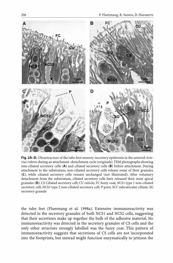
the tube feet (Flammang et al. 1998a). Extensive immunoreactivity wasdetected in the secretory granules of both NCS1 and NCS2 cells, suggestingthat their secretions make up together the bulk of the adhesive material. Noimmunoreactivity was detected in the secretory granules of CS cells and theonly other structure strongly labelled was the fuzzy coat. This pattern ofimmunoreactivity suggests that secretions of CS cells are not incorporatedinto the footprints, but instead might function enzymatically to jettison the
P. Flammang, R. Santos, D. Haesaerts206
Fig. 2A–D. Ultrastructure of the tube foot sensory-secretory epidermis in the asteroid Aste-rias rubens during an attachment–detachment cycle (originals). TEM photographs showingnon-ciliated secretory cells (A) and ciliated secretory cells (B) before attachment. Duringattachment to the substratum, non-ciliated secretory cells release some of their granules(C), while ciliated secretory cells remain unchanged (not illustrated). After voluntarydetachment from the substratum, ciliated secretory cells have released their most apicalgranules (D). CS Ciliated secretory cell; CU cuticle; FC fuzzy coat; NCS1 type 1 non-ciliatedsecretory cell; NCS2 type 2 non-ciliated secretory cell; P pore; SCC subcuticular cilium; SGsecretory granule

fuzzy coat, thereby allowing the tube foot to detach (Flammang 1996; Flam-mang et al. 1998a).
Footprints in echinoderms consist of a sponge-like material deposited as athin layer on the substratum (Thomas and Hermans 1985; Flammang 1996;Flammang et al. 1998a). Although their diameter is easily measured afterstaining of the adhesive material (Flammang 1996), footprint thickness is dif-ficult to estimate. Using an interference-optical profilometer, which generatedthree-dimensional images of the footprint surface, the mean maximum foot-print thickness was found to be 100 nm in the echinoid P. lividus and 230 nmin the asteroid A. rubens (Figs. 1B,C; Santos, Gorb and Flammang, unpubl.data). The chemical composition of the footprint material was analysed in A.rubens. Leaving inorganic residue apart, this material is made up mainly ofproteins and carbohydrates (Flammang et al. 1998a). The protein moiety con-tains significant amounts of both charged (especially acidic) and unchargedpolar residues as well as cysteine. The carbohydrate moiety is also acidic,comprising both uronic acids and sulphate groups.Adhesive interactions withthe substratum could be through ionic bonds, presumably involving theacidic residues of both carbohydrate and protein moieties (Waite 1987),whereas cohesive strength could be achieved by intermolecular disulphidebonds. So far, A. rubens is the only species in which the tube foot adhesive hasbeen studied biochemically and nothing is known about other echinodermspecies. Regarding the asteroids, however, a comparative immunohistochem-ical study of the tube feet from 14 species representing five orders and tenfamilies revealed that the adhesives of all these species are closely related, andthis independently of the taxon considered, of the species habitat and of thetube foot morphotype or function (Santos et al. 2005).
Comparison of the composition of the temporary adhesive of A. rubenswith that of other marine invertebrates shows that it is closer to the transitoryadhesive of limpets, also composed of an association of proteins and acidicglycans, than to the permanent adhesives of mussels and barnacles, madealmost exclusively of proteins (Flammang et al. 1998a; Flammang 2003). Asimilar relationship between non-permanent adhesives is also observed whenone compares the amino acid compositions of the temporary adhesive fromasteroids with the temporary adhesive from monogenean flatworms and thetransitory adhesive from limpets (see Flammang 2003). This relationshipindicates convergence in composition because of common function (i.e. non-permanent attachment to the substratum) and selective pressures.
3Larval Adhesive Organs
For most echinoderms, metamorphosis transforms a bilaterally symmetricaland pelagic larva into a radially symmetrical and benthic postmetamorphicindividual. Settlement always takes place during the so-called perimetamor-
Echinoderm Adhesive Secretions 207

phic period (Gosselin and Jangoux 1998; Haesaerts et al. 2003), but eitherbefore or after the metamorphic stage according to the class considered(Strathmann 1978). In both cases, adhesive organs attach either the competentlarva or the postlarva to the substratum during settlement. In three of the fiveextant echinoderm classes, these organs are the tube feet, viz. the five primarytube feet of competent echinoplutei in echinoids, the five primary tentacles(and, for some species, two posterior tube feet) of pentactulae inholothuroids, the five primary tube feet and the five first pairs of tube feet ofophiuroid postlarvae (Strathmann 1978). These tube feet are similar in struc-ture and function to tube feet of adults (Cameron and Fankboner 1984; Flam-mang et al. 1998b). Larval adhesive organs of crinoids and asteroids are, onthe other hand, unique and have no equivalent in the postmetamorphic stage(Strathmann 1978).
The perimetamorphic period of crinoids comprises three stages: the dolio-laria (free-swimming larval stage), the cystidean (attached metamorphicstage) and the pentacrinoid stages (attached postlarval stage) (Mladenov andChia 1983; Lahaye and Jangoux 1987; Nakano et al. 2003). Competent doliolar-iae are small barrel-shaped larvae. They possess an attachment complex attheir anterior end which consists of a ciliary cap surrounding an apical tuft ofelongated cilia and a ventrally located and slightly depressed adhesive pit(Fig. 3A,B). The ultrastructure of this attachment complex has been studied incomatulids (Chia et al. 1986; Jangoux and Lahaye 1990). It is strictly epidermaland made up of elongated ciliated cells associated with a thick basiepidermalnerve plexus. The four cell types forming the complex are sensory cells, cov-ering cells and two types of secretory cells. Sensory cells and secretory cells ofthe first type occur exclusively in the ciliary cap. The former bear a longvibratile cilium whereas the latter are filled with secretory granules, whichcontain a flocculent mucopolysaccharidic material. Secretory cells of the sec-ond type are restricted to the adhesive pit where they are the most abundantcell type. These cells are filled with secretory granules with an electron-densefibrillar proteinaceous content. At the beginning of the settlement phase, thedoliolaria becomes demersal and brushes the substratum with its apical tuft(sensory structure) (Mladenov and Chia 1983; Lahaye and Jangoux 1988).This implies the occurrence of a mechanism allowing the larva to combineloose adhesion to the substratum with movement. This transitory adhesion isachieved by the combined action of the secretory cells of the ciliary cap whichproduce a thin mucous film retaining the larva at the water–substratum inter-face, and of the covering cells whose cilia beat in this mucus (Jangoux andLahaye 1990; Flammang 1996). When reaching a suitable site, the larva stopsmoving and turns itself round to have its body directed obliquely (the adhe-sive pit facing the substratum). It then becomes permanently fixed and trans-forms into a cystidean larva (Mladenov and Chia 1983; Lahaye and Jangoux1988). Permanent adhesion starts with the release of the proteinaceouscement by the secretory cells of the adhesive pit and continues during bothcystidean and pentacrinoid stages (Chia et al. 1986; Jangoux and Lahaye
P. Flammang, R. Santos, D. Haesaerts208

Echinoderm Adhesive Secretions 209
Fig. 3A–C. Larval adhesive organs of crinoids and asteroids. SEM photographs of the dolio-laria larva of Antedon bifida (A) and of its anterior adhesive pit (B) (from Lahaye 1987), andof the brachiolaria larva of Asterias rubens (C) (original). AD Adhesive disc; AdP adhesivepit; AP apical papilla; AT apical tuft; CC ciliary cap; LBA lateral brachiolar arm; LP lateralpapilla; M mouth; MBA median brachiolar arm; PL preoral lobe; V vestibule; 1–4 ciliarybands

1990). After development of the cirri during this last stage, the juveniledetaches from its cemented stalk (Lahaye and Jangoux 1987).
Competent larvae in asteroids are called brachiolariae because they possessa specialized attachment complex on their anterior part comprising three bra-chiolar arms and an adhesive disc (Fig. 3C; Barker 1978; Haesaerts et al. 2003).Brachiolar arms are hollow tubular structures occupied by an extension of thelarval anterior coelom. Their histological organization comprises four tissuelayers: an inner myomesothelium, a connective tissue layer, a subepidermalnerve plexus and an outer epidermis. Each brachiolar arm is tipped by severalsensory-secretory areas named papillae, where both the epidermis and thenerve plexus are greatly thickened. The papillary epidermis encompassesthree types of secretory cells (cell types A, B and C), sensory cells and supportcells (Barker 1978; Haesaerts et al. 2005). Type A and B secretory cells arenumerous and occupy most of the volume of the papilla, while type C secre-tory cells are scarce and occur only at the base of the papilla. Type A secretorycells bear an apical cilium and contain large ovoid granules that enclose anelectron-dense heterogeneous material staining histochemically as neutralmucopolysaccharides. Type B secretory cells bear a subcuticular cilium andare filled with small granules containing a homogeneous electron-densematerial. The adhesive disc is a round, concave structure lying between thebrachiolar arms. It is an epidermal structure composed of two main celltypes: ciliated secretory cells and support cells (Barker 1978; Haesaerts et al.2005). The former are full of large secretory granules enclosing a fibrous pro-teinaceous content of woven aspect.When exploring the substratum, the com-petent larva orients itself ventral side down and successively attaches anddetaches its brachiolar arms (Barker 1978; Strathmann 1978; Haesaerts et al.2003). Papillae, when making contact with the substratum, are responsible forsensory testing and temporary adhesion. Like adult tube feet, they function asa duo-glandular system, with type A and B secretory cells acting as adhesiveand de-adhesive cells, respectively (Hermans 1983; Flammang 1996; Hae-saerts et al. 2005). In addition, the contents of type A secretory cells cross-react with antibodies raised against tube foot adhesive of A. rubens, indicat-ing that adhesives from both brachiolar arms and podia are related to eachother and probably share identical molecules, or, at least, identical epitopes ontheir constituents (Haesaerts et al. 2005). Once the larva has found a suitablesite for metamorphosis, brachiolar arms are gradually splayed out, enablingthe disc to release its cement (Barker 1978; Haesaerts et al. 2003). This attachesthe larva permanently to the substratum and marks the onset of the meta-morphic stage. During this stage, tube feet become functional and ultimatelyhelp the newly formed postlarva to detach from the cemented disc (Haesaertset al. 2003).
Among marine invertebrates, crinoids and asteroids are unique in usingnon-permanent adhesion during settlement (transitory adhesion for thedoliolariae and temporary adhesion for the brachiolariae), permanent adhe-sion for fixation during metamorphosis,and then reversing to non-permanent
P. Flammang, R. Santos, D. Haesaerts210

adhesion for their whole postmetamorphic life (mechanical attachment for thecomatulids and temporary adhesion for the asteroids). Indeed, in general,invertebrates that remain mobile as adults use a single type of adhesionthroughout their perimetamorphic period. For example, during settlement,pediveligers of gastropod molluscs adhere to the substratum through a viscousfilm of mucus produced by their foot on which they creep (transitory adhesion;Koehl and Hadfield 2004). This type of adhesion is then conserved up until theadult form (Walker 1987). Sessile invertebrates, on the other hand, cannot relyon a single type of adhesion during their perimetamorphic period. Theseorganisms, which as adults use permanent adhesion and live cemented to thesubstratum, need a non-permanent type of adhesion during settlement toenable them to move around while exploring the substratum (Crisp 1984). Forpremetamorphic attachment,they can therefore use either transitory adhesionlike bryozoan larvae or temporary adhesion like barnacle cyprids. From meta-morphic attachment onwards, all these organisms then rely on permanentadhesion to remain cemented to the substratum (Crisp 1984).
Adhesion strength of marine invertebrate larvae is difficult to measure dueto the small size of these organisms. It can be estimated, however, from thewater current required to wash larvae off a glass substratum. Using this tech-nique, the nominal wall shear stress needed to dislodge temporarily attachedindividuals of the asteroid Asterina gibbosa was about 1 Pa for brachiolariaeattached by the arms and 6 Pa for postmetamorphic individuals attached bytube feet (Haesaerts, Callow and Flammang, unpubl. data). These values arecomparable to those required to dislodge newly settled barnacle cyprids(0.2–8.7 Pa) and nudibranch larvae (4.26 Pa) (Koehl and Hadfield 2004). Onthe other hand, a nominal wall shear stress of about 40 Pa was needed todetach metamorphic larvae of A. gibbosa permanently attached by the disc(Haesaerts, Callow and Flammang, unpubl. data), showing the very highadhesive strength of this permanent adhesive.
4Cuvierian Tubules
Cuvierian tubules are peculiar organs found in several species of holothuroids(sea cucumbers), all belonging exclusively to the family Holothuriidae.Tubules (Fig. 4A) occurring in holothuroids of the genera Bohadschia,Holothuria and Pearsonothuria are expelled as sticky white threads that func-tion as a defence mechanism against predators (Hamel and Mercier 2000;Flammang et al. 2002). Cuvierian tubule adhesion is a typical example ofinstantaneous adhesion, adhesion being achieved in a matter of seconds (lessthan 10 s; Zahn et al. 1973).
Cuvierian tubules occur in great numbers (between 200 and 600 in H.forskali; VandenSpiegel and Jangoux 1987) in the posterior part of the bodycavity of the holothuroid. Proximally they are attached to the basal part of the
Echinoderm Adhesive Secretions 211

left respiratory tree and their distal, blind ends float freely in the coelomicfluid. When disturbed, the sea cucumber directs its aboral end toward thestimulating source and undergoes a general body contraction. The anusopens, the wall of the cloaca tears, and the free ends of a few tubules (usually10 to 20 in H. forskali; VandenSpiegel and Jangoux 1987), together withcoelomic fluid, are expelled through the tear and the anus. As water from therespiratory tree is forcefully injected into their lumen, the emitted tubuleselongate up to 20 times their original length (VandenSpiegel and Jangoux1987). Upon contact with any surface, the elongated tubules become instantlysticky. The adhesiveness of Cuvierian tubules combined with their tensilestrength make them very efficient for entangling and immobilizing mostpotential predators (VandenSpiegel and Jangoux 1987; Hamel and Mercier2000). Finally, the expelled tubules autotomize at their attachment point onthe left respiratory tree and are left behind as the holothuroid crawls away(VandenSpiegel and Jangoux 1987). After expulsion and autotomy, Cuvieriantubules are readily regenerated. Cuvierian tubules thus constitute an efficientdefensive mechanism. Their large number, sparing use and regenerationdynamics make them a formidable line of defence (Hamel and Mercier 2000;VandenSpiegel et al. 2000).
P. Flammang, R. Santos, D. Haesaerts212
Fig. 4A, B. Morphology of Cuvierian tubules of Holothuria impatiens (originals). SEMphotograph of a transversely sectioned tubule (A), and longitudinal histological sectionshowing arrangement of tissue layers (B). CTL Connective tissue layer; IE inner epithelium;L lumen; ML muscle layer; M mesothelium

Cuvierian tubule adhesive strength on glass has been measured in sevenspecies of sea cucumbers belonging to the genera Bohadschia, Holothuria andPearsonothuria (Flammang et al. 2002). The mean normal tenacities observedvaried from about 30 to 135 kPa. These tenacities fall within the range ofadhesive strengths described for marine organisms. They are, however,among the lowest observed values (Flammang 2003).
Cuvierian tubules consist, from the inside to the outside, of an epitheliumsurrounding the narrow lumen, a thick connective tissue layer and a mesothe-lium lining the surface of the tubule that is exposed to the coelomic cavity(Fig. 4). The mesothelium is responsible for adhesion. In quiescent tubules, itis a pseudostratified epithelium made up of two superposed cell layers, anouter layer of peritoneocytes and an inner layer of granular cells which ishighly folded along the long axis of the tubule (Fig. 4B). Granular cells arefilled with densely packed membrane-bound granules enclosing a proteina-ceous material (Endean 1957; VandenSpiegel and Jangoux 1987). During elon-gation, the structure of the mesothelium is modified: the protective outerlayer of peritoneocytes disintegrates and the granular cell layer, nowunfolded, thus becomes outermost on the tubule. Granular cells empty thecontents of their granules when the elongated tubule comes into contact witha surface, resulting in adhesion (VandenSpiegel and Jangoux 1987; De Moor etal. 2003).
In H. forskali, tubule print material, i.e. the adhesive left on the substratumafter mechanical detachment of the tubule, is composed of 60 % protein and40 % neutral carbohydrate, a composition that is unique among the adhesivesecretions of marine invertebrates (De Moor et al. 2003). The proteinaceousnature of the adhesive material is confirmed by the observation that prote-olytic enzymes reduce the adhesive strength of Cuvierian tubules in H.forskali (Zahn et al. 1973). The amino acid compositions of the protein frac-tion in H. forskali, H. leucospilota, B. subrubra and P. graeffei indicate that theiradhesives are closely related. All are rich in small side-chain amino acids,especially glycine, and in charged and polar amino acids (Table 1). Their com-positions differ, however, from those of every other marine bioadhesive(Flammang 2003). Only a small fraction of the Cuvierian tubule adhesive canbe extracted using denaturing buffers. This soluble fraction contains severalproteins with different molecular masses but with closely related amino acidcompositions, resembling the one of the whole adhesive (De Moor et al. 2003).As for the tube foot adhesive, charged and polar amino acids are probablyinvolved in adhesive interactions with the substratum through hydrogen andionic bonding (Waite 1987). Small side-chain amino acids, on the other hand,are often found in large quantities in elastomeric proteins (Tatham andShewry 2000). These proteins are able to withstand significant deformationswithout rupture before returning to their original state when stress isremoved (Smith et al. 1999). The composition of Cuvierian tubule adhesivehas therefore all the characteristics of a strong and resistant underwateradhesive.
Echinoderm Adhesive Secretions 213

5Applications of Marine Bioadhesives
In addition to fundamental interests in marine bioadhesives, a substantialimpetus behind understanding these adhesives is the potential technologicalapplications that can be derived from more detailed knowledge. These appli-cations cover two broad fields of applied research: design of water-resistantadhesives and development of new antifouling strategies.
Sessile organisms such as mussels, barnacles and tube-dwelling worms areimportant macro-foulers and their adhesives are secreted as a fluid whichthen gradually solidifies to form a cement possessing high adhesive and cohe-sive strength (Walker 1987). Most studies of invertebrate adhesive systemshave therefore focused on the characterization of the permanent adhesivesfrom these organisms (see, e.g., Taylor and Waite 1997; Kamino et al. 2000).The best-characterized permanent adhesive is that from the blue mussel,Mytilus edulis. In this species, several proteins have been identified and char-acterized that co-occur as a complex blend in the byssal adhesion plaques
P. Flammang, R. Santos, D. Haesaerts214
Table 1. Amino acid compositions of adhesive prints from the Cuvierian tubules of severalspecies of holothuroids (values in residues per thousand)
Amino acid Holothuria Holothuria Bohadschia Pearsonothuria forskalia leucospilotab subrubrab graeffeib
HYP 0 24 8 8 ASX 78 74 64 62THR 87 69 65 80 SER 60 42 58 58GLX 91 122 106 124PRO 55 74 69 63GLY 266 267 298 254ALA 88 115 91 85CYS/2 14 3 9 4VAL 38 29 35 37MET 10 9 1 9ILE 28 24 25 32LEU 37 31 37 38TYR 20 14 17 17PHE 20 16 20 20HIS 26 13 8 20HLYS 0 5 12 3LYS 31 12 29 22ARG 50 57 46 63
a De Moor et al. (2003)b Flammang and Waite (unpubl. data)

(Waite 2002). So far, however, only one of these proteins (Mytilus edulis footprotein 1; Mefp-1) has been used in biotechnological applications: in the formof crude or recombinant preparations of the protein and in the form of chem-ically synthesized, derived peptides (Burzio et al. 1997; Deming 1999; Taylorand Weir 2000).
5.1Design of Water-Resistant Adhesives
The fact that marine invertebrates produce adhesives that act in the presenceof water has aroused increasing scientific and technological attention becausewater, including moist air, is usually the most common subverter of man-made adhesive joints (Kinloch 1987; Waite 1987). Biomimetic materialsinspired by marine adhesives are therefore sorely needed for applications inwet environments. Such materials could be used in underwater construction,of course, but the most important applications are certainly to be found in thebiomedical field (Strausberg and Link 1990; Peppas and Langer 1994; Taylorand Waite 1997). Indeed, the environment of the sea is similar in many ways tothe internal environment of mammalian organisms. Tissues are bathed in flu-ids with pH and ionic composition similar to salt water. Theoretically, theattachment mechanisms that some marine invertebrates have evolved to sur-vive can be useful as biological adhesives for in vitro as well as in vivo uses.
For in vitro techniques, a crude preparation of Mefp-1 has been developedas a cell and tissue adhesive (Cell-Tak, BD Biosciences) for immobilization ofbiologically active moieties to inert substrata (Benedict and Picciano 1989;Taylor and Waite 1997). Cell-Tak is used to attach cells or tissue sections tomany types of surfaces, including plastic, glass, metal and biological materi-als. It can simplify the manipulation of biological samples for a number oftechniques, including in situ hybridization, immunoassays, microinjection,immunohistochemistry and establishing primary cells in culture (Benedictand Picciano 1989; Burzio et al. 1997; Taylor and Waite 1997). Cell-Tak hasbeen used successfully with a variety of healthy cell types, tumour cells, per-meabilized cells and subcellular components (Taylor and Waite 1997). Thisbroad range of applications is explained by the fact that Mefp-1, in contrast tocell adhesion molecules, acts as a non-specific attachment factor that fastensonto a variety of functional groups present and accessible on the surface of allcells and tissues (Taylor and Waite 1997).
Surgical or topical reconnection of severed tissues is essential for restora-tion of their structure and function. The most widely used methods for join-ing tissues focus on mechanical fasteners such as sutures and staples. Surgicaladhesives, however, provide attractive alternatives to mechanical fastening(Strausberg and Link 1990; Albala 2003; Ninan et al. 2003; Singer and Thode2004). In addition to their rapid application, they are particularly useful in tis-sues that are difficult to reach, too difficult to cauterize or too delicate to with-
Echinoderm Adhesive Secretions 215

stand suturing. Moreover, they are relatively painless to apply and may notrequire the use of painful local anesthetics. They are also biodegradable, elim-inating the need for suture removal. Currently, two types of surgical adhesivesare commercially available – fibrin-based adhesives (see, e.g., Albala 2003)and cyanoacrylate-based adhesives (see, e.g., Singer and Thode 2004) – whichhave been used successfully in a growing number of surgical procedures.There are still several applications, however, for which these adhesives cannotbe used, e.g. in areas continuously bathed in body fluids (mucous membranes,bladder, etc) (Albala 2003; Ninan et al. 2003; Singer and Thode 2004). Marinebioadhesives could be ideal candidates for such applications because theyfunction in an aqueous environment, they possess the appropriate adhesiveand cohesive properties and they are ultimately biodegradable (Strausbergand Link 1990; Ninan et al. 2003). However, any adhesive material targeted formedical applications should also be biocompatible (non-toxic, low immuno-genicity) and should not interfere with the natural healing process (Straus-berg and Link 1990). Mefp-1 seems to be biocompatible and non-toxic in vitro(Benedict and Picciano 1989). However, much has still to be learned beforeclinical trials are performed for human applications (Burzio et al. 1997).
Dentistry is another field in which there is current demand for non-toxicbioadhesives able to form durable adhesive bonds in the aqueous environ-ment of the mouth (Peppas and Langer 1994; Burzio et al. 1997).
5.2Development of New Antifouling Strategies
Biofouling is one of the most important problems currently facing marinetechnology. In the marine environment, any solid surface will become fouled.Materials submerged in seawater experience a series of discrete physical,chemical and biological events, which result in the formation of a complexlayer of attached organisms known as biofouling (Abarzua and Jakubowski1995; Callow and Callow 2002). Until now, antifouling methods involved theuse of toxic self-polishing paints releasing tributyltin (TBT) or cuprous oxide.However, the impact of these compounds on the environment has led to a banon the use of TBT-containing paints and to close monitoring of copper dis-charge from antifouling paints (Callow and Callow 2002). Fouling control istherefore increasingly a problem of managing adhesion, and molecularunderstanding of how marine bioadhesives work should open up novel tech-nologies intended to specifically intervene in organism attachment (Taylorand Waite 1997; Callow and Callow 2002). One of these technologies is the useof enzyme-leaching paints. Indeed, it has been shown that enzymes can beactive in a paint coating and hydrolyze the attachment glue which is secretedby the fouling organisms (Abarzua and Jakubowski 1995). Moreover, enzymesare completely degraded in seawater within several days and they are non-toxic for marine life.Another technology is based on the development of foul-
P. Flammang, R. Santos, D. Haesaerts216

ing-release coatings such as silicone coatings (Callow and Callow 2002). Thesecoatings, which exhibit low critical surface tensions, function by minimizingthe adhesion strength of attached organisms. Fouling organisms can settle onthe coating but are then removed by either mechanical or hydrodynamiccleaning. Paradoxically, a recently developed fouling-release coating wasbased on a marine bioadhesive possessing strong fouling characteristics.Dalsin et al. (2003) synthesized hybrid molecules by combining a decapeptidederived from Mefp-1 and poly(ethylene glycol), and used these molecules tomodify surfaces. The strategy exploits the adhesive characteristics of thedecapeptide to anchor poly(ethylene glycol) onto surfaces, rendering the sur-faces resistant to cell attachment in vitro. In the future, it may also beemployed in preventing mussels, barnacles and other fouling organisms fromattaching to ship hulls, piers and other man-made structures (Dalsin et al.2003).
5.3The Potential of Echinoderm Adhesives
In view of the examples mentioned above, it appears that Mefp-1 has beenused successfully in several applications. However, there are some limitations,such as requirement of post-translational modifications to certain aminoacids and the need for a separate enzyme for curing (Strausberg and Link1990). New adhesive molecules that could overcome these problems are there-fore sought. In this context, echinoderm adhesives could offer novel featuresor performance characteristics for biotechnological applications. For exam-ple, the rapidly attaching adhesive of Cuvierian tubules, the releasable adhe-sive of tube feet or the powerful adhesive of asteroid larvae could each be use-ful to address particular bioadhesion problems. If the tube foot de-adhesivesecretion proves effectively to be an enzyme, its incorporation into antifoul-ing paints would be particularly relevant as many fouling larvae use tempo-rary adhesion during settlement (Crisp 1984).
Work is currently in progress to identify, purify and characterize the differ-ent molecules involved in echinoderm adhesion. The complete elucidation oftheir structure and physico-chemical characteristics is an obligatory prereq-uisite before any application can be envisaged.
Acknowledgements. Thanks are extended to Dr. M. Jangoux and Dr. J.H. Waite for usefulcomments on the manuscript. This work was supported by the US Office of Naval Research(grant no.°N00014-99-1-0853). D.H. benefited from an FRIA doctoral grant (Belgium) andR.S. from a doctoral grant of the Foundation for Science and Technology of Portugal(no.°SFRH/BD/4832/2001). P.F. is a Research Associate for the National Fund for ScientificResearch of Belgium (FNRS). This study is a contribution of the “Centre Interuniversitairede Biologie Marine” (CIBIM).
Echinoderm Adhesive Secretions 217

References
Abarzua S, Jakubowski S (1995) Biotechnological investigation for the prevention of bio-fouling. I. Biological and biochemical principles for the prevention of biofouling. MarEcol Prog Ser 123:301–312
Albala DM (2003) Fibrin sealants in clinical practice. Cardiovascular Surg 11(S1):5–11Barker MF (1978) Structure of the organs of attachment of brachiolaria larvae of Stichaster
australis (Verrill) and Coscinasterias calamaria (Gray) (Echinodermata: Asteroidea). JExp Mar Biol Ecol 33:1–36
Benedict CV, Picciano PT (1989) Adhesives from marine mussels. In: Hemingway RW, Con-ner AH, Branham SJ (eds) Proc ACS Symp Adhesives from Renewable Resources, vol 385.American Chemical Society, Washington, pp 465–483
Burzio LO, Burzio VA, Silva T, Burzio LA, Pardo J (1997) Environmental bioadhesion: themesand applications. Curr Opin Biotechnol 8:309–312
Callow ME, Callow JA (2002) Marine biofouling: a sticky problem. Biologist 49:1–5Cameron JL, Fankboner PV (1984) Tentacle structure and feeding processes in life stages of
the commercial sea cucumber Parastichopus californicus (Stimpson). J Exp Mar BiolEcol 81:193–209
Chia FS, Burke RD, Koss R, Mladenov PV, Rumrill SS (1986) Fine structure of the doliolarialarva of the feather star Florometra serratissima (Echinodermata: Crinoidea), with spe-cial emphasis on the nervous system. J Morphol 189:99–120
Crisp DJ (1984) Overview of research on marine invertebrate larvae, 1940–1980. In: CostlowJD, Tipper RC (eds) Marine biodeterioration: an interdisciplinary study. E & FN Spon,London, pp 103–126
Dalsin JL, Hu B-H, Lee BP, Messersmith PB (2003) Mussel adhesive protein mimetic poly-mers for the preparation of nonfouling surfaces. J Am Chem Soc 125:4253–4258
De Moor S,Waite JH, Jangoux M, Flammang P (2003) Characterization of the adhesive fromthe Cuvierian tubules of the sea cucumber Holothuria forskali (Echinodermata,Holothuroidea). Mar Biotechnol 5:37–44
Deming TJ (1999) Mussel byssus and biomolecular materials. Curr Opin Biol Chem3:100–105
Eeckhaut I, Flammang P, Lo Bue C, Jangoux M (1997) Functional morphology of the tenta-cles and tentilla of Coeloplana bannworthi (Ctenophora, Platyctenida), an ectosymbiontof Diadema setosum (Echinodermata, Echinoidea). Zoomorphology 117:165–174
Endean R (1957) The Cuvierian tubules of Holothuria leucospilota. Q J Microsc Sci98:455–472
Flammang P (1996) Adhesion in echinoderms. In: Jangoux M, Lawrence JM (eds) Echino-derm studies, vol 5. AA Balkema, Rotterdam, pp 1–60
Flammang P (2003) The glue of sea cucumber Cuvierian tubules: a novel marine bioadhe-sive. In: Colliec-Jouault S, Bergé JP, Guézennec J, Fleurence J, Le Gal Y, Roy P (eds) Marinebiotechnology: an overview of leading fields. Actes Colloq Ifremer 36:176–185
Flammang P, Walker G (1997) Measurement of the adhesion of the podia in the asteroidAsterias rubens (Echinodermata). J Mar Biol Assoc UK 77:1251–1254
Flammang P, Demeuleneare S, Jangoux M (1994) The role of podial secretions in adhesionin two species of sea stars (Echinodermata). Biol Bull 187:35–47
Flammang P, Michel A,Van Cauwenberge A,Alexandre H, Jangoux M (1998a) A study of thetemporary adhesion of the podia in the sea star Asterias rubens (Echinodermata, Aster-oidea) through their footprints. J Exp Biol 201:2383–2395
Flammang P, Gosselin P, Jangoux M (1998b) The podia, organs of adhesion and sensory per-ception in larvae and postmetamorphic stages of the echinoid Paracentrotus lividus(Echinodermata). Biofouling 12:161–171
P. Flammang, R. Santos, D. Haesaerts218

Flammang P, Ribesse J, Jangoux M (2002) Biomechanics of adhesion in sea cucumberCuvierian tubules (Echinodermata, Holothuroidea). Integr Comp Biol 42:1107–1115
Franc JM (1978) Organization and function of ctenophore colloblasts: an ultrastructuralstudy. Biol Bull 155:527–541
Gosselin P, Jangoux M (1998) from competent larva to exotrophic juvenile: a morphofunc-tional study of the perimetamorphic period of Paracentrotus lividus (Echinodermata,Echinoidea). Zoomorphology 118:31–43
Haesaerts D, Jangoux M, Flammang P (2003) Study of the perimetamorphic period of thesea star Asterias rubens by scanning electron microscopy. In: Féral JP, David B (eds)Echinoderm research 2001. AA Balkema, Lisse, pp 155–159
Haeserts D, Jangoux M, Flammang P (2005) The attachment complex of brachioloaria lar-vae of the sea star Asterias rubens (Echinodermata): An unstructural and immunocyto-chemical study. Zoomorphology (in press)
Hamel J-F, Mercier A (2000) Cuvierian tubules in tropical holothurians: usefulness and effi-ciency as a defence mechanism. Mar Fresh Behav Physiol 33:115–139
Hermans CO (1983) The duo-gland adhesive system. Oceanogr Mar Biol Ann Rev21:281–339
Jangoux M, Lahaye M-C (1990) The attachment complex of the dololaria larvae of Antedonbifida (Echinodermata, Crinoidea). In: De Ridder C, Dubois P, Lahaye M-C, Jangoux M(eds) Echinoderm research. AA Balkema, Rotterdam, pp 99–105
Kamino K, Inoue K, Maruyama T, Takamatsu N, Harayama S, Shizuri Y (2000) Barnaclecement proteins. Importance of disulfide bonds in their insolubility. J Biol Chem275:27360–27365
Kinloch AJ (1987) Adhesion and adhesives, science and technology. Chapman and Hall,London
Koehl MAR, Hadfield MG (2004) Soluble settlement cue in slowly moving water withincoral reefs induces larval adhesion to surfaces. J Mar Syst 49:75–88
Lahaye M-C (1987) Comportement larvaire et ontogénèse postembryonnaire chez la comat-ule Antedon bifida (Echinodermata, Crinoidea). PhD Thesis, Université Libre de Brux-elles, Brussels
Lahaye M-C, Jangoux M (1987) The skeleton of the stalked stages of the comatulid crinoidAntedon bifida (Echinodermata). Fine structure and changes during growth. Zoomor-phology 107:58–65
Lahaye M-C, Jangoux M (1988) Morphologie externe et comportement des larves doliolariad’Antedon bifida (Echinodermata, Crinoidea). Ann Soc R Zool Belg 118:183–189
Mladenov PV, Chia FS (1983) Development, settling behaviour, metamorphosis and pen-tacrinoid feeding and growth of the feather star Florometra serratissima. Mar Biol73:309–323
Nachtigall V (1974) Biological mechanisms of attachment. The comparative morphologyand bioengineering of organs for linkage, suction, and adhesion. Springer, Berlin Heidel-berg New York
Nakano H, Hibino T, Oji T, Hara Y, Amemiya S (2003) Larval stages of a living sea lily(stalked crinoid echinoderm). Nature 421:158–160
Ninan L, Monahan J, Stroshine RL,Wilker JJ, Shi R (2003) Adhesive strength of marine mus-sel extracts on porcine skin. Biomaterials 24:4091–4099
Paine VL (1926) Adhesion of the tube feet in starfishes. J Exp Zool 45:361–366Peppas NA, Langer R (1994) New challenges in biomaterials. Science 263:1715–1720Ridgway SH, Lindner E, Mahoney KA, Newman WA (1997) Gray whale barnacles Cryp-
tolepas rhachianecti infest white whales, Delphinapterus leucas, housed in San DiegoBay. Bull Mar Sci 61:377–385
Echinoderm Adhesive Secretions 219

Santos R (2003) Etude biomécanique et morphologique de l’adhérence chez les oursins(Echinodermata, echinoidea) et sa relation avec la distribution des espèces dans lemilieu naturel. Master Thesis, Université de Mons-Hainaut, Belgium
Santos R, Haesaerts D, Jangoux M, Flammang P (2005) Comparative histological andimmunohistochemical study of sea star tube feet (Echinodermata, Asteroidea). J Mor-phol 263:259–270
Singer AJ, Thode HC (2004) A review of the literature on octylcyanoacrylate tissue adhesive.Am J Surg 187:238–248
Smith BL, Schäffer TE,Viani M, Thompson JB, Frederick NA, Kindt J, Belcher A, Stucky GD,Morse DE, Hansma PK (1999) Molecular mechanistic origin of the toughness of naturaladhesives, fibres and composites. Nature 399:761–763
Strathmann RR (1978) Larval settlement in echinoderms. In: Chia FS, Rice ME (eds) Settle-ment and metamorphosis of marine invertebrate larvae. Elsevier-North Holland, NewYork, pp 235–246
Strausberg RL, Link RP (1990) Protein based medical adhesives. Trends Biotechnol 8:53–57Tatham AS, Shewry PR (2000) Elastomeric proteins: biological roles, structures and mecha-
nisms. Trends Biochem Sci 25:567–571Taylor CM, Weir CA (2000) Synthesis of the repeating decapeptide unit of Mefp1 in orthog-
onally protected form. J Org Chem 65:1414–1421Taylor SW, Waite JH (1997) Marine adhesives: from molecular dissection to application. In:
McGrath K, Kaplan D (eds) Protein-based materials. Birkhäuser, Boston, pp 217–248Thomas LA, Hermans CO (1985) Adhesive interactions between the tube feet of a starfish,
Leptasterias hexactis, and substrata. Biol Bull 169:675–688Tyler S (1988) The role of function in determination of homology and convergence – exam-
ples from invertebrates adhesive organs. Fortsch Zool 36:331–347Vaitilingon D, Eeckhaut I, Fourgon D, Jangoux M (2004) Population dynamics, infestation
and host selection of Vexilla vexillum (Gmelin, 1791), an ectoparasitic muricid of echi-noids, in Madagascar. Dis Aquat Org 61:241–255
VandenSpiegel D, Jangoux M (1987) Cuvierian tubules of the holothuroid Holothuriaforskali (Echinodermata): a morphofunctional study. Mar Biol 96:263–275
VandenSpiegel D, Jangoux M, Flammang P (2000) Maintaining the line of defense: regener-ation of Cuvierian tubules in the holothuroid Holothuria forskali (Echinodermata). BiolBull 198:34–49
Waite JH (1987) Nature’s underwater adhesive specialist. Int J Adhesion Adhesives 7:9–14Waite JH (2002) Adhesion à la moule. Integr Comp Biol 42:1172–1180Walker G (1987) Marine organisms and their adhesion. In: Wake WC (ed) Synthetic adhe-
sives and sealants. Wiley, Chichester, pp 112–135Whittington ID, Cribb BW (2001) Adhesive secretions in the Platyhelminthes. Adv Parasitol
48:101–224Zahn RK, Müller WEG, Michaelis M (1973) Sticking mechanisms in adhesive organs from a
Holothuria. Res Mol Biol 2:47–88
P. Flammang, R. Santos, D. Haesaerts220

Mutable Collagenous Tissue: Overview and Biotechnological Perspective
I.C. Wilkie
Abstract. The mutable collagenous tissue (MCT) of echinoderms canundergo extreme changes in passive mechanical properties within a timescaleof less than 1 s to a few minutes, involving a mechanism that is under directneural control and coordinated with the activities of muscles. MCT occurs ata variety of anatomical locations in all echinoderm classes, is involved inevery investigated echinoderm autotomy mechanism, and provides a mecha-nism for the energy-sparing maintenance of posture. It is therefore cruciallyimportant for the biology of extant echinoderms. This chapter summarisescurrent knowledge of the physiology and organisation of MCT, with particu-lar attention being given to its molecular organisation and the molecularmechanism of mutability. The biotechnological potential of MCT is discussed.It is argued that MCT could be a source of, or inspiration for, (1) new pharma-cological agents and strategies designed to manipulate therapeutically con-nective tissue mechanical properties and (2) new composite materials withbiomedical applications.
1Introduction
1.1Collagenous Tissue
The collagens are a family of at least 19 proteins characterised by the presenceof triple helical regions composed of repeating Gly-X-Y triplets. The majorityof collagens occur in the extracellular matrix (ECM) of connective tissue, themost prevalent of these being types I, II and III, which self-assemble into par-
Progress in Molecular and Subcellular BiologySubseries Marine Molecular BiotechnologyV. Matranga (Ed.), Echinodermata© Springer-Verlag Berlin Heidelberg 2005
I.C. Wilkie (e-mail: [email protected])Department of Biological and Biomedical Sciences, Glasgow Caledonian University, 70Cowcaddens Road, Glasgow G4 0BA, Scotland, UK
allel aggregations known as fibrils, and type IV, which forms a network in thelamina densa of all basement membranes. In the ECM, collagen fibrils areaccompanied by a variety of other components, which may include otherfibrous structures, such as elastin fibres or microfibril aggregations, proteo-glycans, glycoproteins and glycosaminoglycans. Some of these other compo-nents link together adjacent collagen fibrils and are thereby responsible forinterfibrillar cohesion (Kannus 2000; Sarras and Deutzmann 2001).
Connective tissue in which the mechanically dominant component is colla-gen is the most widespread and evolutionarily ancient structural material inthe animal kingdom. With the exception of possibly only the phyla Rotifera(Clément 1993) and Placozoa (Syed and Schierwater 2002), it is present in thebodies of all multicellular animals, including sponges, the internal ‘mesohyl’of the latter being histologically and functionally a collagenous connectivetissue that, unlike the situation in other animals, is not separated from itsinvesting epithelia by basement membranes (Garronne 1978; Bonasoro et al.2001). (It is possible that the absence of basement membranes from mostsponges is due to secondary loss: see Maldonado 2004.)
1.2Mechanical Adaptability of Collagenous Tissue
In its role as a structural material, the principal functions of collagenous tis-sue are to transmit, resist and dissipate mechanical forces and to store andrelease elastic strain energy. These functions depend on the tissue as a wholepossessing certain ‘fit for purpose’ mechanical properties (tensile strength,stiffness, resilience, etc.), which are the result of a combination of the mechan-ical properties of its individual structural components, the micro-architec-tural organisation of these components and the nature of the interactionsbetween them.
The net mechanical properties of most collagenous structures are rela-tively stable within a physiological timescale (seconds to hours). They maychange during maturation and ageing, partly as a result of alterations in col-lagen fibril diameter and the composition of the interfibrillar phase mediatedby adjustments in the synthetic activities of fibroblasts and, in the case of age-ing, partly as a result of the non-adaptive biochemical modification of theECM, e.g. by non-enzymatic glycation (Vogel 1980; Bruel and Oxlund 1996;Reddy et al. 2002; Silver et al. 2003). The mechanical properties of collagenousstructures can also be modified in response to a long-term shift in the patternof force to which they are subjected. For example, exercise increases the ten-sile strength and stiffness of tendons, again probably through fibroblast-mediated changes in fibril diameter and interfibrillar composition (Buchananand Marsh 2002; see also Chiquet 1999 for a discussion of the regulatory path-ways that might be involved). These age-related and adaptive changes occurover timescales of days to years and are quantitatively undramatic. However,
I.C. Wilkie222

some collagenous structures undergo more drastic changes in mechanicalproperties over a shorter timescale, which result in a qualitative modificationof the mechanical functioning of the tissue and thus justify it being regardedas a mechano-effector. In Homo sapiens and other mammals, this phenome-non is demonstrated by various collagenous structures associated with thefemale reproductive tract. For example, at the end of pregnancy, the compli-ance of the uterine cervix increases temporarily by a factor of 12. It switchesfrom being tough and inextensible, in which state it helps to prevent expulsionof the conceptus, to being soft and easily extensible, a condition that permitsthe dilatation and effacement required for the passage of the fetus through thebirth canal. Cervical ‘relaxation’ depends on two separate processes: (1)changes in the synthetic activity of the stromal cells, which secrete less colla-gen types I and III and small proteoglycans and secrete more of the large pro-teoglycan versican; and (2) the enzymatic degradation of the ECM by matrixmetalloproteinases (MMPs) including MMP-1, MMP-3 and MMP-8, togetherwith increased expression of the tissue inhibitors of metalloproteinasesTIMP-1 and TIMP-2 (these presumably acting as a brake on the degradativeprocess). The resulting change in mechanical properties occurs over hours todays and is controlled by a combination of hormonal events and the directeffect of stretch on the stromal cells (Uldbjerg 1994; Westergren-Thorsson etal. 1998; Yoshida et al. 2002; Sennström et al. 2003). Similar mechanisms andcontrolling factors are involved in the rupture of the fetal membranes at term(Bryant-Greenwood 1998) and of the ovarian follicle at ovulation (Robker etal. 2000).
The above examples of connective tissue mechanical adaptability havebeen given in order to highlight the distinctiveness of the subject of this chap-ter – the mutable collagenous tissue (MCT) of echinoderms. MCT canundergo extreme alterations of mechanical properties within a timescale ofless than 1 s to a few minutes, involving a mechanism that is under directneural control and coordinated with the activities of other mechano-effec-tors, viz. muscles. The rest of this chapter provides a review of current knowl-edge of the physiology and organisation of MCT and discusses its biotechno-logical potential.
2Mutable Collagenous Tissue: Physiology and Organisation
2.1Overview
MCT is present at a variety of anatomical locations in all living echinodermclasses, is involved in every investigated autotomy (defensive self-detach-ment) mechanism and provides a mechanism for the energy-sparing mainte-nance of posture (Wilkie 1996, 2001, 2002; Wilkie et al. 2004a). MCT is there-
Mutable Collagenous Tissue: Overview and Biotechnological Perspective 223

fore crucially important for the biology of extant echinoderms and, in view ofits apparent absence from other animals, has been regarded as one of the dis-tinguishing characteristics of the phylum (Byrne 2001).
MCT occurs in the form of dermal connective tissue, interossicular liga-ments, tendons linking muscles to skeletal elements and the connective tissuelayer in the walls of tubular structures. It performs the same mechanical func-tions as the collagenous connective tissue at analogous locations in the bod-ies of vertebrates. Although there have been few attempts to correlate thesupramolecular organisation of mutable collagenous structures with their‘conventional’ mechanical functions (O’Neill 1989; O’Neill and Withers 1995),there is no reason to doubt that, despite their additional property of variabletensility, all such structures are as exquisitely designed to meet the subtly dif-ferent functional demands of each anatomical location as are their vertebrateequivalents (see, e.g., Bauer et al. 1989; Munns et al. 1994; Gupte et al. 2002).
2.2Mechanical Adaptability of MCT
2.2.1Passive Mechanical Properties
Each mutable collagenous structure exhibits one of three patterns of changein its passive tensile properties: (1) only reversible changes (e.g. in viscosity orstiffness); (2) irreversible destabilisation (always associated with autotomy) aswell as reversible changes; or (3) only irreversible destabilisation (Wilkie2002).
The most extreme manifestation of MCT mechanical adaptability is therapid and irreversible loss of tensile strength undergone by collagenousstructures that cross echinoderm autotomy planes. For example, when an armof the ophiuroid Ophiocomina nigra is autotomised, the ultimate tensile
I.C. Wilkie224
Fig. 1A–I. Passive mechanical behaviour of MCT. A Creep test (in which extension of sam-ple under constant load is recorded): in response to 100 mM K+ (K) syzygial ligament ofcrinoid Antedon mediterranea shows sudden decrease in viscosity culminating in rupture(star). Horizontal scale bar 1 min; vertical scale bar 1 mm (adapted from Wilkie et al. 1999).B, C Creep tests: viscosity of dental ligament of echinoid Diadema setosum is increasedreversibly by 1 mM acetylcholine (ACh) and 100 mM K+ (K). ASW Artificial seawater(adapted from Birenheide et al. 1996). D Stress relaxation tests (in which samples are sub-jected to constant deformation and force is recorded): average force relaxation curves of 20dental ligaments of echinoid Dendraster excentricus treated with artificial seawater (ASW)and 15 ligaments treated with divalent cation-free seawater (DCF) (adapted from Ellers andTelford 1996). E, F Stress-strain tests (in which force is recorded while samples are stretchedat fixed extension rate) conducted on spine ligament of echinoid Anthocidaris crassispina:Examples of stress-strain curves produced by samples treated with E 0.1 mM acetylcholine

Mutable Collagenous Tissue: Overview and Biotechnological Perspective 225
(ACh) and F 0.1 mM adrenaline (Adr). Horizontal scale bar Strain of 10 % in both cases; ver-tical scale bar 10 MPa in E and 1 MPa in F (adapted from Hidaka and Takahashi 1983).G–I Dynamic stress-strain tests (in which force is recorded while samples are subjected tooscillating strain): hysteresis loops produced by repetitive testing of dermis of holothurianActinopyga mauritiana in three mechanical states. During these tests, maximum strain(indicated by number above each curve) was increased incrementally; note that sample in‘soft’ state showed strain-induced softening. (Adapted from Motokawa and Tsuchi 2003)

strength of the intervertebral ligament at the autotomising joint drops to lessthan 0.1 % of the normal value in a timescale of 0.4–5.4 s (Wilkie 1988). Thesedrastic changes in mechanical properties can also be demonstrated experi-mentally using isolated tissue preparations undergoing creep tests (in whichtheir extension under constant load is recorded). Treatment with neuro-activeagents such as elevated [K+] or appropriate neurotransmitter chemicalscauses an abrupt decrease in viscosity (stress-strain rate) culminating in tis-sue rupture (Fig. 1A).
Reversible changes in mechanical properties have been quantified andanalysed by means of various testing methods. Reversible changes in viscos-ity have been demonstrated in creep tests and stress relaxation tests (in whichspecimens are stretched to a particular length, which is fixed whilst forcedecay is recorded) (Fig. 1B–D). Reversible changes in tensile strength, tensilestiffness, dynamic shear stiffness, relative damping, etc. have been investi-gated by means of standard stress-strain tests (Fig. 1E,F) and by dynamic test-ing methods in which samples are subjected to oscillating strain (Fig. 1G–I).Evidence of mutability is often apparent in the wide variability in themechanical properties of untreated tissues. Motokawa (1983), for example,found a 200-fold difference in the viscosity of untreated central spine liga-ments of Diadema setosum taken from different joints of the same animal. Inorder to overcome this problem and compare tissues in predictable mechani-cal states, a common strategy has been to subject them to different treatmentsthat induce either maximal or minimal values of stiffness, viscosity, etc. (seeFig. 1G–I). Although biologically relevant stimuli, such as mechanical com-pression, have been used in these investigations, the artificial nature of sometreatments, especially those employed to bring about a compliant state,engender some uncertainty about the physiological relevance of the resultsthus obtained. Nevertheless, these methods no doubt give an indication of themagnitude of the reversible changes that MCT can accommodate in vivo.Hidaka and Takahashi (1983), for instance, found that at a low strain rate theultimate tensile strength and elastic modulus of an echinoid spine ligament inthe compliant condition (induced by 0.1 mM adrenaline) were around 1 % ofthe values measured in the stiffened condition (induced by 0.1 mM acetyl-choline), the latter falling within the range reported for mammalian tendon(Redaelli et al. 2003; Fig. 1E,F).
Data from these biomechanical studies have been used to generate modelswith the ultimate intention of specifying the contribution of different extra-cellular components to net mechanical properties and determining which ofthem contribute to variable tensility. Due to the diversity of testing regimesemployed, the conclusions from different investigations have been incompat-ible (see, e.g., Szulgit and Shadwick 2000; Motokawa and Tsuchi 2003) and aredifficult to interpret in the light of the increasingly complex picture of theMCT extracellular matrix that is emerging (see Sect. 2.3 below). It seems likelythat the full potential of this methodology will not be realised until it can beapplied to tissues from which specific components have been eliminated
I.C. Wilkie226

chemically, enzymatically or, ideally, by genetic knockout (see Bornstein et al.2000 and Chakravarti 2002 for examples of this last approach applied to mam-malian connective tissues).
2.2.2Active Contractility
Certain mutable collagenous structures can generate tensile force. In the caseof the capsular ligament, or ‘catch apparatus’, of the echinoid spine-test joint,this is attributable to the presence of fine (diameter 0.1–1.0 µm) muscle fibresthat are distributed between the bundles of collagen fibrils and constitute1–3 % of the total cross-sectional area of the ligament. The neurotransmitteracetylcholine increases the tensile strength and stiffness of this ligament(Hidaka and Takahashi 1983; Morales et al. 1989), but also causes isolatedpreparations of it to shorten and develop mechanical force, which relaxes assoon as the acetylcholine is removed. Both the contraction and relaxationphases of this response can be very fast, the former in some cases reaching90 % of the maximum in under 1 s (Fig. 2A; Vidal et al. 1993). The functionalsignificance of the muscle fibres is at present unknown. Del Castillo et al.(1995), ignoring incontrovertible evidence that the spine ligament consists ofMCT (see, e.g., Hidaka and Takahashi 1983; Szulgit and Shadwick 1994),hypothesised that they are responsible for varying its passive stiffness, whichthey achieve by adjusting the frictional forces between the ligament fibres andthe skeletal ossicles into which they are inserted. Having provoked a frankexchange of views (see Del Castillo and Smith 1996; Wilkie 1996, 2002; Pérez-Acevedo et al. 1998; Elphick and Melarange 2001), this hypothesis was testedand disproved by Takemae and Motokawa (2002). The muscle fibres also donot seem to be involved in straightening out the wrinkles that form tran-siently in regions of the ligament that are compressed by contraction of thespine muscle (Pérez-Acevedo et al. 1998). This leaves the possibilities that theyassist the reshortening of stretched ligament fibres or that they operate syner-gistically with the spine muscle, perhaps during specific manoeuvres such asre-erection of the spine.
The reputation of echinoderms for being an inexhaustible mine of biolog-ical novelty has been enhanced by the discovery that, as well as varying theirpassive mechanical properties, ligaments in the cirri and arms of crinoidshave the capacity for active contractility, though they lack myocytes. Cirri arefinger-like appendages supported by a single series of interarticulating ossi-cles. The only mechanically significant structures connecting adjacent ossi-cles are myocyte-free collagenous ligaments. Cirri attached to the stalk of sealilies bend upwards, against gravity, when the stalk or the cirri themselves arestimulated mechanically. In stress relaxation tests, isolated cirri display slowforce production in response to the cholinergic agonists muscarine andmethacholine at concentrations as low as 0.1 µM (Fig. 2B). Since force produc-
Mutable Collagenous Tissue: Overview and Biotechnological Perspective 227

I.C. Wilkie228
Fig. 2A–F. Force generation by MCT. A Contraction of spine ligament of echinoid Eucidaristribuloides induced by 0.1 mM acetylcholine (arrow) which was removed as soon as forcepeaked (adapted from Vidal et al. 1993). B Contraction of cirral ligaments of crinoidMetacrinus rotundus induced by 0.1 µm methacholine (M) (adapted from Birenheide et al.2000). C–F Responses to 100 mM K+ (K) of arm ligaments of M. rotundus. In each case, uppertrace shows upward displacement of arm tip (caused by shortening of ligaments) and lowertrace shows stiffness changes in ligament. Horizontal scale bar 3 min in all cases. C Contrac-tion associated with stiffening. D Contraction without stiffness change in 100 mM K+ andcontraction with stiffening when excess K+ was removed. E No contraction, but withmarked stiffening. F No contraction, but with destiffening. (Adapted from Motokawa et al.2004)
Time (min)

tion can be induced in preparations that have undergone some stress relax-ation prior to stimulation, it cannot be explained in terms of the passive recoilof a previously stretched elastic element (Birenheide and Motokawa 1995,1998; Birenheide et al. 2000).
The mobility of crinoid arms depends on the presence of muscular articula-tions between adjacent arm ossicles. Below the fulcral ridge of each articula-tion is a single aboral ligament and above it are paired oral ligaments andpaired muscles.Contraction of the muscles bends the arm orally (upwards),yetthe power stroke for locomotion by swimming, crawling or climbing is gener-ated by the aboral (downward) flexion of the arm and must be effected by theligaments. It had long been assumed that this involved the purely passive elas-tic recoil of stretched aboral ligaments, and perhaps of compressed oral liga-ments, these functioning like expanded and compressed springs respectively(see, e.g.,Young and Emson 1995). However, Birenheide and Motokawa (1996,1998) established that the aboral ligament can shorten slowly and generate atension of up to ca. 5.6 kPa. Because this ligament is a mutable collagenousstructure, it is feasible that its apparent contractility results from its becomingstiff whilst it is stretched (by muscle-mediated oral flexion); it would then storestrain energy until appropriate stimulation induced its destiffening andthereby allowed it to recoil elastically and reshorten. This ‘spring-with-a-lock’hypothesis was shown to be untenable by Motokawa et al. (2004) who, byrecording simultaneously stiffness and shortening, demonstrated the inde-pendence of passive mechanical properties and contraction: contraction, forexample, does not require the ligament to be in a destiffened condition(Fig. 2C–F). The fact that contracting ligaments are usually destiffened, how-ever, indicates that there is coordination of the passive and active mechanicalproperties, both of which appear to be under cholinergic control.
2.3Organisation of MCT
2.3.1Cells
Myocytes, which were discussed in the preceding section, have been found inonly a small minority of mutable collagenous structures. All of these struc-tures, however, always contain two other types of cell. The first is charac-terised by the presence of heterogeneous vacuoles that appear to be lysosomaland may enclose cytoplasmic debris or collagen fibrils. These cells tend tohave a roughly fusiform outline and long, sometimes branching, processes(see, e.g., Wilkie 1988; Wilkie et al. 1992). They occur also in echinoderm con-nective tissue that is not mutable (Wilkie et al. 2004b) and, given the absenceof obvious fibroblasts, they are likely to be pluripotential cells that can adopta fibrogenic phenotype.
Mutable Collagenous Tissue: Overview and Biotechnological Perspective 229

The second cellular component that is invariably present in MCT are cellbodies and/or processes containing large, electron-dense, membrane-bounded granules. In ophiuroids, these cellular elements, known as ‘juxtaliga-mental cells’, form a complex system of ganglion-like clusters innervated byhyponeural (motor) nerves, with a separate juxtaligamental cluster servingeach collagenous structure, including the autotomy tendons of the interverte-bral muscles (Wilkie 1979). It is assumed that at least some of the granule-con-taining cells associated with MCT in the other echinoderm classes are homol-ogous to the juxtaligamental cells, although the cell processes in holothuriansdiffer from those of the other four classes in being separated from the extracel-lular matrix by a basal lamina (see, e.g., Koob et al. 1999), which presumablyrepresents an ontogenetic rather than a phylogenetic or functional distinction.There is clear morphological evidence for functional contact between motorneurons and juxtaligamental cells in at least ophiuroids, echinoids andcrinoids (Cobb 1985; Peters 1985; Welsch et al. 1995), and so, mainly on thegrounds that they (1) provide a link between the motor nervous system and theextracellular matrix,(2) terminate in MCT and (3) have no possible cellular tar-gets, it has been hypothesised that they are the effector cells that directly alterthe tensile properties of MCT. In all classes it is usual for at least two types ofprocess to co-occur in the same tissue, these being distinguishable by the sizeand shape of their granules (see,e.g.,Welsch et al.1995; Koob et al.1999),and ithas been proposed that these include separate ‘stiffener’ and ‘plasticiser’ celltypes. The recent demonstration by immunological methods that juxtaliga-mental granules of holothurian dermis contain identified chemical factors thatinfluence interfibrillar cohesion provides further evidence that these cells havea role in variable tensility (see below).
2.3.2Extracellular Matrix
Overview
The extracellular matrix of almost all mutable collagenous structures is dom-inated by fibres consisting of parallel assemblages of collagen fibrils. Thesefibres exhibit a range of patterns, including parallel fibre arrays (e.g. echinoidspine ligament: Trotter and Koob 1989), crossed-fibre arrays (e.g. echinoidperistomial membrane: Wilkie et al. 1994) and three-dimensional meshworks(e.g. holothurian dermis: Motokawa 1982), which is comparable in diversity tothat shown by vertebrate connective tissue structures. An important excep-tion to this fibrous organisation is provided by the autotomy tendons of ophi-uroids. These are extensions of muscle cell basal laminae and consist of non-fibrillar collagen related probably to vertebrate type IV (Wilkie and Emson1987), although, since their biochemical composition is not known, this is asupposition based only on their histochemistry and ultrastructure. The fol-
I.C. Wilkie230

lowing sections summarise current knowledge of the molecular constituentsof fibrous MCT alone. A generalised model of the molecular organisation ofMCT is illustrated in Fig. 3.
Collagen
The fibres of which most MCT is composed are parallel aggregations of cross-banded collagen fibrils that are discontinuous, i.e. shorter than the length ofthe fibres. The fibrils of two mutable structures with very different microar-chitectures – the echinoid spine ligament and holothurian dermis – resemblethose of mammalian connective tissue such as rat tail tendon in being com-posed of molecules with a triple helix length of 300 nm which are assembledin parallel arrays with a regular stagger of 67 nm between adjacent molecules,in having cross-striations in similar positions though varying in stain inten-sity (an indication of differences in the charge density associated with their
Mutable Collagenous Tissue: Overview and Biotechnological Perspective 231
Fig. 3. Model of MCT molecular organisation, which is based on current evidence andassumes that the few mutable structures from which that evidence has been derived are rep-resentative. Most MCT consists of parallel aggregates of discontinuous, spindle-shaped col-lagen fibrils to which are attached PGs and other GAG-containing molecules whose func-tions include serving as binding sites for molecules responsible for interfibrillar cohesion.Amongst the latter are the proteins stiparin and tensilin, the fibril-aggregating activities ofwhich are modified by a variety of specific inhibitors. The mechanisms by which stiparinand tensilin cause fibril aggregation are incompletely understood. For simplicity, the modelassumes that they form dimers that act as interfibrillar crossbridges. Fibril bundles aredelimited by loose networks of elastic fibrillin-containing microfibrils that return the tissueto its resting dimensions after it has undergone deformation when in a compliant condi-tion. The functions of fibrosurfin are as yet unknown

constituent amino acids), and in being stabilised by high levels of trivalenthydroxypyridinium intermolecular crosslinks (Trotter and Koob 1989, 1994;Trotter et al. 1994, 1995). The chain compositions of the echinoid andholothurian collagen molecules were not the same, the former being a het-erotrimer of two a1 and one a2 polypeptides, as in mammalian type I andmost other echinoderm collagens (see, e.g., Omura et al. 1996; Robinson1997), and the latter being a homotrimer of three a1 polypeptides. These twocollagens also have different solubility characteristics and amino acid compo-sitions (Trotter and Koob 1994; Trotter et al. 1995). The general conclusionfrom this research is that MCT collagens possess no consistent set of bio-chemical or structural features that distinguish them from the collagens ofother echinoderms or other phyla, or that could be correlated with the muta-bility of their parent tissues.
Data on gene sequence and gene organisation indicate that at least someechinoderm collagen polypeptides are evolutionarily close to those of verte-brate fibrillar collagens (D’Alessio et al. 1989, 1990; Exposito et al. 1992;Tomita et al. 1994; Cluzel et al. 2000). So far, no primary sequence data on col-lagen extracted from any MCT have been published. However, an epitope of afully characterised collagen polypeptide has been detected in two confirmedmutable structures. Cluzel et al. (2001) immunolocalised the amino propep-tide of the 2a collagen chain, which, apart from the amino propeptide itself, isclosely similar to that of vertebrate fibrillar collagen (D’Alessio et al. 1990), inthe mutable peristomial membrane and spine ligament of the echinoid Para-centrotus lividus. Since the amino and carboxyl propeptide regions of colla-gen chains are usually removed by specific proteases during the extracellularmaturation process, their retention in the tissues of adult echinoids may havefunctional significance (Lethias et al. 1997).
Because complete collagen fibrils can be isolated from MCT using mild,non-denaturing extraction methods, more is known about their supramolec-ular organisation than that of vertebrate fibrils. Fibrils from both echinoidspine ligament and holothurian dermis are spindle-shaped with paraboloidaltips. Despite varying greatly in length, they have a constant aspect(length:diameter) ratio in the order of 2000 and are molecularly bipolar, i.e. inboth halves of each fibril the amino termini of the collagen molecules are ori-entated towards the nearer tip, and near the axial midpoint of each fibril thereis a region of symmetrical transition from parallel to antiparallel molecularpacking (Trotter and Koob 1989; Thurmond and Trotter 1994; Trotter et al.1994). As in other fibrillar collagens, this organisation results from the self-assembly of the constituent molecules which occurs automatically after enzy-matic removal of their N- and C-propeptides. More recent work by Trotter etal. (1998, 2000a), using digital scanning-transmission microscopy to deter-mine mass per unit length (and therefore the number of molecules) alongwhole collagen fibrils from both echinoid and holothurian tissues, has pro-vided evidence that the self-assembly mechanism is different from that of ver-tebrate fibrils.
I.C. Wilkie232

Although it is not possible at present to isolate whole collagen fibrils fromnormal adult tissues of animals other than echinoderms, the limited data thatare available on vertebrate fibrils indicate that, despite the different mecha-nism of fibrillogenesis, they are also spindle-shaped with paraboloidal tips(see Trotter et al. 1998). This is the ideal shape for fibrils that reinforce a dis-continuous fibre composite, since it allows the full tensile strength and stiff-ness of the fibril to be exploited along its whole length and avoids shear-stressconcentrations near its ends (Trotter and Koob 1989; Trotter et al. 2000b). Thefusiform shape of the collagen fibrils in MCT is therefore unrelated to its vari-able tensility.
Proteoglycans
Proteoglycans (PGs) are present in the fibrous connective tissue of all animalsand consist of a protein core to which are attached covalently side chains ofpolyanionic sulphated glycosaminoglycans (GAGs). The use of the polyca-tionic dyes cuprolinic and cupromeronic blue has revealed that polyanionsare localised to specific sites in each D-period on the surface of fibrils incrinoid, echinoid and holothurian MCT, as is the case in vertebrate collage-nous tissue (Trotter and Koob 1989; Erlinger et al. 1993; Trotter et al. 1995).Biochemical methods have demonstrated that PGs are attached to the fibrilsnon-covalently or covalently. Non-covalently bound PGs in the chondroitin/dermatan sulphate class are attached to the collagen fibrils of the echinoidspine ligament (Trotter and Koob 1989). Collagen fibrils in holothurian der-mis are associated covalently with three different GAG-containing macromol-ecules. The most abundant of these includes a fucose-containing GAG that isassociated with the fibrils via a non-reducible covalent bond. The structure ofthe highly sulphated fucose branches of chondroitin sulphate E from Asticho-pus japonicus has been characterised fully by Kariya et al. (1997). The othertwo covalently bound GAG-containing molecules in holothurian dermis arehigh molecular weight PGs that are linked to collagen fibrils probably viadisulphide bonds and at least one of which acts as a binding site for the glyco-protein stiparin (see below). In addition to these insoluble PGs, holothuriandermis contains at least two soluble PGs that bind stiparin, inhibit stiparin-fibril binding and may be involved in the regulation of stiparin-fibril binding(Trotter et al. 1995 and unpubl.).
Other Non-Collagenous Proteins
Stiparin is the most abundant soluble glycoprotein in the dermis of theholothurian Cucumaria frondosa and can be extracted from minced tissue byprolonged treatment with seawater alone (which also results in tissue disag-gregation, an indication that the collagen fibrils are normally held together byweak bonds). Trotter et al. (1996) demonstrated that stiparin, which has amolecular weight of about 375 kDa, causes calcium-independent aggregationin vitro of collagen fibrils that have been treated with guanidine-HCl (which
Mutable Collagenous Tissue: Overview and Biotechnological Perspective 233

removes non-covalently bound PGs) but has no effect on the mechanicalproperties of samples of intact dermis (Koob et al. 1999).Whilst it seems likelythat stiparin binds to collagen fibrils via a surface-bound PG, the molecularmechanism of stiparin-induced fibril aggregation has still to be determined.
The dermis of C. frondosa contains a 62-kDa sulphated glycoprotein thatdoes not bind collagen fibrils but does bind stiparin and thereby inhibits sti-parin’s fibril-aggregating activity. This molecule has the highest negativecharge density of all macromolecules extracted from the dermis, and all of itsinhibitory activity is associated with the polygalactose sulphate moiety of themolecule rather than with its protein component. The relative concentrationof stiparin inhibitor is 200 times greater in the loose outer dermis of C. fron-dosa than in the dense inner dermis (Trotter et al. 1999).
Tensilin (also known as ‘stiffener’) is a constituent of the inner dermis of C.frondosa that can be isolated only after treatments that cause cell lysis, such asrepeated freeze–thaw cycles, indicating that it is present mainly in intracellu-lar locations. Like stiparin, it causes aggregation of isolated collagen fibrils,but, unlike stiparin, it stiffens intact inner dermis, both effects being calcium-independent (Koob et al. 1999). The peptide sequence of tensilin deducedfrom a full-length cDNA clone (Fig. 4) suggests significant similarity to thetissue inhibitor of metalloproteinase (TIMP) proteins with 21–36 % identitybetween tensilin and the mammalian TIMPs (Tipper et al. 2003). It seemslikely that tensilin interacts with collagen fibrils via surface GAGs. For exam-ple, it binds to isolated collagen fibrils that have surface GAGs, but does notbind to GAG-free molecular collagen (Trotter et al. 1995; Tipper et al. 2003).The binding activity of tensilin is unaffected by stiparin inhibitor (Trotter etal. 2000b).
‘Plasticiser’ is a cell-sequestered <15-kDa protein that is present in only theouter dermis of C. frondosa and destiffens samples of intact inner dermis. Itappears to act directly on the extracellular matrix, since it is as effective oncell-lysed samples as it is on fresh samples (Koob et al. 1999). Nothing more isknown about its mode of action.
Cluzel et al. (2001) characterised a sea-urchin gene that encodes a multido-main interfibrillar protein they called ‘fibrosurfin’. This contains 17 epidermal
I.C. Wilkie234
Fig. 4. Deduced peptide sequence of tensilin. Leu225 (underlined) represents last residuethat was found to be in alignment with a known TIMP sequence (Drosophila TIMP). Proteindatabase comparison revealed that beyond Leu225 tensilin shows no identity or similarityto any known TIMP sequence or any other sequence. (Tipper et al. 2003)

growth factor (EGF) motifs, 11 of which could potentially bind calcium, andwas detected in protein extracts of mainly the mutable spine ligaments andperistomial membrane of Paracentrotus lividus. Immunogold labelling indi-cated that fibrosurfin occurs between, or close to, the collagen fibrils of thespine ligament. Since proteins that contain EGF domains are often involved inprotein–protein interactions, it is possible that fibrosurfin contributes tointerfibrillar cohesion. Its relevance to mutability is at present unknown.
Microfibrils
Hollow microfibrils 10–14 nm in diameter and sometimes beaded with a peri-odicity of 30–100 nm are ubiquitous in MCT. They can be aggregated intofibres or sheets (see, e.g., Wilkie et al. 1994, 1998, 2004b), but most often formloose sheaths that surround and separate bundles of collagen fibrils. Thur-mond and Trotter (1996) and Thurmond et al. (1997) demonstrated that themicrofibrils of C. frondosa dermis resemble the fibrillin-containing microfib-rils of mammalian connective tissue in their morphology, biochemistry andimmunological properties. Isolated microfibrillar networks from C. frondosapossess long-range elasticity (Thurmond and Trotter 1996). They thus mayconfer elasticity on MCT that is in a compliant state and provide it with a pre-determined set of dimensions to which it returns when external forces areremoved (Trotter et al. 2000b). Microfibrils are also present in echinodermcollagenous structures that are non-mutable (Del Castillo et al. 1995; Wilkie etal. 2004b). There is no evidence that they have a role in the variable tensility ofMCT.
2.4Molecular Mechanism of MCT Mutability
2.4.1Are Collagen Fibrils Involved?
There is no evidence that the variable tensility of MCT involves changes in themechanical properties of the collagen fibrils. This would be highly unlikely ona priori grounds, in view of the similarities between the collagen fibrils ofMCT and those of vertebrate connective tissue in terms of (1) fibril shape,supramolecular organisation and intermolecular crosslink biochemistry, and(2) the structure of their constituent collagen molecules. Furthermore,numerous ultrastructural investigations have failed to provide evidence thatalterations in mechanical properties are accompanied by modification of theshape or organisation of the collagen fibrils. Erlinger et al. (1993), however,interpreted 10–11 nm filaments in a crinoid ligament as being collagen‘protofibrils’ produced by a reversible fibril disaggregation mechanism possi-bly associated with mutability. The ultrastructural observations of Birenheide
Mutable Collagenous Tissue: Overview and Biotechnological Perspective 235

and Motokawa (1994) indicate that these filaments are more likely to behomologous to the fibrillin-containing microfibrils that are ubiquitous inechinoderm connective tissue. It is therefore almost certain that mutabilitydepends on changes not in the tensility of the collagen fibrils, but in the cohe-sive forces holding the fibrils together.
2.4.2Are Calcium Ions Involved?
The mechanical properties of MCT are sensitive to changes in the extracellu-lar calcium ion concentration. Increasing [Ca2+]o stiffens and decreasing[Ca2+]o destiffens almost all mutable structures that have been investigated.These and other findings led to the hypothesis that Ca2+ ions contributedirectly to interfibrillar cohesion in MCT and that the juxtaligamental cellsalter tissue stiffness by controlling the amount of extracellular Ca2+ availablefor such a role (reviewed by Wilkie 1996). This hypothesis was discredited bythe demonstration that certain treatments stiffen MCT in the absence of Ca2+
ions and that agents that interfere with calcium-dependent cellular processescan change MCT tensility in the presence of a normal [Ca2+]o (Szulgit andShadwick 1994; Trotter and Koob 1995; Trotter and Chino 1997). The weight ofevidence now favours the view that the influence of [Ca2+]o manipulation onMCT tensility is due mainly to direct effects on cellular elements rather thanon the extracellular matrix itself, and that, although Ca2+ ions contributedirectly to interfibrillar cohesion in an unknown way (Szulgit and Shadwick2000), variable tensility does not involve modulation of [Ca2+]o.
2.4.3Tensilin–Tensilin Protease Hypothesis
The stiffness of MCT is changed dramatically by a range of treatments thatcause cell membrane lysis, such as freeze–thawing or exposure to deionisedwater or detergents (Szulgit and Shadwick 1994, 2000; Trotter and Koob 1995;Trotter and Chino 1997; Wilkie et al. 1999). Extracts prepared from the dermisof C. frondosa after it has been subjected to freeze–thawing have the sameeffects on isolated tissue samples as freeze–thawing itself (Trotter and Koob1995; Koob et al. 1999; Szulgit and Shadwick 2000), and the analysis of suchextracts resulted in the isolation of the active agents tensilin (‘stiffener’) and‘plasticiser’. The observation that these proteins can be isolated from tissuesonly after cell lysis indicates that they are present mainly in intracellularreservoirs, and led to the hypothesis that they are regulatory molecules whichare secreted from cells and bring about changes in MCT tensility (Koob et al.1999; Trotter et al. 2000b). The case for tensilin being a secreted effector mol-ecule has been strengthened by its recent immunolocalisation in granules of
I.C. Wilkie236

juxtaligamental cells in C. frondosa dermis (D.R. Keene and J.A. Trotter,unpubl.).
It was noted by Tipper et al. (2003) that tensilin tends to undergo proteoly-sis in vitro and that the degraded product neither binds collagen fibrils norinduces fibril aggregation. Since analysis of trypsin digests suggested that theC-terminus, which includes a putative fibril-binding site, is susceptible to pro-teolysis, these authors hypothesised that tensilin-induced stiffening isreversed in vivo by a specific protease. Such a protease could be expressedconstitutively, resulting in ‘automatic’ decay back to the destiffened state, or itcould be secreted or activated in response to specific signals (Fig. 5).
At present, the significance for variable tensility of other recently isolatedmolecules is not clear. Some may have a regulatory and others a constitutiverole. Indirect immunofluorescence and immunogold labelling have revealedthat stiparin is associated much more abundantly with collagen fibrils than istensilin (D.R. Keene and J.A. Trotter, unpubl.). This observation and the factthat stiparin, unlike tensilin and ‘plasticiser’, has no effect on whole tissuesamples, suggest it may be a constitutive factor that is not involved in short-term changes in mechanical properties, but functions to hold collagen fibrilsin a weak association that facilitates the action of effector molecules such astensilin (Trotter et al. 2000b). However, the demonstration by immunocyto-chemistry that stiparin, like tensilin, is present in the juxtaligamental granules
Mutable Collagenous Tissue: Overview and Biotechnological Perspective 237
Fig. 5. Model of the tensilin–tensilin protease hypothesis. MCT plasticisation or destiffen-ing results from (1) the release from, or activation by, a specific type of juxtaligamental cell(JLC 1) of tensilin protease (tp), which (2) cleaves tensilin near its GAG-binding site. This (3)allows fibrils to slide past each other, since they are held together only weakly by stiparin.Restiffening results from (4) the release of fresh tensilin (t) from a second type of juxtaliga-mental cell (JLC 2). MCT constituents represented as in Fig. 3

of C. frondosa (D.R. Keene and J.A. Trotter, unpubl.) raises the possibility thatit also could be a regulatory molecule (or that juxtaligamental cells are asource of both constitutive and regulatory factors).
2.4.4Active Force Generation
No information is available currently on the mechanism of active force gener-ation in crinoid ligaments.
3Mutable Collagenous Tissue: Biotechnological Perspective
3.1Current Commercial Uses of MCT
At the present time, MCT is being exploited commercially for purposes thathave nothing to do with its mechanical adaptability, though they may rely onbiochemical properties that underpin mutability. The best-known example ofthis is the use of holothurian body wall as a food item (‘trepang’ or ‘beche-de-mer’) throughout SE Asia, China and Japan. The demand for trepang is met bythe vigorous fishing of natural populations and by an expanding mariculturesector. It is estimated that in the period 1986–1996 more than 50,000 tonnes oftrepang, valued at over US$ 40 million, was imported into Hong Kong, themain world market (Conand 2001; Jiaxin 2003).
For millennia, holothurian body wall has also been revered for its prophy-lactic and curative properties and it is still a component of many herbal med-icines. It has become apparent that its reputed medicinal properties may havea scientific basis. For example, its efficacy as a remedy for joint pain is thoughtto result from the presence of chondroitin sulphate (which may be an impor-tant determinant of the mechanical behaviour of MCT: see above) and,because of its antiviral properties, holothurian chondroitin sulphate has beenpatented for HIV therapy (Jiaxin 2003). In addition, the body wall of certainholothurians possesses antibacterial activity (Villasin and Pomory 2000) andhas a fatty acid profile that suggests it could be of clinical benefit in promot-ing wound repair (Fredalina et al. 1999).
Echinoids are fished and cultured throughout the world for their ediblegonads (Andrew et al. 2002). It has been suggested that discarded echinoidtests, which include sutural ligaments (which may be mutable: Johnson et al.2002), spine ligaments and peristomial membrane (both of which are muta-ble: Hidaka and Takahashi 1983; Wilkie et al. 1993), could be used as a sourceof collagen for the food, cosmetic and biomedical industries (Nagai andSuzuki 2002).
I.C. Wilkie238

3.2Biotechnological Potential of MCT
Consideration of the biotechnological potential of MCT must of necessity behighly speculative in view of our currently incomplete knowledge of the mol-ecular organisation of MCT and the molecular mechanism underpinning itsvariable tensility. Theoretically MCT could be a source of, or an inspirationfor, (1) new pharmacological agents or strategies and (2) new compositematerials.
3.2.1Pharmacological Agents or Strategies
It hardly needs to be reiterated that the outstanding property of MCT is itscapacity for reversible changes in stiffness. There are certain clinical condi-tions that would benefit from the therapeutic manipulation of connective tis-sue mechanical properties, perhaps as an alternative to surgery or other inter-ventions. Most of these conditions would require the temporary or permanentplasticisation or weakening of the connective tissue, rather than its strength-ening. This applies to problems like joint contractures due to immobilisation,burn scar contractures, breast capsule contractures following enhancementprocedures, Dupuytren’s contracture and peritendinous lesions followingtendon surgery. Previous suggestions for the pharmacological treatment ofsome of these conditions have focused on the suppression of collagen synthe-sis and deposition by, for example, the topical application of lathyrogens suchas b-aminopropionitrile, which inhibits an enzyme – lysyl oxidase – involvedin intermolecular cross-link formation (Chvapil 1988). On the other hand,structures that need to be strengthened include ligaments and tendons weak-ened by immobilisation (Nordin and Frankel 1980) and repair sites in trau-matically or surgically transected tendons, which rarely regain full tensilestrength (Koob 2002).
Is it possible that MCT contains molecules that could affect the mechanicalproperties of mammalian connective tissue? As noted above, holothurianchondroitin sulphate relieves joint pain, though there is at the moment noreason to believe that this is due to anything more than the anti-inflammatoryor anti-oxidant effect exerted by other chondroitin sulphates and other gly-cosaminoglycans (GAGs) (Delehedde et al. 2002; Campo et al. 2003). It isintriguing, however, that GAGs contribute significantly to interfibrillar forcetransfer, and therefore the overall mechanical properties, of mammalian con-nective tissue (Redaelli et al. 2003), and that stiffness changes in the uterinecervix are accompanied by significant shifts in the expression of certain GAGs(Westergren-Thorsson et al. 1998). Whilst this implies that the best way totreat fibrotic lesions might be to engineer in them a GAG composition mim-icking that of the compliant cervix, it is feasible that, since holothurian GAGs
Mutable Collagenous Tissue: Overview and Biotechnological Perspective 239
are components of much more mutable connective tissue, they possess fea-tures that might facilitate the ‘loosening’ of fibrotic tissue and that could beincorporated pharmacologically or genetically into the latter. This illustratesthe need to determine both the chemical structure and the precise role inMCT mutability of GAGs, the proteoglycans of which they are constituents,and other, as yet incompletely characterised, interfibrillar components. Thebenefits that would accrue from a therapeutic strategy that treats successfullythe fibrotic conditions referred to above, as well as other common pathophys-iological processes such as pulmonary fibrosis and connective tissue-relatedstiffening of the walls of hypertensive blood vessels, cannot be exaggerated.
Regarding a completely separate feature of MCT, Szulgit and Shadwick(1998) discovered that the mutable dermis of the holothurian Parastichopusparvimensis has remarkable self-adhesive properties. Dermal autografts orallografts adhered to their implantation site without external pressure orassistance of any sort and shear stresses of 200–500 Pa were required to sepa-rate isolated samples after they had been in contact with each other for only2 h. This property is not due to the entangling of collagen fibres, capillaryadhesion or viscous shear forces, but seems to be based on weak chemicalbonds. It is independent of the mechanical state of the tissue and is not cell-dependent, and so it seems to be unrelated to mutability, although Szulgit andShadwick (1998) speculated that a collagen fibril-aggregating factor, such asstiparin, might be involved. This phenomenon merits further investigation,since its elucidation might lead to the identification of chemical factors ormechanisms that could be exploited to promote the adhesion of tissue graftsor artificial skin to wound areas or could be incorporated into MCT-derivedartificial tissue (see below).
3.2.2New Composite Materials
In a recent review, Langer and Tirrell (2004) have drawn attention to the out-standing impact that biomaterials have had on health care, particularly in thecontext of prosthetic and drug delivery devices, and they commented that theextracellular matrix “provides an important model for biomaterial design”.With regard to the development of new structural materials that have medicalapplications, connective tissue has been employed in three different ways:1. An entire connective tissue, either living (i.e. with cellular elements left in
situ) or with cellular elements removed, may be used as a graft or prosthe-sis. Examples of this include the use of Achilles tendon allografts for thereconstruction of cruciate ligaments (DeFrate et al. 2004) and skin repairusing dermis from different species prepared by various methods (Ramos-e-Silva and Ribeiro de Castro 2002). Obviously, it would be ideal if themechanical properties of each implant could be adjusted precisely to matchthe needs of the respective implantation site. For example, skin replace-
I.C. Wilkie240

ments need to be more extensible and elastic over the extension sides ofjoints, such as those of the finger, than over the flexion sides. It has to beadmitted, however, that there is unlikely to be much scope for using wholeMCT in this way, due to the immunological challenge it would present andthe low probability that any echinoderm structure would have a micro-architecture more suitable than that of a mammalian alternative. Tech-niques would also have to be developed to fix the MCT xenograft in theoptimal mechanical state. Furthermore, because of the labile nature of thebonds upon which the integrity of MCT depends, many mutable collage-nous structures become unmanageably friable in the softened state. It ispossible, though, that some of those that demonstrate a limited range oftensile changes and never become compliant to the point of disintegrationcould be exploited. The echinoid compass depressor ligament and peristo-mial membrane are examples of such structures (Wilkie et al. 1992, 1993).
2. Components may be isolated from the connective tissue and then reassem-bled with other biological or synthetic elements to form a novel composite.Examples of biomaterials produced in this way are Integra a combinationof bovine collagen, shark chondroitin-6-sulphate and a silicone sheet (thelast acting as an artificial epidermis), which is used as a skin substitute(Ramos-e-Silva and Ribeiro de Castro 2002), and acellular blood vesselgrafts that consist of intestinal submucosa and bovine collagen (Huynh etal. 1999). It is as a source of such components that MCT could make themost direct contribution to biomaterial design, largely by virtue of theextractability of its collagen fibrils. It is notoriously difficult to extractintact collagen fibrils from the post-fetal connective tissue of vertebrates(Trotter et al. 1997). For this reason, the collagen used in existing reassem-bled biomaterials is in the form of disaggregated molecules. These have theadvantageous property of aggregating spontaneously to form fibrils, but,due to the absence of covalent intermolecular bonds, the fibrils have a lowtensile strength. Koob (2002) has developed a method for chemicallycrosslinking such reconstituted fibrils to make a product that could be usedto bridge gaps in damaged tendons. In stark contrast to the vertebrate situ-ation, intact collagen fibrils can be isolated easily from MCT by mild non-denaturing techniques. A solution containing 0.5 M NaCl, 0.05 M EDTA,0.2 M b-mercaptoethanol and 0.1 M TRIS buffer (pH 8.0) disaggregatesholothurian dermis, asteroid body wall and echinoid spine ligaments (Mat-sumura 1973; Matsumura et al. 1973; Trotter and Koob 1989). Even moreremarkably, Trotter et al. (1996) discovered that fibrils could be isolatedfrom holothurian dermis using sequential 24-h extractions in artificial sea-water alone. This is evidently a consequence of the weak nature of the inter-actions that maintain interfibrillar cohesion in MCT, and it means thatholothurian dermis and other mutable collagenous structures represent acheap and easily accessible source of intact collagen fibrils that retain theirtensile strength and stiffness and could be used for the manufacture of arti-ficial tissues.
Mutable Collagenous Tissue: Overview and Biotechnological Perspective 241

However, a more exciting way in which MCT could contribute to the designof new biomaterials, whether these incorporate MCT-derived componentsor not, is through the mimicking of the mechanisms responsible for itsvariable tensility and contractility. There is a clinical need for artificial tis-sues with site-specific micro-architectures and mechanical properties thatare either pre-set or continuously adjustable and responsive to changingphysiological parameters or to therapeutic manipulation (Langer and Tir-rell 2004). Amongst possible applications for such ‘smart’ materials (i.e.which combine the functions of sensors and actuators) would be vascularimplants or cuffs that controlled blood pressure or local blood flow, andwhose stiffness and/or contractile state were directly sensitive to blood bio-chemistry or to precisely targeted pharmacological agents, thereby offeringan alternative to systemic antihypertensive drugs and their spectrum ofunwanted side effects.Trotter et al. (2000b) have already examined the possibility of developing asimple ‘hybrid’ biomaterial assembled from collagen fibrils extracted fromholothurian dermis and a synthetic interfibrillar matrix. The interactionbetween the fibrils and the matrix, and therefore the tensile properties ofthe whole system, would depend on a pair of synthetic molecules that havebeen shown to selectively and reversibly associate with each other in phys-iological conditions. These are a catechol and a phenylboronic acid, whichcomplex to form a boronic ester. This interaction can be reversed by oxidiz-ing the catechol to orthoquinone which does not bind to boronic acid(Fig. 6). The synthetic material would consist of collagen fibrils, to whichcatechol groups had been attached chemically, linked by a soluble poly-acrylamide polymer complexed with phenylboronic acid groups. Trotter etal. envisaged that the association between fibrils and matrix could berepeatedly switched on and off by sequential oxidations and reductionscontrolled perhaps by optical or electrical signals that change the redoxpotential of the matrix (Fig. 7). The only MCT-derived elements in thisdevice are collagen fibrils. It is possible that further investigation of MCTwill reveal other components that could be built into new controllable bio-materials.
I.C. Wilkie242
Fig. 6. Interaction between catechol and phenylboronic acid, which provides the reversiblecross-links in proposed hybrid biomaterial. (Adapted from Trotter, unpubl.)

3. Connective tissue may provide inspiration for entirely synthetic materials.A simple example resulting from this approach is an artificial tendon whichis manufactured from poly(ethylene terephthalate) fibres embedded in aswollen hydrogel matrix and can be designed to have mechanical proper-ties that suit specific implantation sites (Kolarik 1995). Trotter (unpubl.)has speculated that MCT could serve as a model system for the construc-tion of dynamically controllable ligaments with adjustable stiffness andadjustable damping. Trotter has also suggested that these might be incor-porated into energy-efficient robots, and it is therefore relevant that therehas been interest in the design of ‘compliant’ robots for specialist purposessuch as pipeline inspection (Suzumori 1996). As for reassembled biomate-rials (see above), both the variable passive mechanical properties and thecontractility of MCT may yield design principles applicable to the develop-ment of fully synthetic devices.
4Concluding Remarks
Recent research has done much to elucidate the molecular organisation andfunctioning of MCT. Whilst a number of interfibrillar molecules have beenisolated, only tensilin (‘stiffener’) has been characterized fully, and the signif-
Mutable Collagenous Tissue: Overview and Biotechnological Perspective 243
Fig. 7A, B. Model ofproposed hybrid bio-material. Collagen fib-rils (horizontally stri-ated; only one shown)with attached catecholgroups are embeddedin polyacrylamidepolymer (stippled)complexed withphenylboronic acid. AStiff condition, inwhich cross-links areformed by interactionof catechol andphenylboronic acid. BCompliant condition,in which cross-linksare reversed by oxida-tion of catechol toorthoquinone.(Adapted from Trot-ter, unpubl.)

icance of none of these molecules for either interfibrillar cohesion or variabletensility is fully understood. It would be particularly interesting to find outmore about the proteoglycans in view of their critical influence on thesupramolecular organisation and mechanical properties of vertebrate col-lagenous tissue (Redaelli et al. 2003). Further investigation of the biochem-istry and molecular biology of MCT could take advantage of the presence insome echinoderms of adjacent structures that differ significantly in theircapacity for undergoing tensile changes. Examples include the autotomy andnon-autotomy tendons of ophiuroid intervertebral muscles (only the formerare mutable: Wilkie and Emson 1987), the distal and proximal oral arm plateligaments of ophiuroids (the former show both reversible and irreversiblechanges in mechanical properties and the latter only reversible changes:Wilkie 1992), and the capsular and central ligaments of cidaroid echinoidspines (only the former are mutable: Del Castillo et al. 1995). Qualitative andquantitative comparison of these structures might help to identify the molec-ular correlates of variable tensility.
A fascinating recent discovery is the homology between tensilin and thetissue inhibitors of metalloproteinases (TIMPs) (Tipper et al. 2003). Matrixmetalloproteinases (MMPs) are ubiquitous, connective tissue degradingenzymes and TIMPs are important modulators of MMP activity. One mam-malian TIMP (TIMP-3) binds strongly to the extracellular matrix via a GAG-binding site (Yu et al. 2000), as does tensilin. This similarity and the involve-ment of MMPs and TIMPs in the mechanical changes undergone by femalemammalian reproductive tract collagenous tissues suggest that the mecha-nism underpinning MCT mutability could have evolved from a MMP-TIMPsystem. There are also surprising similarities between MCT and the collage-nous mesohyl of demosponges, which also shows reversible changes in stiff-ness (Wilkie et al. 2004 c). Mutability may therefore be an ancestral propertyof the extracellular matrix, which was lost during the evolution of most ani-mals.
It has emerged that MCT is more versatile than previously suspected. Aswell as showing adaptable passive mechanical properties, some mutable col-lagenous structures are actively contractile, and another has been found todemonstrate non-cell-mediated self-adhesiveness. This chapter has providedsome initial thoughts on the biotechnological potential of these properties,which would seem to be considerable in view of the currently expandinginterest in biological systems as a source of molecules and mechanisms thatcould be developed for biomedical and other applications (Langer and Tirrell2004). Improved knowledge of the basic biology of MCT should lead to betterunderstanding of how its unique properties could be exploited for such pur-poses.
Acknowledgement. I am particularly indebted to Professor J.A. Trotter for providing mewith unpublished information.
I.C. Wilkie244

References
Andrew NL, Agatsuma Y, Ballesteros E, Bazhin AG, Creaser EP, Barnes DKA, Botsford LW,Bradbury A, Campbell A, Dixon J, Einarsson S, Gerring PK, Hebert K, Hunter M, Hur SB,Johnson CR, Juinio-Menez MA, Kalvass P, Miller RJ, Moreno CA, Palleiro JS, Rivas D,Robinson SML, Schroeter SC, Steneck RS, Vadas RL, Woodby DA, Xiaoqi Z (2002) Statusand management of world sea urchin fisheries. Oceanogr Mar Biol Annu Rev 40:343–425
Bauer AM, Russell AP, Shadwick RE (1989) Mechanical properties and morphological cor-relates of fragile skin in gekkonid lizards. J Exp Biol 145:79–102
Birenheide R, Motokawa T (1994) Morphological basis and mechanics of arm movement inthe stalked crinoid Metacrinus rotundus (Echinodermata, Crinoida). Mar Biol121:273–283
Birenheide R, Motokawa T (1995) Motility and stiffness of cirri of the stalked crinoidMetacrinus rotundus. In: Emson RH, Smith AB, Campbell AC (eds) Echinoderm research1995. AA Balkema, Rotterdam, pp 91–94
Birenheide R, Motokawa T (1996) Contractile connective tissue in crinoids. Biol Bull191:1–4
Birenheide R, Motokawa T (1998) Crinoid ligaments: catch and contractility. In: Mooi R,Telford M (eds) Echinoderms: San Francisco. AA Balkema, Rotterdam, pp 139–144
Birenheide R, Tsuchi A, Motokawa T (1996) To be stiff or to be soft – the dilemma of theechinoid tooth ligament. II. Mechanical properties. Biol Bull 190:231–236
Birenheide R, Yokoyama K, Motokawa T (2000) Cirri of the stalked crinoid Metacrinusrotundus: neural elements and the effect of cholinergic agonists on mechanical proper-ties. Proc R Soc Lond B 267:7–16
Bonasoro F, Wilkie IC, Bavestrello G, Cerrano C, Candia Carnevali MD (2001) Dynamicstructure of the mesohyl in the sponge Chondrosia reniformis (Porifera, Demospongiae).Zoomorphology 121:109–121
Bornstein P,Armstrong LC, Hankenson KD, Kyriakides TR,Yang Z (2000) Thrombospondin2, a matricellular protein with diverse functions. Matrix Biol 19:557–568
Bruel A, Oxlund H (1996) Changes in biomechanical properties, composition of collagenand elastin, and advanced glycation endproducts of the rat aorta in relation to age. Ath-erosclerosis 127:155–165
Bryant-Greenwood GD (1998) The extracellular matrix of the human fetal membranes:structure and function. Placenta 19:1–11
Buchanan CI, Marsh RL (2002) Effects of exercise on the biomechanical, biochemical andstructural properties of tendons. Comp Biochem Physiol 133A:1101–1107
Byrne M (2001) The Echinodermata. In: Anderson DT (ed) Invertebrate zoology, 2nd edn.Oxford University Press, Sydney, pp 366–395
Campo GM, Avenoso A, Campo S, Ferlazza A, Altavilla D, Micali C, Calatroni A (2003) Aro-matic trap analysis of free radicals production in experimental collagen-induced arthri-tis in the rat: protective effect of glycosaminoglycans treatment. Free Radical Res37:257–268
Chakravarti S (2002) Functions of lumican and fibromodulin: lessons from knockout mice.Glycoconjugate J 19:287–293
Chiquet M (1999) Regulation of extracellular matrix gene expression by mechanical stress.Matrix Biol 18:417–426
Chvapil M (1988) Method of treatment of fibrotic lesions by topical administration of lath-yrogenic drugs. In: Nimni ME (ed) Collagen, vol 2. CRC Press, Boca Raton, pp 161–175
Clément P (1993) The phylogeny of rotifers: molecular, ultrastructural and behaviouraldata. Hydrobiologia 255/256:527–544
Cluzel C, Lethias C, Garrone R, Exposito J-Y (2000) Sea urchin fibrillar collagen 2a chainparticipates in heterotrimeric molecules of (1 a)2 a stoichiometry. Matrix Biol19:545–547
Mutable Collagenous Tissue: Overview and Biotechnological Perspective 245

Cluzel C, Lethias C, Humbert F, Garrone R, Exposito J-Y (2001) Characterization of fibro-surfin, an interfibrillar component of sea urchin catch connective tissue. J Biol Chem276:18108–18114
Cobb JLS (1985) Motor innervation of the oral plate ligament in the brittlestar Ophiuraophiura (L.). Cell Tissue Res 242:685–688
Conand C (2001) Overview of sea cucumbers fisheries over the last decade – what possibil-ities for a durable management? In: Barker MF (ed) Echinoderms 2000. Swets andZeitlinger, Lisse, pp 339–344
D’Alessio M, Ramirez F, Suzuki HR, Solursh M, Gambino R (1989) Structure and develop-mental expression of a sea urchin fibrillar collagen gene. Proc Natl Acad Sci USA86:9303–9307
D’Alessio M, Ramirez F, Suzuki HR, Solursh M, Gambino R (1990) Cloning of a fibrillar col-lagen gene expressed in the mesenchymal cells of the developing sea urchin embryo. JBiol Chem 265:7050–7054
DeFrate LE, van der Ven A, Gill TJ, Li G (2004) The effect of length on the structural proper-ties of an Achilles tendon graft as used in posterior cruciate ligament reconstruction.AmJ Sports Med 32:993–997
Del Castillo J, Smith DS (1996) We still invoke friction and Occam’s razor to explain catch inthe spines of Eucidaris tribuloides. Biol Bull 190:243–244
Del Castillo J, Smith DS, Vidal AM, Sierra C (1995) Catch in the primary spines of the seaurchin Eucidaris tribuloides: a brief review and a new interpretation. Biol Bull188:120–127
Delehedde M,Allain F, Payne SJ, Borgo R,Vanpouille C, Fernig DG, Deudon E (2002) Proteo-glycans in inflammation. Curr Med Chem Anti-inflam Anti-allergy Agents 1:89–102
Ellers O, Telford M (1996) Advancement mechanics of growing teeth in sand dollars (Echin-odermata, Echinoidea): a role for mutable collagenous tissue. Proc R Soc Lond B263:39–44
Elphick MR, Melarange M (2001) Neural control of muscle relaxation in echinoderms. J ExpBiol 204:875–885
Erlinger R, Welsch U, Scott JE (1993) Ultrastructural and biochemical observations on pro-teoglycans and collagen in the mutable connective tissue of the feather star Antedonbifida (Echinodermata, Crinoidea). J Anat 183:1–11
Exposito J-Y, D’Alessio M, Solursh M, Ramirez F (1992) Sea urchin collagen evolutionarilyhomologous to vertebrate pro-a2(I) collagen. J Biol Chem 267:15559–15562
Fredalina BD, Ridzwan BH, Zainal Abidin AA, Kaswandi MA, Zaiton H, Zali I, Kittakoop P,Mat Jais AM (1999) Fatty acid composition in local sea cucumber, Stichopus chloronotus,for wound healing. Gen Pharmacol 33:337–340
Garronne R (1978) Phylogenesis of connective tissue. Morphological aspects and biosyn-thesis of sponge intercellular matrix. In: Robert L (ed) Frontiers of matrix biology, vol 5.S Karger, Basel
Gupte CM, Smith A, Jamieson N, Bull AMJ, Thomas RD, Amis AA (2002) Meniscofemoralligaments – structural and material properties. J Biomech 35:1623–1629
Hidaka M, Takahashi K (1983) Fine structure and mechanical properties of the catch appa-ratus of the sea-urchin spine, a collagenous connective tissue with muscle-like holdingcapacity. J Exp Biol 103:1–14
Huynh T, Abraham G, Murray J, Brockbank K, Hagen PO, Sullivan S (1999) Remodeling ofan acellular collagen graft into a physiologically responsive neovessel. Nat Biotechnol17:1083–1086
Jiaxin C (2003) Overview of sea cucumber farming and sea ranching practices in China. SPCBeche-de-mer Info Bull 18:18–23
Johnson AS, Ellers O, Lemire J, Minor M, Leddy HA (2002) Sutural loosening and skeletalflexibility during growth: determination of drop-like shapes in sea urchins. Proc R SocBiol Sci 269:215–220
I.C. Wilkie246

Kannus P (2000) Structure of tendon connective tissue. Scand J Med Sci Sports 10:312–320Kariya Y, Watabe S, Kyogashima M, Ishihara M, Ishii T (1997) Structure of fucose branches
in the glycosaminoglycans from the body wall of the sea cucumber Stichopus japonicus.Carbohydr Res 297:273–279
Kolarik J (1995) Composite artificial tendons with hydrogel matrix. Coll Czech Chem Com-mun 60:1995–2005
Koob TJ (2002) Biomimetic approaches to tendon repair. Comp Biochem Physiol133A:1171–1192
Koob TJ, Koob-Emunds MM, Trotter JA (1999) Cell-derived stiffening and plasticizing fac-tors in sea cucumber (Cucumaria frondosa) dermis. J Exp Biol 202:2291–2301
Langer R, Tirrell DA (2004) Designing materials for biology and medicine. Nature428:487–492
Lethias C, Exposito JY, Garrone R (1997) Collagen fibrillogenesis during sea urchin develop-ment. Retention of SURF motifs from the N-propeptide of the 2a chain in mature fibrils.Eur J Biochem 245:434–440
Maldonado M (2004) Choanoflagellates, choanocytes, and animal multicellularity. Inver-tebr Biol 123:1–22
Matsumura T (1973) Shape, size and amino acid composition of collagen fibril of thestarfish Asterias amurensis. Comp Biochem Physiol 44B:1197–1205
Matsumura T, Shinmei M, Nagai Y (1973) Disaggregation of connective tissue: preparationof fibrous components from sea cucumber body wall and calf skin. J Biochem73:155–162
Morales M, del Castillo J, Smith DS (1989) Acetylcholine sensitivity of the spine-test articu-lar capsule of the sea urchin Eucidaris tribuloides. Comp Biochem Physiol 94C:547–554
Motokawa T (1982) Fine structure of the dermis of the body wall of the sea cucumber, Sti-chopus chloronotus, a connective tissue which changes its mechanical properties.Galaxea 1:55–64
Motokawa T (1983) Mechanical properties and structure of the spine-joint central ligamentof the sea urchin, Diadema setosum (Echinodermata, Echinoidea). J Zool Lond201:223–235
Motokawa T, Tsuchi A (2003) Dynamic mechanical properties of body-wall dermis in vari-ous mechanical states and their implications for the behaviour of sea cucumbers. BiolBull 205:261–275
Motokawa T, Shintani O, Birenheide R (2004) Contraction and stiffness changes in collage-nous arm ligaments of the stalked crinoid Metacrinus rotundus (Echinodermata). BiolBull 206:4–12
Munns SW, Jayaraman G, Luallin SR (1994) Effects of pretwist on biomechanical propertiesof canine patellar tendon. Arthroscopy 10:404–411
Nagai T, Suzuki N (2002) Collagen from the underutilized resources of aquatic organisms.Trends Comp Biochem Physiol 9:259–268
Nordin M, Frankel VH (1980) Biomechanics of collagenous tissues. In: Frankel VH, NordinM (eds) Basic biomechanics of the skeletal system. Lea and Febiger, Philadelphia,pp 87–110
Omura Y, Urano N, Kimura S (1996) Occurrence of fibrillar collagen with structure of (a1)2a2 in the test of sea urchin Asthenosoma ijimai. Comp Biochem Physiol 115B:63–68
O’Neill PO (1989) Structure and mechanics of starfish body wall. J Exp Biol 147:53–89O’Neill PO, Withers PC (1995) An analysis of the load curve of the body wall of Coscinaste-
rias calamaria (Echinodermata: Asteroidea). Mar Fresh Behav Physiol 25:245–260Pérez-Acevedo NL, Marrero H, del Castillo J (1998) Transient wrinkles in a variable length
tendon. In: Mooi R, Telford M (eds) Echinoderms: San Francisco. AA Balkema, Rotter-dam, pp 783–790
Peters BH (1985) The innervation of spines in the sea-urchin Echinus esculentus L. Cell Tis-sue Res 239:219–228
Mutable Collagenous Tissue: Overview and Biotechnological Perspective 247

Ramos-e-Silva M, Ribeiro de Castro MC (2002) New dressings, including tissue-engineeredliving skin. Dermatol Clin 20:715–723
Redaelli A, Vesentini S, Soncini M, Vena P, Mantero S, Montevecchi FM (2003) Possible roleof decorin glycosaminoglycans in fibril to fibril force transfer in relative mature tendons– a computational study from molecular to microstructural level. J Biomech36:1555–1569
Reddy GK, Stehno-Bittel L, Enwemeka CS (2002) Glycation-induced matrix stability in therabbit Achilles tendon. Arch Biochem Biophys 399:174–180
Robinson JJ (1997) Comparative biochemical analysis of sea urchin peristome and rat tailtendon collagen. Comp Biochem Physiol 117B:307–313
Robker RL, Russell DL, Yoshioka S, Sharma CS, Lydon JP, O’Malley BW, Espey LL, RichardsJS (2000) Ovulation: a multi-gene, multi-step process. Steroids 65:559–570
Sarras MP, Deutzmann R (2001) Hydra and Niccolo Paganini (1782–1840) – two peas in apod? The molecular basis of extracellular matrix structure in the invertebrate, Hydra.BioEssays 23:716–724
Sennström MB, Brauner A, Byström B, Malmström A, Ekman G (2003) Matrix metallopro-teinase-8 correlates with the cervical ripening process in humans. Acta Obstet GynecolScand 82:904–911
Silver FH, Freeman JW, Seehra GP (2003) Collagen self-assembly and the development oftendon mechanical properties. J Biomech 36:1529–1553
Suzumori K (1996) Elastic materials producing compliant robots. Robotics Auton Syst18:135–140
Syed T, Schierwater B (2002) The evolution of the Placozoa: a new morphological model.Senckenbergiana Lethaea 82:315–324
Szulgit GK, Shadwick RE (1994) The effects of calcium chelation and cell perforation on themechanical properties of sea urchin ligaments. In: David B, Guille A, Féral JP, Roux M(eds) Echinoderms through time. AA Balkema, Rotterdam, pp 887–892
Szulgit GK, Shadwick RE (1998) Novel non-cellular adhesion and tissue grafting in themutable collagenous tissue of the sea cucumber Parastichopus parvimensis. J Exp Biol201:3003–3013
Szulgit GK, Shadwick RE (2000) Dynamic mechanical characterization of a mutable col-lagenous tissue: response of sea cucumber dermis to cell lysis and dermal extracts. J ExpBiol 203:1539–1550
Takemae N, Motokawa T (2002) Is muscular contraction necessary for the stiffness changesof catch apparatus? Zool Sci 19:1460
Thurmond FA, Trotter JA (1994) Native collagen fibrils from echinoderms are molecularlybipolar. J Mol Biol 235:73–79
Thurmond FA, Trotter JA (1996) Morphology and biomechanics of the microfibrillar net-work of sea cucumber dermis. J Exp Biol 199:1817–1828
Thurmond FA, Koob TJ, Bowness JM, Trotter JA (1997) Partial biochemical and immuno-logical characterization of fibrillin microfibrils from sea cucumber dermis. Conn TissueRes 36:211–222
Tipper JP, Lyons-Levy G,Atkinson MAL, Trotter JA (2003) Purification, characterization andcloning of tensilin, the collagen-fibril binding and tissue stiffening factor fromCucumaria frondosa dermis. Matrix Biol 21:625–635
Tomita M, Kinoshita T, Izumi S, Tomino S, Yoshizato K (1994) Characterizations of seaurchin fibrillar collagen and its cDNA clone. Biochim Biophys Acta 1217:131–140
Trotter JA, Chino K (1997) Regulation of cell-dependent viscosity in the dermis of the seacucumber Actinopyga agassizi. Comp Biochem Physiol 118A:805–811
Trotter JA, Koob TJ (1989) Collagen and proteoglycan in a sea urchin ligament with muta-ble collagenous properties. Cell Tissue Res 258:527–539
I.C. Wilkie248

Trotter JA, Koob TJ (1994) Biochemical characterization of fibrillar collagen from the muta-ble spine ligament of the sea-urchin Eucidaris tribuloides. Comp Biochem Physiol107B:125–134
Trotter JA, Koob TJ (1995) Evidence that calcium-dependent cellular processes are involvedin the stiffening response of holothurian dermis and that dermal cells contain an organicstiffening factor. J Exp Biol 198:1951–1961
Trotter JA, Thurmond FA, Koob TJ (1994) Molecular structure and functional morphologyof echinoderm collagen fibrils. Cell Tiss Res 275:451–458
Trotter JA, Lyons-Levy G, Thurmond FA, Koob TJ (1995) Covalent composition of collagenfibrils from the dermis of the sea cucumber, Cucumaria frondosa, a tissue with mutablemechanical properties. Comp Biochem Physiol 112A:463–478
Trotter JA, Lyons-Levy G, Luna D, Koob TJ, Keene D,Atkinson MAL (1996) Stiparin: a glyco-protein from sea cucumber dermis that aggregates collagen fibrils. Matrix Biol15:99–110
Trotter JA, Dahners L, de Vente J, Lester G (1997) Isolation of intact collagen fibrils fromhealing ligament. J Electron Microsc 46:353–356
Trotter JA, Chapman JA, Kadler KE, Holmes DF (1998) Growth of sea cucumber collagenfibrils occurs at the tips and centers in a coordinated manner. J Mol Biol 284:1417–1424
Trotter JA, Lyons-Levy G, Chino K, Koob TJ, Keene DR, Atkinson MAL (1999) Collagen fib-ril aggregation inhibitor from sea cucumber dermis. Matrix Biol 18:569–578
Trotter JA, Kadler KE, Holmes DF (2000a) Echinoderm collagen fibrils grow by surface-nucleation-and-propagation from both centers and ends. J Mol Biol 300:531–540
Trotter JA, Tipper J, Lyons-Levy G, Chino K, Heuer AH, Liu Z, Mrksich M, Hodneland C, Dill-more WS, Koob TJ, Koob-Emunds MM, Kadler K, Holmes D (2000b) Towards a fibrouscomposite with dynamically controlled stiffness: lessons from echinoderms. BiochemSoc Trans 28:357–362
Uldbjerg N (1994) Connective tissue changes related to cervical maturation. Eur J ObstetGynecol Reprod Biol 55:8
Vidal AM, del Castillo J, Smith DS (1993) contractile properties of the articular capsule orligament, in the primary spines of the sea-urchin Eucidaris tribuloides. Comp BiochemPhysiol 106C:643–647
Villasin J, Pomory CM (2000) Antibacterial activity of extracts from the body wall of Paras-tichopus parvimensis (Echinodermata: Holothuroidea). Fish Shellfish Immunol10:465–467
Vogel HG (1980) Influence of maturation and aging on mechanical and biochemical prop-erties of connective tissue in rats. Mech Ageing Dev 14:283–292
Welsch U, Lange A, Bals R, Heinzeller T (1995) Juxtaligamental cells in feather stars andisocrinids. In: Emson RH, Smith AB, Campbell AC (eds) Echinoderm research 1995. AABalkema, Rotterdam, pp 129–135
Westergren-Thorsson G, Norman M, Bjorrnsson S, Endresen U, Stjernholm Y, Ekman G,Malmstrom A (1998) Differential expressions of mRNA for proteoglycans, collagens andtransforming growth factor-b in the human cervix during pregnancy and involution.Biochim Biophys Acta 1406:203–213
Wilkie IC (1979) The juxtaligamental cells of Ophiocomina nigra (Abildgaard) (Echinoder-mata: Ophiuroidea) and their possible role in mechano-effector function of collagenoustissue. Cell Tissue Res 197:515–530
Wilkie IC (1988) Design for disaster: the ophiuroid intervertebral ligament as a typicalmutable collagenous structure. In: Burke RD, Mladenov PV, Lambert P, Parsley RL (eds)Echinoderm biology. AA Balkema, Rotterdam, pp 25–38
Wilkie IC (1992) Variable tensility of the oral arm plate ligaments of the brittlestar Ophiuraophiura L. (Echinodermata: Ophiuroidea). J Zool Lond 228:5–26
Mutable Collagenous Tissue: Overview and Biotechnological Perspective 249

Wilkie IC (1996) Mutable collagenous tissues: extracellular matrix as mechano-effector.Echinoderm Stud 5:61–102
Wilkie IC (2001) Autotomy as a prelude to regeneration in echinoderms. Microsc Res Tech55:369–396
Wilkie IC (2002) Is muscle involved in the mechanical adaptability of echinoderm mutablecollagenous tissue? J Exp Biol 205:159–165
Wilkie IC, Emson RH (1987) The tendons of Ophiocomina nigra and their role in autotomy(Echinodermata, Ophiuroida). Zoomorphology 107:33–44
Wilkie IC, Candia Carnevali MD, Bonasoro F (1992) The compass depressors of Paracentro-tus lividus (Lamarck) (Echinodermata, Echinoida): ultrastructural and mechanicalaspects of their variable tensility and contractility. Zoomorphology 112:143–153
Wilkie IC, Candia Carnevali MD, Andrietti F (1993) Variable tensility of the peristomialmembrane of the sea-urchin Paracentrotus lividus (Lamarck). Comp Biochem Physiol105A:493–501
Wilkie IC, Candia Carnevali MD,Andrietti F (1994) Microarchitecture and mechanics of thesea-urchin peristomial membrane. Boll Zool 61:39–51
Wilkie IC, Candia Carnevali MD, Bonasoro F (1998) Organization and mechanical behav-iour of myocyte-ligament composites in a sea-urchin lantern: the compass depressors ofStylocidaris affinis. Zoomorphology 118:87–101
Wilkie IC, Candia Carnevali MD, Bonasoro F (1999) Evidence for the ‘cellular calcium regu-lation hypothesis’ from ‘simple’ mutable collagenous structures: the brachial and cirralsyzygial ligaments of Antedon mediterranea (Lam). In: Candia Carnevali MD, BonasoroF (eds) Echinoderm research 1998. AA Balkema, Rotterdam, pp 119–125
Wilkie IC, Candia Carnevali MD, Trotter JA (2004a) Mutable collagenous tissue: recentprogress and an evolutionary perspective. In: Heinzeller T, Nebelsick J (eds) Proc 11thInt Echinoderm Conf, Munich 2003 (in press)
Wilkie IC, McKew M, Candia Carnevali MD (2004b) Functional morphology of the com-pass-rotular ligament of Echinus esculentus (Echinodermata: Echinoida): a non-mutablecollagenous component of Aristotle’s lantern. Zoomorphology (in press)
Wilkie IC, Bonasoro F, Bavestrello G, Cerrano C, Candia Carnevali MD (2004 c) Mechanicalproperties of the collagenous mesohyl of Chondrosia reniformis: evidence for physiolog-ical control. Boll Mus Istit Univ Genova (in press)
Yoshida M, Sagawa N, Itoh H, Yura S, Takemura M, Wada Y, Sato T, Ito A, Fujii S (2002)Prostaglandin F2a, cytokines and cyclic mechanical stretch augment matrix metallopro-teinase-1 secretion from cultured human uterine cervical fibroblast cells. Mol HumanReprod 8:681–687
Young CM, Emson RH (1995) Rapid arm movements in stalked crinoids. Biol Bull 188:89–97Yu WH,Yu SC, Meng Q, Brew K, Woessner JF (2000) TIMP-3 binds to sulfated glycosamino-
glycans of the extracellular matrix. J Biol Chem 275:31226–31232
I.C. Wilkie250

Bioresources from Echinoderms
Y. Yokota
Abstract. More than 6,500 species have been recorded in the phylum Echin-odermata. A variety of biologically active substances have been isolated fromthe echinoderm species: saponins, glycolipids, carotenoids, porphyrins, naph-thoquinones, venoms and others. Several substances unique to the echino-derm have also been reported and some of them showed high potentiality asa new medicament. This chapter gives an overview of the history of theexploitation of echinoderm species in the Orient, presents studies on the bio-logically active substance obtained from them, and discusses questionsrelated to the exploitation of the echinoderm and prospects of development ofnew medications.
1Introduction
The sea occupies 71 % of the terrestrial globe. Its average depth is approxi-mately 3,800 m. It is an entire complex of environmental conditions ratherthan a huge uniform hydrosphere. Marine faunas and floras resulted fromadaptation to a variety of its environmental conditions. There is almost nolight in the sea at depths below 150–200 m, as the seawater absorbs visibleradiation. The dark environment, where visual communication is more disad-vantageous than under terrestrial conditions, has forced organisms to evolveother means of communication, e.g., chemical communication. Marineorganisms may contain substances that are not found in terrestrial organ-isms.
Marine organisms are anticipated as new resources of biologically activesubstances, e.g., as pharmacological reagents. The Echinodermata phylum
Progress in Molecular and Subcellular BiologySubseries Marine Molecular BiotechnologyV. Matranga (Ed.), Echinodermata© Springer-Verlag Berlin Heidelberg 2005
Y. Yokota (e-mail: [email protected])Department of Applied Information Technology, Aichi Prefectural University, Aichi 480-1198, Japan

consists of five major classes: Ophiuroidea, 1,900 species; Crinoidea, 600species; Echinoidea, 1,000 species; Asteroidea, 1,500 species; andHolothuroidea, 500 species. Less pharmaceutical exploitation of marineorganisms has taken place than that of terrestrial animals and plants, becausewe are not as familiar with marine life as with terrestrial life. Research anddevelopment of novel chemicals from unexplored echinoderms are currentconcerns, e.g., chemicals to fight tumors and microbes, depressors (hypoten-sives), pesticides and agrichemicals with low environmental impacts.
When we exploit echinoderm species as a new biological resource, we haveto consider the economic and ecological standpoints. Historically and cur-rently, the most fished animal among the echinoderms is the sea urchin. SomeRegularia species have long been consumed and exploited as a gastronomicor an exotic food. In addition, the sea cucumber is fished and consumed inJapan and China. Therefore, market prices of new products from the ediblesea urchin and sea cucumber must be higher than those of raw sea urchin orraw sea cucumber as a food. The price of sea urchin roes of the highest gradeis 2,000–3,000 Japanese yen per 100 g. Judging only from the current situationof the sea urchin market, development of new products from sea urchinslooks unprofitable, although most echinoderm species are non-edible andunexplored. The economic bottleneck described above will be solved by mak-ing use of non-edible species.
From an ecological viewpoint, we must not duplicate the error of overex-ploitation of sea urchins which caused a decrease in sea urchin resources inJapan and the Mediterranean Sea. However, the use of unexplored echino-derm species such as starfish may contribute to the management of fisheryresources. Most asteroid species are carnivorous and final predators in thebenthos population. In the 1960s, outbreaks of Acanthaster planci damagedthe Great Barrier Reef; similarly in the 1970s, the coral reef in Okinawa, Japan,was also preyed upon seriously by A. planci. Blooms of asteroids in the Frigidand Temperate zones and the consequent impacts on shell fishery are alsooften reported. Exploitation of asteroids as a biological resource will con-tribute to the management of the benthic ecosystem. Although aquaculturalfarms of scallops, clams, and other shellfish remove starfish as a measure ofdamage control, the countermeasure is not intended to eradicate the starfishpopulation. The landed starfish usually generates a new problem of industrialwaste, and its disposal is an expense for aquacultural farms.When useful sub-stances are prepared from collected starfish, two problems – how to decreaseboth predation by asteroids and the cost of waste disposal – are simultane-ously solved.
Man has used a variety of organisms for medicines from time immemorial.Furthermore, marine organisms, in addition to terrestrial organisms, havealso been employed as medicine in Japan and China. In this chapter, biologi-cally active substances from echinoderms are described and their potentialapplication is discussed.
Y. Yakota252

2Oriental Medicine and Historical Background
In Oriental medicine, traditional ideology has been inherited, expressing thatthe meal equals the medication. According to this ideology, every food is con-sidered to be physiologically effective. For instance, in the Orient, foods abun-dant in collagen and chondroitin sulfate have long been recognized to con-tribute to maintaining smooth skin and preventing senescence andarteriosclerosis. The dried body wall of sea cucumber has been used as a cor-roborant and nutritious diet.
The sea cucumber is called “haishen” in Chinese, which literally meansmarine ginseng. Ginseng, Panax ginseng, is the most famous plant in Orien-tal medicine, best known for its multipotent pharmaceutical activities. Itcontains saponins which show depression effects on the central nervous sys-tem, and vasodilator effects. Ancient Japanese names “umiko” and“nameriko” for the sea cucumber were found as medications in an antiquemedical book, “Daidoruijuho” (808 A.D.). In the 17th century, a Chinesescholar, Xie Qi (1567–1624), reported that sea cucumbers were comparableto ginseng with respect to pharmaceutical activities in a book named“Wuzazhu”. In Japan, dried and roasted sea cucumbers have been employedas a hemostatic for bleeding such as in hemophilia, a relaxant for convul-sions and other remedies since the 11th century. It is recognized in Japanand China that sea cucumbers contain medicinal substances. The historicalbackground described above seems to support pharmaceutical investiga-tions into the Holothuroidea.
3Saponins
It has long been known that some holothurian species contain toxins. In thesouthern Pacific and Tokara Islands (islands of southern Japan), a peculiarfishing method using sea cucumbers is employed. The autochthones catchparalyzed fish after they throw extracts or fragments of sea cucumbers intotidal pools. This toxic effect results from saponins. Cooper (1880) reportedthat some holothurians discharged white filamentous structures, calledCuvierian tubules. Contact with these tubules also caused skin irritation.Saponins concentrate in Cuvierian tubules. On the other hand, in holothurianspecies without these structures, saponins are distributed in the body wall.Dried sea cucumbers were used as a home remedy for skin diseases, and pow-dered starfish was employed as a pesticide in Japan. The pharmacologicalactivity described is assumed to be due to saponins. In the animal kingdom,the occurrence of saponins is limited to only two echinoderm classes, namelyholothurians and asteroids; in contrast, saponins are widely distributed in theplant kingdom. Saponins have been used as an expectorant, a cure for conges-
Bioresources from Echinoderms 253

tive heart failure and as a component of contraceptives and antirheumaticagents. The surfactant activity of saponins has been known and utilized for along time in medicine. Studies on toxic and pharmaceutical activities ofholothurians were initiated by Yamanouchi in the 1940s (Yamanouchi 1942,1943a, b). Later, Nigrelli and Zahl (1952) named the toxic substance fromCuvierian tubules of Actinpya agassizi holothurin, and initiated a series offurther investigations into the chemical and pharmacological properties ofthe substance. They demonstrated its lethal activities towards various organ-isms in e.g., Protozoa, Cnidaria, Nematoda, Mollusca, Annelida, andAmphibia. Antifungal activity of the holothurian saponin, holotoxin, wasreported by Shimada (1969). He patented holotoxin as a cure for athlete’s footdisease and commercialized it. Kitagawa et al. (1976a, b) showed that holo-toxin is in fact composed of three molecules, holotoxin A, B and C. Theinhibitory activities on fungal growth were much higher than those of variousplant saponins (Table 1). Aglycons of holothurian saponins are terpenoids(Fig. 1), whereas those of asteroid saponins are sterols (Fig. 2). The moleculardifference in saponins between the asteroid and holothurian seems to reflectphylogenic differences. Recently, novel cytotoxic triterpene glycosides havebeen isolated from sea cucumbers Pentamera calcigera (Avilov et al. 2000),Staurocucumis liouvillei (Maier et al. 2001), Hemoiedema spectabilis (Chludilet al. 2002) and Mensamaria intercedens (Zou et al. 2003). The glycosides fromP. calcigera, S. liouvillei and M. intercedens showed antineoplastic activityagainst mammalian cancer cells. Terpene glycosides deserve to be investi-gated as anticancer pharmaceuticals. Two triterpene glycosides from H.spectabilis also showed antifungal activity against the phytopathogenic fun-gus. Antifungal activities of substances from sea cucumbers are thought to bedue to their terpenoid structures.
Y. Yakota254
Table 1. Antifungal activities of holotoxins. Minimum inhibitoryconcentration for growth of microorganisms (µg/ml)
Species A B C
Trichophyton rubrum 0.78 0.78 6.25Trichophyton mentagrphyte 1.56 1.56 12.5Mictosporum gypseum 3.12 1.56 12.5Candida albicans 6.25 6.25 25.0Candida utilis 3.12 3.12 12.5Tomla utilis 3.12 3.12 12.5Aspergillus oryzae 6.25 12.52 5.0Penicillium chrysogenum 3.12 6.25 12.5Trishomonas vaginalis 3.12 1.56 3.12

Bioresources from Echinoderms 255
Fig. 1. Structuralformulae of holotoxinA and B
Fig. 2. Structural formula of asterosaponin

4Glycolipids
Glycosphingolipids, which are important constituents of a variety of cellmembranes of vertebrates, are also reported to be contained abundantly inechinoderms (Kubo et al. 1990; Higuchi et al. 1991, 1994). Glycosphingolipidshave various physiological functions, e.g. as receptors of cytotoxins, cells,viruses, transmitters and hormones, in the regulation of cell proliferation anddifferentiation, cell recognition regulatory function of the cell membranes,signal transduction, etc. Complex carbohydrates have been revealed to playimportant roles in cancer and neuropathy. Some glycosphingolipids such ascerebrosides and gangliosides are believed to be useful in new medications.
Recently, the bioactive glycolipids of echinoderms have been intensivelyinvestigated, focusing on the development of new medicinal materials(Yamada 2002). Higuchi and coworkers examined neuritogenic activity ofgangliosides isolated from the echinoderms Stichopus japonicus, Holothurialeucospilota (Yamada et al. 2001), Asterias amurensis (Higuchi et al. 1993) andHolothuria pervicas (Yamada et al. 2000), on cultured rat pheochromocytomacells. They also demonstrated antitumoral activity of gangliosides from Aster-opecten latespinosus (Higuchi et al. 1995). It was revealed that neuritogenicactivity of echinoderm gangliosides is dependent on the amount of sialicacids in the carbohydrate moieties. Holothurians contain sphingoglycolipidswith molecular structures different from those of mammalian sphingoglycol-ipids. A ganglioside, which shows a pharmaceutical activity higher than thatof a mammalian ganglioside employed for the therapy of neuropathy, wasfound among holothurian gangliosides. The holothurian ganglioside appearscapable of being employed in a new cure for neuropathy, since the chemicalsynthesis of sphingoglycolipids is less advantageous than isolation fromechinoderms from an economic standpoint.
5Pigments
Marine animals, including echinoderms, attract our attention because oftheir fantastic and bizarre colors. The coloration of echinoderms, in mostcases, is due to chemical pigments. Carotenoids, melanins, porphyrins andnaphthoquinones contribute to the pigmentation of the integument. In addi-tion, the appetizing color of sea urchin roes is precisely that of carotenes.Carotenoids are known as antioxidants and widely used as a food supple-ment. As echinoderms are not capable of synthesizing carotenoids de novo,they obtain carotenoids from algae or animals that take them from algae.The occurrence of major carotenoids in echinoderms was documented byFox and Hopkins (1966). The well-known antitumor promoter b-caroteneand its derivatives are the most abundant carotenoids in echinoids and
Y. Yakota256

asteroids. Preparations of major carotenoids from plants seem more eco-nomical than from echinoderms. Pigments other than major carotenoids,which are specific to echinoderm species, will be a focus of pharmaceuticaldevelopment. Tsushima et al. (1995) carried out extensive studies on echin-oderm carotenoids from the viewpoints of comparative biochemistry andpharmacology. They examined 51 carotenoids with different structures forthe inhibitory effects on Epstein-Barr virus activation. A novel marinecarotenoid from Cucumaria japonica, cucumariaxanthin C, showed such aneffect (Tsushima et al. 1996). Quinone sulfates isolated from the crinoids Tro-piometra afra macrodisucus and Oxycomanthus japonicus have shownantifeedant activity on fish (Takahashi et al. 2002). Spinochrome andechinochrome are assumed to show hypotensive activity (Kuzuya et al. 1973;Fig. 3). Pharmaceutical investigation into echinodermal quinones is requiredfor the development of new anticancer reagents.
6Venoms
Venoms occur in two echinoderm classes, namely echinoids and asteroids.Their distribution is limited to three echinoid families, Echinothuriidae,Diadematidae and Toxopneustidae. Acanthaster planci is the only speciesthat has been reported to be venomous among starfish. A few studies on seaurchin venoms have been reported. Peditoxin, purified from the pedicellar-iae of Toxopneustes pileolus, is composed of a protein called pedin and aprosthetic group called pedoxin (Kuwabara 1994). Venomous activity resultsfrom pedoxin, which has a molecular mass of 206 Da. It causes sedation andanesthetic coma accompanied by muscular relaxation at sublethal doses.Another toxin, which was named contractin A, from the venom of T. pileoluspedicellaria, has been purified and characterized (Nakagawa et al. 1991).Contractin A, having an apparent molecular weight of 18,000 Da for a totalof 138 amino-acid residues, caused contraction of the tracheal smooth mus-cle. Spine venom from A. planci has been extensively investigated by Japan-
Bioresources from Echinoderms 257
Fig. 3. Structures of echinochrome A and spinochrome A

ese researchers. They demonstrated that A. planci venom has various toxicactivities. The lethal factor was shown to be a potent hepatotoxic basic gly-coprotein with a molecular weight of 20,000–25,000 Da (Shiomi et al. 1988,1990). A new anticoagulant peptide with a native molecular mass of 7,500 Dafrom the spine venom of A. planci, plancinin, inhibits factor X activation inthe human blood coagulation cascade (Koyama et al. 1998). A fraction ofvenom from the crown-of-thorns starfish causes smooth muscle contractionmediated by prostaglandins (Karasudani et al. 1996). In addition, the venomshowed vasorelaxing and hypotensive effects, which were assumed to be dueto the release of a platelet-activating factor or a factor-like substance (Yaraet al. 1992). The fact that the molecular mass of pedoxin from T. pileolus is206 Da may suggest that the development of its synthesis is possible. Further,pedoxin seems to show low antigenicity because of its low molecular masswhen it is subcutaneously or intramuscularly injected. There are still manysea urchin species to be investigated, the venoms of which are not yet char-acterized.
7Hemagglutinins
In echinoderms, hemagglutinins were reported in a starfish and sea urchins.Hemagglutinin occurs in the coelomic fluid of Anthocidaris crassispina (Gigaet al. 1987) and Hemicentrotus pulcherrimus (Yamada and Aketa. 1982), and ineggs of Anthocidaris crassispina (Ozeki et al. 1991). Hemagglutinins of A. cras-sispina have been extensively investigated and their complete amino-acidsequence has been reported. Also in holothurians, hemagglutinin wasreported (Hatakeyama et al. 1994; Matsui et al. 1994).
In general, hemagglutinins from echinoderms do not seem advantageousfor clinical use, as most of them have not shown any blood group specificity.
8Prospects
Organisms are capable of synthesizing many kinds of biologically active sub-stances with diverse molecular structures. Various antibiotics have been dis-covered and incorporated in the treatment of infection in the last century.Development of biologically active chemicals from the half million species ofmarine organisms is one interest. In order to efficiently fulfill biological andpharmaceutical surveys of substances, a globally standardized system for bio-logical screening must be established. Biologically active substances are gen-erally divided into two categories: those that are antigenic and those that arenon-antigenic. When an antigenic substance is subcutaneously or intramus-cularly administered as a medication, there is a conflict with the host immune
Y. Yakota258

system. It is not to say that oral administration of peptides and proteins isimpossible. This is why antitumoral proteins found in the sea urchinsStrongylocentrotus purpuratus, S. intermedius and S. nudus (Pettit et al. 1979;Sasaki and Endo 1987) are not yet employed in therapy. However, analyses ofthe action mechanism and the notion of active sites in biologically active pep-tides may contribute to the development of novel pharmaceuticals withoutthe side effect of antigenicity.
A large variety of toxic substances have been used for a long time as drugs.In addition, a number of useful biologically active substances such as ano-dynes have been synthesized based on the molecular structure of toxins. Thefact that saponins and terpenoids occur only in Echinodermata in the animalkingdom suggests that echinoderms are capable of synthesizing unknownsubstances. Saponins from asteroids and holothurians display a variety ofpharmaceutical activities. The utilization of synthetic activities specific toasteroids and holothurians is one way of producing new medications. Novelbiologically active substances may act as potential leading compounds eventhough their isolation for such use is not commercially practical.
Finally, the blooming of starfish and sea cucumber, Cucumaria echinata, isa serious obstacle to fishery and the only countermeasure against it is theirextirpation. The 2000 annual catch of starfish in Hokkaido, Japan, wasreported to be 16,000 t. The disposal of extirpated animals is a serious prob-lem for the fishery sectors. Exploitation of chemicals from these animalswould be beneficial regarding both disposal of industrial waste and produc-tion of useful substances.
9Appendix: Antique Illustrations of Echinoderms in Japan
We can go back to an encyclopedia, Wakansansaizue, edited by the physicianRyoan Terashima in 1712, to the scholarly description and illustration of theechinoderm.A sea urchin, a supposed starfish, a sand dollar and a sea cucum-ber were described and illustrated (Figs. 4–6). The encyclopedia is composedof 105 volumes. It was edited on the basis of an antique idea that human dis-eases were generated by three components: the heavens, earth (including ani-mals and plants) and humans. Wakansansaizue literally means Japanese–Chi-nese encyclopedia on the three components.
In 1762,Yoritaka Matsudaira completed a series of picture books on naturalhistory entitled Shurinshukan in order to enhance the local activity of fisheryand related industries. Four of them, called Shurinzu and dealing with fishes,are highly valued for the quality of their drawings. Even echinoderms, seacucumbers, starfish and brittle stars are illustrated in the third issue of Shur-inzu (Figs. 7 and 8).
Senchufu (1811) by Tanshu Kurimoto (1756–1834) describes 27 species ofechinoderms. This number seems incorrect because some species appeared
Bioresources from Echinoderms 259

Y. Yakota260
Fig. 4. Cover of Wakansansaizue(from the reprint published in 1902 byChugai Shuppan, Tokyo)
Fig. 5. Illustration anddescription of starfish andsand dollar in Wakansan-saizue (from the reprintpublished in 1902 byChugai Shuppan, Tokyo).The author classified thesetwo animals into differentanimal groups, although afamous Chinese scholar, LiShizhen (1518–1594),described them as belong-ing to the same categoryin his pharmaceuticalbook Bencaogangmu. Astarfish, named Takono-makura, is a blue-grey ani-mal with five podia. Asand dollar, calledMochikai, has a thin circu-lar body, on which apenta-radiated pattern isseen. A starfish is assumedAsteropecten sp. judgingfrom the explanationgiven. An animal calledTakonomakura is todayClypeaster japonicus

Bioresources from Echinoderms 261
Fig. 6. Description of seaurchin in Wakansan-saizue (from the reprintpublished in 1902 byChugai Shuppan, Tokyo).The body is round andsimilar to a bur. The ani-mal moves its spines inresponse to contact stim-uli. Localities of seaurchin production andquality of sea urchin roeswere explained. Thedescription of the animalin the text suggests thatthe species is Anthoci-daris crassispina
Fig. 7. Sea cucumber in Shurinzu drawn by Bun-ryu Miki (1762) (from a publication of theKagawa History Museum, 2003). The animalappears to be Apostichopus japonicus. Originaldrawing is in color
Fig. 8. Starfish and brittle star in Shurinzu (from apublication of the Kagawa History Museum, 2003).The starfish is recognized as Asterina pectinifera.Original in color

Y. Yakota262
Fig. 9. Description of Heterocentrotus mamillatus in Senchufu (1811) (from the reprintpublished by Kowa Shuppan, 1982). It was described that this species occurred in southernJapan

two or more times in the different sections and the identification of specieswas unclear. Stichopus japonicus, Asteropecten, Diadema setosum, Asterinapectinifera, Strongylocentrotus intermedius, Heterocentrotus mamillatus,Astriclypeus manni, Asterias amurensis, Gorgonocephalus eucnemus, etc. wereillustrated (Figs. 9 and 10). The original book of Senchufu was lost due to fireand only some reproductions remain.
References
Avilov SA,Antonov AS, Drozdova OA, Kalinin VI, Kalinovsky AI, Stonik VA, Riguera R, LenisLA, Jimenez C (2000) Triterpene glycosides from the Far-Eastern sea cucumber Pentam-era calcigera. 1. Monosulfated glycosides and cytotoxicity of their unsulfated derivatives.J Nat Prod 63:65–71
Bioresources from Echinoderms 263
Fig. 10. Illustration of Gorgonocephalus eucnemus from Senchufu (1811) (from the reprintpublished by Kowa Shuppan, 1982). This animal was cited as a peculiar species of starfish. Itwas written that powdered Gorgonocephalus was used as a medication for bruising. The ani-mal was reported to have fundamentally five large tentacles separating into a large numberof small tentacles. A penta-radiated pattern like a cherry blossom was described in the cen-ter of the body. The animal is illustrated on two facing pages

Chludil HD, Muniain CC, Seldes AM, Maier MS (2002) Cytotoxic and antifungal triterpeneglycosides from the Patagonian sea cucumber Hemoiedema spectabilis. J Nat Prod65:860–865
Cooper HS (1880) Coral sands, vol 2. Bentley and Sons, LondonFox DL, Hopkins TS (1966) The comparative biochemistry of pigments. In: Boolootian RA
(ed) Physiology of Echinodermata. Interscience, New York, pp 277–300Giga Y, Ikai A, Takahashi K (1987) The complete amino acid sequence of echinoidin, a lectin
from the coelomic fluid of the sea urchin Anthocidaris crassispina. Homologies withmammalian and insect lectins. J Biol Chem 262:6197-6203
Gurst JE, Sheikh YM, Djerassi C (1973) Synthesis of corticosteroids from marine sources. JAm Chem Soc 95:628–629
Hatakeyama T, Kohzaki H, Nagatomo H, Yamasaki N (1994) Purification and characteriza-tion of four Ca(2+)-dependent lectins from the marine invertebrate, Cucumaria echinata.J Biochem 116:209–214
Higuchi R, Inagaki M, Natori T, Komori T, Kawajiri S (1991) Biologically active glycosidesfrom Asteroidea. XXV Glycosphingolipids from the starfish Asterina pectinifera. 2 Struc-ture of three ganglioside molecular species and a homogeneous ganglioside, and biolog-ical activity of the ganglioside. Liebigs Ann Chem 1991:1–10
Higuchi R, Inukai K, Jhou JX, Honda M, Komori T, Tsuji S, Nagai Y (1993) Biologically activeglycosides from Asteroidea. XXXI Glycosphingolipids from the starfish Asterias amuren-sis versicolor Slade. 2 Structure and biological activity of ganglioside molecular species.Liebigs Ann Chem 1993:359–366
Higuchi R, Inagaki M, Togawa K, Miyamoto T, Komori T (1994) Isolation and structure ofthree new cerebrosides, CE-2b, CE-2 c and CE-2d, from the sea cucumber Cucumariaechinata. Liebigs Ann Chem 1994:79–81
Higuchi R, Matsumoto S, Fujita M, Komori T, Sasaki T (1995) Biologically active glycosidesfrom Asteroidea. XXXII Glycosphingolipids from the starfish Asteropecten latespinosus.Structure of two new ganglioside molecular species and biological activity of the gan-glioside. Liebigs Ann 1995:545–550
Ikegami S, Kamiya Y, Tamura S (1972) A new sterol from asterosaponins A and B. Tetrahe-dron Lett 13:1601–1604
Karasudani I, Koyama T, Nakandakari S,Aniya Y (1996) Purification of anticoagulant factorfrom the spine venom of the crown-of-thorns starfish, Acanthaster planci. Toxicon34:871–879
Kitagawa I, Sugawara T, Yosioka I, Kuriyama K (1976a) Saponin and sapogenol. XIV. Anti-fungal glycosides from the sea cucumber Stichopus japonicus Selenka. (1) Structure ofStichopogenin A4, the genuine aglycon of holotoxin A. Chem Pharm Bull 24:266–274
Kitagawa I, Sugawara T,Yosioka I (1976b) Saponin and sapogenol. XV.Antifungal glycosidesfrom sea cucumber Stichopus japonicus Selenka. (2) Structure of holotoxin A and holo-toxin B. Chem Pharm Bull 24:275–284
Koyama T, Noguchi K,Aniya Y, Sakanashi M (1998) Analysis for sites of anticoagulant actionof plancinin, a new anticoagulant peptide isolated from the starfish Acanthaster planci,in the blood coagulation cascade. Gen Pharmacol 31:277-282
Kubo H, Irie A, Inagaki F, Hoshi M (1990) Gangliosides from the eggs of the sea urchin,Anthocidaris crassispina. J Biochem 108:185–192
Kuwabara S (1994) Purification and properties of peditoxin and the structure of its pros-thetic group, pedoxin, from the sea urchin Toxopneustes pileolus (Lamarck). J Biol Chem269:26734–26738
Kuzuya H, Ikuta K, Nagatsu T (1973) Inhibition of dopamine-beta-hydroxylase by spin-ochrome A and echinochrome A, naphthoquinone pigments of echinoids. BiochemPharmacol 22:2722–2724
Y. Yakota264

Maier MS, Roccatagliata AJ, Kuriss A, Chludil H, Seldes AM, Pujol CA, Damonte EB (2001)Two new cytotoxic and virucidal trisulfated triterpene glycosides from the Antarctic seacucumber Staurocucumis liouvillei. J Nat Prod 64:732–736
Matsui T, Ozeki Y, Suzuki M, Hino A, Titani K (1994) Purification and characterization oftwo Ca(2+)-dependent lectins from coelomic plasma of sea cucumber, Stichopus japoni-cus. J Biochem 116:1127–1133
Nakagawa H, Tu AT, Kimura A (1991) Purification and characterization of Contractin Afrom the pedicellarial venom of sea urchin, Toxopneustes pileolus.Arch Biochem Biophys284:279–284
Nigrelli R, Zahl PA (1952) Some biological characteristics of holothurin. Proc Soc Exp BiolMed 81:379–380
Ozeki Y, Matsui T, Titani K (1991) Amino acid sequence and molecular characterization ofa D-galactoside-specific lectin purified from sea urchin (Anthocidaris crassispina) eggs.Biochemistry. 30:2391–2394
Pettit GR, Herald CL, Vanell LD (1979) Isolation and characterization of strogylostatin 1. JNat Prod 42:407–409
Sasaki T, Endo Y (1987) Antineoplastic substances from marine animals (in Japanese). In:Yasumoto T, Kamiya H (eds) Bioactive marine natural products. Koseishakoseikaku,Tokyo, pp 21–31
Shimada S (1969) Antifungal steroid glycoside from sea cucumber. Science 163:1462Shiomi K, Yamamoto S, Yamanaka H, Kikuchi T (1988) Purification and characterization of
a lethal factor in venom from the crown-of-thorns starfish (Acanthaster planci). Toxicon26:1077–1083
Shiomi K, Yamamoto S, Yamanaka H, Kikuchi T, Konno K (1990) Liver damage by thecrown-of-thorns starfish (Acanthaster planci) lethal factor. Toxicon 28:469–475
Takahashi D, Maoka T, Tsushima M, Fujitani K, Kozuka M, Matsuno T, Shingu T (2002) Newquinone sulfates from the crinoids Tropionetra afra macrodiscus and Oxycomathusjaponicus. Chem Pharm Bull 50:1609–1612
Tsushima M, Maoka T, Katsuyama M, Kozuka M, Matsuno T, Tokuda H, Nishino H,Iwashima A (1995) Inhibitory effect of natural carotenoids on Epstein-Barr virus activa-tion activity of a tumor promoter in Raji cells.A screening study for anti-tumor promot-ers. Biol Pharm Bull 18:227–233
Tsushima M, Fujiwara Y, Matsuno T (1996) Novel marine di-Z-carotenoids: cucumariaxan-thins A, B, and C from the sea cucumber Cucumaria japonica. J Nat Prod 59:30–34
Yamada K (2002) Chemo-pharmaceutical studies on the glycosphingolipid constituentsfrom echinoderm, sea cucumbers, as the medicinal materials (in Japanese) YakugakuZasshi 122:1133–1143
Yamada Y, Aketa K (1982) Purification and partial characterization of hemagglutinins inseminal plasma of the sea urchin, Hemicentrotus pulcherrimus. Biochim Biophys Acta709:220–226
Yamada K, Harada S, Miyamoto T, Isobe R, Higuchi R (2000) Constituents of Holothuroidea.9. Isolation and structure of a new ganglioside molecular species from the sea cucumberHolothuria pervicax. Chem Pharm Bull 48:157–159
Yamada K, Matsubara R, Kaneko M, Miyamoto T, Higuchi R (2001) Constituents ofHolothuroidea. 10. Isolation and structure of a biologically active ganglioside molecularspecies from the sea cucumber Holothuria leucospilota. Chem Pharm Bull 49:447–452
Yamanouchi T (1942) On the poisonous extracts from holothurians (in Japanese). TeikokuGakushiin Hokoku 73
Yamanouchi T (1943a) On the distribution of poison in the body of holothurians (in Japan-ese). Zool Mag Tokyo 55:87
Yamanouchi T (1943b) On the poison contained in holothurians (in Japanese). Zool MagTokyo 55:115
Bioresources from Echinoderms 265

Yara A, Noguchi K, Nakasone J, Kinjo N, Hirayama K, Sakanashi M (1992) Cardiovasculareffects of Acanthaster planci venom in the rat: possible involvement of PAF in itshypotensive effect. Toxicon 30:1281–1289
Zou ZR, Yi YH, Wu HM, Wu JH, Liaw CC, Lee KH (2003) Intercedensides A–C, three newcytotoxic triterpene glycosides from the sea cucumber Mensamaria intercedens Lam-pert. J Nat Prod 66:1055–1060
Y. Yakota266

Subject Index
4-NP-exposure 181
Aabnormal development 189abnormalities 121, 124AchE 100actin 94ADAMs 20adhesion molecules 92, 93agglutinins 92alkali-labile sites 114, 126amassin 93amino acids 207, 213, 214, 217amoebocyte 88amputation 180analytical techniques 183antibacterial activity 2anticholinergic compounds 61anticoagulant 258antimicrobial compounds 155antitumoral protein(s) 259apextrin 24apurinic sites 126apyrimidinic sites 126aquaculture 158arm regeneration 171artificial diet 148asexual reproduction 170asterosaponin 255attachment complex 208, 210autotomy 212axial organ 90
Bbeche-de-mer 238benthic invertebrates 113benzo[a]pyrene 129bioaccumulation 194bioactive compounds 153-156
biochemical responses 191biocompatible adhesives 216biodegradable adhesives 216biofouling substances 216, 217bioindicators 115, 116bioluminescence 4biomarkers 106biomaterials 240 et seq.biomedical field 215biomedical research 11biomimetic material 215biosensor(s) 63, 116, 121blastemal process 184blastocoel matrix 26blastomere interaction 60BMPs homologues 34
Ccadherins 31cadmium 115, 116, 118, 120, 121, 123, 124,
128calcium 52, 56, 236calcium dynamics 55Cambrian explosion 115carbohydrate 207, 208, 210, 213carotenoid(s) 149, 256CdCl2 exposure 101, 102cell biology 1cell communication 14, 47cell junctions 31cell migration 189cell proliferation 189cellular response 103cellular toxicity 79chemiluminescence 71, 73, 74, 83cholinergic molecules distribution 57chondroitin sulphate 154, 233, 238, 239classic embryology 15cleavage 59Cockayne’s syndrome 129

coelomocyte cell cultures 98coelomocyte culture medium 97, 98coelomocyte(s) 81-83, 91, 105, 116-121,
126, 128collagen 24, 231collagen fibril extraction 241colourless amoebocyte/red amoebocyte
89, 104Comet assay 117complement proteins 91, 92connective tissue 221 et seqcontaminants 174contamination 71-73, 75-82contractile connective tissue 227corals 113, 130Cuvierian tubule(s) 253cyclobutane pyrimidine dimer 114, 129cytochrome P-450 193cytological disorder 194cytotoxic activity 2
Dde-differentiated cells 172defence 203, 211, 212dentistry 216development 167developmental biology 14developmental physiology 193disease 150, 153dissociation/reaggregation 27, 28DNA damage 114, 117-119, 122, 124, 126-
130DNA excision repair 114, 120, 122, 129DNA photoproducts 114DNA protein cross-links 114DNA repair 114, 116, 120, 122, 128, 129DNA strand breaks 114, 115, 118, 129,DNA-synthesis 29duo-gland system 204, 210
Eechinocrome 257echinoderm endocrinology 176echinoderm genome 35echinoderm(s) 153, 169, 251echinodermata classes 8echinonectin 23ECM3 25ecological viepoint 252economy 158ecotoxicological tests 175ecto-mesoderm induction 32, 33
egg activation 52, 53elecrical events 52EMAP 13embryo 116, 121-124embryogenesis 168endocrine disrupters 167endocrine dysfunctions 174environment 157, 158enzyme 206, 216, 217epimorphosis 169epithelium ultrastucture 21evolution 114experimental approach 180explants 173exposure concentrations 183exposure effects 184extracellular matrix 221 et seqextracellular matrix patterning 19
Ffasciclin-I 13fast micromethod 117, 118, 126, 129ferritin 96fertilisation 55fertilised eggs: role of cholinergic mole-
cules 58FGF receptor 34fibronectin-like proteins 23fibropellins 21fibrosurfin 234fishery 140, 141footprint 204-207fossil 8, 35full solar spectrum lamp 117, 118, 128
Gganglioside(s) 256gastrulation 18genotoxins 129globins 93glycosaminoglycans 154, 233, 239glycosphingolipid(s) 3, 256growth 153, 186
Hharvesting 149hatchery 146health risks 62heat shock protein 116, 121heavy metals 105, 115-118, 124helicase 129
Subject Index268

hemagglutin(s) 258histological alterations 188holothurian 254holotoxin 254, 255hormonal regulatory mechanisms 176Hsp70 100-103, 105, 116, 121Hsp90 131hyalin 22
Iimmune 71-75, 78-80, 82, 83immune cells 87immune molecules 91immune response 105immune stem cells 90immune system 86, 87immunocytochemistry 182immunoreactivity 206innate immunity 106instantaneous adhesion 203, 211, 212integrins 26, 27, 93interleukins 95intracellular pathways 32
Jjuvenile 146juxtaligamental cells 230
Kkinesin 94
Llamin 94laminin 25larviculture 143-145, 151, 152leading compound(s) 259lectins 4, 92lipophilic xenobiotics 193
Mmajor yolk protein 30malformations 124MAP kinase 34marine ecosystem 177marine invertebrates 175marine pollution 106matrix metalloproteinases (MMPs) 19,
20, 223, 244mechanical properties 224 et seq.medicament 251metal 71-73, 75-78, 80-82metallothioneins 116, 124
metamorphosis 152, 203, 207, 210microalgae 144, 145microfibrils 235mixed pollution 119morphallaxis 169morphological features 10morphological malformations 188mortality 185muscarinic receptors 56muscular dedifferentiation 189mutable collagenous tissue (MCT) 221 et
seqmyocytes 224myosin 94
Nn-butanol 28neuritogenic activity 155NK antigens 95nonylphenols 179nucleotide excision repair 114
Oolfactomedin 93organotin compounds 178oriental medicine 253ozone 112, 114, 115, 119ozone depletion 112, 117, 130
PP. lividus coelomocytes 96, 97P. lividus cultures 99P. lividus development 17pamlin 25parasitism 71, 75, 80PCB-exposure 181peditoxin 257perimetamorphic period 207, 208, 211permanent adhesion 202, 203, 207, 208,
210, 211, 214petaloid phagocytes 89phagocyte 88pharmaceutical exploitation 252pharmacological agents 239philopodial phagocytes 89photolyase 114, 116, 129, 130photoreactivation repair 114phylogenetic tree 9Pl-200K 25plasticiser 234Pl-ercc3 123Pl-nectin 22, 23
Subject Index 269

pluripotential cells 171primary mesenchyme cells (PMCs) 17podia 203, 205, 210pollution studies 103-105polychlorinated biphenyl (PCB) 71, 73,
78-82, 179polycystin 1-2 13porifera 115, 125post-irradiation repair 127primmorph 117, 125-127, 130, 131profilin 94protein 207, 208, 213-215proteoglycans 233pseudo-hormonal contaminants 177pyrimidine(6–4)pyrimidone dimer 114
Qquantitative analysis 186
Rreactive oxygen species (ROS) 71-82, 114,
115regenerating Crinoids 178regeneration 156, 168, 212regenerative phenomena 167regulation 141regulatory factors 171repair/regenerative phases 185research 157RT-PCR 123, 124
Ssalinity 71, 75, 80saponin 253sea cucumber 140, 153sea cucumber culture 150-152sea urchin 116, 118-124, 126, 128, 130,
131, 140, 143-146sea urchin coelomocytes 99sea urchin culture 142, 150sea urchin diets 148,149sea urchin grow-out systems 147sea urchin in biosensors 64sea urchin model 46secretory granule 204, 206, 208, 210, 213self/non self model 86self/non self recognition 87self-adhesive dermis 240semi-static conditions 181Senchufu 262sensitive test species 178settlement 203, 207, 208, 211, 217
Shurinzu 261skeletogenesis 18skeleton defective embryos 23, 33smart materials 242sodium 53, 54solar UV radiation 113, 114, 116, 130somatic growth 147specific effects 194sperm: acetylcholine receptors 51sperm: cholinergic signalling 50sperm: electrical events 48, 49sperm-egg interaction 54spinochrome 257sponge 113, 115, 117, 124-131sponge connective tissue 244stem cells 172steroids 174steroid glycosides 3sterol 254stiffener 234stiparin 233stiparin inhibitor 234strand scission factor 117stratospheric ozone 112, 114, 130stress 71, 75, 78, 83, 103stress response 96sulfated-polysaccharides 3surgical adhesive 215, 216sustainable aquaculture 157
Ttemperature 71, 74, 75, 80, 81temperature stress 100, 101temporary adhesion 202, 203, 207, 210,
211, 217tenacity 204, 213tensilin 234tensilin-tensilin protease hypothesis 236terpenoid 254tissue inhibitors of matrix metallopro-
teinases (TIMPs) 223, 244tissue recycling 191tissue/cell analysis 182TNT-containing bombs 104toposome 29, 93toposome precursor 30TPT-Cl-exposure 181transferrin family 30transitory adhesion 202, 203, 207, 208,
210, 211trepang 238tributyltin 115
Subject Index270

triphenyltin 179triterpene glycosides 154tumour necrosis factor 95
Uultrastructural features 191univin 32uterine cervix 223UV-A 112-114, 117, 118, 125-127, 130UV-B 112-123, 125-131UV-B exposure 101, 102UV-B lamp 118, 119, 127-129UV-B radiation 112-115, 118, 121-123, 125UVS-related protein 131
Vvenoms 4vibratile cell 88vitellogenin 30
WWakansansaizue 260whole-organism analysis 182
XXPB/ERCC-3 122, 123, 129Xeroderma pigmentosum 129zygote 56
Subject Index 271

Taxonomic Index
AAcanthaster planci 4, 252, 257, 258Achnanthes 152Actinopyga mauritania 151Actinopyga mauritiana 225Actinopyga miliaris 155Actinpya agassizi 254Aeromonas hydrophila 155Amphipolis squamata 4Amphora 152Antedon bifida 209Antedon mediterranea 156, 167, 171, 172,
178-180, 184-186, 190, 224Anthocidaris crassispina 224, 258, 261Aphocallistes vastus 129, 130Apostichopus japonicus 261Arbacia lixula 204Arbacia punctulata 95Aspergillus 155Aspergillus oryzae 254Aspidochirotida 150Asterias amurensis 155, 256, 263Asterias forbesi 95Asterias rubens 2, 74-76, 79, 90, 105, 155,
204, 206, 207, 209, 210Asterias vulgaris 204Asterina gibbosa 211Asterina pectinifera 27, 155, 261, 263Asteroidea 8, 11, 204, 207, 208, 210, 211,
217, 252Asteropecten 260, 263Astichopus japonicus 233Astriclypeus manni 263Astrocyclus 3Astropecten 3Astropecten latespinosus 155, 256
BBohadschia 2, 211, 213Bohadschia subrubra 213, 214Brachiolaria 209, 210, 211
CCaenorhabditis elegans 123Candida albicans 254Candida utilis 254Caudina arenicola 93Chaetoceros calcitrans 151Chaetoceros gracilis 144Ciona intestinalis 123Clypeaster japonicus 260Concentricycloidea 8Cricosphaera carterae 144Cricosphaera elongata 144Crinoidea 8, 208, 210, 252Cucumaria 150Cucumaria echinata 4, 259Cucumaria frondosa 2, 94, 155, 233-238Cucumaria japonica 257
DDendraster excentricus 224Dendrochirotida 150Dendronephthya klunzigeri 130Diadema 4Diadema setosum 224, 226, 263Dictyostelium discoideum 122, 123Doliolaria 208-210Drosophila melanogater 122, 123Dunaliella tertiolecta 144, 145
EEchinoidea 8, 11, 139, 142, 204, 207, 208,
252Echinus esculentus 141, 143Enterococcus 155Escherichia coli 129, 130, 150, 155Eucidaris tribuloides 228Evechinus chloroticus 143
GGeodia cydonium 123Gorgonocephalus eucnemus 263

HHeliocidaris erythrogramma 24Hemicentrotus pulcherrimus 25, 30, 258Hemipholis elongata 93Hemoiedema spectabilis 254Henricia downeyae 2, 3Heterocentrotus mamillatus 262, 263Holothuria 153, 155, 211, 213Holothuria atra 155Holothuria forskali 211-214Holothuria fuscogilva 151Holothuria impatiens 2, 212Holothuria leucospilota 156, 213, 214, 256Holothuria pervicas 256Holothuria polii 92Holothuria scabra 139, 151-153, 155Holothuriidae 150Holothuroidea 8, 139, 203, 208, 211, 214,
252Homo sapiens 123, 223Hymenomonas elongata 144
IIsocrysis galbana 144
KKlebsiella pneumoniae 155
LLoxechinus albus 140, 143Luidia 3Lytechinus pictus 53, 55, 56Lytechinus variegatus 23, 27, 95, 97, 143,
145MMellita quinquiesperforata 88Mensamaria intercedens 254Metacrinus rotundus 228Micrococcus luteus 74Mictosporum gypseum 254Mus musculus 123Mya arenaria 76Mytilus edulis 214, 215
NNavicula 152Nitzchia 152
OOphiocoma wendtii 4Ophiocomina nigra 224Ophioderma brevispina 178
Ophiopholis aculeata 2Ophiuroidea 8, 11, 208, 252Oxycomanthus japonicus 257
PPanax ginseng 253Paracentrotus lividus 3, 15-18, 22, 25, 30,
31, 35, 46-48, 50-52, 54-57, 60, 63, 65,78, 88, 89, 95-97
100, 104, 105, 111, 116, 121-124, 140, 143,177, 204, 205, 207, 232, 235
Paramoeba invadens 80Parastichopus parvimensis 240Pearsonothuria 211, 213Pearsonothuria graeffei 213, 214Pecten maximus 97Penicillium chrysogenum 254Pentamera calcigera 254Phaeodactylum tricornutum 144Pharia pyramidata 2Pisaster ochraceus 95Psammechinus miliaris 141, 143, 145, 147Pseudocentrotus depressus 23, 30Pseudoconus californica 2Pseudomonas aeruginosa 155
RRegularia 252Rhodomonas salina 151
SSaccharomyces cerevisiae 122, 123Salmonella typhi 155Sphaerechinus granularis 204Spisula polynyma 76Staphylococcus aureus 155Staurocucumis liouvillei 254Stichopodidae 150Stichopus japonicus 151-153, 155, 156,
256, 263Strongylocentrotus drobachiensis 2, 89,
94, 141, 143, 150, 155Strongylocentrotus franciscanus 143Strongylocentrotus intermedius 121, 142,
259, 263Strongylocentrotus nudus 2, 259Strongylocentrotus purpuratus 25-27, 30,
34, 35, 53, 89, 92, 143, 259Suberites domuncula 124-126, 128
Taxonomic Index274

TTemnopleurus hardwickii 22Thallassia hemprichii 152Tomla utilis 254Toxopneustes pileolus 4, 257, 258Trichophyton mentagrphyte 254Trichophyton rubrum 254
Tripneustus gratilla 30, 148Trishomonas vaginalis 254Tropiometra afra macrodisucus 257
VVibrio diazatrophicus 92Vibrio harveyi 155
Taxonomic Index 275

Authors Index
Aluigi M.G. 45Angelini C. 45Bonaventura R. 85, 111Candia Carnevali M.D. 167Cavalcante C. 7Celi M. 85Costa C. 7Coteur G. 71Danis B. 71Di Bella G. 111Dubois P. 71Falugi C. 45Flammang P. 201Haesaerts D. 201Janipour N. 111Kelly M.S. 139
Matranga V. 7, 85, 111Muller W.E.G. 85, 111Natoli A. 85Petzelt C. 1Pinsino A. 85Poma V. 7Russo R. 111Santos R. 201Schroder H.C. 85, 111Sciarrino S. 7Sgro M. 45Thielecke H. 45Trombino S. 45Wilkie I.C. 221Yokota Y. 251Zito F. 7