Manual of Temporal Bone Exercises - ReadingSample · The depth of the dissection can be con-trolled...
26
Manual of Temporal Bone Exercises Bearbeitet von Henning Hildmann, Holger Sudhoff, Stefan Dazert, Rudolf Hagen 1. Auflage 2011. Taschenbuch. IX, 41 S. Paperback ISBN 978 3 642 19497 9 Format (B x L): 15,5 x 23,5 cm Weitere Fachgebiete > Medizin > Chirurgie Zu Inhaltsverzeichnis schnell und portofrei erhältlich bei Die Online-Fachbuchhandlung beck-shop.de ist spezialisiert auf Fachbücher, insbesondere Recht, Steuern und Wirtschaft. Im Sortiment finden Sie alle Medien (Bücher, Zeitschriften, CDs, eBooks, etc.) aller Verlage. Ergänzt wird das Programm durch Services wie Neuerscheinungsdienst oder Zusammenstellungen von Büchern zu Sonderpreisen. Der Shop führt mehr als 8 Millionen Produkte.
-
Upload
duongduong -
Category
Documents
-
view
220 -
download
1
Transcript of Manual of Temporal Bone Exercises - ReadingSample · The depth of the dissection can be con-trolled...

Manual of Temporal Bone Exercises
Bearbeitet vonHenning Hildmann, Holger Sudhoff, Stefan Dazert, Rudolf Hagen
1. Auflage 2011. Taschenbuch. IX, 41 S. PaperbackISBN 978 3 642 19497 9
Format (B x L): 15,5 x 23,5 cm
Weitere Fachgebiete > Medizin > Chirurgie
Zu Inhaltsverzeichnis
schnell und portofrei erhältlich bei
Die Online-Fachbuchhandlung beck-shop.de ist spezialisiert auf Fachbücher, insbesondere Recht, Steuern und Wirtschaft.Im Sortiment finden Sie alle Medien (Bücher, Zeitschriften, CDs, eBooks, etc.) aller Verlage. Ergänzt wird das Programmdurch Services wie Neuerscheinungsdienst oder Zusammenstellungen von Büchern zu Sonderpreisen. Der Shop führt mehr
als 8 Millionen Produkte.

5H. Hildmann et al., Manual of Temporal Bone Exercises, DOI:10.1007/978-3-642-19498-6_2, © Springer-Verlag Berlin Heidelberg 2011
2.1 Mastoid
2.2 Antrostomy
2.2.1 Partial Opening of the Mastoid and Antrum
A wide access to the mastoid provides a good overview and reduces the danger of damaging important structures. Select the size of the burr according to actual anat-omy. At the beginning of the procedure a large-sized burr is preferable. Irrigation is necessary to remove bone dust and soft tissue from the burr to keep it sharp. Test the drill for fixation, rotation and spinning prior to actual bone work. The bone should be securely fixed in a temporal bone holder and should not move when touched with a rotating burr, to avoid damage to important structures (Fig. 1.1).
We recommend outlining a wide approach posterior to Henle’s spine (landmark 1), parallel to the temporal line (landmark 2), and posterior to the meatal wall (landmark 3). Start outlining the opening with a large cutting burr. Work paral-lel but inferior to the temporal line, along the supposed position of the lateral sinus and behind the posterior meatal wall (Fig. 2.1).
Follow the mastoid air cells from lateral to medial, keeping the mastoid opening as wide as possible (Figs. 2.2 and 2.3), i.e., “saucerization.” The degree of pneuma-tization is very variable. Remember that the posterior meatal wall bends slightly forward medially. The sinus is more superficial in its upper portion and curves medi-ally forming the jugular bulb (Fig. 2.4). The depth of the dissection can be con-trolled by inspecting the external ear canal.
When approaching the antrum, a bony plate lateral to the antrum (Körner’s septum) may lead to confusion but may also facilitate anatomical orientation.
2Basic Surgery of the Temporal Bone

6 2 Basic Surgery of the Temporal Bone
Fig. 2.1
Fig. 2.2
Fig. 2.3

72.2 Antrostomy
Medially, the mastoid air cells are generally larger. Expose the lateral portion of the lateral semicircular canal (Fig. 2.4). The colour of the labyrinthine bone appears more yellow.
Remember that the posterior meatal wall bends slightly forward. Keep the poste-rior wall evenly thin (Fig. 2.4). The beginner tends to drill in the wrong direction and his drill hole tends to become narrower the closer he approaches the antrum. This is dangerous because in a hole not much wider than the burr, the underlying structures, lateral canal or facial nerve, are harder to identify.
The dura of the middle cranial fossa is a reliable landmark in a poorly pneuma-tized temporal bone. Some air cells may be safely opened with a House curette.
The next landmark is the short process of the incus. It is exposed when the pos-terior wall is thinned out superiorly. The cranial extension of mastoid pneumatization toward the middle fossa varies. In a well-pneumatized mastoid, the head of the mal-leus and the epitympanum can be identified. If the bony plate of the middle fossa is low, access to the antrum is limited and the head of the malleus may not be visual-ized (Fig. 2.5).
The approach to the antrum is more difficult in the case of a protruding sigmoid sinus, especially in a temporal bone with little pneumatization. Extensive removal of bone lateral to the sinus must be undertaken to obtain a better view. In cases of extreme protrusion, the bony coverage of the sinus should be removed and the sinus compressed.
A wide opening of the antrum is recommended not only in temporal bone prepa-ration but also in surgery. It provides a sufficient overview of the relevant landmarks and does not compromise the patient or further surgery.
Lateral to the horizontal semicircular canal, the incus and the facial nerve are usually not in danger except in extremely rare abnormalities. Be careful not to touch the incus or the malleus with the rotating burr. This would cause noise-induced trauma in real surgery.
Fig. 2.4

8 2 Basic Surgery of the Temporal Bone
2.2.2 Clinical Application
This procedure is indicated in a coalescent mastoiditis. Depending upon the exten-sion of the disease, the antrostomy may be extended into a mastoidectomy. In these cases, we follow the disease. This surgical approach may be also used to access the tympanic cavity via a posterior tympanotomy to position implantable hearing devices (e.g., cochlear implant, implantable hearing aids).
Fig. 2.5
LandmarksMiddle fossaLateral or sigmoid sinusLateral semicircular canalShort process of the incus

92.3 Mastoidectomy
2.3 Mastoidectomy
Follow the same steps described above for the antrostomy procedure starting from a saucer-shaped wide triangular opening, limited by the bony cover of the medial fossa, the sigmoid sinus, and the posterior meatal wall. Extend the cavity by remov-ing all air cells toward the sino-dural angle, the dura of posterior cranial fossa and the tip of the mastoid. Here, identify the digastric ridge after removing the air cells medially and laterally. The bony covering of the sigmoid sinus and the dura to the middle and posterior fossa remains as a compact bone layer after completely remov-ing the mastoid cells. Identify and delineate the sino-dural angle.
Follow the sigmoid sinus downward as far as possible to identify the jugular bulb. If you are not sure if you have exposed the facial nerve or a mastoid cell, drill around the structure. If the exposed tissue is limited to a cavity, it is a cell. If the exposed tissue extends into a longitudinal structure, the facial nerve must be anticipated. Rarely the facial nerve may lay dehiscent in a cell. Abnormal courses of the facial nerve are exceptional but possible, especially with craniofacial malformation.
The facial nerve is found more laterally in its mastoid than in its tympanic por-tion. As in surgery for extensive cholesteatoma, identify the nerve under its bony cover by drilling parallel to the its course using a large diamond burr. Slowly remove the surrounding cells under continuous irrigation. A thin layer of bone should remain on the nerve before decompressing it later. The chorda tympani branching off the facial nerve must be identified and preserved (Fig. 2.6).
Even in young children, the inner ear structures and the ossicles are more or less completely developed at the time of birth. The tympanic bone is still a ring and the mastoid cavity and the mastoid tip are small. Therefore, the facial nerve is posi-tioned more laterally and will be met earlier compared to the adult temporal bone. Care must be taken even when making the skin incision because the facial nerve is not yet protected by the developing mastoid process in a child.
Fig. 2.6

10 2 Basic Surgery of the Temporal Bone
At the end of the procedure, you should have outlined the sigmoid sinus, the sino-dural angle, and the thin bone covering the middle and posterior fossa. The yellow shining bone of the lateral and posterior canals should be visible. The facial nerve should be covered by a thin layer of bone and appear white in the temporal bone specimen. The chorda tympani is branching off the facial nerve. The body of the incus should be partially visible. In a well-pneumatized mastoid, the head of the malleus may also be exposed (Fig. 2.7).
The air cells might extend beyond the sigmoid sinus. Posteriorly and anteriorly, they may continue into the zygomatic process and around the labyrinth and the cochlea.
2.3.1 Clinical Application
In coalescent mastoiditis the affected bone is often softened due to inflammation. If involvement of the dura or the sinus is suspected, these structures should be exposed and assessed for pathologic alterations (e.g., granulation tissue, pus, thrombosis).
The mastoidectomy is the fundamental approach to several procedures described below.
Fig. 2.7
LandmarksOutline your field of surgery:Temporal line, Henle’s spine, posterior meatal wallTegmen of the middle fossaSigmoid sinusLateral semicircular canalBody of incusFacial nerveDigastric ridge

112.4 Identification of the Semicircular Canals
2.4 Identification of the Semicircular Canals
Remove the cells behind and around the lateral semicircular canal (Fig. 2.8). The bone of the canals is always solid and yellow. The subarcuate fossa is located behind the lateral canal and under the superior canal. In ita centre, the subarcuate artery may be seen (Fig. 2.9, arrow).
Progress with your bone work until the posterior canal is identified. It is more medial than the lateral canal and is often separated from the posterior fossa bone by a narrow tract of air cells. The inferior end may be close to the facial nerve. To clearly identify the semicircular canals, the blue lines should be exposed without opening the perilymphatic space.
Identify the superior semicircular canal. The ampullae of the lateral and superior canal are situated close to each other, below the body of the incus. They should be left intact. The superior semicircular canal arches deep medially toward the posterior
Fig. 2.8
( A. subarcuata)Fig. 2.9

12 2 Basic Surgery of the Temporal Bone
fossa where it meets the posterior canal to form a common crus (Fig. 2.10). The beginner often has difficulties following the superior semicircular canal backward because a large amount of cellular bone has to be removed lateral to the posterior and inferior part. At the end of this step, the canals should be sculptured clearly and the subarcuate fossa be partly excavated.
Identify the sino-dural angle and Trautmann’s triangle. This is a triangular area of the bony plate covering the posterior fossa between the sigmoid sinus, the supe-rior petrosal sinus and the bony labyrinth.
2.4.1 Clinical Application
In the clinical context, this step may be necessary in a more extensive mastoiditis, in extensive cholesteatoma, and as a first step to labyrinthectomy, or on the way to the internal auditory canal for translabyrinthine vestibular nerve section or acoustic neu-roma surgery.
2.5 Posterior Tympanotomy
The intent in this approach is to expose the posterior portion of the middle ear cavity.
( A. subarcuata)
Fig. 2.10
LandmarksFacial nerveLateral semicircular canalSubarcuate fossaPosterior semicircular canalSuperior semicircular canal
132.5 Posterior Tympanotomy
Thin out the posterior meatal wall and identify the posterior incudal ligament. The chorda tympani can be seen and preserved in the majority of cases. The space between the facial nerve, chorda tympani, and the short process of the incus should be opened until there is sufficient room to identify the long process of the incus, the pyramidal eminence, the posterior part of the stapes with its tendon, and the poste-rior part of the promontory. The niche of the round window can be visualized but cannot necessarily be exposed completely due to the position of the facial nerve (Fig. 2.11).
The facial nerve is generally positioned further medially than the lateral semicir-cular canal. According to Anson and Donaldson, the median distance between the lateral semicircular canal and the facial nerve is 1.77 mm (range 0.98–2.29 mm) and the median distance between the lateral semicircular canal and the short process of the incus is 1.25 mm (range 0.92–1.70 mm) (Fig. 2.12).
2.5.1 Clinical Application
This procedure is used for excision of small cholesteatomas (combined approach technique according to Jansen) and is the most common approach to insert a cochlear implant electrode into the cochlea or to couple an implantable hearing aid to the ossicular chain.
Fig. 2.11
LandmarksFacial nerveShort process of the incusChorda tympani

14 2 Basic Surgery of the Temporal Bone
Median, 1.77 mm
Range, 2.29−0.98 mm
Median, 1.25 mm
Range, 1.70−0.92 mm
Median, 2.36 mm
Range, 3.02−.1.38 mm
Fig. 2.12

152.7 Cochleostomy
2.6 Identification of the Facial Nerve in Its Mastoid Portion
This exercise is practically part of the previous exercise. The mastoidectomy has been completed. Find the facial nerve in front of the lateral semicircular canal, slightly medial (median 1.77 mm, Donaldson), and arching down toward the stylo-mastoid foramen. It is slightly more lateral in its inferior course. Use a large dia-mond burr to remove bone until the nerve can be identified as a white structure under the bone. Drilling should be performed parallel to the expected course of the nerve. The soft tissue (origin of the stapedius muscle) in an air cell may be wrongly identified to be the nerve. Drill carefully around the structures and see if they extend up ward and downward. You may also probe with an instrument to see if you have exposed a continuous structure, which would be the facial nerve. In live surgery, facial nerve monitoring may help to follow the course of the nerve. Identify the Chorda tympani branching from the facial nerve. For decompression, the thin bone can be carefully elevated exposing the perineurium. To achieve complete nerve decompression the perineurium may be incised (Fig. 2.13).
2.7 Cochleostomy
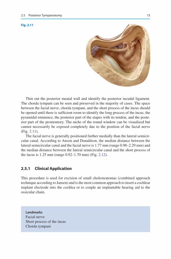
After performing a posterior tympanotomy, a pillar of bone caudal to the short pro-cess of the incus was originally preserved (the so-called buttress). Note the projection of the cochlea (Fig. 2.14). The buttress may protect the incus from damage whilst drilling (important to prevent noise damage when positioning implantable hearing aids) and is also used by some surgeons to fix a cohlear implant electrode to prevent extrusion. A posterior tympanotomy may be performed as described above.
Identify the head of the stapes and the edge of the round window niche. The site of the cochleostomy is determined by doubling the width of the stapes footplate in
Fig. 2.13
16 2 Basic Surgery of the Temporal Bone
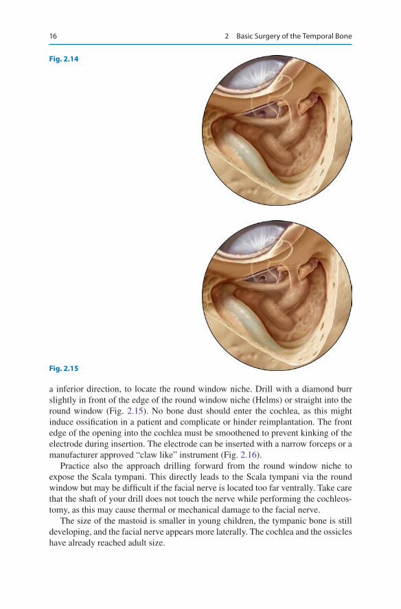
a inferior direction, to locate the round window niche. Drill with a diamond burr slightly in front of the edge of the round window niche (Helms) or straight into the round window (Fig. 2.15). No bone dust should enter the cochlea, as this might induce ossification in a patient and complicate or hinder reimplantation. The front edge of the opening into the cochlea must be smoothened to prevent kinking of the electrode during insertion. The electrode can be inserted with a narrow forceps or a manufacturer approved “claw like” instrument (Fig. 2.16).
Practice also the approach drilling forward from the round window niche to expose the Scala tympani. This directly leads to the Scala tympani via the round window but may be difficult if the facial nerve is located too far ventrally. Take care that the shaft of your drill does not touch the nerve while performing the cochleos-tomy, as this may cause thermal or mechanical damage to the facial nerve.
The size of the mastoid is smaller in young children, the tympanic bone is still developing, and the facial nerve appears more laterally. The cochlea and the ossicles have already reached adult size.
Fig. 2.14
Fig. 2.15
172.8 Endolymphatic Sac Surgery
2.7.1 Clinical Application
Severe hearing loss, when hearing aids cannot achieve sufficient hearing.
2.8 Endolymphatic Sac Surgery
The endolymphatic sac is a space within the dural sheets of the posterior cranial fossa. It is connected with the inner ear by the endolymphatic duct, leading to the vestibule. As it is part of the dura of the posterior fossa, it is usually removed with the dura when the temporal bone specimen is cleaned for preparation. However, the site of the duct, and the external aperture of the vestibular aqueduct can be observed.
After performing a mastoidectomy, identify the lateral and the posterior semicir-cular canals. Outline the sigmoid sinus preserving a thin bony cover. If the position of the lateral and posterior semicircular canal is uncertain, the labyrinthine bone can be removed until the blue lines of the canals are visualized. The perilymphatic space should not be opened. The position of the posterior semicircular canal should always
Fig. 2.16
LandmarksFacial nerveChorda tympaniHandle of malleusIncus, long process and bodyStapedius tendonRound window nicheRound windowScala tympani

18 2 Basic Surgery of the Temporal Bone
be identified by exposing the blue line. In a normally pneumatized mastoid, the facial nerve is close to the inferior part of the posterior canal. Identify the facial nerve without removing the covering bone completely.
Remove the bony coverage toward the posterior fossa until the dura (gray) and the sac (white) can be identified. A line from the lateral canal to the posterior fossa (Donaldson’s line) crosses the arch of the posterior canal in its centre. Remove the bone from the dura. The endolymphatic sac is found within the dura slightly under-neath this line where it meets the dura of the posterior fossa. Pushing the dura toward the posterior fossa, the duct can be seen entering its canal from the endolym-phatic sac (Fig. 2.17).
If the posterior canal is not too close to the posterior fossa, the beginning of the endolymphatic duct can be seen in the bone behind the posterior canal after remov-ing the cells between the posterior canal and the posterior fossa. The endolymphatic sac may be either decompressed or opened with a sickle knife, followed by the insertion of a silastic triangle or catheter. This last step is not possible in a temporal specimen where the dura of the posterior fossa has been removed (Fig. 2.18).
Fig. 2.18
Fig. 2.17

192.9 Epitympanotomy
2.8.1 Clinical Application
Endolymphatic sac surgery is performed to decompress endolymphatic pressure in patients with Menière’s disease.
2.9 Epitympanotomy
After removing the bone from the upper wall of the external auditory canal covering the incus and the head of the malleus, the position of the ossicles and the ligaments can be identified. Since cholesteatomas often extend into this region, the anatomy should be studied in detail (Fig. 2.19).
Fig. 2.19
LandmarksSigmoid or lateral sinusDura of the posterior fossaLateral semicircular canalBlue line of the posterior semicircular canalFacial nerveEndolymphatic duct

20 2 Basic Surgery of the Temporal Bone
2.9.1 Clinical Application
In cases of attic cholesteatoma, this is a safe way to remove disease, following the cholesteatoma sac. The epitympanotomy is the first step and may be sufficient in small cholesteatomas.
2.10 Open Mastoid Cavity
The radical mastoid cavity (canal wall down) was a classic operation before tym-panoplasty was established. It was an open cavity including an open middle ear, generally with removal of the malleus and incus for the treatment of cholesteatoma. Secretion from the open middle ear was often a problem. Today, removal of the disease is combined with closure of the middle ear by a tympanoplasty with or with-out reconstruction of the ossicular chain.
Remove the posterior external meatal wall. This is with minimal risk to the facial nerve, if the nerve is safely identified during the preceding steps. If not, remove the bone over the nerve with a diamond burr until it can be seen through a thin layer of bone. The floor of the external auditory canal must merge into the lower mastoid without irregularities. The roof of the external auditory canal must merge smoothly into the epitympanic roof. This facilitates cleaning of the cavity. A persisting partial posterior meatal wall (facial ridge) is the result of incomplete drilling and compli-cates postoperative care and wound healing. In cases of extensive pneumatization of the mastoid and the mastoid tip, you should remove the tip of the mastoid to reduce the size of the cavity. The so-called facial ridge does not contain the facial nerve (Fig. 2.20).
2.10.1 Clinical Application
A mastoid cavity and closure of the middle ear space with or without reconstruction of the ossicular chain is performed for the treatment of extended cholesteatomas. Alternatively preservation of the posterior meatal wall (canal wall-up technique and
LandmarksOval window with stapesFacial nerveRound windowCochleariform processMastoidal tubal orifice

212.11 Facial Nerve Decompression
posterior tympanotomy) or wall reconstruction after removal of disease may be considered.
2.11 Facial Nerve Decompression
Perform a complete exposure of the tympanic and mastoid part of the facial nerve, from the geniculate ganglion to the stylomastoid foramen, leaving a thin bony cov-ering on the nerve tissue. This bone is removed at the end of the procedure.
The facial nerve is found in front of the lateral semicircular canal slightly medial and arching down towards the stylomastoid foramen. Use a large diamond burr to remove bone until the nerve can be identified as a white structure under the bone. Drilling should proceed along the expected course of the nerve (arrows). For decom-pression, the thin bony layer can finally be elevated (Fig. 2.21).
Further decompression toward the geniculate ganglion in the second portion of the nerve can only be performed after removing the incus and the malleus (preserve the ossicles for later reconstruction exercises). Remove the posterior meatal wall. Now the nerve can be followed ventrally (anteriorly) to the lateral semicircular canal, above the oval window and the cochleariform process to the geniculate ganglion. Finally, remove the thin bony cover with a Plester knife exposing the
Fig. 2.20
LandmarksFacial nerveLateral semicircular canalBone covering the medial cranial fossaSigmoid or lateral sinusBone covering the posterior cranial fossaMiddle ear structures

22 2 Basic Surgery of the Temporal Bone
perineurium. Complete the procedure by incising the perineurium with a sickle knife. Identify the stapedius muscle. It is generally found underneath, and medial to the nerve in its mastoid portion (Fig. 2.22).
2.11.1 Clinical Application
Facial nerve decompression was mainly performed for the treatment of Bell’s palsy. Currently, it is performed to ensure the continuity of the nerve after trauma, for the removal of bone fragments or decompressing a perineural hematoma in the nerve canal.
Rerouting of the facial nerve is sometimes necessary to access structures medial to it e.g., in glomus tumour surgery. The nerve is exposed from the geniculate
Fig. 2.22
Fig. 2.21

232.12 Labyrinthectomy
ganglion to the parotid, and then carefully taken out of its canal and displaced for-ward in to the anterior middle ear. Prior to lifting the nerve out of its position, thin the bone 270° around the nerve and remove the bony covering (Fig. 2.22).
2.12 Labyrinthectomy
The initial steps have been described under the section on “Posterior Tympanotomy”.Remove the cells behind and around the lateral semicircular canal. The bone of
the canal is always solid and yellow. Behind the lateral canal and under the superior canal, identify the subarcuate fossa with the subarcuate artery in its centre. Continue the bony work until the posterior semicircular canal is sufficiently exposed. It is located more medial than the lateral canal, and is often separated from the posterior cranial fossa by a narrow tract of air cells. The inferior end might be close to the facial nerve. If the position of any semicircular canal is uncertain, the “blue lines” of the canals can be exposed but without opening the lumen.
Next, identify the superior semicircular canal (Fig. 2.23).
( A. subarcuata)Fig. 2.23
LandmarksTympanic portion of the facial nerveChochleariform processTubal orificeOval windowLateral semicircular canal

24 2 Basic Surgery of the Temporal Bone
The ampullae of the lateral and superior canal are situated close together under the body of the incus. They should not be opened at this point (Fig. 2.24).
The superior semicircular canal arches deep medially toward the posterior cranial fossa where it meets the posterior canal in the common crus. The beginner generally has difficulties following the superior semicircular canal backwards because a large amount of cellular bone has to be removed above the posterior part. At the end of this step, the semicircular canals should be sculptured clearly and the subarcuate fossa partly excavated.
Now open the perilymphatic space of the canals using the cutting burr. Their structures must be recognizable for as long as possible, to provide topographical orientation. First, the lateral semicircular canal is followed from the ampulla as far down as possible. The bone of the canal is always solid and yellowish. The facial nerve is situated in front of the canal. Some bone should be left for nerve protection. Behind the lateral canal and under the superior canal, the subarcuate fossa with the subarcuate artery in its centre can be identified when the posterior canal is opened. The inferior end might be close to the facial nerve. Follow it downwards where it meets the common crus, and upwards to its ampulla.
Now completely open the posterior semicircular canal, from its ampulla to the common crus. Finally, open the superior canal from its ampulla to the common crus. At this stage, the canals are open but not completely removed in order to study the three-dimensional anatomy of the temporal bone.
You may now perform stepwise removal of the semicircular canalsCompletely remove the lateral semicircular canal, leaving a bony protection for
the facial nerve. Open the vestibule. Drilling the posterior canal, the endolymphatic duct can be identified, leading to the vestibule. The exposure of the common crus is ensured.
The end branches of the vestibular nerve can be identified entering the lateral and superior ampulla cranially, and the inferior ampulla caudally. The ampulla of the superior canal protects the facial nerve underneath. The ampullae of the superior and posterior canals outline the fundus of the internal auditory canal.
( A. subarcuata)
Fig. 2.24

252.13 The Translabyrinthine Approach to the Internal Auditory Canal
2.12.1 Clinical Application
Labyrinthectomy is performed for labyrinthitis, vertigo, translabyrinthine neurectomy of the vestibular nerves and in the translabyrinthine approach for acoustic neuroma surgery.
2.13 The Translabyrinthine Approach to the Internal Auditory Canal
The first steps are mastoidectomy, identification of the semicircular canals, and stepwise labyrinthectomy as described above. The mastoid must be opened widely to give a good overview. Remove bone up to the middle cranial fossa and posteriorly toward the posterior fossa. A protruding sigmoid sinus can be pushed downward and a high jugular bulb can be lowered.
Exposure of the internal auditory canal requires extensive drilling resulting in a circumference around the internal auditory canal of about 300°. While the fundus is immediately under the vestibule, more than 1 cm of hard bone must be removed to expose the orifice to the posterior cranial fossa. Beginners usually do not expose the canal sufficiently (Fig. 2.25).
Fig. 2.25
LandmarksFacial nerveSemicircular canalsSubarcuate fossaAfter opening the canals: Facial nerveThe opened semicircular canalsAmpullae of the superior and inferior canalsEndolymphatic ductVestibule

26 2 Basic Surgery of the Temporal Bone
Opening procedure:Before removing the last thin layer of bone over the dura of the internal auditory
canal, the surrounding bone must be removed downward toward the jugular bulb. The cochlear aqueduct can often be identified as a landmark to protect the lower cranial nerve group. Cranially, the bone above the canal should be removed up to the middle fossa. Above and below the solid labyrinthine bone, you find softer bone with mastoid cells. The jugular bulb varies in its position. It is located medial to the facial nerve, and might even be found in the hypotympanum.
The facial nerve is found under the ampulla of the superior semicircular canal. It can be localized by thinning the bone underneath and towards the fundus. Identify the vertical crest (Bill’s bar) with the nerve entering its canal and open the dura after the internal canal has been exposed in its total length (Fig. 2.26, arrow).
The vertical crest is part of the opening for the entrance of the facial nerve into the temporal bone at the fundus of the inner auditory canal. The horizontal crest separates the superior and the inferior vestibular nerves.
If the nerves are not torn out during the removal of the specimen, the vestibular nerves can be removed after identifying the facial nerve (Fig. 2.27a, b).
2.13.1 Clinical Application
Vestibular vertigo and non-serviceable hearingApproach for translabyrinthine surgery of acoustic neuromas
Vertical crest
Fig. 2.26
LandmarksFacial nerveVertical crest – entrance of facial nerve into the temporal boneHorizontal crest

272.14 Transtemporal Approach to the Internal Auditory Canal (IAC)
2.14 Transtemporal Approach to the Internal Auditory Canal (IAC)
The transtemporal approach to the IAC is performed from the temporal plane. Identify the subarcuate eminence and the greater petrosal nerve leading to the genic-ulate ganglion. Expose the “blue line” of the superior semicircular canal that usually does not coincide with the eminence. Expect it to be up to about 10 mm distant. The superior semicircular canal and the internal auditory canal form an angle of approx-imately 60° opening toward the posterior fossa, varying between 34° and 75° (Fig. 2.28).
When drilling the superior semicircular canal, damage to the facial nerve and the cochlea must be avoided. Identify the superior petrosal sinus. The groove of the greater petrosal nerve leading to the geniculate ganglion and the arcuate eminence can be identified.
Air cells might be found above the superior semicircular canal before the yellow labyrinthine bone is seen. The “blue line” is identified in a longer stretch without fenestration of the canal. Finding the superior semicircular canal is difficult in a well-pneumatized temporal bone since air cells can be found above the semicircular canal. The cells have to be drilled away before the yellow labyrinthine bone is seen. Air cells can be identified by the exposure and course of “blue circles,” the superior canal by the appearance of the “blue line.”
a b
Vestibular nervesTransverse crest
Facial nerve
Vertical crest
Facial nerve
Cochlear nerve
Fig. 2.27

28 2 Basic Surgery of the Temporal Bone
Remove bone in a triangular area outlined by the superior petrosal sinus, the superior semicircular canal, and the 60° line from the anterior crus of the superior canal toward the posterior fossa. Take care not to fenestrate the superior canal and the cochlea. A considerable amount of bone has to be removed to reach the internal auditory canal. Its orifice is situated about 1.2 cm below the superior petrosal sinus. A wide exposure is possible. The internal auditory canal appears as a grey change in colour beneath the bone. The facial nerve is closer to the surface ascending from the internal auditory canal toward the geniculate ganglion. The canal must be identi-fied in its total length and must be exposed at least in 180° of its circumference before removeing the last layer of the remaining thin bone. In actual surgery, this technique avoids cerebrospinal fluid (CSF) displacing the nerves into the field of drilling.
Geniculate ganglion
Facial nerve
46°lsc
ssc
psc
Vestibular nerve
Cochlea
Greater superficialpetrosal nerve
Fig. 2.28

292.14 Transtemporal Approach to the Internal Auditory Canal (IAC)
Identify the nerves control the position of the facial nerve, and section the vestibular nerves (Fig. 2.29a, b).
2.14.1 Clinical Application
Vestibular vertigo with serviceable hearing•Small acoustic neuromas•
Superior vestibular nerve
a b
Vertical crest
Facial nerve
Fig. 2.29
LandmarksGreater petrosal nerveArcuate eminenceBlue line of the superior semicircular canal