Mangiferin from Salacia chinensis Prevents Oxidative Stress and Protects Pancreatic β -Cells in...
9
Mangiferin from Salacia chinensis Prevents Oxidative Stress and Protects Pancreatic b-Cells in Streptozotocin-Induced Diabetic Rats Periyar Selvam Sellamuthu, 1,2 Palanisamy Arulselvan, 3 Balu Periamallipatti Muniappan, 1 Sharida Fakurazi, 3,4 and Murugesan Kandasamy 1 1 Center for Advanced Studies in Botany, University of Madras, Chennai, India. 2 Department of Food Process Engineering, SRM University, Chennai, India. 3 Laboratory of Vaccines and Immunotherapeutics, Institute of Bioscience; 4 Department of Human Anatomy, Faculty of Medicine and Health Sciences; Universiti Putra Malaysia, Selangor, Malaysia. ABSTRACT Oxidative stress in diabetic tissues is a consequence of free radical accumulation with concurrently impaired natural antioxidants status and results in oxidative tissue damage. The present study investigated the protective effects of mangiferin against pancreatic b-cell damage and on the antioxidant defense systems in streptozotocin (STZ)-induced diabetic rats. Diabetes was experimentally induced by a single intraperitoneal injection of STZ. Oxidative stress biomarkers such as tissue malondialdehyde, hydroperoxides, reduced glutathione (GSH) content, and nonenzymatic antioxidants were measured. Biochemical observations were further substantiated with histological examination and ultrastructural studies in the pancreas of diabetic, glibenclamide and mangiferin-treated diabetic rats (dosage of 40mg/kg body weight daily for 30 days). Oral ad- ministration of mangiferin and glibenclamide to diabetic rats significantly decreased the level of blood glucoseand increased levels of insulin. Additionally, mangiferin treatment significantly modulated the pancreatic nonenzymatic antioxidants status (vitamin C, vitamin E, ceruloplasmin, and reduced GSH content) and other oxidative stress biomarkers. The histoarchitecture of diabetic rats showed degenerated pancreas with lower b-cell counts, but mangiferin treatment effectively regenerated insulin secreting islet cells. The electron microscopic study revealed damaged nuclear envelope and mitochondria and fewer secretory granules in pancreas of diabetic rats; however, mangiferin treatment nearly normalized pancreatic architecture. The present findings suggest that mangiferin treatment exerts a therapeutic protective nature in diabetes by decreasing oxidative stress and protecting against pancreatic b-cell damage, which may be attributable to its antioxidative properties. KEY WORDS: antioxidants natural products oxidative stress reduced glutathione thiobarbituric acid–reactive oxygen species INTRODUCTION D iabetes is an endocrine metabolic disorder character- ized by persistent elevated blood glucose levels and deficiencies in insulin action, secretion, or both. 1 Diabetes is one of the most important disease causes of morbidity and mortality and also considered as one of the five leading causes of death in the world, especially in Asian countries and the United States. Almost 150 million or more than 1.3% of the population suffer from diabetes throughout the world, which is about five times more than the estimates done 10 years ago; and moreover, this may be double by 2030. 2 Major consequences of high blood glucose are often due to oxidative stress, which is one of the mechanisms causing chronic diabetic complications. 3 Free radicals are highly reactive oxygen species and causes oxidative injury to the organisms by damaging macromolecules. In normal physi- ological conditions, there is an acute equilibrium in the generation of oxygen free radicals and antioxidant defense systems, which leads to deactivation of the free radicals and protects the organisms against free radical toxicity. 4 Mo- lecular and cellular tissue damages are mainly due to oxi- dative stress in a wide range of human diseases/disorders. 5,6 The treatment/management of diabetes is generally via diet control, exercise, and use of insulin and/or oral hypo- glycemic agents. Yet, they usually have reduced efficacy over time, are ineffective against long-term diabetic com- plications, and also very costly. 7 Because of perceived ef- fectiveness, fewer side effects in clinical trial experience, and relatively low cost, herbal drugs are a valuable source of natural medicines. 8 Although there are numerous known antidiabetic drugs available in the market, remedies from natural sources, es- pecially from the traditional medicinal plants have demon- strated efficacy for managing diabetes. 9,10 There are various kinds of modern drugs available to treat several ailments including diabetes; however, nearly 80% of the people all Manuscript received July 8, 2012. Revision accepted May 5, 2013. Address correspondence to: Periyar Selvam Sellamuthu, PhD, Lab No: 102, Centre for Advanced Studies in Botany, University of Madras, Chennai, 600025, India, E-mail: [email protected] JOURNAL OF MEDICINAL FOOD J Med Food 16 (8) 2013, 719–727 # Mary Ann Liebert, Inc., and Korean Society of Food Science and Nutrition DOI: 10.1089/jmf.2012.2480 719
Transcript of Mangiferin from Salacia chinensis Prevents Oxidative Stress and Protects Pancreatic β -Cells in...

Mangiferin from Salacia chinensis Prevents Oxidative Stressand Protects Pancreatic b-Cells in Streptozotocin-Induced Diabetic Rats
Periyar Selvam Sellamuthu,1,2 Palanisamy Arulselvan,3 Balu Periamallipatti Muniappan,1
Sharida Fakurazi,3,4 and Murugesan Kandasamy1
1Center for Advanced Studies in Botany, University of Madras, Chennai, India.2Department of Food Process Engineering, SRM University, Chennai, India.
3Laboratory of Vaccines and Immunotherapeutics, Institute of Bioscience; 4Department of Human Anatomy,Faculty of Medicine and Health Sciences; Universiti Putra Malaysia, Selangor, Malaysia.
ABSTRACT Oxidative stress in diabetic tissues is a consequence of free radical accumulation with concurrently impaired
natural antioxidants status and results in oxidative tissue damage. The present study investigated the protective effects of
mangiferin against pancreatic b-cell damage and on the antioxidant defense systems in streptozotocin (STZ)-induced diabetic
rats. Diabetes was experimentally induced by a single intraperitoneal injection of STZ. Oxidative stress biomarkers such as
tissue malondialdehyde, hydroperoxides, reduced glutathione (GSH) content, and nonenzymatic antioxidants were measured.
Biochemical observations were further substantiated with histological examination and ultrastructural studies in the pancreas of
diabetic, glibenclamide and mangiferin-treated diabetic rats (dosage of 40 mg/kg body weight daily for 30 days). Oral ad-
ministration of mangiferin and glibenclamide to diabetic rats significantly decreased the level of blood glucose and increased
levels of insulin. Additionally, mangiferin treatment significantly modulated the pancreatic nonenzymatic antioxidants status
(vitamin C, vitamin E, ceruloplasmin, and reduced GSH content) and other oxidative stress biomarkers. The histoarchitecture of
diabetic rats showed degenerated pancreas with lower b-cell counts, but mangiferin treatment effectively regenerated insulin
secreting islet cells. The electron microscopic study revealed damaged nuclear envelope and mitochondria and fewer secretory
granules in pancreas of diabetic rats; however, mangiferin treatment nearly normalized pancreatic architecture. The present
findings suggest that mangiferin treatment exerts a therapeutic protective nature in diabetes by decreasing oxidative stress and
protecting against pancreatic b-cell damage, which may be attributable to its antioxidative properties.
KEY WORDS: � antioxidants � natural products � oxidative stress � reduced glutathione � thiobarbituric acid–reactive
oxygen species
INTRODUCTION
D iabetes is an endocrine metabolic disorder character-ized by persistent elevated blood glucose levels and
deficiencies in insulin action, secretion, or both.1 Diabetes isone of the most important disease causes of morbidity andmortality and also considered as one of the five leadingcauses of death in the world, especially in Asian countriesand the United States. Almost 150 million or more than 1.3%of the population suffer from diabetes throughout the world,which is about five times more than the estimates done 10years ago; and moreover, this may be double by 2030.2
Major consequences of high blood glucose are often dueto oxidative stress, which is one of the mechanisms causingchronic diabetic complications.3 Free radicals are highlyreactive oxygen species and causes oxidative injury to the
organisms by damaging macromolecules. In normal physi-ological conditions, there is an acute equilibrium in thegeneration of oxygen free radicals and antioxidant defensesystems, which leads to deactivation of the free radicals andprotects the organisms against free radical toxicity.4 Mo-lecular and cellular tissue damages are mainly due to oxi-dative stress in a wide range of human diseases/disorders.5,6
The treatment/management of diabetes is generally viadiet control, exercise, and use of insulin and/or oral hypo-glycemic agents. Yet, they usually have reduced efficacyover time, are ineffective against long-term diabetic com-plications, and also very costly.7 Because of perceived ef-fectiveness, fewer side effects in clinical trial experience,and relatively low cost, herbal drugs are a valuable source ofnatural medicines.8
Although there are numerous known antidiabetic drugsavailable in the market, remedies from natural sources, es-pecially from the traditional medicinal plants have demon-strated efficacy for managing diabetes.9,10 There are variouskinds of modern drugs available to treat several ailmentsincluding diabetes; however, nearly 80% of the people all
Manuscript received July 8, 2012. Revision accepted May 5, 2013.
Address correspondence to: Periyar Selvam Sellamuthu, PhD, Lab No: 102, Centre forAdvanced Studies in Botany, University of Madras, Chennai, 600025, India, E-mail:[email protected]
JOURNAL OF MEDICINAL FOODJ Med Food 16 (8) 2013, 719–727# Mary Ann Liebert, Inc., and Korean Society of Food Science and NutritionDOI: 10.1089/jmf.2012.2480
719

over the world still rely on medicinal plants for their primaryhealth care.11 The World Health Organization (WHO) hasemphasized the rational usage of traditional and naturalindigenous medicines for treatment of diabetes mellitus.12
Focusing on these we have chosen one of the well-knowntraditional medicinal plants (Salacia chinensis) and its pri-mary bioactive compound (mangiferin) to investigate itsefficacy against chemically induced diabetes in rats.
S. chinensis Linn is an important medicinal plant be-longing to the family Hippocrateaceae and contains theactive phytochemical mangiferin in the root.13 Mangiferin isa C-glycosyl xanthone and traditionally used to treat manydiseases in India, and it is consequently reported to havevarious pharmacological effects.14 The effect of mangiferinmay be mediated by the stimulation of antioxidant path-ways, which decrease cellular oxidative stress in the im-mune system.15 Mangiferin acts as an effective antioxidantmolecule, and it has the potency to treat immunopatholog-ical disorders such as inflammatory diseases atherosclerosis,or septic shock.16 In addition, mangiferin has numerouseffects on macrophage function, comprising the inhibitionof phagocytic activity and free radical activity.16 Mangiferineffectively decreased blood glucose and increased insulinlevels through increased utilization of glucose in the liver.17
Currently, no systematic investigations exist in the scien-tific literature on the beneficial effect of mangiferin on pan-creatic tissue oxidative stress in streptozotocin (STZ)-induceddiabetes. The present study aimed to characterize the antiox-idant and protective nature of mangiferin on hyperglycemia-mediated lipid peroxidation (LPO), in STZ-induced diabeticrats. These beneficial effects were compared with the well-known antidiabetic agent, glibenclamide.
MATERIALS AND METHODS
Chemicals
STZ was obtained from Sigma Chemicals (St. Louis, MO,USA), stored at - 4�C, and protected from light. All otherchemicals were analytical grade.
Plant material and isolation of mangiferin
The S. chinensis roots were collected from Veenanga-puttu, Karumpakkam, Thangal, and Kurumpuram, Pu-ducherry, India. The plant material was identified by a planttaxonomist and has been deposited in the Center for Ad-vanced Studies in Botany (voucher specimen no: 778;University of Madras) for future reference. Isolation ofmangiferin was done by column chromatography as previ-ously described.13 The purity of mangiferin (99.4%) wasconfirmed through high performance liquid chromatographyby authentic standard (Sigma Aldrich, St. Louis, MO, USA).The structure of isolated mangiferin was characterized byNMR studies.
Animals
Wistar albino male rats weighing around 160–180 g wereprocured from the Tamil Nadu Veterinary and Animals
Sciences University, Chennai, India. Animals were accli-matized to standard animal house conditions for about 1week and given free access to standard rat chow (HindustanLever Ltd., Bangalore, India) and water. The animal ex-periments were conducted in accordance with the ethicalnorms approved by the Ministry of Social Justices andEmpowerment, Government of India and Institutional Ani-mal Ethics Committee Guidelines (IAEC No. 02/004/06).
Experimental protocol
Diabetes was induced in rats with a single intraperitonealinjection of freshly prepared solution of STZ (55 mg/kgbody weight) in 0.1 M cold citrate buffer, pH 4.5.17 The ratswere allowed to drink a 5% glucose solution overnight toovercome the drug-induced hypoglycemia. After allowing 1week for the development and aggravation of diabetes, theexperimental rats with moderate diabetes having persistentglycosuria and hyperglycemia (blood glucose range ofabove 250 mg/dL) were considered as diabetic rats and usedfor the experiment. The phytocompound and standard drugtreatment were started on the 8th day after STZ injection andthis was considered as the 1st day of treatment. A total of 24animals (18 diabetic rats and 6 control rats) were separatedinto four groups of six animals in each group as follows:
Group 1: Control ratsGroup 2: Diabetic control ratsGroup 3: Diabetic rats treated with mangiferin (40 mg/kg
body weight daily)Group 4: Diabetic rats treated with glibenclamide
(600 lg/kg body weight daily)
The animals were fasted overnight and then sacrificed bycervical decapitation after the end of the experimental pe-riod (30 days). Blood sample was collected in tubes con-taining EDTA for the estimation of glucose by O-toluidinemethod.18 The assay of plasma insulin was carried out byRIA assay kit according to the standard protocol (for rats)supplied by Linco Research, Inc., (St. Charles, MO, USA)and estimated vitamin C,19 vitamin E20, and ceruloplas-min.21 The pancreatic tissue from control and experimentalgroups was excised, rinsed in ice-cold saline, and homoge-nized in Tris-HCl buffer, pH 7.4 with a Teflon Homogenizerat 4�C. Protein content present in the tissue homogenate wasestimated by the method of Lowry et al.22
Concentrations of TBARS (thiobarbituric acid reactivesubstances) and hydroperoxides were estimated by themethod of Ohkawa et al.23 and reduced glutathione (GSH)was estimated by the method of Ellman24 in plasma andtissue homogenate.
Histological observation of pancreas
A slice of pancreatic tissue was fixed in 10% formalinsolution for histopathological analysis at room temperature.After fixation, tissues were dehydrated in a graded series ofethanol, cleared in xylene, and embedded in paraffin wax;solid sections were cut at 4 lm thickness using a rotarymicrotome for further staining process. Sections were
720 SELLAMUTHU ET AL.

stained for pancreatic b-cells by standard staining protocol.The stained sections were examined under light microscopeand photomicrographs were taken.
Transmission electron microscopic studies
A portion of pancreas (about 1 mm3) from each rat wasfixed in 3% glutaraldehyde and then postfixed in osmiumtetroxide and embedded in araldite (epoxy resin). One mi-cron sections were cut and then stained with toluidine blue.Suitable areas for ultrastructural study were chosen afterexamining one micron sections under a light microscope.The sections (60–90 nm) were cut on an LKBUM4 ultramicrotome using a diamond knife and sections weremounted on a copper grid and stained with uranyl acetateand Reynolds lead citrate.25 The grids were examined undera Phillips EM201C transmission electron microscope.
Statistical analysis
Results were expressed as mean – SD for six rats in eachgroup. All the grouped data were statistically evaluated withSPSS/10.0 software. Hypothesis testing methods included
one-way analysis of variance followed by least significantdifference test. Values were considered statistically signifi-cant at P < .05.
RESULTS
Identification of mangiferin
The high performance liquid chromatography analysisrevealed that authentic mangiferin had a retention value of5.883 (79.9%; Fig. 1A), while purified mangiferin had aretention value of 5.890 (80.04%; Fig. 1B). When comparedwith authentic mangiferin, the purified sample closely re-sembled the standard by 99.4%. The structure of mangiferinwas also characterized by spectral studies.
1H NMR spectrum analysis
The 1H NMR spectrum of authentic mangiferin and iso-lated compound are shown in Figure 2. The multiplet ap-pearing at 3.15–3.23 ppm are accounted for by the followingthree protons present in the mangiferin, (H-30), (H-40), and(H-50). The multiplet at 3.8 ppm accounted for the twoprotons (H-60). A double-doublet centered at 4.05 ppm aredue to the one proton (H-20). The doublet presented at4.59 ppm was due to (H-10) proton. The singlets at 6.37,
FIG. 1. (A) High performance liquid chromatography (HPLC)analysis of authentic mangiferin. (B) HPLC analysis of purifiedmangiferin from Salacia chinensis.
FIG. 2. (A) 1H NMR spectrum of authentic mangiferin. (B) 1HNMR spectrum of purified mangiferin from S. chinensis.
PANCREATIC TISSUE–PROTECTIVE NATURE OF MANGIFERIN 721
6.86, and 7.37 accounted for the protons (H-4), (H-5), and(H-8), respectively.
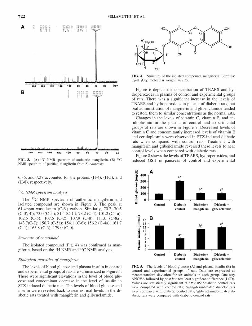
13C NMR spectrum analysis
The 13C NMR spectrum of authentic mangiferin andisolated compound are shown in Figure 3. The peak at61.4 ppm was due to (C-60) carbon. Similarly, 70.2, 70.5(C-30, 40); 73.0 (C-50); 81.4 (C-10); 73.2 (C-4), 101.2 (C-1a);102.5 (C-5); 107.5 (C-2); 107.9 (C-8); 111.6 (C-8a);143.7(C-7); 150.7 (C-5a); 154.1 (C-6); 156.2 (C-4a); 161.7(C-1); 163.8 (C-3); 179.0 (C-0).
Structure of compound
The isolated compound (Fig. 4) was confirmed as man-giferin, based on the 1H NMR and 13C NMR analysis.
Biological activities of mangiferin
The levels of blood glucose and plasma insulin in controland experimental groups of rats are summarized in Figure 5.There were significant elevations in the level of blood glu-cose and concomitant decrease in the level of insulin inSTZ-induced diabetic rats. The levels of blood glucose andinsulin were reverted back to near normal levels in the di-abetic rats treated with mangiferin and glibenclamide.
Figure 6 depicts the concentration of TBARS and hy-droperoxides in plasma of control and experimental groupsof rats. There was a significant increase in the levels ofTBARS and hydroperoxides in plasma of diabetic rats, butoral administration of mangiferin and glibenclamide tendedto restore them to similar concentrations as the normal rats.
Changes in the levels of vitamin C, vitamin E, and ce-ruloplasmin in the plasma of control and experimentalgroups of rats are shown in Figure 7. Decreased levels ofvitamin C and concomitantly increased levels of vitamin Eand ceruloplasmin were observed in STZ-induced diabeticrats when compared with control rats. Treatment withmangiferin and glibenclamide reversed these levels to nearcontrol levels when compared with diabetic rats.
Figure 8 shows the levels of TBARS, hydroperoxides, andreduced GSH in pancreas of control and experimentalFIG. 3. (A) 13C NMR spectrum of authentic mangiferin. (B) 13C
NMR spectrum of purified mangiferin from S. chinensis.
FIG. 4. Structure of the isolated compound, mangiferin. Formula:C19H18O11; molecular weight: 422.35.
FIG. 5. The levels of blood glucose (A) and plasma insulin (B) incontrol and experimental groups of rats. Data are expressed asmean – standard deviation for six animals in each group. One-wayANOVA followed by post hoc test least significant difference (LSD).Values are statistically significant at *P < .05: adiabetic control ratswere compared with control rats; bmangiferin-treated diabetic ratswere compared with diabetic control rats; cglibenclamide-treated di-abetic rats were compared with diabetic control rats.
722 SELLAMUTHU ET AL.
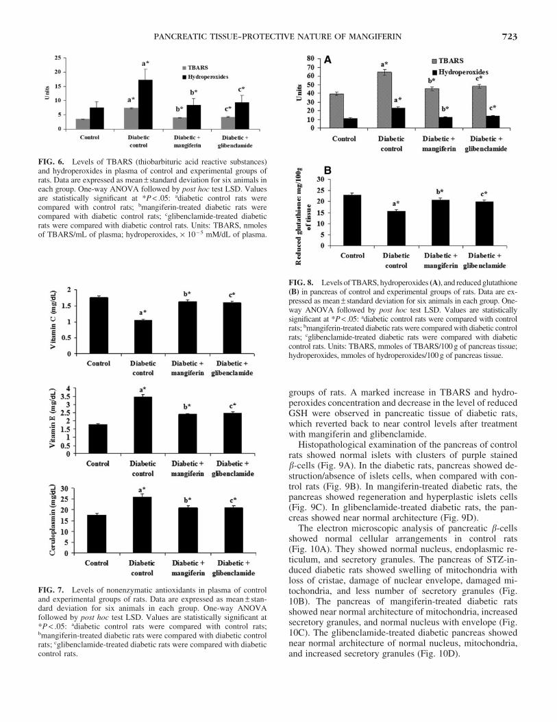
groups of rats. A marked increase in TBARS and hydro-peroxides concentration and decrease in the level of reducedGSH were observed in pancreatic tissue of diabetic rats,which reverted back to near control levels after treatmentwith mangiferin and glibenclamide.
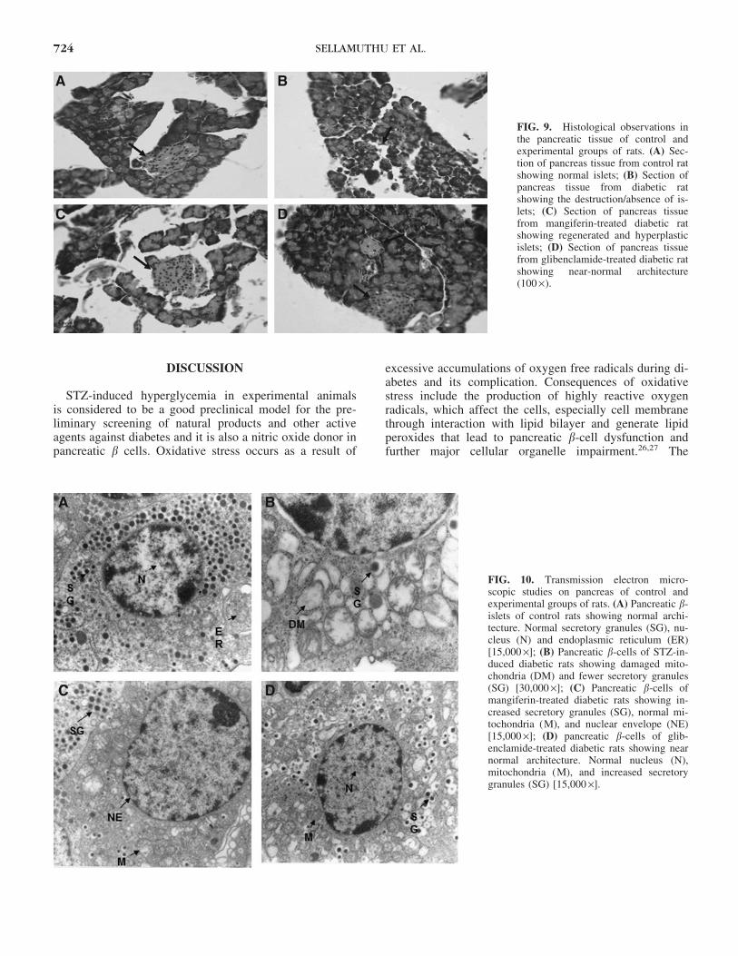
Histopathological examination of the pancreas of controlrats showed normal islets with clusters of purple stainedb-cells (Fig. 9A). In the diabetic rats, pancreas showed de-struction/absence of islets cells, when compared with con-trol rats (Fig. 9B). In mangiferin-treated diabetic rats, thepancreas showed regeneration and hyperplastic islets cells(Fig. 9C). In glibenclamide-treated diabetic rats, the pan-creas showed near normal architecture (Fig. 9D).
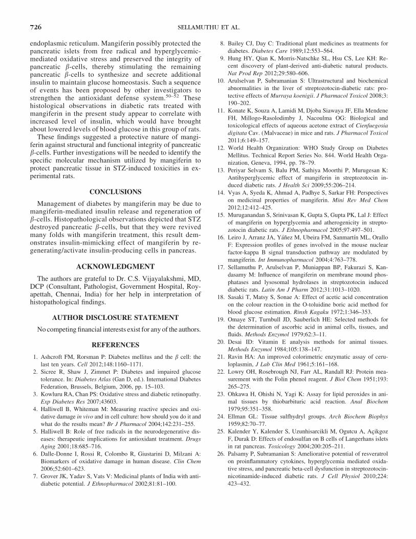
The electron microscopic analysis of pancreatic b-cellsshowed normal cellular arrangements in control rats(Fig. 10A). They showed normal nucleus, endoplasmic re-ticulum, and secretory granules. The pancreas of STZ-in-duced diabetic rats showed swelling of mitochondria withloss of cristae, damage of nuclear envelope, damaged mi-tochondria, and less number of secretory granules (Fig.10B). The pancreas of mangiferin-treated diabetic ratsshowed near normal architecture of mitochondria, increasedsecretory granules, and normal nucleus with envelope (Fig.10C). The glibenclamide-treated diabetic pancreas showednear normal architecture of normal nucleus, mitochondria,and increased secretory granules (Fig. 10D).
FIG. 7. Levels of nonenzymatic antioxidants in plasma of controland experimental groups of rats. Data are expressed as mean – stan-dard deviation for six animals in each group. One-way ANOVAfollowed by post hoc test LSD. Values are statistically significant at*P < .05: adiabetic control rats were compared with control rats;bmangiferin-treated diabetic rats were compared with diabetic controlrats; cglibenclamide-treated diabetic rats were compared with diabeticcontrol rats.
FIG. 8. Levels of TBARS, hydroperoxides (A), and reduced glutathione(B) in pancreas of control and experimental groups of rats. Data are ex-pressed as mean – standard deviation for six animals in each group. One-way ANOVA followed by post hoc test LSD. Values are statisticallysignificant at *P < .05: adiabetic control rats were compared with controlrats; bmangiferin-treated diabetic rats were compared with diabetic controlrats; cglibenclamide-treated diabetic rats were compared with diabeticcontrol rats. Units: TBARS, mmoles of TBARS/100 g of pancreas tissue;hydroperoxides, mmoles of hydroperoxides/100 g of pancreas tissue.
FIG. 6. Levels of TBARS (thiobarbituric acid reactive substances)and hydroperoxides in plasma of control and experimental groups ofrats. Data are expressed as mean – standard deviation for six animals ineach group. One-way ANOVA followed by post hoc test LSD. Valuesare statistically significant at *P < .05: adiabetic control rats werecompared with control rats; bmangiferin-treated diabetic rats werecompared with diabetic control rats; cglibenclamide-treated diabeticrats were compared with diabetic control rats. Units: TBARS, nmolesof TBARS/mL of plasma; hydroperoxides, · 10 - 5 mM/dL of plasma.
PANCREATIC TISSUE–PROTECTIVE NATURE OF MANGIFERIN 723
DISCUSSION
STZ-induced hyperglycemia in experimental animalsis considered to be a good preclinical model for the pre-liminary screening of natural products and other activeagents against diabetes and it is also a nitric oxide donor inpancreatic b cells. Oxidative stress occurs as a result of
excessive accumulations of oxygen free radicals during di-abetes and its complication. Consequences of oxidativestress include the production of highly reactive oxygenradicals, which affect the cells, especially cell membranethrough interaction with lipid bilayer and generate lipidperoxides that lead to pancreatic b-cell dysfunction andfurther major cellular organelle impairment.26,27 The
FIG. 9. Histological observations inthe pancreatic tissue of control andexperimental groups of rats. (A) Sec-tion of pancreas tissue from control ratshowing normal islets; (B) Section ofpancreas tissue from diabetic ratshowing the destruction/absence of is-lets; (C) Section of pancreas tissuefrom mangiferin-treated diabetic ratshowing regenerated and hyperplasticislets; (D) Section of pancreas tissuefrom glibenclamide-treated diabetic ratshowing near-normal architecture(100 ·).
FIG. 10. Transmission electron micro-scopic studies on pancreas of control andexperimental groups of rats. (A) Pancreatic b-islets of control rats showing normal archi-tecture. Normal secretory granules (SG), nu-cleus (N) and endoplasmic reticulum (ER)[15,000 ·]; (B) Pancreatic b-cells of STZ-in-duced diabetic rats showing damaged mito-chondria (DM) and fewer secretory granules(SG) [30,000 ·]; (C) Pancreatic b-cells ofmangiferin-treated diabetic rats showing in-creased secretory granules (SG), normal mi-tochondria (M), and nuclear envelope (NE)[15,000 ·]; (D) pancreatic b-cells of glib-enclamide-treated diabetic rats showing nearnormal architecture. Normal nucleus (N),mitochondria (M), and increased secretorygranules (SG) [15,000 ·].
724 SELLAMUTHU ET AL.

damage to b-cells is mainly due to nitric oxide and freeradicals during diabetes, because they decrease the activitiesof free radical scavenging enzymes.28
The treatment of diabetes with various natural extractsand their active compounds has been shown to effectivelydecrease hyperglycemia by decreasing free radical accu-mulation in experimental animals.29–31 Administration ofmangiferin exerted a significant anti-diabetic effect, proba-bly due to the stimulation of insulin secretion/action fromremnant pancreatic b-cells, which enhanced glucose utili-zation/metabolism in peripheral tissues of experimentallyinduced diabetic rats.32 Glibenclamide is a well-knownsecond-generation standard anti-hyperglycemic agent, be-longing to the class of sulfonylureas. The sulfonylureaagents are widely used for treating diabetes and its com-plications. It undergoes first-pass activity and the most fre-quently reported adverse effects are gastric disturbances likenausea, vomiting, anorexia, and increased appetite after oraltreatment.
STZ is effectively used for the induction of diabetesmellitus in experimental animals. It is postulated to inducediabetes by the degeneration and necrosis of b cells of pan-creatic islets, leading to the decline in insulin release.33 In thepresent investigation, we observed that increased oxidativestress in diabetes due to STZ-induction decreased the releaseof insulin secretion from pancreatic b-cells (Figs. 6–9).
Ascorbic acid is one of the water-soluble antioxidants thatinhibits oxidative damage in the cell membrane caused byaqueous radicals and facilitates the maintenance of vitaminE level at optimal concentrations.34 Alpha tocopherol (vi-tamin E) is the most important lipid soluble antioxidant andis involved in the sequence terminator of LPO and pro-tecting cellular structures from attack of free radicals.35 Inthe present study, significantly lower levels of vitamin C andincreased level of vitamin E were noted in plasma of un-treated diabetic rats compared with control rats, which areconsistent with those of earlier researchers.30,36 Oral ad-ministration of mangiferin significantly altered the levels ofvitamins C and E in STZ-induced diabetic rats comparedwith diabetic untreated rats. These findings suggest thatmangiferin effectively decreases oxidative stress throughenhancing nonenzymatic antioxidant status due to scav-enging of free radicals.
Ceruloplasmin is one of the most abundant proteins inplasma and consists of 95% of the total circulating copper innormal adults. In normal conditions, it acts as a potent freeradical scavenger that oxidizes iron from the ferrous state toferric state. Elevated levels of ceruloplasmin specifies thedegree of oxidative stress owing to its ferroxidase activityand also generation of oxygen products comprising hydro-gen peroxides.37,38 The decreased level of ceruloplasminwere observed in diabetic rats administered with isolatedmangiferin, which may be a tissue protective response to adecrease in circulating unbound Fe2 + and act as a knowninhibitor for further free radical-induced oxidative damageand associated mechanisms.
GSH is an abundant tripeptide nonenzymatic biologicalantioxidant present in the liver, kidney, and other vital tissues.
GSH plays a key role in the antioxidant defense system thatprotects the cellular system against the toxic effects of LPO. Itfunctions as a direct scavenger of free radicals and also as acosubstrate for peroxide detoxification through GSH peroxi-dases.39 The depletion of tissue GSH is due to either de-creased synthesis or increased deprivation of GSH byoxidative stress.40 A noticeable depletion in the GSH contentof pancreas was observed in diabetic rats. In the present study,it was observed that mangiferin was able to increase level ofGSH in comparison to the diabetic control rats (Fig. 8B).
LPO is a specific feature of chronic diabetes and affectsthe cell membrane fluidity and alters the membrane-boundenzymes activities and their receptors, leading to membranemalfunction.41 The secondary product of LPO is mal-ondialdehyde (MDA), which is an indicator of tissue dam-age.23,42 The observed elevation of MDA in diabeticuntreated rats in the present investigation possibly resultedfrom increased intensity of LPO arising due to increasedfree radical production, as reported earlier43 and also alteredmembrane-bound enzymes activities in STZ-induced dia-betic rats.18 Oral administration of mangiferin to diabeticrats brought about a significant decrease in the mean level ofLPO. Mangiferin possibly scavenges free radicals, therebystabilizing the endogenous antioxidant defense network anddecreasing the level of LPO, as has been investigated forother natural antioxidants such as quercetin,44 lycopene,45
and curcumin.46
Dietary natural supplements contribute to the preventionof diabetes and their complications by decreasing LPO andimproving antioxidant status. Li et al.47 reported that man-giferin significantly decreased LPO levels in kidney tissuesand improved their antioxidant status. Further, mangiferineffectively protects renal tissues from cytotoxin throughvarious antioxidant mechanisms. In the current investiga-tion, we observed MDA formation and the LPO index wassignificantly increased in pancreatic tissue of STZ-treatedanimals. Treatment with mangiferin potently abrogatedMDA levels, suggesting that mangiferin might act as anatural antioxidant principle to reduced oxidative damage.
The histological observation of pancreatic tissues pro-vided additional support that mangiferin has a protectivenature against oxidative damage. The STZ induction pro-duced severe pancreatic injury such as a decrease of pan-creatic islets diameter that was perhaps due to the decreasednumber of b cells. It is highly specific to b-cell toxicity andconsequently causes diabetes; hence it is widely used tostudy b-cell damage in in vivo animal experiments.48 STZmainly eliminates the b-cell response in pancreatic islets toglucose. A temporary return of responsiveness to glucosethen seems to be a permanent responsiveness, due to coin-cident damage of b-cells.49
In the present investigation, shrinkage and vacuoliza-tion of pancreatic islets and decreases in the b-cell masswere observed in pancreatic tissue of diabetic rats throughhistological and ultrastructural studies. However, oraladministration of mangiferin to diabetic rats appeared topreserve the remaining b-cell mass and to reduce vacuo-lization of pancreatic islets and swollen mitochondria and
PANCREATIC TISSUE–PROTECTIVE NATURE OF MANGIFERIN 725
endoplasmic reticulum. Mangiferin possibly protected thepancreatic islets from free radical and hyperglycemic-mediated oxidative stress and preserved the integrity ofpancreatic b-cells, thereby stimulating the remainingpancreatic b-cells to synthesize and secrete additionalinsulin to maintain glucose homeostasis. Such a sequenceof events has been proposed by other investigators tostrengthen the antioxidant defense system.50–52 Thesehistological observations in diabetic rats treated withmangiferin in the present study appear to correlate withincreased level of insulin, which would have broughtabout lowered levels of blood glucose in this group of rats.
These findings suggested a protective nature of mangi-ferin against structural and functional integrity of pancreaticb-cells. Further investigations will be needed to identify thespecific molecular mechanism utilized by mangiferin toprotect pancreatic tissue in STZ-induced toxicities in ex-perimental rats.
CONCLUSIONS
Management of diabetes by mangiferin may be due tomangiferin-mediated insulin release and regeneration ofb-cells. Histopathological observations depicted that STZdestroyed pancreatic b-cells, but that they were revivedmany folds with mangiferin treatment, this result dem-onstrates insulin-mimicking effect of mangiferin by re-generating/activate insulin-producing cells in pancreas.
ACKNOWLEDGMENT
The authors are grateful to Dr. C.S. Vijayalakshmi, MD,DCP (Consultant, Pathologist, Government Hospital, Roy-apettah, Chennai, India) for her help in interpretation ofhistopathological findings.
AUTHOR DISCLOSURE STATEMENT
No competing financial interests exist for any of the authors.
REFERENCES
1. Ashcroft FM, Rorsman P: Diabetes mellitus and the b cell: the
last ten years. Cell 2012;148:1160–1171.
2. Sicree R, Shaw J, Zimmet P: Diabetes and impaired glucose
tolerance. In: Diabetes Atlas (Gan D, ed.). International Diabetes
Federation, Brussels, Belgium, 2006, pp. 15–103.
3. Kowluru RA, Chan PS: Oxidative stress and diabetic retinopathy.
Exp Diabetes Res 2007;43603.
4. Halliwell B, Whiteman M: Measuring reactive species and oxi-
dative damage in vivo and in cell culture: how should you do it and
what do the results mean? Br J Pharmacol 2004;142:231–255.
5. Halliwell B: Role of free radicals in the neurodegenerative dis-
eases: therapeutic implications for antioxidant treatment. Drugs
Aging 2001;18:685–716.
6. Dalle-Donne I, Rossi R, Colombo R, Giustarini D, Milzani A:
Biomarkers of oxidative damage in human disease. Clin Chem
2006;52:601–623.
7. Grover JK, Yadav S, Vats V: Medicinal plants of India with anti-
diabetic potential. J Ethnopharmacol 2002;81:81–100.
8. Bailey CJ, Day C: Traditional plant medicines as treatments for
diabetes. Diabetes Care 1989;12:553–564.
9. Hung HY, Qian K, Morris-Natschke SL, Hsu CS, Lee KH: Re-
cent discovery of plant-derived anti-diabetic natural products.
Nat Prod Rep 2012;29:580–606.
10. Arulselvan P, Subramanian S: Ultrastructural and biochemical
abnormalities in the liver of streptozotocin-diabetic rats: pro-
tective effects of Murraya koenigii. J Pharmacol Toxicol 2008;3:
190–202.
11. Konate K, Souza A, Lamidi M, Djoba Siawaya JF, Ella Mendene
FH, Millogo-Rasolodimby J, Nacoulma OG: Biological and
toxicological effects of aqueous acetone extract of Cienfuegosia
digitata Cav. (Malvaceae) in mice and rats. J Pharmacol Toxicol
2011;6:149–157.
12. World Health Organization: WHO Study Group on Diabetes
Mellitus. Technical Report Series No. 844. World Health Orga-
nization, Geneva, 1994, pp. 78–79.
13. Periyar Selvam S, Balu PM, Sathiya Moorthi P, Murugesan K:
Antihyperglycemic effect of mangiferin in streptozotocin in-
duced diabetic rats. J Health Sci 2009;55:206–214.
14. Vyas A, Syeda K, Ahmad A, Padhye S, Sarkar FH: Perspectives
on medicinal properties of mangiferin. Mini Rev Med Chem
2012;12:412–425.
15. Muruganandan S, Srinivasan K, Gupta S, Gupta PK, Lal J: Effect
of mangiferin on hyperglycemia and atherogenicity in strepto-
zotocin diabetic rats. J Ethnopharmacol 2005;97:497–501.
16. Leiro J, Arranz JA, Yanez M, Ubeira FM, Sanmartın ML, Orallo
F: Expression profiles of genes involved in the mouse nuclear
factor-kappa B signal transduction pathway are modulated by
mangiferin. Int Immunopharmacol 2004;4:763–778.
17. Sellamuthu P, Arulselvan P, Muniappan BP, Fakurazi S, Kan-
dasamy M: Influence of mangiferin on membrane mound phos-
phatases and lysosomal hydrolases in streptozotocin induced
diabetic rats. Latin Am J Pharm 2012;31:1013–1020.
18. Sasaki T, Matsy S, Sonae A: Effect of acetic acid concentration
on the colour reaction in the O-toluidine boric acid method for
blood glucose estimation. Rinsh Kagaku 1972;1:346–353.
19. Omaye ST, Turnbull JD, Sauberlich HE: Selected methods for
the determination of ascorbic acid in animal cells, tissues, and
fluids. Methods Enzymol 1979;62:3–11.
20. Desai ID: Vitamin E analysis methods for animal tissues.
Methods Enzymol 1984;105:138–147.
21. Ravin HA: An improved colorimetric enzymatic assay of ceru-
loplasmin, J Lab Clin Med 1961;5:161–168.
22. Lowry OH, Rosebrough NJ, Farr AL, Randall RJ: Protein mea-
surement with the Folin phenol reagent. J Biol Chem 1951;193:
265–275.
23. Ohkawa H, Ohishi N, Yagi K: Assay for lipid peroxides in ani-
mal tissues by thiobarbituric acid reaction. Anal Biochem
1979;95:351–358.
24. Ellman GL: Tissue sulfhydryl groups. Arch Biochem Biophys
1959;82:70–77.
25. Kalender Y, Kalender S, Uzunhisarcikli M, Ogutcu A, Acikgoz
F, Durak D: Effects of endosulfan on B cells of Langerhans islets
in rat pancreas. Toxicology 2004;200:205–211.
26. Palsamy P, Subramanian S: Ameliorative potential of resveratrol
on proinflammatory cytokines, hyperglycemia mediated oxida-
tive stress, and pancreatic beta-cell dysfunction in streptozotocin-
nicotinamide-induced diabetic rats. J Cell Physiol 2010;224:
423–432.
726 SELLAMUTHU ET AL.
27. Tomlinson KC, Gardiner SM, Hebden RA, Bennett T: Functional
consequences of streptozotocin-induced diabetes mellitus, with
particular reference to the cardiovascular system. Pharmacol Rev
1992;44:103–150.
28. Spinas GA: The role of nitric oxide in b-cells. News Physiol Sci
1999;14:49–54.
29. Matough FA, Budin SB, Hamid ZA, Alwahaibi N, Mohamed J:
The role of oxidative stress and antioxidants in diabetic com-
plications. Sultan Qaboos Univ Med J 2012;12:5–18.
30. Arulselvan P, Subramanian SP: Beneficial effects of Murraya
koenigii leaves on antioxidant defense system and ultra structural
changes of pancreatic beta-cells in experimental diabetes in rats.
Chem Biol Interact 2007;165:155–164.
31. Subramanian S, Arulselvan P: Evaluation of hypolipidemic
properties of Murraya koenigii leaves studied in streptozotocin
induced diabetic rats. Biomedicine 2009;29:220–225.
32. Saravanan G, Ponmurugan P, Senthilkumar GP, Rajarajan T:
Antidiabetic properties of S-allylcysteine on streptozotocin-in-
duced diabetes in rats. J App Biomed 2009;7:151–159.
33. Kavalali G, Tuncel H, Goksel S, Hatemi HH: Hypoglycemic
activity of Urtica pilulifera in streptozotocin-diabetic rats.
J Ethnopharmacol 2003;84:241–245.
34. Thomas CE, McLean LR, Parker RA, Ohlweiler DF: Ascorbate
and phenolic antioxidant interactions in prevention of liposomal
oxidation. Lipids 1992;27:543–550.
35. Burton GW, Cheng SC, Webb A, Ingold KU: Vitamin E in young
and old human red blood cells. Biochim Biophys Acta 1986;860:
84–90.
36. Ravi K, Ramachandran B, Subramanian S: Effect of Eugenia
jambolana seed kernel on antioxidant defense system in strep-
tozotocin-induced diabetes in rats. Life Sci 2004;75:2717–2731.
37. Jung CH, Lee WJ, Yu JH, Hwang JY, Shin MS, Koh EH, Kim
MS, Park JY: Elevated serum ceruloplasmin levels are associated
with albuminuria in Korean men with type 2 diabetes mellitus.
Diabetes Res Clin Pract 2011;94:e3–e7.
38. Atanasiu RL, Stea D, Mateescu MA, Vergely C, Dalloz F, Briot
F, Maupoil V, Nadeau R, Rochette L: Direct evidence of caer-
uloplasmin antioxidant properties. Mol Cell Biochem 1998;189:
127–135.
39. Soon YY, Tan BK: Evaluation of the hypoglycemic and anti-
oxidant activities of Morinda officinalis in streptozotocin-in-
duced diabetic rats. Singapore Med J 2002;43:077–085.
40. Loven D, Schedl H, Wilson H, Daabees TT, Stegink LD, Diekus
M, Oberley L: Effect of insulin and oral glutathione on glutathione
levels and superoxide dismutase activities in organs of rats with
streptozocin-induced diabetes. Diabetes 1986;35:503–507.
41. Halliwell B: Lipid peroxidation, antioxidants and cardiovascular
disease: how should we move forward? Cardiovasc Res 2000;47:
410–418.
42. Shanmugam KR, Mallikarjuna K, Reddy KS: Effect of alcohol
on blood glucose and antioxidant enzymes in the liver and kidney
of diabetic rats. Indian J Pharmacol 2011;43:330–335.
43. Maritim AC, Sanders RA, Watkins JB 3rd: Diabetes, oxidative stress,
and antioxidants: a review. J Biochem Mol Toxicol 2003;17:24–38.
44. Coskun O, Kanter M, Korkmaz A, Oter S: Quercetin, a flavonoid
antioxidant, prevents and protects streptozotocin-induced oxida-
tive stress and beta-cell damage in rat pancreas. Pharmacol Res
2005;51:117–123.
45. Gao JX, Li Y, Zhang HY, He XL, Bai AS: Lycopene ameliorates
erectile dysfunction in streptozotocin-induced diabetic rats.
Pharmazie 2012;67:256–259.
46. Suryanarayana P, Satyanarayana A, Balakrishna N, Kumar PU,
Reddy GB: Effect of turmeric and curcumin on oxidative stress
and antioxidant enzymes in streptozotocin-induced diabetic rat.
Med Sci Monit 2007;13:BR286–BR292.
47. Li X, Cui X, Sun X, Li X, Zhu Q, Li W: Mangiferin prevents
diabetic nephropathy progression in streptozotocin-induced dia-
betic rats. Phytother Res 2010;24:893–899.
48. Bolaffai L, Nagamatsu S, Harris J, Grosdsky GM: Protection by
thymidine, an inhibitor of polyadenosine diphosphate ribosyla-
tion, of streptozotocin inhibition of insulin secretion. Endocrinol
1987;120:2117–2112.
49. West E, Simon OR, Mirrison EY: Streptozotocin alters pancre-
atic beta cell responsiveness to glucose within six hours of in-
jection into rats. W Ind Med J 1996;45:60–62.
50. Newsholme P, Rebelato E, Abdulkader F, Krause M, Carpinelli
A, Curi R: Reactive oxygen and nitrogen species generation,
antioxidant defenses, and b-cell function: a critical role for amino
acids. J Endocrinol 2012;214:11–20.
51. Kang KA, Kim JS, Zhang R, Piao MJ, Maeng YH, Kang MY,
Lee IK, Kim BJ, Hyun JW: KIOM-4 protects against oxidative
stress-induced mitochondrial damage in pancreatic b-cells via its
antioxidant effects. Evid Based Complement Alternat Med
2011;978682.
52. Sefi M, Fetoui H, Lachkar N, Tahraoui A, Lyoussi B, Boudawara
T, Zeghal N: Centaurium erythrea (Gentianaceae) leaf extract
alleviates streptozotocin-induced oxidative stress and b-cell
damage in rat pancreas. J Ethnopharmacol 2011;135:243–250.
PANCREATIC TISSUE–PROTECTIVE NATURE OF MANGIFERIN 727