Manganese-enhanced MRI of the mouse auditory pathway
3
Manganese-Enhanced MRI of the Mouse Auditory Pathway Takashi Watanabe, * Jens Frahm, and Thomas Michaelis Functional mapping of the lateral lemniscus and the superior oli- vary complex as part of the auditory pathway was accomplished for the first time in mice in vivo using manganese-enhanced MRI (2.35T, 3D FLASH, 117 m isotropic resolution). These and other auditory centers in the brainstem presented with pronounced sig- nal enhancements after systemic administration of manganese chloride when animals were exposed to acoustic stimuli for 48 hr, but not when kept in a quiet environment. The results indicate an activation-dependent accumulation of manganese in the neural circuit composed of the cochlear nucleus, the superior olivary complex, the lateral lemniscus, and the inferior colliculus. The marked enhancement of the lateral lemniscus suggests that the stimulus-related accumulation of manganese reflects not only a regional uptake from extracellular fluid but also a concurrent de- livery by axonal transport within the auditory system. Magn Reson Med 60:210 –212, 2008. © 2008 Wiley-Liss, Inc. Key words: auditory pathways; brain mapping; manganese chloride; magnetic resonance imaging Manganese-enhanced MRI is increasingly used for func- tional characterization of a wide variety of neural systems in animal brain (for a review, see Ref. (1)). More specifi- cally, a recent study mapped sound-evoked activity in the inferior colliculus of young mice during development (2). Because the inferior colliculus is only one (early) gate to the pathways processing auditory information in the mam- mal brain, the purpose of this study was to examine whether evoked activity can be observed in other struc- tures of the auditory system, for example, by exposing the animals to intense acoustic stimuli for a long period of time. According to anatomical data, the auditory pathway is composed of the cochlea, cochlear nucleus, superior olivary complex, lateral lemniscus, inferior colliculus, me- dial geniculate body, and auditory cortex (3). A schematic outline of the connectivity of these centers is sketched in Fig. 1 for input from the cochlea of one side (4). Hence, the primary aim was a more extensive functional mapping and morphological delineation of the auditory system in adult mice in vivo with use of local MRI signal enhancements that are due to the activation-dependent accumulation of manganese (Mn 2 ) ions. MATERIALS AND METHODS Animals Eight female mice (NMRI, 8 –12 weeks old, 28 –38 g) were studied in accordance with German animal protection laws after approval by the responsible governmental au- thority. Animals received manganese chloride (0.5 mmol/ kg body weight; Sigma, Taufkirchen, Germany) dissolved in distilled water via subcutaneous injection. They were returned to a chamber with unlimited access to food and water. For a period of 48 hr before MRI, four mice were kept in a quiet environment, while four animals were ex- posed to acoustic stimuli generated by an ultrasound wave generator (Conrad Electronic, Germany). The ultrasonic stim- uli involved noise bursts within a frequency range of 25– 65 kHz and an acoustic pressure of about 110 dB SPL. The relatively long duration of the acoustic stimulation used here was chosen because pilot experiments demon- strated a less pronounced signal enhancement in all audi- tory areas for shorter exposures (e.g., 24 hr). On the other hand, because long-term exposure to high-pressure noise might induce seizures, animals were tightly monitored during the stimulation period. No obvious abnormal be- havior was seen in either group. MRI For MRI the mice were anesthetized and placed in the magnet as previously described (5,6). All measurements were carried out at 2.35T using a 4.7/400 mm magnet (Magnex Scientific, Abingdon, UK) equipped with 200 mT m –1 gradients (Bruker Biospin MRI GmbH, Ettlingen, Ger- many). RF excitation and signal reception was accom- plished with use of a Helmholtz coil (inner diameter 100 mm) and an elliptical surface coil (inner diameter 20 14 mm), respectively. T 1 -weighted 3D MRI datasets (RF-spoiled 3D FLASH, TR/TE 17/7.6 ms, flip angle 25°, field of view 15 30 30 mm 3 , matrix 128 256 256, 5 averages, measuring time 93 min) were acquired at 117 m isotropic resolution. For quantitative evaluations the signal-to-noise ratio (SNR, defined as the mean MRI signal intensity of a brain region divided by the standard deviation of the noise) was deter- mined using software supplied by the manufacturer. The analysis followed a previously described strategy (5,6). Briefly, anatomical cross-sections were obtained by multipla- nar reconstructions from the original 3D MRI datasets. In close accordance with resolved anatomical structures, stan- dardized regions of interest (ROIs) were selected in the infe- rior colliculus, the lateral lemniscus, the superior olivary complex, the cochlear nucleus, and the cerebral cortex re- Biomedizinische NMR Forschungs GmbH am Max-Planck-Institut fu ¨ r bio- physikalische Chemie, Go ¨ ttingen, Germany. *Correspondence to: T. Watanabe, PhD, Biomedizinische NMR Forschungs GmbH, 37070 Go ¨ ttingen, Germany. E-mail: [email protected] Received 7 December 2007; revised 22 February 2008; accepted 12 March 2008. DOI 10.1002/mrm.21645 Published online in Wiley InterScience (www.interscience.wiley.com). 210 © 2008 Wiley-Liss, Inc. Magnetic Resonance in Medicine 60:210 –212 (2008) NOTES
-
Upload
takashi-watanabe -
Category
Documents
-
view
215 -
download
1
Transcript of Manganese-enhanced MRI of the mouse auditory pathway
Manganese-Enhanced MRI of the Mouse AuditoryPathway
Takashi Watanabe,* Jens Frahm, and Thomas Michaelis
Functional mapping of the lateral lemniscus and the superior oli-vary complex as part of the auditory pathway was accomplishedfor the first time in mice in vivo using manganese-enhanced MRI(2.35T, 3D FLASH, 117 �m isotropic resolution). These and otherauditory centers in the brainstem presented with pronounced sig-nal enhancements after systemic administration of manganesechloride when animals were exposed to acoustic stimuli for 48 hr,but not when kept in a quiet environment. The results indicate anactivation-dependent accumulation of manganese in the neuralcircuit composed of the cochlear nucleus, the superior olivarycomplex, the lateral lemniscus, and the inferior colliculus. Themarked enhancement of the lateral lemniscus suggests that thestimulus-related accumulation of manganese reflects not only aregional uptake from extracellular fluid but also a concurrent de-livery by axonal transport within the auditory system. MagnReson Med 60:210–212, 2008. © 2008 Wiley-Liss, Inc.
Key words: auditory pathways; brain mapping; manganesechloride; magnetic resonance imaging
Manganese-enhanced MRI is increasingly used for func-tional characterization of a wide variety of neural systemsin animal brain (for a review, see Ref. (1)). More specifi-cally, a recent study mapped sound-evoked activity in theinferior colliculus of young mice during development (2).Because the inferior colliculus is only one (early) gate tothe pathways processing auditory information in the mam-mal brain, the purpose of this study was to examinewhether evoked activity can be observed in other struc-tures of the auditory system, for example, by exposing theanimals to intense acoustic stimuli for a long period oftime. According to anatomical data, the auditory pathwayis composed of the cochlea, cochlear nucleus, superiorolivary complex, lateral lemniscus, inferior colliculus, me-dial geniculate body, and auditory cortex (3). A schematicoutline of the connectivity of these centers is sketched inFig. 1 for input from the cochlea of one side (4). Hence, theprimary aim was a more extensive functional mapping andmorphological delineation of the auditory system in adultmice in vivo with use of local MRI signal enhancementsthat are due to the activation-dependent accumulation ofmanganese (Mn2�) ions.
MATERIALS AND METHODS
Animals
Eight female mice (NMRI, 8–12 weeks old, 28–38 g) werestudied in accordance with German animal protectionlaws after approval by the responsible governmental au-thority. Animals received manganese chloride (0.5 mmol/kg body weight; Sigma, Taufkirchen, Germany) dissolvedin distilled water via subcutaneous injection. They werereturned to a chamber with unlimited access to food andwater. For a period of 48 hr before MRI, four mice werekept in a quiet environment, while four animals were ex-posed to acoustic stimuli generated by an ultrasound wavegenerator (Conrad Electronic, Germany). The ultrasonic stim-uli involved noise bursts within a frequency range of 25–65 kHz and an acoustic pressure of about 110 dB SPL.
The relatively long duration of the acoustic stimulationused here was chosen because pilot experiments demon-strated a less pronounced signal enhancement in all audi-tory areas for shorter exposures (e.g., 24 hr). On the otherhand, because long-term exposure to high-pressure noisemight induce seizures, animals were tightly monitoredduring the stimulation period. No obvious abnormal be-havior was seen in either group.
MRI
For MRI the mice were anesthetized and placed in themagnet as previously described (5,6). All measurementswere carried out at 2.35T using a 4.7/400 mm magnet(Magnex Scientific, Abingdon, UK) equipped with 200 mTm–1 gradients (Bruker Biospin MRI GmbH, Ettlingen, Ger-many). RF excitation and signal reception was accom-plished with use of a Helmholtz coil (inner diameter100 mm) and an elliptical surface coil (inner diameter20 � 14 mm), respectively. T1-weighted 3D MRI datasets(RF-spoiled 3D FLASH, TR/TE � 17/7.6 ms, flip angle 25°,field of view 15 � 30 � 30 mm3, matrix 128 � 256 � 256,5 averages, measuring time 93 min) were acquired at117 �m isotropic resolution.
For quantitative evaluations the signal-to-noise ratio (SNR,defined as the mean MRI signal intensity of a brain regiondivided by the standard deviation of the noise) was deter-mined using software supplied by the manufacturer. Theanalysis followed a previously described strategy (5,6).Briefly, anatomical cross-sections were obtained by multipla-nar reconstructions from the original 3D MRI datasets. Inclose accordance with resolved anatomical structures, stan-dardized regions of interest (ROIs) were selected in the infe-rior colliculus, the lateral lemniscus, the superior olivarycomplex, the cochlear nucleus, and the cerebral cortex re-
Biomedizinische NMR Forschungs GmbH am Max-Planck-Institut fur bio-physikalische Chemie, Gottingen, Germany.*Correspondence to: T. Watanabe, PhD, Biomedizinische NMR ForschungsGmbH, 37070 Gottingen, Germany. E-mail: [email protected] 7 December 2007; revised 22 February 2008; accepted 12 March2008.DOI 10.1002/mrm.21645Published online in Wiley InterScience (www.interscience.wiley.com).
210© 2008 Wiley-Liss, Inc.
Magnetic Resonance in Medicine 60:210–212 (2008)NOTES

mote from the auditory cortex. The caudal surface of theinferior colliculus was chosen as a reference plane follow-ing the histological convention for the mouse auditorysystem (3).
RESULTS
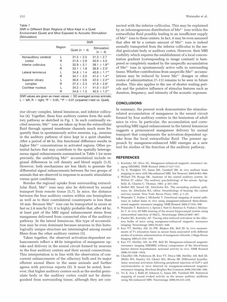
As reported previously (1,2,5), systemic administration ofmanganese led to a generalized signal enhancement in thebrain of all mice. While there were similar enhancementsfor the two groups (quiet vs. stimulation) in the forebrain,the brainstem revealed marked differences. First of all,strong differential—that is, stimulus-related—enhance-ments were observed in the ventral part of the inferiorcolliculus, which were most prominent about 1.5 mmlateral from the midline, as shown in Fig. 2a. Below theinferior colliculus, as shown in coronal sections in Fig. 2b,even more pronounced stimulus-related enhancementswere observed as a curved band. In agreement with histol-ogy (3), this enhanced structure represents the lateral lem-niscus. The band can be tracked further ventrally until itends in another enhanced structure located on the ventralsurface of the pons, as shown in Fig. 2c. It represents thesuperior olivary complex (3). Lastly, in Fig. 2d the co-chlear nucleus can be more clearly delineated in stimu-lated animals in comparison to mice kept in quiet condi-tions. These qualitative observations in all animals of ei-ther group were confirmed by a quantitative evaluation ofselected ROIs summarized in Table 1. While there is nodifference in SNR between groups in nonauditory cerebralcortex, the SNR of various auditory structures in stimu-lated mice is statistically significantly higher than in ani-mals kept in a quiet environment.
DISCUSSION
The present work demonstrates morphological and func-tional characteristics of the lateral lemniscus and the su-perior olivary complex in the mouse brain in vivo in anunprecedented way. These structures are important com-ponents of the auditory pathway, which hitherto have onlybeen described by histology, that is, in brain slices post-
mortem (3). In electrophysiology, these structures havebeen much less intensively studied than the inferior col-liculus, which is located just beneath the skull and there-fore offers convenient experimental access. Manganese-enhanced MRI now offers an alternative in vivo approachwhich is expected to add complementary information tohistology and electrophysiology when performing func-tional investigations of mouse models with disorders ormutations affecting the auditory pathway.
In agreement with manganese-enhanced MRI of devel-oping mice (2), the present results demonstrate an MRIsignal enhancement of the inferior colliculus due to thelocal accumulation of Mn2� ions. In addition, however, thedata reveal a stimulus-related enhancement in the laterallemniscus and the superior olivary complex. Apart fromthe use of adult animals, this new finding may be ascribedto the long exposure to intense acoustic stimuli. When thecochlea is stimulated, neurons in the cochlear nucleus areexcited via the spiral ganglion cells. Subsequently, theexcitation expands to the neurons in the connected supe-
FIG. 1. Schematic outline of the auditory system. The ascendingpathway from the cochlea of one side to the main auditory centerscomprises major (thick lines), moderate (thin), and minor projections(dashed). AC � auditory cortex, C � cochlea, CN � cochlearnucleus, IC � inferior colliculus, LL � lateral lemniscus, MGB �medial geniculate body, SOC � superior olivary complex.
FIG. 2. (Right) Stimulus-related Mn2�-induced MRI signal enhance-ments in mice exposed to acoustic stimulation in comparison to (left)animals kept in a quiet environment (data from eight different animals,scale bars � 1 mm). The regions-of-interest used for quantitativeevaluations are indicated by dotted circles. a: Left-hemispheric para-sagittal sections 1.5 mm from the midline identify the ventral part of theinferior colliculus (IC, arrow) and demonstrate otherwise similar en-hancements of the lateral olfactory tract (arrowhead) and olfactorybulb. b: Coronal sections 2.0 mm rostral from the caudal surface of theIC identify the IC (arrow) and lateral lemniscus (LL, arrowhead). c:Oblique sagittal sections cutting through the LL identify the LL (arrow-head) and its connection to the superior olivary complex (SOC, arrow)in a curved band. d: Coronal sections cutting through the SOC identifythe SOC (arrow) and the cochlear nucleus (CN, arrowhead).
Manganese-Enhanced MRI of Mouse Auditory Pathway 211
rior olivary complex, lateral lemniscus, and inferior collicu-lus (4). Together, these four auditory centers form the audi-tory pathway as sketched in Fig. 1. In such continually ex-cited neurons, Mn2� ions are taken up from the extracellularfluid through opened membrane channels much more fre-quently than in spontaneously active neurons, e.g., neuronsin the auditory pathway of mice kept in a quiet chamber.After a period of 48 hr, this functional difference results inhigher Mn2� concentrations in activated regions. Other po-tential factors that may contribute to the spatially heteroge-neous signal enhancements summarized in Table 1 (or moreprecisely, the underlying Mn2� accumulation) include re-gional differences in cell density and blood supply (1,5).However, both mechanisms are less likely to generate thedifferential signal enhancements between the two groups ofanimals that are observed in response to acoustic stimulationversus quiet conditions.
Besides the regional uptake from surrounding extracel-lular fluid, Mn2� ions may also be delivered by axonaltransport from remote tissue (5,7). In mice, the distancebetween the four auditory centers within one hemisphereas well as to their contralateral counterparts is less than10 mm. Because Mn2� ions can be transported in axons asfast as 2.8 mm/hr (5), it is highly probable that, after 48 hr,at least part of the MRI signal enhancement stems frommanganese delivered from connected sites of the auditorypathway. In the lateral lemniscus, this type of accumula-tion may be even dominant because neurons in this histo-logically unique structure are intermingled among axonalfibers from the other auditory centers (3).
Taken together, the observed activation-dependent en-hancements reflect a 48-hr integration of manganese up-take and delivery in the neural circuit formed by neuronsin the four auditory centers and their axonal connections.This interpretation is in line with the observation of con-current enhancements of the olfactory bulb and its majorefferent axonal fibers in the same animals and furtheragrees with previous reports (5). It must be noted, how-ever, that higher auditory centers such as the medial genic-ulate body or the auditory cortex could not be distin-guished from surrounding tissue, although they are con-
nected with the inferior colliculus. This may be explainedby an inhomogeneous distribution of Mn2� ions within theextracellular fluid possibly leading to an insufficient supplyof Mn2� ions to these centers. In fact, it may be even assumedthat after 48 hr a certain amount of Mn2� ions is indeedaxonally transported from the inferior colliculus to the me-dial geniculate body or auditory cortex. However, their MRIvisibility which requires the establishment of a local concen-tration gradient (corresponding to image contrast) is ham-pered or completely masked by the unspecific accumulationof Mn2� ions in spontaneously active cells in surroundingtissue. Whether contributions from unspecific Mn2� accumu-lations may be reduced by lower Mn2� dosages or otherroutes of administration (7–11) remains to be seen in futurestudies. This also applies to the use of shorter waiting peri-ods and the putative influence of stimulus features such asduration, frequency, and intensity of the acoustic exposure.
CONCLUSIONS
In summary, the present work demonstrates the stimulus-related accumulation of manganese in the neural circuitformed by four auditory centers in the brainstem of adultmice in vivo. In particular, the accumulation and corre-sponding MRI signal enhancement in the lateral lemniscussuggests a pronounced manganese delivery by axonaltransport that complements the activation-dependent up-take from the local extracellular fluid. The present ap-proach by manganese-enhanced MRI emerges as a newtool for studies of the function of the auditory pathway.
REFERENCES
1. Koretsky AP, Silva AC. Manganese-enhanced magnetic resonance im-aging (MEMRI). NMR Biomed 2004;17:527–531.
2. Yu X, Wadghiri YZ, Sanes DH, Turnbull DH. In vivo auditory brainmapping in mice with Mn-enhanced MRI. Nat Neurosci 2005;8:961–968.
3. Willard TH, Ryugo DK. Anatomy of the central auditory system. In:Willott JT, editor. The auditory psychobiology of the mouse. Spring-field, IL: Charles C. Thomas; 1983. p 201–303.
4. Helfert RH, Snead CR, Altschuler RA. The ascending auditory path-ways. In: Altschuler RA, editor. Neurobiology of hearing: the centralnervous system. New York: Raven Press; 1991. p 1–25.
5. Watanabe T, Frahm J, Michaelis T. Functional mapping of neural path-ways in rodent brain in vivo using manganese-enhanced three-dimen-sional magnetic resonance imaging. NMR Biomed 2004;17:554–568.
6. Watanabe T, Radulovic J, Spiess J, Natt O, Boretius S, Frahm J, Michae-lis T. In vivo 3D MRI staining of the mouse hippocampal system usingintracerebral injection of MnCl2. Neuroimage 2004;22:860–867.
7. Pautler RG, Koretsky AP. Tracing odor-induced activation in the olfac-tory bulbs of mice using manganese-enhanced magnetic resonanceimaging. Neuroimage 2002;16:441–448.
8. Kuo YT, Herlihy AH, So PW, Bhakoo KK, Bell JD. In vivo measure-ments of T1 relaxation times in mouse brain associated with differentmodes of systemic administration of manganese chloride. J Magn ResonImaging 2005;21:334–339.
9. Kuo YT, Herlihy AH, So PW, Bell JD. Manganese-enhanced magneticresonance imaging (MEMRI) without compromise of the blood-brainbarrier detects hypothalamic neuronal activity in vivo. NMR Biomed2006;19:1028–1034.
10. Chaudhri OB, Parkinson JR, Kuo YT, Druce MR, Herlihy AH, Bell JD,Dhillo WS, Stanley SA, Ghatei MA, Bloom SR. Differential hypotha-lamic neuronal activation following peripheral injection of GLP-1 andoxyntomodulin in mice detected by manganese-enhanced magneticresonance imaging. Biochem Biophys Res Commun 2006;350:298–306.
11. Yu X, Zou J, Babb JS, Johnson G, Sanes DH, Turnbull DH. Statisticalmapping of sound evoked activity in the mouse auditory midbrainusing Mn-enhanced MRI. Neuroimage 2008;39:223–230.
Table 1SNR in Different Brain Regions of Mice Kept in a QuietEnvironment (Quiet) and Mice Exposed to Acoustic Stimulation(Stimulation)
Region
SNR
Quiet (n � 4)Stimulation
(n � 4)
Nonauditory cerebralcortex
L 31.5 � 2.5 31.3 � 2.7R 31.6 � 2.9 30.9 � 4.0
Interior colliculus L 32.9 � 2.1 38.1 � 1.8**R 33.1 � 1.8 38.8 � 2.5*
Lateral lemniscus L 34.8 � 1.4 40.9 � 1.1**R 34.7 � 2.0 41.4 � 1.4**
Superior olivarycomplex
L 36.6 � 0.6 42.0 � 2.2**R 37.4 � 2.3 41.8 � 2.6*
Cochlear nucleus L 34.2 � 1.1 41.0 � 0.5**R 34.6 � 1.3 40.5 � 1.2**
SNR values are given as mean values � SD averaged across animals;L � left, R � right; *P � 0.05, **P � 0.01 (unpaired t-test vs. Quiet).
212 Watanabe et al.