
Mandibular nerve
20
Mandibular Nerve www.facebook.com/notesdental
-
Upload
deepak-kumar-gupta -
Category
Health & Medicine
-
view
141 -
download
2
Transcript of Mandibular nerve
Mandibular Nerve
www.facebook.com/notesdental
Introduction
• Its the largest branch of trigeminal nerve. It is a mixed nerve with two roots : – large sensory root– small motor root
• The sensory root of the mandibular division originates at the inferior angle of the trigeminal ganglion
• The motor arises in the motor cells located in the pons & medulla oblongata.
• The two roots emerge from the cranium separately through the foramen ovale , the motor root lying medial to sensory .
• They unite just outside the skull and form the main trunk of the third division .
• This trunk remains undivided for only 2 to 3 mm before it splits into – a small anterior– a large posterior division
www.facebook.com/notesdental
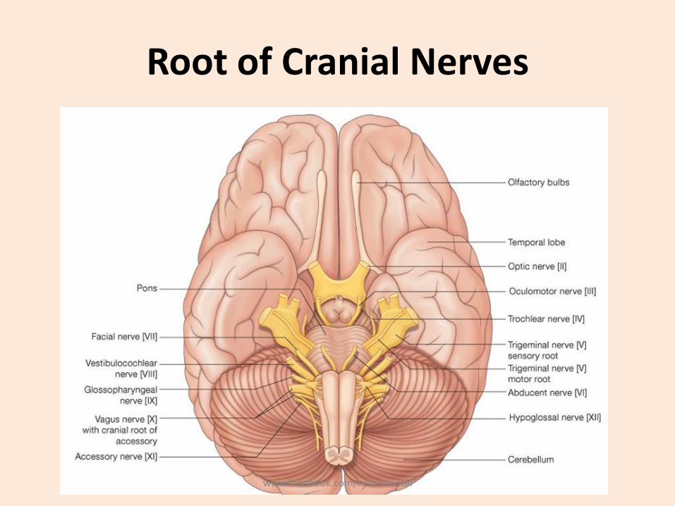
Root of Cranial Nerves
www.facebook.com/notesdental
Mandibular Nerve: Supply
• It supplies – the teeth and gums of the mandible,
– the skin of the temporal region,
– the auricula,
– the lower lip,
– the lower part of the face,
– muscles of mastication;
– it also supplies the mucous membrane of the anterior two-thirds of the tongue
www.facebook.com/notesdental

www.facebook.com/notesdental
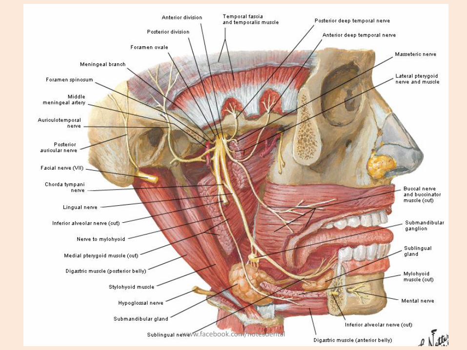
Branches from Undivided Nerve
• NERVOUS SPINOSUS– It arises outside the skull
– then passes into the middle cranial fossa to supply the dura & mastoid cells.
• NERVE TO MEDIAL PTERYGOID MUSCLE – A branch of the motor root passes to innervate
the medial pterygoid muscle.
– This branch passes without interruption to inervate the tensor veli palatini and the tensor tympani muscles.
www.facebook.com/notesdental

Anterior Division
• Nerve to lateral pterygoid muscle:The pterygoid nerve enters the medial side of the lateral pterygoid muscle to provide its motor nerve supply.
• Nerve to masseter muscle: the masseter nerve passes above the lateral pterygoid to transverse the mandibularnotch.
• Nerve to temporal muscles : its again divided in 2 branch
– Anterior deep temporal nerve - It supplies deep part of the temporal the anterior portion of the temporal muscle.
– Posterior deep temporal nerve- It passes upwards to the deep part of the temporal muscle.
www.facebook.com/notesdental
Anterior Division
• Long buccal nerve- it passes downward, anteriorly and laterally between the two heads of lateral pterygoid muscle. It supplies
– Buccinator muscle
– Mucous membrane of cheek
www.facebook.com/notesdental
www.facebook.com/notesdental
POSTERIOR DIVISION
• It is mainly sensory but also carries some motor components. This division extends downwards and medially and then branches into:
– Auriculotemporal nerve
– Lingual nerve
– Inferior alveolar nerve
www.facebook.com/notesdental
Auriculotemporal nerve
• it arises by a medial and lateral root.
• These roots embrance the middle meningealartery and unite behind the artery just below the foramen spinosum.
• It passes with superficial temporal artery in its upward course and divides into numerous branches – Tragus of the pinna of external ear ,
– Scalp about the ear
– as far as upward as the vertex of the skull
www.facebook.com/notesdental
Lingual nerve
• It first passes medially to lateral pterygoid muscle
• As it decends , lies between the internal pterygoidmuscle and the ramus of the mandible.
• The nerve lies parallel to the inferior alveolar nerve but medial and anterior to it .
• It then passes deep to reach the side of the base of the tongue.
• At the side of the tongue it lies below the lateral lingual sulcus.
• It has communications with the chorda tympani of facial nerve
www.facebook.com/notesdental
Inferior alveolar nerve
• It is the largest branch of the posterior division of mandibular part of the trigeminal nerve.
• It descends with the inferior alveolar artery, at first beneath the Pterygoideus plexus,
• Then between the sphenomandibular ligament and the ramus of the mandible to the mandibular foramen.
• It then passes forward in the mandibular canal, beneath the teeth, as far as the mental foramen, where it divides into two terminal branches, – incisive and mental.
• The branches of the inferior alveolar nerve are the mylohyoid, dental, incisive, and mental.
www.facebook.com/notesdental
Inferior alveolar nerve
• Mylohyoid nerve– derived from the inferior alveolar just before it enters the mandibular
foramen. – It descends in a groove on the deep surface of the ramus of the
mandible,– Reaching the under surface of the Mylohyoideus supplies this muscle
and the anterior belly of the digastric
• Dental branches– Supply the molar and premolar teeth. – They correspond in number to the roots of those teeth; – each nerve entering the orifice at the point of the root, and supplying
the pulp of the tooth;– above the alveolar nerve they form an inferior dental plexus.– This divideds in to 2 branch i.e incisive nerve and mental nerve
www.facebook.com/notesdental
Inferior alveolar nerve
• Incisive branch• Continuation of dental nerve - onward within the bone, and
supplies the canine and incisor teeth
• Mental nerve (n. mentalis) – Emerges at the mental foramen,– Divides beneath the Triangularis muscle into three
branches; • one descends to the skin of the chin• two ascend to the skin and mucous membrane of the lower
lip
– These branches communicate freely with the facial nerve
www.facebook.com/notesdental
Otic Ganglion (ganglion oticum)
• The otic ganglion is a small, ovalshaped, flattened ganglion of a reddish-gray color,
• Situated immediately below the foramen ovale; • Lies on the medial surface of the mandibular nerve,
and surrounds the origin of the nerve to medial pterygoid
• It is in relation, – Laterally, with the trunk of the mandibular nerve at the
point where the motor and sensory roots join; – Medially, with the cartilaginous part of the auditory tube,
and the origin of the Tensor veli palatini;– Posteriorly, with the middle meningeal artery.
www.facebook.com/notesdental
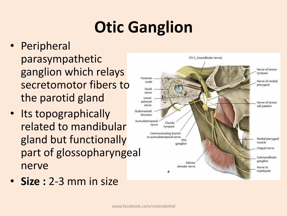
Otic Ganglion• Peripheral
parasympathetic ganglion which relays secretomotor fibers to the parotid gland
• Its topographically related to mandibulargland but functionally part of glossopharyngealnerve
• Size : 2-3 mm in size
www.facebook.com/notesdental
Otic Ganglion : Distribution• Motor and parasympathetic root
– Formed by petrosal nerve– Preganglionic fibers – derived from inferior salivary nucleus of
9th nerve– Postganglionic fibers – pass through auricotemporal nerve to
parotid gland
• Sympathetic root– Plexus of middle meningeal artery– It contains postganglionic fibrs arising in the superior cervical
ganglion– Fibers pass through the ganglion without relay and reach the
parotid gland via auricotemporal nerve– They are vasomotor in function
• Sensory root - auricotemporal nerve and its sensory to the parotid gland
www.facebook.com/notesdental
Submandibular ganglion
• Parasympathetic root
– Preganglionic parasympathetic fibers from the facial nerve (CN VII) travel to the ganglion in the chorda tympani, facial nerve, and lingual nerve (CN V3).
• Sympathetic root
– Sympathetic fibers from the superior cervical ganglion ascend (via the internal carotid plexus) and travel in a plexus on the facial artery
www.facebook.com/notesdental
References
• Grays Anatomy for Students 2nd Edition
• Head and Neck Anatomy for Dental Medicine
• Head, Neck and Dental Anatomy, 4th Edition
• Netter’s Head and Neck Anatomy for Dentistry, 2nd Edition Neil S norton
www.facebook.com/notesdental


