Management of Retained Common Bile Duct Stones of Retained Common Bile Duct Stones Nefertiti A....
40
Management of Retained Common Bile Duct Stones Nefertiti A. Brown, MD SUNY Downstate Medical Center Morbidity and Mortality Conference October 25, 2012 www.downstatesurgery.org
Transcript of Management of Retained Common Bile Duct Stones of Retained Common Bile Duct Stones Nefertiti A....

Management of Retained Common Bile Duct Stones
Nefertiti A. Brown, MD SUNY Downstate Medical Center
Morbidity and Mortality Conference October 25, 2012
www.downstatesurgery.org
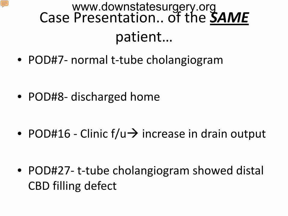
Case Presentation.. of the SAME patient…
• POD#7- normal t-tube cholangiogram
• POD#8- discharged home
• POD#16 - Clinic f/u increase in drain output
• POD#27- t-tube cholangiogram showed distal CBD filling defect
www.downstatesurgery.org
Presenter
Presentation Notes
She had a postop T-tube cholangiogram on POD#7 and was read as normal. It was the increase in the drain output during the clinic F/U made me, the astute surgeon, (you need to emphasize that I did not do the original operation) suspected that she had a retained stone. 4 week after the initial operation, she had a outpatient T-tube cholangiogram which confirmed a single retained large CBDS.

Case Presentation.. of the SAME patient…
• Readmitted POD#36 with cholangitis
- discharged 5 days later
• Barriers to ERCP- duodenal diverticulum
• Plan: OR for Percutaneous biliary exploration
www.downstatesurgery.org
Presenter
Presentation Notes
She developed cholangitis after the study and was admitted for anitobiotics.��The delay is to wait for the T-tube tract to mature further. GI did the ERCP before the initial operation but did not see any stones. They were not consulted before I took the patient to the OR since the stone is quite large and the patient also has a duodenal diverticulum. I don't think that they can do an adequate sphincterotomy.

Case Presentation
• OR
- Percutaneous biliary exploration, Intraoperative Cholangiogram (IOC)
- IOC demonstrated stone, choledochoscope advanced through the biliary tree to the duodenum
-no stone was visualized
• Repeat on-table cholangiogram showed no evidence of stone
www.downstatesurgery.org
Presenter
Presentation Notes
A little over a month later she was taken to the OR for Percutaneous exploration through the fistulous tract. Repeat on-table cholangiogram showed no evidence of stone. So the case was concluded

Operative films www.downstatesurgery.org
Presenter
Presentation Notes
I do have the intraop images but they are not that good since none of them clearly demonstrated the stone. I can email you some images to point out the technical aspect/difficulties due to the path of the t-tube tract and why we had a difficult time deflecting the ureteroscope to visualize the stone since it was already maximally "passively" deflected in a candy cane fashion.

But…
• POD#1: formal T-tube cholangiogram
- large impacted stone in the distal CBD just proximal to the ampulla of Vater
www.downstatesurgery.org
Presenter
Presentation Notes
formal T-tube cholangiogram in the IR suite. The study, however, showed a large impacted stone in the distal CBD just proximal to the ampulla of Vater.
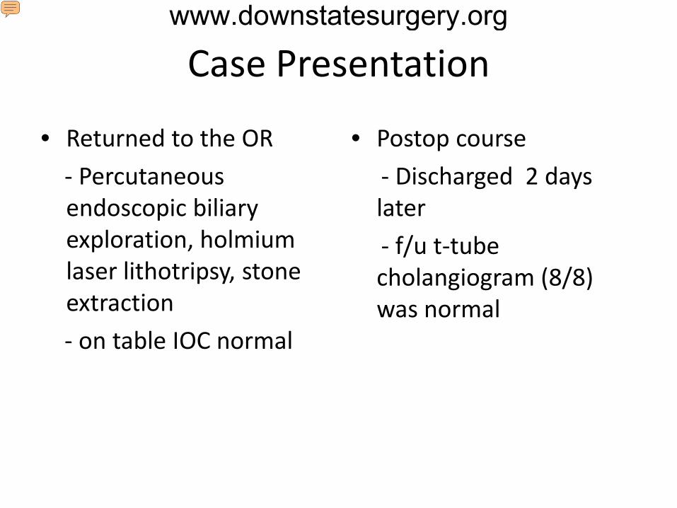
Case Presentation
• Returned to the OR
- Percutaneous endoscopic biliary exploration, holmium laser lithotripsy, stone extraction
- on table IOC normal
• Postop course
- Discharged 2 days later
- f/u t-tube cholangiogram (8/8) was normal
www.downstatesurgery.org
Presenter
Presentation Notes
POD#2 we took her back to the OR and finally were able to visulaize the stone which we removed via lithotripsy and stone extraction. The on table IOC appeared normal. The procedure was otherwise uncomplicated and she was d/c’d home 2 days later. The following week she had a formal study which was normal.

Goals
• History • Classifying stones • The problem • Preoperative, Intraoperative, and Postoperative
identification of CBD stones and approaches in management
• Complications in management • Tailoring decision making to patient’s
circumstances
www.downstatesurgery.org
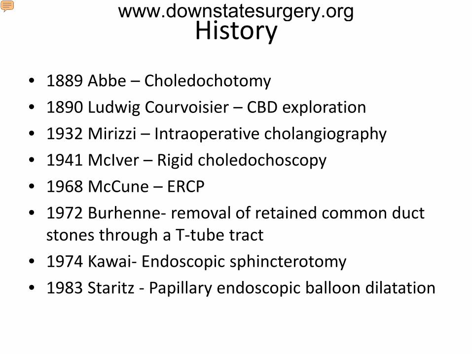
History
• 1889 Abbe – Choledochotomy
• 1890 Ludwig Courvoisier – CBD exploration
• 1932 Mirizzi – Intraoperative cholangiography
• 1941 McIver – Rigid choledochoscopy
• 1968 McCune – ERCP
• 1972 Burhenne- removal of retained common duct stones through a T-tube tract
• 1974 Kawai- Endoscopic sphincterotomy
• 1983 Staritz - Papillary endoscopic balloon dilatation
www.downstatesurgery.org
Presenter
Presentation Notes
History of invasive biliary interventions. Although surgery is the treatment of choice for symptomatic gallstones, most bile duct stones can be treated with nonsurgical methods. Many percutaneous and endoscopic techniques for stone elimination have been published since 1972 when Burhenne described removal of retained common duct stones through a T-tube tract with stone baskets and Kawai in 1974 described endoscopic clearance of stones through sphincterotomy. These techniques are still common interventional and endoscopic procedures. Papillary endoscopic balloon dilatation was introduced by Staritz et al. [13] in 1983 as an alternative method for gaining access to the common bile duct for the removal of stones. It fell out of favor (perceived increased risk of pancreatitis) and appears to be entering back into favor offering an effective and safe alternative to endoscopic sphincterotomy (bleeding, ppreservation of sphincter function). Note: Lap chole by Muhe (1986), (1882) Cholecystectomy by Langenbuch

Describing stones
• Primary stones (usually brown pigment stones), which form in the bile ducts
• Secondary stones (usually cholesterol), which form in the gallbladder but migrate to the bile ducts
• Residual stones, which are missed at the time of cholecystectomy (evident < 3 yr later)
• Recurrent stones, which develop in the ducts > 3 yr after surgery
www.downstatesurgery.org
Presenter
Presentation Notes
In developed countries, > 85% of common duct stones are secondary; affected patients have additional stones located in the gallbladder. Up to 10% of patients with symptomatic gallstones also have associated common bile duct stones. After cholecystectomy, brown pigment stones may result from stasis (eg, due to a postoperative stricture) and the subsequent infection. The proportion of ductal stones that are pigmented increases with time after cholecystectomy. Cholesterol gallstones make up 75% of all gallstones; the remaining 25% are pigment stones. They are associated with obesity, diabetes, female gender, and childbearing; pigment stones are associated with hemolysis and cirrhosis of the liver
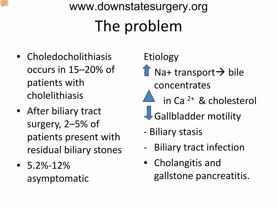
The problem
• Choledocholithiasis occurs in 15–20% of patients with cholelithiasis
• After biliary tract surgery, 2–5% of patients present with residual biliary stones
• 5.2%-12% asymptomatic
Etiology
- Na+ transport bile concentrates
in Ca 2+ & cholesterol
- Gallbladder motility
- Biliary stasis
- Biliary tract infection
• Cholangitis and gallstone pancreatitis.
www.downstatesurgery.org
Presenter
Presentation Notes
Choledocholithiasis occurs in 15–20% of patients with cholelithiasis and, after biliary tract surgery, 2–5% of patients present with residual biliary stones in the bile ducts. asymptomatic CBDS between 5.2% and 12%Cause if you have your GB: Active sodium transport by the epithelium of the gallbladder causes concentration of the bile into a form that is up to 10 times more concentrated than when first excreted by the liver. This concentration process leads to changes in the solubility of the calcium and cholesterol components of the bile. Decreased gallbladder motility with bile stasis contributes to stone formation, but biliary tract infection can also lead to stone formation. Two serious complications of CBDS are cholangitis and gallstone pancreatitis.

Preoperative Diagnosis
• Blood tests (elevated LFT’s)
• Abdominal U/S
-15-30% sensitivity, If CBD >10mm90% • EUS
- Sensitivity and specificity 92-100%
• MRCP
- 90% sensitive, 100% specificity
• ERCP
www.downstatesurgery.org
Presenter
Presentation Notes
Dx can be established preoperatively via these modalities, atients exhibiting the described symptoms require diagnostic investigation to assess for the presence of CBDS [12]. Liver function tests (LFTs) can be used to screen for CBDS . Elevated serum bilirubin and alkaline phosphatase typically reflect biliary obstruction, but these are neither highly sensitive nor specific for CBDS.Transabdominal ultrasound, sensitivity for the detection of CBD stones is only 15% to 30% CBD diameter greater than 10 mm in a jaundiced patient predicts CBD stones in more than 90% of cases Endoscopic ultrasound :The sensitivity and specificity for the diagnosis of CBD stones by EUS ranges from 92% to 100% and 95% to 100%, respectively Several studies have shown that MRCP can diagnose CBD stones with a sensitivity of 90%, a specificity of 100%. Cholangiography is the gold standard for the diagnosis of CBD stones. Skilled endoscopists can successfully cannulate the CBD in approximately 90% to 95% of patients.

ERCP Diagnostic and therapeutic Endoscope into 2nd portion of duodenum
Papilla visualized & cannulated
– Radioopaque dye injected under fluroscopy – Stones appear as filling defects
Performed in conjunction with sphincterotomy and stone extraction
Stats: 99% success rate, 6% morbidity, 0.2% mortality
www.downstatesurgery.org
Presenter
Presentation Notes
ERCP as previously mentioned by Marilyn involves an endoscope is passing into the duodenum. The papilla of Vater is cannulated and radiopaque liquid contrast is injected into the biliary ducts, providing excellent contrast on radiographic images. Stones in bile appear as filling defects in the opacified ducts. Currently, ERCP is usually performed in conjunction with endoscopic retrograde sphincterotomy and gallstone extraction. If a stricture is present, brushings may be done along with either balloon dilation or stent placement.

Complications •Pancreatitis (3.5%)
•Cholangitis (<1%) • Duodenal perforation (0.1 to 0.6%) • Bleeding (1.3%) 3-10% not suitable for ERCP
Contrast related
www.downstatesurgery.org
Presenter
Presentation Notes
Duodenal perf presents as retro or intraperitoneal free air. Most ERCP-associated bleeding is intraluminal, although intraductal bleeding can occur and hematomas (hepatic,splenic, and intra-abdominal) have been reported. Hemorrhage is primarily a complication related to sphincterotomy rather than diagnostic ERCP. ERCP is not possible in 3% to 10% of all patients. Previous operations, cholangitis, anatomic abnormalities, and stone impaction were the principal reasons for failure of endoscopic retrograde cholangiopancreatography (ERCP). Pancreatitis is the most common serious ERCP complication.7-15 Although transient increase in serum pancreatic enzymes may occur in as many as 75% of patients,16 such an increase does not necessarily constitute pancreatitis. A widely used consensus definition for post- ERCP pancreatitis (PEP) is (1) new or worsened abdominal pain, (2) new or prolongation of hospitalization for at least 2 days, and (3) serum amylase 3 times or more the upper limit of normal, measured more than 24 hours after the procedure.17 By using this or similar definitions, the incidence of PEP in a meta-analysis of 21 prospective studies was approximately 3.5%18 Perforation rates with ERCP range from 0.1% to 0.6%.7,8,10,15,63 Three distinct types of perforation have been described: guidewire-induced perforation, periampullary perforation during sphincterotomy, and luminal perforation at a site remote from the papilla Prompt recognition of periampullary perforation and treatment with aggressive biliary and duodenal drainage (by means of nasobiliary and nasogastric tubes) coupled with broadspectrum antibiotics can result in clinical resolution without the need for operative intervention in as many as 86% of patients. The management of perforation will depend on many factors, such as the site and location, clinical status, and radiographic imaging. Early identification and expeditious management of a perforation have been shown to decrease associated morbidity and mortality.65 Perforations related to endoscopy are best approached in collaboration with surgical colleagues Most ERCP-associated bleeding is intraluminal, although intraductal bleeding can occur and hematomas (hepatic, splenic, and intra-abdominal) have been reported.56-58 Hemorrhage is primarily a complication related to sphincterotomy rather than diagnostic ERCP. In a meta-analysis of 21 prospective trials, the rate of hemorrhage as a complication of ERCP was 1.3% (95% CI, 1.2%-1.5%) with 70% of the bleeding episodes classified as mild
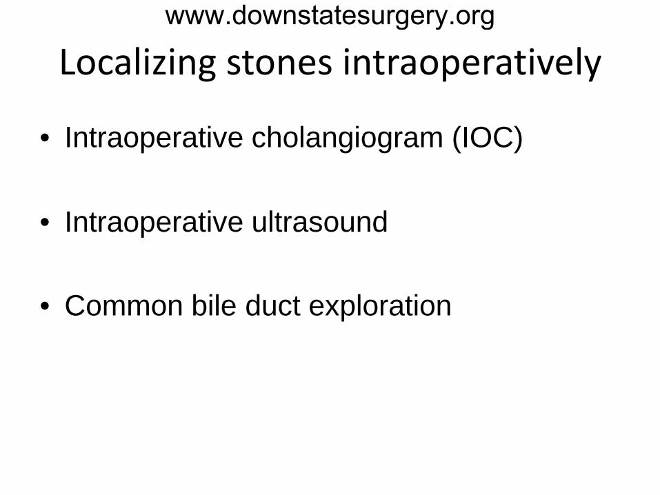
Localizing stones intraoperatively
• Intraoperative cholangiogram (IOC)
• Intraoperative ultrasound
• Common bile duct exploration
www.downstatesurgery.org

STATIC DYNAMIC filling defect
Intraoperative Cholangiogram (IOC)
www.downstatesurgery.org

IOC
• Time consuming (>16 min)
• Film often inadequate
• Lower success rate (47%)
• Visualization of anatomy more difficult
• Difficulty in differentiation between stones and air bubbles
STATIC
www.downstatesurgery.org
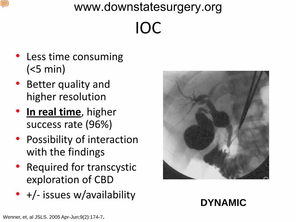
IOC
• Less time consuming (<5 min)
• Better quality and higher resolution
• In real time, higher success rate (96%)
• Possibility of interaction with the findings
• Required for transcystic exploration of CBD
• +/- issues w/availability DYNAMIC
Wenner, et, al JSLS. 2005 Apr-Jun;9(2):174-7.
www.downstatesurgery.org
Presenter
Presentation Notes
Issues with availability (depending on your institution with rad techs, etc) Interactions= surgeons vs. radiologists interpretations vary there can be a significant difference of LIOC interpretation between surgeons and radiologist specially in the detection of defects of fillings although this variability did not affect the clinical outcome. A second strategy is to do IOC on patients with risk factors, and to do intraoperative stone removal if stones are detected. The problem with this, as mentioned is that IOC, is time-consuming and associated with up to 12% false positive rate. Subsequent intraoperative stone removal is both time consuming and risky, and often subjects the patient to an open procedure. Cholangiography was successfully completed in 96% of patients. The mean time added to laparoscopic cholecystectomy by the addition of dynamic fluoroscopic intraoperative cholangiography was 4.3 minutes. The median time was 3.0 minutes. The times ranged from 2.0 minutes to 16.0 minutes. Choledocholithiasis was present in 15.4% of these patients. The false-positive rate was zero in this study.

IOC complications
• Bleeding
• Infection
• Pancreatitis
• Damage to the common bile duct
www.downstatesurgery.org

Intraoperative U/S
• Success rate ~90%
• High sensitivity and specificity (~94%)
• Safer
• Procedure time <10 min
• Low resolution
• Operator dependent
Machi, et al Surg Endosc 2007;21(2):270
www.downstatesurgery.org
Presenter
Presentation Notes
This technique offers the advantages of cholangiography, but is noninvasice, repeatable, fast and inexpensive. It’s limitations are that it’s clearly dependent on a experienced user, the resoultion of the image, and a standard/reproducible technique. During laparoscopy, an ultrasound probe is inserted into the peritoneal cavity though a 10-mm trochar and is used to scan the bile ducts. The reported sensitivity and specificity are over 90 percent and it has been suggested that the routine use of intraoperative ultrasound followed by selective IOC leads to the accurate diagnosis of CBD stones, while reducing the need for IOC
CBD exploration (CBDE)
• Laparoscopic vs. Open
-Lap: Transcystic vs. transductal approach
- Open
• Surgeon’s comfort
www.downstatesurgery.org
Presenter
Presentation Notes
The successful laparoscopic management of CBD stones depends on several factors including surgical expertise, adequate equipment, the biliary anatomy, and the number and size of CBD stones [86]. With advancing technology and minimally invasive surgery, laparoscopic biliary surgery has become safe, efficient, and cost effective [87–89]. Laparoscopic common bile duct exploration (LCBDE) was associated with successful stone clearance rates ranging from 85% to 95%, a morbidity rate of 4%–16% and a mortality rate of around 0%–2% [90, 91]. Laparoscopic exploration is very effective for clearing difficult CBD stones. Tai et al. reported that the clearance rate was 100%, and no recurrence was discovered during a mean followup period of 16 months [76]. Golipour et al. showed LCBDE to be an effective procedure as the initial modality of management for acute gallstone cholangitis [92]. Complications from this method include CBD laceration, stricture formation and bile leak [93]. Patients treated with LCBDE had a significantly shorter hospital stay and lower hospital costs as compared with ERCP/EST [88].

Transcystic: • Stone < 6 mm • Cystic duct > 4 mm • CBD < 6 mm • Stone location distal to
the cystic duct/CBD Junction • Fewer than 6 to 8 stones
within the CBD
Petelin, Surg Endosc, 2003
Laparoscopic CBD Exploration www.downstatesurgery.org
Presenter
Presentation Notes
The size of the stones to be removed dictates the approach to the CBD: stones smaller than 4 mm can usually be retrieved in fluoroscopically directed baskets and generally do not necessitate cystic duct dilatation; larger stones (4 to 8 mm) are retrieved under direct vision with the choledochoscope.�� A hydrophilic guide wire is inserted through the cholangiogram catheter into the CBD under fluoroscopic guidance. The cholangiogram catheter is then removed. If the largest stone is larger than the cystic duct, dilatation of the duct is necessary, not only for passage of the stone but also to allow passage of the choledochoscope, which may be 3 to 5 mm in diameter. The cystic duct should not be dilated to a diameter greater than 8 mm. Larger stones in the CBD may be either fragmented with electrohydraulic or mechanical lithotripsy, if available, or removed via choledochotomy.�� Once dilatation is complete, the guide wire may be removed or left in place to guide passage of a choledochoscope or baskets. When the choledochoscope is used, a second incision in the cystic duct, close to the CBD, avoids the Heister valves and allows removal of the guide wire. If baskets are used, a 6 French plastic introducer sheath may be inserted through the trocar used for cholangiography into the cystic duct.

Laparoscopic CBD Exploration
Transductal Irrigation + Glucagon
Basket +/- T-tube, endobiliary stent
Fogarty
Choledochoscope Lithotripsy
Transductal: • Failed laparoscopic
transcystic exploration or preoperative endoscopic stone extraction
• Stone > 6 mm • Cystic duct < 4 mm • CBD > 6 mm • Multiple stones • Stone location proximal to
the cystic duct/CBD junction
www.downstatesurgery.org
Presenter
Presentation Notes
Large stones (> 1 cm), as well as most stones in the common hepatic ducts, are not retrievable with the techniques described above. Ductal clearance can be achieved via choledochotomy if the duct is dilated and the surgeon is sufficiently experienced.46,47 The anterior wall of the CBD is bluntly dissected for a distance of 1 to 2 cm. When small vessels are encountered, it is preferable to apply pressure and wait for hemostasis rather than use the electrocautery in this area. Adrenaline-soaked gauzes placed through the 12 mm umbilical port are very effective for this purpose. Two stay sutures are placed in the CBD. An additional 5 mm trocar is placed in the right lower quadrant for insertion of an additional needle driver. A small longitudinal choledochotomy (a few millimeters longer than the circumference of the largest stone) is made with curved microscissors on the anterior aspect of the duct while the stay sutures are elevated. A choledochoscope is then inserted, and warm saline irrigation is initiated. In most cases, baskets should suffice for stone retrieval; however, lithotriptor probes and lasers are available for use through the working channel of the choledochoscope. The choice of approach depends on availability and individual surgical experience.�� Subsequently, a 12 or 14 French latex T tube is fashioned with short limbs, placed entirely intraperitoneally to prevent CO2 from escaping, and positioned in the CBD. The choledochotomy is then closed with fine interrupted absorbable sutures. The first suture is placed right next to the T tube, securing it distally, and the second is placed at the most proximal end of the choledochotomy; lifting these two sutures facilitates placement of additional sutures. Intracorporeal knots are preferred to avoid sawing of the delicate tissues. The end of the T tube is then pulled out through a trocar, and cholangiography is performed after completion of the procedure.
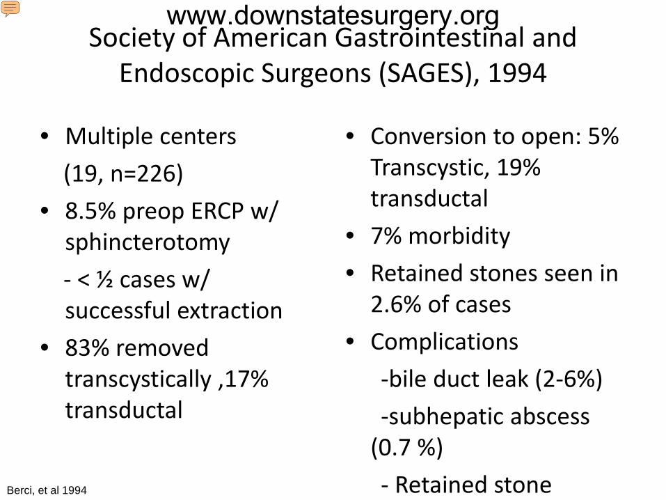
Society of American Gastrointestinal and Endoscopic Surgeons (SAGES), 1994
• Multiple centers
(19, n=226)
• 8.5% preop ERCP w/ sphincterotomy
- < ½ cases w/ successful extraction
• 83% removed transcystically ,17% transductal
• Conversion to open: 5% Transcystic, 19% transductal
• 7% morbidity
• Retained stones seen in 2.6% of cases
• Complications
-bile duct leak (2-6%)
-subhepatic abscess (0.7 %)
- Retained stone Berci, et al 1994
www.downstatesurgery.org
Presenter
Presentation Notes
Laparoscopic common bile duct exploration (CBDE) was the subject of a multi-institutional study on 226 patients from 19 major hospital centers. Female patients predominated (2.3:1); the average age was 54; 75% of cases were chronic, and the remainder were acute. Although 97% had preoperative ultrasonograms, only 12% showed a stone in the dilated common bile duct. The alkaline phosphatase was elevated in 41% and the serum bilirubin in 28% of cases. Preoperative endoscopic retrograde cholangiography with sphincterotomy (ERC-ES) was performed in 8.5%; there was a successful stone extraction in less than half the cases. Cholangiography was performed in 99.5%, and in 94% of those cases, stones were found. In 83% of cases, stones were removed through the transcystic approach, and in 17% removal was throughout the CBD. In the majority of cases, the choledochoscope and wire basket (34%), irrigation (33%), or a combination of both was employed. In the transcystic group, 5% were converted to open procedures due to technical difficulty, as contrasted with the trans-CBD route, where the conversion rate was 19%. There were two ductal injuries. Minor complications occurred in 5.7% within 24 h; there was one death (0.4%). Within 30 days, the morbidity rate was 7% and there were no deaths. Retained stones were discovered in 2.6% of cases. Laparoscopic CBDE is a feasible approach for CBD stones which permits a definitive procedure in one stage, without pre- or postoperative ES.

Level 2 evidence • Transcystic common bile duct exploration in the management of patients with
choledocholithiasis. J Gastrointest Surg. 2003 May-Jun;7(4):492-6.
Rojas-Ortega S, Arizpe-Bravo D, Marín López ER, Cesin-Sánchez R, Roman GR, Gómez C.
• All-comers policy for laparoscopic exploration of the common bile duct. Br J Surg. 2002 Dec;89(12):1608-12.
Thompson MH, Tranter SE.
• Laparoscopic exploration of common bile duct in difficult choledocholithiasis. Surg Endosc. 2004 Jun;18(6):910-4. Epub 2004 Apr 21.
Tai CK, Tang CN, Ha JP, Chau CH, Siu WT, Li MK.
• National analysis of in-hospital resource utilization in choledocholithiasis management using propensity scores. Surg Endosc. 2006 Feb;20(2):186-90. Epub 2005 Dec 9.
Poulose BK, Arbogast PG, Holzman MD.
- Stone clearance rates ranging from 85% to 95%, - vs. ERCP, less cost, <LOS
-Morbidity rate of 4%–16% , - CBD laceration, stricture,
- Mortality rate of around 0%–2% bile leak
www.downstatesurgery.org
Presenter
Presentation Notes
Multiple studies have provided level 2 evidence that Laparoscopic common bile duct exploration (LCBDE) was associated with successful stone clearance rates ranging from 85% to 95%, a morbidity rate of 4%–16% and a mortality rate of around 0%–2%. Tai et al. reported that the clearance rate was 100%, and no recurrence was discovered during a mean followup period of 16 months. Poulouse demonstrated that patients treated with LCBDE had a significantly shorter hospital stay and lower hospital costs as compared with ERCP. Complications include: CBD laceration, stricture formation and bile leak Poulose: Mean total hospital costs were less for CBDE (25,200 dollars +/- 1,800 dollars) than for ERCP (29,900 dollars +/- 800 dollars, p < 0.05). Mean LOS was less for CBDE (4.9 +/- 0.2 days) than for ERCP (5.6 +/- 0.1 days, p < 0.05). PS adjusted analysis revealed an estimated overall cost savings of 4,500 dollars +/- 1,600 dollars and reduced LOS (0.6 +/- 0.2 days) per hospitalization for CBDE Level II (B= high level) Evidence from controlled trials without randomization Or Cohort or case-control studies Or Multiple time series, dramatic uncontrolled experiments

Summary of randomized trials comparing endoscopic common duct clearance plus surgery against surgery alone
Reference (year)
Treatment n Successful
duct clearance
Mortality Morbidity
(Total) Morbidity
(Major)
Additional procedures
required
Median hospital
stay (days)
Neoptolemos ES 55 50 2 18 9 1 9 (1987) S 59 54 1 13 5 0 11 Stain ES 26 17 0 4 1 n·a· 5
(1991) S 26 23 0 7 1 n·a· 6 Stiegmann ES 16 5 0 3 0 1 n·a·
(1992) S 18 6 0 3 0 0 n·a· Hammarstrom ES 39 35 0 7 3 4 n·a·
(1995) S 41 37 0 9 4 4 n·a· Targarona ES 50 44 3 8 5 n·a· 5
(1996) S 48 45 2 11 4 n·a· 11 Kapoor ES 16 11 0 5 4 2 10.6 (1996) S 17 13 0 5 3 3 11.3
Suc ES 97 67 3 13 13 28 12 (1998) S 105 75 1 13 5 8 16
Rhodes ES 40 37 0 6 4 10 3.5 (1998) S 40 30 0 7 2 10 1
Cuschieri ES 133 82 2 17 9 17 9 (1999) S 133 92 1 21 9 17 6
Sgourakis ES 42 27 1 6 3 5 9 (2002) S 36 24 1 5 2 4 7.4
Nathanson ES 45 43 0 11 6 3 7.7 (2005) S 41 40 0 12 7 3 6.4 Hong ES 93 85 0 8 1 1 4.2 (2006) S 141 126 0 22 1 3
Total ES 652 503 (77.1%)
11 (1.69%)
106 (16.25%)
58 (8.89%)
72 (12.5%) 4.6
S 705 565 (80.1%)
6 (0.85%)
128 (18.15%)
43 (6.1%)
52 (8.2%)
www.downstatesurgery.org
Presenter
Presentation Notes
Patients with CBDS undergoing laparoscopic cholecystectomy may be managed by laparoscopic common bile duct exploration (LCBDE) at the time of surgery, or undergo peri-operative ERCP. There is no evidence of a difference in efficacy, morbidity or mortality when these approaches are compared, though LCBDE is associated with a shorter hospital stay. It is recommended that the two approaches are considered equally valid treatment options, and that training of surgeons in LCBDE is to be encouraged.
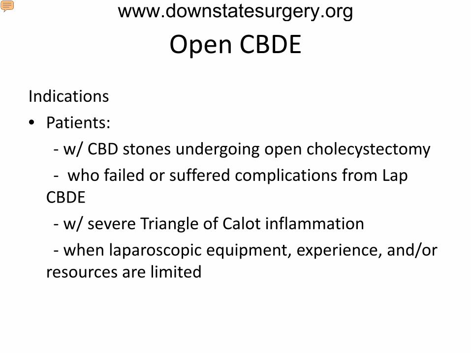
Open CBDE
Indications
• Patients:
- w/ CBD stones undergoing open cholecystectomy
- who failed or suffered complications from Lap CBDE
- w/ severe Triangle of Calot inflammation
- when laparoscopic equipment, experience, and/or resources are limited
www.downstatesurgery.org
Presenter
Presentation Notes
Open CBD exploration remains an important technique and should be part of every gastrointestinal surgeon's armamentarium for treating hepatobiliary diseases. Surgeons performing laparoscopic cholecystectomy should be prepared to convert to open CBD exploration if necessary. Open CBD exploration should be performed in the following situations: Patients with CBD stones who are undergoing open cholecystectomy Patients who have failed or suffered complications from laparoscopic CBD exploration Patients with severe inflammation in the triangle of Calot When laparoscopic equipment, experience, and/or resources are limited

Open CBDE • Anterior duct exposed
• Stay sutures laterally
• CBD opened vertically
• Catheter irrigation
• +/- Fogarty, basket, stone forceps, scope
• Place t-tube
• Close choledochotomy
www.downstatesurgery.org
Presenter
Presentation Notes
The anterior aspect of the duct is exposed over a distance of 1 to 2 cm, avoiding electrocautery during dissection. Two stay sutures of a 3-0 monofilament are placed lateral to the midline of the duct. The common hepatic duct is sharply opened with a No. 11 or No. 15 scalpel and longitudinally incised further with a Potts arteriotomy or similar scissors. When performing these maneuvers, the surgeon must respect the arterial blood supply of the duct, which courses laterally on either side of the duct in the 3 o'clock and 9 o'clock positions [see Figure 24]. In some cases, stones are immediately visible and can simply be plucked from the duct once it is opened. Flushing the duct with saline, proximally and then distally, through a 12 or 14 French Foley or red rubber catheter may also clear the duct of stones. The intravenous administration of 1 to 2 mg of glucagon will relax the sphincter of Oddi, which may help in the flushing of stones from the duct. In some cases, stones will be impacted within the duct and will require additional maneuvers. The Kocher maneuver (liberally mobilizing the lateral duodenum and head of the pancreas) will allow the surgeon to hold and palpate the duodenum, the head of the pancreas, and stones within the duct, facilitating instrumentation. Stone retrieval forceps, biliary Fogarty catheters, and wire baskets can all be employed to retrieve stones. A choledochoscope can also be used, either at the outset of exploration or for stone retrieval, if simpler maneuvers are not successful. The common bile duct is opened vertically between laterally positioned stay sutures. (b) A catheter is then used to irrigate and flush stones from the duct. If stones are impacted within the duct, they can be retrieved with Fogarty catheters, wire stone retrieval baskets, or stone retrieval forceps. The choledochoscope can be used if any of these methods fail or as the initial method of exploration.](a) After common bile duct exploration, a 12 or 14Fr T tube is fashioned and is placed into the duct. (b) Interrupted 4-0 absorbable sutures are used to close the choledochotomy snug around the tube. Completion cholangiography may then be performed. The T tube, is brought out through a separate stab incision in the right lateral abdominal wall and secured to the skin. Several days later, cholangiography is repeated. If it shows good flow into the duodenum without obstruction, the tube may be clamped and removed at the 2-week mark. If there are retained stones, a more mature tract must be allowed to develop over 4 to 6 weeks for future instrumentation and stone retrieval. Retained stones may require ERCP, percutaneous transhepatic instrumentation, T tube tract instrumentation, or combinations of these for removal.
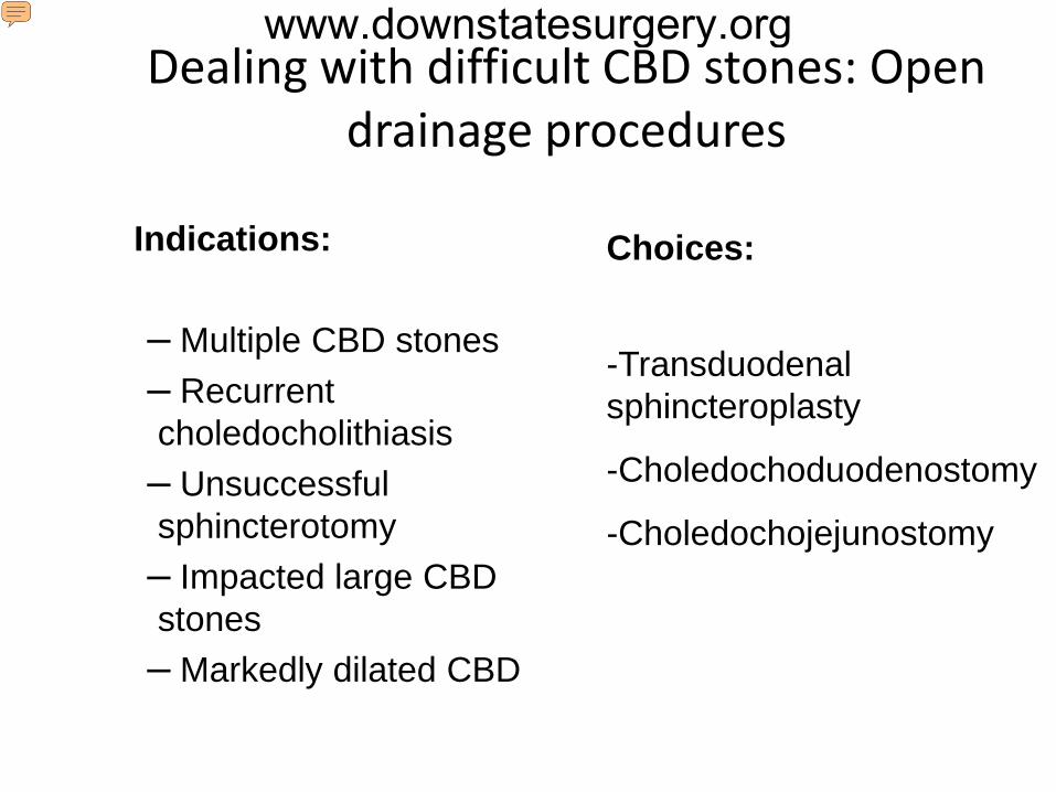
Dealing with difficult CBD stones: Open drainage procedures
Indications: – Multiple CBD stones – Recurrent choledocholithiasis – Unsuccessful sphincterotomy – Impacted large CBD stones – Markedly dilated CBD
Choices:
-Transduodenal sphincteroplasty
-Choledochoduodenostomy
-Choledochojejunostomy
www.downstatesurgery.org
Presenter
Presentation Notes
Sphincterotomy consists of incising the distal part of the sphincter musculature over a length of approximately 1 cm. This incision should not extend beyond the outer wall of the duodenum [96]. After the choledochotomy, a catheter or dilator is passed distally and a Kocher maneuver is performed, then duodenotomy is performed at the level of the ampulla. The dilator is advocated to bring the ampulla into the operative field, where it is then incised sufficiently along the anterosuperior border (opposite the pancreatic duct orifice) to permit removal of the impacted calculus [96]. Choledocoenterostomy is the most commonly performed as a side-to-side choledochoduodenostomy, usually in the setting of a dilated CBD with multiple stones [96], a recurrence of CBDS in the Vater's papilla occurred after ES and dilated CBD (≥2.0 cm). These patients require drainage for good long-term results without recurrence of jaundice or cholangitis [121]. The technique most commonly used is that of a side-to-side hand-sutured anastomosis between the supraduodenal common bile duct and the duodenum [122]. A Kocher maneuver is performed and the distal CBD is exposed. Choledochotomy is made within 2-3 cm of the lateral border of the duodenum. A diamond-shaped anastomosis is performed with interrupted absorbable sutures. One potential complication is the “sump syndrome” caused by food or other debris caught in the distal CBD [123]. This complication is rare (1%) and can be managed with ERC/ES [124]. The alternative operation, transection choledochoduodenostomy, excludes the distal (transpancreatic) segment of the bile duct from the end-to-side anastomosis of the transected common bile duct with the second part of the duodenum. The long-term results of this procedure are excellent [122]. Another optimal option is the choledochojejunostomy with a roux-en-Y loop.

Postoperative Management
•Post-op ERCP
• Dissolution -Ursodeoxycolic acid -Methyl tert-buthyl ether (MBTE)
•Lithotripsy -Mechanical (crushing technique) -Extra-corporeal shock wave (electromagnetic) -Intra-corporeal (laser)
www.downstatesurgery.org
Presenter
Presentation Notes
A third strategy is to do postoperative ERCP if the IOC shows stones. Again, the problem here is that up to a 15% failure risk associated with ERCP would subject the patient to another surgical procedure to remove the stones.These solutions have few toxic side effects and do not cause irritation of the biliary tree. Every dissolution therapy will last for several weeks, therefore the ideal solvent has not yet been produced. The use of ursodeoxycholic acid (UDCA) and chenodeoxycholic acid has only been shown to dissolve cholesterol-containing stones Methyl-Tert-butyl-Ether (MTBE) is an excellent cholesterol solvent that has been shown to work faster, but it is toxic to liver and duodenal mucosa. It has been proposed by several studies that using dissolution in combination with endoscopic retrieval or lithotripsy has better outcomes

Lithotripsy
• Electrohydraulic Lithotripsy (EHL)
-direct high voltage
- cholangioscopy or under fluoroscopy
-reserved for CBD packed with multiple stones or a large impacted stone
- Tissue damage, bleeding
• Extracorporeal Shockwave Lithotripsy (ESWL)
-Percutaneous sound waves
-done before ERCP
-clearance rates of 83% to 90%
-not common approach in US
www.downstatesurgery.org
Presenter
Presentation Notes
EHL uses direct high voltage to generate a shockwave through a liquid medium to fragment the bile duct stone. The procedure has been performed successfully under cholangioscopic guidance [10, 125] or under fluoroscopic control using a balloon catheter [126]. Typically, its use is reserved for cases of CBD packed with multiple faceted stones or a single large impacted stone. For EHL to be successful the stone must be targeted under direct sight, otherwise there is increased risk of damaging the bile duct wall [127]. This method is rarely used because of its high potential for tissue damage and bleeding. Extracorporeal Shockwave Lithotripsy (ESWL) ESWL was first used treating gallstones in 1980s following its successful use in fragmenting renal calculi [10]. ESWL involves the percutaneous administration of sound waves directed at the liver and bile duct. It is not performed during endoscopy, but rather before an ERCP in hopes of shattering large stones into smaller, more manageable fragments [127]. European studies evaluating ESWL report duct clearance rates of 83% to 90%, but its acceptance in the United States has been slow.

Laser lithotripsy
• amplified light energy
• under direct vision with cholangioscopy or under fluoroscopic control
• rate of duct clearance for retained CBDS using is 64-97%
www.downstatesurgery.org
Presenter
Presentation Notes
ser lithotripsy uses amplified light energy at a particular wavelength, which is focused into a single beam and directed onto a stone within the bile duct [10]. Laser lithotripsy can be performed under direct vision with cholangioscopy using mini scopes or can be performed under fluoroscopic control using standard equipment [10]. The success rate of duct clearance for retained CBDS using laser lithotripsy is between 64% and 97% in several studies
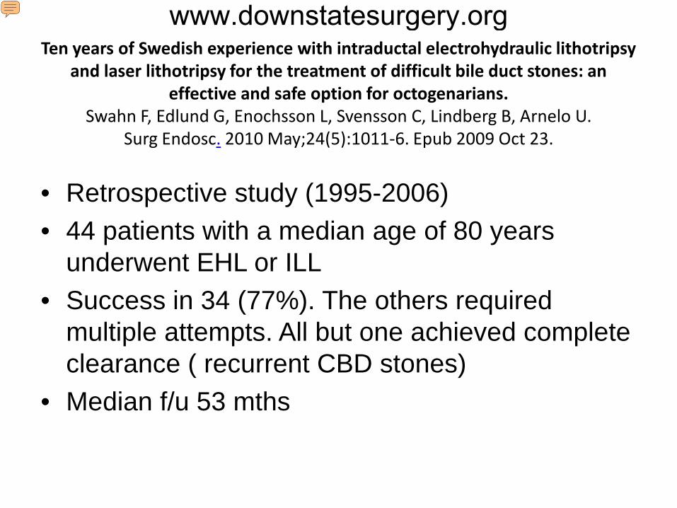
Ten years of Swedish experience with intraductal electrohydraulic lithotripsy and laser lithotripsy for the treatment of difficult bile duct stones: an
effective and safe option for octogenarians. Swahn F, Edlund G, Enochsson L, Svensson C, Lindberg B, Arnelo U.
Surg Endosc. 2010 May;24(5):1011-6. Epub 2009 Oct 23.
• Retrospective study (1995-2006) • 44 patients with a median age of 80 years
underwent EHL or ILL • Success in 34 (77%). The others required
multiple attempts. All but one achieved complete clearance ( recurrent CBD stones)
• Median f/u 53 mths
www.downstatesurgery.org
Presenter
Presentation Notes
Final stone clearance after EHL or ILL treatment with or without additional conventional endoscopic retrograde cholangiopancreatography (ERCP) was achieved for 34 (77%) of 44 patients. The results for 10 patients (23%) were defined as failures. Complete or partial stone fragmentation and definitive duct clearance were achieved in one session for 23 patients (52%). A second EHL or ILL attempt made in five cases of primary failure led to definitive stone clearance in three cases. Two patients experienced perioperative complications (stone basket impaction). Mild post-ERCP pancreatitis occurred for one patient and cholangitis for two patients. During long-term follow-up evaluation, recurrent CBD stones were found in one patient. CONCLUSIONS: Peroral endoscopic EHL or ILL, under direct cholangioscopic visualization by a mother-baby endoscopic system, is an effective treatment for difficult CBD stones. The technique can be used safely even in frail and elderly patients.

CBDS Algorithm
biliary sphincterotomies (BS) endoscopic extraction (ESE)
Williams, et al, 2008
www.downstatesurgery.org
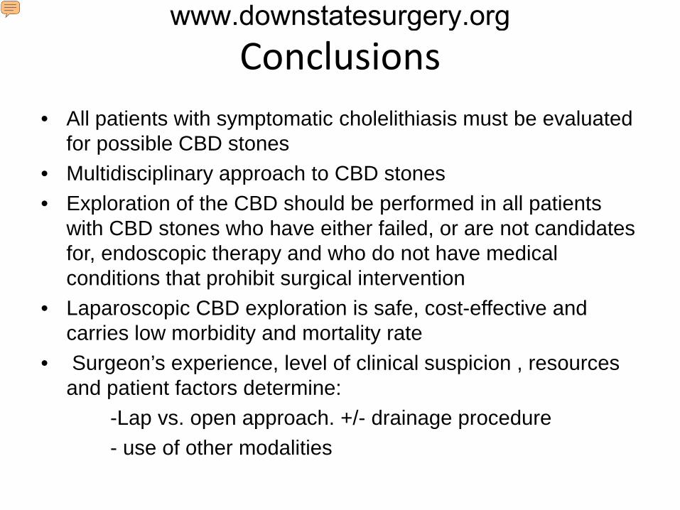
Conclusions • All patients with symptomatic cholelithiasis must be evaluated
for possible CBD stones • Multidisciplinary approach to CBD stones • Exploration of the CBD should be performed in all patients
with CBD stones who have either failed, or are not candidates for, endoscopic therapy and who do not have medical conditions that prohibit surgical intervention
• Laparoscopic CBD exploration is safe, cost-effective and carries low morbidity and mortality rate
• Surgeon’s experience, level of clinical suspicion , resources and patient factors determine:
-Lap vs. open approach. +/- drainage procedure - use of other modalities
www.downstatesurgery.org
Presenter
Presentation Notes
Common bile duct (CBD) stones are identified in 10 to 15 percent of patients undergoing surgery for symptomatic cholelithiasis, so all patients with symptomatic cholelithiasis must be evaluated for possible CBD stones. The availability of multiple diagnostic modalities with differing levels of sensitivity, specificity, and invasiveness provides clinicians with various options in the evaluation of patients with suspected choledocholithiasis. The aim of the diagnostic evaluation is to confirm or exclude the presence of common bile duct (CBD) stones using the least invasive, most accurate, and most cost-effective imaging modality Read 3+ bullets.
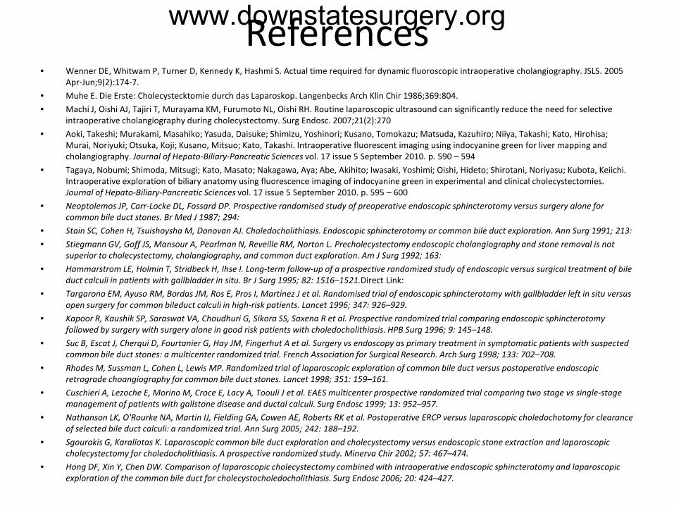
References • Wenner DE, Whitwam P, Turner D, Kennedy K, Hashmi S. Actual time required for dynamic fluoroscopic intraoperative cholangiography. JSLS. 2005
Apr-Jun;9(2):174-7.
• Muhe E. Die Erste: Cholecystecktomie durch das Laparoskop. Langenbecks Arch Klin Chir 1986;369:804.
• Machi J, Oishi AJ, Tajiri T, Murayama KM, Furumoto NL, Oishi RH. Routine laparoscopic ultrasound can significantly reduce the need for selective intraoperative cholangiography during cholecystectomy. Surg Endosc. 2007;21(2):270
• Aoki, Takeshi; Murakami, Masahiko; Yasuda, Daisuke; Shimizu, Yoshinori; Kusano, Tomokazu; Matsuda, Kazuhiro; Niiya, Takashi; Kato, Hirohisa; Murai, Noriyuki; Otsuka, Koji; Kusano, Mitsuo; Kato, Takashi. Intraoperative fluorescent imaging using indocyanine green for liver mapping and cholangiography. Journal of Hepato-Biliary-Pancreatic Sciences vol. 17 issue 5 September 2010. p. 590 – 594
• Tagaya, Nobumi; Shimoda, Mitsugi; Kato, Masato; Nakagawa, Aya; Abe, Akihito; Iwasaki, Yoshimi; Oishi, Hideto; Shirotani, Noriyasu; Kubota, Keiichi. Intraoperative exploration of biliary anatomy using fluorescence imaging of indocyanine green in experimental and clinical cholecystectomies. Journal of Hepato-Biliary-Pancreatic Sciences vol. 17 issue 5 September 2010. p. 595 – 600
• Neoptolemos JP, Carr-Locke DL, Fossard DP. Prospective randomised study of preoperative endoscopic sphincterotomy versus surgery alone for common bile duct stones. Br Med J 1987; 294:
• Stain SC, Cohen H, Tsuishoysha M, Donovan AJ. Choledocholithiasis. Endoscopic sphincterotomy or common bile duct exploration. Ann Surg 1991; 213:
• Stiegmann GV, Goff JS, Mansour A, Pearlman N, Reveille RM, Norton L. Precholecystectomy endoscopic cholangiography and stone removal is not superior to cholecystectomy, cholangiography, and common duct exploration. Am J Surg 1992; 163:
• Hammarstrom LE, Holmin T, Stridbeck H, Ihse I. Long-term follow-up of a prospective randomized study of endoscopic versus surgical treatment of bile duct calculi in patients with gallbladder in situ. Br J Surg 1995; 82: 1516–1521.Direct Link:
• Targarona EM, Ayuso RM, Bordas JM, Ros E, Pros I, Martinez J et al. Randomised trial of endoscopic sphincterotomy with gallbladder left in situ versus open surgery for common bileduct calculi in high-risk patients. Lancet 1996; 347: 926–929.
• Kapoor R, Kaushik SP, Saraswat VA, Choudhuri G, Sikora SS, Saxena R et al. Prospective randomized trial comparing endoscopic sphincterotomy followed by surgery with surgery alone in good risk patients with choledocholithiasis. HPB Surg 1996; 9: 145–148.
• Suc B, Escat J, Cherqui D, Fourtanier G, Hay JM, Fingerhut A et al. Surgery vs endoscopy as primary treatment in symptomatic patients with suspected common bile duct stones: a multicenter randomized trial. French Association for Surgical Research. Arch Surg 1998; 133: 702–708.
• Rhodes M, Sussman L, Cohen L, Lewis MP. Randomized trial of laparoscopic exploration of common bile duct versus postoperative endoscopic retrograde choangiography for common bile duct stones. Lancet 1998; 351: 159–161.
• Cuschieri A, Lezoche E, Morino M, Croce E, Lacy A, Toouli J et al. EAES multicenter prospective randomized trial comparing two stage vs single-stage management of patients with gallstone disease and ductal calculi. Surg Endosc 1999; 13: 952–957.
• Nathanson LK, O'Rourke NA, Martin IJ, Fielding GA, Cowen AE, Roberts RK et al. Postoperative ERCP versus laparoscopic choledochotomy for clearance of selected bile duct calculi: a randomized trial. Ann Surg 2005; 242: 188–192.
• Sgourakis G, Karaliotas K. Laparoscopic common bile duct exploration and cholecystectomy versus endoscopic stone extraction and laparoscopic cholecystectomy for choledocholithiasis. A prospective randomized study. Minerva Chir 2002; 57: 467–474.
• Hong DF, Xin Y, Chen DW. Comparison of laparoscopic cholecystectomy combined with intraoperative endoscopic sphincterotomy and laparoscopic exploration of the common bile duct for cholecystocholedocholithiasis. Surg Endosc 2006; 20: 424–427.
www.downstatesurgery.org

Question 1
The most common gallstones in the developed world are:
A) Brown pigment
B) Black pigment
C) Cholesterol
D) Quartz
www.downstatesurgery.org
Presenter
Presentation Notes
85%

Question 2
The most common complication of ERCP is:
A) Perforation
B) Pancreatitis
C) Cholangitis
D) Bleeding
www.downstatesurgery.org
Question 3
Which of the following statements is true?:
A) Laparoscopic CBDE carries low morbidity and mortality rate, but is not cost effective
B) There is potential use for lithotripsy in elderly & frail patients with CBDS w/acceptable results
C) Surgeon’s comfort means favorite OR, not resources to operate
www.downstatesurgery.org
Question 4
Why didn’t this patient undergo ERCP with sphincterotomy post-cholecystectomy?
A) Previous h/o cholangitis
B) Duodenal diverticulum
C) Abnormal anatomy
D) Age
www.downstatesurgery.org