Management of Neonatal Hyperbilirubinemia - … · Management of Neonatal Hyperbilirubinemia...
15
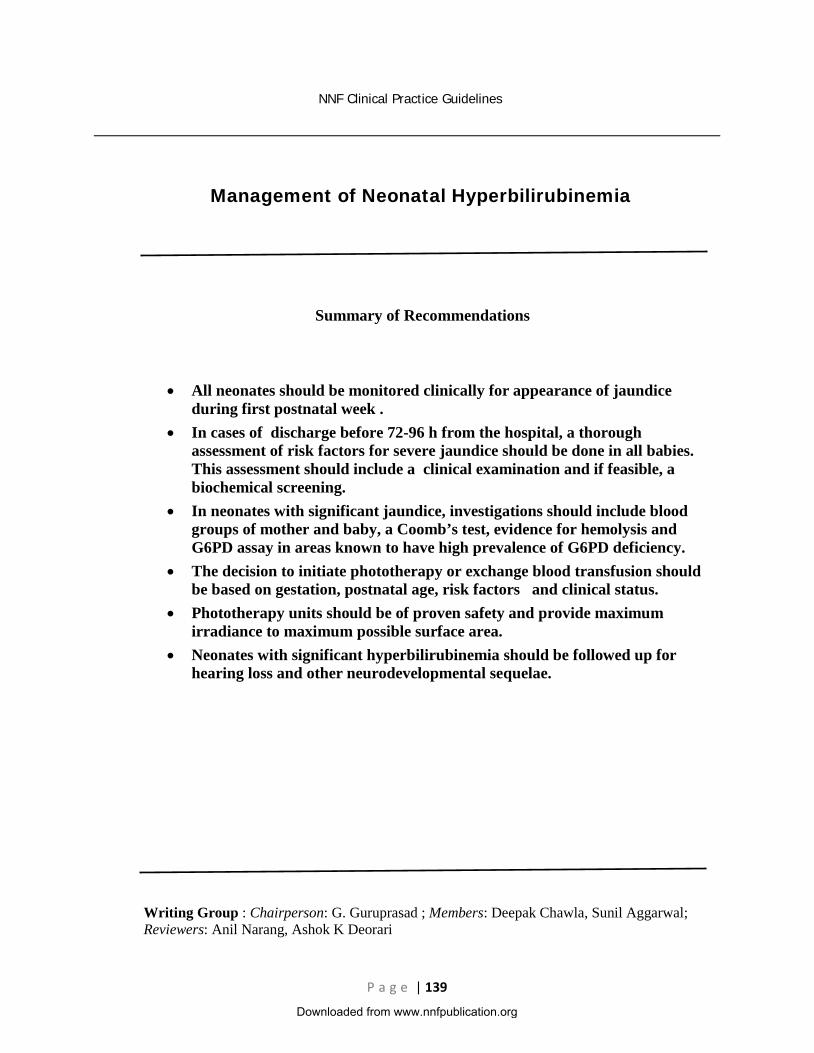
NNF Clinical Practice Guidelines Page | 139 Management of Neonatal Hyperbilirubinemia Summary of Recommendations • All neonates should be monitored clinically for appearance of jaundice during first postnatal week . • In cases of discharge before 72-96 h from the hospital, a thorough assessment of risk factors for severe jaundice should be done in all babies. This assessment should include a clinical examination and if feasible, a biochemical screening. • In neonates with significant jaundice, investigations should include blood groups of mother and baby, a Coomb’s test, evidence for hemolysis and G6PD assay in areas known to have high prevalence of G6PD deficiency. • The decision to initiate phototherapy or exchange blood transfusion should be based on gestation, postnatal age, risk factors and clinical status. • Phototherapy units should be of proven safety and provide maximum irradiance to maximum possible surface area. • Neonates with significant hyperbilirubinemia should be followed up for hearing loss and other neurodevelopmental sequelae. Writing Group : Chairperson: G. Guruprasad ; Members: Deepak Chawla, Sunil Aggarwal; Reviewers: Anil Narang, Ashok K Deorari Downloaded from www.nnfpublication.org
Transcript of Management of Neonatal Hyperbilirubinemia - … · Management of Neonatal Hyperbilirubinemia...
NNF Clinical Practice Guidelines
P a g e | 139
Management of Neonatal Hyperbilirubinemia
Summary of Recommendations
• All neonates should be monitored clinically for appearance of jaundice during first postnatal week .
• In cases of discharge before 72-96 h from the hospital, a thorough assessment of risk factors for severe jaundice should be done in all babies. This assessment should include a clinical examination and if feasible, a biochemical screening.
• In neonates with significant jaundice, investigations should include blood groups of mother and baby, a Coomb’s test, evidence for hemolysis and G6PD assay in areas known to have high prevalence of G6PD deficiency.
• The decision to initiate phototherapy or exchange blood transfusion should be based on gestation, postnatal age, risk factors and clinical status.
• Phototherapy units should be of proven safety and provide maximum irradiance to maximum possible surface area.
• Neonates with significant hyperbilirubinemia should be followed up for hearing loss and other neurodevelopmental sequelae.
Writing Group : Chairperson: G. Guruprasad ; Members: Deepak Chawla, Sunil Aggarwal; Reviewers: Anil Narang, Ashok K Deorari
Downloaded from www.nnfpublication.org
NNF Clinical Practice Guidelines
P a g e | 140
Introduction
Hyperbilirubinemia is a common problem in neonates with an incidence of 70-80%.1 A significant proportion of these neonates develop pathological hyperbilirubinemia (defined as hyperbilirubinemia requiring treatment) during the first week of life.2-4 Premature babies have much higher incidence of neonatal jaundice requiring therapeutic intervention more commonly than term newborns.5 Although the outcome for the majority is benign, infants with untreated, severe hyperbilirubinemia (defined as serum total bilirubin level >20 mg/dL) can develop signs of acute bilirubin encephalopathy (ABE). If not treated immediately, they might go on to develop kernicterus, a chronic, neurologically devastating condition resulting from bilirubin toxicity. Management of hyperbilirubinemia includes detection of at-risk neonates, investigating the cause of pathological hyperbilirubinemia, deciding thresholds for starting and stopping treatment and follow-up of neonates with severe hyperbilirubinemia.
This guideline reviews the evidence for the following issues :
• Optimum timing for discharge & follow-up and assessment policy • Pre-discharge stratification for risk of developing significant jaundice • Universal serum or transcutaneous bilirubin estimation for risk assessment • Laboratory investigations to be conducted in a baby needing treatment • Use of phototherapy (PT) & blood exchange transfusion (BET) for treatment • Monitoring of a baby with jaundice and assessment of response to treatment • Additional therapies in prevention/treatment of jaundice • Long term morbidities and follow-up plan of baby with jaundice
Optimum timing for discharge & follow-up and assessment policy to minimize the risk of severe hyperbilirubinemia and kernicterus
In India, healthy neonates are usually discharged within 24-48 h after a normal delivery. Due to continuing rise of bilirubin and absence of supervision for ensuring optimal feeding, neonates discharged home before completing 48-72 h of age are at high risk of developing undetected significant jaundice. In India, this risk may further be aggravated due to the absence of any formal system of follow-up home-visits by health care personnel (e.g. public health nurse) and due to traditional practice of confinement of mother-baby dyad at home for first few weeks after delivery.
Evidence: There is no prospective population-based or birth cohort-based study on epidemiology (rise, peak and fall of bilirubin) of significant jaundice in Indian neonates. Therefore it is difficult to define the time-period during which newborn needs to be followed up for significant jaundice. American Academy of Pediatrics (AAP) recommends follow-up of all neonates based on the postnatal age at discharge. This strategy also helps in assessment of newborn for feeding adequacy. Although desirable, this approach may not be feasible uniformly in India due to relative shortage of health care personnel and inability of some families to return for follow-up. Therefore follow-up plan may be devised based on pre-discharge risk assessment (Table 1).
Downloaded from www.nnfpublication.org
NNF Clinical Practice Guidelines
P a g e | 141
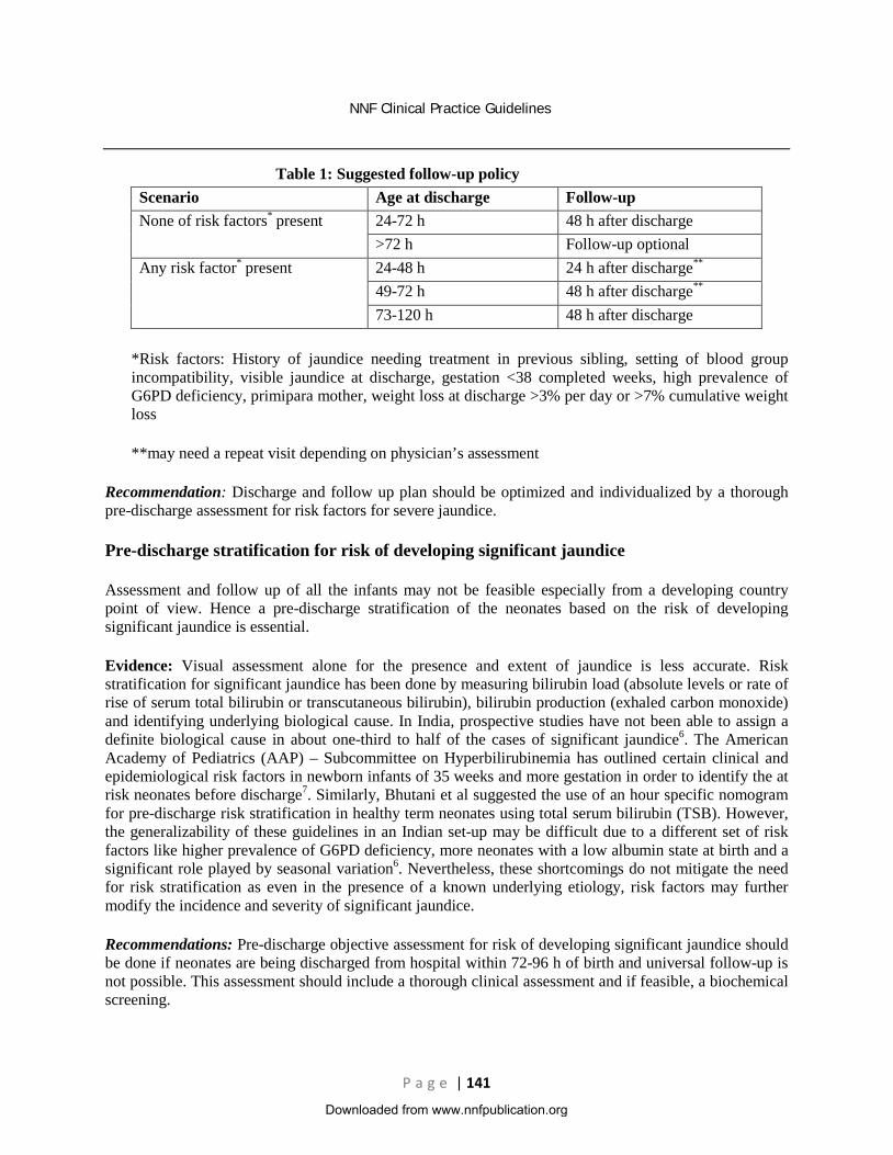
Table 1: Suggested follow-up policy Scenario Age at discharge Follow-up None of risk factors* present 24-72 h 48 h after discharge
>72 h Follow-up optional Any risk factor* present 24-48 h 24 h after discharge**
49-72 h 48 h after discharge**
73-120 h 48 h after discharge
*Risk factors: History of jaundice needing treatment in previous sibling, setting of blood group incompatibility, visible jaundice at discharge, gestation <38 completed weeks, high prevalence of G6PD deficiency, primipara mother, weight loss at discharge >3% per day or >7% cumulative weight loss
**may need a repeat visit depending on physician’s assessment
Recommendation: Discharge and follow up plan should be optimized and individualized by a thorough pre-discharge assessment for risk factors for severe jaundice.
Pre-discharge stratification for risk of developing significant jaundice
Assessment and follow up of all the infants may not be feasible especially from a developing country point of view. Hence a pre-discharge stratification of the neonates based on the risk of developing significant jaundice is essential.
Evidence: Visual assessment alone for the presence and extent of jaundice is less accurate. Risk stratification for significant jaundice has been done by measuring bilirubin load (absolute levels or rate of rise of serum total bilirubin or transcutaneous bilirubin), bilirubin production (exhaled carbon monoxide) and identifying underlying biological cause. In India, prospective studies have not been able to assign a definite biological cause in about one-third to half of the cases of significant jaundice6. The American Academy of Pediatrics (AAP) – Subcommittee on Hyperbilirubinemia has outlined certain clinical and epidemiological risk factors in newborn infants of 35 weeks and more gestation in order to identify the at risk neonates before discharge7. Similarly, Bhutani et al suggested the use of an hour specific nomogram for pre-discharge risk stratification in healthy term neonates using total serum bilirubin (TSB). However, the generalizability of these guidelines in an Indian set-up may be difficult due to a different set of risk factors like higher prevalence of G6PD deficiency, more neonates with a low albumin state at birth and a significant role played by seasonal variation6. Nevertheless, these shortcomings do not mitigate the need for risk stratification as even in the presence of a known underlying etiology, risk factors may further modify the incidence and severity of significant jaundice.
Recommendations: Pre-discharge objective assessment for risk of developing significant jaundice should be done if neonates are being discharged from hospital within 72-96 h of birth and universal follow-up is not possible. This assessment should include a thorough clinical assessment and if feasible, a biochemical screening.
Downloaded from www.nnfpublication.org
NNF Clinical Practice Guidelines
P a g e | 142
Universal serum or trans-cutaneous bilirubin estimation for risk assessment
Evidence: In a study conducted on term and near term infants without Rh hemolytic disease, Agarwal et al found that absence of TSB >6 mg/dL at 24±6 h of age virtually ruled out the possibility of subsequent significant jaundice (likelihood ratio of negative test 0.07) within 5 days of birth.8 In another Indian study, a cut off of 3.99 mg/dL at 18-24 h was found to have sensitivity and specificity of 67% each for prediction of subsequent bilirubin level >15 mg/dL.9 However, in both the studies only those neonates who stayed in the hospital, either due to their illness or due to certain maternal indications, were followed up. Moreover about 50% of infants, who were healthy and thus discharged early, were not followed up. As serum total bilirubin (TSB) estimation is invasive and relatively labor-intensive, non-invasive transcutaneous bilirubin (TcB) assessment has been investigated and was reported to correlate and agree closely with TSB in European and American origin infants.7 Reports from India suggest heterogeneous performance with declining accuracy at TSB >13 mg/dL.10 However, TcB used judiciously may serve as a good screening tool for jaundice prediction during first 24-48 of life in neonates with relatively lower levels of bilirubin. Bhat YR et al have reported good predictive ability of TcB index using cut-off of 7 at 24 h and 10 at 48 h of age in healthy term breastfed neonates.11
Hour-specific serum bilirubin nomogram published by Bhutani et al has been recommended by the APP for a pre-discharge risk assessment and are used widely in the Western population.12 However, direct extrapolation of this nomogram to Indian neonates may not be possible as <1% of the neonates included in this study were of Asian origin. Mishra et al have provided normative data for TcB in healthy term and late-preterm Indian neonates during first 72 h of age using a multiwavelength reflectance transcutaneous bilimeter13. However, the diagnostic utility of this nomogram for predicting hyperbilirubinemia needs to be tested in a separate validation cohort.
Recommendations: Universal measurement of serum total bilirubin or transcutaneous bilirubin may help in identifying and stratifying neonates based on their risk of developing significant jaundice. However, more studies are required in this regard from a wider population base especially involving Indian neonates and the feasibility of this approach at the community level remains to be evaluated .
Laboratory investigations to be conducted in a baby needing treatment
Evidence: Laboratory investigations primarily intend to identify the presence of clinically significant jaundice, the severity of jaundice, the underlying etiology and possible adverse effects due to therapy. Literature search has not revealed any particular set of investigations universal for all infants. Investigations should be designed based on the region, risk factors present and clues from examination. Consensus opinion from the subcommittee for hyperbilirubinemia of the AAP has outlined investigations which are, to a large extent, applicable to Indian infants. Table 2 depicts a list of proposed investigations in an infant with jaundice. Other rare causes like galactosemia, hypothyroidism and intrauterine infections may be looked for depending on the clinical presentation. Bacterial infections have been implicated as a cause in 5-7% cases of significant jaundice.6 However, with the present body of evidence, in absence of others sign(s) indicative of bacterial infection, investigations for sepsis is not warranted in a jaundiced infant.
Some of the investigations to diagnose presence of hemolysis may not be available round-the-clock in some settings. For the purpose of clinical management significant jaundice should be assumed to be due to hemolysis in following circumstances: mother blood group not known or O positive, high prevalence of G6PD deficiency, onset of jaundice within first 24 h after birth and presence of signs of Bilirubin Induced
Downloaded from www.nnfpublication.org
NNF Clinical Practice Guidelines
P a g e | 143
Neurological Damage (BIND). Universal screening for G6PD deficiency in communities with high prevalence of this enzyme deficiency (eg. Panjabis in North India & Lambani tribal population in South India) needs to be investigated.
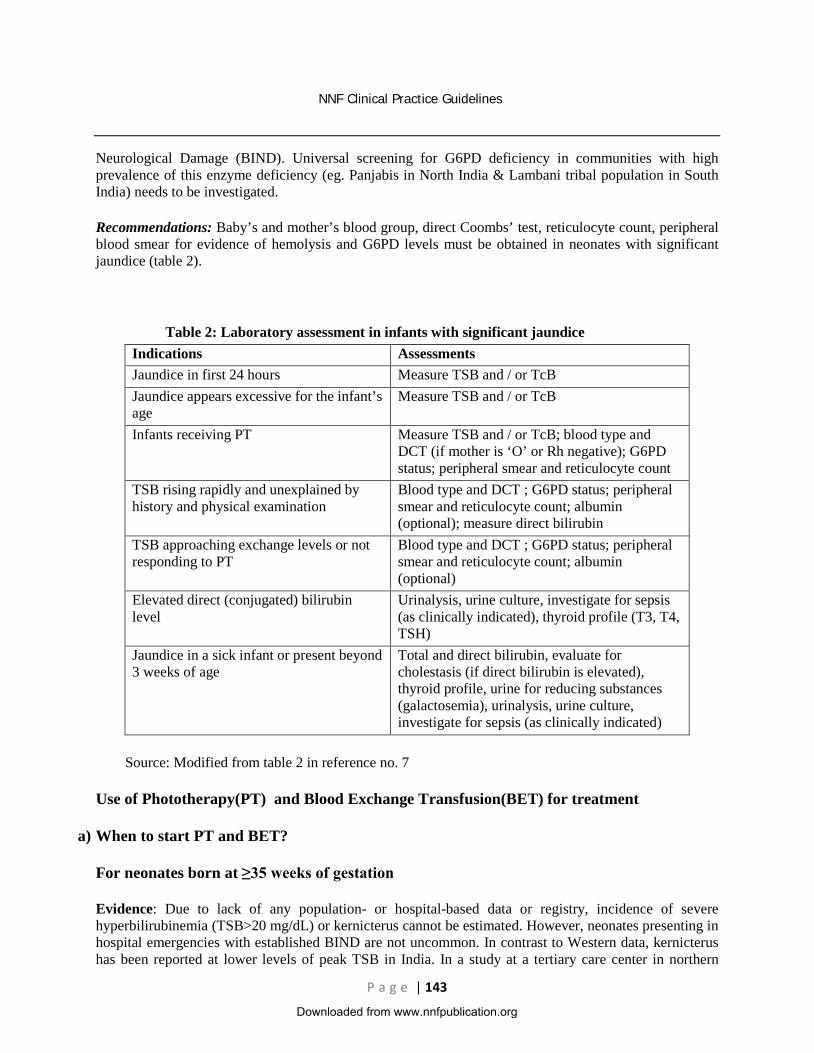
Recommendations: Baby’s and mother’s blood group, direct Coombs’ test, reticulocyte count, peripheral blood smear for evidence of hemolysis and G6PD levels must be obtained in neonates with significant jaundice (table 2).
Table 2: Laboratory assessment in infants with significant jaundice Indications Assessments Jaundice in first 24 hours Measure TSB and / or TcB Jaundice appears excessive for the infant’s age
Measure TSB and / or TcB
Infants receiving PT Measure TSB and / or TcB; blood type and DCT (if mother is ‘O’ or Rh negative); G6PD status; peripheral smear and reticulocyte count
TSB rising rapidly and unexplained by history and physical examination
Blood type and DCT ; G6PD status; peripheral smear and reticulocyte count; albumin (optional); measure direct bilirubin
TSB approaching exchange levels or not responding to PT
Blood type and DCT ; G6PD status; peripheral smear and reticulocyte count; albumin (optional)
Elevated direct (conjugated) bilirubin level
Urinalysis, urine culture, investigate for sepsis (as clinically indicated), thyroid profile (T3, T4, TSH)
Jaundice in a sick infant or present beyond 3 weeks of age
Total and direct bilirubin, evaluate for cholestasis (if direct bilirubin is elevated), thyroid profile, urine for reducing substances (galactosemia), urinalysis, urine culture, investigate for sepsis (as clinically indicated)
Source: Modified from table 2 in reference no. 7
Use of Phototherapy(PT) and Blood Exchange Transfusion(BET) for treatment
a) When to start PT and BET?
For neonates born at ≥35 weeks of gestation
Evidence: Due to lack of any population- or hospital-based data or registry, incidence of severe hyperbilirubinemia (TSB>20 mg/dL) or kernicterus cannot be estimated. However, neonates presenting in hospital emergencies with established BIND are not uncommon. In contrast to Western data, kernicterus has been reported at lower levels of peak TSB in India. In a study at a tertiary care center in northern
Downloaded from www.nnfpublication.org
NNF Clinical Practice Guidelines
P a g e | 144
India, 21.8% neonates with non-hemolytic jaundice and TSB ≥18 mg% had kernicterus when brought to the hospital.14 In another study from the same hospital, about 10% neonates with TSB of 20-25 mg/dL presented with established BIND.15 In a study on term infants with hyperbilirubinemia of mixed etiology, 10 out of 15 neonates with TSB of 21-25 mg/dL developed transient abnormalities in brainstem auditory evoked response (BERA) and 11% had developmental delay at 1 year of age.15, 16 In a large cross-sectional study, kernicterus was identified as the underlying cause in 16.7% cases of cerebral palsy.17 Reasons for increased propensity to BIND in Indian neonates need to be investigated. Proposed mechanisms include prolonged exposure to high TSB due to late referrals, concomitant morbidities, higher incidence of glucose-6-phosphate dehydrogenase (G6PD) deficiency, increased production of bilirubin and genetically altered blood-brain barrier permeability. Need for PT and BET should ideally be based on estimates of risk: benefit and cost: benefit ratio. Unfortunately, there is little such evidence on which to base these recommendations. As a result, treatment guidelines rely more on uncertain estimates and extrapolations.
Factors which may modify the risk of BIND in Indian neonates include:
1. Possible differences in genetic make-up including higher incidence of G6PD deficiency in certain geographical areas of India: In areas with documented higher incidence of BET, all neonates should be considered to be G6PD deficient unless proven otherwise and therefore risk-stratification for PT/BET must be done accordingly. Similarly, history of sibling with unexplained neonatal jaundice requiring treatment should be considered as a risk factor while deciding treatment.
2. Significantly higher proportion of small-for-gestation age (SGA) neonates: About two-thirds of low birth weight neonates born in India are born SGA.18 However, there is no data regarding their differential risk of BIND. In the absence of supporting evidence it is not possible to formulate guidelines for this group of neonates. However, it may be prudent to consider them for appropriate treatment based on their birth weight rather than gestation.
3. Non-availability of intensive PT in many neonatal units: AAP recommends use of intensive PT with irradiance in blue-green spectrum of at least 30µW/cm2 per nm and delivered to as much of the infant’s surface area as possible.7 However, this seems to be very difficult to achieve in Indian scenario. In a study done on 58 PT units across 24 centers in India, it was observed that only 9% of the units are of the special-blue lights and 31% of the units were giving irradiance of at least 15 µW/cm2 per nm.19 Although, standard-light fluorescent tubes have been demonstrated to be as efficacious as special blue compact fluorescent lamps (CFL) in moderate hyperbilirubinemia20, their efficacy has not yet been demonstrated in hyperbilirubinemia nearing threshold for BET.
For neonates born at <35 weeks of gestation
Evidence: Several physiological differences from term infants indicate an increased risk for bilirubin toxicity in the immature newborn. It is therefore generally recommended to treat hyperbilirubinemia at lower levels in low birth weight (LBW) infants in comparison to term infants. The general guide is to start PT when TSB is 0.5% & 0.75% of the body weight in healthy and sick infants respectively and to do a EBT when TSB is ≥ 1% of the body weight in grams (table 3). However, it is important to remember that most of the algorithms do not take into consideration the lower concentration of albumin and the diminished albumin binding ability in a sick preterm neonate. More aggressive approach towards starting PT in extremely low birth weight (ELBW) neonates has not demonstrated benefit in terms of improvement in composite outcome of death and neurodevelopment impairment.21
Downloaded from www.nnfpublication.org
NNF Clinical Practice Guidelines
P a g e | 145
Recommendations:
1) The postnatal age, gestation and risk based recommendations by AAP for starting PT and doing BET in infants ≥35 weeks of gestation thus can be used, but need certain modifications according to the Indian scenario.
2) In using the guidelines for PT and BET, one should use TSB. The direct reacting or conjugated bilirubin level should not be subtracted from the total bilirubin unless it is greater than 50% of the total. Immediate EBT is recommended if infant shows signs of ABE or if TSB is ≥5 mg/dl above the recommended lines.
3) AAP guidelines present PT and BET thresholds up to seven days of post natal age. Due to ongoing maturation of blood brain barrier, the thresholds for treatment are expected to be higher as post natal age advances. However due to lack of outcome studies, thresholds presented on seventh day may be used for rest of the neonatal period.
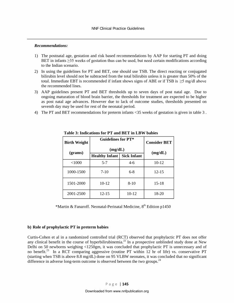
4) The PT and BET recommendations for preterm infants <35 weeks of gestation is given in table 3 .
Table 3: Indications for PT and BET in LBW babies
Birth Weight
(grams)
Guidelines for PT*
(mg/dL)
Consider BET
(mg/dL) Healthy Infant Sick Infant
<1000 5-7 4-6 10-12
1000-1500 7-10 6-8 12-15
1501-2000 10-12 8-10 15-18
2001-2500 12-15 10-12 18-20
*Martin & Fanaroff. Neonatal-Perinatal Medicine, 8th Edition p1450
b) Role of prophylactic PT in preterm babies
Curtis-Cohen et al in a randomized controlled trial (RCT) observed that prophylactic PT does not offer any clinical benefit in the course of hyperbilirubinemia.22 In a prospective unblinded study done at New Delhi on 50 newborns weighing <1250gm, it was concluded that prophylactic PT is unnecessary and of no benefit.23 In a RCT comparing aggressive (routine PT within 12 hr of life) vs. conservative PT (starting when TSB is above 8.8 mg/dL) done on 95 VLBW neonates, it was concluded that no significant difference in adverse long-term outcome is observed between the two groups.24
Downloaded from www.nnfpublication.org
NNF Clinical Practice Guidelines
P a g e | 146
c) Administration of PT
Three types of PT units are in common use:
i) Special blue tube-lights (PHILIPS TL52, 20W) ii) Special blue CFT lamps (OSRAM 18 W) iii) High intensity gallium nitride blue light emitting diode (LED)
In a multi-centric, open-label RCT comparing efficacy of single surface LED vs. CFT lamps, it was concluded that both were equally efficacious in the management of non-hemolytic hyperbilirubinemia in healthy term and late-preterm neonates.25 In an another RCT done at a level III tertiary newborn center, it was concluded that CFT PT has no superiority over standard tube-light PT in terms of efficacy and adverse effects on the neonate and effects on nursing staff.20 There is no evidence to support one above the other in moderate hyperbilirubinemia. Intensive PT must be ensured for neonates nearing BET threshold. It is not necessary to measure spectral irradiance before each use of PT; however it is important to perform periodic checks of PT units to make sure that an adequate irradiance is being delivered. Double surface PT should be started whenever possible and there is evidence to suggest its superiority over single surface PT.26 Use of locally made wooden devices/devices without electrical fitting certification has been associated with fatal accidents and is therefore strongly discouraged.
d) Increasing the delivered irradiance
Irradiance can be increased by decreasing the distance between baby and the PT unit to as low as 10 cm (only if using CFL/LED lamps). Sides of bassinet, incubator or warmer can be lined with aluminum foil or white material to increase the irradiance, but the evidence for clinical benefit is not conclusive. In a RCT done at a tertiary care centre it was concluded that, though hanging of white reflective sling on sides of CFT PT equipment resulted in marginal increase in irradiance, it did not decrease the duration of PT.27 However in an another RCT done at Malaysia on term newborns with uncomplicated neonatal jaundice it was concluded that hanging white curtains around PT units significantly increases efficacy of PT in the treatment of neonatal jaundice without evidence of increased adverse effects.28
e) Stopping of PT
PT may be discontinued when serum bilirubin level has fallen below 2mgs/dL lower than the PT threshold for that postnatal age. Discharge from the hospital need not be delayed to observe the infant for rebound provided investigations have shown a non-hemolytic etiology for the jaundice and an early follow up after discharge is assured. If PT is used for infants with hemolytic diseases or is initiated early and discontinued before the infant is 3 to 4 days old, a follow-up bilirubin measurement within 24 hours after discharge is recommended. For infants who are readmitted with hyperbilirubinemia and then discharged, significant rebound is rare, but a repeat TSB measurement or follow-up 24 hours after discharge is an option. In a prospective observational study it was observed that post-PT neonatal bilirubin rebound to clinically significant levels may occur, especially in cases of prematurity, direct Coombs test positivity, and those treated < 72 hours. These risk factors should be taken into account when planning post-PT follow up.29
Downloaded from www.nnfpublication.org
NNF Clinical Practice Guidelines
P a g e | 147
f) Care of newborns under PT
Breastfed infants should continue breastfeeding every 2-3 hourly. Infants with inadequate oral intake, excessive weight loss (>10%), or clinical or biochemical evidence of dehydration should receive supplemental breast milk or formula. Intravenous fluids should be given if enteral feeding is unsuccessful and the infant is dehydrated. The infant should be nursed naked except for diapers (use diapers and nappy pads only if deemed necessary and cut them to the minimum possible size), and the eyes should be covered with an opaque cloth (soft, preferably made of cotton) to reduce the risk of retinal damage. Older data suggested that PT was associated with increased insensible water loss; therefore, many clinicians have routinely added a certain percentage to the infant's estimated basic fluid requirements. Newer data suggest that if temperature homeostasis is maintained, fluid loss is not significantly increased by PT. Rather than instituting blanket increases of fluid supplements to all infants receiving PT, fluid supplementation should be tailored to the infant's individual needs, as measured through evaluation of daily weight, urine output measurements, urine specific gravity, and fecal water loss supplemented by biochemical tests, wherever essential.
g) Failure of PT
Failure of PT has been defined as an inability to observe a decline in bilirubin of 1-2 mg/dL after 4-6 hours and/or to keep the bilirubin below the BET level. BET is recommended if the TSB rises to these levels despite intensive PT. For readmitted infants, if the TSB level is above the exchange level, intensive PT should be started pending arrangement for BET. One may consider a repeat TSB measurement just prior to the procedure to confirm the TSB levels are still above the exchange level. However, a BET should be performed at the suspicion of bilirubin encephalopathy irrespective of the bilirubin value.
h) Indications for a BET
The decision to do exchange is based on the TSB value for that postnatal age, level of sickness of the baby, the likely etiology of jaundice, and presence or absence of bilirubin encephalopathy. Important risk factors to consider exchange at lower TSB levels include presence of Acidosis, low Albumin level, Blood brain barrier disruption (e.g. intracranial hemorrhage, asphyxia, sepsis, meningitis), Coomb’s positive jaundice, G6PD deficiency, Displacers of bilirubin (e.g. FFA from intralipid, ibuprofen, ceftriaxone) and Encephalopathy.
One should consider an early BET in case of hydrops (may require a initial partial exchange followed by a double volume BET), history of previous sibling requiring exchange because of Rh isoimmunisation, cord Hb <11 gm/dL, cord TSB >5mg/dL, rate of rise of TSB >1mg/dL/hr despite PT or rise >0.5mg/dL/hr despite PT if Hb is between 11-13gm/dL, any TSB >12mg/dL in first 24hrs and TSB >20mg/dL in the neonatal period.
i) Procedure of BET
Type and volume of blood: For ‘Rh’ isoimmunization, the best choice would be O negative packed cells suspended in AB positive plasma. O negative whole blood or cross-matched baby’s blood group (Rh negative) may also be used. For ‘ABO’ isoimmunization, O group (Rh compatible) packed cells suspended in AB plasma or O group whole blood (Rh compatible with baby) should be used. In other situations baby’s blood group should be used. All blood must be cross matched against maternal plasma.
Downloaded from www.nnfpublication.org
NNF Clinical Practice Guidelines
P a g e | 148
Blood volume: 2 x (80-100 ml/kg) x birth weight in Kg. In a prospective observational study done at a tertiary care centre it was concluded that BET with G6PD-deficient donor blood leads to a lesser drop in post-exchange TSB.30 It prolongs the duration of PT and increases the need for repeat BETs. However, due to absence of round the clock availability of the G6PD measurements in most of health facilities, routine screening of donor blood cannot be recommended at present.
Single versus double volume BET: A recent Cochrane review on the topic concluded that there was insufficient evidence to support or refute the use of single volume BET as opposed to double volume BET in jaundiced newborns.31 A change from the current practice of double volume BETs for severe jaundice in newborns infant, cannot be recommended based on current evidence.
Route of exchange (peripheral vs. umbilical): A retrospective study done to compare the efficiency and safety of BET by using peripheral arteries and veins with that of conventional BET via the umbilical vein in treating neonatal pathologic hyperbilirubinemia, concluded that BET using peripheral arteries and veins is efficient and effective in reducing serum bilirubin from circulation and is associated with few adverse events.32 However, placing a peripheral arterial line may require more expertise in comparison to an umbilical venous line.
Intravenous albumin infusion before blood exchange: In an RCT done on intravenous albumin infusion before blood exchange on south Iranian term neonates, it was concluded that infusion of 20% albumin (1g/kg) one hour prior to blood exchange can significantly reduced the post exchange total serum bilirubin and duration of PT. However there are concerns about safety of this approach and routine use of albumin cannot be recommended based on the current evidence.
j) Complications of BET
Death associated with BET has been reported in approximately 3 in 1000 procedures. Significant morbidity (apnea, bradycardia, cyanosis, vasospasm, thrombosis, necrotizing enterocolitis) occurs in as many as 5% of BETs, and the risks associated with the use of blood products must always be considered.7 Incidence of complications may be higher in developing countries. Hence BET should be performed only by trained personnel in a neonatal nursery with full monitoring including ECG and resuscitation capabilities.
Recommendations:
1. PT and BET cut off should be decided based on the baby’s gestation/birth weight, hours of life and presence or absence of risk factors (using cut-offs published by AAP).
2. Intensive PT covering maximum possible surface area should be used in cases of TSB approaching critical levels.
3. Use of locally made wooden devices/devices without certified electrical fittings has been associated with fatal accidents and is therefore strongly discouraged.
4. Different light sources including standard length tube-lights, CFT lights and LED lights may be used as long as the desired irradiance is delivered and source-specific precautions are followed.
5. There is no role of prophylactic PT in preterm neonates. 6. BET should be done by central or peripheral route aiming replacement of double the baby’s blood
volume and should be done by skilled personnel in a well-equipped centre.
Downloaded from www.nnfpublication.org
NNF Clinical Practice Guidelines
P a g e | 149
Monitoring for progression of jaundice and response to treatment
Assessment of the severity of jaundice
a) Clinical assessment: Clinical judgment, which is widely used for the initial assessment of jaundice, is based on the cephalo-caudal progression of jaundice (Kramer’s rule). Visual assessment of jaundice should be performed in an adequately illuminated room (day light or white fluorescent light). Skin should be blanched by digital pressure, revealing the underlying color of skin and subcutaneous tissue. Level of total serum bilirubin (TSB) is based on extent of yellowish discoloration (light or deep) and dermal zone of icterus.33 Extent of jaundice thus detected gives a rough estimate of serum bilirubin. Once bilirubin levels are more than 15 mg/dl, it results in staining of soles and palms. But questions have been raised about the utility of clinical assessment especially in dark colored infants or when TSB is more than 15 mg%. In a study by Lodha R et al, clinical examination was found to have a sensitivity of 52.2% in detecting hyperbilirubinemia >13 mg%.10 AAP advises frequent clinical assessment during early neonatal period but cautions against completely relying on the clinically assessed level of TSB.7 World Health Organization (WHO) in its guide for managing newborn problems relies heavily on clinical assessment for deciding need for starting PT or referral. Guidelines label jaundice as severe if it appears on day 1, involves arms and legs on day 2 and hands and feet on day 3 or thereafter.34 These guidelines may be useful in certain settings like assessment during home visits by peripheral health workers or in a district hospital/first referral unit when facility for TSB measurement is not available during ‘off-hours’. However, validity and reliability of clinical assessment of bilirubin in different cadres of health workers needs testing.
b) Transcutaneous bilirubin (TcB) estimation
Transcutaneous bilimeter although an objective method of assessing the degree of jaundice cannot substitute for TSB estimation particularly for babies with serum bilirubin >13 mg/dl.10 When used as screening tool TcB measurement has been shown to reduce the need of blood sampling in healthy term and near-term north Indian neonates.35 Further studies are needed to assess cost-effectiveness of this strategy, especially in Asian neonates.
c) Total Serum Bilirubin (TSB) estimation
TSB estimation can be done by various methods including laboratory dependent High Performance Liquid Chromatography (HPLC) or Diazo methods and bedside estimation using a spectrophotometer. Later method uses very small amount of blood sample, is rapid and accurate if routine calibrations are carried out. Till now, the laboratory estimation by HPLC is considered to be the gold standard followed by estimation by Diazo method.
Monitoring response to therapy
Studies performed on Indian neonates under PT for moderately high levels of TSB due to non-hemolytic significant jaundice have demonstrated a rate of decline of about 0.2 mg/dL/h.20, 25, 27. In neonates with higher TSB levels this decline may be faster. If initial TSB levels are near BET range, repeat measurement may be done after 4-6 h of intensive PT. Once a declining trend has been documented and levels are no longer near BET threshold, TSB may be monitored every 8-12 h. Due to blanching of skin, clinical assessment and TcB measurement are not useful when baby is under PT.
Downloaded from www.nnfpublication.org
NNF Clinical Practice Guidelines
P a g e | 150
Rebound rise in bilirubin has been reported in about 7% neonates after stopping PT.29 Neonates with gestation at birth<35 weeks, birth weight<2000 gm and onset of jaundice at <60 h of postnatal age are at higher-risk.29 Post-PT discharge and follow-up planning should take these risk factors into account.
Recommendations:
1. All neonates should be monitored clinically for appearance of jaundice during first postnatal week. Frequency of monitoring should be twice daily during first 72 h and daily thereafter.
2. TcB measurements can reduce the need for estimating TSB. TSB measurement should be considered if clinical and/or TcB assessment is within 2-3 mg/dL or 80% of age-specific threshold for starting PT. If TcB is use, a value of >13 mg/dL should be confirmed with a TSB estimation.
3. A point of care testing instrument e.g. a spectrophotometer which negates the effect of hemoglobin and other serum solutes while estimating TSB should be available in neonatal nurseries.
4. WHO clinical criteria for severe jaundice (outlined above) may be used by peripheral health workers for assessment during home visits or while deciding the need of treatment/referral if facility for TSB measurement is not available.
5. During PT, depending on severity of hyperbilirubinemia, TSB should be monitored every 4-12 h. 6. Post-PT discharge and follow-up planning should take into account the possibility of rebound rise in
bilirubin after stopping PT.
Role of additional therapies in prevention/treatment of jaundice
In addition to PT and BET various other pharmacological agents/approaches have been investigated to prevent or treat significant jaundice. These include phenobarbitone, immunoglobulin, tin mesoporphyrins, clofibrate, zinc and fluid supplementation. These therapeutic modalities either decrease the peak TSB or the duration of hyperbilirubinemia thereby decreasing the duration of PT and the need for EBT. In addition, a decrease in interventions may decrease morbidities like fluid and electrolyte imbalance, patent ductus arteriosus, intraventricular hemorrhage and nosocomial infection.
a) Phenobarbitone: Phenobarbitone induces the activity of uridine-di-phosphate glucuronyl transferase (UDPGT) enzyme. Three RCTs (one conducted in Indian neonates) have investigated the efficacy of phenobarbitone in very low birth weight (VLBW) neonates.36 A meta-analysis of these three studies has concluded that phenobarbitone reduces peak serum bilirubin, duration and need of PT and need of BET in preterm VLBW neonates.37 Although no major adverse events have been reported, reporting on neurodevelopmental outcome is lacking. Arya et al investigated use of prophylactic phenobarbitone in neonates with cord bilirubin >2.5 mg/dL and did not find any difference in the need of PT or incidence of bilirubin level >13 mg/dL.38 Similarly, Murki S et al studied the role of prophylactic phenobarbitone in neonates with G6PD deficiency and did not observe any significant difference in need of PT or BET.39
b) Fluid supplementation: Subclinical dehydration due to evaporative losses and poor intake of breast milk can lead to an increased incidence and severity of jaundice in newborns. In the only published RCT, Mehta S et al have investigated role of intravenous fluid supplementation in treating extreme hyperbilirubinemia in term neonates and they observed a decrease in the need of BET and duration of PT 40. These findings and incidence of severe hyperbilirubinemia attributable to dehydration need to be confirmed in further studies. Furthermore, IV fluid administration has been reported to be a risk factor for development of nosocomial infection, though in this study, there was no increase in sepsis rates.
Downloaded from www.nnfpublication.org
NNF Clinical Practice Guidelines
P a g e | 151
c) Intravenous immunoglobulins (IVIG): IVIG therapy inhibits hemolytic breakdown of red blood cells by causing non-specific blockade of Fc receptors in the reticulo-endothelial system. Significant reduction in maximum TSB and the need for BET has been reported in a meta-analysis which included 3 studies that investigated the role of IVIG in hemolytic anemia.41 However, there was a trend towards increased need for packed cell transfusion due to anemia after first week of life. No significant difference was observed in a small RCT comparing 0.5 and 1.0 gm/kg dose of IVIG.42 With present body of evidence use of IVIG can be recommended if TSB is reaching exchange threshold (within 2-3 mg/dL) in proven cases of iso-immune hemolytic anemia (positive direct Coomb's test). If neonate is not already under PT, a trial of intensive PT for 4-6 h may obviate the need of IVIG therapy. In addition, it must be emphasized that not all newborns with a positive DCT will necessarily develop hyperbilirubinemia and IVIG should not be administered until it is apparent that hyperbilirubinemia is progressing. There are limitations in design of published studies and short- and long-term benefits/harms of IVIG need to be investigated in a well-designed RCT. Furthermore, there are many other unanswered questions regarding optimum timing (prophylactic after or before BET or when bilirubin levels are reaching exchange threshold) and the optimal dose of IVIG.
d) Other agents: These include Clofibrate, tin-mesoporphyrins, agar and zinc. Due to lack of consistent effect or concern about long-term side effects, these agents are not recommended at present.
Recommendations:
1. IVIG may be used in the dose of 0.5-1g/kg for decreasing the need of EBT in neonates with proven isoimmune hemolytic jaundice (established hyperbilirubinemia with positive direct Coomb’s test or hydrops fetalis). In case BET is imminent, IVIG may be given after completing the procedure.
2. Supplemental intravenous fluids in neonates presenting with severe hyperbilirubinemia in the emergency may have a role in decreasing the need of BET but needs careful monitoring for potential complications.
3. Other agents like phenobarbitone and clofibrate are not recommended due to either lack of efficacy or concerns regarding medium and long term adverse effects.
Long-term morbidities associated with pathological hyperbilirubinemia and followup plan
Major long-term morbidities associated with pathological hyperbilirubinemia include sensori-neural hearing loss (SNHL) and cerebral palsy. Transient abnormalities in brainstem auditory evoked response has been observed during severe hyperbilirubinemia, usually with levels >20 mg/dL.16,43 With rapid institution of treatment, these changes were usually reversed. Similarly, early clinical markers of BIND may be observed during severe hyperbilirubinemia and were reversed as TSB falls with institution of treatment. In absence of overt signs of BIND, significant jaundice has not been associated with other ‘milder’ forms of brain damage like intellectual function, learning disability or behavioral changes.
Recommendations:
1. Based on the existing evidence, it is recommended that all neonates with peak TSB > 20 mg/dL or where there was a need for BET should be followed up and screened for SNHL and abnormalities of tone, posture and movements.
2. Hearing screening should preferably be conducted before 3 months of age by BAER as oto-acoustic emission may be normal in some cases.
Downloaded from www.nnfpublication.org
NNF Clinical Practice Guidelines
P a g e | 152
3. Clinical examination for motor dysfunction should be conducted at 3, 6, 9, 12 and 18-24 months of age.
References 1.Ip S, Chung M, Kulig J, et al. An evidence-based review of important issues concerning neonatal hyperbilirubinemia. Pediatrics 2004;114:e130-53 2.Report 2002-2003: National Neonatal Perinatal Database Network. New Delhi: National Neonatology Forum of India; 2004. 3.Report 2002-2003: National Neonatal Perinatal Database Human Reproduction Research Centre Network. New Delhi:National Neonatology Forum of India; 2006. 4.Report 2000: National Neonatal Perinatal Database Network. New Delhi: National Neonatology Forum of India; 2001. 5.Narang A, Kumar P, Kumar R. Neonatal jaundice in very low birth weight babies. Indian J Pediatr 2001;68:307-9. 6.Narang A, Gathwala G, Kumar P. Neonatal jaundice: an analysis of 551 cases. Indian Pediatr 1997;34:429-32. 7.Management of hyperbilirubinemia in the newborn infant 35 or more weeks of gestation. Pediatrics 2004;114:297-316. 8.Agarwal R, Kaushal M, Aggarwal R, Paul VK, Deorari AK. Early neonatal hyperbilirubinemia using first day serum bilirubin level. Indian Pediatr 2002;39:724-30. 9.Awasthi S, Rehman H. Early prediction of neonatal hyperbilirubinemia. Indian J Pediatr 1998;65:131-9. 10.Lodha R, Deorari AK, Jatana V, Paul VK. Non-invasive estimation of total serum bilirubin by multi-wavelength spectral reflectance in neonates. Indian Pediatr 2000;37:771-5. 11.Bhat YR, Rao A. Transcutaneous bilirubin in predicting hyperbilirubinemia in term neonates. Indian J Pediatr 2008;75:119-23. 12.Bhutani VK, Johnson L, Sivieri EM. Predictive ability of a predischarge hour-specific serum bilirubin for subsequent significant hyperbilirubinemia in healthy term and near-term newborns. Pediatrics 1999;103:6-14. 13.Mishra S, Chawla D, Agarwal R, Deorari AK, Paul VK. Indian J Pediatr 2010; 77:45-50 14.Murki S, Kumar P, Majumdar S, Marwaha N, Narang A. Risk factors for kernicterus in term babies with non-hemolytic jaundice. Indian Pediatr 2001;38:757-62. 15.Dhaded SM, Kumar P, Narang A. Safe bilirubin level for term babies with non-hemolytic jaundice. Indian Pediatr 1996;33:1059-60. 16.Agrawal VK, Shukla R, Misra PK, Kapoor RK, Malik GK. Brainstem auditory evoked response in newborns with hyperbilirubinemia. Indian Pediatr 1998;35:513-8. 17.Banerjee TK, Hazra A, Biswas A, et al. Neurological disorders in children and adolescents. Indian J Pediatr 2009;76:139- 46. 18.State of India's Newborns. National Neonatology Forum of India and Save the Children US. Washington DC; 2004. 19.Pejaver RK, Vishwanath J. An audit of PT units. Indian J Pediatr 2000;67:883-4. 20.Sarin M, Dutta S, Narang A. Randomized controlled trial of compact fluorescent lamp versus standard PT for the treatment of neonatal hyperbilirubinemia. Indian Pediatr 2006;43:583-90. 21.Morris BH, Oh W, Tyson JE, et al. Aggressive vs. conservative PT for infants with extremely low birth weight. N Engl J Med 2008;359:1885-96. 22.Curtis-Cohen M, Stahl GE, Costarino AT, Polin RA. Randomized trial of prophylactic PT in the infant with very low birth weight. J Pediatr 1985;107:121-4. 23.Tripathi S, Saili A. Effect of prophylactic PT on neonatal hyperbilirubinemia of prematures. Indian J Med Sci 2006;60:385-7. 24.Jangaard KA, Vincer MJ, Allen AC. A randomized trial of aggressive versus conservative PT for hyperbilirubinemia in infants weighing less than 1500 g: Short- and long-term outcomes. Paediatr Child Health 2007;12:853-8. 25.Kumar P, Murki S, Malik GK, et al. Light-emitting diodes versus compact fluorescent tubes for PT in neonatal jaundice: A multi-center randomized controlled trial. Indian Pediatr 2009:PII:S097475590800565-1.
Downloaded from www.nnfpublication.org
NNF Clinical Practice Guidelines
P a g e | 153
26.Sarici SU, Alpay F, Unay B, Ozcan O, Gokcay E. Double versus single PT in term newborns with significant hyperbilirubinemia. J Trop Pediatr 2000;46:36-9. 27.Sivanandan S, Chawla D, Misra S, Agarwal R, Deorari AK. Effect of sling application on efficacy of PT in healthy term neonates with nonhemolytic jaundice: a randomized conrolled trial. Indian Pediatr 2009;46:23-8. 28.Djokomuljanto S, Quah BS, Surini Y, et al. Efficacy of PT for neonatal jaundice is increased by the use of low-cost white reflecting curtains. Arch Dis Child Fetal Neonatal Ed 2006;91:F439-42. 29.Bansal A, Jain S, Parmar V, Chawla D. Bilirubin rebound after intensive PT for neonatal jaundice. In: XXVIII Annual conference of National Neonatology Forum of India; 2008; Kolkata; 2008. 30.Samanta S, Kumar P, Kishore SS, Garewal G, Narang A. Donor blood glucose 6-phosphate dehydrogenase deficiency reduces the efficacy of BET in neonatal hyperbilirubinemia. Pediatrics 2009;123:e96-e100. 31.Thayyil S, Milligan DW. Single versus double volume BET in jaundiced newborn infants. Cochrane Database Syst Rev 2006:CD004592. 32.Chen HN, Lee ML, Tsao LY. BET using peripheral vessels is safe and effective in newborn infants. Pediatrics 2008;122:e905-10. 33.Kramer LI. Advancement of dermal icterus in the jaundiced newborn. Am J Dis Child 1969;118:454-8. 34.Managing newborn problems: A guide for doctors, nurses, and midwives. Geneva: World Health Organization; 2003. 35.Mishra S, Chawla D, Agarwal R. Comparison of transcutaneous bilirubinometry and visual assessment of level of jaundice in term and near-term neonates of deciding the need for blood sampling: A randomized trial and economic evaluation. In: XXVII Annual conference of National Neonatology Forum of India; 2007; Pune; 2007. 36.Kumar R, Narang A, Kumar P, Garewal G. Phenobarbitone prophylaxis for neonatal jaundice in babies with birth weight 1000-1499 grams. Indian Pediatr 2002;39:945-51. 37.Chawla D, Parmar V. Phenobarbitone for prevention and treatment of unconjugated hyperbilirubinemia in preterm neonates: A systematic review and meta-analysis. Indian Pediatr 2009:PII:S097475590800485-1. 38.Arya VB, Agarwal R, Paul VK, Deorari AK. Efficacy of oral phenobarbitone in term "at risk" neonates in decreasing neonatal hyperbilirubinemia: a randomized double-blinded, placebo controlled trial. Indian Pediatr 2004;41:327-32. 39.Murki S, Dutta S, Narang A, Sarkar U, Garewal G. A randomized, triple-blind, placebo-controlled trial of prophylactic oral phenobarbital to reduce the need for PT in G6PD-deficient neonates. J Perinatol 2005;25:325-30. 40.Mehta S, Kumar P, Narang A. A randomized controlled trial of fluid supplementation in term neonates with severe hyperbilirubinemia. J Pediatr 2005;147:781-5. 41.Alcock GS, Liley H. Immunoglobulin infusion for isoimmune haemolytic jaundice in neonates. Cochrane Database Syst Rev 2002:CD003313. 42.Girish G, Chawla D, Agarwal R, Paul VK, Deorari AK. Efficacy of two dose regimes of intravenous immunoglobulin in Rh hemolytic disease of newborn: a randomized controlled trial. Indian Pediatr 2008;45:653-9. 43.Deorari AK, Singh M, Ahuja GK, et al. One year outcome of babies with severe neonatal hyperbilirubinemia and reversible abnormality in brainstem auditory evoked responses. Indian Pediatr 1994;31:915-21.
Downloaded from www.nnfpublication.org