Management of nasal foreign bodies at Kenyatta national ...
62
TITLE: MANAGEMENT OF NASAL FOREIGN BODIES AT KENYATTA NATIONAL HOSPITAL. A PROSPF.CTIVF. STUDY FROM AUGUST 2005 TO FEBRUARY 2006. This thesis is submitted in partial fulfilment of the requirements for the Degree of Master of Medicine in ENT Surgery, University of Nairobi. BY DR. GEOFFREY ONDEYO OTOMU, MBchB (UON).^~ UNIVERSITY OF NAIROBI MEDICAL LIBRARY i imwor^itV ol NAIROBI Library
Transcript of Management of nasal foreign bodies at Kenyatta national ...

TITLE:
MANAGEMENT OF NASAL FOREIGN BODIES
AT
KENYATTA NATIONAL HOSPITAL.
A PROSPF.CTIVF. STUDY FROM AUGUST 2005 TO FEBRUARY 2006.
This thesis is submitted in partial fulfilment of the requirements for the Degree of Master of Medicine in ENT Surgery, University of Nairobi.
BY
DR. GEOFFREY ONDEYO OTOMU, MBchB (UON).^~
UNIVERSITY OF NAIROBIMEDICAL LIBRARY
i imwor itV ol NAIROBI Library
DECLARATION:
This thesis is my original work and to the best o f my knowledge, has not been presented for a degree in any university.
SignedCandidate
Dr. Geoffrey Ondeyo Otomu, MBchB (UON).
This thesis has been submitted for examination with my approval as the
MBchB, M.Med (ENT Surgery), Associate professor.Department of Surgery, University of Nairobi.
ii

ACKNOWLEDGEMENTS
I would like to express my sincere appreciation to my supervisor.
Professor Macharia for his guidance and valuable advice in the writing of
this thesis.
I am also grateful to my senior colleagues for their valuable criticisms
and suggestions.
I am also indebted to Mr Momanyi for his help in data analysis and Mr
Albert Orenge for typing and editing this work.
Finally, I wish to thank the Kenyatta National Hospital Ethical
Committee tor allowing me to carry out the studv.
iii

DEDICATION
This work is dedicated to my beloved wife Mercy for her material and
emotional support, my daughter Happiness and son Leon, for their
tolerance and encouragement during the time I was away doing the study.
And to my parents, Mr and Mrs Alifacksad Otomu for their inspiration
and encouragement.
IV

CONTENTS:
Title...................................................................................................... i
Declaration.......................................................................................... ii
Acknowledgement...............................................................................iii
Dedication............................................................................................. iv
Table of contents..................................................................................v
Abbreviations...................................................................................... vi
List of tables and Figures................................................................. vii
Summary.............................................................................................. ix
1.0 Introduction............................................................................... 1
Literature review................................................................. 4
Rationale of study...............................................................14
2.0 Aims and objectives.................................................................... 15
3.0 Study design and methodology................................................. 16
4.0 Ethical issues................................................................................ 19
5.0 Results...........................................................................................20
6.0 Discussion.................................................................................... 41
7.0 Conclusion...................................................................................45
8.0 Recommendations......................................................................46
9.0 Appendix 1..................................................................................47
10.0 Appendix II............................................................................52
UNIVERSITY of NAIROBIm e d ic a l l i b r a r y

ABBREVIATIONS:
ENT -
ORE -
KNH -
NFBs -
FBs -
CT scan-
HIT-
PNS-
FESS
ER-
Ear, Nose and Throat
Otorhinolaryngology
Kenyatta National Hospital..
Nasal Foreign Bodies
Foreign Bodies.
Computerised tomography scan.
Hypertrophied inferior turbinate.
Post nasal space.
Functional endoscopic sinus surgery
Emergency Room
VI

LIST OF TABLES AND FIGURES
Table 1-Age -sex distribution of the 279 patients............... -.................. 20.
Table 2-Educational level........-........................... ........ .............-................21
Table 3-Patients' care taker..........................................................................22
Table 4- Lag period between NFB insertion and presentation at KNH--25
Table 5-NFB inserter.............................................................................-.......26
Table 6-Who inserted the FB......................................... —.......... -............... 27
Table 7-Nostril of lodgement........-.............................................................. 28
Table 8-Place of attempted removal..............-........... -........... -.................. 30
Table 9-Person who attempted removal.............................................. .......31
Table 10-state of nasal cavity before removal............... ........................... 32
Table 11-Site of NFB lodgement....................................-.......-................— 33
Table 12-Methods of NFB removal............ ........ .......-................-.............34
Table 13-Type of anaesthesia used.................. —........... —.......................-35
Table 14-Indications of General Anaesthesia............................................ 36
Table 15- State of nasal cavity after removal........... ................................. 37
Table 16-Types of FBs........................................................ -........................ 38
Table 17 -Complications of attempted removal.................. ..................... 39
Table 18-Complications of FB impaction..........................-.......................40
FIGURES
Figure 1 - Common sites of FB lodgement................................................1
Figure 2- Lateral wall of the nose—........................................................... 4
vii
Figure 4- history of disease................................................................ — 24
Figure 5 - Attempted removal................................................................. 29
Figure 3-Residence at the time o f insertion-............................ -..............-2 1
viii

SUMMARY:
A prospective ,crossectional descriptive study was carried out on
279 patients with nasal foreign bodies at the Ear, Nose and Throat clinic
and casualty of Kenyatta National Hospital for seven months from 1st
August 2005 to 28th February 2006.
The aim of the study was to determine the diagnostic pointers and
therapeutic measures for nasal foreign bodies. It was noted that 75.3% of
the patients were children less than three years of age whereas the age
range was from 6 months to 21 years. The peak age was between 0 and 3
years. The sex distribution revealed that 52.3% of the patients were
female and 47.7% were male.
The highest number of patients presented to hospital within hours
of insertion (80.3%) with history of FB insertion (52.7%).Most
patients(77.1%) accepted inserting themselves whereas 11.8% were
inserted by playmate and 6.8% was unknown.
The NFBs were noticed by parents in 57% of the patients while self-
reporting accounted for 27.2%.majority of NFBs were lodged in the right
nostril(60.6%) and left nostril (36.9%) and not found were 2.1% and
most of them had been attempted before presentation (69.2%) with such
methods as tobacco sniffing,match sticks,pins and needles.
The commonest FBs were the beads (34.4%),cereals (22.9%) and
one case of rhinolith.They were mostly lodged in between the septum and
IX

inferior turbinate (68.1%) and septum and middle turbinate
(25.8%).Removal was accomplished with hooks(51.5%) and forceps
(44.4%) without anaesthesia (65.2%).Only 34.8% of patients had FBs
retrieved under general anaesthesia for unco-operation (41.4%),firm
lodgement (26.7%) and pain (20.9%).With skilled manpower and the
right instruments, no complications were sustained in 42.7% of the
patients but due to repeated attempted removal some sustained ulceration
(12.5%), nasal bleeding (44.8%) and unilateral foul discharge(16.1%) in
occult presentations.
x

CHAPTER I
1 n INTRODUCTION:
Nasal foreign bodies are commonly encountered in Otorhinolaryngology practice
particularly among children and the mentally retarded patients. (1, 10) Although not life
threatening, the placement or presence of foreign bodies in the nose and their subsequent
removal can be a source of significant morbidity. This is particularly true in children
because of smaller anatomic dimensions and a variable level of co-operation.
Nasal foreign bodies can be found in any portion of the nasal cavity but more
often are discovered around the floor of the nose just below the inferior turbinate or
between the septum and inferior turbinate. Another common location is immediately
anterior to the middle turbinate (32, 33).
Figure 1: Common sites of foreign bodies in the nasal cavity (IT-inferior
turbinate, M I-middle turbinate, SS-sphenoid sinus, ST-superior turbinate)
A loose foreign object in the postnasal space can accidentally be aspirated or pushed back
in an attempt at removal and may result in acute respiratory obstruction .Foreign bodies
in the nose have been implicated as carriers of the causative organisms of diphtheria and
other infections .It therefore appears that foreign bodies in the nose can create fatal
problems and should be taken seriously (33).
Nasal foreign bodies are either inanimate or less commonly, animate objects. The
inanimate foreign bodies are further divided into organic and inorganic FBs and the ones
commonly identified include rubber erasers, paper wads, pebbles, beads, marbles, beans,
1

safety beans, nuts, sponges and chalk. Other authors have reported pieces of wood, a door
handle, metal hooks, pieces of cloth, bullets, thimbles, iron bolts, corks and coins (20,
33).Organic materials e.g. sponge, cereals provoke intense inflammatory reaction
from the nasal mucosa which sometimes obscures their visibility. Thus successful
diagnosis and treatment of nasal foreign bodies requires a careful and thorough
examination of the nasal cavity under good illumination and skilled removal. (1)
Endogenous materials like bone and pieces of cartilage have been left behind in
the nasal cavity after surgical intranasal manipulations. Trauma to structures adjacent to
the nose such as orbits, paranasal sinuses and palate can force bone spicules and cartilage
fragments into the nose. Supernumerary teeth have erupted in the floor of the nose,
presented like an osteoma and cause nasal obstruction (33).
Myiasis of the nose is common in the warm tropical climates with the infestation
primarily related to the poor hygiene of the inhabitants. The most common of all the
infestations is the fly maggot. The ordinary maggot represents the larval stage and it
thrives in dead tissue and does not destroy living material. Larvae of other flies like those
of aestrous, hypoderma, cochliomva macellaria and cochliomyia homnivorax also infest
the nose. These infestations occur more commonly in patients suffering from ozaena and
nasal syphilis (29, 33).
Ascaris lumbricodes is a species of nematode and will find lodgement in the nose
when regurgitated or coughed up. It is the most common intestinal helminthes of man and
flourishes best in warm, tropical climates associated with low standards of personal
hygiene (32, 33)
In adults, foreign bodies can be removed in an outpatient setting, with or without
local anesthesia. Foreign bodies that are impacted or those that have been present for
some time and have become encrusted or those that have been impacted with force
usually are challenging to remove and because of the difficulty in extracting nasal foreign
bodies and the lack of co-operation among the paediatric patients, general anesthesia
should be considered if there is any question concerning the adequacy of nasal
examination. (1, 33).
2

Sometimes, extraction of nasal foreign bodies can be difficult especially if they are
spherical and necessitate other manoeuvres for successful removal (2, 11. 17. and 20).
The purpose ot this study was to determine the diagnostic pointers and the therapeutic measures of nasal foreign bodies at Kenyatta National Hospital.
mScl~! Z^roai
3

i 1 I ITERATURE REVIEW:
APPLIED ANATOMY:
The nose consists of the external nose and the nasal cavity (35).
External Nose:
The external nose has a skeletal framework that is partly bony and partly
cartilaginous. The bones are the nasal bones, which form the bridge of the nose together
with frontal processes of the maxillae. The cartilages are the superior and inferior nasal
cartilages, the septal cartilages and some small cartilages (34, 35).
Nasal Cavity:
The nasal cavity extends from the nostrils in front to the choanae behind. It is
divided into right and left halves by the nasal septum. Each half has a floor, a roof, a
lateral wall and medial wall (34, 35).
Each half measure about 5cm in height, 5cm to 7 cm in length and 1.5 cm in
width near the floor and 1 mm to 2 mm near the roof. The width is further reduced bv the
conchae, which project into the cavity from the lateral wall and nearly fill it (34. 35).
The roof is about 7cm long and 2 mm wide. It slopes downwards, both in front
and behind. The middle horizontal part is formed by the cribiform plate of the ethimoid
and is nearly horizontal. The anterior slope is formed by the nasal part of the frontal bone,
nasal bone and the nasal cartilages. The posterior lope is formed by the inferior surface of
the body of the sphenoid bone (34, 35).
The floor is about 5 cm long and 1.5 cm wide. The palatine process of the maxilla
and the horizontal plate of the palatine bone form it. It is concave from side to side and
slightly higher anteriorly than posteriorly (34, 35).
The lateral wall is marked by the superior, middle and inferior turbinate (conchae)
projecting medially into the nasal cavity subdividing it into superior, middle and inferior meati (34, 35).
4

Figure2:LateraInasalwall
Vestibule
Superior turbinate
■middle turbinate
middle meatusInterior turbinateInferior meatus
Blood Supply:
Arterial supply: Facial artery via superior labial branch to anteroinferior part of the nasal
septum and lateral nasal branch to the ala and dorsum of the nose.
The nasal septum is supplied by anterior ethimoidal artery and superior labial
branch of the facial artery anterosuperiorly and posteroinferiorly by the sphenopalatine
artery. Their anastomosis forms a rich capillary network, a common site for epistaxis.
The lateral wall is divided to four quadrants and anterosuperior quadrant is
supplied by anterior ethmoidal artery and posterior ethmoidal and facial arteries,
anteroinferior quadrant by facial and greater palatine aeteries, posterosuperior quadrant
by sphenopalatine and posteroinferior quadrant by geater palatine artery.
Venous drainage: Veins form plexuses that drain anteriorly to the facial vein and
posteriorly to the pterigoid plexus.
Lymphatic drainage: The anterior half of the nose drains to the submandibular nodes and
posterior half to the retropharyngeal and deep cervical nodes.
Nerve supply: General sensory nerves arise from the trigeminal nerve and special sensory
(.olfactory) are confined to the upper 1/3 of the nasal cavity.
The upper one third of the nasal cavity is lined by olfactory mucosa while the rest of the
nasal cavity is lined by the respiratory mucosa that is highly vascular and contains numerous glands.
5

Functions of the Nose (29, 33, 35):
1. Olfaction
2. Air passage
3. Filtration of air.
4. Air conaitioning and humidification.
5. Vocal resonance.
6. Outlet for lacrimal and paranasal sinuses' secretions.
7. Receptive field for nasal reflex functions.
Epidemiology
Many cases of nasal foreign bodies have been reported in the otolaryngologic
literature. They occur more often in the pediatric population than in adults often as a
result of self-introduction. (25, 26). When they occur in adults, more often the patients are
mentally retarded (1).
Age:
Nasal foreign bodies are common among children aged between 2 and 5 five years (16.
17, 20,33).
Nostril:
The right nostril is more commonly involved in unilateral nasal foreign bodies(17).
Type of foreign body:
Sponge fragments were the commonest NFBs in a study by Balbani et al when he
examined 187 children aged between 1 and 5 years (16).Beads, papers, cotton and foam
are reported as the most frequent foreign bodies as documented by Franycois M. et al
after studying 68 children aged between 1 and 12.5 years (6).Other authors have reported
both inanimate and animate objects with pebbles, beads, beans sponges being more
common(16,17,33).
6

Mortality:Attempts to remove loosely held toreign body in the nasal cavity in a crying child may
push it posteriorly and be aspirated, a complication that has been implicated in between
500 and 3000 deaths annually in the United States Of America (33).
Pathophysiology:
Some foreign bodies are inert and can remain in the nose for many years without
mucosal changes. However, most inanimate objects cause congestion and swelling of the
nasal mucosa with pressure necrosis causing ulceration, mucosal erosion .septal
perforation and epistaxis(l 1,33).
The retained secretions, the decomposed foreign body and the accompanying
ulceration lead to foul odour.These changes further impact the foreign body because of
surrounding oedema,granulations and discharge especially vegetable foreign bodies
which absorb water from the tissues and swell and initiate a very brisk inflammatory
reaction which may be sufficient to produce toxaemia(33).
A foreign body can act as a nucleus for concretion if it is firmly impacted or is
buried in the granulation tissue by receiving a coating of calcium, magnesium phosphate
and carbonate and becomes a rhinolith. Occasionally this process may occur around an
area of inspissated mucopus or blood clot. Rhinoliths usually form near the floor of the
nose and are radio-opaque (33).
Button batteries, which are abundantly available in our domestic environment,
may result in severe destruction of the nasal septum. They contain various types of heavy
metals like mercury, zinc, nickel, cadmium and lithium. Leakage of these substances
causes various types of lesions depending on the localization with an intense local tissue
reaction and liquefaction necrosis thus causing septal perforations, synaechie,
constriction and stenosis of the nasal cavity (3, 22, 33).
Maggots and screw worms in the nose initiate varying degrees of inflammatory
reaction from a mild localized infection to maximum destruction of the nasal bones both
cartilaginous and bony with formation of deep, stinking suppurating caverns. The larvae
harch in these caverns and a new cycle is repeated (29, 33).
7

Clinical studies have revealed that irritation caused by pre-existing diseases of the
nose e.g. acute or chronic rhinnitis, chronic vestibulitis, presence of dried, thick, sticky
mucus in the vestibule of the nose in the resolving stage of a common cold are major
etiological factors in the in the placement of foreign bodies in the nose. Boredom,
curiosity, whims to explore the natural cavities of the body, habit, act of imitation, fun
making and mental retardation are only minor etiological factors (3).
While majority of nasal foreign bodies are inserted through the anterior nares, patients
with uvelopharyngeal insufficiency from cleft palate, palate tumors and soft palate
paralysis are at a great risk of regurgitated nasal foreign bodies especially food items.
Clinical features:
fhc symptomatology depends on whether the l;B is organic or inorganic material
and the duration of retention in the nasal cavity thus patients ma\ present acutelv.
subacutely or many years later in adulthood (32).
Acutely, nasal foreign bodies will usually have been noticed by an adult or
reported by the child. Nasal foreign bodies are generally painless, however pain and
headache have been experienced on the involved side with intermittent epistaxis and
sneezing reported by others (33).
In subacute presentation, the presence of intranasal foreign body should be
suspected when patients present with the typical accompanying nasal symptoms of
unilateral, foul-smelling, purulent and at times, bloody nasal discharge and excoriation
around the affected naris . Infact the ensuing unilateral vestibulitis,specific of the
paediatric age group, is diagnostic. In children it may require examination of both nasal
cavities under general anaesthesia before the presence of a foreign body is ruled out
(Ul,25,33).
In patients with animate nasal foreign bodies, the symptoms tend to be bilateral
because of their constant movement . Nasal occlusion, headaches and sneezing with
serosanguinous discharge are usually the presenting symptoms. A rise in body
temperature occurs and a disagreeable fetid odour emanates from the nasal passages.
Examination of the nasal cavity may reveal extensive destruction of the surrounding
mucous membranes, bone and cartilage and the mucosa is fragile and bleeds easily.
Constant motion and masses of different worms may be observed (33).
8

Rhinoliths are initially symptomless and later cause nasal obstruction only if they
become enlarged. Calcium and magnesium compounds deposit around the foreign body
forming a rhinolith which must be removed under general anaesthesia. (12, 17,21, 33).
In complicated intranasal foreign body, a patient can present with fever,
unilateral nasal discharge and sinusitis in late presentation especially among adults. In
some rare cases, a patient can present with chronic facial pain and headache .
Early signs of nasal obstruction in children may pass unnoticed by the
unsuspecting physicians because these signs mimic innocuous problems such as the
common cold, sinusitis and allergic rhinnitis especially if the child has bilateral foreign
bodies(4).
In patients with parasitic and larvae infestation of the nose diagnosis is easily
confirmed as the organisms are directly visualized(33).
Physical Examination:
It is often diagnostic and should include a general examination and a full ENT-
HN examination with more emphasis on the nasal cavities. The patient is usually
examined in the upright sitting position carried out for routine otorhinological
examination. A child may best be examined by tilting the head back slightly so that the
floor of the nose is visible to the examiner. For this an adult may need to restrain the
child and hold the head steady(29,33).
The examination of the nose involves anterior rhinoscopy and use of either a
fibreoptic nasopharyngoscope or a 0 degree rigid endoscope which often reveals the
foreign body. However in cases where mucosal oedema or granulations obscure it, the
nose should be sprayed with a vasoconstrictor agent to shrink the mucosa before re
examination. In younger or apprehensive children it may be necessary for the
examination to be done under general anaesthesia (33).
In patients with rhinolith, examination of the nasal cavity shows a grayish irregular mass,
usually along the floor of the nose that feels bony, hard, and gritty on probing.
Radiography usually confirms the diagnosis and reveals the extent of the rhinolith (14, 29, 33).
9

Investigations:
1) Radiological investigations:
Radiological examination of the nasal cavities and nasopharynx is helpful in
demonstrating radio-opaque foreign bodies(18,19,21,29).Lateral view x- rays of the head
can be useful in showing the shape and position of the foreign bodies. Coronal
computerized tomography (CT ) scans can help to delineate accurately the size and site
of the foreign bodies that are buried in nasal soft tissues or granulation tissues which
attempted removal has failed for planning of removal(30).
2) Laboratory investigations:
Other investigations that can be done are a full haemogram especially in suspected
secondary infection to help in planning treatment. Culture and sensitivity of nasal
secretions are useful in the choice of antibiotics (33).
Differential diagnosis:
There are several disorders that may present like NFBs especially in children. They
include:-
> Adenoid hypertrophy
> Unilateral sinusitis
> Unilateral nasal polyp
> Hypertrophied inferior turbinates
> Septal heamatoma
> Unilateral choanal atresia
> Benign and malignant tumours of the nasal cavity
> Infections like syphilis and diphtheria
Management:
A co-operative patient is needed to visualize and remove a nasal foreign body
successfully. The attending clinician should always be ready with removal instruments
during examination as there may be only one chance before the patient becomes uncooperative.
The success in removing the nasal foreign bodies depends on certain factors which
include nature of the foreign body, size and consistency, probable time of retention, site
of lodgement, state of the nasal cavity, co-operation of the patients and adequate lighting
10

and visualization. These factors should be considered when a foreign body is to be
removed to avoid the object being pushed further into the postnasal space or causing
mucosal trauma.
The following methods are commonly used in the removal of nasal foreign bodies: -
• Positive pressure technique-nasal blowing may expel small foreign bodies in the
nose. A patient takes a deep breath through the mouth and then forcibly exhales
through the nose with the uninvolved nostril occluded. However, success of this
manoeuvre depends on how firmly the object is lodged and the co-operation of
the patients. Nebulised adrenaline can help loosen firmly impacted foreign bodies
thereby facilitating removal, minimizing upset of the patient and avoiding
anesthetic risk (22).Ambu bag insufflations and mouth to mouth ventilation also
called ‘mothers’ kiss' have also been tried with various degrees of
success(l 3,24,37).
• Angled hook- a curved hook is passed beyond the foreign body ,the tip rotated to
rest just behind it and foreign body is then pulled forward gently along the floor
of the nose. However, if difficulties are encountered, general anesthesia should be
used. It is best suited for rounded or spherical objects (32,33,37,38).
• Forceps - forceps such as Hartman’s microforceps and crocodile forceps are
useful for those objects that can be grasped. The forceps can also be used for
friable objects which are grasped firmly and gently withdrawn(29,32,33,37,38).
• Surgical removal- this method utilizes the lateral rhinotomy approach and is used
for those objects that are large like rhinoliths and prove difficult to remove
through the nostrils(29).
• Other methods of NFB removal that have beqn tried include :-
o Foley catheter or Fogarty biliary catheter have been used to remove
nasal foreign bodies. After ensuring that the balloon is intact, the catheter
is passed into the nose beyond the foreign body. The balloon is then
inflated with 0.5mls of water and the catheter is withdrawn back through
the nose, pulling the foreign body in front of the balloon( 33.37).
11

o Sometimes a foreign body may be pushed posteriorly into the pharynx but
the patient should be under general anaesthesia with endotracheal
intubation done to protect the airway(33,38).
o Stick and glue-cyanocrylate glue also called ’superglue’ is applied the
end of wooden or plastic applicator stick and pressed against the FB for
about one minute while taking care against accidental nasal mucosal
adhesions. The FB can be extracted by gently pulling on the stick
(33,37,38).
o Suction catheter-a suction catheter tip is held against the FB and
machine turned on to suction pressures of between 100 and
140mmHg.The method is suited for rounded objects and application of
surgical lubricant enhances the suction tip adherent properties (37).
o Different techniques are employed to remove animate foreign bodies.
■ In case of larvae and maggots, a weak solution of 25% chloroform
is instilled into the nasal spaces to kill the larvae. This may be
repeated two to three times a week for about six weeks until all the
larvae are killed. After each treatment ,removal can be
accomplished by blowing the nose if the patient is awake and by
suction, irrigation or curettage if the patient is asleep(33).
■ Ascaris lumbricoides can be removed manually or with help of
forceps thus avoiding the need to kill them before removal to
prevent further nasal lodgement , the patients should be treated
with oral levamisole or mebendazole (33).
Complications of Nasal Foreign Bodies:
Approximately 90% of all foreign bodies can be removed without significant
complications with simple equipments (10, 23).
Some of the complications that can occur include: -
PERSISTENT RHINNORHEA:
This is the commonest complication where a patient develops unilateral, foul
smelling nasal discharge that doesn't heal despite treatment. With mucosal erosions, the
discharge becomes bloody(17).
12

RHINNOLITH FORMATION:
A rhinoliths forms in the nose as a result of a foreign body getting buried in
granulations and remains neglected, especially if asymptomatic. The foreign body forms
a nucleus around which a coating of calcium and magnesium phosphate and carbonate
occurs. The different layers have various degrees of porosity and red staining probably
due to traces of amorphous iron oxide (14, 29, 32, 33).
SEPTAL PERFORATIONS:
Septal bums and severe mucosal damage from leakage of corrosive chemicals
from miniature batteries can lead to septal perforation. Traumatic and unskilled removal
also causes septal perforations (10, 12, 33).
NASAL BLEEDING:
Traumatic removal ot nasal foreign bodies leads to mucosal lacerations and
bruises that lead to bleeding. Unskilled removal predisposes to trauma( 10,23).
ASPIRATION OF FOREIGN BODY:
Unskilled removal can lead to dislodgement of the foreign body into the
nasopharynx and finally into the tracheo-bronchial tree, thus becoming a more serious
emergency(l 1,33).
INGESTION OF THE FOREIGN BODY:
The dislodged foreign body into the nasopharynx can be swallowed to become a
foreign body in the gastrointestinal tract(23).
ULCERATION:
Foreign body impaction will lead to pressure-induced ischaemia, thus necrosis of
the lateral nasal wall, which will slough off leaving an ulcer(10,l 1).
SYNAECHIE :
Traumatized nasal mucosa heal by adhering to adjacent mucosa and other soft tissues
leading to synaechie and nasal cavity stenosis (33).
13

1 7 RATIONALE OF THE STUDY:
Nasal foreign bodies are generally accepted to be a common problem in children.
Despite this reported frequency, there is surprisingly little written information regarding
the treatment of nasal foreign bodies in Africa and Kenya in particular. This study intends
to document the diagnostic pointers and management of nasal foreign bodies in the ENT
and casualty departments at KNH. It is hoped that the findings of this study will
therefore be helpful in highlighting the magnititude of the problem of nasal foreign
bodies thus help in policy formulation in management protocol of these patients so as to
help reduce the morbidity and mortality that may be associated with them.
14

CHAPTER II
TO AIM AND OBJECTIVES:
The aim of this study was to provide information on the diagnostic pointers and
therapeutic measures of nasal foreign bodies in patients at KNH.
THE SPECIFIC OBJECTIVES:
1. To determine the socio-demographic pattern of patients who present with nasal
foreign bodies.
2. To determine the clinical presentation , complications and management outcomes
of FBs in the nose.
3. To determine the common types of NFBs and their sites of lodgement.
v15

CHAPTER III
3.0 STUDY DESIGN AND METHODOLOGY:
3.1 Study design:
The study design was a descriptive cross-sectional, prospective study.
3.2 Study Area:
The study was conducted in the ENT and casualty Departments, Kenvatta National
Hospital, which is a teaching and referral hospital. The hospital is located in Nairobi,
the capital city of Kenya and offers both outpatient and inpatient services.
3.3 Study Period:
The study period was 7 months from 1st August 2005 to 28th February 2006.
3.4 Study Population:
All patients that were seen at KNH, ENT and casualty Departments with nasal
foreign bodies during the study period.
3.5 Sample Size:
The sample size was determined from the formula below (36):
n=z2 p( 1 -p)/d2
Where n=sample size to be determined.
z=1.645 at 95% confidence limits (C.L). Ip=prevalence of nasal foreign bodies.
d=precision required by the investigator, in this case 0.05.
Because p was not known ,the recommended p=50% or 0.5 was used.
Substitution:
n=( 1.645)2 x0.5x0.5/0.052
= 271 patients
16

3.6 Sampling Method:
All patients with nasal foreign bodies who were consecutively seen over a period of
seven months.
3.7 Inclusion Criteria:
All patients with NFBs who or whose parent/guardian consented to participate in the
study.
3.8 Exclusion Criteria:
All patients who or whose parent(s)/guardian declined to consent.
All patients with uvelopharyngeal insufficiency clue to malignancy, cleft palate or soft
palate paralysis.
3.9 Recruitment:
The principal investigator with the assistance of resident doctors in ENT-HN
department identified and inducted the patients.
The recruited patients were examined and treated by the principal investigator and
/or the research assistants and the findings were entered in a study Proforma after due
consent was obtained.
3.10 Consent:
The consent for participation was sought from the patient, if over 18 years or the
parent(s)/guardian accompanying the patient selected to participate in the study. This
was done by the principle investigator with the assistance of the other resident
doctors.
3.11 Equipment:
For adequate examination and management, the following equipment were
used. They included Thudicum or Killian nasal speculums, probes, hooks and
forceps, suction machine, good head light and examination chair. All these equipment
are available at the ENT clinic and main theatre.
17

3.12 Questionnaire Filling:
Filling of the questionnaires was done by the principal investigator with the#
assistance of the attending resident doctor. The patients once treated, stable, been
explained to about the study and consented, were interviewed using a pre-structured
questionnaire (see appendix I ). The variables that were collected included age, sex.
nostril FB is inserted, site of lodgement, type of FB, time lag between insertion and
hospital visit, state of nasal cavity, method of removal and removal outcome.
3.13 Data Analysis:
The data was analyzed with help of a statistical package (SSPS) and presented in
pie-charts, bar graphs and tables.Help of a statistician was sought.
V
18

CHAPTER IV
4.0 ETHICAL ISSUES:
1. Authority was sought from the Kenyatta National Hospital Ethical and
Research committee.
2. The study was undertaken after formal approval from the committee.
3. Informed consent was sought from the participating subjects or
their parent(s)/guardian.
4. Confidentiality of the participating subjects was maintained. The name of the
subject was to appear on the questionnaire only for the purpose of follow-up,
if and when necessary.
19

5.0 RESULTS:CHAPTER V
A prospective, crossectional descriptive study was done on 279 patients presenting with
NFBs, at the ENT clinic and casualty department of KNH between 1st August 2005 and
28th February 2006.
FIGURE 1: AGE-SEX DISTRIBUTION OF THE 279 PATIENTS
A G E S E X D IS T R IB U T IO NFREQUENCY
□ MALE BE FEMALE
0-3 4__7 8_11 1 2 1 5 16>
AGE(Yrs)/SEX
a u . u u / o 80.00% 70.00% 60.00% 50.00% 40.00% 30.00% 20 .00% 10 .00%
n nno,'
The ages of the patients presenting with NFBs ranged from 6 months and 21 years.
Children below the age of 3 years comprised (210) 75.3 % of all the patients and those
between 4 and 7 years (57) 20.4%.The patients above 16 years were 3 comprising 2.1 %.
Female patients accounted for 52.5 % while male patients were 47.7% with an overall
ratio of 1:1.
UNIVERSITY OF NAIROBIMEDICAL !
20

FIGURE 3: RESIDENCE AT TIME OF INSERTION
R E S ID E N C E
D Rura l-35.5%■ U rban-64 .2%D Boarding School-0% D D aycare-0 .3%■ O thers-0%
Majority of the patients lived in urban homes comprising 64.2% and those in rural
homes accounted for 35.5%.Only a small percentage were in daycare centres at the time
of insertion( 0.3%).

TABLE 2:EDUCATIONAL LEVEI
Educationalstatus
Preschool
Nursery Standard1-3
Standard4-8
Others Totals
Frequency 193 62 21 1 2 279Percentage 69.2% ?? ">% 7.5% 0.4% 0.7% 100%
Pre-school going patients represented the highest number of cases with a frequency of
193 constituting 69.2% while nursery going pupils represented a frequency of 62
(22.2%). Those in standard 4-8 present the lowest number of cases of 1 patient making
0.4% ol the total study population. Others comprising of 2 cases making up 0.7% of the
study were school leavers .
22

TABLE 3 :PATIENTS’ CARE TAKER
Caretaker Mother Oldersiblings
Househelp
Self Grandmother Father totals
Frequency 198 9 52 5 9 6 279Percentage 71% 3.2% 18.6% 1.8% 3.2% 2.2% 100%
Children under the care of the mothers were the majority of the patients comprising 71%
whereas patients under the care of house help represented 18.6% of the patients. Patients
under the care of older sibling and the grandmother were represented by 3 cases each
giving rise to 3.2%. 1.8% of the cases were patients who took care of themselves.
Patients under the care of their fathers comprised of 2.2% of the study population.
23
l'Kil.RF, 4:PRF.SF,NTING SV!\IPTOMS
120.00%
100 00%
80 00%
F R E Q U E N C Y
60 00%
40 00%
20 00%
0 00 %
□ Yes■ No
History of Unilateralinsertion Nasal
discharge
Nasal pain UnilateralB ilateral Nasal Bilateral
Nasal b lockage Nasaldiscnarge blockage
S Y M P T O M S
Frontal Respiratory headache sym ptom s
others
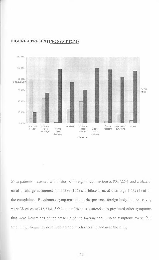
Most patients presented with history of foreign body insertion at 80.3(224) and unilateral
nasal discharge accounted for 44.8% (I25) and bilateral nasal discharge l .4% (4) of all
the complaints. Respiratory symptoms due to the presence foreign body in nasal cavity
were 38 cases of (16.6%). 5.0% (14) of the cases attended to presented other symptoms
that were indications of the presence of the foreign body. These symptoms were, foul
smell, high frequency nose rubbing, too much sneezing and nose bleeding.

I ABI.E 4 :LACi PERIOD BETWEEN NEB INSERTION AND PRESENTATION IN KNH
Period TotalsHours Days Weeks Unknown
Frequency 147 S4 30 18 279Percentage 52.7% 30.1% 10.8% 6.4% 100%
Majority of patients presented to hospital within 24 hours of NFB insertion comprising
52.2% (147) and less than 7 days constituted 30.1% (84) of all tha patients. Some
patients with incidental NFBs with unknown duration comprising 6.4% (18) of all the
patients.
25

TABLE 5: NFB INSERTER
Inserter Self Playmate H/girl Sibling Vomiting Unknown TotalsFrequency 215 JJ) 0 7 5 19 279Percentage 77.1% 11.8% 0% 2.5% 1.8% 6.8% 100%
Highest number of patients 77.1% (215) were those who inserted the foreign bodies by
themselves, whereas 33(11.8%) had the foreign bodies inserted by their playmates.
House girls played no role of inserting the foreign body with 0% prevalence. Patients
with foreign bodies who the inserter was not known were 19 cases (6.8%) of the study
population.
2 6

TABLE 6: WHO NOTICED THE FB
Noticed Parents Sibling H/girl S/reporting Teacher Doctor HAvorker TotFreq uency 159 9 25 76 2 5 279percentage 57.0% 3.2% 9.0% 27.2% 0.7% 1.1% 1.8% 100
The parents 57% (159) noticed most of the presence of nasal foreign body and self
reporting had the second highest frequency of 27.2% (76) cases. Teachers noticed two
(2) 0.7% cases . Those cases identified by doctors constituted 1.1% (3) of all the patients.
Those identified by other health workers were 1.8% (5) cases.
2 7

TABLE 7: NOSTRIL OF LODGEMENT
Nostril of lodgment Patients (frequency) percentageRight Nostril 169 60.6%Left Nostril 103 36.9%FB not found 6 2.1%Others 1 0.4%Totals 279 100%
Unilateral right nostril lodgement comprised of 60.6% (169) cases. Patients with FBs in
their left nostril were 36.9% (103) cases. Those presented to the hospital with history of
foreign body but the foreign body could not found were 2.1% (6) cases. One case
(0.4%) had the foreign body lodged in the nasopharynx.
28

FIGURE 5: ATTEMPTED REMOVAL
The patients with attempted removal before presentation at KNH were 69.2% (193)
cases of the total study population compared to those without attempted removal which
were 30.2% (86) patients.
29

TABLE 8: PLACE OF ATTEMPTED REMOVAL
Place of attempt No. of attempts % of total attemptsHome 75 38.9%Dispensary 42 21.8%District Hospital 12 6.2%Private clinics 62 32.1%School 2 1.0%Totals 193 100%
Attempted removal at home were 38.9% (75) cases of the total attempts. Others were
tried at various health facilities i.e. private clinics which attempted 32.1% (62) cases,
dispensary had 21.8% (42) cases, and district hospitals attempted 6.2% (12) of the total
attempts. There were two (2) (1.0%) cases that were attempted at the school. All the
above cases were attempts made before presentation to Kenyatta National Hospital.

TABLE 9: PERSON WHO ATTEPTED REMOVAL
Who attempted Frequency PercentageParents 60 31.1%Siblings 8 4.1%House help 10 5.2%Health worker 115 59.6%Teacher 2 1.0%Totals 193 100%
Most attempts were done at health facilities by health workers who had a frequency of
59.6% (115) of all attempts made. Parents who attempted had a frequency of 31.1% (60)
cases, siblings had 4.1% (8) cases and house helps 5.2% (10) cases. Those tried in school
were 1.0% (2) having the least prevalence in the study.
Those attempts made at home used different methods that included, nose blowing,
tobacco sniffing, and use of other crude items like ware, crotchet, needle and pins which
are more dangerous. Others included match sticks, ear buds, free hand/finger force and
mouth sucking. Attempts done at health facilities used tools like hooks and forceps to try
and remove the foreign bodies.
31

TABLE 10: STATE OF NASAL CAVITY BEFORE REMOVAL
Observation No. of occurrence percentageNasal discharge bloody 68 13.1%Mucopurulent 124 23.8%Watery discharge 24 4.6%Inflamed nasal mucosa 80 15.4%Foreign body visualized 188 36.2%Laceration from previous attempts
36 6.9%
Totals 520 100%
Easily visualized foreign bodies comprised of 36.2% (188) whereas those immersed in
mucopurulent discharge represented 23.8% (124) of the total cases. Nasal cavity with FB
immersed in watery nasal discharge were 4.6% (24) and bloody nasal discharge were
13.1% (68). Those with inflamed nasal mucosa comprised of 15.4% (80) cases and
those with laceration from the previous attempts were 6.9% (36) cases.
Of the total population investigated, there were a total of 520 observations made as some
patients had more than one sign.
Other nasal state signs observed were hyperemia of septal mucosa and foul smell.
32

TABLE 11: SITE OF NFB LODGEMENT
Site No. of cases % studyVestibule 7 2.5%Between the septum & inferior turbinate
190 68.1%
Between the septum & middle turbinate
72 25.8%
In the postnasal space (PNS)
1 0.4%
Others 2 0.7%None 6 2.1%In the throat 1 0.4%Totals 279 100%
The study revealed that the commonest site lodgement of foreign bodies in the nasal
cavity is between the septum and the inferior turbinate with 68.1% (190) cases of the total
study population. Those with foreign bodies lodged between the septum and middle
turbinate were 25.8% (72) cases. 2.5% (7) cases presented foreign bodies lodged in the
vestibule while those foreign bodies found in other sites i.e. posterior choana were two
(2) representing 0.7%
There was one case of foreign body in the postnasal space (PNS) accounting tor 0.4%.
2.1% (6) patients who presented to the hospital with history of nasal foreign body and
after thorough examinations , no foreign bodies were found. It is possible that the foreign
bodies were ingested by the complainants (patients). There was only one (1) case where
the foreign body was found in the throat representing 0.4% of the study.
33

TABLE 12: METHODS OF NFB REMOVAL
Method of removal Frequency of usage %Forceps 124 44.4%Hook 146 52.3%Positive pressure 2 0.7%Lateral rhinotomy incision 0 0%Not found 6 2.1%Others-FESS 1 0.4%TOTALS 279 100%
The most commonly used instrument was the hook with the highest frequency of (146)
52.3% followed by forceps which had a frequency of (124) 44.4%. Other methods
applied were positive pressure that was applied in two (2) 0.7% cases and lateral
rhinotomy was not used in any of the patients who participated in the study and therefore
had a frequency of 0%.
There were (6) 2.1%cases in which the foreign bodies were not found on explorative
rigid nasal endoscopy and it is possible that the foreign bodies were ingested by the
patients.
34

TABLE 13: TYPE OF ANAESTHESIA USED
Type of anaesthesia No. of patientsGeneral anaesthesia 97 (34.8%)None 182 (65.2%)TOTALS 279 (100%)
Patients who were not administered with any form of anaesthesia were 65.2% (182) of
the total study population. Those who were administered with general anaesthesia were
34.8% (97) cases of the study population. The study reveals that topical anaesthesia is
not used in the Emergency Room.
35

TABLE 14 INDICATIONS OF GENERAL ANAESTHESIA
Indications Frequency PercentagePatient unco-operative 87 (41.4%)Foreign body firmly lodged 56 (26.7%)Patients in lots of pain 44 (20.9%)FB not seen on anterior rhinoscopy
23 (11.0%)
Totals 210 (100.0%)
The figures in the number of indications were much higher than the number of patients
administered with anaesthesia because some of the patients had more than one indication.
General anaesthesia was administered to most patients who wrere unco-operative with a
frequency of 41.4% (87) cases of the total indications while those indications with
foreign body firmly lodged were 26.7% (56) cases. Patients who were administered
anaesthesia because of being in a lot of pain were 20.9% (44) of total indications. 11.0%
(23) patients underwent examination under anaesthesia because the foreign bodies were
not seen on anterior rhinoscopy.
36
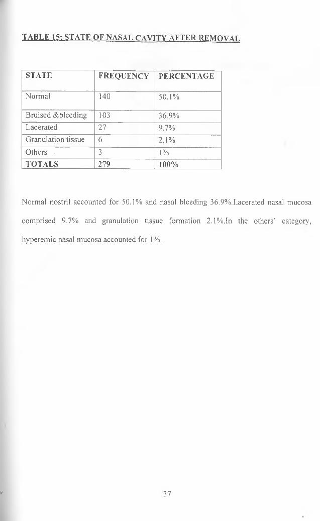
TABLE 15: STATE OF NASAL CAVITY AFTER REMOVAL
STATE FREQUENCY PERCENTAGE
Normal 140 50.1%
Bruised &bleeding 103 36.9%Lacerated 27 9.7%Granulation tissue 6 2.1%Others 3 1%TOTALS 279 100%
Normal nostril accounted for 50.1% and nasal bleeding 36.9%.Lacerated nasal mucosa
comprised 9.7% and granulation tissue formation 2.1%.In the others’ category,
hyperemic nasal mucosa accounted for 1%.
V 37

TABLE 16: TYPES OF FOREIGN BODIES
TYPE FREQUENCY PERCENTAGE
Bead 96 34.4%Cereals 64 22.9%Paper 31 11.1%Safety pin 1 0.4%Sponge 28 10.0%Piece of vegetable 29 10.4%Button 6 2.1%Rubber 10 3.6Stone (1 rhinolith) 4 1.4%Battery j 1.1%Crayon 1 0.4%No FB found 6 2.2%
TOTALS 279 100%
The commonest NFB was the bead at 34.4% followed by cereals at 23.3%.The other FBs
were seen the following order of frequency: paper 11.1%,sponge 10.0%,piece of
vegetable 10.4%,button 2.1% rubber 3.6%,stone 1.4% ,battery l.l%,cryon 0.4% and
those no FB was found accounted for 2.2%.An open safety pin was retrieved from an
adult female (0.4%).
38

TABLE 17 :COMPLICATIQNS OF ATTEMPTED REMOVAL
COMPLICATION FREQUENCY PERCENTAGENone 119 42.7%Ulceration 35 12.5%Nasal bleeding 125 44.8%TOTALS 279 100%
Commonest complication of attempted removal was nasal bleeding at 44.8% and those
no complication was observed were 42.7%.Ulceration accounted for 12.5%.No FB
ingestion or aspiration was observed.
39
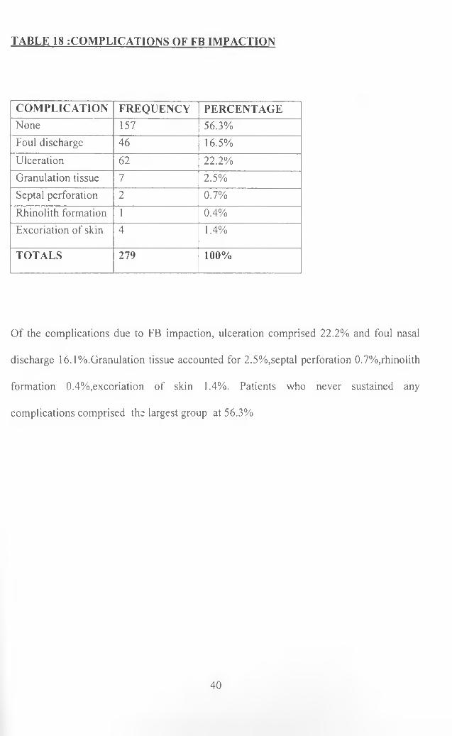
TABLE 18 :COMPLICATIONS OF FB IMPACTION
COMPLICATION FREQUENCY PERCENTAGENone 157 56.3%Foul discharge 46 16.5%Ulceration 62 22.2%Granulation tissue 7 2.5%Septal perforation 2 0.7%Rhinolith formation 1 0.4%Excoriation of skin 4 1.4%
TOTALS 279 100%
Of the complications due to FB impaction, ulceration comprised 22.2% and foul nasal
discharge 16.1%.Granulation tissue accounted for 2.5%,septal perforation 0.7%,rhinolith
formation 0.4%,excoriation of skin 1.4%. Patients who never sustained any
complications comprised the largest group at 56.3%
40

Majority of patients presented to KNH within 24 hours (52.7%) of FB insertion
and this is similar to a series of cases by Balbani A P et al where he found 45.8%
presented to hospital within 24 hours. Those who took days (30.1%) had presented in
some primary health facility within hours of insertion with resultant failed attempted
removal and patients took time to travel to KNH. Patients who took weeks, are those on
anterior rhinoscopy .were reassured of no NFB only for complications like persistent
foul discharge to set in and necessitating presentation at KNH.
The NFBs were self-inserted in 77.1% of the cases and by playmate accounted for
11.8%, vomiting through nasal regurgitation 1.8% and those not known comprised 6.8%
.Self insertion still remains the most important aetiologic factor and by use of hawk eye
of an house help, the problem may significantly be reduced. Most patients are reported in
literature to insert FBs deliberately to relieve mucosal irritation or epistaxis(38).This
study had not been designed to look at risk factors and therefore there will be need in
future for a study to investigate risk factors in NFBs.
Despite majority of NFBs being inserted under the care of mothers , they noticed
them(159) 57% during routine cleaning or on examination when children behaved
unusually. Self-reporting accounted for 27.2% (76), a fairly low figure because majority
of the patients were less than 3 years old and therefore not yet fluent in speech. Most
NFBs were lodged in the right nostril (169) 60.6% compared to left nostril (103)
36.9%.Similar findings are reported by Franycois M et al who found unilateral FB in
right nostril 67.6% and two bilateral NFBs .No bilateral NFBs were found in this study.
Many of the patients had attempted removal (193) 69.2% before presentation at
KNH with majority being done at home 38.9% (75) and private clinics 62 (32.1%) but
were unsuccessful, may be for lack of the right instruments and removal skills that are
key to successful removal. At home the parents (31.1%) tried such methods as nose
blowing, tobacco sniffing and such instruments as wires, crotchets, needles and pins,
match sticks, ear buds and fingernails. From this study is difficult to comment on the
success of these methods, if any, as the patients will ordinarily not present to hospital
after removal. Therefore there will seem to be need for a controlled study to assess their
success as they seemed popular with parents. Health workers in private clinics attempted
in 59.6% of the cases and failed to remove the FBs. This is a big number and therefore
42

there will be need to ascertain the level of training of [he workers as the clinics were
convenient both in accessibility and affordability going by high number of patients who
presented there.
In most of the cases, the NFBs were visualized (36.2%) on anterior rhinoscopy
while others were covered in bloody discharge (13.1%) from injuries of previous
attempts, mucopurulent discharge (23.8%) and in some cases there were lacerations
(6.9%).Majority of the FBs were lodged between the septum and inferior turbinate
(68.1%) and septum and middle turbinate (25.8%) and a similar trend is reported by
Kalan et al (31)
A variety of techniques and instruments have been used to remove foreign bodies
from the nasal passages. The methods used most successfully include the angled hooks
(52.3%) and forceps (44.4%).There was one case of rhinolith which had presented with
progressive nasal blockage and was taken to theatre for examination under anaesthesia
for a granulomatous disease and was found to be stony hard and was removed piece meal
with help of nasal endoscope and tissue forceps. Flistology confirmed inflammatory
reaction to a foreign body. In 2.1% of the cases, no FB was seen on anterior rhinoscopy
and were taken to theatre for explorative rigid nasal endoscopy as flexible fibre-optic
endoscopy was technically not possible on this patients who were already uncooperative.
Only one patient who had skull x-ray, as the safety pin lodged in her nostrils could not be
visualized on anterior rhincscopy. Flowever many materials such as food, sponge, beads
may not be visible on radiographs and therefore patients who there was doubt, were taken
to theatre for explorative rigid nasal endoscopy under general anaesthesia.
Topical anaesthesia was not used and majority of the foreign bodies were
retrieved by proper positioning in 65.2% of the patients. This ensured that NFBs were
removed with minimal attempts and without loosing the patient’s co-operation as will
commonly happen due to the unpleasant irritation of topical anaethesia. In 34.8% of the
patients the foreign bodies were removed under general anaesthesia because they were
firmly lodged, 26.7% were unco-operative, 41.4% were in severe pain and 20.9% of the
FBs could not be seen on anterior rhinoscopy. It is important to note that removal can be
quite simple and successful removal rates in the Emergency Room of over 90% have
43

been documented in literature(38) .Previous failed attempted removal seem to make the
patients apprehensive and therefore lower the success rates.
Procedural or conscious sedation for removal of nasal foreign bodies is
increasingly being used in the Emergency Room other centers and is reported to provide
important anxiolytic and sedation for safe removal of nasal foreign bodies in young
children .However special caution is advised given the proximity of the NFBs to the
airway when using medications that can blunt airway and respiratory reflexes. In this
study, conscious sedation was not used.
No abnormality were noted after NFB removal in 50.1% of the nostrils though
36.9% had sustained bruises and bleeding and granulation tissue formation in 2.1%.No
patient in this study aspirated objects lodged in their nostrils though the possibility exists
especially in loosely held objects in unco-operative patient. The complications that
occurred such as ulceration 12.5%, nose bleeding 44.8% were usually related to repeated
attempts at removal. The FBs that were never found after comprehensive nasal
examination may have been ingested. However since oesophagoscopies were not done,
that could not be confirmed. Synaechie and nasal stenosis were not observed in this study
population though a prospective, longitudinal study will need to done to follow up the
patients who sustain lacerations, bruises and granulation tissue formation to confirm if
nasal patency is comprised in any way.
In majority of the patients (56.3%),there were no immediately recognizable
complications though there was rhinolith formation in 0.4% of the patients, ulceration in
22.2% of the patients in the firmly lodged FBs. Septal perforation was observed in 0.7%
of the patients.Generally, most NFB impactions can be safely retrieved without much
ado if the right techniques and procedures are used.
UNIVERSITY OF NAIROBIm e d i c a l l i b r a r y
44

CHAPTER VII
7.0 CONCLUSION:
Younger children are more prone to insert foreign bodies which are objects
usually found in our domestic environments and most nasal foreign bodies can be
successfully removed in the Emergency Room by a skilled clinician utilizing any number
of simple techniques depending on the type of nasal foreign body. Because of the many
different nasal foreign bodies found, the primary clinicians should be skilled in the
numerous techniques of removal.
A few cases may require multiple attempts or multiple techniques for successful
removal and with a few exceptions, difficult cases can be removed under general
anaesthesia. Complications may occur as a result of attempts to remove the foreign
bodies without help of skilled personnel or proper conditions. However most patients can
be successfully managed without complications if correct procedures are adopted.
45

OJ
CHAPTER VIII
8.0 RECOMMENDATIONS:
1. Mothers should use bigger beads in plaiting children's hair which can not
easily fit in nasal orifices. Or totally get rid of beads from the domestic
environment by keeping them out of reach of children.
2. Clinicians consulted, if in doubt of their centres’ capacity and skills at
removal, should refer the patients without attempted removal to centres
with the skilled personel and facilities.
Use of conscious or procedural sedation will reduce the number patient
exposed to general anaesthesia for unco-operation and pain.
46

APPENDIX 1: PROFORMA
NASAL FOREIGN BODIES:
1.0 PART A
1.1 Patient's socio-demographic Particulars:
a) Name of Patient:.......................................... Date...............................
b) Age of Patient:................................
c) Hospital N o:....................................
d) Study N o:.......................................
e) Contact Address...........................T el..........................
f) Gender Male () Female ()
g) Residence at the time of NFB insertion.
i) Rural ( ) ii) Urban () iii) Boarding school ( ) iv) day care
centre () v) Other................
h) Level of education
i) Non-school going ( ) ii) Nursery ( ) ii) Between standard 1-3 ( )
iii) Between standard 4-8 () iv)Other .....................
i) .Patients’ caretaker.
i) Mother () ii) Older sibling () iii) House help () iv) Self ()
v) Other ( specify)........
1.2 Disease History :
1.21 Presenting complaints:
i. History of FB insertion Yes () No ()
ii. Nasal discharge -unilateral Yes () No ()
-bilateral Yes () No ()
iii. Nasal pain Yes () No ()
iv. Nasal blockage -unilateral Yes () No ()
-bilateral Yes () No ()
V. Frontal headache Yes () No ()
vi. Respiratory symptoms(cough, wheeze) Yes () No ()
vii. Other (specify) —
47
1.22 Duration of complaints in:
i. Hours ()
ii. Days ()
iii. Weeks ()
iv. Not known ()
v. Other (specify).......................
NB: Specify exact duration e.g. (2) days
1.23 Who inserted the foreign body
i. Self ()
ii. Playmate ()
iii. House girl ()
iv. Siblings ()
v. Regurgitation/vomiting ()
vi. Other (specify).....................
1.24 Who noticed the presence of the foreign body?
i. Parents ()
ii. Siblings ()
iii. House girl ()
iv. Self reporting ()
v. Other (specify)...................
1.25 Was there attempted removal before presentation at KNH?
Yes () No ()
If yes, where?
a. Home ()
b. Dispensary ()
c. District hospital ()
d. Private clinics ()
e. Other (specify)......................
48

By whom?
i. Parents 0ii. Siblings 0
iii. House girl 0iv. Health worker ()
V. Other (specify).......................
What method was used in the attempted removal (specify)............................
2.0 PART B
2.1 State of the nose before removal.
i. Nasal discharge Bloody (), Mucopurulent (), watery ()
ii Inflamed nasal mucosa ()
iii FB clearly visualized ()
iv. Lacerations from previous attempts ()
v. Other (specify).......................
2.2 In which nostril was the FB?
i) Right ( ) ii) Left () iii) Others (specify)
2.3 Site of lodgement of the FB?
i. Vestibule ()
ii. Between the septum and the inferior turbinate. ()
iii. Between the septum and middle turbinate ()
iv. In the postnasal space (PNS) ()
v. Other (Specify)............................... ()
49
2.4 Method used to remove the foreign body?
i. Forceps 0ii. Hook 0
iii. Positive pressure technique 0iv. Lateral rhinotomy incision 0V. Other (specify)...........................
2.5 Which form of anaesthesia was used?
i None ()
ii Topical anaesthesia ()
iii General Anaesthesia ()
2.6 If general anaesthesia was used, what was the indication?
i. Patient unco-operative ()
ii. Foreign body firmly lodged ()
iii. Patient in a lot of pain ()
iv. FB not seen on anterior rhinoscopy ()
v. Other (specify)..................
2.7 State of the nasal cavity walls after removal?
i. Normal ()
ii. Bruised and bleeding ()
iii. Lacerated ()
iv. Granulation tissue ()
v. Other (specify).......................
2.8 Type of foreign body?
i. Bead ()
ii. Miniature battery ()
iii. Piece of vegetable ()
iv. Paper ()
v. Other (specify)...............
50

APPENDIX II
REFERENCES:
1. Werman, HA: Removal of Foreign Body of the Nose: Emergency clinics of
North America; 5 (i) 253-63, 1987.
2. Baker MD: Foreign Bodies of the Ear and Nose in childhood: Pediatric
emergency care; 3 (i) 67-70: 1987.
3. Das SK: Aetiological evaluation of foreign bodies in the ear and nose; Journal
of laryngology and Otology: 98 (10): 989-91, 1984.
4. Myer CM, Cotton RT: Nasal obstruction in the paediatric patient: Paediatrics
72 (6): 766-77, 1983.
5. Forarelli P et al: An unusual untranasal foreign body; Paediatric emergency
care, 4(2): 117-8, 1988.
6. Bhatia PL: Otolaryngological foreign bodies; a study in Jos, Nigeria: Tropical
Doctor, 19(2): 62-4, 1998.
7. Ritter J, Berghansa; Iatragenic foreign body of the nose. Laryngo-Rhino-
Otologie: 68(5)299-300, 1989.
8. Stoney P, Binghan B et al: Diagnosis of rhinolith with rigid endoscopy; Journal
of otolaryngology, 20(6): 408-11, 1991.
9. Harun S et al; An unusual oronasal foreign body; Journal of Laryngology and
Otology, 105(12): 1118-9, 1991.
10. Tong MC et al: The hazards of button batteries in the nose; Journal of
otolaryngology 21 (6): 458-60, 1992.
11. Cohen HA et al: Removal of nasal foreign body in children: Clinical
paediatrics, 32 (3): 192, 1993.
12. Zealand; Intranasal button battery causing septal perforation: a case report;
Journal of Laryngology and Otology: 108(7): 586-90, 1994.
13. Backlin SA; positive pressure technique for nasal foreign body removal in
children; Annal of Emergency Medicine, 25(4): 554-5, 1995.
14. Loh WS; Hazardous foreign body; Complications and management of button
batteries in nose: Annal of Otology, Rhinology and Laryngology, 112(4); 379-
83, 2003.
52

15. Vink Bw et al; A case of Rhinolithiasis in Bostwana; a mineralogical,
microscopic and chemical study: Journal ol Laryngology and Otology, 116(12):
106-40, 2002.
16. Balbani AP et al; Ear and Nose foreign body removal in children; Int, J,
Paediatri Otorhinolaryngol: 1998 No 15.46 (1-2) P37-42.
17. Franycois M et al; Nasal foreign bodies in children; Eur, Arch.
Otorhinolaryngol, 1998. 255(3): PI32-4.
18. Mu-noz A et al; Eraseroma as a cause of rhinolith: CT and MRI in a child;
neuroradiology. 1997, 39 (11), P824-6.
19. Al, Ayoubi A, Ray J; An unusual foreign body in the postnasal space: a case
report: J Laryngol.Otol 1997 118 (8) P775.
20. Kadish Ha, Correli Hm; Removal of nasal foreign bodies in the paediatric
population; AM, J Emerg. Med. 1997, 15(1): P54-6.
21. De Carpentier et al; An unusual cause of facial pain: J. Larvngol. 1996, 110(8)
P796-8.
22. Doudlas AR; Use of nebulised adrenaline to aid expulsion of itranasal foreign
bodies in children; J. Laryngol.Otol. 1996, 110 (6). P559-60.
23. Tong Me et al; Nasal foreign bodies in children; Int.J.Pedatri.Otolaryngol.
1996:95:961-2.
24. Finkelstein JA; Oral Ambu-bag insufflation to remove unilateral nasal
foreign bodies. Am.J.Emerg.Med. 1996, 14(1). P57-8.
25. Lawton H et al; Nasal foreign body: Removal of an open safety pin from the
left nostril; ENT: ear, nose and Throat Journal: 2000, 84:824-6.
26. John F et al; Treatment of aural foreign bodies in children; Pediatrics, 1998,
35:207-11.
27. Aijaz Alvi et al; Miniature Disc Battery in nose: A dangerous foreign body:
clinical paediatrics, july 1997, 56:403-6.
28. Isabella Fini-storch et al; A typical intranasal foreign body; Ear, Nose and
Throat journal December, 2001, 95:961-2.
29. Mohd Maqbool; Foreign bodies in the Nose; Textbook of Ear, Nose and
Throat Diseases, JayPee brothers medical publisher (P) Ltd, 9th ed. PI 58-9.
53

UNIVERSi l Y OF NAIROBIm e d i c a l l i b r a r y
30. Stoney P., Bingham B, Okuda I et al: Diagnosis of Rhinoliths with rigid
endoscopy; janol of Otolaryngology 20(6): 408-11, 1991.31. Utrata j. Erosion of the soft palate by a foreign body of the nose.Ear, Nose, Throat J: 1977; 56:403-432. Sukhbir Ahluwalia, Anthony A N; Foreign bodies in the ear, nose and throat:
Surgery International, 2004; 66:182-183.
33. Kalan A, Tarq M: Foreign Bodies in the nasal cavities: A comprehensive review
of the aetiology, diagnostic pointers and therapeutic measures: Postgrad. Med J 2000
(8):76:484-487
34. Romanes G.J: Cunningham’s manual of practical Anatomy: volume 3: head and
Neck and Brain 15th edition Pg 150-153, Oxford university presses.
35. Spell S.R: Clinical Anatomy for medical students, 6th edition; p743-7, Lippincott
Williams & Wilkins.
36. Barker D.J.P, Hall .A.J. Populations and samples; Practical epidemiology. 4th
edition, 1991, 30-43.
37. A Ngo,K C Ng: Otorhinolarvngeal foreign bodies in children presenting to
emergency department; Singapore Medical Joumal:2005;46(4):172
38. Theodore C, Jacob et al: Nasal foreign body removal: The journal of Emergency
Medicine, Vol 26.No.4,pp.441-445,2004
54