Management of high-energy tibial pilon fractures
11
REVIEW Management of high-energy tibial pilon fractures Nebu Jacob 1,3 • Amit Amin 1 • Nikolaos Giotakis 2 • Badri Narayan 2 • Selvadurai Nayagam 2 • Alex J. Trompeter 1 Received: 3 March 2015 / Accepted: 23 August 2015 / Published online: 25 September 2015 Ó The Author(s) 2015. This article is published with open access at Springerlink.com Abstract Tibial pilon fractures result from high-energy trauma unlike usual ankle fractures. Their management provides numerous challenges to the orthopaedic surgeon including obtaining anatomic reduction of articular surface and the management of associated soft tissue injuries. This article aims to review major advances and principles that guide our practice today. We also discuss a treatment algorithm based on a staged approach to the fracture: initial spanning external fixation followed by definitive fixation. Keywords Pilon fractures Á Management Á Strategy Á Algorithm Á Reconstruction Á Bone defects Introduction Pilon is the French word for a pestle. Etienne Destot, a French Radiologist, is credited for using the term to describe the fracture in 1911. He compared the talus to a pestle. High-energy tibial ‘pilon’ fractures are due to axial loading with the talus driven into the distal tibia, exploding the distal tibial articular surface with impaction of the comminuted metaphyseal bone, and with occasional proximal diaphyseal extensions. These commonly result from falls from a height or from motor-vehicle-related accidents [1]. The degree of trauma to the surrounding soft tissue envelope cannot be underestimated; there is limited muscle cover between the skin and bone at this level of the lower limb, and the energy of the injury is transferred directly to these soft tissue structures. Open fractures are common, and even in the absence of an open lesion, sig- nificant soft tissue damage must be appreciated in closed injuries [2]. The treatment objectives are to restore articular con- gruency and mechanical alignment and to allow early functional rehabilitation whilst minimising soft tissue complications. Two-stage management with initial span- ning external fixation allows soft tissue resuscitation prior to definitive management and has gained acceptance by most surgeons [1, 3–7]. Microscopic articular cartilage damage that occurs at the time of injury has significant bearing on the long-term prognosis even in the presence of anatomical joint reduc- tion [8, 9]. The challenge lies in minimising complications, such as deep infection, whilst optimising clinical outcome through appropriate and well-timed surgery. This article focuses on the controversies in the management of high- energy pilon fractures, and we present a treatment algo- rithm based on the authors’ collective clinical experience. Classification The two main X-ray classification schemes are those of Ruedi and Allgower [10] and the AO Foundation and Orthopaedic Trauma Association (AO/OTA) [11]. Ruedi and Allgower described three groups, specific to tibial pilon fractures, based on the size and displacement of articular fragments: type I represents non-displaced & Nebu Jacob [email protected] 1 Department of Trauma and Orthopaedic Surgery, St Georges Healthcare NHS Trust, Blackshaw Road, Tooting, London SW17 0QT, UK 2 Limb Reconstruction Unit, Department of Trauma and Orthopaedic Surgery, Royal Liverpool and Broadgreen University Hospital NHS Trust, Liverpool L7 8XP, UK 3 1 Locke Gardens, Slough, Berkshire SL3 7BE, UK 123 Strat Traum Limb Recon (2015) 10:137–147 DOI 10.1007/s11751-015-0231-5
Transcript of Management of high-energy tibial pilon fractures
REVIEW
Management of high-energy tibial pilon fractures
Nebu Jacob1,3 • Amit Amin1 • Nikolaos Giotakis2 • Badri Narayan2 •
Selvadurai Nayagam2• Alex J. Trompeter1
Received: 3 March 2015 / Accepted: 23 August 2015 / Published online: 25 September 2015
� The Author(s) 2015. This article is published with open access at Springerlink.com
Abstract Tibial pilon fractures result from high-energy
trauma unlike usual ankle fractures. Their management
provides numerous challenges to the orthopaedic surgeon
including obtaining anatomic reduction of articular surface
and the management of associated soft tissue injuries. This
article aims to review major advances and principles that
guide our practice today. We also discuss a treatment
algorithm based on a staged approach to the fracture: initial
spanning external fixation followed by definitive fixation.
Keywords Pilon fractures � Management � Strategy �Algorithm � Reconstruction � Bone defects
Introduction
Pilon is the French word for a pestle. Etienne Destot, a
French Radiologist, is credited for using the term to
describe the fracture in 1911. He compared the talus to a
pestle. High-energy tibial ‘pilon’ fractures are due to axial
loading with the talus driven into the distal tibia, exploding
the distal tibial articular surface with impaction of the
comminuted metaphyseal bone, and with occasional
proximal diaphyseal extensions. These commonly result
from falls from a height or from motor-vehicle-related
accidents [1]. The degree of trauma to the surrounding soft
tissue envelope cannot be underestimated; there is limited
muscle cover between the skin and bone at this level of the
lower limb, and the energy of the injury is transferred
directly to these soft tissue structures. Open fractures are
common, and even in the absence of an open lesion, sig-
nificant soft tissue damage must be appreciated in closed
injuries [2].
The treatment objectives are to restore articular con-
gruency and mechanical alignment and to allow early
functional rehabilitation whilst minimising soft tissue
complications. Two-stage management with initial span-
ning external fixation allows soft tissue resuscitation prior
to definitive management and has gained acceptance by
most surgeons [1, 3–7].
Microscopic articular cartilage damage that occurs at the
time of injury has significant bearing on the long-term
prognosis even in the presence of anatomical joint reduc-
tion [8, 9]. The challenge lies in minimising complications,
such as deep infection, whilst optimising clinical outcome
through appropriate and well-timed surgery. This article
focuses on the controversies in the management of high-
energy pilon fractures, and we present a treatment algo-
rithm based on the authors’ collective clinical experience.
Classification
The two main X-ray classification schemes are those of
Ruedi and Allgower [10] and the AO Foundation and
Orthopaedic Trauma Association (AO/OTA) [11].
Ruedi and Allgower described three groups, specific to
tibial pilon fractures, based on the size and displacement
of articular fragments: type I represents non-displaced
& Nebu Jacob
1 Department of Trauma and Orthopaedic Surgery, St Georges
Healthcare NHS Trust, Blackshaw Road, Tooting,
London SW17 0QT, UK
2 Limb Reconstruction Unit, Department of Trauma and
Orthopaedic Surgery, Royal Liverpool and Broadgreen
University Hospital NHS Trust, Liverpool L7 8XP, UK
3 1 Locke Gardens, Slough, Berkshire SL3 7BE, UK
123
Strat Traum Limb Recon (2015) 10:137–147
DOI 10.1007/s11751-015-0231-5
intra-articular fractures without loss of articular congru-
ency; type II represents displaced fractures with loss of
articular congruency; and type III represents those
severely comminuted fractures with impaction of the distal
tibia.
The AO/OTA group use an alphanumeric system to
describe all fractures. The first number represents the bone:
in this case, tibia is ‘4’; the second represents the segment
of bone which in this case is ‘3’ for distal. Following this,
‘A’ represents extra-articular fractures within 5 cm of the
ankle joint and ‘B’ represents partial articular injuries, both
not included in this review. Group ‘C’ denotes complete
articular injury where there is no direct continuity between
the diaphysis and the articular segment. This group
accounts for the majority of high-energy pilon fractures.
The final numbers in this classification represent the sub-
groups of each type and refer to the degree of comminution
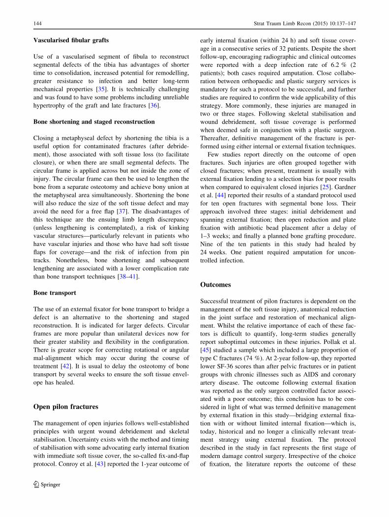
of the articular component and the metaphysis (Fig. 1).
Swiontkowski et al. [12] raised concern about classifi-
cation systems in general when reporting on the inter-ob-
server reliability of the AO/OTA system. They found
moderate correlation for groups A, B or C and poor cor-
relation between subgroup detection. They concluded that
compartmentalising fracture severity, which behaves as a
continuous and not a dichotomous variable, should be
avoided.
Topliss et al. [13] reviewed a consecutive series of 126
pilon fractures with 115 cases classified as AO/OTA ‘C’
type injuries. Of these, 67 patients (52 %) had the more
complex C3 injuries. It is this subgroup, which comprises
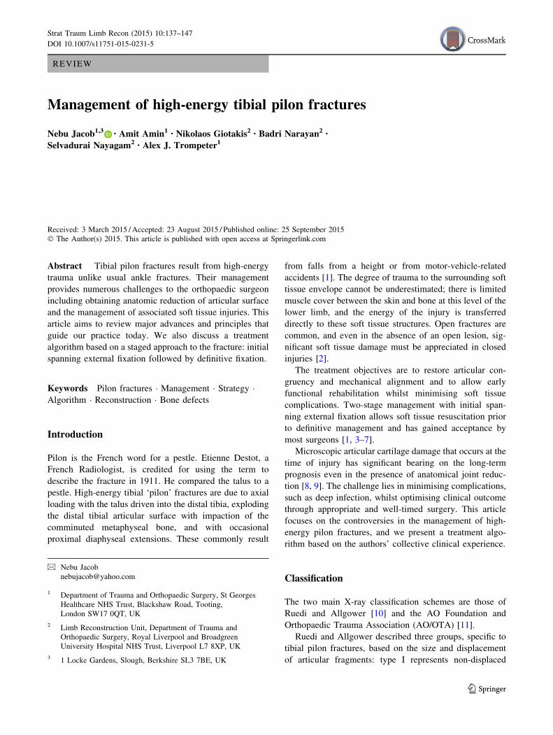
Fig. 1 AO classification of distal tibial fractures (Muller AO Classification of Fractures-Long Bones, Copyright by AO Foundation,
Switzerland)
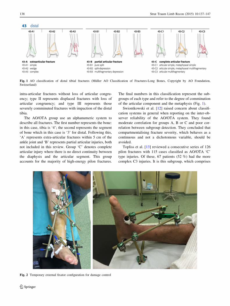
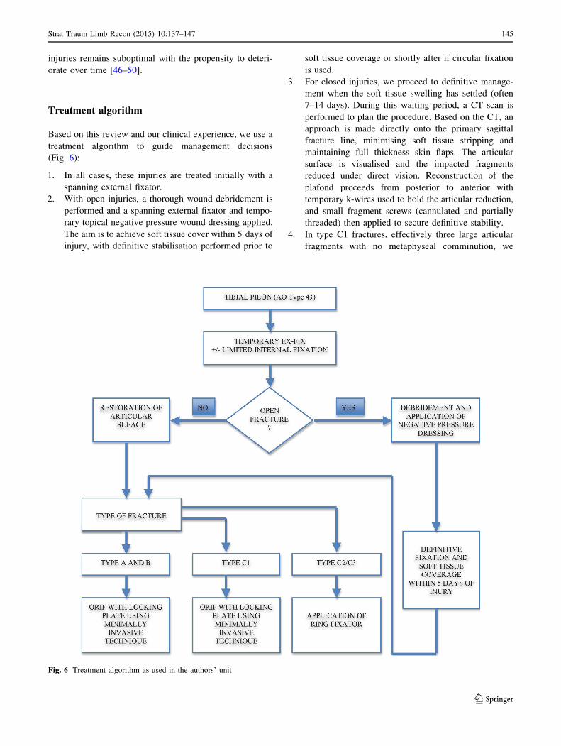
Fig. 2 Temporary external fixator configuration for damage control
138 Strat Traum Limb Recon (2015) 10:137–147
123
true high-energy pilon fractures, where significant discrep-
ancy and disagreement exist in the literature over manage-
ment. Their study provided a CT-based classification
segregating fracture patterns into two main families, which
were termed ‘sagittal’ and ‘coronal’ based on the primary
fracture line seen on axial cuts at the level of the plafond.
These subtypes were assessed for patient and deformity
characteristics, noting that sagittal plane fractures tended to
present in varus and had resulted from higher-energy inju-
ries in younger individuals. The coronal plane fractures
tended to present in valgus and were associated with lower-
energy injuries in older patients. This study offered an
interesting insight into the spectrum of fracture pattern
variability. Although the authors reported good inter-ob-
server reliability, their findings have yet to be replicated.
Initial management
Pilon fractures managed akin to polytrauma
with damage control strategies
Early operative management through a tenuous soft tissue
envelope risks wound healing problems, invites infection
and can potentially lead to limb amputation. Temporary
spanning external fixation, with or without fibular stabili-
sation at index surgery, has gained acceptance as the first-
line intervention and is considered a local ‘damage control’
strategy (Fig. 2).
Patterson and Cole [5] first described the two-stage
management of pilon injuries with definitive management
undertaken at 10–14 days following external fixation and
with all patients having formal open reduction and internal
fixation. Sirkin et al. [3] popularised this protocol in two
subsequent publications stating that the technique was
successful in both closed and open fractures.
Temporary stabilisation should be performed as soon as
possible but preferably during daylight hours on a desig-
nated operating list. A careful restoration of alignment with
the external fixator must be considered at this early stage.
Fixator constructs vary with ‘delta’ and ‘A’ frames
assemblies being most common. An extension of the fix-
ator onto the forefoot (usually the first metatarsal) is
helpful to avoid an equinus contracture. This method of
skeletal stabilisation has superseded calcaneal traction as it
permits patient mobilisation albeit non-weight bearing. In
some centres, patients are sent home whilst awaiting soft
tissue recovery and definitive management.
CT scan
CT scanning is a prerequisite for planning definitive
management and is best performed after application of
spanning external fixation and restoration of overall
alignment through ligamentotaxis. The axial cuts at the
level of the plafond accurately define fracture plane ori-
entation, whilst sagittal and coronal reformatting allows a
full assessment of fracture morphology.
Tornetta et al. [14] correlated radiographs and CT scans
in 22 patients with pilon fractures. Based on the CT find-
ings, they altered their surgical approach in 64 % of their
patients. In 12 patients, the major fracture line exited lat-
erally, and in 10 patients, it exited medially. The identifi-
cation of this major fracture line dictated the surgical
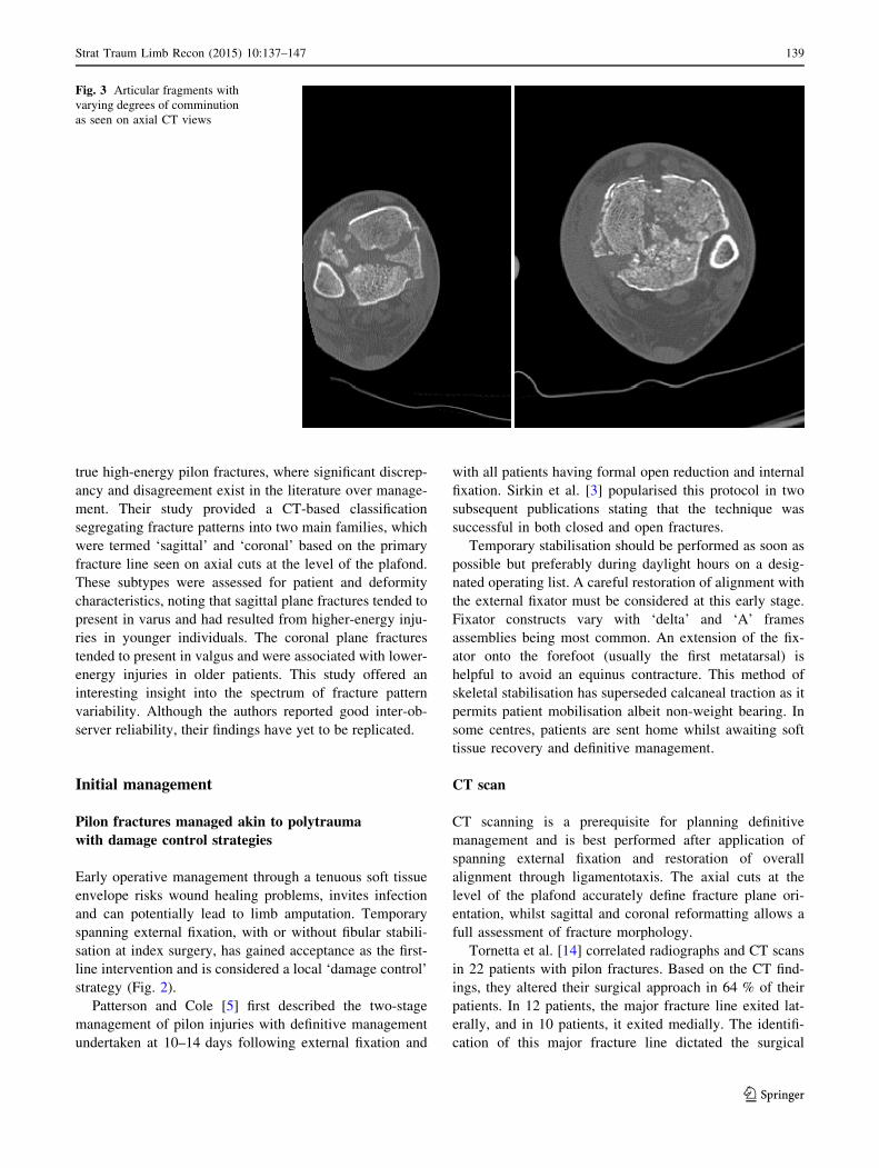
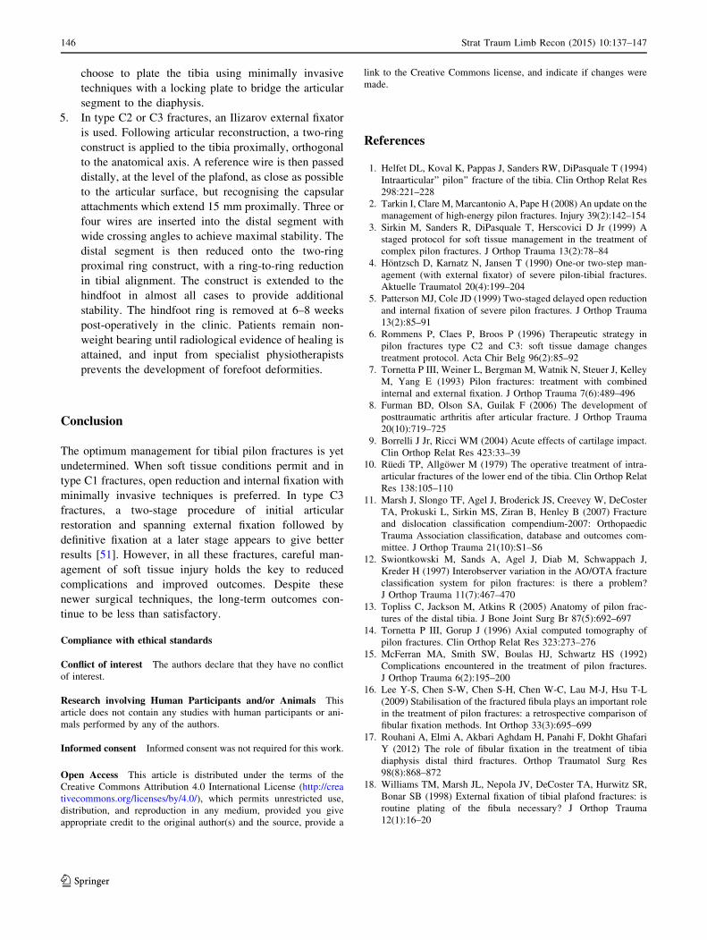
Fig. 3 Articular fragments with
varying degrees of comminution
as seen on axial CT views
Strat Traum Limb Recon (2015) 10:137–147 139
123
approach to the fracture. Where the fracture line exited
medially, an anteromedial approach based on the tibialis
anterior was used. In the others with a lateral exit of the
fracture line, a lateral approach between the extensor dig-
itorum communis and peroneus tertius was used. The CT
scan provided vital information on metaphyseal com-
minution which in five patients was fixed percutaneously.
Images from the CT are essential as axial cuts demonstrate
fracture lines common to all pilon fractures, knowledge of
which is vital for pre-operative planning, incision place-
ment and articular reduction.
There are three typical articular fragments: anterolateral,
posterolateral and medial, with variations in size and
comminution (Fig. 3).
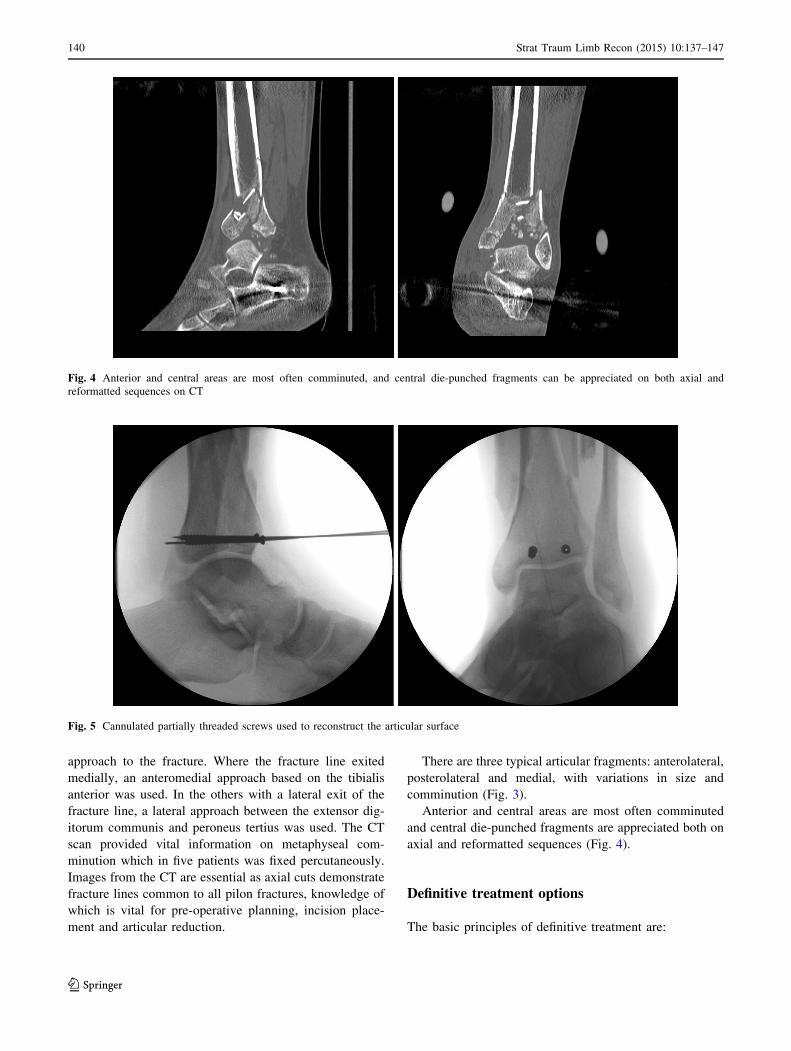
Anterior and central areas are most often comminuted
and central die-punched fragments are appreciated both on
axial and reformatted sequences (Fig. 4).
Definitive treatment options
The basic principles of definitive treatment are:
Fig. 4 Anterior and central areas are most often comminuted, and central die-punched fragments can be appreciated on both axial and
reformatted sequences on CT
Fig. 5 Cannulated partially threaded screws used to reconstruct the articular surface
140 Strat Traum Limb Recon (2015) 10:137–147
123
1. Articular reduction and stabilisation.
2. Restoration of alignment by reduction in the recon-
structed articular block to the diaphysis.
3. Management of bone loss at primary surgery or as a
planned late intervention (C3 injuries).
4. Respect for the soft tissue envelope
5. Early restoration of motion
Treatment choice is based upon the severity of the soft
tissue injury, fracture pattern and the treating surgeon’s
experience. There is no level I evidence currently for
optimal management with both internal and external fixa-
tion techniques, alone or in combination, commonly
employed.
Traditional open reduction and internal fixation (ORIF)
of complex type ‘C’ fractures, with direct exposure of the
metadiaphyseal region, extensive surgical dissection and
handling of all fracture fragments was associated with an
unacceptably high soft tissue complication rate [15].The
early good results of ORIF, reported by Reudi and Allgo-
wer [10], were based on a different patient and fracture
population, many of whom sustained lower-energy distal
tibial fractures with extension to the tibial plafond sec-
ondary to skiing injuries. Nevertheless, their four classic
principles of treatment: plating the fibula to length, artic-
ular reconstruction, bone grafting of metaphyseal defects
and providing a medial buttress to the tibia, still remain
important orthopaedic concepts.
The role of initial fibula fixation is controversial. The
proposed benefits include restoration of length, indirect
reduction in the tubercle of Chaput (anterolateral) and
Volkmann (posterolateral) tibial fragments in the case of
distal fibula fractures, and a faster soft tissue recovery.
Conversely, ignoring the fibula fracture allows the option
of tibial shortening to improve fracture contact at the
metaphysis, especially in the type C3 fracture where the
metaphysis is comminuted and prone to delayed healing. In
pilon fractures, Lee et al. [16] found a lower rate of
malunion and ankle arthrosis in 6 years of follow-up when
the fibula was fixed by plating compared to pin fixation.
Rouhani et al. [17] and Williams et al. [18] found no
clinical difference at 6-month and at 2-year follow-ups,
respectively, in patients treated with ankle-bridging exter-
nal fixation, with or without fibula plating. The plating
group suffered more wound complications, and the non-
plating group had more with angular malunion. However,
this may have resulted from the bridging technique used in
their study which did not provide any direct metadiaphy-
seal stability.
If fibular fixation is undertaken, careful pre-operative
planning for the approach to the tibial pilon and fibula is
needed to avoid a high wound complication rate. Ideally,
these fractures should to be referred to experts early.
Restoration of articular surface
Reconstruction of high-energy type C fractures should be
performed when the soft tissue conditions allow safe sur-
gical dissection. Direct exposure of the articular segment
through planned limited or formal approaches is advocated.
Percutaneous techniques can be used with simple articular
patterns, but C3 injuries require direct reduction.
The common approaches are either anterolateral or
anteromedial depending on the axial CT images at the level
of the plafond as described in the study by Tornetta et al.
[14]. The incisions allow direct articular reduction, but not
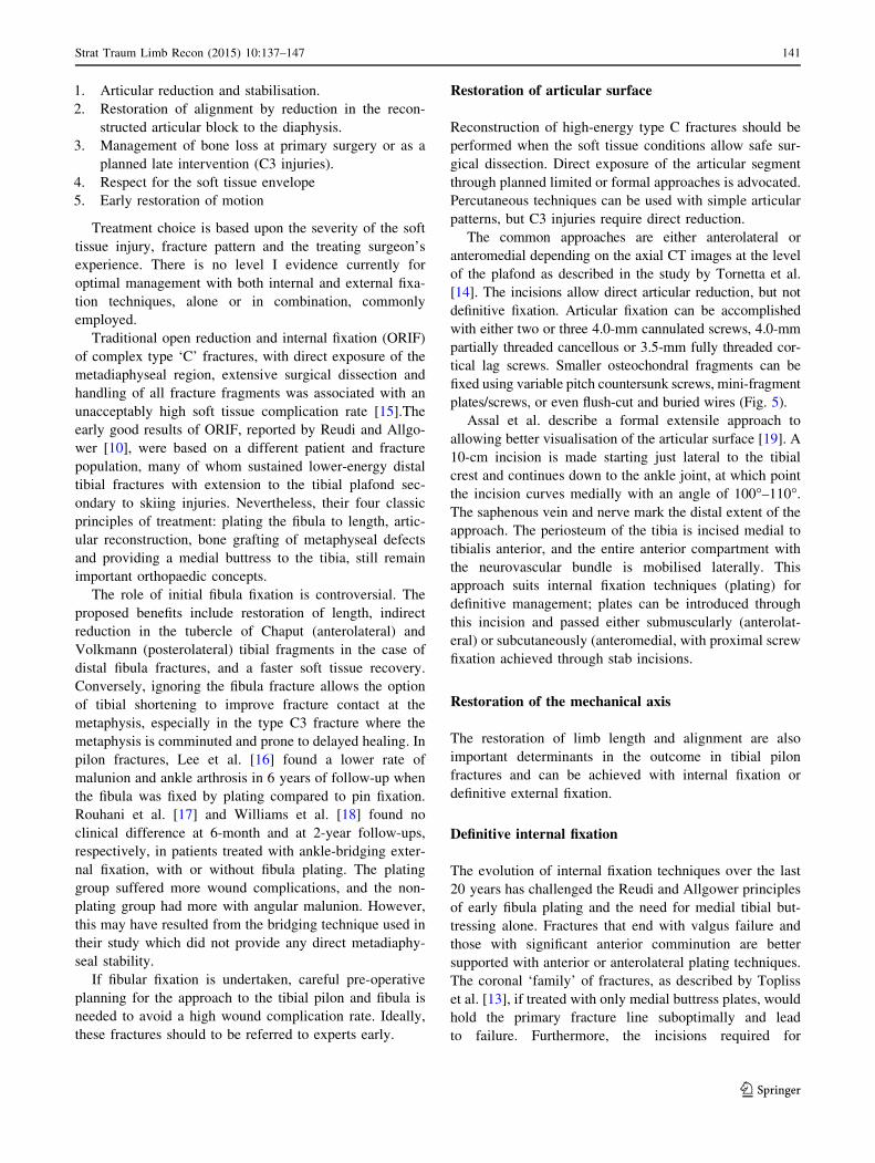
definitive fixation. Articular fixation can be accomplished
with either two or three 4.0-mm cannulated screws, 4.0-mm
partially threaded cancellous or 3.5-mm fully threaded cor-
tical lag screws. Smaller osteochondral fragments can be
fixed using variable pitch countersunk screws, mini-fragment
plates/screws, or even flush-cut and buried wires (Fig. 5).
Assal et al. describe a formal extensile approach to
allowing better visualisation of the articular surface [19]. A
10-cm incision is made starting just lateral to the tibial
crest and continues down to the ankle joint, at which point
the incision curves medially with an angle of 100�–110�.The saphenous vein and nerve mark the distal extent of the
approach. The periosteum of the tibia is incised medial to
tibialis anterior, and the entire anterior compartment with
the neurovascular bundle is mobilised laterally. This
approach suits internal fixation techniques (plating) for
definitive management; plates can be introduced through
this incision and passed either submuscularly (anterolat-
eral) or subcutaneously (anteromedial, with proximal screw
fixation achieved through stab incisions.
Restoration of the mechanical axis
The restoration of limb length and alignment are also
important determinants in the outcome in tibial pilon
fractures and can be achieved with internal fixation or
definitive external fixation.
Definitive internal fixation
The evolution of internal fixation techniques over the last
20 years has challenged the Reudi and Allgower principles
of early fibula plating and the need for medial tibial but-
tressing alone. Fractures that end with valgus failure and
those with significant anterior comminution are better
supported with anterior or anterolateral plating techniques.
The coronal ‘family’ of fractures, as described by Topliss
et al. [13], if treated with only medial buttress plates, would
hold the primary fracture line suboptimally and lead
to failure. Furthermore, the incisions required for
Strat Traum Limb Recon (2015) 10:137–147 141
123
anterolateral plating often mean that a standard lateral
incision for fibula fixation cannot be utilised.
Sirkin et al. [3], in their landmark paper that popularised
the staged approach to management, found that in their
closed group of pilon injuries of 29 patients, five developed
some form of wound necrosis which did not escalate to
deep infection; only one patient developed a late compli-
cation which was a chronic draining sinus that resolved
with fracture consolidation and metal removal. The open
fracture group included 17 patients with two late deep
infections: one patient underwent limb reconstruction with
an aggressive protocol and one patient had a below knee
amputation.
The concept of two-stage management is established
with a trend towards minimally invasive plating techniques
to reduce further wound healing complications. The con-
cept of biological plating with minimally invasive appli-
cation of pre-contoured implants is a further evolution in
internal fixation which enables epiphyseal and metadia-
physeal contact and alignment without extensive periosteal
stripping. However, as with any new technology, achieving
consistent results requires multiple refinements often with
respect to the implant design and surgical technique.
Fracture reduction with indirect techniques is more difficult
to master, and the view that the implant will compensate
for an inadequate reduction will lead to either a mal- or
non-union.
Despite an increasing use of ‘biological plating’ in
orthopaedic trauma, there is a paucity of evidence on the
outcome when applied to patients with ‘C’ type pilon
fractures. Most studies refer to a heterogeneous group of
patients that include type A and B injuries. Using a two-
stage minimally invasive protocol, Borens et al. [20]
reported on 17 patients with good to excellent radiographic
results at 17-month follow-up although 41 % had devel-
oped moderate arthritis at this time. Five of the patients had
low-energy trauma, and 12 fractures were classified as
either C2 or C3 injuries. This subgroup of higher-energy
injuries did not have any serious wound healing problems.
The plate used in this study was a non-locking low-profile
implant, termed a ‘scallop’ plate, designed to pass through
the soft tissues with minimal trauma. Little evidence sup-
ports the use of locking plates over standard plates when
used in patients with good bone quality. Pre-contoured
low-profile non-locking plates, such as those used by
Borens et al. [20], can be applied with limited incisions and
placed either subcutaneously or submuscularly. These are
less bulky and kinder to the soft tissues especially over the
medial subcutaneous border of the tibia.
Blauth et al. [21] compared three methods of treatment in
a cohort of 51 patients with 47 type C fractures. Twenty-
eight patients were treated with one-stage articular reduc-
tion and bridging external fixation. Fifteen patients were
treated with primary plate fixation, and eight patients had a
two-stage minimally invasive intervention, with application
of a medial plate when the soft tissues had recovered. The
latter option yielded the best results although two compar-
ative groups used in their study, that of definitive bridging
external fixation and primary plate fixation are not now
considered as reliable management options.
Definitive external fixation
With increasing comminution of the metaphysis (C3 inju-
ries), restoration of mechanical alignment and achieving
stable fixation becomes increasingly difficult. The meta-
physeal component of the injury may lead to a non-union
or malunion, and these injuries are prone to wound healing
complications and infection. Proponents of internal fixation
argue that pilon fractures treated by external fixation often
result such complications. This leads to the debate sur-
rounding pilon fractures whether definitive management of
C2 and C3 injuries are better treated definitively by
external or internal fixation. External fixation constructs
described in the literature include simple bridging frames,
ankle-articulating devices, and hybrid or circular frames
which are used mostly in conjunction with limited internal
fixation of the articular surface through percutaneous or
small incisions [2, 7, 21–23]. The ability of articulating
devices to offer useful range of movement during treatment
has been questioned and may be due to the difficulty in
reproducing movement about the ankle joint axis [24].
The evidence used against external fixation as a valuable
definitive option is based largely upon historical techniques
where definitive management consisted of bridging the
ankle joint with a fixator, without direct control of the
metaphyseal component of the injury. Additionally, the
more severe injuries are treated with external fixation and
introduced a patient selection bias into these studies.
Anglen et al. [25] reported dismal results associated with
hybrid external fixation when compared to internal fixation
for type C fractures. This retrospective study was based on
the more severe injuries, including more C2 and C3 types
and open injuries that were chosen for treatment with a
hybrid fixator as a one-stage intervention. This study
demonstrated that one-stage management of high-energy
injuries was not effective.
Pin-site infection has been reported to be a serious
complication with prolonged external fixation. Whilst this
is a recognised complication of fine wire fixation in gen-
eral, it can be controlled and managed with an integrated
multidisciplinary approach [26]. Deep infection rates vary
significantly in the literature and are biased by a higher
proportion of open injuries treated definitively with exter-
nal fixation. Papadokostakis et al. [27] reviewed the merits
of spanning versus non-spanning frames and found, in their
142 Strat Traum Limb Recon (2015) 10:137–147
123
systematic review, that the overall deep infection rate with
non-spanning frames was 2.7 %. The deep infection rate in
the spanning group was 3.9 % which may be related to the
larger proportion of open injuries in this group. The con-
clusion from this review suggested that there were no
statistically significant differences with either technique
with respect to infection, non-union or time to union. There
was a higher rate of malunion in the spanning group. The
groups were heterogeneous, and the relative merits of
external fixation as definitive management for these inju-
ries were not determined clearly.
A few studies have reported the outcome of circular ring
fixation as definitive management. McDonald et al. [28]
retrospectively reviewed 13 pilon fractures, of which 12
were true high-energy injuries. The technique involved
application of a non-bridging three-ring circular frame,
with a minimally invasive approach to articular reduction.
Eleven fractures were healed by 16 weeks. There was one
delayed union that required bone grafting and one non-
union treated with an arthrodesis. Importantly, there were
no deep infections.
Leung et al. [29] reviewed 31 distal tibial fractures with
16 cases classified as C type injuries. A protocol similar to
McDonald et al. was employed with mostly non-bridging
circular frames. Two patients with very comminuted C3
fractures had bridging frames to the calcaneus for 2 weeks
for additional stability. All but one fracture united at an
average of 13.8 weeks. One fracture was complicated by
infection and required an arthrodesis. Only five patients
(38 %) had good results (clinical rating system of Teeny
and Wiss) possibly a reflection of the poor outcome asso-
ciated with these injuries.
Vidyadhara and Rao [30] reported on 21 pilon fractures
with 13 cases classified as C type injuries. Minimally
invasive techniques were used for joint reduction, with
limited approaches where necessary, and circular ring fix-
ation used. The authors bridged to the calcaneus for
6 weeks in all patients with the half ring removed in the
outpatient setting. All fractures united with frame removal
at an average of 26.6 weeks. Seven patients developed pin-
site infections which settled with local care, and one patient
required pin removal at 3 months due to persistent infec-
tion. There were no deep infections.
Watson et al. [31] reviewed 107 pilon fractures treated
according to a staged protocol which included initial sta-
bilisation with calcaneal traction. Definitive treatment was
based on the degree of soft tissue compromise. Forty-one
patients with Tscherne grade 0 and I injuries underwent
open reduction and internal fixation, with minimal inci-
sions and low-profile implants, with most cases managed
within 5 days of presentation. Sixty-four patients with
Tscherne grade II and III injuries, and all open fractures
underwent limited internal fixation of the articular
fragments through small incisions and fine wire external
fixation as definitive management. For the type C fractures
in both groups, there was a significantly higher rate of
complications including non-union, malunion and wound
complications. They recommended small wire circular
fixators for the subgroup of type C fractures. Some would
argue that internal fixation when performed within 5 days
of injury might have accounted for the higher complication
rate, but this group was selected on the basis of the less
severe soft tissue injuries.
Wang et al. [32] performed a meta-analysis of compli-
cations associated with ORIF versus limited internal fixa-
tion combined with external fixation. They included nine
studies with 498 fractures. The meta-analysis found no
significant differences in bone healing complications, non-
union, malunion or delayed union, superficial and deep
infections, arthritis symptoms or chronic osteomyelitis
between the two groups.
These studies offer some perspective when dealing with
type C fracture patterns and demonstrate the low incidence
of serious complications, offering some support to the use
of circular ring fixation as a definitive management for
these injuries.
Management of bone defects
Segmental bone defects associated with pilon fractures
have been treated with different methods. This includes
bone grafting, either acutely or staged (Masquelet), vas-
cularised fibular grafts, bone transport and acute shortening
followed by lengthening.
Bone grafting
Autologous bone grafting is used commonly for smaller
bone defects and is limited primarily by the amount that
can be harvested from the donor site. Allograft has been
used in certain conditions in conjunction with bone mor-
phogenetic protein (BMP); this has been demonstrated to
be of value in cases of non-union with bone defects by
Johnson et al. [33].
The two-staged technique described by Masquelet et al.
[34] has gained popularity. During the first stage, stabili-
sation is performed following the bone resection and a
cement spacer is inserted followed by soft tissue repair. An
osteoinductive membrane is formed around the spacer. The
second stage is performed a few weeks later with removal
of the spacer, bone decortication and use of cancellous
bone graft packing the cavity within the induced mem-
brane. Reports for its use have been encouraging but for a
mixed group of conditions; evidence for use in pilon
fractures is as yet lacking.
Strat Traum Limb Recon (2015) 10:137–147 143
123
Vascularised fibular grafts
Use of a vascularised segment of fibula to reconstruct
segmental defects of the tibia has advantages of shorter
time to consolidation, increased potential for remodelling,
greater resistance to infection and better long-term
mechanical properties [35]. It is technically challenging
and was found to have some problems including unreliable
hypertrophy of the graft and late fractures [36].
Bone shortening and staged reconstruction
Closing a metaphyseal defect by shortening the tibia is a
useful option for contaminated fractures (after debride-
ment), those associated with soft tissue loss (to facilitate
closure), or when there are small segmental defects. The
circular frame is applied across but not inside the zone of
injury. The circular frame can then be used to lengthen the
bone from a separate osteotomy and achieve bony union at
the metaphyseal area simultaneously. Shortening the bone
will also reduce the size of the soft tissue defect and may
avoid the need for a free flap [37]. The disadvantages of
this technique are the ensuing limb length discrepancy
(unless lengthening is contemplated), a risk of kinking
vascular structures—particularly relevant in patients who
have vascular injuries and those who have had soft tissue
flaps for coverage—and the risk of infection from pin
tracks. Nonetheless, bone shortening and subsequent
lengthening are associated with a lower complication rate
than bone transport techniques [38–41].
Bone transport
The use of an external fixator for bone transport to bridge a
defect is an alternative to the shortening and staged
reconstruction. It is indicated for larger defects. Circular
frames are more popular than unilateral devices now for
their greater stability and flexibility in the configuration.
There is greater scope for correcting rotational or angular
mal-alignment which may occur during the course of
treatment [42]. It is usual to delay the osteotomy of bone
transport by several weeks to ensure the soft tissue envel-
ope has healed.
Open pilon fractures
The management of open injuries follows well-established
principles with urgent wound debridement and skeletal
stabilisation. Uncertainty exists with the method and timing
of stabilisation with some advocating early internal fixation
with immediate soft tissue cover, the so-called fix-and-flap
protocol. Conroy et al. [43] reported the 1-year outcome of
early internal fixation (within 24 h) and soft tissue cover-
age in a consecutive series of 32 patients. Despite the short
follow-up, encouraging radiographic and clinical outcomes
were reported with a deep infection rate of 6.2 % (2
patients); both cases required amputation. Close collabo-
ration between orthopaedic and plastic surgery services is
mandatory for such a protocol to be successful, and further
studies are required to confirm the wide applicability of this
strategy. More commonly, these injuries are managed in
two or three stages. Following skeletal stabilisation and
wound debridement, soft tissue coverage is performed
when deemed safe in conjunction with a plastic surgeon.
Thereafter, definitive management of the fracture is per-
formed using either internal or external fixation techniques.
Few studies report directly on the outcome of open
fractures. Such injuries are often grouped together with
closed fractures; when present, treatment is usually with
external fixation lending to a selection bias for poor results
when compared to equivalent closed injuries [25]. Gardner
et al. [44] reported their results of a standard protocol used
for ten open fractures with segmental bone loss. Their
approach involved three stages: initial debridement and
spanning external fixation; then open reduction and plate
fixation with antibiotic bead placement after a delay of
1–3 weeks; and finally a planned bone grafting procedure.
Nine of the ten patients in this study had healed by
24 weeks. One patient required amputation for uncon-
trolled infection.
Outcomes
Successful treatment of pilon fractures is dependent on the
management of the soft tissue injury, anatomical reduction
in the joint surface and restoration of mechanical align-
ment. Whilst the relative importance of each of these fac-
tors is difficult to quantify, long-term studies generally
report suboptimal outcomes in these injuries. Pollak et al.
[45] studied a sample which included a large proportion of
type C fractures (74 %). At 2-year follow-up, they reported
lower SF-36 scores than after pelvic fractures or in patient
groups with chronic illnesses such as AIDS and coronary
artery disease. The outcome following external fixation
was reported as the only surgeon controlled factor associ-
ated with a poor outcome; this conclusion has to be con-
sidered in light of what was termed definitive management
by external fixation in this study—bridging external fixa-
tion with or without limited internal fixation—which is,
today, historical and no longer a clinically relevant treat-
ment strategy using external fixation. The protocol
described in the study in fact represents the first stage of
modern damage control surgery. Irrespective of the choice
of fixation, the literature reports the outcome of these
144 Strat Traum Limb Recon (2015) 10:137–147
123
injuries remains suboptimal with the propensity to deteri-
orate over time [46–50].
Treatment algorithm
Based on this review and our clinical experience, we use a
treatment algorithm to guide management decisions
(Fig. 6):
1. In all cases, these injuries are treated initially with a
spanning external fixator.
2. With open injuries, a thorough wound debridement is
performed and a spanning external fixator and tempo-
rary topical negative pressure wound dressing applied.
The aim is to achieve soft tissue cover within 5 days of
injury, with definitive stabilisation performed prior to
soft tissue coverage or shortly after if circular fixation
is used.
3. For closed injuries, we proceed to definitive manage-
ment when the soft tissue swelling has settled (often
7–14 days). During this waiting period, a CT scan is
performed to plan the procedure. Based on the CT, an
approach is made directly onto the primary sagittal
fracture line, minimising soft tissue stripping and
maintaining full thickness skin flaps. The articular
surface is visualised and the impacted fragments
reduced under direct vision. Reconstruction of the
plafond proceeds from posterior to anterior with
temporary k-wires used to hold the articular reduction,
and small fragment screws (cannulated and partially
threaded) then applied to secure definitive stability.
4. In type C1 fractures, effectively three large articular
fragments with no metaphyseal comminution, we
Fig. 6 Treatment algorithm as used in the authors’ unit
Strat Traum Limb Recon (2015) 10:137–147 145
123
choose to plate the tibia using minimally invasive
techniques with a locking plate to bridge the articular
segment to the diaphysis.
5. In type C2 or C3 fractures, an Ilizarov external fixator
is used. Following articular reconstruction, a two-ring
construct is applied to the tibia proximally, orthogonal
to the anatomical axis. A reference wire is then passed
distally, at the level of the plafond, as close as possible
to the articular surface, but recognising the capsular
attachments which extend 15 mm proximally. Three or
four wires are inserted into the distal segment with
wide crossing angles to achieve maximal stability. The
distal segment is then reduced onto the two-ring
proximal ring construct, with a ring-to-ring reduction
in tibial alignment. The construct is extended to the
hindfoot in almost all cases to provide additional
stability. The hindfoot ring is removed at 6–8 weeks
post-operatively in the clinic. Patients remain non-
weight bearing until radiological evidence of healing is
attained, and input from specialist physiotherapists
prevents the development of forefoot deformities.
Conclusion
The optimum management for tibial pilon fractures is yet
undetermined. When soft tissue conditions permit and in
type C1 fractures, open reduction and internal fixation with
minimally invasive techniques is preferred. In type C3
fractures, a two-stage procedure of initial articular
restoration and spanning external fixation followed by
definitive fixation at a later stage appears to give better
results [51]. However, in all these fractures, careful man-
agement of soft tissue injury holds the key to reduced
complications and improved outcomes. Despite these
newer surgical techniques, the long-term outcomes con-
tinue to be less than satisfactory.
Compliance with ethical standards
Conflict of interest The authors declare that they have no conflict
of interest.
Research involving Human Participants and/or Animals This
article does not contain any studies with human participants or ani-
mals performed by any of the authors.
Informed consent Informed consent was not required for this work.
Open Access This article is distributed under the terms of the
Creative Commons Attribution 4.0 International License (http://crea
tivecommons.org/licenses/by/4.0/), which permits unrestricted use,
distribution, and reproduction in any medium, provided you give
appropriate credit to the original author(s) and the source, provide a
link to the Creative Commons license, and indicate if changes were
made.
References
1. Helfet DL, Koval K, Pappas J, Sanders RW, DiPasquale T (1994)
Intraarticular’’ pilon’’ fracture of the tibia. Clin Orthop Relat Res
298:221–228
2. Tarkin I, Clare M, Marcantonio A, Pape H (2008) An update on the
management of high-energy pilon fractures. Injury 39(2):142–154
3. Sirkin M, Sanders R, DiPasquale T, Herscovici D Jr (1999) A
staged protocol for soft tissue management in the treatment of
complex pilon fractures. J Orthop Trauma 13(2):78–84
4. Hontzsch D, Karnatz N, Jansen T (1990) One-or two-step man-
agement (with external fixator) of severe pilon-tibial fractures.
Aktuelle Traumatol 20(4):199–204
5. Patterson MJ, Cole JD (1999) Two-staged delayed open reduction
and internal fixation of severe pilon fractures. J Orthop Trauma
13(2):85–91
6. Rommens P, Claes P, Broos P (1996) Therapeutic strategy in
pilon fractures type C2 and C3: soft tissue damage changes
treatment protocol. Acta Chir Belg 96(2):85–92
7. Tornetta P III, Weiner L, Bergman M, Watnik N, Steuer J, Kelley
M, Yang E (1993) Pilon fractures: treatment with combined
internal and external fixation. J Orthop Trauma 7(6):489–496
8. Furman BD, Olson SA, Guilak F (2006) The development of
posttraumatic arthritis after articular fracture. J Orthop Trauma
20(10):719–725
9. Borrelli J Jr, Ricci WM (2004) Acute effects of cartilage impact.
Clin Orthop Relat Res 423:33–39
10. Ruedi TP, Allgower M (1979) The operative treatment of intra-
articular fractures of the lower end of the tibia. Clin Orthop Relat
Res 138:105–110
11. Marsh J, Slongo TF, Agel J, Broderick JS, Creevey W, DeCoster
TA, Prokuski L, Sirkin MS, Ziran B, Henley B (2007) Fracture
and dislocation classification compendium-2007: Orthopaedic
Trauma Association classification, database and outcomes com-
mittee. J Orthop Trauma 21(10):S1–S6
12. Swiontkowski M, Sands A, Agel J, Diab M, Schwappach J,
Kreder H (1997) Interobserver variation in the AO/OTA fracture
classification system for pilon fractures: is there a problem?
J Orthop Trauma 11(7):467–470
13. Topliss C, Jackson M, Atkins R (2005) Anatomy of pilon frac-
tures of the distal tibia. J Bone Joint Surg Br 87(5):692–697
14. Tornetta P III, Gorup J (1996) Axial computed tomography of
pilon fractures. Clin Orthop Relat Res 323:273–276
15. McFerran MA, Smith SW, Boulas HJ, Schwartz HS (1992)
Complications encountered in the treatment of pilon fractures.
J Orthop Trauma 6(2):195–200
16. Lee Y-S, Chen S-W, Chen S-H, Chen W-C, Lau M-J, Hsu T-L
(2009) Stabilisation of the fractured fibula plays an important role
in the treatment of pilon fractures: a retrospective comparison of
fibular fixation methods. Int Orthop 33(3):695–699
17. Rouhani A, Elmi A, Akbari Aghdam H, Panahi F, Dokht Ghafari
Y (2012) The role of fibular fixation in the treatment of tibia
diaphysis distal third fractures. Orthop Traumatol Surg Res
98(8):868–872
18. Williams TM, Marsh JL, Nepola JV, DeCoster TA, Hurwitz SR,
Bonar SB (1998) External fixation of tibial plafond fractures: is
routine plating of the fibula necessary? J Orthop Trauma
12(1):16–20
146 Strat Traum Limb Recon (2015) 10:137–147
123
19. Assal M, Ray A, Stern R (2007) The extensile approach for the
operative treatment of high-energy pilon fractures: surgical tech-
nique and soft-tissue healing. J Orthop Trauma 21(3):198–206
20. Borens O, Kloen P, Richmond J, Roederer G, Levine DS, Helfet
DL (2009) Minimally invasive treatment of pilon fractures with a
low profile plate: preliminary results in 17 cases. Arch Orthop
Trauma Surg 129(5):649–659
21. Blauth M, Bastian L, Krettek C, Knop C, Evans S (2001) Surgical
options for the treatment of severe tibial pilon fractures: a study
of three techniques. J Orthop Trauma 15(3):153–160
22. Walker C, Garg A, McQueen M (1999) Half-ring external fixa-
tion in the management of tibial plafond fractures. J Orthop
Trauma 13(3):200–206
23. Barbieri R, Schenk R, Koval K, Aurori K, Aurori B (1996)
Hybrid external fixation in the treatment of tibial plafond frac-
tures. Clin Orthop Relat Res 332:16–22
24. Marsh JL, Bonar S, Nepola JV, Decoster TA, Hurwitz SR (1995)
Use of an articulated external fixator for fractures of the tibial
plafond. J Bone Joint Surg Am 77(10):1498–1509
25. Anglen JO (1999) Early outcome of hybrid external fixation for
fracture of the distal tibia. J Orthop Trauma 13(2):92–97
26. Davies R, Holt N, Nayagam S (2005) The care of pin sites with
external fixation. J Bone Joint Surg Br 87(5):716–719
27. Papadokostakis G, Kontakis G, Giannoudis P, Hadjipavlou A
(2008) External fixation devices in the treatment of fractures of
the tibial plafond: a systematic review of the literature. J Bone
Joint Surg Br 90(1):1–6
28. McDonald MG, Burgess RC, Bolano LE, Nicholls PJ (1996)
Ilizarov treatment of pilon fractures. Clin Orthop Relat Res
325:232–238
29. Leung F, Kwok HY, Pun TS, Chow SP (2004) Limited open
reduction and Ilizarov external fixation in the treatment of distal
tibial fractures. Injury 35(3):278–283
30. Vidyadhara S, Rao SK (2006) Ilizarov treatment of complex
tibial pilon fractures. Int Orthop 30(2):113–117
31. Watson JT, Moed BR, Karges DE, Cramer KE (2000) Pilon
fractures: treatment protocol based on severity of soft tissue
injury. Clin Orthop Relat Res 375:78–90
32. Wang D, Xiang J-P, Chen X-H, Zhu Q-T (2015) A meta-analysis
for postoperative complications in tibial plafond fracture: open
reduction and internal fixation versus limited internal fixation
combined with external fixator. J Foot Ankle Surg
54(4):646–651. doi:10.1053/j.jfas.2014.06.007
33. Johnson EE, Urist MR, Am Finerman G (1992) Resistant non-
unions and partial or complete segmental defects of long bones:
treatment with implants of a composite of human bone mor-
phogenetic protein (BMP) and autolyzed, antigen-extracted,
allogeneic (AAA) bone. Clin Orthop Relat Res 277:229–237
34. Pelissier P, Masquelet A, Bareille R, Pelissier SM, Amedee J
(2004) Induced membranes secrete growth factors including
vascular and osteoinductive factors and could stimulate bone
regeneration. J Orthop Res 22(1):73–79
35. Hertel R, Pisan M, Jakob R (1995) Use of the ipsilateral vascu-
larised fibula for tibial reconstruction. J Bone Joint Surg Br
77(6):914–919
36. DeCoster TA, Gehlert RJ, Mikola EA, Pirela-Cruz MA (2004)
Management of posttraumatic segmental bone defects. J Am
Acad Orthop Surg 12(1):28–38
37. Simpson A, Andrews C, Giele H (2001) Skin closure after acute
shortening. J Bone Joint Surg Br 83(5):668–671
38. Ilizarov GA (1989) The tension-stress effect on the genesis and
growth of tissues—part I: the influence of stability of fixation and
soft-tissue preservation. Clin Orthop Relat Res 238:249–281
39. Ilizarov GA (1989) The tension-stress effect on the genesis and
growth of tissues—part II: the influence of the rate and frequency
of distraction. Clin Orthop Relat Res 239:263–285
40. Dagher F, Roukoz S (1991) Compound tibial fractures with bone
loss treated by the Ilizarov technique. J Bone Joint Surg Br
73(2):316–321
41. Saleh M, Rees A (1995) Bifocal surgery for deformity and bone
loss after lower-limb fractures: comparison of bone-transport and
compression-distraction methods. J Bone Joint Surg Br 77(3):
429–434
42. Keating J, Simpson A, Robinson C (2005) The management of
fractures with bone loss. J Bone Joint Surg Br 87(2):142–150
43. Conroy J, Agarwal M, Giannoudis P, Matthews S (2003) Early
internal fixation and soft tissue cover of severe open tibial pilon
fractures. Int Orthop 27(6):343–347
44. Gardner MJ, Mehta S, Barei DP, Nork SE (2008) Treatment
protocol for open AO/OTA type C3 pilon fractures with seg-
mental bone loss. J Orthop Trauma 22(7):451–457
45. Pollak AN, McCarthy ML, Bess RS, Agel J, Swiontkowski MF
(2003) Outcomes after treatment of high-energy tibial plafond
fractures. J Bone Joint Surg 85(10):1893–1900
46. Teeny SM, Wrss DA (1993) Open reduction and internal fixation
of tibial plafond fractures: variables contributing to poor results
and complications. Clin Orthop Relat Res 292:108–117
47. Okcu G, Aktuglu K (2004) Intra-articular fractures of the tibial
plafond: a comparison of the results using articulated and ring
external fixators. J Bone Joint Surg Br 86(6):868–875
48. Sands A, Grujic L, Byck DC, Agel J, Benirschke S, Swion-
tkowski MF (1998) Clinical and functional outcomes of internal
fixation of displaced pilon fractures. Clin Orthop Relat Res
347:131–137
49. Chen S-H, Wu P-H, Lee Y-S (2007) Long-term results of pilon
fractures. Arch Orthop Trauma Surg 127(1):55–60
50. Marsh JL, Weigel DP, Dirschl DR (2003) Tibial plafond fractures
how do these ankles function over time? J Bone Joint Surg
85(2):287–295
51. Calori G, Tagliabue L, Mazza E, de Bellis U, Pierannunzii L,
Marelli B, Colombo M, Albisetti W (2010) Tibial pilon fractures:
which method of treatment? Injury 41(11):1183–1190
Strat Traum Limb Recon (2015) 10:137–147 147
123


![The mid-term results of treatment for tibial pilon fractures · 7-10% of all tibia fractures are pilon fractures.[1-3] The usual mechanism of injury is axial loading of the limb through](https://static.fdocuments.net/doc/165x107/6018c5987d71101a3a4e4d4b/the-mid-term-results-of-treatment-for-tibial-pilon-fractures-7-10-of-all-tibia.jpg)