ADHD WORKSHOP FOR PAEDIATRICIANS UCT Paediatric Refresher Course Feb 2010.
Upload
truongduongCategory
view
231download
0
NATIONAL GUIDELINES
MANAGEMENT OFCENTRAL NERVOUS SYSTEM
INFECTIONS IN CHILDREN

GUIDELINES FOR THE MANAGEMENT OF CNS INFECTIONS36
CLINICAL GUIDELINES ONCENTRAL NERVOUS SYSTEM INFECTIONS
GUIDELINES ON MENINGITIS
Introduction
These guidelines are aimed at achieving the best possible paediatric care forchildren with CNS infection within Sri Lanka. It should be utilized together withthe clinical input and discretion of the managing paediatrician. Each patientshould be individually evaluated and a decision made as to appropriatemanagement in order to achieve the best clinical outcome. In the interests ofpatient care it is critical that contemporaneous, accurate and completedocumentation is maintained during the course of patient management fromarrival to discharge. Parental anxiety should not be discounted: it is often ofsignificance even if the child does not appear especially unwell.
Suspected bacterial meningitis is a medical emergency. If untreated it is invariablyfatal. Even with treatment, bacterial meningitis in childhood is associated withhigh morbidity and mortality. It is also important to treat meningitis promptly.Delay of treatment even for a few hours can make a huge difference to the finaloutcome.
It is anticipated that modifications may be required for local practice. Variationsin management may also be required in individual cases. It is stressed that theguidelines are, by necessity, general in nature and are not intended as a substitutefor clinical judgment. One has to be also mindful of evolving new knowledgethat a document of this cannot fulfill.
Key points in the acute management of bacterial meningitis in children
First principles
Early recognition and prompt management are the goals.

GUIDELINES FOR THE MANAGEMENT OF CNS INFECTIONS 37
Early consultation with a senior paediatric or senior emergencydepartment staff member should occur in all cases of suspectedmeningitis. Any delay in making the correct decisions and the lost goldenhours will have lasting consequences for the affected patient.
Clinical presentation
Not all patients will have fever, neck stiffness and altered mental status.
The younger the patient, the more subtle the symptoms and signs andthe higher should be the level of suspicion.
Fever is not always present at presentation in acute bacterial meningitis.(50% of infants and 45% of older children in small case series.)
Neck stiffness is more useful above 3 yrs of age and seen in 60-80% ofcases.
Clinical presentations can vary from being acute (hours to 1–2 days) toinsidious (over a few days).
Preceding upper respiratory tract infection is often present (~75 percent).
Seizures occur in 20–30 per cent.
Papilloedema is rare in acute uncomplicated bacterial meningitis andwhen present it should be a reason to arrange for imaging without delay
Bulging or tense anterior fontanelle is a useful sign in young infantswho should be examined in the upright position when not crying.
Prior antibiotics modify presentation and diagnostic yield, and shouldalways be part of the history.
Initial management
The priorities are ensuring the adequacy of the Airway, Breathing,Circulation, Disability (level of consciousness) and Exposure (rashassessment and environmental control) i.e. ‘ABCDE’.
The risks from inadequate cerebral circulation may be higher than therisks of cerebral oedema so volume expanders should be titrated againstthe patient’s perfusion.
If venous access is significantly difficult, an intraosseous needle can beused.
Seizures should be treated urgently.

GUIDELINES FOR THE MANAGEMENT OF CNS INFECTIONS38
A bedside whole blood glucose reading (reflectance meter) e.g.‘Dextrostix’, should be performed as part of the early assessment,especially in infants. If the level is less than 50mg/dl(and less than 40mg/dl in the neonate) action should be taken to correct it immediately. (x)
Electrolyte abnormalities should also be addressed.
Diagnosis
Ensuring the adequacy of the ABCDEs has priority over establishing aprecise diagnosis.
CSF examination provides the definitive diagnosis. Blood cultures mayprovide supportive evidence. Ideally, CSF via lumbar puncture (LP)and blood cultures should be taken prior to antibiotic therapy. Otherinvestigations are helpful for acute management of a sick child, but notdefinitive in the diagnosis of meningitis.
The initiation of appropriate antibiotic therapy assumes high priority. Ifthe patient is too sick or unstable for immediate definitive investigations,then appropriate antibiotics should be commenced after taking bloodfor culture. (x)
The LP should be performed when the patient is resuscitated andstable. (x)
A cerebral computed tomogram (CT) scan is not part of the routineworkup and is indicated only in specific situations (see text). It shouldonly be done when the patient is stable.
The limits of sensitivity of the CSF diagnostic tests, especially if pre-treated with parenteral antibiotics, should be recognized.
CSF samples should be expeditiously transported to the laboratory andurgently analyzed. In case of delay in the transport of CSF, samplesshould not be refrigerated but kept at room temperature.
Steroid therapyA recent systematic review supports the early use (just before or with
first dose of antibiotics) of adjunctive steroid therapy provided children havenot been pre-treated with antibiotics. The benefit is an approximate two-thirdsreduction in severe hearing loss (Hib and non-Hib). The impact on long termcognitive function remains unanswered. There is insufficient information to becertain about the benefit of steroids in the 0–3 month age group given thedifferent aetiological agents and unknown potential adverse effects of steroidsin this age group. The benefit is also less clear in children who present late inthe illness or in severe sepsis.

GUIDELINES FOR THE MANAGEMENT OF CNS INFECTIONS 39
Suggested criteria for steroid use in acute bacterial meningitis:
> three months of age Not pre-treated with parenteral antibiotics. Due to insufficient
information, no clear recommendations can be given about steroids inthe < 3 month age group and those presenting with advanced meningitisor severe sepsis.
Suggested regimen:
Dexamethasone, 0.15 mg/kg/dose, IV, six-hourly for four days. Given as a ‘push’, followed by first dose of antibiotics for practical
purposes.
Antibiotic therapy
Recommended empiric antibiotic therapy is:Neonates to three months: ampicillin plus cefotaxime (not ceftriaxonein neonates). If GBS is isolated high dose C Penicillin could be usedalone (x)> three months: cefotaxime or ceftriaxone. (x)
Fluid Management
Patients with evidence of shock should be treated with a rapid infusionintravenous/interosseous crystalloid (Normal saline/Hartmann’s solution)20ml/kg. Fluid restriction (for SIADH) is not routinely recommended in theinitial management but should be considered in specific circumstances; e.g.patients with recurrent seizures or when there is other evidence such ashyponatraemia.
Further careOnce the patient is resuscitated and stabilized, refining management andfurther investigations can be arranged under the direction of the responsibleteam.
The decision to transfer the patient or to continue to provide care locallywill be determined by the local resources and the patient’s needs.

GUIDELINES FOR THE MANAGEMENT OF CNS INFECTIONS40
Algorithm: Acute management of suspected bacterialmeningitis in children
Child clinically suspected of having meningitis
Assess and attend to airways, breathing, circulation and level of consciousness +/– seizures
Patient stable
Indication to delay LP ?
Lumbar puncture#
No❡ other tests FBC, CRP, blood culture, RBS, SE, Assess need for empiric antibiotic cover before further investigations ❡ Consult senior staff
Yes
Plus other essential tests: # Expeditious* lab assay of the CSF ❡ M/C/S: urgent microscopy, culture and sensitivity ❡ Protein ❡ Glucose – best interpreted with concurrent serum glucose *any delay keep CSF in room temperature, NOT IN REFRIGERATOR NEVER USE INTRATHECAL ANTIBIOTICS ❡ Turbid CSF and/or
❡ High clinical suspicion
❡ ?Steroids (See ‘Key points’) ❡ Start empiric antibiotics by age group
Yes
Reassess at later stage and LP when safe
If no other indications to delay LP, proceed to LP
Start antibiotics (with or without LP) if with LP start immediately
No
Normal or Equivocal CSF Abnormal CSF
High clinical suspicion for bacterial meningitis
Low clinical suspicion for bacterial meningitis
Consistent with bacterial meningitis
❡ ?Steroids (See ‘Key Points’) ❡ Continue empiric antibiotics ❡ Discuss further management with senior staff
ADMIT
Discuss further management with senior staff
❡ ?Steroids (See ‘Key Points’) ❡ Continue empiric antibiotics
ADMIT
Consider taking blood for investigation at the time of establishing IV access if practical
Pt unstable
High clinical suspicionfor bacterial meningitis
Low clinical suspicionfor bacterial meningitis
Normal or Equivocal CSF Abnormal CSF

GUIDELINES FOR THE MANAGEMENT OF CNS INFECTIONS 41
Clinical presentations
While viral meningitis occurs more commonly than bacterial meningitis, it isoften difficult to distinguish from bacterial meningitis. Mycobacteriumtuberculousis (MTB) meningitis should also be considered in the differentialdiagnosis. MTB meningitis is rare and if suspected such patients are bestmanaged by sending them to a specialized centre.
However initial management of suspected meningitis should be done as forbacterial meningitis until proven otherwise.
Common presentations
Clinical presentations of bacterial meningitis vary, depending on; age, durationof illness, the patient’s response to infection, whether prior antibiotics havebeen used and the infecting organism. The presentations could be insidious(over a few days), acute and sometimes fulminant (a few hours).
Overall, severity of the illness at presentation appears the most predictive ofoutcome.
The history and examination of a child presenting with suspected meningitis isthe same as any acutely unwell child, with attention paid to neurological signsand associated complications.
Apart from the clinical presentations, the history should include the followingkey elements:
age (~90 per cent of bacterial meningitis occurs at age < five years)vaccination historyrecent use of antibioticsDrug allergies.
0–2 monthsThe diagnosis may be more difficult in the very young as history andpresentations can be non-specific. Features include:
fever or hypothermiabulging fontanelleirritabilityhigh pitched crylethargyaltered mental stateseizuresapnoeapoor feedingvomiting.

GUIDELINES FOR THE MANAGEMENT OF CNS INFECTIONS42
A high index of suspicion for meningitis must exist in sick, febrile or hypothermicnewborns with or without the above features.
>2 monthsSymptoms become more CNS specific after this age. Acute presentations include:
fever (not always present at presentation time ~50 % in infants and in~45 % in older children in small case series)
neck stiffness (60- 80%, more useful in children > 3 years) Kernig’s sign (inability to completely extend the leg) in older children.
Absence does not exclude meningitis Brudzinki’s sign (flexion at the hip and knee in response to forward
flexion of the neck) in older children. Absence does not excludemeningitis
irritability or lethargy altered mental state (highly variable) anorexia, nausea and/or vomiting (a common/non–specific symptom) photophobia (older children) seizures (about 20–30 per cent incidence)
NB: Papilloedema in uncomplicated early bacterial meningitis is rare. Thepresence of papilloedema suggests complications like venous sinus thrombosis,abscess, or subdural empyema, It is a reason to arrange imaging without delay
Children who have received prior antibiotics
The clinical presentations and CSF findings in children who have receivedprevious antibiotics may be modified. Some features include:
less frequent presentations with temperature > 38.5°Cless frequent alterations in mental statusthe relationship between polymorphonuclear cells and lymphocytesin CSF may be reversed.Culture and gram stain may be negative.

GUIDELINES FOR THE MANAGEMENT OF CNS INFECTIONS 43
Complications
Patients may uncommonly present with early complications of sepsis or raisedintracranial pressure:
Septic shockDisseminated intravascular coagulopathyCerebral abscess or subdural effusionsAcute hydrocephalusCranial nerve palsiesPurpura fulminansWaterhouse-Friderichsen syndromeCerebral herniation
Minimizing delay in diagnosis
To avoid a delay in the diagnosis of meningitis, the following important pointsmust be noted.
The early diagnosis of bacterial meningitis can be difficult even forexperienced clinicians – a high index of suspicion should bemaintained. If doubts exist about a diagnosis, consultation with asenior staff member is strongly advocated. Meningitis must beconsidered in any child with unexplained fever
Meningitis needs to be considered in all children presenting withseizures in association with fever, particularly in children aged < 12months, or the fever is prolonged in nature or refractory tomanagement. Not all children presenting with fever and convulsionswill have meningitis.
The presence of an apparent explanation for fever eg. pharyngitis orotitis media does not rule out the possibility of meningitis. Apreceding history of an upper respiratory tract infection may presentin about 75 %.
Maculopapular, petechial or purpuric rashes may sometimes beassociated with Neisseria meningitidis meningitis/ septicaemia.Petechiae and/or purpura have also (less commonly) been observedin Haemophilus influenzae type b or Streptococcus pneumoniaesepsis.
Prior oral antibiotics for unexplained fever or other focus mayconfuse and delay diagnosis.

GUIDELINES FOR THE MANAGEMENT OF CNS INFECTIONS44
Apparent improvement with paracetamol is not helpful in excludingthe diagnosis.
Examination of any CSF samples taken is urgent. Thus, appropriatelabeling of requests, facilitation of delivery of specimens and directcommunication with the pathology laboratory is recommended. CSFFR(full report) examined after one hour is unreliable
In case of delay in sending the CSF, samples to be sent for cultureshould not be refrigerated but kept at room temperature.(However, CSF samples sent for viral or mycobacterial studies orsamples submitted only for bacterial antigen detection should berefrigerated)
Initial management
The assessment of any critically unwell child must always focus initially onresuscitation. The diagnostic test for meningitis is the lumbar puncture (LP).However, this test should not be undertaken until the patient has beenresuscitated and stabilized. Assessing ABCDE, the ‘Airway, Breathing,Circulation, Disability (level of consciousness) and Environment (presence ofrash, temperature control)’ is thus the first priority. Once the patient has beenstabilized, then the examination should include general assessment looking forfeatures of sepsis and meningitis.
Resuscitation
Airway and BreathingEnsure that there is an open airway and adequate ventilation is established.Supplemental oxygen should always be administered. If ventilation oroxygenation is inadequate, then respiratory support should be commenced inthe form of bag and mask technique, followed by endotracheal intubation.
CirculationFluid restriction is not an issue in the initial stabilization of children withmeningitis. Patients with evidence of shock should be treated with a rapidinfusion intravenous/interosseous crystalloid (Normal saline) 20ml/kg. Anyconsiderations for fluid restriction (for syndrome of inappropriate anti–diuretichormone, SIADH) should only be undertaken once the patient is no longershocked.(see notes below on fluid therapy) (x)

GUIDELINES FOR THE MANAGEMENT OF CNS INFECTIONS 45
Disability (level of consciousness)If there are signs of cerebral oedema (decreasing level of consciousness, bulgingfontanelle, papilloedema, rising blood pressure with falling heart rate), mannitol(0.5–1.5g/kg = 2.5–7.5 mL/kg of 20% solution) should be given. It could berepeated if necessary 1–2 times after 4–8 hours. The bed should be elevated to30° and ventilation controlled to maintain PaCO2 between 30–35mmHg. (y)
EnvironmentThe presence of a rash may be indicative of meningococcal sepsis. Regulationof temperature is important in the acute management of children presentingwith sepsis. (x)
SeizuresSeizures should be treated immediately with a rapid injection of a benzodiazepine(eg midazolam, 0.15mg/kg, IV). Alternatively, IM midazolam (0.15mg/kg) or rectaldiazepam(0.5mg/kg ) could be used. Considerations to a loading dose ofphenytoin (20mg/kg over 20 minutes) should given if seizures continue.Phenytoin has the benefit of avoiding sedation, although phenobarbitone (20mg/kg) is more commonly used in neonates. Paraldehyde may also be used IM orrectally(mixed with olive oil or sunflower oil
Blood glucose, urea and electrolytesThese must be checked early in the management and corrected if necessary. Abedside whole blood glucose (reflectance meter) e.g. ‘Dextrostix™’ should beperformed in the early assessment, especially in infants.
Diagnostic testsThe laboratory gold standard for establishing the diagnosis of bacterialmeningitis is the isolation of the causative bacteria from the cerebrospinal fluid(CSF). However, laboratory diagnosis is often made using the combination ofblood and/or CSF cultures along with Gram stain and chemical analysis of theCSF.
Investigations
1. Routine investigations for all patients with suspected bacterial meningitisBlood tests - FBC, with differential WCC, blood film (A WCC of <10 x
109/L does not exclude meningitis. Thrombocytopenia canoccur in DIC)Blood cultureCRP – (A trap to watch for is a low or normal CRP that mayoccur early in severe infection)Blood urea, electrolytes, glucose, LFTs (Na to detectSIADH; renal and liver impairment can occur with sepsis)

GUIDELINES FOR THE MANAGEMENT OF CNS INFECTIONS46
CSF analysis- Full Report - includes protein, cell count and gram stain(cells, specially polymorphs disintegrate fast; if notexamined within one hour cell counts are unreliable)Glucose - to be interpreted with blood glucose measuredsimultaneouslyCulture - In case of delay, specimens should not berefrigerated but kept at room temperature
2. Possible additional tests based on clinical presentation(as and when indicated )
Blood - JE serology (IgM and IgG- IgG require convalescentserology tooHerpes simplex serology
CSF - Bacterial antigen detection- Mycobacterium tuberculosis- AFB, PCR and culture- Cryptococcal stain and antigen- immuno-compromised
patients- Viral cultures – (only for enteroviruses at MRI – specimen
should be taken within 1st 4 days of illness. Stool specimenshould also be sent for enteroviral culture. Both specimensshould be sent on ice
- Herpes simplex PCR- Antibodies
JE/HSV Ab (should be taken within 05 days of illness)Mumps Ab (should be taken within 05 days of illness alongwith a blood/serum sample for Ab)Measles Ab (should be taken within 05 days of illness alongwith a blood/serum sample for Ab)
Skin - scrapings of skin lesions for microscopy + cultureWhen petechiae are seen gently deroof the lesion with aneedle and roll the sterile swab over the base of the lesionand then on to a glass slide for microscopy and then anotherswab for culture
EEG - for Herpes simplexencephalitis – focal changes speciallyin the temporal lobe

GUIDELINES FOR THE MANAGEMENT OF CNS INFECTIONS 47
CT brain - is of very limited use and is only indicated when there isdoubt about other causes of meningism such as posteriorfossa tumour or complications like abscess are clinicallysuspected. Any decision to perform a CT should not delayantibiotics. A CT scan cannot rule out raised intracranialpressure and a normal CT does not absolutely excludesubsequent risk of herniation.
The Lumbar Puncture (LP)
An LP for CSF analysis should be performed once the diagnosis of meningitisis suspected and after the patient is stabilised. If there are reasons to delay LP(see below) and bacterial meningitis is clinically suspected, antibiotics shouldbe given prior to the LP. Early lumbar puncture rapidly confirms or excludesbacterial meningitis in most cases and should be performed when meningitis issuspected unless there is a specific contraindication. Antibiotics may sterilisethe CSF within one hour in meningococcal meningitis and within four hours inpneumococcal meningitis. However, instituting antibiotics 1–2 hours prior toLP does not decrease the diagnostic sensitivity of the CSF culture if done inconjunction with blood cultures and CSF bacterial antigens. The decision toperform cranial computed tomogram (CT) before the LP is one factor contributingto delayed diagnosis. Although concerns about herniation following an LPexist, herniation is unlikely in children unless they have focal neurologicalfindings or are comatose.
Indications to delay the LP
Signs of raised intracranial pressure—altered pupillary responses, absentDoll’s eye reflex, decerebrate or decorticate posturing, abnormalrespiratory pattern, papilloedema, hypertension and bradycardiaRecent (within 30 minutes) or prolonged (over 30 minutes) convulsiveseizuresFocal or tonic seizuresOther focal neurological signs—hemi/monoparesis, extensor plantarresponses, ocular palsiesGlasgow Coma Score < 13 or deteriorating level of consciousnessStrong suspicion of meningococcal infection (typical purpuric rash in anill child)State of shockLocal superficial infectionCoagulation disorder.

GUIDELINES FOR THE MANAGEMENT OF CNS INFECTIONS48
Interpreting the CSF
1. If there is difficulty in interpreting the LP, a clinical microbiologist,infectious disease physician or senior staff should be consulted.
2. No CSF test is fully reliable in distinguishing bacterial from non-bacterialmeningitis. In rare instances (<3%), ‘normal’ CSF findings have beenassociated with culture proven bacterial meningitis. However, in mostcases, clinical indicators of meningitis or sepsis will be present. (Veryrarely initial LP could still be normal but with evidence in LP donesubsequently. A repeat LP at 24–48 hours may be indicated when clinicalindicators of meningitis are present but CSF examination is normal. Thisshould be decided by a senior person in special circumstances)
3. Post-ictal CSF abnormalities (pleocytosis or raised protein) are rare,and should not be readily accepted as a cause for an abnormal CSF.
Guide to distinguishing a traumatic tap from CSF pleocytosis
A simple rule is that for every 500 RBC in the CSF, it is acceptable to haveone WBC.
However this depends on the peripheral white and red cell counts. A moreprecise formula to estimate the WCC in CSF has been described.
1. The ratio of RBC to WBC in the periphery is generally 1000 RBC: 1–2 WBC x 106/L.
2. Thus, number of WCs introduced into the CSF per L = WBC(Peripheral) x RBC(CSF) x 106/L
RBC(Peripheral)
3. Compare result with actual number of WCC in CSF.
4. 1000 x 106/L RBC in CSF raises CSF protein by about 0.015g/L.

GUIDELINES FOR THE MANAGEMENT OF CNS INFECTIONS 49
White cells in CSF
1. The presence of polymorphonuclear (PMN) cells is always abnormaland if present, usually suggests bacterial meningitis. It may also occurin the early phase of viral meningitis, but lymphocytosis is morecommonly seen.
2. In partially treated bacterial meningitis, the relationship between PMNsand lymphocytes may be reversed.
3. In tuberculous (TB) meningitis, the total WCC is usually < 500 x 109/Land lymphocytes predominate (although PMNs may predominate in theearly stages). Characteristically, the CSF glucose is low (< 50% of serumglucose) and CSF protein raised (often 1.0–3.0 g/L), but normal valuesdo not exclude TB meningitis.
Normal Values Cell
count Predominant
cells Protein (g/l) Glucose (% of blood
sugar) Neonates <30 /ml neutrophils 0.4 – 1.2 2/3rd (66%) Children <10/ml lymphocytes 0.2 – 0.8 60% Adults <10/ml lymphocytes 0.2 – 0.5 60%
Cellular response and biochemical properties in meningitis
Normal ranges and typical findings in patients with meningitis
Polymorphs Lymphocyte Protein Glucose Glucose (x 106/L) (x 106/L) (g/L) mmol/L (CSF:Blood
ratio) Normal ≤1 month 0 ≤11 < 1.0 ≥2.1 ≥0.6 of age Normal > 1 month 0 ≤5 < 0.4 ≥2.5 ≥0.6 of age Bacterial meningitis 100–10,000 Usually > 1.0 Usually < 0.4
(but may < 100 (but may decreased (but may be normal) be normal) be normal)
Viral meningitis Usually <100 10–1000 0.4–1 (but Usually Usually (but may be may be normal normal normal) normal)
Bacterial meningitis
Viral meningitis TB / cryptococcal meningitis
Cell count / mm3 100-3000 10-500 100-500 Differential count Mainly neutrophils Mainly lymphocytes Mainly lymphocytes Protein g/l 0.5-3.0 0.5-1.0 1.0-6.0 Glucose % Blood sugar expected range
<60% (0.0-2.2mmol/l)
Normal
<60% (0.0-2.2mmol/l)

GUIDELINES FOR THE MANAGEMENT OF CNS INFECTIONS50
CSF glucose concentration
Blood glucose levels obtained at the time of the LP enables proper interpretationof the CSF glucose as changes in the CSF glucose level follow changes in theblood glucose by about 30 minutes. CSF glucose < 2.2 mmol/L is found in about2/3rds of patients with bacterial meningitis. CSF: blood ratio <0.3 is found in70%. However, a normal glucose does not exclude meningitis. While the CSFglucose seldom influences treatment decisions, the CSF glucose level wasfound useful in the following situations.
Patients pre-treated with antibioticsCSF pleocytosis – to aid in differentiating between the most likely classof organismPatients > eight weeks of agePatients at risk of unusual organisms.
CSF protein concentration
About 90 % of patients with bacterial meningitis will have elevated proteinlevels. The protein levels may be elevated in a traumatic tap. There will be anapproximate 0.01–0.015 g/L increase in protein levels for every 1000 RBCs inuncentrifuged CSF samples.
Gram stain
This is the best single test for rapidly diagnosing bacterial meningitisand initiating appropriate therapy.The Gram stain will identify bacteria in 60–90 % of cases. Occasionally,the Gram stain will be positive despite the absence of pleocytosis.Gram stain yields are reduced if there has been prior treatment withantibiotics, but other CSF indices may still indicate a likely bacterialinfection.

GUIDELINES FOR THE MANAGEMENT OF CNS INFECTIONS 51
Gram stain results of common bacteria causing community acquiredbacterial meningitis
Organism CSF Gram StainGroup B streptococcus ………… Gram positive cocci resemblingstreptococciStreptococcus pneumoniae ........ Gram positive diplococci or GPCresembling streptococciNeisseria meningitides………… Gram negative diplococci or gram
negative cocciHaemophilus influenzae……..… Gram negative cocco-bacilliEnterobacteriaceae eg E coli....... Gram negative rodsListeria monocytogenes…...…… Gram positive rods
Gram variable rods**Discuss with microbiologist
Antibiotic management
General
Empiric antibiotics selection is dependent on the likely bacterial organismand modified by factors such as antibiotic resistance patterns. Subsequenttherapy is based on culture and sensitivities.
Empiric antibiotic selection
Age Group Common Organisms Antibiotic 0–1 months Group B streptococcus, Ampicillin (or benzyl penicillin)
Escherichia coli, + cefotaxime Listeria monocytogenes *NB: Ceftriaxone is contraindicated in
neonates. 1- 2 months Neonatal organisms, Haemophilus Benzyl Penicillin + Pneumococcus, Meningococcus Cefotaxime/Ceftriaxone > 2 months–5 years Meningococcus, Haemophilus, Cefotaxime or ceftriaxone Pneumococcus > 5 years Pneumococcus, Meningococcus Cefotaxime or ceftriaxone Myelomeningocele, CSF shunts, post neurosurg Vancomycin + ceftazidime -ery, penetrating trauma, Vancomycin + cefotaxime basal skull fracture

GUIDELINES FOR THE MANAGEMENT OF CNS INFECTIONS52
Adjunctive therapy – Corticosteroids
Systematic reviews/studies examining the efficacy and safety of early adjuvantcorticosteroid therapy in children (and adults) with acute bacterial meningitishas recently become available. The studies have been heterogenous with respectto study protocol and study population. Several issues were not addressable.These include the role of steroids in children presenting with both meningitisand severe sepsis, the impact of steroids on long term cognitive function, andpotential adverse impact of treating viral meningitis with steroids. However thecurrent consensus opinion is that early steroids in acute bacterial meningitiscaused by either Hib or non–Hib in children reduce the risk of severe hearingloss by about two thirds. Impact on neurological sequelae remains uncertain.Steroids do not increase mortality and is not associated with increased adverseevents.
Based on the findings, it seems reasonable to use dexamethasone in acutebacterial meningitis in children > three months of age, provided that childrenhave not been pre-treated with parenteral antibiotics. There is insufficientinformation about steroids in infants < three months of age and in thosepresenting with severe sepsis or delayed or advanced meningitis.
If steroids are indicated:
Steroids should be given early. For practical reasons, steroids shouldbe given at the time antibiotics are administered as a single push tominimise potential delay in antibiotic administration. The suggesteddosing regimen is 0.15mg/kg, IV, every six hours for four days
If steroids are used and resistant pneumococcus is found (or suspected),careful monitoring of patients during therapy for indications of failureof drug therapy should done. Consideration should be given to a repeatLP to document a sterile CSF 48–72 hours after the start of therapy.
Antibiotic doses1. Benzylpenicillin 60mg/kg (max 2.4g) IV 4-hourly2. Ampicillin 50mg/kg (max 2g) IV 4-hourly3. Cefotaxime 50mg/kg (max 2g) IV 6-hourly4. Ceftazidime 50mg/kg (max 2g) IV 6-hourly5. Ceftriaxone 100mg/kg (max 4g) IV Daily6. Vancomycin 15mg/kg (max 500 mg) IV 6-hourly (not for routine use)

GUIDELINES FOR THE MANAGEMENT OF CNS INFECTIONS 53
Chemoprophylaxis
1. Haemophilus influenzae
1. Chemoprophylaxis is recommended for all household contacts ,irrespective of age when at least one unvaccinated contact is youngerthan four years of age.
2 . When index case is under 2 years of age commence a full course ofHib vaccination as soon as possible after recovery regardless of anyprevious immunization. Unvaccinated contacts under 5 years of ageshould be immunized as soon as possible.
Drugs for prophylaxis
RifampicinNeonate 10mg/kg daily for 4daysChild 20mg/kg daily for 4daysAdult 600mg/kg daily for 4days
If Rifampicin is considered unsuitable
CefuroxoneChild 50mg/kg IM daily for 2 daysAdult 1g IM daily for 2 days
2. Neisseria meningitidis
1. Chemoprophylaxis is recommended for all household and day carecontacts , irrespective of age.
2. Prophylaxis is not recommended for healthcare workers unlessdirect contact with respiratory secretions of patients withsuspected or proven Nisseria menningitidis has occurred.
RifampicinNeonate-1year 5mg/kg 12 for 2daysChild ( 1-12years) 10mg/kg 12hrly for 2daysAdult 600mg/kg 12 hrly for 2days
CefuroximeChild 125mg as single dose IMAdult 250mg as single dose IM (preferred option during pregnancy)
Ciprofloxacin500mg orally as single dose

GUIDELINES FOR THE MANAGEMENT OF CNS INFECTIONS54
Isolation
Haemophilus influenzae – Patient becomes noncommunicable within 24-48hrs after starting effective antibiotic therapy.1
Meningococcal meningitis – Isolaton for 24 hrs after start of chemotherapy.1
GUIDELINES ON ENCEPHALITIS
Key Points
• Suspected encephalitis is a paediatric emergency.• Early recognition and prompt management and early consultation/
involvement of senior staff member is important as with meningitis• Encephalitis is an acute inflammatory process that affects brain tissue
and is almost always accompanied by inflammation of the adjacentmeninges.
• Encephalitis presents as diffuse and/or focal neuropsychologicaldysfunction. From an epidemiologic and pathophysiologic perspective,encephalitis is distinct from meningitis, though on clinical evaluationthe two often coexist with signs and symptoms of meningeal inflammation,such as photophobia, headache, or a stiff neck.
• Majority of encephalitis are viral though it could also be caused bybacterial infection
• Causative agents include arbovirses, enteroviruses, herpes simplex types1 and 2, varicella zoster (VZE), rabies, etc. though 25%- 50% of casesdo not have a specific pathogen isolated.
• Japanese encephalitis(JE) is the commonest form of elinical encephalitisknown to occur in epidemic form.
• Herpes eimplex encephalitis (HSE) is the most common non-epidemicencephalitis and account for 10% of all encephalitis world over.
• Most of the time viral encephalitis is asymptomatic or sub-clinical. Onlya small % develops clinically overt disease. In JE only about 1 per 250infections results in symptomatic disease.
• Encephalitis resulting from viral infection manifests as either acute viralencephalitis or post-infectious encephalomyelitis. A distinction betweenthese two is important because management and prognosis are different.Acute viral encephalitis is caused by direct viral infection of neural cells
Patients with menigococcal or Hib disease should be given rifampicinprior to discharge from hospital to assure elimination of the organism.

GUIDELINES FOR THE MANAGEMENT OF CNS INFECTIONS 55
with associated perivascular inflammation and destruction of gray matter.Post-infectious encephalomyelitis(also known as acute disseminatedencephalomyelitis-ADEM) follows infection with various viral orbacterial agents; the primary pathologic finding is demyelination of whitematter. This illness often occurs 2 to 3 weeks following an initial infectionsuch a respiratory viral syndrome or a vaccination.
• No satisfactory specific treatment exists for most common acute viralencephalitides. Clinically distinguishing these from HSE and VZE whichcould be effectively treated with acyclovir is an important step in themanagement.
Clinical presentation
• Encephalitis frequently begins as an acute febrile illness or withnonspecific systemic features such as malaise, anorexia, vomiting,abdominal pain, myalgia, mild headache, chills, or low-grade fever.Additional features that may be noted during specific viral syndromesinclude pharyngitis, rash, diarrhea, cough, or other respiratory symptoms.
• The interval between the onset of nonspecific, systemic complaints andthe appearance of overt neurologic symptoms or signs ranges from a fewhours to several days and vary according to the cause and severity ofthe infection
• Early features of mild encephalitis consist of headache, malaise,somnolence, and mild cognitive or behavioral disturbances
• In more severe infections, alterations in the sensorium, drowsiness,confusion and restlessness which progress rapidly could be seen.
• Seizure are common and in JE convulsions occur in nearly 90% ofpatients.
• Findings from physical examination are not usually diagnostic. Focalneurological deficits (eg, opisthotonos, pareses, tremors, ataxia,hypotonia, diplopia), accentuated reflexes, and extensor plantar responsesmay be observed. Abnormal movements and, rarely, tremor may be seen.Increased intracranial pressure can also lead to papilloedema and VIcranial nerve palsy.
• Signs of meningeal irritation such as neck stiffness are common butcomparatively less severe compared to the rapidly deteriorating level ofconsciousness,
• Focal features such as paralysis or aphasia increase the probability ofHSE but focal features may be seen in other encephalitis too (eg EBV).However about 20% of HSE cases may be relatively mild and atypicalwithout the typical focal features
GUIDELINES FOR THE MANAGEMENT OF CNS INFECTIONS56
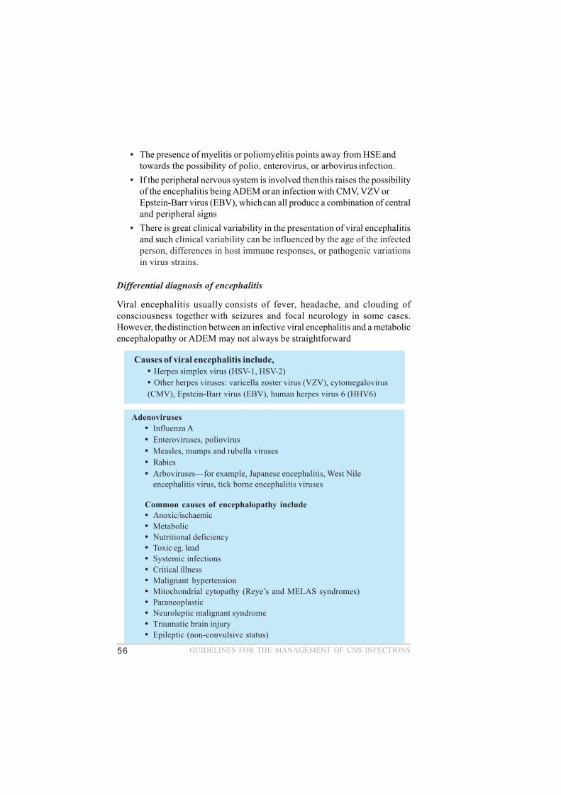
• The presence of myelitis or poliomyelitis points away from HSE andtowards the possibility of polio, enterovirus, or arbovirus infection.
• If the peripheral nervous system is involved then this raises the possibilityof the encephalitis being ADEM or an infection with CMV, VZV orEpstein-Barr virus (EBV), which can all produce a combination of centraland peripheral signs
• There is great clinical variability in the presentation of viral encephalitisand such clinical variability can be influenced by the age of the infectedperson, differences in host immune responses, or pathogenic variationsin virus strains.
Differential diagnosis of encephalitis
Viral encephalitis usually consists of fever, headache, and clouding ofconsciousness together with seizures and focal neurology in some cases.However, the distinction between an infective viral encephalitis and a metabolicencephalopathy or ADEM may not always be straightforward
Causes of viral encephalitis include,• Herpes simplex virus (HSV-1, HSV-2)• Other herpes viruses: varicella zoster virus (VZV), cytomegalovirus(CMV), Epstein-Barr virus (EBV), human herpes virus 6 (HHV6)
Adenoviruses• Influenza A• Enteroviruses, poliovirus• Measles, mumps and rubella viruses• Rabies• Arboviruses—for example, Japanese encephalitis, West Nile
encephalitis virus, tick borne encephalitis viruses
Common causes of encephalopathy include• Anoxic/ischaemic• Metabolic• Nutritional deficiency• Toxic eg. lead• Systemic infections• Critical illness• Malignant hypertension• Mitochondrial cytopathy (Reye’s and MELAS syndromes)• Paraneoplastic• Neuroleptic malignant syndrome• Traumatic brain injury• Epileptic (non-convulsive status)

GUIDELINES FOR THE MANAGEMENT OF CNS INFECTIONS 57
Viral encephalitis vs post – infectious encephalomyelitis
ADEM, also known as postinfectious encephalomyelitis, usually follows eithera vaccination within the preceding four weeks, or an infection which may be achildhood exanthema such as measles, rubella or chickenpox, or else a systemicinfection characteristically affecting the respiratory or gastrointestinal systems
Comparison between ADEM and viral encephalitis
Differences between encephalopathy and viral encephalitis
CLINICAL FEATURES ENCEPHALOPATHY ENCEPHALITIS
Fever Uncommon Common
Headache Uncommon Common
Depressed mental status Steady deterioration May fluctuate
Focal neurologic signs Uncommon Common
Types of seizures Generalised Generalised or focal
Laboratory findings
Blood Leucocytosis uncommon Leucocytosis common
CSF Pleocytosis uncommon Pleocytosis common
EEG Diffuse slowing Diffuse slowing and focal abnormalities
MRI Often normal Focal abnormalities
Clinical features ADEM ENCEPHALITIS Age
Children Any age
Recent vaccination Common Uncommon Prodromal illness Usual Occasional Fever
May occur Common
Visual loss (one or both eyes)
May occur Uncommon
Spinal cord signs May occur Rare Laboratory findings Blood Leucocytosis occasionally occurs
Leucocytosis common
MRI Multiple focal areas of hyper intensity that are the same and may involve white matter of both hemispheres, basal ganglia, brainstem, cerebellum, and spinal cord
One or more diffuse areas of hyper intensity involves the grey matter of both cerebral cortices and its underlying white matter and, to a lesser extent, basal ganglia, brainstem, and cerebellum
CSF Lymphocytic pleocytosis, elevated protein, normal glucose, and negative cultures. Red blood cells seen in acute haemorrhagic leucoencephalitis.
Lymphocytic pleocytosis, elevated protein, normal glucose, and negative cultures. Red blood cells may be seen in herpes simplex encephalitis.

GUIDELINES FOR THE MANAGEMENT OF CNS INFECTIONS58
Investigations
General1. Haematological - FBC – leucocytosis common in ADEM; relative
lymphocytosis may occur in viral enephalitis ESR – when very highD/D include tuberculosis/malignancy
2. Biochemical – metabolic encephalopathy
3. Drug screen and urine analysis
4. Cold agglutinins – mycoplasma
5. Serology- Ab to arbovirusus – take acute and convalescent samplesfor viral antibody titres - these are of little value in the acutemanagement of the patient because of the delay in obtaining theresults
6. Chest X ray
Neurological
Electroencephalogram(EEG) – should be obtained in all cases of suspectedencephalitis if possible.
• In general except a few cases most viral encephalitis will have an“abnormal” EEG
• In viral encephalitis, EEG usually shows slowing of backgroundrhythms or epileptiform discharges that can be focal or diffuse
• In patients with biopsy-proven HSE, 80% will show focalabnormalities on the EEG.
Focal EEG changes may be seen with other types of encephalitis too

GUIDELINES FOR THE MANAGEMENT OF CNS INFECTIONS 59
• In severe cases of viral encephalitis, irrespective of aetiology, the EEGmay demonstrate a burst suppression pattern or severe depression ofbackground activity that may progress to electrocerebral silence
Imaging studies
• CNS imaging studies should be done to exclude space occupyinglesions like tumour or abscess and to identify abnormalities that areindicative of HSV encephalitis.
• MRI is the imaging of choice in suspected cases of viral encephalitis,although CT scanning may be used where MRI facilities are notavailable.
• CT brain –
Changes takes 3-4 days to appear even in HSE; - i.e. CT normalearly in the illness
In HSE, 2/3rd have CT abnormalities;characteristically showsreduced attenuation in one or both temporal lobes or areas of
hyperintensity.
MRI brain
MRI is sensitive even in the early stages of HSE although rarely itmay be normal
• EEG in HSESlow waves, spikes, or spike-wave discharges, including periodic lateralizingepileptiform discharges that localize to the temporal regions increase theprobability of HSE but their absence does not exclude HSE.
Contrary to the diffuse slow waves seen in other encephalitides, tracings in HSEcases are usually very asymmetrical and often show clear foci of spikes on aabnormally slow background. Low amplitude in one or more regions especiallyover the temporal lobes is not unusual.
Periodic complexes 1-3 seconds apart are frequent in the same area but are oftentransient. Periodic complexes occur most commonly between 2nd &15th day ofthe disease and are rare after day 15. They are not specific and may occur withothers like EBV or mycoplasma too.

GUIDELINES FOR THE MANAGEMENT OF CNS INFECTIONS60
The typical MRI features in HSE are areas of focal oedema in thetemporal lobes and orbital surface of the frontal lobes as well asthe insular cortex and angular gyrus
Some other types of encephalitis like JE are also associated withparticular MRI abnormalities
Midline shift may be present in cases with significant cerebraloedema
T2 MRI alone shows many changes
Though these neuroimaging techniques demonstrate significant changes, theyare too non-specific to be used for aetiological diagnosis
Abnormalities seen in patients with postinfectious encephalomyelitis includeareas of demyelination (often symmetric) of spinal cord, white matter, and basalganglia.
Lumbar puncture and CSF analysisProvided cranial imaging has excluded any contraindications such as aspace occupying lesion or severe cerebral oedema and brain shift, aCSF analysis by lumbar puncture should be carried out in all cases ofsuspected encephalitis.The CSF in viral encephalitis typically shows a lymphocyticpleocytosis, elevated protein content, and normal or mildly depressedglucose content. However, these parameters vary considerably, and somepatients may have an entirely normal CSF examination early in theirdisease. Patients with certain viral disorders, can have a CSF profile thatresembles that of bacterial disease.The total leukocyte count usually ranges from 10 to 1000 cells/mm3, butcounts as high as 8000 cells/mm3 can be observed. Some patients lackCSF pleocytosis early in their illness; approximately 5%-10% of patientswith biopsy-proven HSV encephalitis have normal cell counts on theinitial CSF examinationAlthough the CSF usually shows a lymphocytosis, neutrophils frequentlypredominate in the early stages of viral encephalitis. Other leukocytes,including monocytes (histiocytes), plasmacytoid cells (activated B cells),and eosinophils, can be detected.The typical features of HSE are a lymphocyte cell of 10–200/mm3 andan increased protein of 0.6–6 g/l

GUIDELINES FOR THE MANAGEMENT OF CNS INFECTIONS 61
In most cases of viral encephalitis, the protein content is normal or mildlyelevated, usually less than 100mg/dL but occasionally greater than 500mg/dL.CSF glucose is usually normal and when low it is important to excludetuberculous meningoencephalitisXanthochromia of CSF may be present in HSE it is of no specificdiagnostic valueVirus-induced abnormalities of the CSF can persist for extended periods.CSF for polymerase chain reaction (PCR) is very useful and detects viralDNA or RNA in CSF. It has greater than 95% sensitivity and 100%specificity for HSE DNA within the 1st week of illness. False negativesare most likely in 1st 24-48 hrs and after 10-14 days.PCR remains positive up to 5 days after initiation of treatment in HSE.CSF should be cultured for bacteria, viruses, fungi, and mycobacteria.Oligoclonal bands are sometimes present in the CSF of patients withpost-infectious encephalomyelitis.
Management of encephalitis
• As mentioned in the guidelines on meningitis initial management consistof ensuring the adequacy of the Airway, Breathing, Circulation, etc.
• Initial resuscitation also include treatment of shock, correction ofhypoglycaemia if present and control of seizures.
• After initial resuscitation management include general and specificmeasures.
General & supportive care
• The general management is essentially supportive and in severe casespatients should be managed in a high dependency medical environmentor an intensive care unit(ICU).

GUIDELINES FOR THE MANAGEMENT OF CNS INFECTIONS62
Control of increased intracranial pressure (ICP)
• The control of increased intracranial pressure(ICP) and cerebral oedemais essential. The most important aspect of managing intracranialhypertension is early identification and initiation of appropriatetherapeutic measures. Most therapeutic measures fail if instituted late, asirreversible cerebral damage often occurs before these agents start theiraction.
• The control of ICP include- Avoidance of situations that increase ICP- Therapeutic measures to reduce ICP
Control of factors aggravating ICP
Positioning of head.- Keep head in midline position with a tilt up at 15°–30°(x)
Temperature control. – Fever worsens ICP by increase cerebral metabolism.As physical measures like sponging cause shivering, which can aggravateintracranial hypertension, they should always be used in conjunction withantipyretics.
While in the ward/ICU, monitoring of a suspected encephalitispatient should include
• Glasgow Coma scale score.(GCS)• Seizure type and frequency.• Clinical signs of intracranial hypertension.• Blood pressure (continuous/hourly):• Urine output every four hours: maintained at 0.5 ml/kg/hour.• Calculated serum osmolality: every 24 hours (preferably every
12 hours).
Se Osmolality mosm/kg= 2(Na+K in mEq) + Glucose (mmol/L)+Urea (mmol/L)
• Continuous oxygen saturation monitoring, if not possible atleast hourly monitoring.
• Central venous pressure(CVP) for severe coma.
GUIDELINES FOR THE MANAGEMENT OF CNS INFECTIONS 63
Sedation- Pain and arousal cause raised ICP by increasing cerebral bloodflow. Sedatives are useful to prevent worsening of intracranial hypertensionby this mechanism. Conventionally, sedatives were avoided, due to fear of“clouding the neurological examination”. This is not a justifiable reason toavoid proper control of intracranial pressure through sedation. Sedationcan be with diazepam 0.1-0.3 mg/kg/dose or phenobarbitone 3–5 mg/kg/dayin two divided doses (y)
Seizure control.- Seizures increase intracranial pressure by increasingcerebral metabolism and cerebral blood flow and by Valsalva. Henceanticonvulsants should be administered prophylactically or therapeutically.PHENYTOIN: Loading dose 15-20 mg/kg to be infused at a rate of 1.0 mg/kg/min. (x)
Maintenance dose - 5-8 mg/kg/d. If the seizures persist, then anotherloading dose of phenobarbitone 10-20mg/kg can be given. DIAZEPAMINFUSION: If the seizures still persists, then diazepam infusion to bestarted at a rate of not more than 5 mg/min followed by infusion at rateof 0.1-0.4 mg/kg/hr. Diluents to be used - sterile water, normal saline. Ifthe seizures are yet to be controlled, then any of the following optionscan be tried.
MIDAZOLAM INFUSION: Loading dose of 0.05-0.2 mg/kg stat followedby infusion at a rate of 1-5 µg/kg/min
Appropriate fluid and electrolyte therapy - Hypovolaemia often accompaniesviral encephalitis due to decreased intake and increased loss (vomiting,sweating). Hence any initial dehydration should be corrected first. Continuedfluid therapy should be with N/2 saline 2/3rd to 3/4th of the maintenance withallowance for temperature, hyperventilation, and urine output. With increasedtemperature fluid requirement is increased and if the urine output is more than50% of the total input, then add the excess fluid to the maintenance
Therapeutic measures to reduce ICP
Hyperventilation- Can be done with bag and mask ventilation or after intubation.To decrease further rise in ICPduring intubation either 4% lignocaine as localspray /adequate sedation is needed. When hyperventilation is used for themanagement of raised ICP, then aim is to achieve a Paco2 of 25mmHg. Intracranialpressure begins to diminish 10–30 seconds after inception of hyperventilation,reaches a peak in 30 minutes, and returns to the original value in less than hour.The level of PCO2 should be raised to 30-35mmHg after 1hour. Prolongedhyperventilation may worsen the outcome.

GUIDELINES FOR THE MANAGEMENT OF CNS INFECTIONS64
Hence, hyperventilation should be gradually withdrawn (rapid withdrawalcauses rebound intracranial hypertension) after a period of 30–60 minutes so asto raise PCO2 with institution of other modes of therapy simultaneously.
Mannitol- Response to mannitol depends on original intracranial pressure,dose given over the previous three hours (the lesser, the better effect),and rateof administration. Rapid administration is more effective in reducing intracranialpressure, but the action has a much shorter duration. A slower infusion rateproduces a lesser degree of decrease that lasts longer. With mannitol significantside effects can occur if used inappropriately. Chronic continuous therapy shouldbe avoided as the brain adapts to the sustained hyperosmolality and producesrebound cerebral oedema after withdrawal of anticerebral oedema measures).Initial bolus to be given over 30 min, DOSE: 2.5- 5ml/kg of 20% solution (0.5-1 g/kg) Repeat mannitol to be given only if serum osmolality is <=300 mOm/kg(tocalculate osmolality-see above) and could be given every 4-6 hourly, at a doseof 2.5ml/kg, given over 30 minutes.
Frusemide (furosemide) alone causes a slow reduction in intracranial pressure,but when combined with mannitol, the fall in intracranial pressure is rapid and issustained for a considerably longer period than when either agent is used alone.Dose: 1mg/kg/dose every 12th hrly can be given (potassium levels & bloodpressure to be monitored while giving diuretics.)
Thiopentone: To be used only when the above measures fail, but can becombined with hyperventilation. Dosage: loading dose of 5 mg/kg given over30 - 60 min with a maintenance dose of 1 mg/kg/hr as infusion. Maximummaintenance dose is 5 mg/kg/hr. Whenever the maintenance dose is increasedby 1 mg/kg/hr, a loading dose of 5 mg/kg to be given. Caution:hypotension
Specific measures & treatment
1. Until a bacterial cause of CNS inflammation is excluded, parenteralantibiotics should be given. Treatment with a third-generationcephalosporin, such as cefotaxime sodium + c. penicillin or ceftriaxonesodium is recommended.
2. Antiviral therapy- If antiviral therapy is not given to patients with HSEthe mortality is over 70% and this can be brought down to 20% if treatedearly using I.V Acyclovir. If HSE is a possibility then acyclovir, 10 mg/kg I.V three times daily, should be started as soon as possible. When thediagnosis is confirmed by CSF PCR,(HSE PCR is available in the privatesector at present) and/or where there are characteristic MRI changes,acyclovir should be continued for 14 days. Acyclovir should only be

GUIDELINES FOR THE MANAGEMENT OF CNS INFECTIONS 65
stopped where there is a definite alternative diagnosis made. The situationof still suspected HSE in the presence of a negative CSF PCR and MRIacyclovir for 10 days is still justified. Acyclovir is a very safe andrelatively non-toxic however during IV acyclovir therapy renal functionshould be closely monitored because of the rare development of renalimpairment. The risk of renal toxicity is reduced by adequately hydratingthe patient (eg, 1 mL fluid per day for each 1 mg/d of acyclovir) Becauseof its high pH, IV acyclovir may cause phlebitis and local inflammationif extravasation occurs
Prognosis
Supportive care and rehabilitation are important after the patient recovers.Because some sequelae of encephalitis may be subtle, neurodevelopmentaland audiologic evaluations should be part of routine follow-up.
Most patients completely recover from viral encephalitis; however, prognosisdepends on the cause and severity of the illness and the patient’s age. If theclinical illness is severe and substantial parenchymal involvement is evident,prognosis is poor. Potential deficits include intellectual, motor, psychiatric,epileptic, visual, and auditory abnormalities. The prognosis of HSE is better inyounger patients, and those having a disease duration of four days or less anda Glasgow coma score of 6 or less at the time of acyclovir initiation.
Cerebral malaria• Detailed clinical guidelines on cerebral malaria is not included in this
document. The presentation is varied and may suggest other conditions,such as meningitis, encephalitis, or epilepsy. Thus, cerebral malariashould be considered in the differential diagnosis of a febrileneurological illness if a history of residence or travel through amalarious area exists.
• Seizures and coma are common in a child with malaria, (sometimeswithout cerebral malaria) and prolonged postictal state should raisesuspicion of this dangerous entity. Cerebral malaria can be rapidlyfatal, prompt diagnosis and immediate treatment is important.
• Quinine is still the DOC for severe and complicated malaria thoughresistance could be a problem. 15-20 mg/kg IV diluted in IV infusionfluid to 1 mg/mL; infuse over at least 4 h; followed by 10 mg/kg/doseq8-12h if continuation beyond 48 h is needed; reduce dose to 5 mg/kgq8h; switch to PO therapy as soon as possible

GUIDELINES FOR THE MANAGEMENT OF CNS INFECTIONS66
Tuberculous Meninigitis(TBM)
This document does not include clinical guidelines for the management ofTBMTBM continues to be an important disease and should be considered inthe differential diagnosis in any patient presenting with fever and a change insensorium.Tuberculous meningitis may present in acute, subacute, or chronicform.

GUIDELINES FOR THE MANAGEMENT OF CNS INFECTIONS 67
Prepared by the Guidelines Committeeof
the Sri Lanka College of Paediatricians comprising
Dr Lakkumar Fernando Consultant Paediatrician, Chilaw(Coordinator) Hospital.
Prof. S P Lamabadusuriya Senior Professor, Dept. of Paediatrics,Faculty of Medicine University ofColombo.
Prof. Manouri Senanayake Professor, Dept. of Paediatrics, Facultyof Medicine University of Colombo.
Dr Deepthi Samarage Senior Lecturer in Paediatrics, Faculty ofMedical Sciences, University ofSri Jayawardenapura, Nugegoda.
Dr Kumudu Karunaratne Consultant Microbiologist, Lady RidgewayHospital Colombo.
Dr Wasanthi Thevanesan Consultant Microbiologist
Dr H T Wickramasinghe Consultant Paediatrician.

NATIONAL GUIDELINES