Management of breast lumps with awareness to breast carcinoma إyusor (1)
60
Management of breast lump with awareness to breast carcinoma Presented by : Yusor Jaafar Mariam Talal Sherin Raad
-
Upload
home -
Category
Health & Medicine
-
view
23 -
download
2
Transcript of Management of breast lumps with awareness to breast carcinoma إyusor (1)

Management of breast lump with awareness to breast carcinoma
Presented by:Yusor JaafarMariam TalalSherin Raad
Management of breast lump with awareness to breast carcinom
Introduction.Evaluation of breast lump.
By: Yusor Jaafar
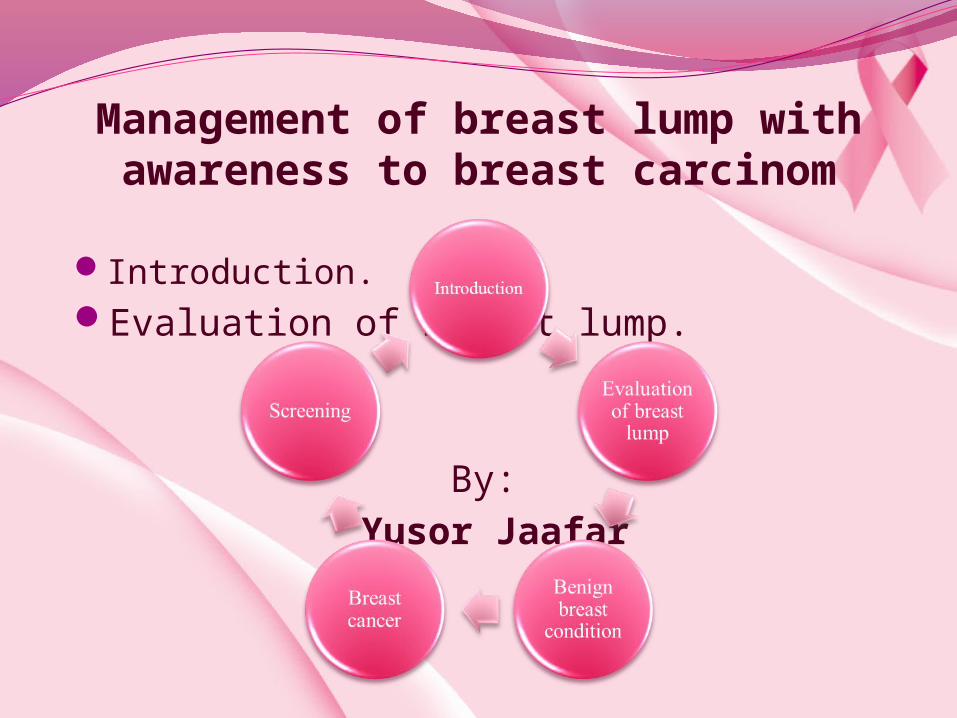
IntroductionThe complaining of breast lump ranging frm 40_70%
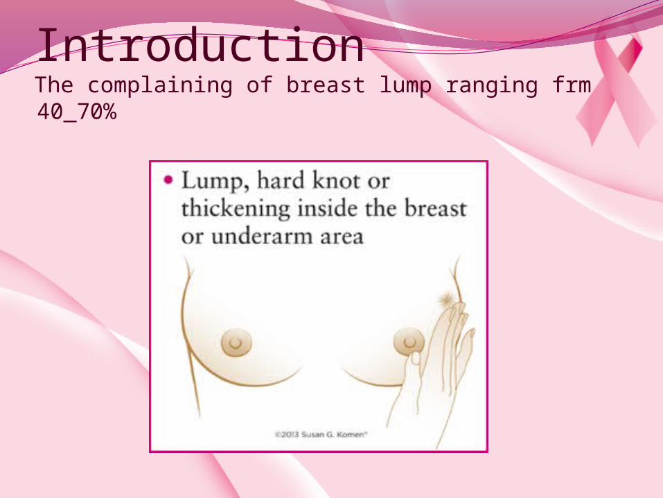
Evaluation of breast lump
Diagnosis
History and examination
imaging
Cytology and
histology

History taking
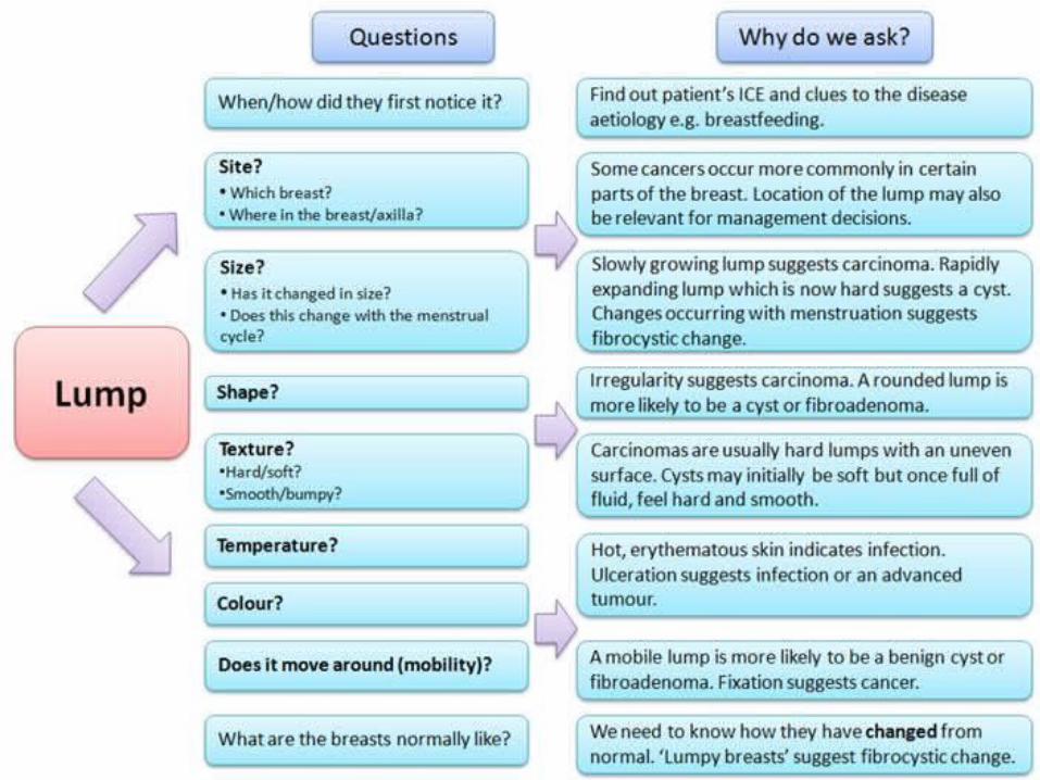

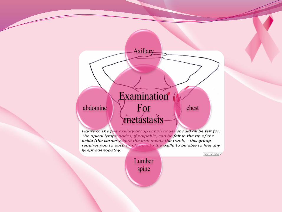
Physical examinationGeneral examinationPosition
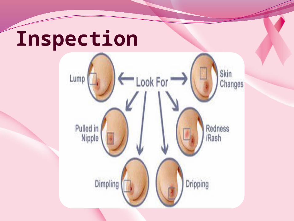
Inspection
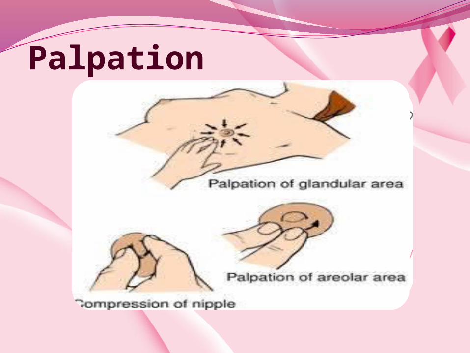
Palpation
Primary analysisPainless lump:CarcinomaCystFibroadenomaArea of fibroadenosis

Painful lump:An area of fibroadenosisCystPeriductal mastitisAbscessCarcenoma

Nipple changesDestructionDepressionDiscolourationDisplacementDeviationDischargeDuplication
Nipple dischargeRed (blood)Pink (serum+blood)Clear pale yellowBrownGreenBlackCreamy white or yellowThin white
Nipple dischargeDuct ectasia
Intraduct papilloma
Ductal carcinoma in-situ
Associated with cyst
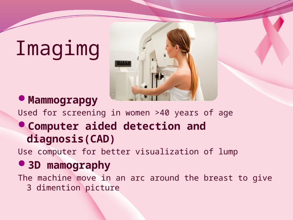
Imagimg
MammograpgyUsed for screening in women >40 years of age
Computer aided detection and diagnosis(CAD)
Use computer for better visualization of lump
3D mamographyThe machine move in an arc around the breast to give 3
dimention picture

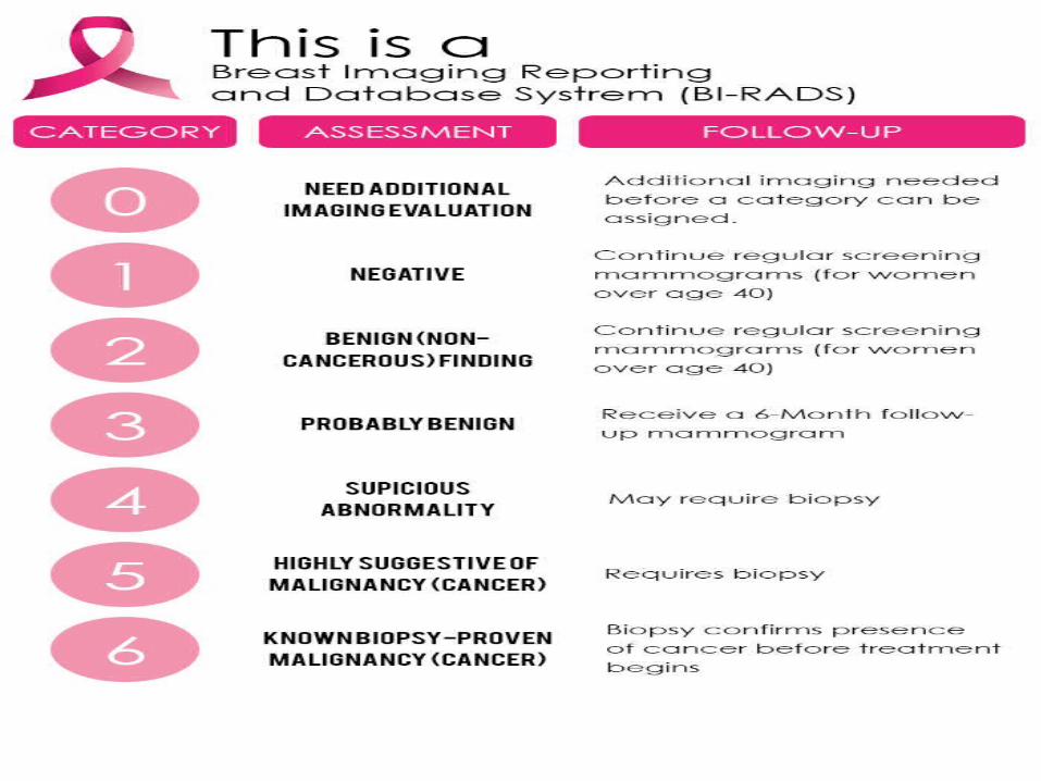
BI-RADS system categories:
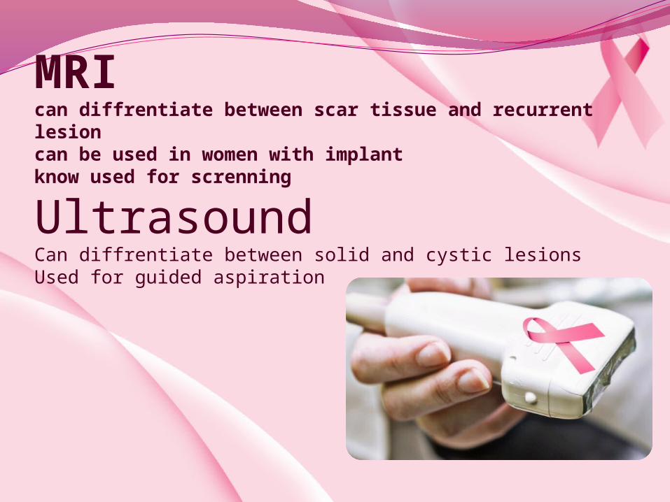
MRIcan diffrentiate between scar tissue and recurrent lesioncan be used in women with implant know used for screnning
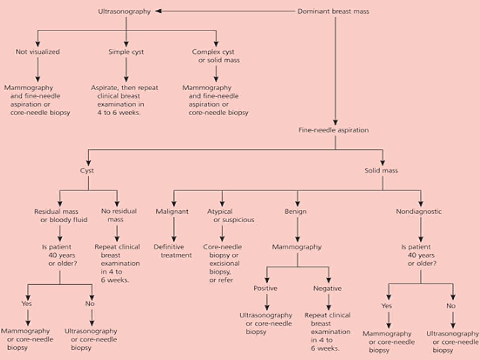
Ultrasound Can diffrentiate between solid and cystic lesions Used for guided aspiration
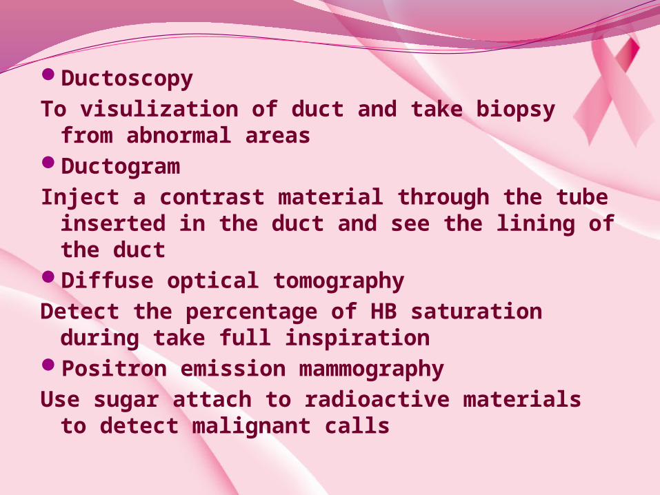
DuctoscopyTo visulization of duct and take biopsy from abnormal areas DuctogramInject a contrast material through the tube inserted in the duct and
see the lining of the ductDiffuse optical tomographyDetect the percentage of HB saturation during take full inspirationPositron emission mammographyUse sugar attach to radioactive materials to detect malignant calls
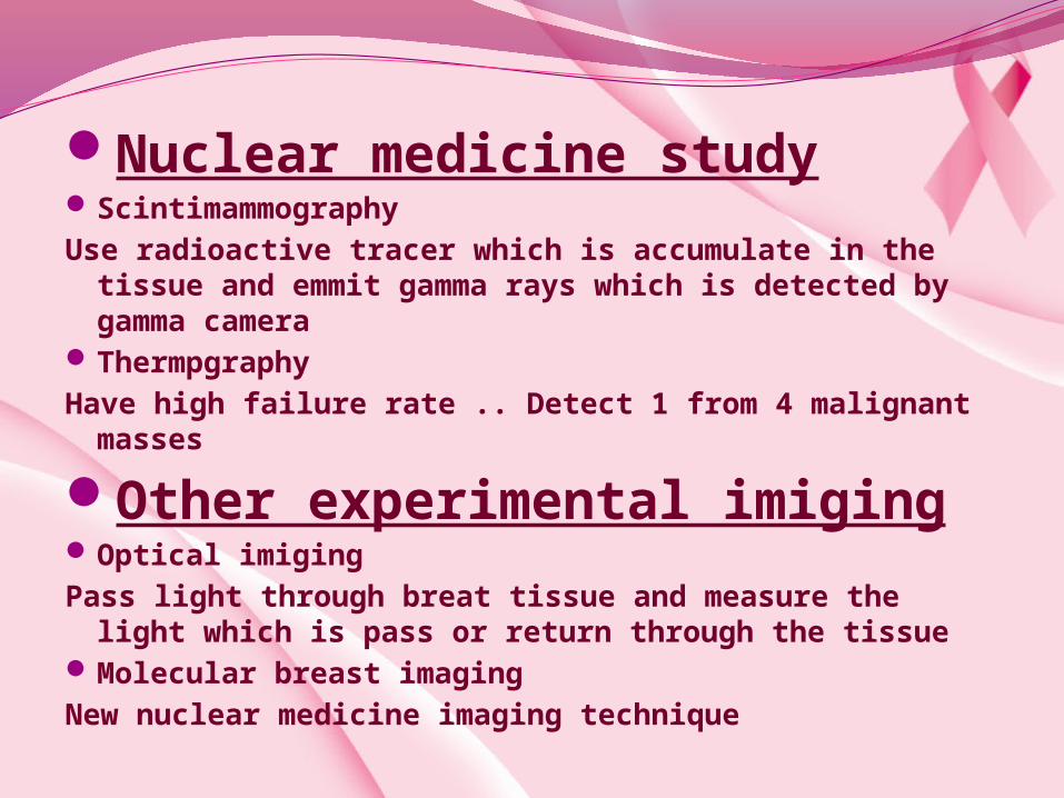
Nuclear medicine studyScintimammography Use radioactive tracer which is accumulate in the tissue and emmit gamma rays
which is detected by gamma camera ThermpgraphyHave high failure rate .. Detect 1 from 4 malignant masses
Other experimental imigingOptical imigingPass light through breat tissue and measure the light which is pass or return
through the tissueMolecular breast imagingNew nuclear medicine imaging technique
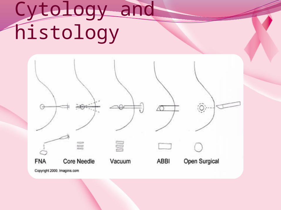
Cytology and histology
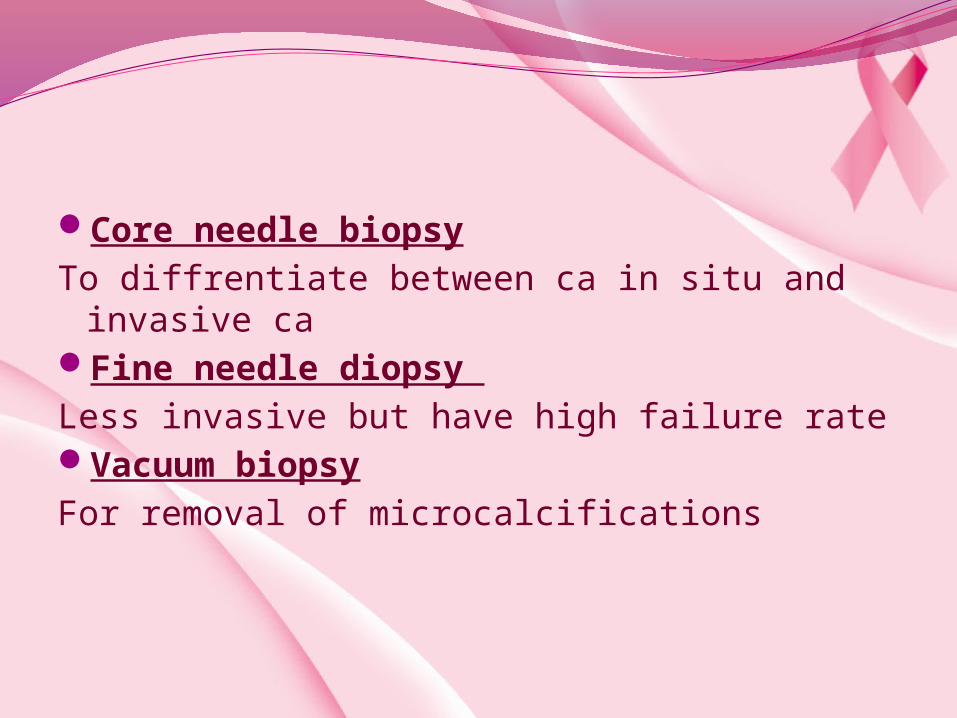
Core needle biopsy
To diffrentiate between ca in situ and invasive ca Fine needle diopsy
Less invasive but have high failure rateVacuum biopsy
For removal of microcalcifications

Pathological classification of breast lumps
Epithelial: Duct papilloma
Connective tissue:Neurofibroma
Lipoma
Mixed:
Fibroadenoma
Phyllodes tumor
Benign Invasive duct
carcinoma
Inflammatory carcinoma
Malignant
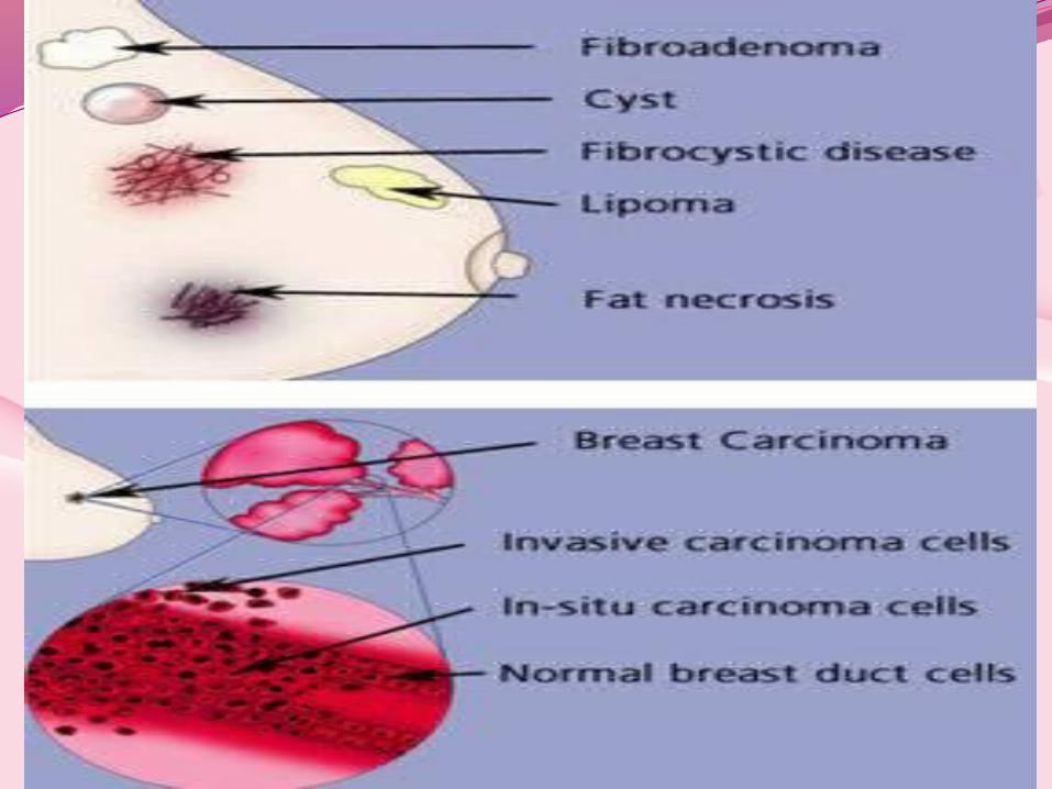
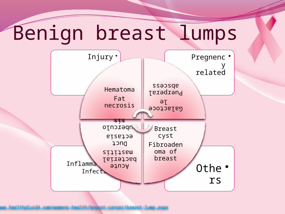
Benign breast lumps
•Others •Inflammation•Infection
•Pregnency related
• Injury
Hematoma
Fat necrosis Galactocele
Puerperal abscess
Breast cyst
Fibroadenoma of breast
Acute bacterial mastitis
Duct ectasia
tuberculosis
http://www.healthplus24.com/womens-health/breast-cancer/breast-lump.aspx
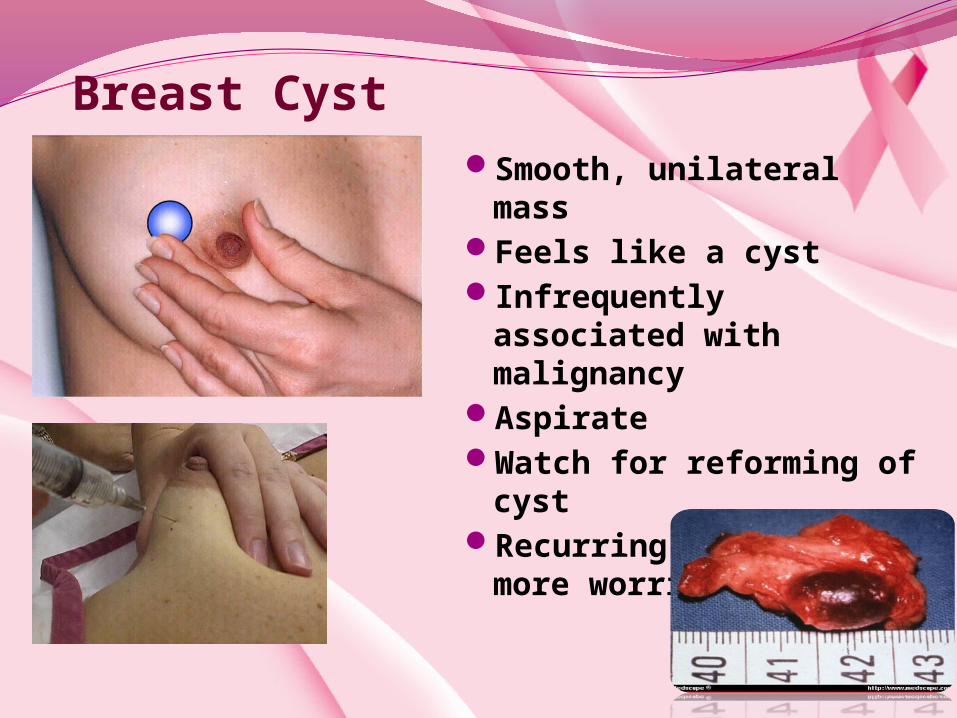
Breast CystSmooth, unilateral
massFeels like a cystInfrequently
associated with malignancy
AspirateWatch for reforming of
cystRecurring cysts are
more worrisome.

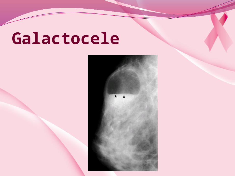
Galactocele
Milk-filled cyst
Usually follows lactation
Firm, tender mass
Usually in upper quadrants
Diagnostic aspiration often curative

Galactocele
Galactocele
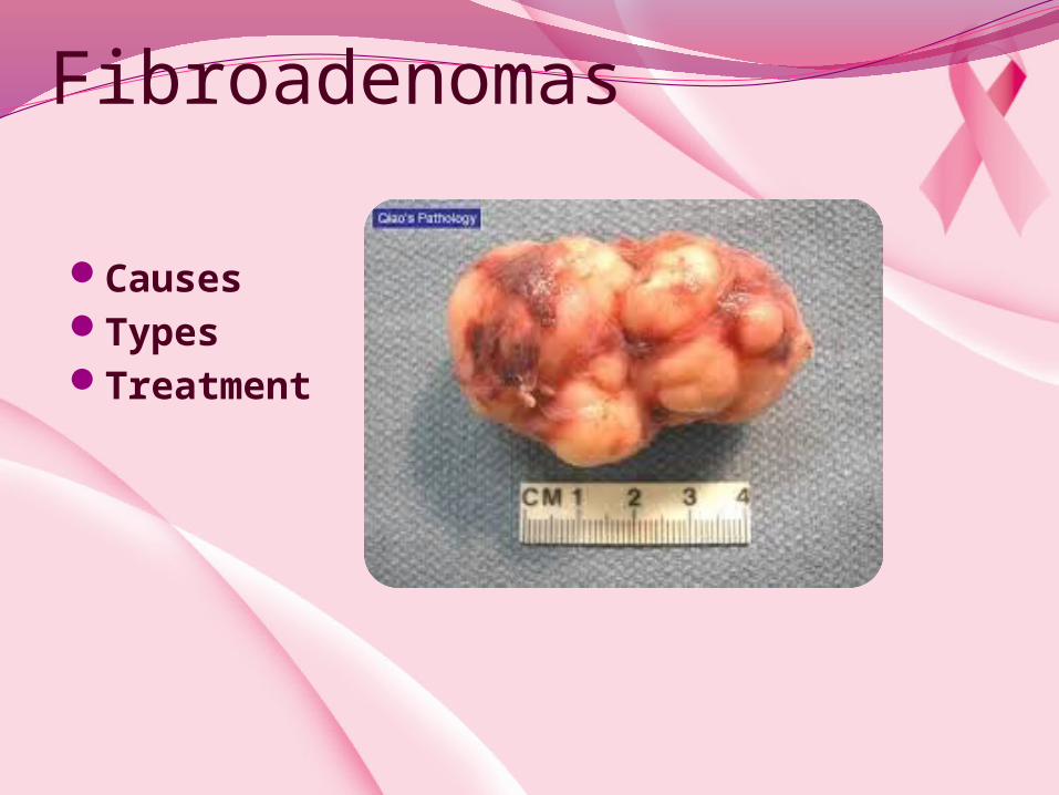
Fibroadenomas
CausesTypesTreatment
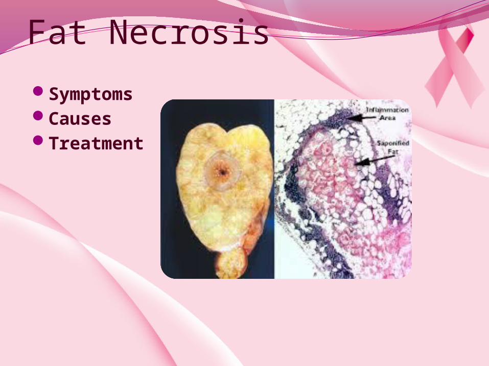
Fat Necrosis
SymptomsCauses Treatment

Lipoma
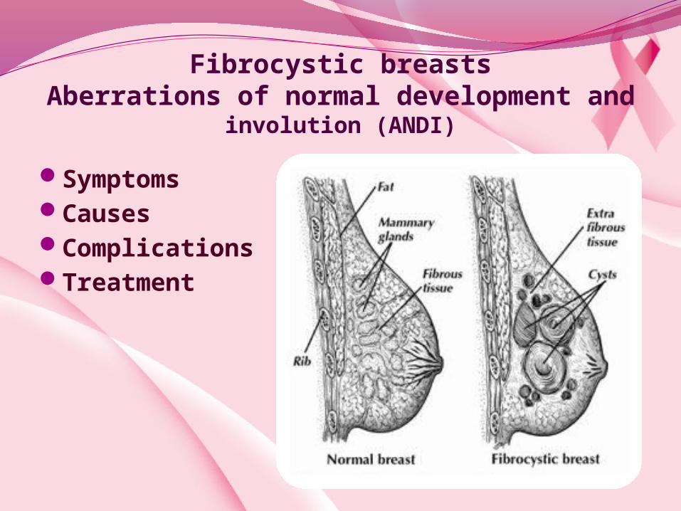
Fibrocystic breastsAberrations of normal development and
involution (ANDI)
SymptomsCausesComplicationsTreatment
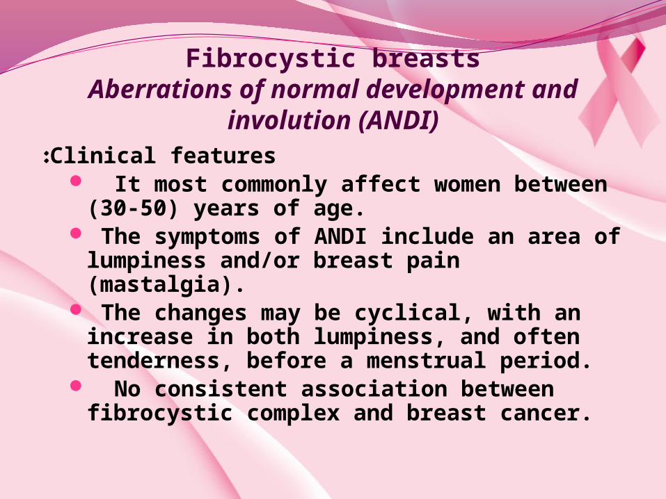
Fibrocystic breastsAberrations of normal development and
involution (ANDI)Clinical features: It most commonly affect women between
(30-50) years of age. The symptoms of ANDI include an area of
lumpiness and/or breast pain (mastalgia). The changes may be cyclical, with an
increase in both lumpiness, and often tenderness, before a menstrual period.
No consistent association between fibrocystic complex and breast cancer.
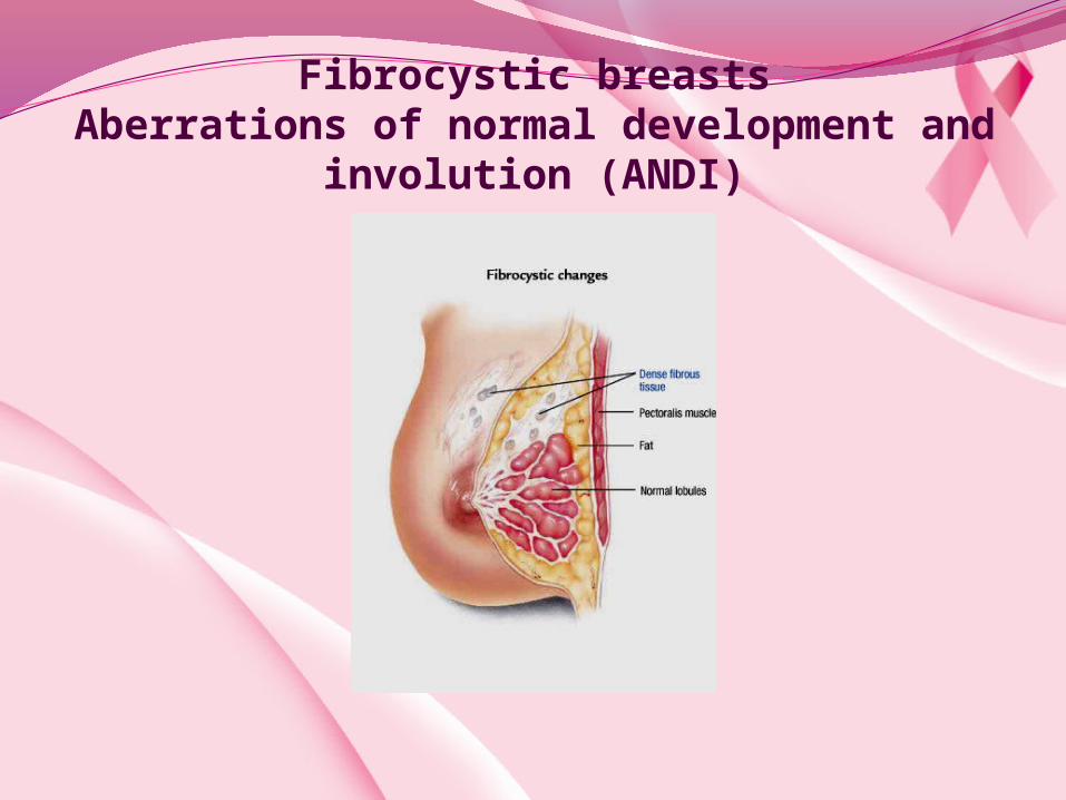
Fibrocystic breastsAberrations of normal development and
involution (ANDI)
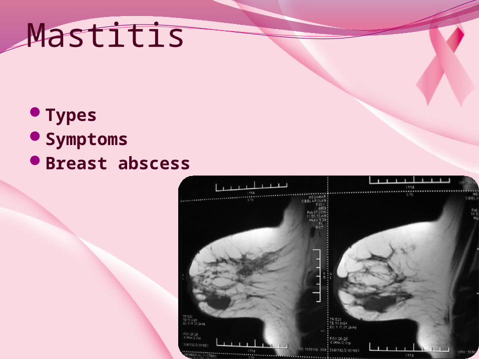
Mastitis
TypesSymptomsBreast abscess
CausesTreatmentRisk factorsWeaning
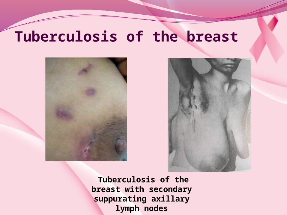
Tuberculosis of the breast
Tuberculosis of the breast with secondary suppurating axillary
lymph nodes

Breast CancerEpidemology
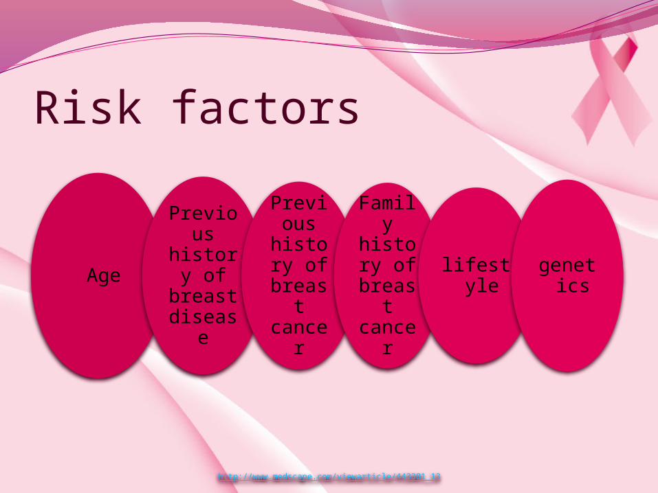
Risk factors
Age Previous history of
breast disease
Previous history of
breast cancer
Family history of
breast cancer
lifestyle genetics
http://www.medscape.com/viewarticle/443381_13
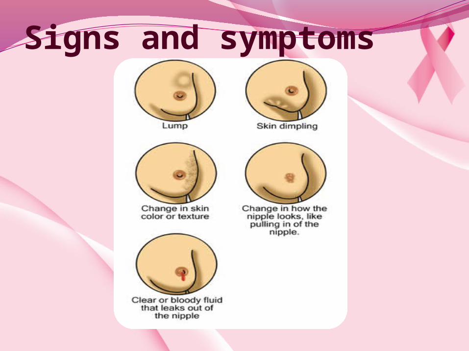
Signs and symptoms

Clinical presentationbreast cancer commences most
frequently in the upper, outer quadrant
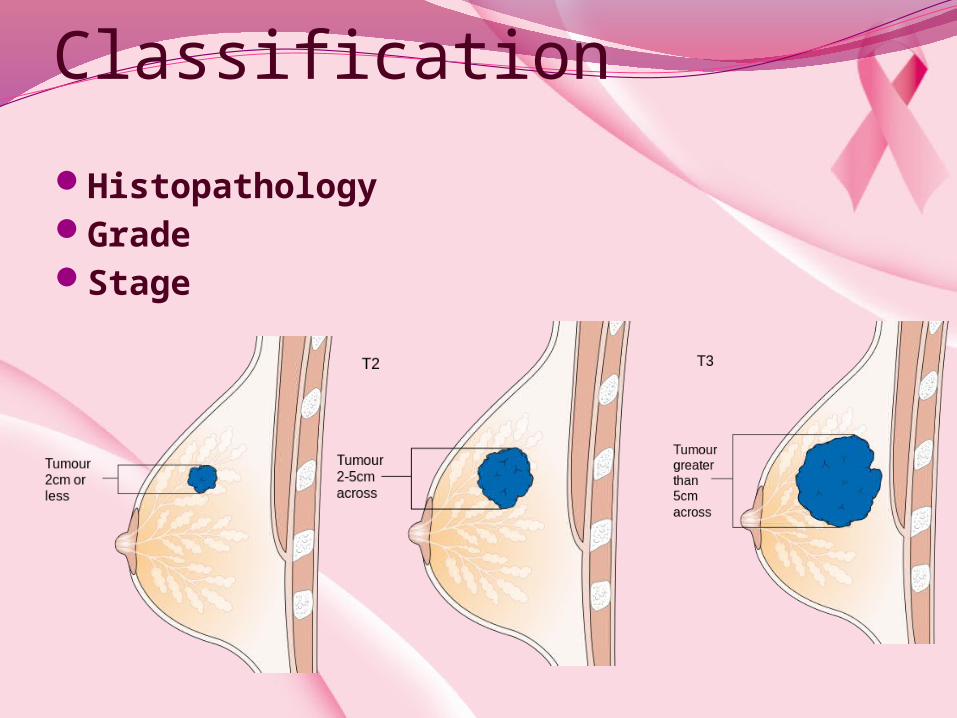
Classification
HistopathologyGradeStage

Stage 1A
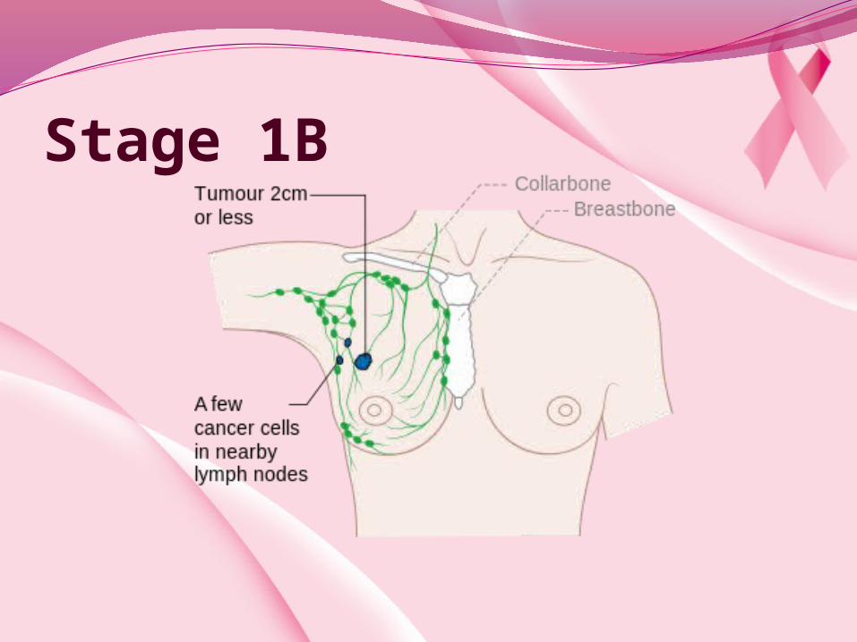
Stage 1B
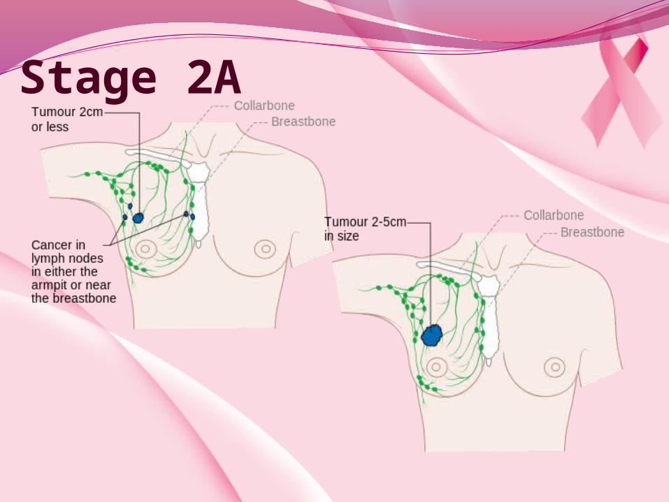
Stage 2A

Stage 2B

Stage 3A
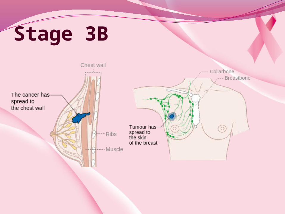
Stage 3B
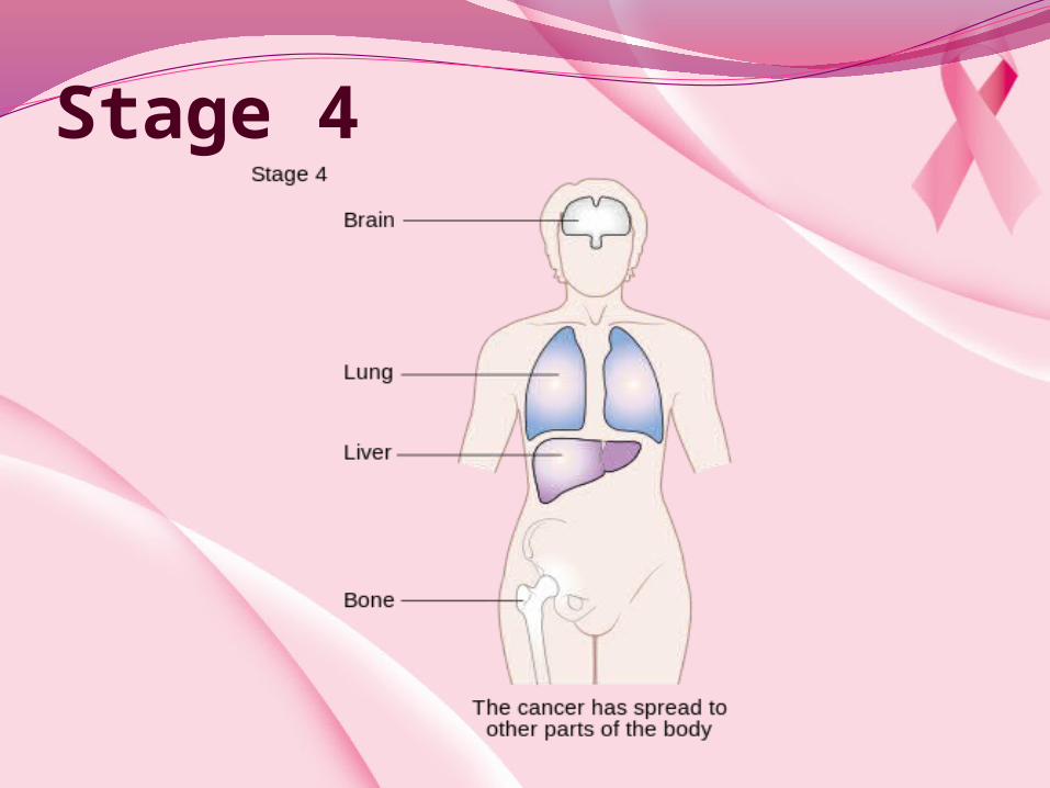
Stage 4

Treatment Medication
Hormone therapy

ChemotherapyCMFCAF (or FAC)TACAC → TFEC → TTCTCH

Monoclonal antibodiesNaked monoclonal antibodiesConjugated monoclonal antibodies

Radiation
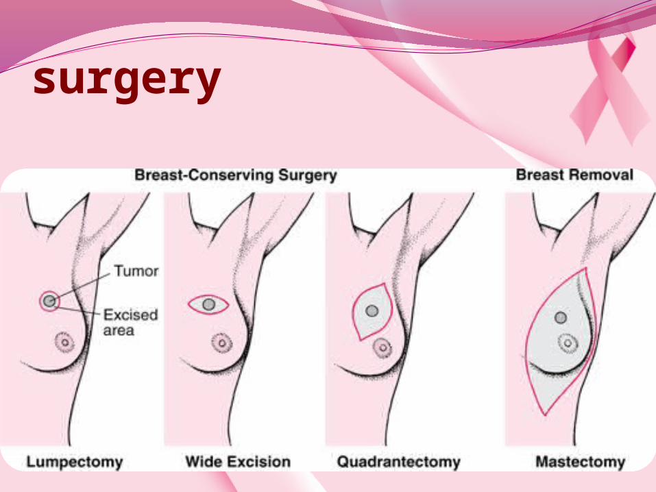
surgery

Screening Average-risk womenAbove-average risk women
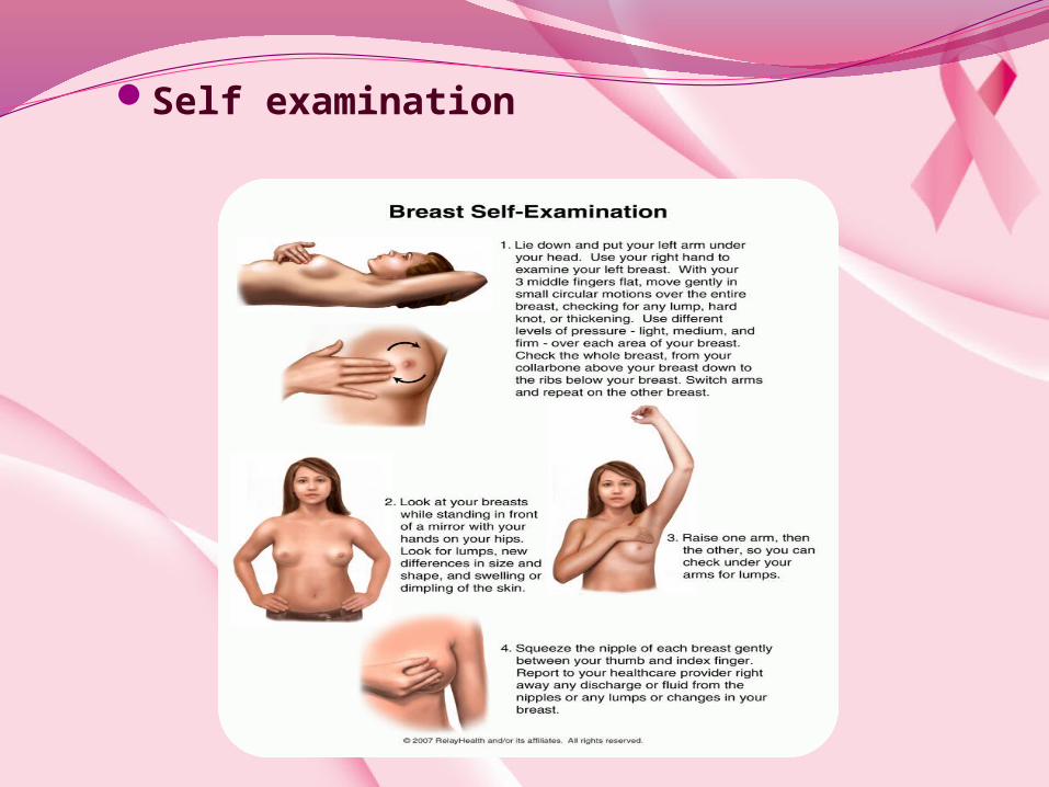
Self examination

Clinical Breast ExaminationMammographyMRI