
HANDOUT - Comprehensive Echocardiographic Assessment of Adults with Congenital Heart Disease.pdf

Dr Margreet Bink-BoelkensUniversity Hospital
Groningen, The Netherlands
Dr Jack ColmanMount Sinai Hospital
Toronto, Ontario
Dr Michael ConnellyThe Peter Lougheed Centre
Adult Congenital Cardiac ClinicCalgary, Alberta
Dr Luciano DalientoUniversity of Padua Medical School
Padova, Italy
Dr Annie DoreMontreal Heart Institute
Montreal, Quebec
Dr Michael GatzoulisRoyal Brompton HospitalLondon, United Kingdom
Dr Welton GersonyBabies Hospital
New York, New York
Dr Thomas GrahamVanderbilt Medical Center
Nashville, Tennessee
Dr John HessDeutsches Herzzentrum Munchen
Munchen, Germany
Dr Andreas HoffmannUniversity of BaselBasel, Switzerland
Dr Laurence IserinHopital Necker
Paris, France
Dr Richard LiberthsonMassachusetts General Hospital
Boston, Massachusetts
Dr Ariane MarelliMcGill University Hospital Center
Montreal, Quebec
Dr Folkert MeijboomErasmus Medical Centre Rotterdam
Rotterdam, The Netherlands
Dr Barbara MulderAcademic Medical Center
Amsterdam, The Netherlands
Dr Koichiro NiwaChiba Cardiovascular Center
Chiba, Japan
Dr Erwin OechslinUniversity HospitalZurich, Switzerland
Dr Joseph PerloffUCLA Adult Congenital Heart
Disease CenterUCLA Center for the Health Sciences
Los Angeles, California
Dr Patricia PresbiteroHumanitas Mirasole SpA
Milan, Italy
Dr Reed PyeritzUniversity of Pennsylvania School
of MedicinePhiladelphia, Pennsylvania
Dr Jane SomervilleMiddlesex Hospital/UCLLondon, United Kingdom
Dr Dylan TaylorUniversity of Alberta
Walter MacKenzie CentreEdmonton, Alberta
Dr Judith TherrienSir Mortimer B Davis Jewish
General HospitalMcGill UniversityMontreal, Quebec
Dr Ulf ThilenLund University Hospital
Lund, Sweden
Dr Carole WarnesMayo Clinic
Rochester, Minnesota
Dr Gary WebbToronto Congenital Cardiac Centre
for AdultsThe Toronto General Hospital
Toronto, Ontario
MANAGEMENT OF ADULTS WITH CHD
PRIMARY PANELISTS
Yvonne BalonThe Peter Lougheed Centre
Calgary, Alberta
Dr Michael CY ChanStanford University Medical Center
Stanford, California
Dr Louise HarrisToronto Congenital Cardiac
Centre for AdultsThe Toronto General Hospital
Toronto, Ontario
Dr Jonathan HowlettQEII Health Sciences Centre
Halifax, Nova Scotia
Dr Alasdair HunterChildren’s Hospital of Eastern Ontario
Ottawa, Ontario
Dr Philip KilnerRoyal Brompton HospitalLondon, United Kingdom
Dr Peter McLaughlinToronto Congenital Cardiac
Centre for AdultsThe Toronto General Hospital
Toronto, Ontario
Dr Mathew SermerMount Sinai Hospital
Toronto, Ontario
Dr Samuel SiuToronto Congenital Cardiac
Centre for AdultsThe Toronto General Hospital
Toronto, Ontario
Dr Christo TchervenkovMontreal Children’s Hospital
McGill UniversityMontreal, Quebec
Dr WG WilliamsHospital for Sick Children &Toronto Congenital Cardiac
Centre for AdultsToronto, Ontario
SECONDARY PANELISTS

Introduction 943
General Recommendations 944
Section I – Atrial Septal Defect 947
Section II – Ventricular Septal Defect 950
Section III – AVSD 952
Section IV – PDA 955
Appendix I – Canadian Adult Congenital Heart Network centres 957and contact persons
Appendix II – Levels of evidence used in developing the 957management recommendations for adults with congenital heart disease
Appendix III Types of disorders that should be seen at national or 957regional adult congenital heart disease centres
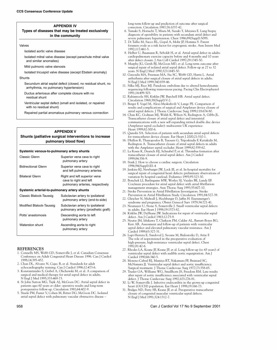
Appendix IV – Types of diseases that may be treated exclusively 958in the community
Appendix V – Shunts (palliative surgical interventions to increase 958pulmonary blood flow)
THE CANADIAN JOURNAL OF
CARDIOLOGYSeptember 2001 Volume 17 No 9

Can J Cardiol Vol 17 No 9 September 2001 943
The authors of this second Consensus Conference Reporton Adult Congenital Heart Disease (ACHD) are grate-
ful to the Canadian Cardiovascular Society (CCS) and itsCouncil for the opportunity to update and assemble thedocument, which follows.
Important advances have been made in the field of adultcongenital cardiology since the 1996 ConsensusConference Report was published (1), which led to thedecision to update the recommendations. As well, over 160new references have been added. Sections on marfan syn-drome, single ventricle and cyanotic patients have beenadded. Prevalence, genetics, pregnancy and arrhythmias foreach specific cardiac lesion have been incorporated. Therecommendations that follow are the best available giventhe present knowledge.
These recommendations have been written for cardiolo-gists, cardiac surgeons and other health care professionals
who are not experts in this field. This is important to state,because an audience more or less knowledgeable about thesubject would require a different amount of backgroundinformation and depth of treatment of the material.
The panelists are convinced that the interests of any butthe most simple patients are best served by involving whatare called ‘national or regional ACHD centres’. The knowl-edge and experience in the care of these patients should befocused, so that competence and skills become available asquickly as possible. This recommendation is not intended tostand in the way of involving local physicians in the care ofthese patients as collaborating members of a team with thebest interests of the patients at heart. Almost all of thesepatients require primary care. Many would benefit fromperiodic contact with a cardiologist in their community,along with their specialist at the national or regionalACHD centre.
SPECIAL ARTICLE
CCS Consensus Conference2001 update:
Recommendations for theManagement of Adults with
Congenital Heart DiseasePart I
Judith Therrien MD, Annie Dore MD, Welton Gersony MD,Laurence Iserin MD, Richard Liberthson MD, Folkert Meijboom MD,
Jack M Colman MD, Erwin Oechslin MD, Dylan Taylor MD, Joseph Perloff MD,Jane Somerville MD, Gary D Webb MD
This Consensus Conference on the Management of Adults with Congenital Heart Disease is an update of the previous document published in The Canadian Journal of Cardiology 1998;14:395-452. The entire consensus statement will be published in three consecutive issues of The Canadian Journal of Cardiology
The Congenital Heart Disease Committee of the American College of Cardiology, the Council on Cardiovascular Disease in the Young of theAmerican Heart Association, the Grown-Up Congenital Heart Working Group of the European Society of Cardiology, the InternationalSociety for Adult Congenital Cardiac Disease and the Japanese Society for Adult Congenital Heart Disease have all endorsed this document

One of the problems and challenges of ACHD is thelarge number of different lesions and situations that may beencountered. Those specializing in the area have workedhard to attain the competence and confidence that theyhave, yet regularly continue to be unclear about managingindividual patients. We have great respect for the seeminglyendless scenarios that we encounter.
Patients with congenital heart disease (CHD) are inter-esting to have in one’s practice, but they should be acceptedeither collaboratively with a national or regional ACHDcentre, or after one has concluded that the patient does notneed a referral to such a centre. The natural interest in ‘col-lecting a few congenitals’ should be resisted until this testhas been run. This principle applies as much in a surgical asin a medical practice.
Canada is fortunate to have a nationwide group ofnational and regional ACHD centres called the CanadianAdult Congenital Heart (CACH) Network (Appendix 1).We encourage Canadian readers to make use of these facil-ities, and the skills and experience they represent. Moreinformation can be obtained on the Internet atwww.cachnet.org.
Another aspect of this consensus conference update isthat it will remain available on the Internet at www.cachnet.org, at www.achd-library.com and at www.ccs.ca.
In keeping with the origins of the panelists, this docu-ment has been endorsed by the most important societieswith an interest in this field. We have written this materialin as user-friendly a fashion as possible. We envisaged a cli-nician looking up a lesion, and wishing to see the recom-mendations at a glance, rather than having to refer to othersections of the report. This has led to some repetition forthe reader who begins at the beginning and ends at the end.The repetitive portions are printed in italics to reduce frustrationsresulting from this style. We also committed to not writing atextbook, even though a good and current one is needed foran audience such as ours. We have focused on the principlesof management of these patients, leaving latitude wherepossible for the clinician to exercise judgement. We wish toguide, but not to constrain unduly. We have given weight toour management recommendations. The scales that weused are shown in Appendix II. We have used standardssimilar to those used in past CCS Consensus Conferences.
We hope that these recommendations will be helpful tothe patients in whose interests we have written them, andto those who care for them. Canadians have made impor-tant contributions to the management of patients withCHD. We join with our international colleagues in hopingthat this report will follow in this tradition.
CHOICE OF PANELISTSThe CCS invited Dr Gary Webb (president of the
CACH Network) and Dr Judith Therrien to lead theprocess, and endorsed the membership of the primary andsecondary panels. As is apparent from the panel member-ships, the Grown-Up Congenital Heart Working Group ofthe European Society of Cardiology and the International
Society for Adult Congenital Cardiac Disease contributedmany members to this process. While published in Canada,this is truly an international document. Further, the panelswere selected to receive input from various interestedgroups (adult/pediatric cardiology, cardiac surgery, obstet-rics, genetics, nurse practitioner), and from the variousregions of Canada, the United States, Europe and Japan.
Most of the panelists worked very hard reviewing manydrafts and offering suggestions for improvement. The panelhad almost no difficulty in reaching agreement on the state-ments made. Debate occurred only where there were insuf-ficient data to decide a point.
A glossary defining the many unusual terms used in thisfield has been prepared as a companion document; spacedoes not permit publishing this in this journal. We refer youto www.ccs.org and www.achd-library.com.
GENERAL RECOMMENDATIONSPart I – Levels of care for adult
patients with CHDCare of adult patients with CHD should be available at
several different levels. A national ACHD centre is onethat can provide all needed services to ACHD patients.The CACH Network has recommended the maintenanceor establishment of five ‘national’ centres in Canada (pop-ulation 31 million), one in each of the five regions of thecountry. A regional ACHD centre is one that has the essen-tial resources required for an ACHD centre (two ACHDcardiologists and excellent echocardiography facilities), plusany other resources that they may have beyond these. Suchcentres would provide most patient care, but would refer toa national centre when their resources are required (eg, con-genital heart surgery, special electrophysiology services).
A national ‘full service’ ACHD centre:
• Should have all (or almost all) of the componentsdescribed below in the ideal national centre
• May provide care to any patient with congenital orheritable cardiovascular disease
• Would usually serve a population base of three to 10 million
A regional ACHD centre:
• Will have a minimum of two cardiologists (eitheradult or pediatric) with special skills, training orexperience in the care of adult patients with CHD,and highly skilled echocardiographic services;beyond this, other components of the nationalACHD centre described below may be available,depending on local resources and needs
• May provide care to any patient with congenital orheritable cardiovascular disease within theconstraints of available resources
• Would usually serve a population base of up totwo million
CCS Consensus Conference Update
Can J Cardiol Vol 17 No 9 September 2001944

An individual specialist/cardiologist:
• Provides specialist care to patients with the types ofdisorders listed in Appendix III, without there beinga need to involve an ACHD centre; when suchpatients require special interventions (eg, cardiacsurgery, electrophysiology study), referral to anational or regional ACHD centre is stillrecommended, unless these matters have nothing to do with the CHD
• Participates in the care of patients with the types ofdisorders listed in Appendix III in collaboration withthe staff of a regional or national ACHD centre
An individual primary caregiver:• May reasonably provide cardiovascular follow-up for
some of these patients (see categories with asterisksin Appendix IV) without specialist referral unlesscircumstances warrant
• Should only manage other patients with congenitaland heritable cardiovascular disease in collaborationwith the staff in a regional or national ACHD centre
Part II – Description of an idealnational ACHD centre
The purpose of the ACHD centre is as follows.
• To optimize care for all adult patients with CHD and to reduce errors in care occurring in such patients
• To consolidate specialized resources required for thecare of adult congenital cardiac patients
• To provide sufficient patient numbers to facilitate thetraining of personnel wishing to develop expertise inACHD, and to maintain staff and facultycompetence and special skills in the treatment ofpatients with ACHD
• To facilitate research in this unique population toapproach the ideal of evidence-based care, and topromote a more complete understanding of theprocesses affecting these patients
• To offer educational opportunities to primary caregivers, cardiologists and surgeons so that they may contribute optimally to patientmanagement
• To provide a readily available source of informationand expert opinion for patients and doctors
• To help organize support groups for patients
• To provide information for government and act asthe representative of the specialty
Human resourcesHuman resources should include the following personnel,who have additional experience or training in the manage-ment of adults with CHD, as well as adult cardiology in
general, and knowledge of the terminology and issues ofconcern in pediatric CHD patients.
• Two or more cardiologists trained in adult cardiologyand/or pediatric cardiology and with special trainingor experience in the care of ACHD patient
• At least two surgeons with experience in all aspectsof CHD surgery (usually based in a pediatric unit)
• Two or more electrophysiologists with training orexperience in congenital cardiac electrophysiologyand with relevant pacemaker expertise
• Two or more interventional cardiologists withtraining or experience in noncoronary interventionalprocedures
• Two or more medical imaging specialists (eg,magnetic resonance imaging [MRI], computedtomography [CT], nuclear cardiology)
• Two or more cardiac anesthetists with special ACHDknowledge and skills
• A well functioning intensive care team
• A well functioning transplant team or a formalassociation with a transplant centre
• A social work and vocational counselling service
• Specialized nursing staff (eg, nurse clinicians orspecialists) with experience in dealing with CHDand adult cardiology
• A cardiac pathologist with substantial experience incongenital heart malformations
• Consultants in the fields of obstetrics andgynecology, genetics, nephrology, pulmonarymedicine, hematology, neurology, rheumatology,cardiac rehabilitation, infectious diseases andpsychiatry/psychology
Technical infrastructureThe following resources should be available.• Echocardiography (including transesophageal, intra-
operative and fetal echocardiography)
• A cardiac catheterization laboratory with biplaneangiography for both diagnostic and interventionalprocedures
• An electrophysiology laboratory capable ofsophisticated mapping and radiofrequency ablation
• An operating room and team capable of providingboth pump and nonpump facilities, both electivelyand as emergencies
• Other appropriate inpatient facilities (intensive careunit [ICU]), step-down and inpatient unit)
• Diagnostic imaging with full capabilities (includingcardiovascular radiology, CT, MRI, nuclearcardiology)
Management of Adults with CHD
Can J Cardiol Vol 17 No 9 September 2001 945

• Pacemaker clinic with expertise in advanced pacingand defibrillation technology
• Holter monitoring
• Cardiopulmonary function testing, exercise testingand oxygen saturation capability
• Cardiac pathology
• Data collection system
The functions of the ACHD centre are as follows.• To provide optimal care to adults with CHD
• To work with colleagues at the usually adjacentpediatric centres to optimize the transition and transferof patient care from a pediatric to an adult facility
• To hold regular conferences in which management ofpatients is discussed and decisions are made throughconsensus
• To ensure appropriate and timely communicationwith referring physicians and their staff
• To ensure appropriate links between the nationalACHD centre and support services within theacademic medical centre
• To implement and ensure processes for evaluatingfeedback and continuous quality improvement inpatient care and teaching within the nationalACHD centre
• To ensure cooperation and collaboration with otherACHD centres
• To participate in clinical trials with other centresboth nationally and internationally, and to helpdevelop new knowledge through, when appropriate,the sharing of linked databases established inaccordance with legal and ethical requirements
• To establish and evaluate ongoing training programsfor both cardiologists and surgeons interested indeveloping expertise in the treatment of patientswith ACHD, and for all associated staff, includingtechnologists, nurses, psychologists, physiotherapists,occupational therapists and others
• To maintain a database on all patients managedthrough that centre
Part III – Indications for referral to a national orregional ACHD centre
An adult or older adolescent patient would be referred toan ACHD centre for the following.• Assessment of suspected or known CHD
• Follow up and continuing care of patients withlesions (Appendix III)
• Some types of surgical or nonsurgical intervention
• Assessment regarding noncardiac surgery orpregnancy
Part IV – Specialists involved in the managementof ACHD patients
The following specialists should be involved in the man-agement of patients with ACHD.• Cardiac surgeons operating on adults and
adolescents with CHD should have completedtraining in cardiothoracic or cardiac surgery toprevailing national standards, undergone formaltraining in surgery for congenital heartmalformations and obtained extensive experience in the surgical management of adult patients with CHD.
• Cardiac anesthetists involved in surgery on adults with CHD must have had specialized training and/or extensive experience in thetreatment of patients with CHD, adult patientsundergoing other types of cardiac surgery and theanesthetic management of problems such as cyanosis, elevated pulmonary vascular resistance orsevere outflow obstruction.
• Adult ACHD cardiologists (especially those still tobe trained) should have completed full adultcardiology training, and have taken at least one yearof supplemental training in CHD as it applies toadolescents and adults. Guidelines have beenpublished. Their ability to serve the interests of thesepatients will be in proportion to the amount of timethat they have spent in training, continuingeducation and clinical experience in themanagement of these patients.
• Pediatric ACHD cardiologists (especially those stillto be trained) should have completed pediatriccardiology training and have taken at least one yearof supplemental training in adult cardiology andACHD so as to be able to recognize and deal withnoncongenital issues that will arise in these patients.Their ability to serve the interests of these patientswill be in proportion to the amount of time that they have spent in training, continuing educationand clinical experience in the management of these patients.
• Echocardiographers responsible for recording and interpreting echocardiograms in adults withCHD should be appropriately trained (level 3echocardiography training) and have a thoroughunderstanding of the technical principles ofechocardiography and a thorough knowledge of theanatomy, hemodynamics and pathology of bothacquired and CHD to obtain, correlate and recordefficiently the echocardiographic findings. TheCanadian Society of Echocardiography on Physician Training recommends one year ofechocardiography fellowship to attain level 3training (2). Training in transesophagealechocardiography is also vital.
CCS Consensus Conference Update
Can J Cardiol Vol 17 No 9 September 2001946

Part V – Specific issues in the care ofpatients with ACHD
Noncardiac surgeryPerformance of any surgical procedures in most adultpatients with CHD carries a greater risk than in the generalpopulation. Evaluation in an ACHD centre before surgeryis recommended, and in the case of unoperated or complexACHD, it is recommended (where feasible) that the surgerybe carried out in the ACHD centre by experienced cardiacanesthetists. This is strongly recommended for cyanoticpatients, patients with pulmonary hypertension or patientswith some rhythm abnormalities. Pregnant women withCHD should be managed by the patient’s obstetrician andACHD cardiologist together with a cardiac anesthetist, ifnecessary. In most cases, an obstetrician knowledgeable inthe management of ACHD is optimal. Postoperatively, thepatients with CHD may need ICU or monitoring facilitieseven for relatively minor procedures.
Dental careRegular dental care, often in a hospital setting, is needed bymost adult patients with CHD to decrease the likelihood ofcaries, abscesses or periodontal disease, all of which con-tribute to the increased incidence of infective endocarditis.There is justification for government subsidization of dentalcare in patients unable to afford it. Endocarditis prophy-laxis, both antibiotics and daily teeth and gum care, are rec-ommended.
Informed consentDespite its lifelong presence, most adolescents and youngadults with CHD have inadequate knowledge about theircardiac conditions. Health care providers must assess eachpatient’s knowledge of his or her condition and give appro-priate information to enable independent decision-makingabout choices in care. Adults with CHD should be encour-aged to understand not only their disease, but also the med-ications that they use. They should be involved in majormanagement decisions or decisions involving invasive pro-cedures. Patients should be encouraged to inform their spe-cialists of any new events that may occur. Furtherinvolvement of patients in the evaluation of processes andprograms, and in the planning of research trials within theconstraints of their motivation and capacity to understandthem, is ideal.
Advance directives and palliative carePatients should be made aware of the availability ofadvance directives that are legally binding. Their use mayreduce uncertainty when caring for critically ill individuals.Likewise, the role of nonintervention or of palliative care asa treatment modality should be presented in a realistic,unbiased and acceptable manner as one of the options topatients making decisions about interventions or proce-dures. The probable result of this clinical pathway should beobjectively explained with comparison of outcomes withother interventions when this information is known.
Autodonation of bloodPatients should be made aware of the possibility of autolo-gous donation or directed donation (from family members)of blood before cardiac surgery if such facilities exist.
SECTION I – ATRIAL SEPTAL DEFECTPart I – Background information
Atrial septal defect (ASD) includes ostium secundum,sinus venosus and coronary sinus. Ostium primum (partialatrioventricular septal defect [AVSD]) is discussed inSection III.
A ‘clinically significant’ ASD:
• Causes right heart volume and sometimes pressureoverload
• May cause exercise limitation
• May be associated with atrial arrhythmias (atrialfibrillation, atrial flutter, usually over age 30 years)
• May cause late right heart failure (usually over age 40years)
• May permit paradoxical embolism resulting intransient ischemic attack or stroke
• May lead to pulmonary hypertension, althoughpulmonary hypertension may also develop from othercauses
Part II – Prevalence and geneticsAlthough usually sporadic, some ASDs are inherited as
an autosomal dominant gene and/or are associated withother congenital lesions (eg, Holt-Oram syndrome).
Part III – History and managementof unoperated patients
Most patients with ‘significant’ ASDs (see above) even-tually develop symptoms, although the timing of symptomdevelopment is unpredictable and may be after the fifthdecade. The most common symptoms are exercise intoler-ance (dyspnea and fatigue) and symptomatic supraventricu-lar arrhythmias (atrial fibrillation, atrial flutter or sick sinussyndrome). Any condition causing reduced left ventricularcompliance (eg, left ventricular hypertrophy due to hyper-tension, cardiomyopathy or myocardial infarction) tends toincrease the left-to-right shunt through an ASD andworsen symptoms. Their prevention and/or early treatmentshould be addressed.
In Lutembacher syndrome (congenital ASD withacquired mitral stenosis), the mitral valve obstructionincreases the left-to-right shunt. The combination oflesions is usually poorly tolerated.
Part IV – Diagnostic recommendationsAn adequate diagnostic workup:• Documents the presence and type of ASD(s)
• Determines the size (diameter) of the defect(s)
Management of Adults with CHD
Can J Cardiol Vol 17 No 9 September 2001 947
• Determines the functional importance of the defect by
– shunt size (pulmonary to systemic flow ratio[Qp/Qs]);
– right ventricular size, function and volumeoverload and right atrial size; or
– pulmonary artery pressures and, if elevated,pulmonary vascular resistance
• Identifies other associated conditions that mayinfluence management (eg, anomalous pulmonaryvenous connection, significant valve disease orcoronary artery disease)
The initial workup should include at a minimum:
• A thorough clinical assessment
• Electrocardiogram
• Chest x-ray
• Transthoracic echocardiography (TTE)/Dopplerevaluation by an appropriately trained individual
• Transesophageal echocardiography (TEE)/Dopplerexamination to prove the existence of an ASD,better define its (their) location(s) and size(s) andshape(s), assess pulmonary venous connections, andevaluate the cardiac valves, if this information is notprovided by TTE; a transesophageal examination isessential to determine whether the ASD is suitablefor device closure and must be performed before theprocedure
• Resting oxygen saturation
The diagnostic workup may require:
• Heart catheterization (if determination of pulmonaryartery pressures and resistances is of concern, toassess pulmonary vascular reactivity or to delineateanomalous pulmonary venous connections)
• Coronary angiography in patients at high risk of coronaryartery disease or in patients over the age of 40 years ifsurgical repair is planned
• MRI to prove the existence of an ASD or to assesspulmonary venous connections if doubts remain afterother imaging modalities. MRI can also be used toestimate Qp/Qs
• Oxygen saturation with exercise if there is anysuggestion of pulmonary hypertension. If there issevere pulmonary hypertension or restingdesaturation of less than 85%, the patient should notbe exercised
• Open lung biopsy should only be considered when thereversibility of the pulmonary hypertension is uncertainfrom the hemodynamic data; it is potentially hazardousand should be done only at centres with substantialrelevant experience in CHD
Part V – Indications for interventionIndications for closure are debated. There is little proof
of firm guidelines. We offer a consensus view.
If closure of the atrial septal defects is planned, it is rec-ommended that it be performed without undue delay(before age 25 years for mortality benefit, and probablybefore age 40 years for arrhythmia benefit). As a rule,younger patients have a better outlook after repair (3,9,10).
Part VI – Surgical/interventional technical options
Device closure may be offered as an alternative to surgi-cal closure to patients with secundum ASD of up to 36to 38 mm in diameter. The intervention should be per-formed under general anesthesia with transesophagealguidance in centres and by individuals with a commit-ment to the technique and to its clinical evaluation. Grade: CLevel: VReferences: 13-17
Transvenous pacing should be avoided in patients with unre-paired ASDs because paradoxical emboli may occur. For thesame reason, venous thromboemboli from any source are apotential hazard. Grade: CLevel: VReferences: 11,12
The mere presence of a ‘significant’ ASD may warrantintervention especially if there is a significant shunt(greater than two to one).If pulmonary hypertension is present (pulmonary arterypressure [PAP] greater than two-thirds systemic arterialblood pressure [SABP], or pulmonary arteriolar resistancegreater than two-thirds systemic arteriolar resistance), theremust be a net left-to-right shunt of at least 1.5 to one, orevidence of pulmonary artery reactivity when challengedwith a pulmonary vasodilator (eg, oxygen, nitric oxideand/or prostaglandins) or lung biopsy evidence that pul-monary arterial changes are potentially reversible (HeathEdwards grade II to III or less).A cryptogenic cerebrovascular event in the presence ofa small ASD or patent foramen ovale, and right-to-leftshunting demonstrated on contrast echocardiogrammay warrant ASD closure. This indication, however, is‘softer’ than the others.Grade: CLevel: IIIReferences: 3-8
CCS Consensus Conference Update
Can J Cardiol Vol 17 No 9 September 2001948

Surgical closure may also be offered and may be espe-cially attractive should the patient prefer the time-hon-oured surgical approach, or especially if atrial arrhythmiasurgery (atrial maze procedure for atrial fibrillation andradiofrequency or cryoablation for atrial flutter) may beoffered concurrently.
The availability of an inframammary or right minithora-cotomy or ministernotomy approach to a typical secundumASD should be made known to potentially interestedpatients considering surgery.
Part VII – Surgical/interventional outcomesDevice closureEarly and intermediate follow-up are excellent after deviceclosure. The intermediate results are comparable with those ofsurgery, with a high rate of shunt closure and few major com-plications. The long term outcome is unknown. Longer fol-low-up is needed to determine the incidence of arrhythmiasand thromboembolic complications late after device closure.
Functional capacity improves, and supraventriculararrhythmias are better tolerated and more responsive topharmacological management.
Surgical closureFor secundum ASD without pulmonary hypertension, sur-gical closure should result in a very low (less than 1%) oper-ative mortality. Early and long term follow-up are excellent.
Following surgical repair, preoperative symptoms, if any,should decrease or abate. Pre-existing atrial flutter and fib-rillation may persist unless cryo- or radiofrequency ablation(for the former) or a right atrial maze including pulmonaryvein encirclement (for the latter) has been performed.Likewise, atrial flutter and/or fibrillation may arise de novoafter repair, but are better tolerated and often more respon-sive to antiarrhythmic therapy.
Left ventricular failure may occur in patients with asso-ciated cardiovascular disease (eg, coronary artery disease,hypertension, mitral valve incompetence).
Part VIII – ArrhythmiasLate atrial fibrillation may occur in up to one-third of
patients, especially in adults older than 40 years and/or ifatrial arrhythmias were present preoperatively. Physiciansmay elect to provide anticoagulation therapy with warfarinto these high risk patients for the first six months after oper-ation because of the risk of atrial fibrillation and stroke.Anticoagulation treatment can probably be discontinuedthereafter if the patient remains arrhythmia-free and thereare no other risk factors.
Part IX – PregnancyPregnancy is well tolerated in patients after ASD clo-
sure. Pregnancy is also well tolerated in women with unre-paired ASDs, but the risk of paradoxical embolism isincreased during pregnancy as well as during the postpartumperiod.
Pregnancy is contraindicated in patients with Eisenmengersyndrome because of the high maternal (up to 50%) andfetal (up to 60%) mortality.Grade: CLevel: VReference: 22
If atrial fibrillation occurs, both anticoagulants andantiarrhythmic therapy are usually indicated.Grade: ALevel: IReference: 21
The presence of preoperative atrial flutter or fibrillationmay warrant surgical closure of the defect with con-comitant cryosurgical or ablative therapy, or an atrialmaze procedure.Grade: CLevel: VReference: 20
Postoperative ASD patients are especially prone to car-diac tamponade for the first several weeks after surgery.Grade: Consensus
Patients with a sinus venosus ASD or ostium primumASD cannot be closed by percutaneous devices andshould be surgically repaired by congenital heart sur-geons.Grade: CLevel: VReferences: 18,19
Management of Adults with CHD
Can J Cardiol Vol 17 No 9 September 2001 949

Part X – Follow-up
SECTION II – VENTRICULARSEPTAL DEFECT
Part I – Background information Only isolated ventricular septal defects (VSDs) are con-
sidered.
Hemodynamic severity grading of isolated VSDsin adults:• Small: Pulmonary to aortic systolic pressure ratio
less than 0.3, and Qp/Qs less than 1.4
• Moderate: Systolic pressure ratio greater than 0.3and Qp/Qs 1.4 to 2.2
• Large: Systolic pressure ratio greater than 0.3 andQp/Qs greater than 2.2
• Eisenmenger: Systolic pressure ratio greater than0.9 and Qp/Qs less than 1.5
Physiological classification of isolated VSD in adults:• Restrictive: Right ventricular pressure less than left
ventricular pressure in the absence of rightventricular outflow tract obstruction.
• Nonrestrictive: Equal right and left ventricularpressures in the absence of right ventricular outflowtract obstruction.
Clinical severity grading of isolated VSDs in adults:• Small: Causes negligible hemodynamic changes.
Left ventrular size is usually normal without anypulmonary hypertension.
• Moderate: Causes enlargement of left ventricle andleft atrium, and usually some pulmonaryhypertension (reversible).
• Large: Results in pulmonary vascular obstructivedisease and Eisenmenger physiology unless there is coexistent right ventricular outflow tractobstruction.
Pathological/surgical classification:
• Perimembranous: Bordered by fibrous continuity of an atrioventricular valve and an arterial valve, usually with inlet or outletextension.
• Muscular: Bordered by muscle rim, usually trabecular.
• Doubly committed: Bordered by fibrous continuity of both the aortic and the pulmonaryvalve.
VSDs may coexist with other cardiac lesions (especiallyvalvar or subvalvar pulmonary stenosis) or result in sec-ondary infundibular hypertrophy, right ventricular out-flow obstruction and aortic regurgitation from aortic cuspprolapse.
Part II – Prevalence and geneticsDoubly committed VSDs are more common in Asian
patients.
Part III – History and managementof unoperated patients
Small VSDs are associated with a relatively high risk ofendocarditis, but otherwise patients enjoy a normal lifeexpectancy. Atrial arrhythmias may occur. Spontaneousclosure of VSDs can still occur occasionally in adult life.
Moderate VSDs are unusual in the adult but may occurwhen a prolapsing aortic valve cusp partially obstructs thedefect. They are associated with the development of leftheart dilation and shunt-related pulmonary hypertension(which often reverses with correction of the defect), andresultant congestive heart failure and atrial fibrillation, aswell as the risk of endocarditis.
Large VSDs without pulmonary hypertension exist inadults only when associated with obstruction to right ven-tricular outflow and are rare. Some are cyanotic because ofmore severe right ventricular outflow tract obstruction atthe infundibular or valvular level.
VSD patients with Eisenmenger syndrome (see sectionXV) are at continuous risk of mortality and morbidity. Poorprognostic features are thought to be atrial flutter/fibrillation, syn-cope, heart failure, hemoptysis and aneurysmal dilation of proximalhypertensive pulmonary arteries, which may rupture. Laminatedthrombus in the dilated pulmonary arteries can be found.
Five per cent of VSDs develop aortic valve regurgitation.Patients with doubly committed subarterial VSDs are more
ASD patients with the following characteristics requireperiodic follow up by an ACHD cardiologist.
• Repaired as adults
• Elevated pulmonary artery pressures at the timeof repair
• Atrial arrhythmias pre- or postoperatively
• Ventricular dysfunction preoperatively
• Coexisting heart disease (eg, coronary arterydisease, valvular heart disease, hypertension)
• Those with device closure need follow-up inspecialized centres with serial electrocardiogramsand echocardiograms to determine the lateoutcomes of these new techniques
Endocarditis prophylaxis and acetylsalicylic acid arerecommended for six months following device closure.Grade: Consensus
CCS Consensus Conference Update
Can J Cardiol Vol 17 No 9 September 2001950

likely to develop aortic regurgitation from progressive pro-lapse of the aortic valve cusps than those with a perimem-branous VSD (23).
Part IV – Diagnostic recommendationsAn adequate diagnostic workup
• Documents the number and type(s) of VSD.
• Determines the size (restrictive or nonrestrictive)and functional importance (left-to-right shunt estimate, left and right ventricularsize/function, ventricular volume and pressure overload, pulmonary artery pressure and resistance) of the defect
• Identifies other associated conditions that may influence management (aorticregurgitation, subaortic stenosis, right ventricular outflow obstruction, significant valvedisease, coronary artery disease, coarctation of the aorta)
The initial workup should include at a minimum
• A thorough clinical assessment
• Electrocardiogram
• Chest x-ray
• TTE/Doppler evaluation by an appropriately trainedindividual
The diagnostic workup may require
• Oximetry
• Heart catheterization to determine pulmonary artery pressures and resistances (with or withoutreversibility using oxygen, nitric oxide and/orprostaglandins); to assess intracardiac shunting; toevaluate associated lesions, particularly if aorticregurgitation is present; and to exclude multipleVSDs
• Coronary angiography in patients at risk of coronary artery disease or in patients over the age of 40 years if a surgical repair is planned
• Open lung biopsy should only be considered when the reversibility of the pulmonary hypertension is uncertain from the hemodynamic data. It is potentially hazardous and should be done only at centres with substantial relevant experience in CHD
• MRI occasionally to confirm the presence or absence of other associated lesions or to help define the anatomy of the aortic cusps to eliminate aortic valve prolapse. MRI can also be used to estimate Qp/Qs
Part V – Indications for intervention
Endocarditis (especially recurrent) may be an indication foroperative closure (30).
Part VI – Surgical/interventional technical options
Patients with an isolated VSD with or without associ-ated lesions (right ventricular outflow tract obstruction,aortic valve prolapse, subaortic stenosis or infectiveendocarditis) should be repaired by congenital heartsurgeons.Grade: CLevel: VReferences: 18,19
Transvenous pacing should be avoided where possible in allpatients with VSDs because paradoxical emboli may occur.For the same reason, venous thromboemboli from anysource are a potential hazard. Grade: CLevel: VReference: 11
The following situations warrant operative closure.
• The presence of a ‘significant’ VSD(symptomatic Qp/Qs=2/1, pulmonary arterysystolic pressure greater than 50 mmHg),deteriorating ventricular function due to volume(left ventricle) or pressure (right ventricle)overload.
• Significant right ventricular outflow tractobstruction (peak to peak catheterizationgradient of 50 mmHg, or peak instantaneousgradient greater than 70 mmHg)
• A perimembranous or doubly committed VSDwith more than mild aortic incompetence
• In the presence of severe pulmonary hypertension(PAP greater than two-thirds SABP, or pulmonaryarteriolar resistance greater than two-thirds systemicarteriolar resistance), there must be a net left-to-rightshunt of at least 1.5 to one or evidence of pulmonaryartery reactivity when challenged with a pulmonaryvasodilator (eg, oxygen, nitric oxide and/orprostaglandins) or lung biopsy evidence thatpulmonary arterial changes are potentially reversible(Heath Edwards grade II-III or less).
Grade: CLevel: IVReferences: 24-29
Management of Adults with CHD
Can J Cardiol Vol 17 No 9 September 2001 951
Device closure of VSDs may be performed in the settingof isolated trabecular muscular VSDs but are still consid-ered an experimental procedure for perimembranous VSDs(31,32).
Part VII – Surgical/interventional outcomesSuccessful closure is associated with excellent survival if
ventricular function is normal. Elevated pulmonary arterypressures preoperatively may progress, regress or remainunchanged postoperatively.
Part VIII – ArrhythmiasAtrial fibrillation may occur, especially if there has been
longstanding volume overload of the left heart, or if otherreasons for left atrial dilation are present. Late ventriculararrhythmias and sudden death are potential risks, especiallyin patients repaired late in life (33,34). Complete heartblock may also occur after surgical repair.
Part IX – PregnancyPregnancy is well tolerated in women with small or mod-
erate VSD and in women with repaired VSDs.
Part X – Follow-up
SECTION III – AVSDPart I – Background information
Definition: The terms ‘atrioventricular (septal) defects’,‘atrioventricular canal defects’ and ‘endocardial cushiondefects’ can be used interchangeably to describe thisgroup of defects. AVSD cover a spectrum of anomaliescaused by abnormal development of the endocardialcushions. The defect may be only at the atrial level(ostium primum ASD) or may include an inlet-type ven-tricular septal defect (intermediate AVSD when the VSDis restrictive or complete form of AVSD when the VSD isnonrestrictive). The atrioventricular valves are funda-mentally abnormal, being derived from five leaflets (aright anterosuperior leaflet, a right inferior leaflet, a supe-rior bridging leaflet, an inferior bridging leaflet and a leftmural leaflet). This may result in separate right and leftatrioventricular valves (with the left atrioventricularvalve having a ‘cleft’ at the junction of the superior andinferior bridging leaflets) or a common valve (Table 1).
Part II – Prevalence and geneticsAVSD may coexist with other lesions, both cardiac
and noncardiac. Down syndrome occurs in 35% ofpatients with AVSD. Most complete AVSDs occur inDown syndrome patients (greater than 75%). Patientswith Down syndrome have a premature tendency for pul-monary vascular disease, irrespective of the type ofAVSD. Most partial AVSDs occur in non-Down syn-drome patients (greater than 90%). AVSD may occur inassociation with tetralogy of Fallot and other forms ofcomplex CHD.
Pregnancy is contraindicated in Eisenmenger syndromebecause of high maternal (up to 50%) and fetal (up to60%) mortality.Grade: CLevel: VReference: 22
CCS Consensus Conference Update
Can J Cardiol Vol 17 No 9 September 2001952
TABLE 1Classification of atrioventricular (AV) septal defects(AVSDs)
AVSD Characteristic
Partial The ventricular septum is intactThere is almost always a primum ASD
‘cleft’ in the left AV (mitral) valveThere are two separate AV valve annuli
Intermediate The rarest form and a part of a spectrumbetween complete and partial AVSD
Characterized by a restrictive VSD, a primumASD and a cleft mitral valve
The anterior and posterior bridging leaflets are fused, giving two distinct AV valve components
Complete A nonrestrictive inlet-type VSDUsually a primum ASD (rarely, the atrial
septum may be intact)There is a common AV orifice
Patients with the following problems benefit from peri-odic evaluation by an ACHD cardiologist.
• Patch leaks or residual VSDs (which seldomrequire reoperation)
• Elevated pulmonary vascular resistance at thetime of surgery
• Aortic valve surgery
• Late repair of moderate or large defects
• Significant atrial or ventricular arrhythmias
• Associated cardiac lesions (eg, right ventricular outflow tract obstruction or aorticregurgitation)
Endocarditis prophylaxis is recommended for sixmonths following VSD closure or for life if any residualdefect persists.Grade: Consensus
Part III – History and managementof unoperated patients
Clinical presentation of unoperated patients depends onthe presence and size of the ASD and VSD, and compe-tence of the left atrioventricular (‘mitral’) valve.
Clinical presentation may take several forms:
• Symptoms of heart failure or pulmonary vasculardisease
• Atrial arrhythmias, nodal rhythm or complete heartblock
• Subaortic stenosis may or may not be present initiallybut may develop or progress
• No symptoms
Partial or intermediate AVSDPresentation of an unrepaired partial (ostium primumASD) or intermediate AVSD as an adult is not uncommon.Symptoms include decreased exercise tolerance, fatigue,dyspnea, arrhythmias and recurrent chest infections.Symptoms increase with age, and most adults are sympto-matic by 40 years of age.
Complete AVSDMost patients with complete defects have been repaired ininfancy, although some may have been palliated in thepast with pulmonary artery bands and have variabledegrees of pulmonary vascular obstructive disease. Thehistory of unoperated complete AVSD is that ofEisenmenger syndrome (Section XV). AVSD withEisenmenger syndrome seems to have a worse prognosisthan ASD, VSD or PDA with Eisenmenger. Poor prognos-tic features are thought to be atrial flutter/fibrillation, syncope,heart failure and hemoptysis.
Part IV – Diagnostic recommendationsAn adequate diagnostic workup:
• Documents the presence of each component of the AVSD and whether the ventricular chambersizes are ‘balanced’ (although this is usually apediatric issue)
• Assesses the magnitude and direction of intracardiacshunting
• Documents the pulmonary artery pressure
• Documents abnormalities of the atrioventricularvalves and their connections (straddling of the atrioventricular valves or overriding of the atrioventricular annulus) and assesses the severity of atrioventricular valve regurgitation, if any
• Documents the presence or absence of subaorticstenosis, which may occasionally require provocative
testing with isoproterenol, although it may beimpossible to document a gradient in the presence ofa nonrestrictive VSD
• Identifies the presence of associated abnormalities(cardiac and noncardiac), which may affectmanagement (eg, pulmonary hypertension, tetralogy of Fallot, patent ductus arteriosis [PDA],muscular VSDs, aortic coarctation or Downsyndrome)
The initial workup should include at minimum:
• A thorough clinical assessment paying particular attention to atrioventricular valveregurgitation
• Electrocardiogram
• Chest x-ray
• TTE/Doppler evaluation by an appropriately trainedindividual
The diagnostic work-up may require:
• TEE to determine the exact anatomy (if unclear after TTE), the presence of intracardiacshunts, chordal attachments, the presence and severity of left atrioventricular (‘mitral’) valve regurgitation (or stenosis if previous valve repair has been undertaken), the presence and severity of right atrioventricular valve regurgitation and subaortic stenosis
• Heart catheterization to determine the presence and magnitude of intracardiac shunts, pulmonary artery pressures and resistances, the severity of pulmonary vascular disease (with or without reversibility using oxygen, nitric oxide and/or prostaglandins), the presence and severity of left atrioventricular (‘mitral’) valveregurgitation (or stenosis, if previous valve repair has been undertaken), the presence and severity of subaortic stenosis (provocative testing may be necessary)
• Coronary angiography in patients at risk of coronaryartery disease or in patients over the age of 40 years if asurgical repair is planned
• Open lung biopsy should only be considered when thereversibility of the pulmonary hypertension is uncertainfrom the hemodynamic data. It is potentially hazardousand should be done only at centres with substantialrelevant experience in CHD
• Holter monitoring to assess atrioventricular block orother arrhythmia
• MRI to help define the anatomy. MRI can also beused to estimate Qp/Qs
Management of Adults with CHD
Can J Cardiol Vol 17 No 9 September 2001 953

Part V – Indications for intervention/reintervention
Part VI – Surgical technical options
When mitral valve repair is not possible, mitral valvereplacement may be necessary. It should have a similaroperative risk as routine mitral valve replacement, althoughthe risk of complete atrioventricular block may be higher.
Part VII – Surgical outcomesIn the short term, the results of repair of partial AVSD
are similar to those following closure of secundum ASD, butsequelae of left atrioventricular (‘mitral’) valve regurgita-tion, subaortic stenosis and atrioventricular block maydevelop or progress (38-42).
In general, late results after ‘mitral’ valvuloplasty forthese patients have been excellent, with the need for surgi-cal revision in about 5% to 10% of patients (39-41).Occasionally, repair of the abnormal left atrioventricular(‘mitral’) valve may result in a stenotic valve, which usuallynecessitates reoperation.
The likelihood of a residual left-to-right shunt from leftatrium or left ventricle to right atrium is small. Subaorticstenosis develops or progresses in up to 5% of patients afterrepair, particularly in patients with primum ASD and somecomplete defects, especially if the left atrioventricular(mitral) valve has been replaced.
The long term results of repair of complete AVSD arenot well known, but problems similar to those with partialAVSD are likely.
Part VIII – ArrhythmiasFirst degree atrioventricular block is common, and com-
plete atrioventricular block may occur spontaneously orafter repair. Sinus node dysfunction may also occur, espe-cially after repair, and lead to brady- or tachyarrhythmias.Atrial flutter or fibrillation in the adult is not uncommon.
Part IX – PregnancyPregnancy is well tolerated in patients with complete
repair and no significant residual lesions.
Women in New York Heart Association class I and IIwith unoperated partial AVSD usually tolerate preg-nancy very well, but have an increased risk of paradox-ical embolization. Consideration should be given to closure of any signifi-cant AVSD before pregnancy to minimize the risk ofparadoxical emboli.Grade: Consensus
AVSD patients, including those with ostium primumASD, left atrioventricular (‘mitral’) valve repair,subaortic stenosis or residual defects, should undergooperation by congenital heart surgeons. Grade: CLevel: VReferences: 18,19
Transvenous pacing should be avoided if there are residualinteratrial or interventricular communications because par-adoxical emboli may occur. For the same reason, venousthromboemboli from any source are a potential hazard.Grade: CLevel: VReference: 11
The following situations warrant intervention or rein-tervention.
• The unoperated AVSD with any sustained atrial arrhythmias, impaired ventricular function, right ventricular volume overload,attributable symptoms, heart failure, presumedparadoxic embolism or reversible pulmonaryhypertension
• Persisting or new hemodynamically significantdefects arising after the original repair
• Left atrioventricular (‘mitral’) valve regurgitation(or stenosis from previous repair) causingsymptoms, atrial arrhythmia or deterioration inventricular function.
• Significant subaortic obstruction (peak to peak catheterization gradient greater than 50 mmHg, or peak instantaneous gradientgreater than 70 mmHg) may require intervention
Grade: CLevel: VReferences: 35-37
CCS Consensus Conference Update
Can J Cardiol Vol 17 No 9 September 2001954

Part X – Follow-up
SECTION IV – PDAPart l – Background information
The ductus arteriosus, in utero, connects the proximalleft pulmonary artery to the descending aorta, just distal tothe left subclavian artery. Failure of closure at birth repre-sents a congenital malformation. A PDA in an adult is usu-ally an isolated lesion.
Clinical severity grading of PDA in adults:• Silent: Tiny PDA detected only by nonclinical
means (usually echocardiography).
• Small: Audible continuous murmur. Causesnegligible hemodynamic change. Normal leftventricular size without any pulmonary hypertension.
• Moderate: Audible continuous murmur. Wide pulsepressure (as in aortic regurgitation). Causesenlargement of the left ventricle and somepulmonary hypertension (usually reversible).
• Large: Usually does not exist in adults withoutEisenmenger physiology.
• Eisenmenger: Continuous murmur is absent. Causessubstantial pulmonary hypertension, differentialhypoxemia and often differential cyanosis.
Part II – History and management of unoperated patients
The risk of endarteritis with small silent PDA isunknown but is likely very low (only sporadic case reportsexist).
All other PDAs are associated with a risk of endarteritis(which may increase with increasing age). Patients withsmall PDAs have a normal life expectancy. A moderatePDA is unusual in the adult. It is associated with the devel-opment of left heart dilation and shunt-related pulmonaryhypertension (which often reverses with correction of thedefect). The majority of patients are symptomatic from dys-pnea or palpitations (atrial arrhythmias), although frankheart failure is unusual. A large PDA is rare in the adult,most having been corrected in infancy and childhood.
Pulmonary hypertension is usual and may not reverseentirely with closure of the defect. Most patients are symp-tomatic from dyspnea or palpitations. Aneurysm formationof the duct is an uncommon but important complication.
Eisenmenger PDA has a prognosis similar to that ofEisenmenger VSD, although symptoms may be less markedand exercise tolerance better. Eisenmenger PDA is dis-cussed further in section XV.
Part lII – Diagnostic recommendationsAn initial diagnostic work-up:
• Documents the presence of PDA
• Determines the size (systemic-to-pulmonary shuntestimate) and functional importance (pulmonaryartery pressures) of the defect. Shunt estimates areoften inaccurate because of the difficulty inobtaining a representative pulmonary blood samplefor saturation assessment
• Identifies whether a ductal aneurysm is present
• Identifies whether the duct is calcified if surgicalrepair is planned
The diagnostic work-up should include, at a minimum:
• A thorough clinical assessment
• Electrocardiogram
• Chest x-ray
• TTE/Doppler evaluation by an appropriately trainedindividual
• Oximetry (obtained on both fingers and toes)
The diagnostic work-up may require:
• Heart catheterization (to determine pulmonary arterypressures and resistances with testing of pulmonaryvascular reactivity using prostacyclin, inhaled oxygenand nitric oxide if pulmonary arterial pressures aregreater than two-thirds the systemic pressure)
No intervention is indicated if a small silent PDA isdetected.Grade: Consensus
All patients with AVSD require periodic follow up byan ACHD cardiologist because of the possibility of pro-gressive atrioventricular valve regurgitation (or steno-sis), the development of subaortic stenosis, thedevelopment of significant atrial arrhythmias or pro-gression of the commonly present first degree atrioven-tricular block. Particular attention should be paid tothose with pulmonary vascular disease present preoper-atively. Endocarditis prophylaxis is recommended forsix months following AVSD closure or for life if anyresidual defect persists.Grade: Consensus
Pregnancy is contraindicated in Eisenmenger syndromebecause of the high maternal (up to 50%) and fetal (up to60%) mortality.Grade: CLevel: VReferences: 22
Management of Adults with CHD
Can J Cardiol Vol 17 No 9 September 2001 955
• Coronary angiography in patients at risk for coronaryartery disease or in patients over 40 years if a surgicalrepair is planned
• Open lung biopsy should only be considered when thereversibility of the pulmonary hypertension is uncertainfrom the hemodynamic data. It is potentially hazardousand should be done only at centres with substantialrelevant experience in CHD
• MRI or CT scan to define the anatomy and detectductal aneurysm or calcification. MRI can also beused to estimate Qp/Qs
Part IV – Indications for intervention
Part V – Surgical/interventionaltechnical options
Part VI – Surgical/interventional outcomesDevice closureSuccessful closure is achieved in the majority of attemptsusing a variety of devices (45,49,50). More than 85% ofducts are closed by one year following device placement. Ina small proportion of patients, a second or even a thirddevice may need to be placed. This is usually deferred for atleast six months. Recanalization is rare but can occur.
Surgical closureMore than 95% of ducts can be closed by surgery.Recanalization is unusual but recognized. Postoperativecomplications may include recurrent laryngeal or phrenicnerve damage and thoracic duct damage.
Part VII – Pregnancy
Part VIII – Follow-up
Patients who have been repaired should have periodicevaluation by an ACHD cardiologist because recanal-ization can occur, or residual problems (pulmonaryhypertension, left ventricular dysfunction, atrial fibril-lation) may persist or develop. Patients with devices insitu should be followed up periodically because the nat-ural history of these devices is unknown. Endocarditis prophylaxis is recommended for sixmonths following PDA device closure or for life if anyresidual defect persists.Patients with a silent PDA do not require follow up orendocarditis prophylaxis.Grade: Consensus
Pregnancy is well tolerated in women with silent andsmall PDA, or in patients with functional class 1 or 2before pregnancy.Pregnancy is contraindicated in Eisenmenger syndromebecause of the high maternal (up to 50%) and fetal (up to60%) mortality.Grade: CLevel: VReferences: 22
Surgical closure should be reserved for those with aPDA that is too large for device closure. Examples inwhich the ductal anatomy may be too distorted to beacceptable for device closure include aneurysm andpostendarteritis. Operative repair should probably beundertaken by congenital heart surgeons.Grade: ConsensusReferences: 47,48
Device closure is the preferred method for the smallductus and, when possible, should be planned at thesame time as the diagnostic catheterization. The presence of ductal calcification increases surgicalrisk and favours device closure.Grade: CLevel: VReferences: 45,46
The following situations warrant intervention.• The presence of a PDA (except the silent duct at
one extreme and the presence of severe,irreversible pulmonary vascular disease at theother extreme)
• Closure of a small but audible PDA is usuallyrecommended, although this indication remainscontroversial given the low perceived risk ofendarteritis
• The occurrence of an episode of endarteritis on aclinically silent PDA
• If pulmonary hypertension is present (PAP greater than two-thirds SABP or pulmonaryarteriolar resistance exceeds two-thirds systemicarteriolar resistance), there must be a net left-to-right shunt of at least 1.5 to one, or evidence of pulmonary artery reactivity whenchallenged with a pulmonary vasodilator (eg, oxygen, nitric oxide, and/or prostaglandin1) or lungbiopsy evidence that pulmonary arterial changes arepotentially reversible (Heath Edwards grade ll to IIIor less)
Grade: CLevel: VReferences: 25,43,44
CCS Consensus Conference Update
Can J Cardiol Vol 17 No 9 September 2001956

Management of Adults with CHD
Can J Cardiol Vol 17 No 9 September 2001 957
Dr Anne WilliamsMemorial University
St John's, NewfoundlandDr Catherine Kells
Dalhousie UniversityHalifax, Nova Scotia
Dr Marie-Helene LeblancLaval UniversitySte-Foy, Quebec
Dr Francois MarcotteMcGill UniversityMontreal, Quebec
Dr Annie DoreUniversity of Montreal
Montreal, QuebecDr Gary Burggraf
Queen’s UniversityKingston, Ontario
Dr Kwan ChanUniversity of Ottawa
Ottawa, OntarioDr Gary Webb
University of TorontoToronto, Ontario
Dr Elaine GordonMcMaster University
Hamilton, OntarioDr Lynn Bergin
University of Western OntarioLondon, OntarioDr James Tam
University of ManitobaWinnipeg, ManitobaDr James McMeekin
University of SaskatchewanSaskatoon, Saskatchewan
Dr Nanette AlvarezUniversity of Calgary
Calgary, AlbertaDr Dylan Taylor
University of AlbertaEdmonton, Alberta
Dr Marla KiessUniversity of British ColumbiaVancouver, British Columbia
APPENDIX ICanadian Adult Congenital Heart Network
centres and contact persons
APPENDIX IILevels of evidence used in developing the
management recommendations for adults withcongenital heart disease
Grade of Level of evidence recommendation
Level I: Large randomized trials with Aclear-cut results and low risk of error
Level II: Randomized trials with uncertain Bresults and/or moderate to high risk of error
Level III: Nonrandomized studies with Ccontemporaneous controls
Level IV: Nonrandomized studies with Chistorical controls
Level V: Case series without controls C
APPENDIX IIITypes of disorders that should be seen at nationalor regional adult congenital heart disease centres
Aorto-left ventricular fistulaAtrioventricular septal defectsCoarctation of the aortaComplete transposition of the great arteriesCongenitally corrected transposition of the great arteriesCoronary artery anomalies (except incidental findings)Criss-cross heartCyanotic congenital heart patients (all)Double outlet ventricleEbstein anomalyEisenmenger syndromeFontan procedureHeterotaxy syndromesInfundibular right ventricular outflow obstruction of significanceMitral atresiaOne ventricle (also called double inlet, double outlet, common,
single, primitive)Partial anomalous pulmonary venous connectionPatent ductus arteriosus (not closed)Pulmonary atresia (all forms)Pulmonary hypertension complicating congestive heart diseasePulmonic valve regurgitation (moderate or greater)Pulmonic valve stenosis (moderate to severe)Pulmonary vascular obstructive disease Sinus of Valsalva fistula/aneurysmSubvalvar or supravalvar aortic stenosisTetralogy of FallotTotal anomalous pulmonary venous connectionTricuspid atresiaTruncus arteriosus or hemitruncusValved conduitsVentricular septal defect with
– Absent valves – Aortic regurgitation– Aortic coarctation– Mitral disease– Right ventricular outflow tract obstruction– Straddling tricuspid and/or mitral valve– Subaortic stenosis
CCS Consensus Conference Update
Can J Cardiol Vol 17 No 9 September 2001958
APPENDIX IVTypes of diseases that may be treated exclusively
in the community
Valves
Isolated aortic valve disease
Isolated mitral valve disease (except parachute mitral valveand similar anomalies)
Mild pulmonic valve stenosis
Isolated tricuspid valve disease (except Ebstein anomaly)
Shunts
Secundum atrial septal defect (closed, no residual shunt, noarrhythmia, no pulmonary hypertension)
Ductus arteriosus after complete closure with noresidual shunt
Ventricular septal defect (small and isolated, or repairedwith no residual shunt)
Repaired partial anomalous pulmonary venous connection
APPENDIX VShunts (palliative surgical interventions to increase
pulmonary blood flow)
Systemic venous-to-pulmonary artery shunts
Classic Glenn Superior vena cava to rightpulmonary artery
Bidirectional Glenn Superior vena cava to rightand left pulmonary arteries
Bilateral Glenn Right and left superior venacava to right and left pulmonary arteries, respectively
Systemic arterial-to-pulmonary artery shunts
Classic Blalock-Taussig Subclavian artery to ipsilateral pulmonary artery (end-to-side)
Modified Blalock-Taussig Subclavian artery to ipsilateral pulmonary artery (prosthetic graft)
Potts’ anastomosis Descending aorta to left pulmonary artery
Waterston shunt Ascending aorta to right pulmonary artery
REFERENCES1. Connelly MS, Webb GD, Somerville J, et al. Canadian Consensus
Conference on Adult Congenital Heart Disease 1996. Can J Cardiol1998;14:395-452.
2. Chan DL, Alvarez N, Cujec B, et al. Standards for adultechocardiography training. Can J Cardiol 1996;12:473-6.
3. Konstantinides S, Geibel A, Olschewski M, et al. A comparison ofsurgical and medical therapy for atrial septal defect in adults. N Engl J Med 1995;333:469-73.
4. St John Sutton MG, Tajik AJ, McGoon DC. Atrial septal defect inpatients ages 60 years or older: operative results and long-termpostoperative follow-up. Circulation 1981;64:402-9.
5. Steele PM, Fuster V, Cohen M, Ritter DG, McGoon DC. Isolatedatrial septal defect with pulmonary vascular obstructive disease –
long-term follow-up and prediction of outcome after surgicalcorrection. Circulation 1987;76:1037-42.
6. Yamaki S, Horiuchi T, Miura M, Suzuki Y, Ishizawa E. Lung biopsydiagnosis of operability in patients with secundum atrial defect andsevere pulmonary hypertension. Chest 1986;89(Suppl):509S.
7. Di Tullio M, Sacco RL, Gopal A, Mohr JP, Homma S. Patentforamen ovale as a risk factor for cryptogenic stroke. Ann Intern Med1992;117:461-5.
8. Helber U, Baumann R, Seboldt H, et al. Atrial septal defect in adults:cardiopulmonary exercise capacity before and 4 months and 10 yearsafter defect closure. J Am Coll Cardiol 1997;29:1345-50.
9. Murphy JG, Gersh BJ, McGoon MD, et al. Long-term outcome aftersurgical repair of isolated atrial septal defect. Follow-up at 27 to 32years. N Engl J Med 1990;323:1645-50.
10. Gatzoulis MA, Freeman MA, Siu SC, Webb GD, Harris L. Atrialarrhythmia after surgical closure of atrial septal defects in adults. N Engl J Med 1999;340:839-46.
11. Silka MJ, Rice MJ. Paradoxic embolism due to altered hemodynamicsequencing following transvenous pacing. Pacing Clin Electrophysiol1991;14:499-503.
12. Rahimtoola SH, Kirklin JW, Burchell HB. Atrial septal defect.Circulation 1968;38(Suppl):V-2.
13. Berger F, Vogel M, Alexi-Meskishvili V, Lange PE. Comparison ofresults and complications of surgical and Amplatzer device closure ofatrial septal defects. J Thorac Cardiovasc Surg 1999;118:674-80.
14. Chan KC, Godman MJ, Walsh K, Wilson N, Redington A, Gibbs JL.Transcatheter closure of atrial septal defect and interatrialcommunications with a new self expanding nitinol double disc device(Amplatzer septal occluder): multicentre UK experience. Heart 1999;82:300-6.
15. Qureshi SA. Selection of patients with secundum atrial septal defectsfor transcatheter device closure. Eur Heart J 2000;21:510-1.
16. Dhillon R, Thanopoulos B, Tsaousis G, Triposkiadis F, Kyriakidis M,Redington A. Transcatheter closure of atrial septal defects in adultswith the Amplatzer septal occluder. Heart 1999;82:559-62.
17. La Rosee K, Deutsch HJ, Schnabel P, et al. Thrombus formation aftertranscatheter closure of atrial septal defect. Am J Cardiol1999;84:356-9.
18. Stark J. How to choose a cardiac surgeon. Circulation1996;94(Suppl):II1-4
19. Jenkins KJ, Newburger JW, Lock JE, et al. In-hospital mortality forsurgical repair of congenital heart defects: preliminary observations ofvariation by hospital caseload. Pediatrics 1995;95:323-30.
20. Bonchek LI, Burlingame MW, Worley SJ, Vazales BE, Lundy EF.Cox/maze procedure for atrial septal defect with atrial fibrillation:management strategies. Ann Thorac Surg 1993;55:607-10.
21. Stroke Prevention in Atrial Fibrillation Investigators. StrokePrevention in Atrial Fibrillation Study. Circulation 1991;84:527-39.
22. Gleicher N, Midwall J, Hochberger D, Jaffin H. Eisenmenger’ssyndrome and pregnancy. Obstet Gynecol Surv 1979;34:721-41.
23. Neumayer U, Stone S, Somerville J. Small ventricular septal defectsin adults. Eur Heart J 1998;19:1573-82.
24. Kirklin JW, DuShane JW. Indications for repair of ventricular septaldefect. Am J Cardiol 1963;12:75-9.
25. Neutze JM, Ishikawa T, Clarkson PM, Calder AL, Barratt-Boyes BG,Kerr AR. Assessment and follow-up of patients with ventricularseptal defect and elevated pulmonary vascular resistance. Am JCardiol 1989;63:327-31.
26. Lupi-Herrera E, Sandoval J, Seoane M, Bialostozky D, Attie F. The role of isoproterenol in the preoperative evaluation of high-pressure, high-resistance ventricular septal defect. Chest1982;81:42-6.
27. Rhodes LA, Keane JF, Keane JP, et al. Long follow-up (to 43 years) ofventricular septal defect with audible aortic regurgitation. Am JCardiol 1990;66:340-5.
28. Moreno-Cabral RJ, Mamiya RT, Nakamura FF, Brainard SC,McNamara JJ. Ventricular septal defect and aortic insufficiency.Surgical treatment. J Thorac Cardiovasc Surg 1977;73:358-65.
29. Trusler GA, Williams WG, Smallhorn JS, Freedom RM. Late resultsafter repair of aortic insufficiency associated with ventricular septaldefect. J Thorac Cardiovasc Surg 1992;103:276-81.
30. Li W, Somerville J. Infective endocarditis in the grown-up congenitalheart (GUCH) population. Eur Heart J 1998;19:166-73.
31. Bridges ND, Perry SB, Keane JF, et al. Preoperative transcatheterclosure of congenital muscular ventricular septal defects.N Engl J Med 1991;324:1312-7.

32. Sideris EB, Walsh KP, Haddad JL, Chen CR, Ren SG, Kulkarni H.Occlusion of congenital ventricular septal defects by the buttoneddevice. “Buttoned device” Clinical Trials International Register.Heart 1997;77:276-9.
33. Meijboom F, Szatmari A, Utens E, et al. Long-term follow-up aftersurgical closure of ventricular septal defect in infancy and childhood.J Am Coll Cardiol 1994;24:1358-64.
34. Kidd L, Driscoll DJ, Gersony WM, et al. Second natural history studyof congenital heart defects. Results of treatment of patients withventricular septal defects. Circulation 1993;87:38-51.
35. Studer M, Blackstone EH, Kirklin JW, et al. Determinants of earlyand late results of repair of atrioventricular septal (canal) defects.J Thorac Cardiovasc Surg 1982;84:523-42.
36. King RM, Puga FJ, Danielson GK, Schaff HV, Julsrud PR, Feldt RH.Prognostic factors and surgical treatment of partial atrioventricularcanal. Circulation 1986;74:I42-6.
37. Van Arsdell GS, Williams WG, Boutin C, et al. Subaortic stenosis in the spectrum of atrioventricular septal defects. Solutionsmay be complex and palliative. J Thorac Cardiovasc Surg1995;110:1534-41.
38. Burke RP, Horvath K, Landzberg M, Hyde P, Collins JJ Jr, Cohn LH.Long-term follow-up after surgical repair of ostium primum atrialseptal defects in adults. J Am Coll Cardiol 1996;27:696-9.
39. Bergin ML, Warnes CA, Tajik AJ, Danielson GK. Partialatrioventricular canal defect: long-term follow-up after initial repairin patients > or = 40 years old. J Am Coll Cardiol 1995;25:1189-94.
40. Michielon G, Stellin G, Rizzoli G, et al. Left atrioventricular valveincompetence after repair of common atrioventricular canal defects.Ann Thorac Surg 1995;60:S604-9
41. Bando K, Turrentine MW, Sun K, et al. Surgical management ofcomplete atrioventricular septal defects. A twenty-year experience.J Thorac Cardiovasc Surg 1995;110:1543-52.
42. Gatzoulis MA, Hechter S, Webb GD, Williams WG. Surgery forpartial atrioventricular septal defects in adults. Ann Thorac Surg1999;67:504-10.
43. Ash R, Fischer D. Manifestations and results of treatment of patentductus arteriosus in infancy and childhood. An analysis of 138 cases.Pediatrics 1955;16:695
44. Schrader R, Kadel C. Persistent ductus arteriosus – is closure indicatedalso in asymptomatic adults with small ductus and minor shunt.Zeitschrift fur Kardiologie 1993;82:563-7.
45. Harrison DA, Benson LN, Lazzam C, Walters JE, Siu S, McLaughlin PR. Percutaneous catheter closure of the persistentlypatent ductus arteriosus in the adult. Am J Cardiol 1996;77:1094-7.
46. Fedderly RT, Beekman RH, Mosca RS, Bove EL, Lloyd TR.Comparison of hospital charges for closure of patent ductus. Am J Cardiol 1996;77:776-9.
47. Gross RE, Hubbard JP. Surgical ligation of a patent ductus arteriosus.Report of first successful case. JAMA 1939;112:729.
48. John S, Muralidharan S, Jairaj PS, et al. The adult ductus: review ofsurgical experience with 131 patients. J Thorac Cardiovasc Surg1981;82:314-9.
49. Schenck MH, O’Laughlin MP, Rokey R, Ludomirsky A, Mullins CE.Transcatheter occlusion of patent ductus arteriosus in adults. Am J Cardiol 1993;72:591-5.
50. Bonhoeffer P, Borghi A, Onorato E, Carminati M. Transfemoralclosure of patent ductus arteriosus in adult patients. Int J Cardiol1993;39:181-6.
Management of Adults with CHD
Can J Cardiol Vol 17 No 9 September 2001 959

THE CANADIAN JOURNAL OF
CARDIOLOGYOctober 2001 Volume 17 Number 10
Introduction 1032
Section V – Left ventricular outflow tract obstruction 1032
Section VI – Coarctation of the aorta 1035
Section VII – Right ventricular outflow tract obstruction 1038
Section VIII – Tetralogy of Fallot 1041
Section IX – Ebstein anomaly 1044
Section X – Marfan Syndrome 1046
Appendix I – Canadian Adult Congenital Heart Network 1048centres and contact persons
Appendix II – Levels of evidence used in developing the 1048management recommendations for adults with congenital heart disease

Can J Cardiol Vol 17 No 10 October 20011032
The Congenital Heart Disease Committee of theAmerican College of Cardiology, the Council on
Cardiovascular Disease in the Young of the AmericanHeart Association, the Grown-Up Congenital HeartWorking Group of the European Society of Cardiology, theInternational Society for Adult Congenital CardiacDisease and the Japanese Society for Adult CongenitalHeart Disease have all endorsed this document. Canada isfortunate to have a nationwide group of national andregional ACHD centres called the Canadian AdultCongenital Heart (CACH) Network (Appendix 1).
This material has been written in as user-friendly a fash-ion as possible. We envisaged a clinician looking up alesion, and wishing to see the recommendations at aglance, rather than having to refer to other sections of thereport. This has led to some repetition for the reader whobegins at the beginning and ends at the end. The repetitiveportions are printed in italics to reduce frustrations resultingfrom this style. We have given weight to our managementrecommendations.The scales that we used are shown inAppendix II. We have used standards similar to those usedin past CCS Consensus Conferences.
SECTION V – LEFT VENTRICULAR OUTFLOWTRACT OBSTRUCTION
Part I – Background informationDefinition: This section concerns left ventricular outflowtract obstruction (LVOTO) in the setting of concordantatrioventricular and ventriculoarterial connections.(Neither hypertrophic cardiomyopathy nor interruptedaortic arch will be considered here.)
LVOTO can occur at several levels:
• Supravalvar LVOTO may occur rarely in isolation as an hourglass deformity. It is more often diffuse, how-ever, involving the major arteries to varying degrees,and begins at the superior margin of the sinuses ofValsalva.
• Valvar LVOTO in the adult patient with congenitalheart disease is usually due to a bicuspid aortic valve(rheumatic and trileaflet calcific aortic stenosis areexcluded here). It usually occurs in isolation but is associ-ated with other abnormalities, the most common beingcoarctation of the aorta (which should be sought),patent ductus arteriosus (PDA) or ascending aortopathy.
SPECIAL ARTICLE
CCS Consensus Conference 2001 update:
Recommendations for theManagement of Adults with
Congenital Heart Disease – Part II
Judith Therrien MD, Michael Gatzoulis MD,Thomas Graham MD, Margreet Bink-Boelkens MD, Michael Connelly MD,
Koichiro Niwa MD, Barbara Mulder MD, Reed Pyeritz MD, Joseph Perloff MD,Jane Somerville MD, Gary D Webb MD
This Consensus Conference on the Management of Adults with Congenital Heart Disease is an update of the previous document published in The Canadian Journal of Cardiology 1998;14:395-452. Part I was published in The Canadian Journal of Cardiology 2001;17(9):940-959,and Part III will be published in the November issue of the Journal

• Subvalvar LVOTO is usually either a discretefibromuscular ridge that partially or completelyencircles the left ventricular outflow tract, or a longfibromuscular narrowing beneath the base of theaortic valve. Occasionally, there is a tunnel-likenarrowing of the whole left ventricular outflow tractwith a small aortic root. Rarely, abnormal insertionof the mitral valve or accessory mitral leaflet maycause significant obstruction.The concurrence of both left ventricular inflow tract
obstruction (including supravalvar mitral ring, parachutemitral valve) and LVOTO (including subvalvar LVOTO,bicuspid aortic valve and aortic coarctation) is known asShone syndrome.
Part II – Prevalence and geneticsSupravalvar LVOTO is usually part of Williams syn-
drome, which is a contiguous gene syndrome associated withneurodevelopmental and multisystem manifestations causedby a deletion at chromosome 7q11.23, but may be familialwith normal facies, or associated with rubella syndrome.
Bicuspid aortic valve is the most common congenitalcardiac anomaly occurring in 1% to 2% of the population,with a male predominance (4:1).
Subvalvar LVOTO also has a male predominance (2:1).A genetic predisposition has been suggested because thereare reports of a familial incidence.
Part III – History and management of unoperated patients
Supravalvar LVOTO is usually progressive and aorticregurgitation is common. With Williams syndrome, thereare often associated peripheral pulmonary artery or systemicarterial (including coronary ostial) stenoses, which mayworsen, resolve or remain unchanged. Systemic hyperten-sion is common.
Valvar LVOTO commonly progresses as the patientgrows, but the rate is variable. Some patients with bicuspidaortic valve will not experience any problems, althoughthere is a lifelong risk of endocarditis. Others will developaortic stenosis (especially after calcification of the valve inthe sixth decade), aortic regurgitation, aortic dissection oraneurysmal aortic root dilation (irrespective of alteredhemodynamics or age) (1,2). If there is associated coarcta-tion, this should usually be dealt with first (unless there iscritical LVOTO; if both are severe, they may be dealt withat one operation by an anterior approach with an ascendingto descending aortic graft, and concomitant aortic valvereplacement).
Subvalvar LVOTO often progresses, but the rate is vari-able, and low gradients may remain for many years. It isoften associated with aortic regurgitation (up to 60% ofcases) through an otherwise normal valve that has beendamaged by the subvalvar jet of blood. It may also progress,but seldom becomes more than moderate. There may beassociated small ventricular septal defects (VSDs). Thesepatients are particularly prone to endocarditis. Tunnel-like
subvalvar LVOTO is progressive and requires surgery forrelief of obstruction, although this may be technically diffi-cult because the aortic root is small. Subvalvar LVOTOmay occur with a variety of associated lesions.
Part IV – Diagnostic work-upAn adequate initial workup:
• Documents the level(s) of obstruction
• Quantifies the severity and anatomy of the obstruction(s)
• Identifies associated abnormalities including aorticregurgitation, proximal aortic dilation, aortic coarctation and the associated anomalies of Williams and Shone syndromes
The diagnostic workup should include at a minimum:
• A thorough clinical assessment
• Electrocardiogram (ECG)
• Chest x-ray
• Transthoracic echocardiography (TTE) Doppler examination by an appropriately trained individual todetermine the level(s) of obstruction, septal thickness,and size of both the aortic root and the ascending aorta
The diagnostic workup may require:• Transesophageal echocardiography (TEE) to define
the anatomy precisely if it is unclear from TTE
• Exercise testing
• A heart catheterization with or without provocativetesting to assess the hemodynamics and severity ofobstruction
• Coronary angiography and aortography if surgery isbeing planned
• Magnetic resonance imaging (MRI) to assess associated lesions such as pulmonary artery stenosesor aortic coarctation, and to measure left ventricularmass and function
• Abdominal aortography to identify significant renalor other arterial stenoses
Part V – Indications for intervention
Supravalvar LVOTO may require intervention for apeak to peak catheterization greater than 50 mmHg, oran echocardiography peak instantaneous gradientgreater than 70 mmHg, if the obstruction is discrete.Criteria for intervention for diffuse obstruction are notwell defined but are probably similar because the endeffect on the coronary arteries and the myocardium isthe same.
Grade: CLevel: VReference: 3
Management of Adults with CHD
Can J Cardiol Vol 17 No 10 October 2001 1033

Part VI – Interventional options
Supravalvar LVOTO requires patch aortoplasty or,rarely, replacement of the proximal ascending aorta.
Valvar LVOTO may be treated with the following: bal-loon valvuloplasty (if the valve is noncalcified); open aorticvalvotomy; or valve replacement using a mechanical valve,biological valve or pulmonary autograft (Ross procedure,which consists of replacing the aortic valve with thepatient’s pulmonary valve and implanting a homograft inthe pulmonary position). Aortic valve disease, isolated or incombination with supravalvar or subvalvar stenosis, hasbeen increasingly treated by pulmonary autografts, espe-cially in young adults. The choice depends on the availabil-ity and skills of the team available, and the preference ofthe patient.
Discrete subvalvar LVOTO requires surgical resectionalmost invariably associated with myomectomy ormyotomy. In older patients, the aortic valve may also needto be replaced or repaired because of significant aorticregurgitation.
Tunnel-like subvalvar LVOTO may require augmenta-tion of the LVOTO using the Konno procedure (aorto-ventriculoplasty with aortic valve replacement) or othermodifications for enlargement of the outflow tract. In thepast, a left ventricular apex-to-aorta valved conduit wasimplanted if it was impossible to relieve the LVOTO ade-quately by any other means, but the long term durabilitywas unacceptable and the procedure has been abandoned.Some of these patients are still alive.
Subvalvar LVOTO associated with repair of atrioventric-ular septal defect (AVSD) often recurs if the fibromusculartissue alone is excised. Patch enlargement of the infundibu-lar septum and patch enlargement of the superior bridgingleaflet of the left atrioventricular (mitral) valve or left atrio-ventricular (mitral) valve replacement may be required.
Part VII – Interventional outcomesSupravalvar LVOTO should have a low operative mor-
tality. Recurrence of obstruction is uncommon. The longterm durability of the patches or conduits used to relieve
Patients who require operation for subvalvarLVOTO should be operated on by congenital heartsurgeons with experience with the technique.
Grade: CLevel: VReferences: 20,21
Pulmonary autograft (Ross procedure) and balloonvalvuloplasty for valvar LVOTO should be performedin centres by people with substantial experience inthese procedures.
Grade: Consensus
Patients who require operation for supravalvar LVOTOshould be operated on by congenital heart surgeonswith experience with the technique.
Grade: CLevel: VReferences: 20,21
Reoperation is indicated after valvotomy or aftersurgery for:• Recurrent LVOTO (same criteria as above)
• Severe aortic regurgitation
• Combined restenosis with moderate or greaterregurgitation, especially if symptoms orprogressive left ventricular dilation is present
Grade: CLevel: IV References: 5-8,10,18,19
Subvalvar LVOTO should be considered for interven-tion when there is a resting catheterization peak topeak gradient greater than 50 mmHg, or echocardiog-raphy peak instantaneous gradient greater than70 mmHg, symptoms develop, or if it is combinedwith progressive aortic regurgitation that is more thanmild. If there is an associated VSD, the gradient maybe underestimated and important subvalvar LVOTOmay become manifest only after VSD closure.
Grade: CLevel: IV
Valvar LVOTO requires intervention for symptoms(dyspnea, angina, presyncope or syncope) or, arguably,‘critical’ aortic stenosis (valve area less than 0.6 cm2).Intervention may be indicated occasionally for otherreasons (eg, a person with a lesser degree of obstructionwho wishes to play vigorous sports or wishes to becomepregnant). Bicuspid aortic valves may also requireintervention for moderate or severe regurgitation asso-ciated with exertional symptoms, or left ventricular endsystolic dimensions greater than 55 mm or left ventric-ular ejection fraction of less than 55%; aortic rootreplacement is required for ascending aortic dissectionand should be considered prophylactically for proximalaortic dilation (greater than 55 mm).
Grade: CLevel: IVReferences: 3-11
CCS Consensus Conference Update
Can J Cardiol Vol 17 No 10 October 20011034

the obstruction may be a problem, and surveillance shouldinclude assessment for aneurysm and endocarditis.
Valvar LVOTO treated by valvotomy or valvuloplasty isassociated with progressive recurrent stenosis and calcifica-tion, and/or progressive regurgitation, and may eventuallyrequire valve replacement.
Patients with subaortic stenosis who require valvereplacement will have a course similar to those who havevalve replacement for acquired valvar LVOTO.
Recurrence of fibromuscular subvalvar LVOTO is notuncommon (up to 20% over a decade, particularly if theaortic root is small).
Tunnel-like subvalvar LVOTO with extensive repairwith or without aortic valve replacement has a high recur-rence risk (24) although newer techniques may improvethis risk.
Clinically important aortic regurgitation following sub-valvar LVOTO repair is not uncommon (up to 25% ofpatients).
Part VIII – PregnancyLVOTO lesions associated with increased maternal and
fetal risk during pregnancy include severe LVOTO with orwithout symptoms, aortic regurgitation and in New YorkHeart Association functional class III to IV, LVOTO withsevere left ventricular dysfunction and mechanical prostheticvalves requiring anticoagulation. The latter underscores theimportance, when feasible, of valve reconstruction or consid-eration of a bioprosthesis or pulmonary autograft procedurerather than replacement with a mechanical prosthesis inwomen having preconception cardiac surgery (25).
Patients with mild to moderate LVOTO and normal leftventricular function can usually be managed conservativelythrough the entire pregnancy. Patients with more severeobstruction (catheterization peak to peak gradient greaterthan 50 mmHg, or echocardiography peak instantaneous gra-dient greater than 70 mmHg before pregnancy) or symptomsshould be advised to delay conception until relief of LVOTOis performed. Balloon dilation of a severely stenotic bicuspidvalve during pregnancy can reduce the hazards of gestation,labour and delivery (26,27).
Aortic regurgitation with LVOTO can usually be man-aged medically with a combination of diuretics and/or
vasodilators. Surgery during pregnancy should be contem-plated only for the control of refractory functional class IIIor IV symptoms.
The presence of bicuspid aortic valve and ascending aor-tic medial abnormality may predispose patients to sponta-neous aortic dissection in the third trimester.
Part IX – Follow-up
SECTION VI – COARCTATION OF THE AORTAPart I – Background information
Coarctation of the aorta is a stenosis, usually but notalways, in the region of the ligamentum arteriosum.Rarely, it can occur in the ascending or abdominal aorta.It is usually discrete but may be associated with diffusehypoplasia of the aortic arch and isthmus. The specificanatomy, severity and degree of hypoplasia proximal tothe aortic coarctation are highly variable.
Simple aortic coarctation is discrete coarctation in theabsence of other intracardiac lesions. It is the most com-mon form detected de novo in adults. ‘Complex’ aorticcoarctation is coarctation in the presence of other impor-tant intracardiac anomalies and is usually detected ininfancy. The most common associated abnormalities areVSDs, aortic stenosis, subaortic stenosis or a combination,accounting for two-thirds of cases of ‘complex’ aorticcoarctation. The ratio of ‘simple’ to ‘complex’ aorticcoarctation is approximately 1:1.
A significant aortic coarctation is usually one withright arm hypertension and a peak pull-back gradient ofmore than 20 mmHg across the coarctation site at angiog-raphy. If there is extensive collateral circulation, a signifi-cant aortic coarctation may have minimal or no pressuregradient, and even acquired aortic atresia.
All patients should have regular cardiology follow-up.Patients with Williams or Shone syndrome, and thosewith complex LVOTO with or without repair shouldbe followed up by an adult congenital heart disease(ACHD) cardiologist.
Particular attention should be paid to:
• Progressive and/or recurrent stenosis at any level
• Aortic regurgitation
• Ventricular function and/or dilation
• Aortic root dilation
• Right ventricle-pulmonary artery conduit andpulmonary autograft degeneration, (leading toneoaortic valve regurgitation) and coronaryabnormalities following a Ross procedure
Endocarditis prophylaxis is recommended for pros-thetic valves and for any residual lesions
Grade: Consensus
Patients with pulmonary autografts have excellenthemodynamic characteristics, require no anticoagula-tion and have much reduced risk of thromboembolism.However, the pulmonary autograft may deterioratewith time, as may the pulmonary homograft leading tostenosis and/or regurgitation. These patients need longterm follow-up.
Grade: C Level: V References: 22,23
Management of Adults with CHD
Can J Cardiol Vol 17 No 10 October 2001 1035

Associated cardiovascular abnormalities include:
• Bicuspid aortic valve (up to 85%)
• Berry (intracranial) aneurysms of the circle ofWillis (3% to 5%)
• Anomalies of the brachiocephalic circulation such asanomalous origin of the right subclavian artery (5%)or involvement of the left subclavian artery in thecoarctation
• Collateral circulation both anteriorly (involving theinternal mammary arteries) and posteriorly(involving the intercostal arteries)
• Aortic medial disease in the paracoarctation aorta,and in the ascending aorta if a bicuspid aortic valveis present
• VSD
Noncardiovascular abnormalities, involving the respira-tory, gastrointestinal, genitourinary tracts or musculoskele-tal system have been reported in up to 25% of cases. Thereis good evidence that coarctation is usually associated witha diffuse arteriopathy that persists even after repair of thecoarctation itself.
Part II – Prevalence and genetics Coarctation of the aorta is more common in men, with a
male to female ratio of 1.3:1 to 1.7:1. It is usually sporadic,but genetic influences can play a role (up to 35% of Turnersyndrome [45,X] females have aortic coarctation). Veryrarely, it may be autosomal dominant.
Part III – History and managementof unoperated patients
Presentation in adolescence and/or adulthood is usuallywith upper limb hypertension, differential arm-leg pulses,headaches, exertional leg fatigue or an incidental murmur.Symptoms are often absent. Rarely, presentation may bewith an intracerebral hemorrhage. An occasional patientmay be diagnosed from the typical x-ray appearance.
The mean survival (before widespread surgical repairand modern diagnostic methods) of patients with untreatedaortic coarctation was 35 years, with 75% mortality by46 years of age. Most developed systemic hypertension, typ-ically during childhood, and ultimately, by the fifth decade,left ventricular failure.
Death in untreated aortic coarctation is usually due to:
• Heart failure, usually beyond 30 years of age (28%)
• Aortic rupture and/or dissection (21%)
• Infective endarteritis and/or endocarditis (18%)
• Cerebral hemorrhage (12%)
• Premature coronary artery disease
• Concomitant aortic valve disease (usually involvinga bicuspid aortic valve)
Part IV – Diagnostic recommendations
An initial diagnostic workup should document:
• The location and type of aortic coarctation togetherwith its severity
• The presence (or absence) and severity of otherintracardiac lesions (bicuspid aortic valve, mitralvalve abnormalities, subaortic stenosis, VSD)
• Left ventricular function and the presence (orabsence) of left ventricular hypertrophy
• The presence (or absence) of other extracardiac car-diovascular anomalies such as collateral circulation,involvement of other vessels (subclavian and/orcarotid stenoses) and associated aneurysms
The diagnostic workup should include at a minimum:
• a thorough clinical assessment, including upper and lower limb blood pressure measurement, determination of radiofemoral pulse delay, palpation of femoral and distal pulses, and auscultation for collaterals around the scapula
• ECG, which may show signs of left ventricularhypertrophy with more or less ‘strain’
• Chest x-ray, which may show the ‘three sign’(caused by indentation of the aorta at the site of the aortic coarctation, combined with dilationbefore and after the coarctation) or ‘rib notching’(caused by erosion of the inferior border of the pos-terior ribs by enlarged intercostal arteries)
• Echocardiography Doppler evaluation by an appropriately trained individual. The echocardiographywindow, in particular, the suprasternal arch view,may be difficult in adults
• MRI to delineate the coarctation anatomy, possibleaneurysm formation and with velocity mapping,assess the degree of restenosis. Contrast magneticresonance angiography may allow visualization ofarch geometry and collaterals (28)
The diagnostic workup may require:
• Invasive angiography with hemodynamic measurements to assess the aortic coarctation gradient and nature of the obstruction, and todetermine the presence and/or absence of collateralsor aneurysm formation if appropriate informationcannot be obtained by MRI, if MRI is not availableor if percutaneous intervention is not planned. Ifpercutaneous intervention is planned, angiographycan be performed at the time of procedure
• Digital subtraction angiography (DSA), which provides good anatomical detail and may obviatethe need for invasive arteriography
CCS Consensus Conference Update
Can J Cardiol Vol 17 No 10 October 20011036

• Complete heart catheterization with aortography if associated cardiovascular lesions are present
• Coronary angiography because of the increased risk of premature coronary artery in these patients,if a clinical indication exists, if the patient is over40 years of age (or younger if major coronary riskfactors) or if there is any evidence of left ventricularfailure
Part V – Indications for intervention
Patients with significant aortic valve stenosis may alsorequire valve surgery, which may or may not be done atthe same time as aortic coarctation repair. If operated onseparately, the sequence depends on the severity of eachof the lesions, the more severe one being dealt with first.
Part VI – Surgical and/or interventional optionsFor aortic coarctation or recoarctation, intervention
may be either surgical or percutaneous (30). Surgical repairremains the gold standard against which newer therapiesmust be compared.
Surgical repair may involve:
• Interposition graft
• End-to-end anastomosis (usually the preferredmethod for initial repair)
• Patch aortoplasty
• Arch augmentation
• Jump graft bypassing the aortic coarctation segment
• Subclavian flap aortoplasty (may be used in children but not recommended in adults because of concern about the arterial supply to the arm)
If there is aortic stenosis, and the aortic coarctation isrelieved first, re-evaluation of the aortic gradient is neededafter the procedure. This is usually accomplished withechocardiography Doppler.
Part VII – Surgical/interventional outcomesFollowing surgical repair of simple aortic coarctation, the
obstruction is usually relieved with minimal mortality (lessthan 1%). Mortality is higher for reoperation (5% to 15%).Recurrent coarctation is more common when initiallyrepaired in infancy.
Complications of surgical repair include:
• Paraplegia due to spinal cord ischemia. It is uncommon but recognized, particularly in patientswho do not have well developed collateral circulation
• Rebound paradoxical hypertension in the earlypostoperative phase. It may be due to rebound sympathetic activation and activation of the renin-angiotensin system. It usually responds to beta blockade
• Recurrent laryngeal nerve palsy
• Phrenic nerve injury with diaphragmatic paralysis
• Aneurysm formation following patch aortoplasty (particularly Dacron). It occurs opposite the patch
• Late dissection at the repair site is a rare complication,but false aneurysms may occur
• Arm claudication (rare) if subclavian flap aortoplastyhas been used
Balloon dilation and/or intravascular stenting for nativeaortic coarctation or recoarctation can be as effective assurgical repair in relieving stenoses, with similar mortality(33-35). The long term outcomes are unknown.
The complications of balloon dilation and/or intravascularstenting include:
Balloon dilation and/or stent insertion is being usedincreasingly as an alternative to surgery and, in somecentres, has replaced surgery as the primary manage-ment strategy, unless additional problems coexist. It isnot an appropriate therapy if there is an interpositiongraft or important concomitant arch hypoplasiainvolving the origin of the left common carotid or inthe proximal arch. It should only be performed in cen-tres and by people with a commitment to the tech-nique and to its clinical evaluation.
Grade: ConsensusReferences: 31,32
Surgical repair of aortic coarctation in adults is morehazardous than in children. It should be performed bycongenital heart surgeons.
Grade: CLevel: VReferences: 20,21
All patients with significant aortic coarctation orrecoarctation, including those with proximal systemichypertension (regardless of age) whether symptomaticor asymptomatic, warrant intervention.
Grade: CLevel: VReference: 29
Management of Adults with CHD
Can J Cardiol Vol 17 No 10 October 2001 1037

• Recoarctation. The rate is higher for balloon dilationwithout stenting compared with surgery, particularlyfor younger patients
• Aneurysm formation (6% to 12% with native aorticcoarctation), although this is substantially less withstent use
• Femoral artery injury and/or thrombosis
• Stroke (rare)
• Aortic rupture (rare)
Hemoptysis from a leaking and/or ruptured aneurysmis a life-threatening complication and requires immediateinvestigation and treatment. MRI or DSA are optimalbecause of the risk of rupture with aortography and also,at times, failure to visualize the aneurysm.
Long term follow-up after surgical repair has shown anincreased incidence of premature cardiovascular diseaseand death (36).
Prior hypertension resolves in many patients, but thismay depend on the length of follow-up and age at repair.If hypertension fails to resolve, it is generally responsiveto standard therapy. Systolic hypertension is commonwith exercise, the significance of which is unknown. Itmay be related to residual arch hypoplasia. Persistentexercise-induced upper limb hypertension may occur,even in the absence of any significant residual gradient.
Heart failure usually resolves.Late strokes may occur, notably in patients repaired as
adults and in those with residual hypertension. Cerebralhemorrhage due to a ruptured berry aneurysm can occurlate after repair, even in the absence of systemic hyper-tension.
Endocarditis and/or endarteritis can occur at the aorticcoarctation site or involving associated lesions. If it is atthe coarctation site, embolic manifestations are usuallyrestricted to the abdominal viscera and legs.
Part VIII – Pregnancy
Part IX – Follow-upAll patients require periodic follow-up by an ACHD car-diologist.
SECTION VII – RIGHT VENTRICULAROUTFLOW TRACT OBSTRUCTION
Part I – Background informationSupravalvar right ventricular outflow tract (RVOT)
obstruction seldom occurs in isolation. It may occur intetralogy of Fallot, Williams syndrome, Noonan syn-drome, VSD, arteriohepatic dysplasia or congenitalRubella syndrome.
Valvar RVOTO, the most common form of RVOTO, isalmost always congenital in origin. Typically, the stenoticpulmonic valve is a thin, pliable, dome-shaped structure,with a narrow opening at its apex. In 10% to 15% of cases,the valve is dysplastic with thickened and immobile cusps.In adults, the valve may calcify late in life.
Subvalvar (infundibular) RVOTO usually occurs incombination with other lesions, particularly VSD, and aspart of tetralogy of Fallot.
A separate but somewhat similar entity is ‘double-chambered right ventricle’ with midcavity obstruction,
All patients should have a periodic MRI or angiogramfollowing repair of the aortic coarctation to documentthe postrepair anatomy and mechanical complications(restenosis or aneurysm formation).
Particular attention should be directed toward:
• Residual hypertension, heart failure, coronaryartery disease or other cardiac disease
• Associated bicuspid aortic valve, which maydevelop stenosis or regurgitation later in life
• Recurrent aortic coarctation or significant arm-leg blood pressure gradient at rest
• Ascending aortopathy, especially in the presenceof bicuspid aortic valve
• New or unusual headaches because of the possibility of berry aneurysms
• Late dissection proximal or distal to the repair site
• Aneurysm formation at the site of aortic coarctation repair, especially if either a Dacronpatch or if balloon angioplasty has been used
Endocarditis prophylaxis is recommended for sixmonths following coarctation repair or for life if anyresidual gradient or associated indications persist.
Grade: Consensus
Women with aortic coarctation contemplating preg-nancy should have repair before pregnancy. The man-agement of hypertension in the unoperated pregnantpatient may be problematic because too low a pres-sure below the coarctation site may result in abortionor death of the fetus. The risk of aortic dissection oraneurysm rupture during pregnancy is low, but deathis likely if one of these occurs.
Grade: CLevel: VReferences: 37-40
CCS Consensus Conference Update
Can J Cardiol Vol 17 No 10 October 20011038

often from a prominent moderator band. This may beassociated with a VSD.
RVOTO (either valvar or subvalvar) may rarely occurin association with subaortic stenosis.
Branch pulmonary artery stenosis is not consideredhere.
Hemodynamic severity grading The following is based on peak systolic pressure gradi-
ents at heart catheterization). It is time-honoured anduseful for decisions regarding therapy.
Trivial: less than 25 mmHg
Mild: 25 to 49 mmHg
Moderate: 50 to 79 mmHg
Severe or critical: greater than 80 mmHg
Part II – Prevalence and geneticsPatients with Noonan syndrome (autosomal dominant
inheritance) may present with pulmonary stenosis, atrialseptal defect (ASD) and restrictive cardiomyopathy.Mental retardation, abnormal facies, short stature, tho-racic/penile and/or testicular abnormalities may also bepresent. Williams syndrome is a contiguous gene syn-drome associated with cardiac (pulmonary stenosis, pul-monary artery stenosis, supravalvar aortic stenosis),neurodevelopmental (mental retardation, ‘cocktail per-sonality’) and multisystem manifestations (abnormalfacies, short stature, hypercalcemia) caused by a deletionat chromosome 7q11.23. Patients with Alagille syndrome(autosomal dominant inheritance – also called arterio-hepatic dysplasia) may have pulmonary stenosis, pul-monary arterial stenosis and abnormal facies (prominentoverhanging forehead, deep-set eyes, small pointed chin).
Part III – History and management of unoperated patients
Supravalvar RVOTO may progress in severity andshould be monitored.
Female patients often present to physicians during preg-nancy because of an increase in the loudness of the murmur.Others may present because of enlarged pulmonary arteriesdetected on chest x-ray.
Subvalvar RVOTO usually progresses in severity andoften leads to the development of worsening right ventric-ular hypertrophy, symptoms and critical gradients requiringsurgical repair.
Part IV – Diagnostic work-up
An adequate diagnostic workup:
• Documents the level(s) of obstruction
• Quantifies the severity of the obstruction(s)
• Identifies associated abnormalities such as ASD,PDA, VSD and tetralogy of Fallot
The diagnostic workup should include at a minimum:
• A thorough clinical assessment, paying particularattention to the ‘a’ wave on the venous pulse, the length of the murmur, and the pulmonary component of the second sound and rightventricular hypertrophy
• ECG
• Chest x-ray, paying particular attention to valvarcalcification on the lateral film
• Echocardiography Doppler examination by an appropriately trained individual
The diagnostic workup may require:
• Oximetry (rest and exercise) to determine whetherthere is cyanosis because of associated abnormalities(ASD or VSD)
• Heart catheterization (including angiocardiography)to assess the hemodynamics and severity of obstruction, and pulmonary artery abnormalities
• Coronary angiography in patients at risk of coronaryartery disease, or in patients over the age of 40 years inwhom intervention is being planned
• MRI to assess associated lesions such as pulmonaryartery stenoses, coexisting pulmonary regurgitationand right ventricular function if unable to assessthese properly by echocardiography or angiogram
Mild valvar RVOTO may progress in 20% of unoper-ated patients. Moderate stenosis may progress in up to70% of unoperated patients. Some of these patients willalso become symptomatic later in life because of atrialarrhythmias. When the gradient is moderate to severeor the patient is symptomatic, balloon valvotomy (orrarely surgical valvotomy) is recommended.
Grade: CLevel: IVReferences: 41,42
Patients with trivial valvar RVOTO who are asymp-tomatic do not become worse with time as adults andwill not require treatment, unless endocarditisoccurs.
Grade: CLevel: IVReference: 41
Management of Adults with CHD
Can J Cardiol Vol 17 No 10 October 2001 1039

Part V – Indications for intervention and/or reintervention
Intervention may be indicated following an episode ofendocarditis.
Part VI – Surgical/interventional options
Relief of obstruction in a double-chambered right ven-tricle is accomplished by surgical resection of right ventric-ular muscle bands.
Part VII – Surgical and/or interventional outcomesThe long term results of surgical pulmonary valvotomy
are established. Clinical outcomes are excellent. Relief ofvalvar RVOTO is usually maintained, but residualobstruction may progress. Occasionally, pulmonary regur-gitation may progress and become severe enough to war-rant reintervention. Long term survival in surgicalpatients is close to normal when valvar RVOTO occurs asan isolated lesion. Long term mortality may be increased,however, with greater age (greater than 21 years) at thetime of surgery.
Patients treated with balloon valvuloplasty, in theabsence of a dysplastic valve, have the same prognosis asthose who have had surgical valvotomy, at least in themedium term (47,48).
Subvalvar and supravalvar RVOTO seldom recur afteradequate repair.
Part VIII – PregnancyThe increased hemodynamic load of pregnancy may
precipitate right heart failure, atrial arrhythmias or tricus-pid regurgitation in patients with significant RVOTO, irre-spective of the presence or absence of symptoms beforepregnancy. Patients with moderate to severe RVOTOshould, therefore, be considered for RVOTO relief beforeconception.
Balloon valvuloplasty for valvar pulmonary stenosismay be employed during pregnancy if the stenosis is severeor symptoms due to pulmonary stenosis develop. Whenpossible, intervention should be delayed until afterorganogenesis.
Mild RVOTO or RVOTO that has been alleviated byvalvuloplasty or surgery (with or without pulmonary regur-gitation) is well tolerated.
Patients who require operation for supravalvar orsubvalvar RVOTO should be operated on by congeni-tal heart surgeons.
Grade: CLevel: VReferences: 20,21
Balloon valvuloplasty for valvar RVOTO is an estab-lished technique but should still be performed only incentres and by teams with experience in this tech-nique.
Grade: ConsensusReferences: 45,46
Balloon valvuloplasty is the treatment of choice forvalvar RVOTO. Occasionally, valve replacement maybe necessary.
Grade: CLevel: IVReferences: 45,46
Reintervention is indicated for:
• Recurrent RVOTO after prior surgery or balloon valvotomy (same criteria as above)
• Severe pulmonic regurgitation associated withreduced exercise capacity of cardiovascular causeor deteriorating right ventricular function, orsubstantial tricuspid regurgitation or sustainedatrial flutter and/or fibrillation or sustainedventricular tachycardia
Grade: CLevel: VReference: 44
Intervention is also indicated if:
• There are important arrhythmias (usually sustained atrial flutter)
• There is an associated ASD or VSD, especially if there is right-to-left shunting
• There is a double-chambered right ventriclewith significant midcavity obstruction (pullbackpeak to peak gradient at catheterization greaterthan 50 mmHg)
Grade: ConsensusReference: 43
Intervention is indicated if:
• The combined pullback peak to peak gradient atcatheterization across the RVOT is greater than 50 mmHg at rest
• Symptoms are present (exertional dyspnea,angina, presyncope or syncope)
Grade: CLevel: IVReference: 43
CCS Consensus Conference Update
Can J Cardiol Vol 17 No 10 October 20011040

Part IX – Follow-up
SECTION VIII – TETRALOGY OF FALLOTPart I – Background information
Definition: Tetralogy of Fallot is due to the following:anterocephalad deviation of the outlet septum resulting inan unrestricted large anterior malalignment subaorticVSD; RVOTO, which may be infundibular, valvar,supravalvar or a combination of all; consequent right ven-tricular hypertrophy; and an overriding aorta (less than50%). Accompanying features can include additional mus-cular VSDs, anomalous coronary arteries, a right-sided aor-tic arch, PDA, aortic root dilation and aortopulmonarycollaterals (mainly seen in patients with pulmonary atresiaand/or VSD, which is not discussed here).
The so-called pentalogy of Fallot also has an ASD orpatent foramen ovale (PFO).
Part II – Prevalence and geneticsApproximately 15% of patients with tetralogy of Fallot
have a deletion of chromosome 22q11 (49).The incidenceis especially high in patients with right aortic arch, pul-monary atresia and aortic-to-pulmonary collaterals. Theclinical spectrum is summarized in the 22q11 deletion syn-drome (cardiac defect, abnormal facies, thymic hypoplasia,cleft palate, hypocalcemia and 22q11 deletion). Thesepatients may have an elevated risk of late psychiatric disor-ders. Deletion of 22q11 is usually sporadic. Affected sub-
jects, however, have a 50% risk (autosomal dominant) oftransmitting the deletion to their offspring.
Part III – History and managementof unoperated patients
The pathophysiology varies depending on the degree ofRVOTO.
With relatively mild obstruction, the presentation isof increased pulmonary blood flow and minimal cyanosis,so-called ‘pink tetralogy’ or ‘acyanotic Fallot’. This occa-sionally presents in adulthood.
Most children, however, have significant RVOTO withconsequent right-to-left shunt and cyanosis. Most of themwill have had reparative surgery.
Rarely, adults present who are unoperated. For them,surgical repair is still recommended because the results aregratifying and the operative risk is comparable with pedi-atric series (provided there is good left ventricular functionand no serious coexisting morbidity) (50,51).
Some patients reach adulthood with previous palliationonly. The types of palliative procedures include:
• Blalock-Taussig shunt or modification (subclavianartery-to-pulmonary artery shunt)
• Waterston shunt (ascending aorta-to-right pulmonary artery)
• Potts shunt (descending aorta-to-left pulmonaryartery)
• Central interposition tube graft
• Infundibular resection (Brock procedure) or pulmonary valvotomy
• Right ventricular to pulmonary artery conduit without VSD closure or with fenestrated closure
Reparative surgery involves closing the VSD and relievingthe RVOT obstruction. The latter may involve:
• Resection of infundibular muscle
• Right ventricular subannular outflow tract patch
• Transannular patch (a patch across the pulmonaryvalve annulus that disrupts the integrity of the pulmonary valve and causes substantial pulmonaryregurgitation)
• An extracardiac conduit placed between the right ventricle and pulmonary artery (in cases ofanomalous coronary artery crossing the RVOT)
• Replacement of the pulmonary valve
• Pulmonary valvotomy
• Pulmonary arterioplasty
A PFO or secundum ASD should be closed, and anAVSD repaired, if present.
Additional lesions such as aortic regurgitation or muscu-lar VSDs may also need to be addressed.
Patients with trivial RVOTO (gradient less than25 mmHg) do not require ACHD cardiology follow-up.Follow-up by a general physician and/or internist/cardi-ologist is sufficient unless new findings or symptomsoccur.
Patients with mild or greater RVOTO, or moderateto severe pulmonary regurgitation, require monitoringby an ACHD cardiologist because intervention may berequired.
Particular attention should be paid to:
• Progressive and/or recurrent stenosis, especiallyat the subvalvar level
• Right ventricular size and function in thecontext of significant pulmonary/subpulmonarystenosis and/or regurgitation
• Tricuspid regurgitation (often reflecting rightventricular dysfunction)
• Atrial and occasionally ventricular (usually postoperative) arrhythmias (sustained)
• Evidence of intracardiac shunting, especiallyright-to-left
Endocarditis prophylaxis is recommended.Grade: Consensus
Management of Adults with CHD
Can J Cardiol Vol 17 No 10 October 2001 1041

Part IV – Investigational recommendations in operated patients
Investigations are directed toward the postoperative seque-lae and vary according to the type of operation performed.
All patients should have, at a minimum:
• A thorough clinical assessment
• ECG
• Chest x-ray
• Echocardiography Doppler examination by an appropriatelytrained individual to detect and quantify residual pulmonary stenosis and regurgitation, residual VSD,right and left ventricular size and function, aortic regur-gitation and aortic root size
Patients may require:• Exercise testing to assess functional capacity and to
evaluate possible exertional arrhythmias
• Holter monitoring
• Quantitative lung perfusion scan in patients withsuspected pulmonary artery branch stenosis
• Heart catheterization if adequate assessment of thehemodynamics is not obtainable by noninvasive means,including pulmonary angiography in patients with sus-pected pulmonary artery branch stenosis and coronaryangiography if surgical reintervention is planned
• Electrophysiology study for those being evaluatedbecause of sustained atrial flutter or fibrillation, or for sustained monomorphic ventricular tachycardia or fibrillation
• MRI for the assessment of pulmonary artery or aorticanomalies, pulmonary regurgitant fraction, as well asright ventricular size and function
For patients who have had previous palliation, assessment ofpulmonary artery pressure and anatomy is mandatory at somepoint, because these shunts have inherent complications (dis-tortion of the pulmonary arteries, stenosis or aneurysm in theshunt or at the site of anastomosis, development of pulmonaryhypertension, and volume overloading of the left heart).
The following issues may need to be addressed after a pallia-tive shunt:• Determine whether complete repair is possible
• Explain increasing cyanosis with erythrocytosis
• Determine whether pulmonary hypertension is present(unilateral or bilateral)
• Explain the reduction or absence of the continuousshunt murmur (suspected shunt stenosis, occlusion orpulmonary hypertension)
• Determine whether there is aneurysm formation in theshunt
Patients presenting as adults who have not been repairedmay have elevated pulmonary artery pressures despite severeRVOT obstruction.
Part V – Indications for reintervention
Reoperation is only necessary in approximately 10% to 15% ofpatients following reparative surgery over a 20-year follow up.
The following situations may warrant intervention fol-lowing repair:• Residual VSD with a shunt greater than 1.5:1
• Residual pulmonary stenosis with right ventricularpressure two-thirds of systemic pressure or more(either the native right ventricular outflow or valvedconduit if one is present)
• Free pulmonary regurgitation associated withprogressive or moderate to severe right ventricularenlargement, important tricuspid regurgitation,sustained atrial or ventricular arrhythmias, orsymptoms such as deteriorating exerciseperformance
• Significant aortic regurgitation associated withsymptoms and/or progressive left ventricularsystolic dysfunction
• Aortic root enlargement 55 mm in diameter or more• A large RVOT aneurysm, or evidence of infection
or false aneurysm
• Sustained clinical arrhythmias, most commonlyeither atrial flutter or fibrillation, or sustainedmonomorphic ventricular tachycardia. When anyof these arrhythmias occur, a treatable cause ofhemodynamic deterioration should be sought
• The combination of residual VSD, and/or residualpulmonary stenosis and regurgitation, all mild tomoderate but leading to progressive rightventricular enlargement, reduced right ventricularenlargement function or symptomsGrade: CLevel: VReferences: 44, 52-58
Following palliative surgery, complete intracardiac repairshould be considered in all patients in the absence of severeirreversible pulmonary hypertension or unfavourableanatomy (inadequate pulmonary arteries). The followingsituations particularly warrant complete repair:• Worsening symptoms• Increasing cyanosis with erythrocytosis• Reduction or absence of the continuous shunt
murmur (suspected shunt stenosis, occlusion or pulmonary hypertension)
• Aneurysm formation in the shunt• Left ventricular dilation in the presence of aortic
regurgitation or a left-to-right shuntGrade: ConsensusReference: 51
CCS Consensus Conference Update
Can J Cardiol Vol 17 No 10 October 20011042

Part VI – Surgical and/or interventional options
The following are possible intervention strategies:
• Surgery may be necessary for residual pulmonarystenosis; this may involve resection of residualinfundibular stenosis or placement of a rightventricular outflow or transannular patch.Occasionally, a valved extracardiac conduit may benecessary
• Aortic valve and/or root replacement may benecessary for those with aortic valve regurgitationand/or root dilation
• Reoperation to insert a new pulmonary valve (eitherhomograft or porcine) may be necessary for severepulmonary regurgitation leading to right ventriculardilation, sustained arrhythmias and/or symptoms.Tricuspid valve (TV) annuloplasty may also benecessary when at least moderate tricuspidregurgitation is present
• Suture or patch closure of a residual VSD if theshunt is 1.5:1 or more, or if the patient is undergoingcardiac reoperation for other reasons
• Branch pulmonary artery stenosis may be managedwith balloon dilation with or without stent insertionor surgery
• Radiofrequency or surgical cryoablation for atrialflutter and sustained ventricular tachycardia. Mazeprocedure including pulmonary vein encirclementfor atrial fibrillation
• The role of automatic implantable cardioverter-defibrillator (AICD) for arrhythmias in thesepatients is unclear
• Closure of ASD or PFO, especially if there ispersistent cyanosis or paradoxical embolus
Part VII – Surgical/interventional outcomesThe overall survival of patients who have had operative
repair is excellent, provided that the VSD has been closedand the RVOTO has been relieved. A 36-year survival rateof 85% has been reported (59). Death may occur from reop-eration, endocarditis or congestive heart failure. The cumu-lative risk of sudden death following repair of tetralogy ofFallot seems to be about 1.2% at 10 years, 2.2% at 20 years,4% at 25 years and 6% at 35 years (accounting for approxi-mately one-third of late deaths) (59,60).
Pulmonary valve replacement for chronic significantpulmonary regurgitation can be performed with a low mor-tality and may lead to improvement in right ventriculardimension and performance if completed before markedright ventricular dysfunction supervenes (58,61).
Part VIII – ArrhythmiasWhereas nonsustained ventricular arrhythmia on Holter
is common, sustained monomorphic ventricular tachycardiais relatively uncommon. The latter relates to abnormalhemodynamics, usually from right ventricular dilation sec-ondary to pulmonary regurgitation and/or tricuspid regurgita-tion. QRS duration on the surface ECG correlates to rightventricular size and when prolonged (QRS 180 ms orgreater) is a sensitive (although less specific) predictor of sus-tained ventricular tachycardia and sudden death (62).
Restoration of hemodynamics through pulmonary valveimplantation, TV repair, or RVOT aneurysm resection,with concomitant intraoperative cryoablation, has a posi-tive effect on pre-existing sustained ventricular tachycar-dia (58,63). There is clearly a role for antiarrhythmicdrugs, but addressing the underlying hemodynamics is usu-ally the top priority. AICD implantation may also have arole for secondary prevention of sudden death; particularlyso for patients with advanced ventricular dysfunction,nonresponsive to reoperations or without hemodynamicabnormalities amenable to surgery.
Atrial flutter and fibrillation occur in one-third of theadult patients and contribute to morbidity and even latemortality (64). As with sustained ventricular tachycardia,restoration of acceptable hemodynamics with concomitantcryoablation and antiarrhythmic medication are the maintherapeutic tools (58,63).
Part IX – PregnancyPregnancy in unoperated patients constitutes a consider-
able risk of maternal and fetal complications and death.This risk is greater when resting oxygen saturations are lessthan 85%. The fall in peripheral resistance during preg-nancy, and hypotension during labour and delivery mayincrease the right-to-left shunt and aggravate pre-existingcyanosis.
The risk of pregnancy in repaired patients depends onthe hemodynamic status. The risk is low, approaching thatof the general population, in patients with good underlyinghemodynamics. In patients with significant residualRVOTO, severe pulmonary regurgitation with or withouttricuspid regurgitation and right ventricular dysfunction,the increased volume load of pregnancy may lead to rightheart failure and arrhythmias.
All patients with tetralogy should have cardiology coun-selling preconception and follow-up by an ACHD cardiolo-gist during pregnancy. Preconception assessment of 22q11deletion syndrome using fluorescent in situ hybridization(FISH) is recommended.
Patients who require intervention should be operatedon by congenital heart surgeons.
Grade: CLevel: VReferences: 20,21
Management of Adults with CHD
Can J Cardiol Vol 17 No 10 October 2001 1043

Part X – Follow-up
SECTION IX – EBSTEIN ANOMALYPart I – Background information
Ebstein anomaly is rare. The term encompasses a widespectrum of anatomical and functional abnormalities of themorphological TV, which have certain features in common:
• Apical displacement of the septal and posterolateralleaflets of the TV below the atrioventricular junctioninto the right ventricle
• Resultant ‘atrialization’ of the inflow of the rightventricle to varying degrees and consequently asmaller ‘functional’ right ventricle
• Varying degrees of tricuspid regurgitation (exception-ally, the TV is stenotic)
• Enlargement of the right atrium
• A shunt at atrial level, either PFO or secundumASD, in approximately 50%
• One or more accessory conduction pathways, increasing the risk of atrial tachycardias, in 25% of patients
• Varying degrees of anatomical and physiological right ventricular inflow or outflow tract obstruction
• Varying impairment of left ventricular function (65-67)
• Varying degrees of cyanosis (less than half ofpatients)
Associated lesions may include:
• VSD
• Pulmonary stenosis
• Occasionally others, such as aortic coarctation ormitral valve prolapse
Part II – History and management of unoperated patients
Patients with mild Ebstein anomaly may be asymptomaticwith no functional limitation. Survival to the ninth decadehas been reported. Patients with moderate Ebstein anomalymay become symptomatic during late adolescence or youngadult life. Patients with severe Ebstein usually present atbirth or even in utero.
The most common symptoms in adults are exercise intol-erance (dyspnea and fatigue) and symptomatic supraventric-ular arrhythmias. Heart block occasionally occurs.
When an atrial defect is present, patients may be cyan-otic (to a varying degree – particularly during exercise), andare at risk of a paradoxical embolus resulting in transientischemic attack and/or stroke. Alternatively, they may havea left-to-right shunt at rest, which can reverse on effort.
End-stage disease with severe tricuspid regurgitation andright ventricular dysfunction may manifest as right-sidedcardiac failure. It is usually precipitated by an arrhythmiasuch as atrial flutter or fibrillation. Sudden death (presumedarrhythmic in nature) may occur at any age and is morelikely if accessory pathway(s) is/are present (68-74).
Part III – Diagnostic work-up
An adequate diagnostic workup:• Documents the anatomical severity (degree of apical
displacement of the TV) with resultant degree ofright-sided enlargement, right ventriculardysfunction and degree of tricuspid regurgitation
• Determines whether the TV has the potential forsurgical repair. This depends on the anterior leafletsize and degree of tethering, as well as the relativesize of the ‘functional’ right ventricle
• Documents the presence or absence of an atrialcommunication, and whether there is right-to-leftshunting
• Determines the presence or absence of associatedlesions
• Measures left ventricular function and identifies anymitral valve abnormalities
• Defines, if possible, the presence or absence of anaccessory pathway
• Determines the amount of functional limitation, ifany
The initial workup should include at a minimum:• A thorough clinical assessment
• ECG
• Chest x-ray
• Echocardiography Doppler evaluation by an appropriatelytrained individual
• Oximetry
The diagnostic workup may require:• Exercise test
• TEE Doppler examination if the anatomicalinformation is not provided by TTE
• Holter monitor
• An electrophysiological study if there is a history orECG evidence of arrhythmias or accessorypathway(s)
• Coronary angiography in patients at risk of coronaryartery disease or in patients over the age of 40 years ifsurgical repair is planned
All tetralogy patients should have regular cardiologyfollow-up by an ACHD cardiologist.Endocarditis prophylaxis is recommended.
Grade: Consensus
CCS Consensus Conference Update
Can J Cardiol Vol 17 No 10 October 20011044

Management of Adults with CHD
Can J Cardiol Vol 17 No 10 October 2001 1045
Part IV – Indications for intervention
Part V – Interventional options
When the anterior TV leaflet is mobile and can serve asa monocusp valve, and the functional right ventricle is ofadequate size (greater than one-third of the total right ven-tricle), valve repair may be possible and is preferable tovalve replacement (82).
If the TV is not repairable, valve replacement is neces-sary.
An atrial communication, if present, should be closed.Given normal pulmonary artery pressures, in patients
with an inadequate right ventricle (because of size or func-tion), severe tricuspid regurgitation and chronic supraven-tricular arrhythmias, a bidirectional cavopulmonaryconnection may be used to supplement the intracardiacrepair.
Occasionally, a Fontan operation may be the best optionin patients with tricuspid stenosis and/or hypoplastic rightventricle.
It is controversial whether the atrialized portion of the rightventricle should be plicated to improve hemodynamics andreduce the risk of atrial arrhythmias. Radiofrequency or opera-tive cryoablation have been successful in preventing atrial flut-ter. A maze procedure, including pulmonary vein encirclement,may be helpful to prevent and treat atrial fibrillation.
Part VI – Interventional outcomesWith satisfactory valve repair, with or without bidirec-
tional cavopulmonary connection, medium term prognosisis excellent. Late arrhythmias, most commonly atrial tachy-arrhythmias and seldom complete atrioventricular block,may occur (83,84).
Valve rereplacement may be necessary because of a fail-ing bioprosthesis or thrombosed mechanical valve. There isa high incidence of complete heart block with TV re-replacement.
Part VII – ArrhythmiasIn patients with chronic supraventricular arrhythmias
(atrial fibrillation and/or flutter), concomitant cryoablationand/or right atrial maze procedure at the time of surgery maybe considered (80). Radiofrequency ablation is less success-ful because of the commonly very large right atrium.
If an accessory pathway is present, this should be mappedand can be obliterated either at the time of surgical repair orpreoperatively in the catheter laboratory (85). However,multiple pathways are common, and preoperative ablationmay prove to be difficult.
Part VIII – PregnancyIn the absence of maternal cyanosis, right-sided heart fail-
ure or arrhythmias, pregnancy is usually well tolerated (86,87).
Part IX – Follow up
All Ebstein patients should have regular follow-upwith an ACHD cardiologist. Particular attentionshould be paid to:
• Cyanotic patients
• Substantial cardiomegaly (cardiothoracic ratiogreater than 60%)
• Right-sided ventricular function, which mayworsen and cause congestion
• Tricuspid regurgitation or tricuspid stenosis inthe previously operated patient
• Degeneration and/or infection of a bioprostheticvalve or thrombosis and/or infection of themechanical valve
• Recurrent atrial arrhythmias
• Ventricular arrhythmias
• Complete heart block
Endocarditis prophylaxis is recommended for sixmonths following Ebstein repair or for life if any resid-ual gradient and/or lesions persist or in the presence ofa prosthetic valve.
Grade: Consensus
Ebstein anomaly should only be repaired by congenitalheart surgeons, ideally those with substantial specificexperience in this operation. Every effort should bemade to preserve the native TV.
Grade: CLevel: VReferences: 77-81
The following situations warrant intervention:
• Deteriorating exercise capacity (New York HeartAssociation class greater than II)
• Increasing heart size (cardiothoracic ratio greaterthan 60%)
• Important cyanosis (resting oxygen saturation lessthan 90%)
• Severe tricuspid regurgitation with symptoms
• A transient ischemic attack or stroke
• Sustained atrial flutter or fibrillation
• Atrial arrhythmias secondary to an accessorypathway
Grade: CLevel: VReferences: 75,76

SECTION X – MARFAN SYNDROMEPart I – Background information
Definition: Marfan syndrome is an autosomal dominantlyinherited disorder of connective tissue in which cardiovascu-lar, skeletal, ocular and other abnormalities may be present toa highly variable degree. The prevalence has been estimatedto be one in 3000 to one in 5000.
Part II – Prevalence and geneticsNew mutations account for 25% to 30% of cases of
Marfan syndrome. The clinical features are the result of aweakening of the supporting tissues, due to defects in fib-rillin-1, a glycoprotein and a principal component of theextracellular matrix microfibril. The gene for fibrillin-1(fbn1) is located on chromosome 15. More than 200 muta-tions in fbn1 have been described. The phenotype presentsto a highly variable degree due to varying genotypeexpression (88-90).
Part III – History and management of unoperated patients
The prognosis of patients with Marfan syndrome is mainlydetermined by aortic root abnormalities, which predisposethe aorta to progressive dilation and dissection, and lead toaortic regurgitation. The mean survival of untreated patientsis 40 years, but the variance is large. Not only the aortic root,but also other parts of the aorta and ‘elastic’ arteries may bedilated, and may dissect or rupture, although much less com-monly. Patients with a dilated aorta are usually asympto-matic. The presence of aortic regurgitation or mitral valveprolapse with regurgitation may lead to signs or symptoms ofleft ventricular volume overload.
Both medical and surgical therapies have improved lifeexpectancy substantially, from a mean survival of 40 yearsin 1972 (91) to approximately 60 years in 1993 (92). Therisk of type A dissection clearly increases with increasingaortic root diameter. Nonetheless, aortas with no or onlymild dilation occasionally dissect. A beneficial effect of
beta-adrenergic blockade has been shown in slowing the rateof aortic dilation and reducing the risk of dissection (93,94).
Part IV – Diagnostic work-upAn adequate diagnostic workup:
• Documents the basis for the diagnosis of Marfan syn-drome, using the ‘Ghent criteria’ (95) (Table 1)
• Determines the diameter and searches for dissection ofthe aortic root and all other parts of the aorta
• Determines whether aortic regurgitation is present
• Determines the presence of mitral valve prolapse,mitral regurgitation, calcification of the mitral annulus,presence of TV prolapse, tricuspid regurgitation andthe diameter of the main pulmonary arteryThe accuracy of diagnosis is critical and requires a rigorous
approach. To this point, the diagnosis of Marfan syndrome ismade on clinical grounds. Because of the variability in clini-cal expression, a multidisciplinary evaluation in a centre forMarfan screening is recommended for a complete evaluationof a patient and for screening of the patient’s relatives forMarfan syndrome.
A definite diagnosis requires occurrence of major mani-festations in two different categories, and involvement(presence of criteria) of a third category (Table 1).
Grade: ConsensusReferences: 95
To reduce aortic and arterial stress, the use of beta-blockade is recommended, as well as the avoidance ofmaximal and isometric exertion, and contact sports.
Grade: CLevel : VReferences: 93,94
CCS Consensus Conference Update
Can J Cardiol Vol 17 No 10 October 20011046
TABLE 1Diagnostic criteria for Marfan syndromeCriteria Major Minor
Family history Independent diagnosis in parent, child, sibling NoneGenetics Mutation fbn1 NoneCardiovascular Aortic root dilation;
dissection of ascending aorta Mitral valve prolapse; calcification of the mitral valve (<40 years);
Dilation of the pulmonary artery;Dilation/dissection of the descending aorta
Ocular Ectopia lentis Two needed – flat cornea, myopia, elongated globe
Skeletal (reference 101) Four needed – pectus excavatum needing surgery, Two to three major, pectus carinatum, pes planus, wrist and thumb sign, or one major and two minor signs –
scoliosis >20° or spondylolisthesis, arm span-height ratio >1.05, Moderate pectus excavatum;protrusio acetabulae (x-ray, MRI), diminished extension elbows (<170°) High, narrowly arched palate;
Typical facies; joint hypermobility
Pulmonary Spontaneous pneumothorax; apical bullaSkin Unexplained stretch marks (striae);
Recurrent or incisional herniaeCentral nervous system (references 101,102) Lumbosacral dural ectasia (CT or MRI)CT Computed tomography; MRI Magnetic resonance imaging

When the diagnosis of Marfan syndrome has been estab-lished the diagnostic work-up should include at a minimum:
• A thorough clinical assessment
• ECG
• Chest x-ray
• Echocardiography Doppler evaluation especially formeasurements of the ascending aorta and degree ofmitral regurgitation
• MRI for measurements of the entire aorta and itsbranches and for lumbosacral dural ectasia, or abdom-inal ultrasound for the abdominal aorta; or computedtomography scanning for any of the above
A diagnostic workup may require:
• Coronary arteriography in patients over the age of40 years (or younger if there are severe risk factorsfor coronary artery disease) in whom surgery isbeing planned
• TEE if aortic dissection is suspected
Part V – Indications for intervention
The following situations warrant surgical intervention:
Part VI – Surgical optionsFor aortic root replacement, the surgical options include
the following: a composite graft repair (a modified Bentallprocedure – using a mechanical, bioprosthetic or homograftaortic valve prosthesis) (96), or an aortic valve-sparing pro-cedure (100). If necessary, all other parts of the aorta can bereplaced. Surgery should be performed in a centre and bysurgeons with substantial experience with these types ofsurgery.
Part VII – Surgical outcomesThe five and 10-year survival rates after aortic root
replacement are 80% and 60%, respectively (98). This rel-atively poor outlook is mainly caused by the necessity forreoperation of the aorta because of the presence of pre-existing type 1 dissection, or the development of new dila-tion or dissection of other parts of the aorta. A recent studyreports a high survival rate at five years for selected Marfanpatients who had aortic-valve sparing surgery (100). Longerterm data are not yet available.
Part VIII – ArrhythmiasArrhythmias are not a feature of Marfan syndrome itself.
They may occur as a consequence of mitral regurgitation,myocardial ischemia and/or infarction due to dissection, orventricular dysfunction.
Part IX – PregnancyFor women with Marfan syndrome, pregnancy presents
a two-fold problem. There is a genetic problem (becausethere is a 50% chance that the child will be affected), andthere is an increased (but unquantified) risk of aortic dis-section during pregnancy, and for up to six months post-partum.
Women with an aortic diameter above 44 mmshould be strongly discouraged from becoming preg-nant without repair of the arota. An aortic diameterbelow 40 mm rarely presents a problem, although acompletely safe diameter does not exist.
Grade: CLevel: VReferences: 101,102
• A maximal aortic root diameter greater than 55 mm
• A maximal aortic root greater than 50 mm inpatients with a family history of dissection, rapidaortic root growth greater than 2 mm per year orsevere aortic and/or mitral valve regurgitationthat requires surgery
• Maximal aortic root diameter of greater than 45 to 50 mm if the surgeon believes that theaortic valve can be spared (a more aggressiveview, especially if an aortic valve-sparingprocedure is planned)
• A maximum aortic root dimension greater than44 mm if pregnancy is desired
• Progressive dilation or a diameter ofapproximately 50 mm of other parts of the aorta
• Severe mitral regurgitation associated withsymptoms or progressive left ventricular dilationand/or dysfunction
Grade: CLevel: IVReferences: 96-100
Management of Adults with CHD
Can J Cardiol Vol 17 No 10 October 2001 1047

Part X – Follow-up
Whenever possible, Marfan patients should be underthe care of professionals with specific training and/orexperience in Marfan syndrome. Ideally, this should bethrough a multidisciplinary clinic.
All patients with Marfan syndrome should beadvised to take beta-adrenergic blocking agents, and toremain on them unless intolerable side effects precludetheir use. This is especially true, usually in associationwith other blood pressure-lowering agents, if dissectionhas occurred.
During follow-up, the aortic root and the entireaorta should be regularly evaluated with echocardiog-raphy, MRI, computed tomography and/or abdominalultrasound examinations. This is particularly true if adissection remains and its stability is being monitored.
Patients with mitral valve prolapse and moderateto severe mitral regurgitation should also be followedup with yearly echocardiography.
Endocarditis prophylaxis is recommended for sixmonths following aortic root replacement or for life ifany residual gradient and/or lesions persist, or in thepresence of prosthetic valve or mitral regurgitation.
Grade: ConsensusReferences: 100,102
CCS Consensus Conference Update
Can J Cardiol Vol 17 No 10 October 20011048
REFERENCES1. de Sa M, Moshkovitz Y, Butany J, David TE. Histologic abnormalities
of the ascending aorta and pulmonary trunk in patients with bicuspidaortic valve: clinical relevance to the Ross procedure. J ThoracCardiovasc Surg 1999;118:588-94.
2. Nistri S, Sorbo MD, Marin M, Palisi M, Scognamiglio R, Thiene G.Aortic root dilation in young men with normally functioning bicuspidaortic valves. Heart 1999;82:19-22.
3. Congenital aortic stenosis. Kirklin JW, Barratt-Boyes BG. CardiacSurgery, 2nd edn. New York: Churchill Livingstone, 1993;32-1231.
4. Keane JF, Driscoll DJ, Gersony WM, et al. Second natural historystudy of congenital heart defects: Results of treatment of patients with aortic valvar stenosis. Circulation 1996;87:I16-27.
5. Ross JJ, Braunwald E. Aortic stenosis. Circulation1968;38(Suppl):V61-7.
6. Kelly TA, Rothbart RM, Cooper CM, Kaiser DL, Smucker ML,Gibson RS. Comparison of outcome of asymptomatic to symptomaticpatients older than 20 years of age with valvular aortic stenosis. Am JCardiol 1988;61:123-30.
7. Pellikka PA, Nishimura RA, Bailey KR, Tajik AJ. The natural historyof adults with asymptotic, hemodynamically significant aortic stenosis. J Am Coll Cardiol 1990;15:1012-7.
8. Chizner MA, Pearle DL, deLeon AC. The natural history of aorticstenosis in adults. Am Heart J 1980;99:419-24.
9. ACC/AHA guidelines for the management of patients with valvular heart disease. A report of the American College ofCardiology/American Heart Association. Task force on practiceguidelines (Committee on management of patients with valvularheart disease). J Am Coll Cardiol 1998;37:1486-588.
10. Donofrio MT, Engle MA, O’Loughlin JE, et al. Congenital aorticregurgitation: natural history and management. J Am Coll Cardiol1992;20:366-72.
11. Beppu S, Suzuki S, Matsuda H, Ohmori F, Nagata S, Miyatake K.Rapidity of progression of aortic stenosis in patients with congenitalbicuspid aortic valves. Am J Cardiol 1993;71:322-7.
12. Choi JY, Sullivan ID. Fixed subaortic stenosis: anatomical spectrumand nature of progression. Br Heart J 1991;65:280-6.
13. de Vries AG, Hess J, Witsenberg M, Frohn-Mulder IM, Bogers JJ, Bos E. Management of fixed subaortic stenosis: a retrospective studyof 57 cases. J Am Coll Cardiol 1992;19:1013-7.
14. Coleman DM, Smallhorn JS, McCrindle BW, Williams WG, Freedom RM. Postoperative follow up of fibromuscular subaorticstenosis. J Am Coll Cardiol 1994;24:1558-64.
15. Brauner R, Lacks H, Drinkwater DC, Shvarts O, Eghbali K, Galindo A. Benefits of early surgical repair in fixed subaortic stenosis.J Am Coll Cardiol 1997;30:1835-42.
16. Rohlicek CV, Pino SF, Hosking M, Miro J, Cote JM, Finley J. Natural history and surgical outcomes for isolated discrete subaorticstenosis in children. Heart 1999;82:708-13.
Dr Anne WilliamsMemorial University
St John's, NewfoundlandDr Catherine Kells
Dalhousie UniversityHalifax, Nova Scotia
Dr Marie-Helene LeblancLaval UniversitySte-Foy, Quebec
Dr Francois MarcotteMcGill UniversityMontreal, Quebec
Dr Annie DoreUniversity of Montreal
Montreal, QuebecDr Gary Burggraf
Queen’s UniversityKingston, Ontario
Dr Kwan ChanUniversity of Ottawa
Ottawa, OntarioDr Gary Webb
University of TorontoToronto, Ontario
Dr Elaine GordonMcMaster University
Hamilton, OntarioDr Lynn Bergin
University of Western OntarioLondon, OntarioDr James Tam
University of ManitobaWinnipeg, ManitobaDr James McMeekin
University of SaskatchewanSaskatoon, Saskatchewan
Dr Nanette AlvarezUniversity of Calgary
Calgary, AlbertaDr Dylan Taylor
University of AlbertaEdmonton, Alberta
Dr Marla KiessUniversity of British ColumbiaVancouver, British Columbia
APPENDIX ICanadian Adult Congenital Heart Network
centres and contact persons
APPENDIX IILevels of evidence used in developing the
management recommendations for adults withcongenital heart disease
Grade of Level of evidence recommendation
Level I: Large randomized trials with Aclear-cut results and low risk of error
Level II: Randomized trials with uncertain Bresults and/or moderate to high risk of error
Level III: Nonrandomized studies with Ccontemporaneous controls
Level IV: Nonrandomized studies with Chistorical controls
Level V: Case series without controls C

17. Roughneen PT, DeLeon SY, Cetta F, et al. Modified Konno-RastanProcedure for subaortic stenosis: indications, operative techniques,and results. Ann Thorac Surg 1998;65:1368-76.
18. Serraf A, Zoghby J, Lacour-Gayet F, et al. Surgical treatment of subaortic stenosis: a seventeen-year experience. J ThoracCardiovasc Surg 1999:117:669-78.
19. Rayburn ST, Netherland DE, Heath BJ. Discrete membranoussubaortic stenosis: improved results after resection and myectomy.Ann Thorac Surg 1997;64:105-9.
20. Stark J. How to choose a cardiac surgeon. Circulation1996;94(Suppl):II1-4
21. Jenkins KJ, Newburger JW, Lock JE, et al. In-hospital mortality forsurgical repair of congenital heart defects: preliminary observationsof variation by hospital caseload. Pediatrics 1995;95:323-30.
22. David TE, Omran A, Ivanov J, et al. Dilation of the pulmonaryautograft after the Ross procedure. J Thorac Cardiovasc Surg2000;119:210-20.
23. Gatzoulis MA. Ross procedure: The treatment of choice for aorticvalve disease? Int J Cardiol 1999:71:205-6.
24. van Son JA, Schaff HV, Danielson GK, Hagler DJ, Puga FJ.Surgical treatment of discrete and tunnel subaortic stenosis. Latesurvival and risk of reoperation. Circulation 1993;88:II159-69.
25. Dore A, Somerville J. Pregnancy in patients with pulmonaryautograft valve replacement. Eur Heart J 1997;18:1659-62.
26. Rao TT, Adelman AG, Sermer M, Colman JM. Balloonvalvuloplasty for congenital aortic stenosis in pregnancy. Br JObstet Gynecol 1993;100;1141-2.
27. Banning AP, Peason JF, Hall RJ. Role of balloon dilation of the aortic valve in pregnant patients with severe aortic stenosis. Br Heart J 1993;70:544-5.
28. Therrien J, Thorne SA, Wright A, Kilner PJ, Somerville J. Repairedcoarctation: a “cost-effective” approach to identify complications inadults. J Am Coll Cardiol 2000;35:997-1002.
29. Campbell M. Natural history of coarctation of the aorta. Br Heart J1970;32:633-40.
30. Gibbs JL. Treatment options for coarctation of the aorta. Heart2000;84:11-13.
31. Rao PS, Najjar HN, Mardini MK, Solymar L, Thapar MK. Balloonangioplasty for coarctation of the aorta: immediate and long-termresults. Am Heart J 1988;115:657-65.
32. Hellenbrand WE, Allen HD, Golinko RJ, Hagler DJ, Lutin W, KanJ. Balloon angioplasty for aortic recoarctation: results ofValvuloplasty and Angioplasty of Congenital Anomalies Registry.Am J Cardiol 1990;65:793-7.
33. Ebeid MR, Prieto LR, Latson LA. Use of balloon-expandable stents for coarctation of the aorta: initial results and intermediate-termfollow-up. J Am Coll Cardiol 1997;30:1847-52.
34. Magee AG, Brzezinska-Rajszys G, Qureshi SA, et al. Stentimplantation for aortic coarctation and recoarctation. Heart1999;82:600-6.
35. Fawzy ME, Sivanandam V, Pieters F, et al. Long-term effects ofballoon angioplasty on systemic hypertension in adolescent and adultpatients with coarctation of the aorta. Eur Heart J 1999;20:827-32.
36. Maron BJ, Humphries JO, Rowe RD, Mellits ED. Prognosis ofsurgically corrected coarctation of the aorta. A 20-yearpostoperative appraisal. Circulation 1973;47:119-26.
37. Shime J, Mocarski EJ, Hastings D, Webb GD, McLaughlin PR.Congenital heart disease in pregnancy: short- and long-termimplications. Am J Obstet Gynecol 1987;156:313-22.
38. Szekely P, Julian DG. Heart disease and pregnancy. Curr Prob Cardiol 1979;4:1-74.
39. Whittemore R, Hobbins JC, Engle MA. Pregnancy and its outcome in women with and without surgical treatment of congenital heartdisease. Am J Cardiol 1982;50:641-51.
40. Barash PG, Hobbins JC, Hook R, Stansel HC Jr, Whittemore R,Hehre FW. Management of coarctation of the aorta duringpregnancy. J Thorac Cardiovasc Surg 1975;69:781-4.
41. Hayes CJ, Gersony WM, Driscoll DJ, et al. Second natural historystudy of congenital heart defects. Results of treatment of patientswith pulmonary valvar stenosis. Circulation 1993;87(Suppl l):28-37.
42. Chen CR, Cheng TO, Huang T, et al. Percutaneous balloonvalvuloplasty for pulmonic stenosis in adolescents and adults. N Engl J Med 1996;335:21-5.
43. McElhinney DB, Chatterjee KM, Reddy VM. Double-chamberedright ventricle presenting in adulthood. Ann Thorac Surg2000;70:124-741.
44. Bove EL, Kavey RE, Byrum CJ, Sondheimer HM, Blackman MS,Thomas FD. Improved right ventricular function following latepulmonary valve replacement for residual pulmonary insufficiencyor stenosis. J Thorac Cardiovasc Surg 1985;90:50-5.
45. Stanger P, Cassidy SC, Girod DA, Kan JS, Lababidi Z, Shapiro SR.Balloon pulmonary valvuloplasty: results of the Valvuloplasty andAngioplasty of Congenital Anomalies Registry. Am J Cardiol1990;65:775-83.
46. McCrindle BW, Kan JS. Long-term results after balloon pulmonaryvalvuloplasty. Circulation 1991;83:1915-22.
47. Teupe CH, Burger W, Schrader R, Zeiher AM. Late (five to nineyears) follow-up after balloon dilation of valvular pulmonarystenosis in adults. Am J Cardiol 1997;80:240-2.
48. Sadr-Ameli MA, Sheikholeslami F, Firoozi I, Azarnik H. Late results of balloon pulmonary valvuloplasty in adults. Am J Cardiol1998;82:398-400.
49. Goldmuntz E, Clark BJ, Mitchell LE, et al. Frequency of 22q11deletions in patients with conotruncal defects. J Am Coll Cardiol1998;32:492-8.
50. Mohanty SR, Airan B, Bhan A, et al. Adult cyanotic congenitalheart disease: surgical experience. Indian Heart J 1999;51:186-92.
51. Nollert G, Fischlein T, Bouterwek S, et al. Long-term results oftotal repair of tetralogy of Fallot in adulthood: 35 years follow-up in 104 patients corrected at the age of 18 or older. ThoracCardiovasc Surg 1997;45:178-81.
52. Murphy JG, Gersh BJ, Mair DD, et al. Long-term outcome in patients undergoing surgical repair of tetralogy of Fallot. N Engl JMed 1993;329:593-9.
53. Castaneda AR, Sade RM, Lamberti J, Nicoloff DM. Reoperation forresidual defects after repair of tetralogy of Fallot. Surgery1974;76:1010-7.
54. Finck SJ, Puga FJ, Danielson GK. Pulmonary valve insertion during reoperation for tetralogy of Fallot. Ann Thorac Surg1988;45:610-3.
55. Bonow RO, Lakatos E, Maron BJ, Epstein SE. Serial long-termassessment of the natural history of asymptomatic patients withchronic aortic regurgitation and normal left ventricular systolicfunction. Circulation 1991;84:1625-35.
56. Rosenthal A, Gross RE, Pasternac A. Aneurysms of rightventricular outflow patches. J Thorac Cardiovasc Surg 1972;63:735-40.
57. Downar E, Harris L, Kimber S, et al. Ventricular tachycardia aftersurgical repair of tetralogy of Fallot: results of intraoperativemapping studies. J Am Coll Cardiol 1992;20:648-55.
58. Oechslin EN, Harrison DA, Harris L, et al. Reoperation in adults with repair of tetralogy of Fallot: indications and outcomes. J Thorac Cardiovasc Surg 1999;118:245-51.
59. Nollert G, Fischlein T, Bouterwek S, Bohmer C, Klinner W,Reichart B. Long-term survival in patients with repair of tetralogyof Fallot: 36-year follow-up of 490 survivors of the first year aftersurgical repair. J Am Coll Cardiol 1997;30:1374-83.
60. Silka MJ, Hardy BG, Menashe VD, Morris CD. A population-based prospective evaluation of risk of sudden cardiac death afteroperation for common congenital heart defects. J Am Coll Cardiol1998;32:245-51.
61. Therrien J, Webb GD, Siu S. Pulmonary valve replacement inadults late after repair of tetralogy of Fallot: Are we operating toolate? J Am Coll Cardiol 2000;36:1670-5.
62. Gatzoulis MA, Till JA, Somerville J, Redington AN.Mechanoelectrical interaction in tetralogy of Fallot. QRSprolongation relates to right ventricular size and predicts malignantventricular arrhythmias and sudden death. Circulation1995;92:231-7.
63. Gatzoulis MA, Balaji S, Webber SA, et al. Risk factors forarrhythmia and sudden death in repaired Tetralogy of Fallot: A multi-centre study. Lancet 2000;356:975-81.
64. Roos-Hesselink J, Perlroth MG, McGhie J, Spitaels S. Atrialarrhythmias in adults after repair of tetralogy of Fallot. Correlationswith clinical, exercise, and echocardiographic findings. Circulation1995;91:2214-91.
65. Monibi AA, Neches WH, Lenox CC, Park SC, Mathews RA,Zuberbuhler JR. Left ventricular anomalies associated with Ebstein’smalformation of the tricuspid valve. Circulation 1978;57:303-6.
66. Daliento L, Angelini A, Ho SY, et al. Angiographic andmorphologic features of the left venricle in Ebstein’s malformation.Am J Cardiol 1997;80:1051-9.
Management of Adults with CHD
Can J Cardiol Vol 17 No 10 October 2001 1049

CCS Consensus Conference Update
Can J Cardiol Vol 17 No 10 October 20011050
67. Benson LN, Child JS, Schwaiger M, Perloff JK, Schelbert HR. Left ventricular geometry and function in adults with Ebstein’sanomaly of the tricuspid valve. Circulation 1987;75:353-9.
68. Giuliani ER, Fuster V, Brandenburg RO, Mair DD. Ebstein’sanomaly: the clinical features and natural history of Ebstein’sanomaly of the tricuspid valve. Mayo Clin Proc 1979;54:163-73.
69. Celermajer DS, Bull C, Till JA, et al. Ebstein’s anomaly:presentation and outcome from fetus to adult. J Am Coll Cardiol1994;23:170-6.
70. Attie F, Rosas M, Rijlaarsdam M, Buendia A, Zabal C, Kuri J,Granados N. The adult patient with Ebstein anomaly. Outcome in 72 unoperated patients. Medicine (Baltimore) 2000;79:27-36.
71. Hong YM, Moller JH. Ebstein’s anomaly: a long-term study ofsurvival. Am Heart J 1993;125:1419-24.
72. Radford DJ, Graff RF, Neilson GH. Diagnosis and natural history of Ebstein’s anomaly. Br Heart J 1985;54:517-22.
73. Watson H. Natural history of Ebstein’s anomaly of tricuspid valvein childhood and adolescence. An international co-operative studyof 505 cases. Br Heart J 1974;36:417-27.
74. Kumar AE, Fyler DC, Miettinen OS, Nadas AS. Ebstein’s anomaly. Clinical profile and natural history. Am J Cardiol1971;28:84-95.
75. Danielson GK, Driscoll DJ, Mair DD, Warnes CA, Oliver WC Jr.Operative treatment of Ebstein’s anomaly. J Thorac CardiovascSurg 1992;104:1195-202.
76. Gentles TL, Calder AL, Clarkson PM, Neutze JM. Predictors oflong-term survival with Ebstein’s anomaly of the tricuspid valve.Am J Cardiol 1992;69:377-81.
77. Hetzer R, Nagdyman N, Ewert P, et al. A modified repair techniquefor tricuspid incompetence in Ebstein’s anomaly. J Thorac Cardiovasc Surg 1998;115:857-68.
78. Chauvaud S, Fuzellier JF, Berrebi A, et al. Bi-directionalcavopulmonary shunt associated with ventriculo and valvuloplastyin Ebstein’s anomaly: benefits in high risk patients. Eur JCardiothorac Surg 1998;13:514-9.
79. Carpentier A, Chauvaud S, Mace L, et al. A new reconstructiveoperation for Ebstein’s anomaly of the tricuspid valve. J ThoracCardiovasc Surg 1988;96:92-101.
80. Theodoro DA, Danielson GK, Porter CJ, Warnes CA. Right-sidedmaze procedure for right atrial arrhythmias in congenital heartdisease. Ann Thorac Surg 1998;65:149-53; discussion 153-4.
81. van Son JA, Kinzel P, Mohr FW. Pericardial patch augmentation ofanterior tricuspid leaflet in Ebstein's anomaly. Ann Thorac Surg1998;66:1831-2.
82. Shiina A, Seward JB, Tajik AJ, Hagler DJ, Danielson GK. Two-dimensional echocardiographic-surgical correlation in Ebstein’s anomaly: preoperative determination of patients requiring tricuspid valve plication vs replacement. Circulation1983;68:534-44.
83. Kiziltan HT, Theodoro DA, Warnes CA, O’Leary PW, Anderson BJ, Danielson GK. Late results of bioprosthetic tricuspidvalve replacement in Ebstein’s anomaly. Ann Thorac Surg1998;66:1539-45.
84. Augustin N, Schmidt-Habelmann P, Wottke M, Meisner H,Sebening F. Results after surgical repair of Ebstein’s anomaly.
Ann Thorac Surg 1997;63:1650-6. 85. Smith WM, Gallagher JJ, Kerr CR, et al. The electrophysiologic
basis and management of symptomatic recurrent tachycardia inpatients with Ebstein’s anomaly of the tricuspid valve. Am JCardiol 1982;49:1223-34.
86. Donnelly JE, Brown JM, Radford DJ. Pregnancy outcome andEbstein’s anomaly. Br Heart J 1991;66:368-71.
87. Connolly HM, Warnes CA. Ebstein’s anomaly: outcome ofpregnancy. J Am Coll Cardiol 1994;23:1194-98.
88. Maron BJ, Moller JH, Seidman CE, et al. Impact of laboratorymolecular diagnosis on contemporary diagnostic criteria forgenetically transmitted cardiovascular diseases: hypertrophiccardiomyopathy, long QT syndrome and Marfan syndrome.Circulation 1998;98:1460-71.
89. Robinson PN, Godfrey M. The molecular genetics of Marfan syndrome and related fibrillinopathies. J Med Genet2000;37:9-25.
90. Burn J, Camm J, Davies MJ, et al. The phenotype/genotype relationand the current status of genetic screening in hypertrophiccardiomyopathy, Marfan syndrome, and the long QT syndrome.Heart 1997;78:110-6.
91. Murdoch JL, Walker BA, Halpern BL, et al. Life expectancy andcauses of death in the Marfan syndrome. N Engl J Med1992;286:804-8.
92. Silverman DI, Burton KJ, Gray J et al. Life expectancy in theMarfan syndrome. Am J Cardiol 1995;75:157-60.
93. Pyeritz RE. Effectiveness of beta-adrenergic blockade in the Marfan syndrome: Experience over 10 years. Am J Med Genet1989;32:345.
94. Shores J, Berger KR, Murphy EA, Pyeritz RE. Chronic β-adrenergic blockade protects the aorta in the Marfan syndrome:a prospective, randomized trial of propranolol. N Engl J Med1994;330:1335-41.
95. De Paepe A, Devereux RB, Dietz HC, Hennekam RCM, Pyeritz RE. Revised diagnostic criteria for the Marfan syndrome.Am J Med Genet 1996;62:417-26.
96. Gott VL, Pyeritz RE, Cameron DE, Greene PS, McKusick VA.Composite graft repair of Marfan aneurysms of the ascending aorta:Results in 100 patients. Ann Thorac Surg 1991;52:38-45.
97. Groenink M, Lohuis TAJ, Tijssen JPG, et al. Survival andcomplication/free survival in Marfan’s syndrome: implications ofcurrent guidelines. Heart 1999;82:499-504.
98. Gott VL, Greene PS, Alejo DE, et al. Replacement of the Aorticroot in patients with Marfan’s syndrome. N Engl J Med1999;340:1307-13.
99. Finkbohner R, Johnston D, Crawford ES, Coselli J, Milewicz DM.Marfan syndrome. Long-term survival and complication after aorticaneurysm repair. Circulation 1995;91:728-33.
100. Tambeur L, David TE, Unger M, Armstrong S, Ivanov J, Webb G.Results of surgery for aortic root aneurysm in patients with theMarfan syndrome. Eur J Cardiothoracic Surg 2000;17:415-19.
101. Sponseller PD, Hobbs W, Riley LH III, Pyeritz RE. Thethoracolumbar spine in Marfan syndrome. J Bone Joint Surg1995; 77A:867-76.
102. Fattori R, Nienaber CA, Descovich B, et al. Importance of duralectasia in phenotypic assessment of Marfan’s syndrome. Lancet1999;354:910-3.

THE CANADIAN JOURNAL OF
CARDIOLOGYNovember 2001 Volume 17 Number 11
Introduction 1138
Section XI – Complete transposition of the great arteries 1138
Section XII – Congenitally corrected transposition of the great arteries 1142
Section XIII – Fontan operation 1144
Section XIV – Single ventricle 1147
Section XV – Eisenmenger syndrome 1149
Section XVI – Management of cyanotic patients 1152
Appendix I – Canadian Adult Congenital Heart Network 1155centres and contact persons
Appendix II – Levels of evidence used in developing the 1155management recommendations for adults with congenital heart disease
Appendix III – Shunts (palliative surgical interventions 1155to increase pulmonary blood flow)

Can J Cardiol Vol 17 No 11 November 20011138
The Congenital Heart Disease Committee of theAmerican College of Cardiology, the Council on
Cardiovascular Disease in the Young of the American HeartAssociation, the Grown-Up Congenital Heart WorkingGroup of the European Society of Cardiology, theInternational Society for Adult Congenital Cardiac Diseaseand the Japanese Society for Adult Congenital Heart Diseasehave all endorsed this document. Canada is fortunate tohave a nationwide group of national and regional adult con-genital heart disease (ACHD) centres called the CanadianAdult Congenital Heart (CACH) Network (Appendix 1).
This material has been written in as user-friendly a fash-ion as possible. We envisaged a clinician looking up a lesion,and wishing to see the recommendations at a glance, ratherthan having to refer to other sections of the report. This hasled to some repetition for the reader who begins at thebeginning and ends at the end. The repetitive portions areprinted in italics to reduce frustrations resulting from this style.We have given weight to our management recommenda-tions. The scales that we used are shown in Appendix II. Wehave used standards similar to those used in past CanadianCardiovascular Society (CCS) Consensus Conferences.
SECTION XI – COMPLETE TRANSPOSITION OF THE GREAT ARTERIES
Part I – Background informationDefinition: There is atrioventricular concordance andventriculoarterial discordance – ie, the right atrium con-nects to the morphological right ventricle, which givesrise to the aorta, and the left atrium connects to the mor-phological left ventricle, which gives rise to the pul-monary artery.
Approximately two-thirds of patients have no major associ-ated abnormalities (‘simple’ transposition). Approximatelyone-third of patients have associated abnormalities (‘com-plex’ transposition). The most common associated abnor-malities are ventricular septal defect (VSD) and pulmonaryand/or subpulmonary stenosis.
Part II – History and management Unoperated (simple) transposition is a lethal condition
with 90% mortality in the first year of life. Thus, nearly allpatients seen as adults will have had intervention.
The most common surgical procedure in patients whoare adults is the atrial switch operation in the form of a
SPECIAL ARTICLE
CCS Consensus Conference 2001 update:
Recommendations for theManagement of Adults with
Congenital Heart DiseasePart III
Judith Therrien MD, Carole Warnes MD, Luciano Daliento MD, John Hess MD,
Andreas Hoffmann MD, Ariane Marelli MD, Ulf Thilen MD, Patricia Presbitero MD,
Joseph Perloff MD, Jane Somerville MD, Gary D Webb MD
This Consensus Conference on the Management of Adults with Congenital Heart Disease is an update of the previous document published in The Canadian Journal of Cardiology 1998;14:395-452. Part I was published in The Canadian Journal of Cardiology 2001;17(9):940-959, and Part II was published in The Canadian Journal of Cardiology 2001;17(10):1029-1050

Mustard or Senning procedure. Blood is redirected at theatrial level using a baffle (Mustard operation) or atrial flaps(Senning operation), achieving physiological correction,but the right ventricle continues to support the systemiccirculation.
Now, the atrial switch operation has been supplantedby the arterial switch operation (Jatene), but few of thesepatients have yet become adults. Blood is redirected at thegreat artery level by switching the aorta and pulmonaryarteries such that the left ventricle supports the systemiccirculation. The coronary arteries are translocated to theneoaorta (formerly the pulmonary artery). The tissue lossin the sinuses of the neopulmonary artery is made goodwith a pericardial patch.
In a small proportion of patients (less than 10%) whohave had a VSD and pulmonary and/or subpulmonarystenosis, a Rastelli operation will have been done. Blood isredirected at the ventricular level (with the left ventricu-lar outflow tunnelled to the aorta), and a valved conduit isplaced from the right ventricle to the aorta. The left ven-tricle supports the systemic circulation.
Rarely, in patients with a large VSD and establishedpulmonary vascular disease, a palliative atrial switchoperation will have been done to improve oxygenation.This is an atrial switch operation but the VSD is left open(or enlarged). These patients resemble EisenmengerVSDs and should be managed as such. (Section XV –Eisenmenger Syndrome).
Part III – Investigational recommendations in operated patients
Because most patients will have had an operation,investigations are directed toward postoperative sequelaeand will vary according to the type of operation per-formed.
All patients should have at a minimum:
• A thorough clinical assessment
• Electrocardiogram (ECG)
• Chest x-ray
• Oximetry at rest and possibly with exercise
Patients who have had an atrial switch operation alsorequire:
• Echocardiography Doppler examination by an appropriately trained individual to detect baffleobstruction, baffle leak or atrioventricular valveregurgitation, and to assess systemic ventricularfunction and subpulmonary obstruction
• A Holter monitor because of the high prevalence of sick sinus syndrome and atrial arrhythmias, andpossible ventricular arrhythmias in older patients
and may require:• Transesophageal echocardiogram (TEE) if there is
inadequate visualization of the intra-atrial baffle onthe transthoracic echocardiogram (TTE)
• Nuclear cardiology assessment of myocardialperfusion (if ischemia is suspected), or of ventricularfunction. Radionuclide angiography and magneticresonance imaging (MRI) usually report better rightventricular function than does echocardiography
• MRI to evaluate baffle function (obstruction orleakage) and ventricular volumes, shapes andfunction
• Heart catheterization including coronaryangiography if there are doubts about additionallesions and if surgical reintervention is planned, or if adequate assessment of the hemodynamics is notobtained by noninvasive means
• Exercise testing to evaluate functional capacity(including) heart rate and blood pressure response,and to assess whether arrhythmias may be provoked
Patients who have had an arterial switch operation alsorequire:
• Echocardiography Doppler examination by an appropriately trained individual to assess rightventricular outflow tract obstruction (the most common problem), ventricular function, neoaorticroot dilation, possible neoaortic valve regurgitationand coronary ostial status (although the last may be difficult to see in adults)
• Exercise stress testing periodically because of possiblecoronary ischemia due to reimplantation of the coronary arteries
and may require:
• Holter monitoring if arrhythmia is suspected. (Thelong term outcome following the arterial switch isunknown, but arrhythmias appear to be substantiallyless common than after the atrial switch operation)
• Nuclear cardiology assessment of myocardial perfusion periodically because coronary ischemia is possible due to reimplantation or redirection of the coronary arteries
• Coronary arteriography if ischemia is documented onnoninvasive testing
• Complete heart catheterization if adequate assessment of the hemodynamics is not obtained by noninvasive means or additional lesions are suspected
• MRI to assess right ventricular outflow tractobstruction
Management of Adults with CHD
Can J Cardiol Vol 17 No 11 November 2001 1139

Patients who have had a Rastelli operation also require:
• Echocardiology Doppler examination by an appropriatelytrained individual to assess right ventricle-to-pulmonaryartery conduit stenosis and/or regurgitation, subaorticstenosis, aortic regurgitation, ventricular function andatrioventricular valve regurgitation. Assessment of theconduit gradient may be difficult, but it is usually possi-ble to measure the right ventricular systolic pressurefrom the tricuspid regurgitation jet; this may be a useful surrogate in the absence of pulmonary hypertension
and may require:
• MRI to assess the issues above
• Heart catheterization to determine the severity of conduit stenosis or regurgitation, and the status of the distal pulmonary arteries if inadequate information is obtained from noninvasive testing and surgery is contemplated
Patients who have had a palliative atrial switch operationalso require:
• Echocardiography Doppler examination by an appropriatelytrained individual to detect baffle obstruction, baffleleak or atrioventricular valve regurgitation, and toassess systemic ventricular function
• A Holter monitor because of the high prevalence of sick sinus syndrome and atrial arrhythmias
• Complete blood count, ferritin, clotting profile, renalfunction and uric acid (Section XVI – Managementof Cyanotic Patients)
Part IV – Indications for reintervention
Part V – Surgical and/or interventional options
The following are possible intervention strategies.
• Surgery may be necessary for baffle stenosis or leakagein patients with an atrial switch procedure. Balloondilation of the superior vena cava (SVC) or theinferior vena cava (IVC) stenosis is an option, butsuccess is limited in adults. Pathway obstruction is lesscommon after the Senning operation than after theMustard operation, and is usually amenable to balloondilation. SVC stenosis is usually benign, unlike IVC stenosis, which may be life-threatening. Stentinsertion may be considered for SVC or IVC stenosis
Patients who require reintervention should be treatedby ACHD cardiologists and congenital heart surgeonswith appropriate experience.
Grade: CLevel: VReferences: 17,18
The following situations may warrant reinterventionfollowing the Rastelli procedure.• Significant right ventricle-to-pulmonary artery
conduit stenosis (pullback gradient atcatheterization greater than 60 mmHg) or significant regurgitation
• Significant subaortic obstruction across the left ventricle-to-aorta tunnel
• Residual VSD
• Branch pulmonary artery stenosisGrade: CLevel: VReferences: 15,16
The following situations may warrant intervention following an arterial switch procedure.• Significant right ventricular outflow tract
obstruction at any level (pullback peak to peak gradient at catheterization greather than 50 mmHg or right ventricular/left ventricularpressure ratio greater than 0.6)
• Myocardial ischemia from coronary arteryobstruction
• Neoaortic valve regurgitation
• Aortopulmonary collateral vesselsGrade: CLevel: VReferences: 11-14
The following situations may warrant reinterventionfollowing the atrial switch procedure.
• Significant systemic (tricuspid) atrioventricularvalve regurgitation
• Severe right or left ventricular dysfunction
• Symptomatic bradycardia, tachyarrhythmias orsick sinus syndrome
• Baffle leak resulting in a significant left-to-rightshunt (greater than 1.5:1), any right-to-left shunt,symptoms or ventricular dysfunction
• Superior vena cava or inferior vena cava pathwayobstruction
• Pulmonary venous obstruction (although this isusually seen early and will have been reoperatedupon in childhood)
Grade: CLevel: VReferences: 1-10
CCS Consensus Conference Update
Can J Cardiol Vol 17 No 11 November 20011140

• Patients with an atrial switch procedure and severesystemic (tricuspid) atrioventricular valveregurgitation may need valve replacement if systemicventricular function is adequate, or possiblypulmonary artery banding to improve tricuspidregurgitation by altering septal geometry
• Patients with severe systemic (right) ventriculardysfunction and/or severe systemic (tricuspid)atrioventricular valve regurgitation following anatrial switch procedure may need to be considered forheart transplantation. A conversion procedure to anarterial switch following retraining of the leftventricle with a pulmonary artery band may beconsidered but this is experimental with little dataavailable in adults (19-22)
• Patients who have had an arterial switch operationmay require coronary artery bypass grafting(preferably with arterial conduits) for myocardialischemia
• Patients who have had an arterial switch operationmay require augmentation of the right ventricularoutflow tract for outflow tract obstruction
• Patients who have had a Rastelli operation will needconduit replacement at some time
• Patients who have had a Rastelli operation may needleft ventricle-to-aorta baffle revision because ofobstruction
• Patients who have had a palliative atrial switchoperation may require consideration of heart-lungtransplantation
• The role of afterload reduction with angiotensin-converting enzyme (ACE) inhibitors or beta-blockers to preserve systemic right ventricularfunction is as yet unknown, but a major trial toaddress this question will soon be underway.Meanwhile, many patients are being treatedempirically with ACE inhibitors
Part Vl – Surgical and/or interventional outcomesThe overall survival of patients who have had an atrial
switch procedure is approximately 65% at 25 years of age,with increased likelihood of survival with later year of oper-ation. Patients who have ‘simple’ transposition have a bet-ter survival (80% at 25 years of age) than those with‘complex’ transposition (45% at 25 years of age). Causes ofdeath include sudden unexpected (presumed arrhythmic)death, heart failure and baffle obstruction.
The long term survival data following the arterialswitch are just beginning to emerge (19). Neoaortic rootdilation, neoaortic valve regurgitation, right ventricularoutflow tract obstruction and coronary artery stenosisand/or occlusion are recognized complications.
Following the Rastelli operation, repeated conduitchanges will be necessary and there is a risk of deterioratingventricular function. Sustained monomorphic ventriculartachycardia and supraventricular tachycardias may occur.
Patients who have had a palliative atrial switch probablyhave a prognosis similar to Eisenmenger VSD, but specificinformation is lacking. Quality of life is generally improvedfor a time, however.
Part VII – ArrhythmiaAtrial flutter (intra-atrial re-entry) occurs in 20% of
atrial switch patients by age 20 years of age, and progressivesinus node dysfunction and/or junctional rhythm is seen inhalf of the patients by that time (4,8,9,23,24).
Transvenous pacemaker insertion for symptomaticbradycardia or antitachycardia pacing for some atrialarrhythmias may be required. In patients with an atrialswitch operation, transvenous pacing leads must traversethe upper limb of the atrial switch to enter the morpholog-ical left atrium and/or left ventricle. Active fixation isrequired.
Transcatheter ablation procedures for intra-atrial re-entry tachycardia and/or atrial flutter and atrioventricularnodal re-entry is feasible, with an initial rate of success inthese patients of 60% to 70% (26). Ablation in thesepatients is more complex and associated with a lower curerate because of both the complex anatomy and the previoussurgical scars; ablation should be undertaken by an electro-physiologist with appropriate training and/or experience inthis population.
Part VIII – PregnancyPregnancy in women with a normal functional class fol-
lowing atrial switch operation is usually well tolerated.Worsening of systemic right ventricular function during orshortly after pregnancy, however, is reported in about 10%of patients (27,28). ACE inhibitors should be stoppedbefore pregnancy occurs.
Transvenous pacing for bradyarrhythmias followingintra-atrial repair for transposition can be performedwhen needed (15% to 20% of adult patients) byexperts. Baffle leak must be ruled out by TEE beforetransvenous pacemaker insertion to reduce the risk ofparadoxical embolism, and morphological assessmentof the systemic venous pathway should be performed torule out a stenotic systemic channel. Epicardial leadsare a good alternative when venous access is trouble-some.
Grade: CLevel: VReferences: 25
Management of Adults with CHD
Can J Cardiol Vol 17 No 11 November 2001 1141

Part IX – Follow-up
SECTION XII – CONGENITALLY CORRECTEDTRANSPOSITION OF THE GREAT ARTERIES
Part I – Background informationTransposition of the great arteries (TGA) involves atri-
oventricular discordance and ventriculoarterial discordance(double discordance). Systemic venous return to the rightatrium enters the morphological left ventricle, which ejectsblood into the pulmonary artery. Pulmonary venous returnis to the left atrium and then by the morphological rightventricle to the aorta. The circulation is physiologicallycorrected, but the systemic circulation is supported by themorphological right ventricle (hence the term ‘ventricularinversion’ that has been used for this condition).
Congenitally corrected transposition may exist in thesetting of univentricular heart, but this is not consideredfurther here.
It is rare (less than 1% of congenital heart defects) butaccounts for a high percentage of cyanotic patients under-going surgery as adults. Associated anomalies occur in up to98% in some series and consist of VSD (75% of cases), pul-monary or subpulmonary stenosis (75% of cases), and sys-temic (tricuspid) valve anomalies (Ebstein-like in 30% ofcases). Congenital complete heart block occurs in 5% ofcases.
Part II – Prevalence and geneticsA large survey did not show an elevated rate of cardiac
malformations in parents (0.6%) or siblings (0.8%) ofpatients with different forms of TGA (29). Although singlecase reports of chromosome 22q11 deletion in cases ofTGA exist, a large study did not confirm this association(30).
Part III – History and management of unoperated patients
Patients with no associated abnormalities may surviveuntil the sixth or seventh decade, and may go unrecognizeduntil problems arise. Progressive systemic (tricuspid) atri-oventricular valve regurgitation and systemic (right) ven-tricular dysfunction, which may present as acute pulmonaryedema, tend to occur from the fourth decade onwards. Thepresence of significant systemic tricuspid atrioventricularregurgitation negatively affects the survival of this patientpopulation (31). Atrial arrhythmias are common from thefifth decade onwards. In addition to congenital completeatrioventricular block, acquired complete atrioventricularblock continues to develop at a rate of 2% per year (32),and is especially common at the time of heart surgery.
Pulmonary (mitral) atrioventricular valve regurgitationmay occasionally occur, and subpulmonary (morphologicalleft) ventricular dysfunction or outflow tract obstructionmay also develop and progress.
The outcome of patients with pulmonary stenosis and/orVSD who have a balanced pulmonary circulation (withoutexcessive pulmonary blood flow on the one hand or exces-sive pulmonary stenosis on the other hand) is similar withor without operation.
Part lV – Diagnostic work-up
An adequate diagnostic work-up:• Documents the anatomy described above
• Identifies and quantifies associated abnormalitiesthat may influence management (VSD, pulmonaryand/or subpulmonary stenosis, systemic [tricuspid]atrioventricular valve regurgitation, ventricularfunction and atrioventricular block)
The diagnostic workup should include at a minimum:• A thorough clinical assessment
• ECG
• Chest x-ray
• Echocardiography Doppler examination by an appropriately trained individual
• Exercise or cardiopulmonary testing with oximetry
The diagnostic workup may require:• TEE examination to assess ventricular function,
atrioventricular valve regurgitation and pulmonaryoutflow tract if this information is not provided by a TTE study, particularly in the operated patient.These patients are often very difficult to image onTTE because of a poor echocardiography window
• A complete heart catheterization to assess thehemodynamics, especially in the operated patientwho has a conduit between the left ventricle andpulmonary artery, or the unoperated patient who isbeing considered for surgery
• Coronary angiography in patients at risk of coronary artery disease or if the patient is over the age of 40 years and surgery is planned
• Holter monitor for atrioventricular block and atrialarrhythmia assessment
• Nuclear cardiology assessment of ventricularfunction. Radionuclide angiography and MRI usuallyreport better right ventricular function than doesechocardiography
• MRI to evaluate ventricular volumes, ventricularfunction or conduit function
• Complete blood count, ferritin, clotting profile, renalfunction and uric acid if the patient is cyanosed(Section XVI – Management of Cyanotic Patients)
All patients should have regular cardiology follow-upby an ACHD cardiologist. Endocarditis prophylaxis isrecommended.
Grade: Consensus
CCS Consensus Conference Update
Can J Cardiol Vol 17 No 11 November 20011142

Part V – Indications for interventionPatients with a VSD and pulmonary outflow tract
obstruction are frequently cyanotic and may have been pal-liated with systemic-to-pulmonary artery shunts in child-hood. Significant cyanosis (oxygen saturation less than90%) in the absence of severe pulmonary hypertensionshould be an indication for intracardiac repair.
Part VI – Interventional options
Occasionally, patients may be unsuitable for repairbecause of small pulmonary arteries, small systemic (right)ventricles or straddling atrioventricular valves, and mayrequire palliative shunt procedures.
VSD closure alone is almost always performed in childhoodwith the VSD patch placed to avoid atrioventricular block.
Balloon dilation of pulmonary stenosis may lead to com-plete atrioventricular block and is not recommended.
Repair may involve implantation of a valved conduitfrom the pulmonary (left) ventricle to the pulmonary arteryand repair of the VSD(s). Alternatively, the Ilbawi approachinvolves tunnelling the left ventricle to the aorta, a rightventricle-to-pulmonary artery conduit and an atrial switch(Mustard procedure). Thus, the morphological left ventricleand mitral valve support the systemic circulation. Adultdata are lacking regarding the use of the Ilbawi approach (inthe setting of VSD and pulmonary stenosis) or a doubleswitch (ie, Mustard operation and an arterial switch if thereis no pulmonary stenosis). Such procedures should still beconsidered experimental in adult patients.
Patients with deteriorating systemic (right) ventricularfunction (which commonly appears after surgical repair)should be treated aggressively with medical therapy but mayneed to be considered for transplantation. Deteriorationmay be rapid. The role of ACE inhibitors in preserving sys-temic right ventricular function is as yet unknown, butmany patients are treated empirically with afterload reduc-tion while clinical trial data are awaited.
Complete atrioventricular block is not uncommon aftersurgery and necessitates pacing.
In isolated pulmonary and/or subpulmonary stenosis,direct enlargement of the outflow tract and valve is seldompossible because of the wedging of the outflow tract and theclose relation to the conducting system and left coronaryartery, and a pulmonary (left) ventricle-to-pulmonary arteryconduit is often required.
Part VlI – Interventional outcomesOf patients with congenitally corrected transposition
operated or unoperated who reach adulthood, median sur-vival is 40 years. Survival is better if there are no associatedanomalies, but the overall survival of these patients is stillvery poor compared with that of the general population (41).Usual causes of death are sudden (presumed arrhythmic) andprogressive ventricular dysfunction with systemic (tricuspid)atrioventricular valve regurgitation.
Patients with systemic (tricuspid) regurgitation present-ing for surgery will usually require valve replacement.Repair is usually impractical because the valve is usuallymorphologically abnormal. Surgery should be performedbefore systemic ventricular function deteriorates. TheIlbawi approach, leaving the regurgitant tricuspid valveon the pulmonary side may be an option but is still exper-imental in the adult.
Grade: CLevel: VReferences: 33,37
Patients who require intervention or reinterventionshould be treated by ACHD cardiologists and congeni-tal heart surgeons with appropriate experience.
Grade: CLevel: VReferences: 17,18
The following situations may warrant reintervention.• Residual VSD
• Stenosis across a prior left ventricle-to-pulmonary artery conduit (pullback gradient atcatheterization greater than 60 mmHg)
• Moderate or worse systemic (tricuspid)atrioventricular valve regurgitation followingprior surgical repair
• Deteriorating systemic (right) ventricular function
• Failing pacemaker
• Important pulmonary and/or subpulmonary stenosis
Grade: CLevel: VReferences: 38-40
The following situations may warrant intervention.
• The presence of VSD
• Pulmonary or subpulmonary stenosis (pullback gradient at catheterization greater than 60 mmHg)
• The presence of moderate or greater systemic(tricuspid) atrioventricular valve regurgitation
• Complete atrioventricular block that requires pacemaker implantation for symptoms, progressive or profound bradycardia or poor exercise heart rate response
• Symptomatic deteriorationGrade: CLevel: VReferences: 15,32-37
Management of Adults with CHD
Can J Cardiol Vol 17 No 11 November 2001 1143

Following surgical repair of VSD and/or subpulmonarystenosis, rapidly progressive systemic (tricuspid) atrioventric-ular valve regurgitation is well recognized. Medical therapy isoften tried, but valve replacement is usually required.
Part VIII – Arrhythmias Atrial fibrillation is common in operated patients and may
be related to systemic (tricuspid) atrioventricular valve regur-gitation.
If atrial fibrillation occurs, both anticoagulants and antiar-rhythmic therapy are usually required. Backup pacing mayalso be necessary. Reparative surgery at the tricuspid valvelevel does not seem to prevent recurrence of atrial arrhyth-mias (42).
Complete atrioventricular block requires the insertion of apermanent pacemaker. The optimal modality is DDD but isnot always possible. Active fixation electrodes are required.
Part IX – PregnancyPregnancy may be associated with a marked deterioration
in systemic right ventricular function and/or the developmentor worsening of systemic (tricuspid) atrioventricular valveregurgitation. In two series of 19 and 22 patients with cong-enitally corrected TGA, a total of 105 pregnancies werereported (43,44). There were no maternal deaths, but sub-stantial maternal morbidity and fetal losses were observed.Close supervision of pregnant patients is recommended.
Part X – Follow-up
SECTION XIII – FONTAN OPERATIONPart I – Background information
Definition: The Fontan operation is a palliative proce-dure for patients with a functionally or anatomically sin-gle ventricle or complex malformation consideredunsuitable for biventricular repair. There is diversion ofall of the systemic venous return to the pulmonary arter-ies, usually without employing a subpulmonary ventricle.
Originally described for patients with tricuspid atresia,it has now been extended to most forms of single ventri-cle circulation.
There are numerous variations in the surgicalapproach. The most likely types of Fontan procedure tobe encountered are: the direct right atrium to pulmonaryartery connection; the total cavopulmonary connection(SVC to pulmonary artery and IVC to pulmonary arterythrough an intra-atrial tunnel); the extracardiac conduit(SVC to pulmonary artery and IVC to pulmonary arterythrough an external conduit); and right atrium to rightventricle through a valved conduit when right ventricu-lar size and function are adequate. The Fontan proceduremay be performed as a single or staged procedure (with aclassic or bidirectional Glenn shunt performed as the firstprocedure, followed by the completion of the Fontan as asecond procedure).
Part II – History and management of operated patients
Patients who have had a Fontan operation are at risk fromthe following.
• Arrhythmias: Atrial flutter and/or fibrillation is common and increases with increasing duration offollow-up. This can be associated with profoundhemodynamic deterioration and needs promptmedical attention. Heart block may also occur late and is often associated with hemodynamicdeterioration (45).
• Thromboembolism (both systemic and pulmonary)may be associated with atrial fibrillation; may berelated to a sluggish circulation, especially in thesystemic veins and right atrium; and may be relatedto clotting abnormalities (eg, protein C deficiency)(47).
When arrhythmias are present, an underlying hemo-dynamic cause should always be sought, and in partic-ular, obstruction of the Fontan circuit needs to beexcluded.
Grade: CLevel: VReferences: 46
All patients should have regular cardiology follow-upby an ACHD cardiologist.
Particular attention should be paid to:
• Ventricular function (deteriorating systemic ventricular function may require consideration of afterload reduction and/or transplantation)
• Systemic (tricuspid) atrioventricular valve regur-gitation
• Complete atrioventricular block
• Atrial fibrillation
Endocarditis prophylaxis is recommended.
Grade: Consensus
Transvenous pacing should be avoided if there areintracardiac shunts because paradoxical emboli mayoccur. Epicardial leads are preferred under these cir-cumstances. For the same reason, venous thromboem-boli from any source are a potential hazard.Anticoagulants should be used if a source of venousthromboembolism is found.
Grade: CLevel: VReferences: 25
CCS Consensus Conference Update
Can J Cardiol Vol 17 No 11 November 20011144
• Protein-losing enteropathy (PLE) occurs in up to 10% of postoperative Fontan patients, and is associated with ascites, peripheral edema, pleuraland pericardial effusions, chronic diarrhea andelevated stool alpha1 antitrypsin levels (48)
• Progressive deterioration of ventricular function with or without atrioventricular valve regurgitationmay be part of the natural history of a patient with a single ventricle
• Hepatic dysfunction is usually due to hepaticcongestion
• Right pulmonary vein compression and/orobstruction is due to compression from the enlargedright atrium or atrial baffle bulging into the leftatrium
• Cyanosis: Worsening cyanosis may relate toworsening of ventricular function, the developmentof venous collateral channels draining to the left atrium or the development of pulmonaryarteriovenous malformation (especially if a classicGlenn procedure remains as part of the Fontanoperation)
Part III – Investigational recommendations
Particular attention should be paid to:
• Ventricular function, both systolic and diastolic
• Systemic atrioventricular valve regurgitation
• Obstruction at the Fontan anastomosis
• Residual shunts
• The detection of thrombus within the right atrium
• Increasing cyanosis
• The development of atrial flutter or fibrillation
• The detection of pulmonary arteriovenous malformations resulting in increased cyanosis (especially when a classic Glenn procedure remains)
• Serum protein and albumin levels
• Hepatic function
Investigations are directed toward postoperative seque-lae and will vary according to the type of operation per-formed.
All patients should have at a minimum:
• A thorough clinical assessment
• Oximetry at rest
• ECG
• Chest x-ray
• Echocardiography Doppler examination by anappropriately trained individual to assess systemicventricular function, atrioventricular valveregurgitation, the presence or absence of residualshunts, the presence or absence of obstruction in the Fontan circuit and of spontaneous contrast(‘smoke’) in the atrium
• Serum protein and albumin measurement. If low,increased alpha1 antitrypsin clearance in the stooldocuments the presence of PLE
The diagnostic workup may require:• Echocardiography with a bubble study to rule out
pulmonary arteriovenous malformations
• TEE if there is inadequate visualization of the Fontananastomosis or to exclude thrombus in the atrium
• MRI if the Fontan anastomosis cannot be assessedreliably by TEE or to assess ventricular function
• Nuclear angiography to evaluate ventricular function
• Complete heart catheterization if surgicalreintervention is planned or if adequate assessment ofthe hemodynamics is not obtained by noninvasivemeans. Even small gradients between the atrium andpulmonary artery (or outflow chamber) may suggestimportant obstruction across the Fontan anastomosis
Part IV – Indications for reintervention
The following situations may warrant reintervention.• Residual atrial septal defect (ASD) resulting in a
significant right-to-left shunt, symptoms or cyanosis
• Residual shunt secondary to a previous palliativesurgical shunt or residual ventricle-to-pulmonaryartery connection
• Significant systemic atrioventricular valveregurgitation
• Obstruction in the Fontan circuit
• Development of venous collateral channels orpulmonary arteriovenous malformations
• Development of sustained atrial flutter orfibrillation (an immediate attempt to restore sinusrhythm is crucial once right atrial thrombus hasbeen excluded)
• Development of PLE
• Pulmonary venous obstruction
• High degree atrioventricular block or sick sinussyndrome necessitating pacemaker insertion
• Planned closure of a fenestrated Fontan(transcatheter)
Grade: CLevel: VReferences: 49-52
Management of Adults with CHD
Can J Cardiol Vol 17 No 11 November 2001 1145

Part V – Surgical/interventional options
The following are possible intervention strategies.
• Patients with systemic atrioventricular valve regurgitation may require atrioventricular valverepair or replacement
• Patients with residual shunts of significance may require closure of the residual shunt
• Patients with significant obstruction at the Fontan anastomosis may be candidates for balloon angioplasty, stenting or surgical revision of the Fontan connection
• Patients whose anastomosis is a valved conduit(right atrium to right ventricle connection) mayneed Fontan revision or conversion to a differentform of Fontan
• Patients with venous collateral channels or arteriovenous malformation may need transcatheterocclusion or conversion of a classic Glenn shunt to a bidirectional Glenn, respectively
• Patients with poorly controlled atrial flutter may becandidates for catheter ablation
• Conversion of a classical Fontan to a lateral tunnelor external conduit with concomitant atrial mazeprocedure may be considered for the treatment ofserious refractory atrial arrhythmias
• If permanent pacing is required, epicardial atrioventricular sequential pacing should beemployed whenever possible to reduce the riskof thromboembolism
• Patients with PLE may be candidates for the creation of a fenestration in the atrial septum or revision of the Fontan. Alternatively, subcutaneous heparin, octreotide treatment and prednisone therapy have also been tried with variable success. No therapy seems more successful than the others
• Transplantation may be necessary for systemic ventricular failure or intractable PLE
Part VI – Surgical and/or interventional outcomesThe Fontan operation remains a palliative, not a cura-
tive, procedure.The reported average 10-year survival following Fontan
operation is approximately 60%, rising to 80% under idealcircumstances (55,56).
If PLE develops, the five-year survival is approximately50%. Reoperation following the Fontan procedure carries ahigh mortality, and with PLE the mortality may be as highas 75%. If obstruction in the Fontan circuit is the cause ofthe PLE, however, successful revision of the Fontan anasto-mosis may cure the PLE.
Usual causes of death are those related to ventricularfailure, arrhythmias, reoperation and PLE.
Part VII – ArrhythmiasAtrial flutter and/or fibrillation is common (15% to 20%
at five-year follow up), and increases with duration of follow-up. It carries significant morbidity, can be associated withprofound hemodynamic deterioration and needs promptmedical attention. Patients at greater risk for atrial tachy-arrhythmias are those who were operated on at an older agewith an atriopulmonary connection, who have poor ventric-ular function, systemic atrioventricular valve regurgitation orincreased pulmonary artery pressure. When atrial flutterand/or fibrillation is present, an underlying hemodynamiccause should always be sought, and in particular, evidence forobstruction of the Fontan circuit needs to be sought. Patientsnot anticoagulated, presenting with atrial flutter and/or fib-rillation should have intravenous heparin started immedi-ately and transesophageal echocardiography performed torule out the presence of thrombus. Prompt attempts shouldbe made to restore sinus rhythm if no thrombus is foundand/or if there is hemodynamic compromise. Antiarrhythmicmedications, alone or combined with an antitachycardia pac-ing device, and radiofrequency catheter ablation techniques,have had limited success. Surgical conversion from an atri-opulmonary Fontan to a total cavopulmonary connectionwith concomitant atrial cryoablation (for flutter) therapyand maze procedure (for fibrillation) at the time of surgeryhas been reported with good short term success (57). Patientswith atrial arrhythmias (including paroxysmal) should beanticoagulated with warfarin long term.
Sinus node dysfunction and complete heart block canoccur and require pacemaker insertion. Endovenous ventricu-lar pacing through the coronary sinus is possible, but epicar-
The role of long term anticoagulation is contentious. It isrecommended that patients with a history of docu-mented atrial flutter or fibrillation, fenestration in theFontan connection or spontaneous contrast (‘smoke’) inthe right atrium on echocardiography be anticoagulated.
Grade: ConsensusReferences: 53,54
Patients who require reintervention should betreated by ACHD cardiologists and congenital heartsurgeons who have appropriate training and/or expe-rience.
Grade: CLevel: VReferences: 17,18
CCS Consensus Conference Update
Can J Cardiol Vol 17 No 11 November 20011146

dial atrioventricular sequential pacing should be employedwhenever possible to reduce the risk of thromboembolism.
Part VIII – PregnancyPregnancy carries additional risks to the mother because ofthe increased hemodynamic burden on the single ventricleand atrium (58,59). There is an increased risk of:
• Systemic venous congestion
• Deterioration in ventricular function
• Worsened systemic atrioventricular valveregurgitation
• Atrial arrhythmias
• Thromboemboli
• Paradoxical emboli if the Fontan is fenestratedPregnancy is possible, however, with very careful patientselection, and meticulous cardiac and obstetric supervision(58).
Part IX – Follow-up
SECTION XIV – SINGLE VENTRICLEPart I – Background information
Anatomy: Patients with single ventricles either have an‘anatomically’ single ventricle made up of a single pouch ofindeterminate origin or, more commonly, have a ‘function-ally’ single ventricle with one well-formed ventricle accom-panied by a second underdeveloped or rudimentaryventricle. The atrium can be situs solitus, inversus orambiguous. The atrioventricular valves guarding the inletof the univentricular heart can consist of two separatevalves (double inlet left ventricle, double inlet right ventri-cle [DIRV]), one patent valve and one atretic valve (tricus-pid atresia, mitral atresia), or a common valve (unbalancedatrioventricular septal defect). The well developed ventric-ular chamber can be of the left ventricular type with ananterosuperior right ventricular pouch or, less commonly, ofa right ventricular type with a posterior left ventricularpouch (DIRV). The ventriculoarterial connection can beconcordant or discordant, or the great arteries can arisefrom the same ventricle and be patent or stenotic (60,61).
Part II – History and management of unoperated patients
Patients with an ‘ideal’ anatomy (ie, a functionally singlemorphological left ventricle) with a ‘well balanced’ circula-tion (ie, some degree of pulmonic stenosis to avoid excessivepulmonary blood flow) may achieve late survival with goodventricular function, exercise capacity and minimal symp-toms (62). The prognosis of all patients with unoperated uni-
ventricular hearts, however, is poor with a median survivalof 14 years of age (death rate of 4.8% per year) with themajority being symptomatic with cyanosis and exerciseintolerance (63).
Part III – Diagnostic workup
An initial diagnostic work-up should:• Assess the anatomy and document the situs of the
atria, status of the inlet atrioventricular valves(number, patency, and presence or absence ofstraddling), and morphology of the main ventricularchamber, as well as the position and patency of thegreat arteries
• Document the etiology of cyanosis (decreasedpulmonary blood flow, arteriovenous mixing orpulmonary hypertension) and assess pulmonaryresistance
• Identify other factors affecting the clinical conditionof the patient (see complications and clinicalsequelae of cyanotic heart disease)
The diagnostic work-up should include at a minimum:• A thorough clinical assessment
• ECG
• Chest x-ray
• Echocardiographic Doppler evaluation by anappropriately trained individual
• Oximetry at rest and perhaps with exertion (if the saturation at rest is more than 90%)
• Complete blood count, ferritin, clotting profile, renalfunction and uric acid (Section XVI – Managementof Cyanotic Patients)
The diagnostic work-up may require:
• TEE to visualize the anatomy in terms of atrial situs, atrioventricular connections, ventricular typeand great arteries connections, as well as patency.(Caution should be exercised with sedation)
• MRI to visualize the anatomy, assess ventricular sizesand function, and evaluate associated lesions
• Nuclear angiography to quantify ventricular function
• Cardiopulmonary testing to evaluate functionalcapacity objectively, the degree and basis for exertional limitation, and exercise desaturation
• Heart catheterization to determine pulmonary artery pressures and resistances if these have not been adequately defined by other investigations
Part IV – Indications for interventionSignificant functional limitation, resting saturation less
than 90%, dilated (volume overloaded) systemic ventricleand paradoxical embolism are indications for intervention.
All patients who have had a Fontan operation shouldbe followed up yearly by an ACHD cardiologist.
Endocarditis prophylaxis is often recommended.
Grade: Consensus
Management of Adults with CHD
Can J Cardiol Vol 17 No 11 November 2001 1147

Part V – Interventional optionsAortopulmonary shunt: The aortopulmonary shunt is rarelyperformed as the sole intervention any more.
Bidirectional Glenn: The bidirectional Glenn is usually per-formed in infancy as a staged procedure before the Fontanprocedure (one ventricle repair), or performed as a ‘definitivepalliation’ when patients are too high risk for Fontan surgery.It provides a controlled source of pulmonary blood flow whilevolume unloading the systemic ventricle (64).
Bidirectional Glenn plus additional pulmonary blood flow.An additional source of pulmonary blood flow by the pul-monary artery through a pulmonary artery band or nativepulmonary stenosis, or through a Blalock-Taussig shunt, issometimes added in conjunction with a bidirectional Glennprocedure to increase oxygen saturation at the expense of anincreased volume load on the systemic ventricle (65).
One ventricle repair: Especially when the rudimentary pul-monary ventricle is less than 30% of its normal volume, theFontan procedure allows systemic venous return to enterdirectly into the pulmonary circulation, bypassing the pulmonicventricle or outlet chamber (Section XIII – Fontan Operation).
One and one-half ventricle repair. Sometimes when therudimentary pulmonic ventricle is between 30% and 80% ofits normal volume, the IVC blood flow is permitted to returnto the pulmonary circulation by the pulmonic ventricle,whereas the SVC blood returns directly to the pulmonary cir-culation by a bidirectional Glenn procedure (66).
Two ventricles repair: In some instances, when the pul-monic ventricle is greater than 80% of its normal volume, abiventricular repair or ventricular septation may be feasible.Straddling of the atrioventricular valves and TGA may com-plicate this type of repair.
Transplantation: Heart transplantation for ventricular fail-ure or heart and lung transplantation for ventricular failurewith pulmonary hypertension should be considered whenthe patient is symptomatic and further palliation and/orrepair is not possible.
Part VI – Interventional outcomes Aortopulmonary shunt: Of the patients who survived toadulthood, there is at best a 50% survival at 20 years’ follow-up (67,68). Systemic ventricular dilation and failure, as wellas the development of arrhythmias (mainly atrial fibrillationand/or flutter), occur commonly.
Bidirectional Glenn: There is a 50% survival at 20 yearsfollow-up (68,69). Progressive cyanosis may be due to agreater contribution of IVC blood flow compared with SVCblood flow with somatic growth (as seen in childhood) or,as is typical in adults, from the development of pulmonaryarteriovenous fistulae.
Bidirectional Glenn plus additional pulmonary blood flow:No long term studies are available. By increasing volumeloading on the systemic ventricle, the additional pulmonaryblood flow may confer an actual survival disadvantage inthese patients (63).
One ventricle repair. There is an 81% survival at 10 years for‘the perfect’ Fontan candidate (55) compared with 60% sur-vival at 10 years for all Fontan patients (56). Complicationsafter a Fontan procedure include atrial arrhythmias, thrombusformation, PLE (especially if the Fontan procedure was per-formed in adults [70]), as well as systemic ventricular failureand progressive atrioventricular valve regurgitation (seeFontan section XIII).
One and one-half ventricle repair. No long term studies areavailable. The main long term complication of a bidirectionalGlenn procedure, namely progressive formation of pulmonaryatrioventricular fistulae, has not been documented at four yearsfollow-up following a one and one-half ventricular repair (71).
Two ventricle repair. A complex biventricular repair (need-ing valved conduit or complex intraventricular tunnel) maynot be preferable in the short or intermediate term to a sim-ple one or one and one-half ventricular repair (72).
Transplantation. The outcome of heart transplantation inadult patients with congenital heart disease approaches that ofadult patients without congenital heart problems with a one-year survival of 79% and a five-year survival of 60% (73). Theoutcome of heart and lung transplantation is less with a one-year survival of 60% to 80%, and a 10-year survival of 30%(74,75).
Part VII – ArrhythmiasShunt. Patients palliated with an aortopulmonary shunt willdevelop significantly more atrial fibrillation or flutter at30 years follow-up than patients palliated with a cavopul-monary shunt (35% versus 15%). Progressive systemic ven-tricular dysfunction has been linked to the development ofatrial arrhythmias (68).
One ventricle repair. Section XIII – Fontan Operation.
Part VIII – PregnancyShunt: Pregnancy is often well tolerated in a single ventriclepatient with good functional class, good ventricular functionand an oxygen saturation greater than 85% (Section XVI –Management of Cyanotic Patients). The risk of paradoxicalemboli in these patients is high, and meticulous attentionshould be paid to avoid deep venous thrombosis in thesepatients.One ventricle repair. Section XIII – Fontan Operation.
Part IX – Follow-up
Yearly follow-up by an ACHD cardiologist is recom-mended. Yearly clinical visit to follow functional statusand oxygen saturation, as well as a yearly echocardiog-raphy to assess systemic ventricular function, semilu-nar valve stenosis and atrioventricular valveregurgitation should be performed. Complete bloodcount, ferritin, clotting profile, renal function and uricacid should be obtained on a yearly basis as well(Section XVI – Management of Cyanotic Patients).
Endocarditis prophylaxis is recommended.
CCS Consensus Conference Update
Can J Cardiol Vol 17 No 11 November 20011148

SECTION XV – EISENMENGER SYNDROME Part I – Background information
Eisenmenger syndrome, a term first used by Paul Wood(76), is defined as pulmonary vascular obstructive diseasethat develops as a consequence of a large pre-existing left-to-right shunt such that pulmonary artery pressures approachsystemic levels and the direction of the flow becomes bidi-rectional or right-to-left. Congenital heart defects that canlead to the Eisenmenger syndrome include ‘simple’ defectssuch as ASD, VSD and patent ductus arteriosus (PDA) aswell as more ‘complex’ defects such as atrioventricular septaldefect, truncus arteriosus, aortopulmonary window, complexpulmonary atresia and univentricular heart. The high pul-monary vascular resistance is usually established in infancy(by age two years, except in ASD), and can sometimes bepresent from birth.
Part II – History and management of unoperated patients
Patients with defects that allow free communicationbetween the pulmonary and systemic circuits at the aortic orventricular levels usually have a fairly healthy childhood (ifno early congestive heart failure), and gradually become pro-gressively cyanotic with each succeeding decade. Exerciseintolerance (dyspnea and fatigue) is proportional to thedegree of hypoxemia or cyanosis. In the absence of complica-tions, these patients generally have a good functional capacityup to their third decade (77,78), and thereafter usually expe-rience a slowly progressive decline in their physical abilities.
In patients with medium or large ASDs, Eisenmengerphysiology usually appears later, often associated with preg-nancy, recurrent thromboembolism or the development ofprimary or other secondary causes of pulmonary hypertension.Such additional factors may be required in ASD patients todevelop this physiology, although this is a contentious point.
Complications from Eisenmenger syndrome tend tooccur from the third decade onward. Congestive heart fail-ure, the most serious complication, usually occurs after age40 years (77).
Other complications include:
Most patients with Eisenmenger syndome survive to adult-hood (78-80) with a reported 77% and 42% survival rate at15 and 25 years of age, respectively (78).
The most common modes of death associated withEisenmenger syndrome are sudden death (30%), congestiveheart failure (25%) and hemoptysis (15%). Pregnancy, peri-operative mortality following noncardiac surgery and infec-tious causes (brain abscesses and endocarditis) account formost of the remainder of these deaths (77,78,80).
Part III – Diagnostic work-upAn adequate diagnostic work-up:• Documents the presence of one or more
communications between the systemic and pulmonarycircuits at the great vessel, ventricular or atrial level
• Documents the existence of severe pulmonary hypertension with significant right-to-left shunting(saturation less than 90%)
• Identifies other factors affecting the clinical conditionof the patient (see complications and clinical sequelae)
The diagnostic work-up should include at a minimum:• A thorough clinical assessment including examination
of the toes looking for differential cyanosis
• ECG
• Chest x-ray
• Echocardiography Doppler evaluation by an appropriatelytrained individual
• Oximetry at rest, and occasionally with exertion (if the saturation at rest is more than 90%)
• Blood work (complete blood count, ferritin, clottingprofile, creatinine, uric acid) (Section XVI –Management of Cyanotic Patients)
The diagnostic work-up may require:• MRI to visualize the defect(s) between the pulmonary
and systemic circuits or to define better its/their loca-tion(s) and size(s), to evaluate the size of the proximalpulmonary arteries and to determine the presence ofmural or obstructive thrombi
• TEE (rarely) to visualize defects between the pulmonary and systemic circuits or to define betterits/their location(s) and size(s). Caution should beexercised with sedation because of the risk of systemichypotension with consequent increase in right to leftshunting
• Spiral/high resolution computed tomography (CT) scanof the chest in patients with hemoptysis to rule out thepossibility of major pulmonary hemorrhage, especially inthe setting of a chest x-ray showing pulmonary infiltrate
• Heart catheterization with pulmonary vasodilators primarily to determine pulmonary artery pressures and resistances if these have not been adequatelydefined by other investigations, and to rule out potentially reversible pulmonary vascular disease
Management of Adults with CHD
Can J Cardiol Vol 17 No 11 November 2001 1149
• Angina pectoris
• Arrhythmias (atrial fibrillation and/or flutter)
• Bleeding disorders
• Brain abscess
• Erythrocytosis
• Hemoptysis
• Hyperuricemia and/or gout
• Infective endocarditis
• Paradoxical emboli
• Pulmonary arterial aneurysm and/or calcification
• Progressive valvar stenosis and/or regurgitation
• Renal dysfunction (especially proteinuria)
• Sudden death
• Stroke and/or transient ischemic attack

• Open lung biopsy only when the reversibility of thepulmonary hypertension is uncertain from thehemodynamic data. It is potentially hazardous and shouldonly be performed at centres that have substantialrelevant experience in congenital heart disease
Part IV – Indications for intervention The current underlying management principle in patients
with Eisenmenger syndrome is to avoid any factors that maydestabilize the delicately balanced physiology. In general, anapproach of nonintervention is recommended.
The main interventions, therefore, are directed toward pre-venting complications (eg, influenza shots to reduce the mor-bidity of respiratory infections) or to restore the physiologicalbalance (eg, iron replacement for iron deficiency, antiarrhyth-mic management of atrial arrhythmias, salt restriction anddiuretics for right-sided heart failure, etc).
Hyperviscosity symptoms, in the absence of volume deple-tion, should be treated with isovolumic phlebotomy unlessiron deficiency is present, in which case iron supplementation(and not phlebotomy) becomes the treatment of choice(Section XVI – Management of Cyanotic Patients).
Hypovolemia should be avoided. Any cause of hypov-olemia may lead to hypotension and hypoxemia. Volumeexpansion should be provided immediately.
Noncardiac surgery should be performed only when neces-sary because of its high associated mortality (77,81). An expe-rienced cardiac anesthetist with an understanding ofEisenmenger physiology should administer anesthesia.Eisenmenger patients are particularly vulnerable to alterationsin hemodynamics induced by anesthesia or surgery, such as aminor decrease in systemic vascular resistance that canincrease right-to-left shunting and possibly potentiate cardio-vascular collapse. Local anesthesia should be used wheneverpossible. Avoidance of prolonged fasting and especially dehy-dration, the use of antibiotic prophylaxis when appropriate(82) and careful intraoperative monitoring (sometimes withan arterial line with or without a central venous line to allowearly detection of sudden pressure and volume changes duringsurgery) are recommended (81,83). The choice of general ver-sus epidural-spinal anesthesia is controversial. An experiencedcardiac anesthetist with an understanding of Eisenmengerphysiology should administer anesthesia. Additional risks ofsurgery are excessive bleeding, postoperative arrhythmias anddeep venous thrombosis with paradoxic emboli. An ‘air filter’or ‘bubble trap’ should be used for any intravenous lines. Earlyambulation is recommended (81,83). Postoperative care in anintensive care unit setting is optimal (84,85).
As a general rule, hemoptysis should lead to a chest x-ray, acomplete blood count, a clinical decision as to whether thepatient needs hospital admission and often CT scanning tolook for pulmonary hemorrhage or secondary cause. Bed restshould be implemented and, while usually self-limiting,each such episode should be regarded as potentially life-threatening. A treatable cause should be excluded, althoughit is most commonly due to bleeding bronchial vessels orpulmonary infarction.
Transplantation: Young age at presentation, recent wors-ening in functional class, history of syncope, right-sidedfailure and supraventricular arrhythmias are harbingers ofpoor prognosis and should accelerate the decision-makingprocess regarding timing of transplantation (77,78,86).
Patients with Eisenmenger syndrome should generallybe given the following advice.• Take medication only after consultation with your
physician
• Avoid dehydration
• Avoid smoking
• Tell your ACHD cardiologist whether you neednoncardiac surgery, or have suffered from a serious illness or injury
• Avoid excessive physical activity
• Ask to be referred to a personal physician whounderstands and has experience in themanagement of the Eisenmenger syndrome
• Avoid needless high altitude exposure, especiallywhen combined with significant physical activity
• Flying on commercial airline flights can be safely performed with stable patients and oxyhemo-globin saturation on room air greaterthan 85%
Grade: ConsensusReferences: 87,88,90
The following carry increased risk in patients withEisenmenger syndrome.• Pregnancy (contraindicated)
• General anesthesia
• Dehydration
• Hemorrhage
• Noncardiac surgery
• Cardiac surgery
• Certain drugs eg, vasodilators, diuretics, someoral contraceptive pills, danazol, nonsteroidalanti-inflammatory drugs
• Anemia most commonly due to iron deficiency
• Cardiac catheterization
• Intravenous lines (because of the risk ofparadoxical air embolism and infection)
• Altitude exposure
• Pulmonary infections
Grade: CLevel: VReferences: 85,87,88,89
CCS Consensus Conference Update
Can J Cardiol Vol 17 No 11 November 20011150

Management of Adults with CHD
Can J Cardiol Vol 17 No 11 November 2001 1151
Part V – Interventional options
Transplantation: When patients are severely incapacitatedfrom severe hypoxemia or congestive heart failure, the mainintervention available is lung (plus repair of the cardiacdefect) or heart-lung transplantation. This is generallyreserved for those individuals without contraindicationsthought to have a one-year survival of less than 50%. Suchassessment is fraught with difficulty because of the unpre-dictability of the time course of the disease and the risk ofsudden death. The one-year survival rate for adults under-going lung transplantation with primary intracardiac repairis 70% to 80%, with less than 50% of patients alive fouryears after transplantation (94-97). The outcome afterheart-lung transplantation is no better, with a one-year sur-vival rate of 60% to 80% and a 10-year survival rate of lessthan 30% (94,96). These options, however sobering, maybe relatively attractive to individuals confronting death andthose having an intolerable quality of life.
Investigational therapy – Calcium channel blockers: Thechronic use of nifedipine in a small group of patients withEisenmenger syndrome demonstrated a small but significantincrease in exercise tolerance (98) and a decrease in pul-monary vascular resistance, especially in children (99).This therapy is still considered to be investigational andshould only be prescribed in a clinical research setting.
ACE inhibitors: Data available on a highly selected group of10 patients with cyanotic congenital heart disease showed nochange in oxygen saturation despite a subjective improve-ment in functional capacity (100). Proponents of the use ofACE inhibitors in these patients argue that by decreasing sys-temic vascular resistance one improves the cardiac outputand thus oxygen delivery. The counter-argument is that
these agents are potentially dangerous because they lowersystemic vascular resistance without changing pulmonaryvascular resistance and lead to an increase in right-to-leftshunting. The use of this medication remains highly experi-mental and should only be administered within the bound-aries of a study trial guided by rigorous monitoring.
Prostacyclin: A recent study of chronic prostacyclinadministration in such patients showed improvement inhemodynamics (lower pulmonary vascular resistance andincreased cardiac output) and a somewhat increased exer-cise capacity (101). Further research in this field is neededbefore recommendations on the use of prostaglandins inthese patients can be made.
Pulmonary artery banding: Pulmonary artery banding inone acyanotic patient with biopsy-proven pulmonary vas-cular changes led to regression of pulmonary vascularchanges, which made surgical closure of the defects possible(102). Further data regarding this revolutionary practice areneeded.
Part VI – Pregnancy
Part VII – Follow-up
All patients with Eisenmenger syndrome should be caredfor by an ACHD cardiologist. These patients requireexpert supervision because of the precarious hemodynam-ics. They may also benefit from the involvement of otherspecialists within such an ACHD centre (nursing,respirology, psychology and/or psychiatry, hematology,gynecology, anesthesia, intensive care, social work).
Yearly clinical visits with complete blood count, fer-ritin, clotting profile, renal function, uric acid andechocardiography are recommended.
Endocarditis prophylaxis is recommended.
Grade: Consensus
Pregnancy should be avoided. If it occurs, early termi-nation is advised. If pregnancy is continued, maternalmortality approaches 50% with each pregnancy andfetal loss is similar. Contraception is extremely impor-tant in female patients. Sterilization (by means oflaparoscopy) is generally preferred (but is not withoutrisks), and should be conducted with skilled anestheticand intensive care support after full consultation withthe patient. Intrauterine contraceptive devices and thecombined oral contraceptive pill are best avoided,although progesterone-only pills and depot injectionmay be acceptable after the adolescent period.
Grade: CLevel: VReference: 87
Supplemental oxygen at home may reduce episodes ofdyspnea, although its routine use is not recommendedbecause its effect on survival remains unclear.Psychological dependence may develop.
Grade: ConsensusReferences: 93
Eisenmenger patients should have a hemoglobin andhematocrit level inversely proportional to their satura-tion level. Excessive phlebotomy or blood loss mayresult in a suboptimal hemoglobin level for thesepatients. Phlebotomy with fluid replacement with orwithout iron supplementation should be performedonly in patients who are symptomatic from erythrocy-tosis. Prevention of iron deficiency is important(Section XVI – Management of Cyanotic Patients).
Grade: CLevel: VReferences: 88,91,92

XVI – MANAGEMENT OF CYANOTIC PATIENTSPart I – Background information
Cyanosis is a bluish discoloration of the skin and mucousmembranes resulting from an increased amount of reducedhemoglobin. Central cyanosis in patients with congenitalheart disease occurs when persistent venous to arterial mixingoccurs secondary to a right-to-left shunt, resulting in chronichypoxemia. In the presence of hypoxemia, adaptive mecha-nisms increase oxygen delivery. These include an increase inoxygen content, a rightward shift in the oxyhemoglobin disso-ciation curve and an increase in cardiac output. Oxygen deliv-ery is enhanced at the cost of a higher hematocrit aserythropoietin production is stimulated.
Cyanosis is observed in unoperated and palliated patientswith cyanotic lesions. Cyanotic lesions are summarized inTable1, and palliative shunts aimed at increasing pulmonary bloodflow are described in Appendix III (103,104).
Part II – History and management of unoperated patients
Adult survival into the seventh decade, although rare, isdocumented in cyanotic patients (105,106). Survival is deter-mined by two factors: the underlying cardiac condition and itsrepercussion on the heart and pulmonary circulation; and themedical complications of cyanosis. When cyanosis is notrelieved, chronic hypoxemia and erythrocytosis result inhematological, neurological, renal and rheumatic complica-tions (107).
The hematological complications of chronic hypoxemiaare erythrocytosis, iron deficiency and bleeding diathesis(108). Erythrocytosis is different from polycythemia, which isthe result of an increase in cellular mass, not only of red bloodcells, but also of white blood cells and platelets. Erythrocytosismay cause hyperviscosity symptoms, including headaches,faintness, dizziness, fatigue, altered mentation, visual distur-bances, paresthesias, tinnitus and myalgias. Symptoms areclassified as mild to moderate when they interfere with onlysome activities, or they can be marked to severe when they
interfere with all or most activities. Individual patients havethe same symptoms each time they occur. They are relieved byphlebotomy to an appropriate hematocrit level. In the iron-replete state, moderate to severe hyperviscosity symptoms mayoccur, typically when hematocrit levels are in excess of 65%.
Hemostatic abnormalities have been documented in cyan-otic patients with erythrocytosis and can occur in up to 20%of patients. Bleeding tendency can be mild and superficial,leading to easy bruising, skin petechiae or mucosal bleeding,or the bleeding can be moderate or life-threatening with epis-taxis, hemoptysis or postoperative bleeding (108). An ele-vated prothrombin and partial thromboplastin time;decreased levels of factors V, VII, VIII and IX; and thrombo-cytopenia, qualitative and quantitative platelet disordershave all been described. Findings analogous to type II vonWillebrand’s disease with absence of the largest vWF multi-mers are described. Hemostatic defects correlate with ery-throcytosis. The management of bleeding diathesis isdetermined by the clinical circumstance, the severity and theabnormal hemostatic parameters.
• Acetylsalicylic acid, heparin and warfarin should be avoided unless indicated for chronic atrialfibrillation, the presence of a mechanical prostheticvalve or pulmonary embolus. Platelet transfusions,fresh frozen plasma vitamin K, cryoprecipitate anddesmopressin can be used to treat severe bleeding
• It is recommended that cyanotic patients havingsurgery undergo prophylactic phlebotomy to reducethe hematocrit to less than 65%. When specificfactor deficiencies are documented, fresh frozenplasma can be used as a substitute for volumereplacement during prophylactic preoperativephlebotomy
• Chronic oxygen therapy is unlikely to benefithypoxemia secondary to right-to-left shunting inthe setting of a fixed pulmonary vascularresistance, and may result in mucosal dehydrationwith an increased incidence of epistaxis. It is,therefore, not routinely recommended
Grade: CLevel: III References: 103,108-110
With symptoms of hyperviscosity, in the iron-replete stateand in the absence of dehydration, 250 to 500 mL of bloodshould be removed over 30 to 45 min with concomitantquantitative volume replacement with normal saline ordextran. The procedure may be repeated every 24 h untilsymptomatic improvement occurs.
Grade: CLevel: V References: 108,109
TABLE 1Cyanotic lesions
Isolated shunts with Eisenmenger physiologyVentricular septal defectAtrial septal defectPatent ductus arteriosusAortopulmonary window
Complex lesions without Eisenmenger physiologyComplete transposition of the great arteries (PHT possible)Truncus arteriosus (PHT possible)Tetralogy of Fallot Tetralogy of Fallot with pulmonary atresia (PHT possible)Univentricular heart (PHT possible)Tricuspid atresiaEbstein’s anomaly with atrial septal defectComplete atrioventricular canal defect (PHT possible)
PHT Pulmonary hypertension
CCS Consensus Conference Update
Can J Cardiol Vol 17 No 11 November 20011152
Iron deficiency is a common finding in cyanotic adultpatients, occurring because of phlebotomy or excessivebleeding. Although normochromic erythrocytosis is notusually symptomatic at hematocrit levels of less than 65%,iron deficiency may manifest as symptoms similar to hyper-viscosity at hematocrits well below 65% (111).
Neurological complications, including cerebral hemor-rhage, can occur secondary to hemostatic defects and canbe seen following the use of anticoagulant therapy. Patientswith right-to-left shunts may be at risk for paradoxical cere-bral emboli. Brain abscess should be suspected in a cyanoticpatient with a new or different headache or any neurologi-cal symptoms (112,113). Attention should be paid to theuse of air filters in peripheral and/or central lines to avoidparadoxical emboli through a right-to-left shunt.
Renal dysfunction can manifest itself as proteinuria,hyperuricemia or renal failure (114). Increased blood vis-cosity from erythrocytosis in combination with arteriolarvasoconstriction can lead to renal hypoperfusion with pro-gressive glomerulosclerosis. Hyperuricemia is common andis thought to be due mainly to the decreased reabsorption ofuric acid rather than to overproduction from erythrocytosis.Urate nephropathy, uric acid nephrolithiasis and goutyarthritis are rare but may occur.
Rheumatological complications include gout and hyper-trophic osteoarthropathy, which are thought to be responsi-ble for the arthralgias and bone pain affecting up toone-third of patients.
Gallstones composed of calcium bilirubinate and conse-quent cholecystitis occur with increased frequency inadults, adolescents and children.
Part III – Diagnostic work-up
An initial diagnostic work-up should:
• Establish the cause of cyanosis and the source ofright-to-left shunting
• Document the anatomy of underlying cardiacanomaly and palliative intervention when applicable
• Document the hemodynamic consequences of thelesion
• Document the presence or absence and degree of pulmonary hypertension
• Determine whether the patient may benefitand/or is eligible for intervention
• Document the presence or absence of the medicalcomplications of cyanosis, and determine whethermedical therapy is needed
The diagnostic work-up should include:
• In addition to a full cardiac history, a historydocumenting the presence or absence of symptoms of hyperviscosity, a functional inquiry pertinent tobleeding diathesis, neurological complications, renaldysfunction, gallstones and arthritis should beobtained. The functional capacity and its changeover time should be documented
• An oxygen saturation level at rest in all patients andwith exercise if resting saturation is greater than 90%
• 12-lead ECG
• Chest x-ray
• Baseline complete blood count, ferritin, clottingprofile, renal function and uric acid
• Echocardiography Doppler evaluation by an appropriately trained individual
• Cardiac catheterization with a pulmonary vascularstudy and coronary angiography in patients over theage of 40 years when surgical intervention isconsidered
• MRI for unrestricted anatomical visualization, withcine imaging and velocity mapping for investigationof shunt lesions
Part IV – Indication for interventionInterventions have the goal of either prolonging life or
improving symptoms. There exists controversy as towhether a cyanotic adult survivor who is stable, but eligiblefor complete physiological repair, should be considered forsurgery to improve or prolong life. Outcome varies widelyand depends on the lesion and the surgical expertise andsupport. Symptomatic patients may manifest worseningcyanosis and ensuing medical complications, or decreasingfunctional capacity with or without the occurrence ofsymptomatic arrhythmias.
• Hydration before procedures involving contrastmedia should be prescribed to avoid renaldysfunction
• Asymptomatic hyperuricemia should not betreated. Long term therapy aimed at lowering uricacid levels has not been shown to prevent renaldisease or gout
• Symptomatic hyperuricemia and gouty arthritiscan be treated as necessary with colchicine,probenecid, sulfinpyrazone or allopurinol
• Nonsteroidal anti-inflammatory drugs should beavoided to prevent bleeding events
Grade: CLevel: VReferences: 107,115
If iron deficiency anemia is confirmed, iron replacement should be prescribed.
Grade: CLevel: IIIReferences: 108
Management of Adults with CHD
Can J Cardiol Vol 17 No 11 November 2001 1153

Part V – Interventional optionsPercutaneous closure of intracardiac shunts: A variety ofdevices can be used to close ASDs, PDAs and occasionallyVSDs (Sections I, II and IV).
Palliative surgical interventions: Palliative surgical inter-ventions performed in patients with cyanotic lesions aredefined as operations that serve to increase or decrease pul-monary blood flow while allowing a mixed circulation andcyanosis to persist. Palliative surgical shunts aimed atincreasing pulmonary blood flow are summarized inAppendix III.
Physiological repair: Physiological repair is a term that canbe applied to procedures that result in total or near-totalanatomical and physiological separation of the pulmonaryand systemic circulations in complex cyanotic lesions,thereby resulting in relief of cyanosis. These are describedthroughout this document with reference to specificlesions.
Transplantation: Transplantation of the heart, one or bothlungs with surgical shunt closure and heart-lung transplan-tation have been performed in cyanotic patients with orwithout palliation who are no longer candidates for otherforms of intervention. Pulmonary vascular obstructive dis-ease precludes isolated heart transplantation, but anincreasing number of patients with previous palliation andventricular failure are successfully undergoing cardiac trans-plantation (116). Technical difficulties relate to previousthoracotomies and bleeding tendency in addition to intrac-ardiac and pulmonary anatomical distortion from previousintervention.
Part VI – Interventional outcomesPalliative surgical interventions: Systemic arterial-to-pul-monary artery shunts result in improved saturation levelswith high levels of pulmonary blood flow (117,118). Thelong term complications of these shunts may include pul-monary hypertension, pulmonary artery stenosis and vol-ume overload of the systemic ventricle, often makingfurther surgical intervention more difficult or impossible.
This is particularly true of large central shunts, the Pottsshunt and the Waterston anastomosis. Blalock-Taussigshunts result in more controlled pulmonary flow but maystenose over time and can distort the pulmonary arteryanatomy (118). Pulmonary artery banding can lead to dis-tortion of the pulmonary artery complicating later repair.Transplantation: The results of cardiac transplantation inproperly selected patients with congenital heart disease,with and without previous palliative surgery, have improvedin recent years (116). Lung transplantation (single or dou-ble) (119), with intracardiac repair can be effective inreducing pulmonary hypertension.
Part VII – ArrhythmiasPatients with Eisenmenger syndrome are at risk for sud-
den cardiac death, the etiology of which remains poorlydefined (120). Multiple factors including arrhythmias havebeen described. In cyanotic patients, arrhythmias can besupraventricular or ventricular. In patients with poor ven-tricular function, both are poorly tolerated. The presence ofatrial flutter and/or fibrillation increases the risk of para-doxical emboli and stroke. The choice of antiarrhythmicdrugs are complicated by the presence of ventricular dys-function and lung disease. In addition, there have been nodrug trials in this patient population to determine possibleproarrhythmic effects. The use of pacemakers to treat brady-arrhythmias, which are primary or secondary to antiar-rhythmic therapy, can be complicated by inadequatevenous access. The decision to use anticoagulants inpatients with cyanosis is complicated by the presence ofbleeding diathesis, and difficulty obtaining a true measureof international normalized ratio (INR) due to reducedplasma volume. The use of implantable defibrillators forsymptomatic malignant ventricular arrhythmia has notbeen studied in this patient population.
• Sinus rhythm should be maintained wheneverpossible
• Asymptomatic ventricular ectopy should not betreated with antiarrhythmic therapy
• Reversible hemodynamic lesions should beaddressed to minimize arrhythmia occurrence
• Symptomatic arrhythmias should be treated withindividualized antiarrhythmic therapy
• An implantable defibrillator may be consideredin patients with syncope and documentedconcurrent ventricular arrhythmia. Epicardialleads should be used
• Patients with atrial fibrillation should receivewarfarin therapy with judicious monitoring ofINR levels
Grade: CLevel: VReferences: 108
• Patients with symptoms of worsening cyanosis,decreasing functional capacity or symptomaticarrhythmias should be considered forintervention
• Eligible cyanotic patients should be consideredfor complete physiological repair by congenitalheart surgeons
• Advanced pulmonary vascular obstructivedisease with a resistance that is fixed incombination with the absence of left-to-rightshunting render a patient ineligible for cardiacrepair. These patients may be candidates for apalliative procedure or transplantation
Grade: Consensus
CCS Consensus Conference Update
Can J Cardiol Vol 17 No 11 November 20011154

Part VIII – PregnancyPregnancy in cyanotic congenital heart disease, exclud-
ing Eisenmenger’s reaction, results in a 32% incidence ofmaternal cardiovascular complications and a 37% inci-dence of prematurity. Pregnant women with an oxygen sat-uration greater than 85% fare better than women with anoxygen saturation less than 85% (121).
Part IX – Follow-upAll cyanotic patients should be followed up by an ACHDcardiologist, and particular attention should be paid to:
• Symptoms of hyperviscosity
• Systemic complications of cyanosis
• Change in exercise tolerance
• A change is saturation levels
• Occurrence of arrhythmias
• Cardiovascular risk modification and surveillance for acquired cardiovascular diseases
• Endocarditis prophylaxis
• Perioperative assessment for noncardiac surgery
In stable cyanotic patients, yearly follow-up is recom-mended and should include yearly blood work (completeblood count, ferritin, clotting profile, renal function, uricacid) and echocardiography Doppler studies.
Management of Adults with CHD
Can J Cardiol Vol 17 No 11 November 2001 1155
Dr Anne WilliamsMemorial University
St John’s, Newfoundland
Dr Catherine KellsDalhousie UniversityHalifax, Nova Scotia
Dr Marie-Helene LeblancLaval UniversitySte-Foy, Quebec
Dr Francois MarcotteMcGill UniversityMontreal, Quebec
Dr Annie DoreUniversity of Montreal
Montreal, Quebec
Dr Gary BurggrafQueen’s UniversityKingston, Ontario
Dr Kwan ChanUniversity of Ottawa
Ottawa, Ontario
Dr Gary WebbUniversity of Toronto
Toronto, Ontario
Dr Elaine GordonMcMaster University
Hamilton, Ontario
Dr Lynn BerginUniversity of Western Ontario
London, Ontario
Dr James TamUniversity of ManitobaWinnipeg, Manitoba
Dr James McMeekinUniversity of SaskatchewanSaskatoon, Saskatchewan
Dr Nanette AlvarezUniversity of Calgary
Calgary, Alberta
Dr Dylan TaylorUniversity of AlbertaEdmonton, Alberta
Dr Marla KiessUniversity of British ColumbiaVancouver, British Columbia
APPENDIX ICanadian Adult Congenital Heart Network
centres and contact persons
APPENDIX IILevels of evidence used in developing the
management recommendations for adults withcongenital heart disease
Grade of Level of evidence recommendation
Level I: Large randomized trials with Aclear-cut results and low risk of error
Level II: Randomized trials with uncertain Bresults and/or moderate to high risk of error
Level III: Nonrandomized studies with Ccontemporaneous controls
Level IV: Nonrandomized studies with Chistorical controls
Level V: Case series without controls C
APPENDIX IIIShunts (palliative surgical interventions to increase
pulmonary blood flow)
Systemic venous-to-pulmonary artery shunts
Classic Glenn Superior vena cava to rightpulmonary artery
Bidirectional Glenn Superior vena cava to rightand left pulmonary arteries
Bilateral Glenn Right and left superior venacava to right and left pulmonary arteries, respectively
Systemic arterial-to-pulmonary artery shunts
Classic Blalock-Taussig Subclavian artery to ipsilateral pulmonary artery (end-to-side)
Modified Blalock-Taussig Subclavian artery to ipsilateral pulmonary artery (prosthetic graft)
Potts’ anastomosis Descending aorta to left pulmonary artery
Waterston shunt Ascending aorta to right pulmonary artery
REFERENCES1. Williams WG, Trusler GA, Kirklin JW, et al. Early and late results of a
protocol for simple transposition leading to an atrial switch (Mustard)repair. J Thorac Cardiovasc Surg 1988;95:717-26.
2. Warnes CA, Somerville J. Transposition of the great arteries: late results in adolescents and adults after the Mustard procedure. Br Heart J 1987;58:148-55.
3. Cochrane AD, Karl TR, Mee RB. Staged conversion to arterial switchfor late failure of the systemic right ventricle. Ann Thorac Surg1993;56:854-62.
4. Flinn CJ, Wolff GS, Dick M, et al. Cardiac rhythm after the Mustardoperation for complete transposition of the great arteries. N Engl JMed 1984;310:1635-8.
5. Hagler DJ, Ritter DG, Mair DD, Davis GD, McGoon DC. Clinical,angiographic, and hemodynamic assessment of late results afterMustard operation. Circulation 1978;57:1214-20.
6. Driscoll DJ, Nihill MR, Vargo TA, Mullins CE, McNamara DG. Latedevelopment of pulmonary venous obstruction following Mustard’soperation using a Dacron baffle. Circulation 1977;55:484-8.
CCS Consensus Conference Update
Can J Cardiol Vol 17 No 11 November 20011156
7. Wilson NJ, Clarkson PM, Barratt-Boyes BG, et al. Long-term outcomeafter the Mustard repair for simple transposition of the great arteries.28-year follow-up. J Am Coll Cardiol 1998;32:758-65.
8. Puley G, Siu S, Connelly M, et al. Arrhythmia and survival in patients>18 years of age after the Mustard procedure for complete transpositionof the great arteries. Am J Cardiol 1999;83:1080-4.
9. Myridakis DJ, Ehlers KH, Engle MA. Late follow-up after venousswitch operation (Mustard procedure) for simple and complextransposition of the great arteries. Am J Cardiol 1994;74:1030-6.
10. Rhodes LA, Wernovsky G, Keane JF, et al. Arrhythmias andintracardiac conduction after the arterial switch operation. J ThoracCardiovasc Surg 1995;109:303-10.
11. Bical O, Hazan E, Lecompte Y, et al. Anatomic correction oftransposition of the great arteries associated with ventricular septal defect: midterm results in 50 patients. Circulation 1984;70:891-7.
12. Jenkins KJ, Hanley FL, Colan SD, Mayer JE Jr, Castaneda AR,Wernovsky G. Function of the anatomic pulmonary valve in thesystemic circulation. Circulation 1991;84:III173-9.
13. Martin RP, Ettedgui JA, Qureshi SA, et al. A quantitative evaluationof aortic regurgitation after anatomic correction of transposition of thegreat arteries. J Am Coll Cardiol 1988;12:1281-4.
14. van Son JA, Reddy VM, Silverman NH, Hanley FL. Regression oftricuspid regurgitation after two-stage arterial switch operation forfailing systemic ventricle after atrial inversion operation. J ThoracCardiovasc Surg 1996;111:342-7.
15. Ciaravella JM Jr, McGoon DC, Danielson GK, Wallace RB, Mair DD,Ilstrup DM. Experience with the extracardiac conduit. J ThoracCardiovasc Surg 1979;78:920-30.
16. Vouhe PR, Tamisier D, Leca F, Ouaknine R, Vernant F, Neveux JY.Transposition of the great arteries, ventricular septal defect and rightventricular outflow tract obstruction: Rastelli or Lecompte procedure?J Thorac Cardiovasc Surg 1992;103:4.
17. Stark J. How to choose a cardiac surgeon. Circulation1996;94(Suppl):II1-4.
18. Jenkins KJ, Newburger JW, Lock JE, et al. In-hospital mortality forsurgical repair of congenital heart defects: preliminary observations of variation by hospital caseload. Pediatrics 1995;95:323-30.
19. Haas F, Wottke M, Poppert H, Meisner H. Long-term survival andfunctional follow up in patients after the arterial switch operation.Ann Thorac Surg 1999;68:1692-7.
20. Helvind MH, McCarthy JF, Imamura M, et al. Ventriculo-arterialdiscordance: switching the morphologically left ventricle into thesystemic circulation after 3 months of age. Eur J Cadiothorac Surg 1998;14:173-8.
21. Mee RB. Severe right ventricular failure after Mustard or Senningoperation. Two-stage repair: pulmonary artery banding and switch. J Thorac Cardiovasc Surg 1986;92:385-90.
22. Chang AC, Wernovsky G, Wessel DL, et al. Surgical management of late right ventricular failure after Mustard or Senning repair.Circulation 1992;86:140-9.
23. Gewillig M, Cullen S, Mertens B, Lesaffre E, Deanfield J. Risk factors for arrhythmia and death after Mustard operation for simple transposition of the great arteries. Circulation 1991;84:187-92.
24. Gelatt M, Hamilton RM, McCrindle BW, et al. Arrhythmia andmortality after the Mustard procedure: a 30-year single-centerexperience. J Am Coll Cardiol 1997;29:194-201.
25. Silka MJ, Rice MJ. Paradoxic embolism due to altered hemodynamicsequencing following transvenous pacing. Pacing Clin Electrophysiol1991;14:499-503.
26. Kanter RJ, Papagiannis J, Carboi MP, Ungerleider RM, Sanders WE, Wharton JM. Radiofrequency catheter ablation ofsupraventricular tachycardia substrates after Mustard and Senningoperations for transposition of the great arteries. J Am Coll Cardiol2000;35:428-41.
27. Megerian G, Bell JG, Hunta JC, Bottalico JN, Weiner S. Pregnancyoutcome following Mustard procedure for transposition of the greatarteries: a report of five cases and review of the literature. ObstetGynecol 1993;82:676-772.
28. Genoni M, Jenni R, Hoerstrup SP, Vogt P, Turina M. Pregnancy after atrial repair for transposition of the great arteries. Heart1999;81:276-7.
29. Becker TA, Van Amber R, Moller JH, Pierpont ME. Occurrence ofcardiac malformations in relatives of children with transposition of the great arteries. Am J Med Genet 1996;66:28-32.
30. Goldmuntz E, Clark BJ, Mitchell LE, et al. Frequency of 22q11deletions in patients with conotruncal defects. J Am Coll Cardiol1998;32:492-8.
31. Presbitero P, Somerville J, Rabajoli F, Stone S, Conte MR. Corrected transposition of the great arteries without associated defects in adult patients: clinical profile and follow up. Br Heart J1995;74:57-9.
32. Huhta JC, Maloney JD, Ritter DG, Ilstrup DM, Feldt RH. Completeatrioventricular block in patients with atrioventricular discordance.Circulation 1983;67:1374-7.
33. Williams WG, Suri R, Shindo G, Freedom RM, Morch JE, Trusler GA. Repair of major intracardiac anomalies associated with atrioventricular discordance. Ann Thorac Surg 1981;31:527-31.
34. Lundstrom U, Bull C, Wyse RK, Somerville J. The natural and“unnatural” history of congenitally corrected transposition. Am JCardiol 1990;65:1222-9.
35. Prieto LR, Hordof AJ, Secic M, Rosenbaum MS. Progressive tricuspidvalve disease in patients with congenitally corrected transposition ofthe great arteries. Circulation 1998;98:997-1005.
36. Westerman GR, Lang P, Castaneda AR, Norwood WI. Correctedtransposition and repair of associated intracardiac defects. Circulation1982;66:197-202.
37. van Son JA, Danielson GK, Huhta JC, et al. Late results of systemicatrioventricular valve replacement in corrected transposition. J ThoracCardiovasc Surg 1995;109:642-53.
38. Connelly MS, Liu PP, Williams WG, Webb GD, Robertson P,McLaughlin PR. Congenitally corrected transposition in the adult:Functional status and complications. J Am Coll Cardiol 1996;27:1238-43.
39. Kloevekorn WP, Meisner H, Paek SU, Sebening F. Long-term resultsafter right ventricular outflow tract reconstruction with porcinebioprosthetic conduits. J Card Surg 1991;6:624-6.
40. Schaff HV, DiDonato RM, Danielson GK, et al. Reoperation forobstructed pulmonary ventricle-pulmonary artery conduits. Early and late results. J Thorac Cardiovasc Surg 1984;88:334-43.
41. Voskuil M, Hazekamp MG, Kroft LJ, et al. Postsurgical course ofpatients with congenitally corrected transposition of the great arteries.Am J Cardiol 1999;83:558-62.
42. Overgaard CB, Harrison DA, Siu SC, Williams WG, Webb G, Harris L. Outcome of previous tricuspid valve operation andarrhythmias in adult patients with congenital heart disease. AnnThorac Surg 1999;68:2158-63.
43. Therrien J, Barnes I, Somerville J. Outcome of pregnancy in patientswith congenitally corrected transposition of the great arteries. Am JCardiol 1999;84:820-4.
44. Connolly HM, Grogan M, Warnes CA. Pregnancy among women with congenitally corrected transposition of the great arteries. J AmColl Cardiol 1999;33:1692-5.
45. Durongpisitkul K, Porter CJ, Cetta F, et al. Predictors of early- and late-onset supraventricular tachyarrhythmias after Fontan operation.Circulation 1998;98:1099-107.
46. Peters NS, Somerville J. Arrhythmias after the Fontan procedure. Br Heart J 1992;68:199-204.
47. Rosenthal DN, Friedman AH, Kleinman CS, Kopf GS, Rosenfeld LE,Hellenbrand WE. Thromboembolic complications after Fontanoperations. Circulation 1995;92:II287-93.
48. Mertens L, Hagler DJ, Sauer U, Somerville J, Gewillig M. Protein-losing enteropathy after the Fontan operation: an internationalmulticenter study. PLE study group. J Thorac Cardiovasc Surg1998;115:1063-73.
49. Fernandez G, Costa F, Fontan F, Naftel DC, Blackstone EH, Kirklin JW. Prevalence of reoperation for pathway obstruction afterFontan operation. Ann Thorac Surg 1989;48:654-9.
50. Vargas FJ, Mayer JE Jr, Jonas RA, Castaneda AR. Atrioventricularvalve repair or replacement in atriopulmonary anastomosis: surgicalconsiderations. Ann Thorac Surg 1987;43:403-5.
51. Girod DA, Fontan F, Deville C, Ottenkamp J, Choussat A. Long-termresults after the Fontan operation for tricuspid atresia. Circulation1987;75:605-10.
52. Vitullo DA, DeLeon SY, Berry TE, et al. Clinical improvement after revision in Fontan patients. Ann Thorac Surg 1996;61:1797-804.
53. Monagle P, Cochrane A, McCrindle B, Benson L, Williams W, Andrew M. Thromboembolic complications after fontan procedures –the role of prophylactic anticoagulation. J Thorac Cardiovasc Surg1998;115:493-8.
54. Balling G, Vogt M, Kaemmerer H, Eicken A, Meisner H, Hess J.Intracardiac thrombus formation after the Fontan operation. J Thorac Cardiovasc Surg 2000;119:745-52.
55. Fontan F, Kirklin JW, Fernandez G, Costa F, Naftel DC, Tritto F,Blackstone EH. Outcome after a “perfect” Fontan operation.Circulation 1990;81:1520-36.
56. Driscoll DJ, Offord KP, Feldt RH, Schaff HV, Puga FJ, Danielson GK. Five- to fifteen-year follow up after Fontanoperation. Circulation 1992;85:469-96.
57. Deal BJ, Mavroudis C, Backer CL, Johnsrude CL, Rocchini AP.Impact of arrhythmia circuit cryoablation during Fontan conversionfor refractory atrial tachycardia. Am J Cardiol 1999;83:563-8.
58. Canobbio MM, Mair DD, nander Velde M, Koos BJ. Pregnancyoutcomes after the Fontan repair. J Am Coll Cardiol 1996;28:763-7.
59. Oakley CM. Pregnancy in congenital heart disease. Heart1997;78:12-4.
60. Perloff JK. The Clinical Recognition of Congenital Heart Disease. The Univentricular Heart, 4th edn. Philadelphia: WB SaundersCompany, 1994; 635-57.
61. Anderson RH, Ho SY. Which hearts are unsuitable for biventricularcorrection? Ann Thorac Surg 1998;66:621-6.
62. Ammash NM, Warnes CA. Survival into adulthood of patientswith unoperated single ventricle. Am J Cardiol 1996;77:542-4.
63. Moodie DS, Ritter DG, Tajik AJ, O’Fallon WM. Long-term followup in the unoperated univentricular heart. Am J Cardiol1984;53:1124-8.
64. Knott-Craig CJ, Fryar-Dragg T, Overholt ED, Razook JD, Ward KE, Elkins RC. Modified Hemi-Fontan operation: analternative definitive palliation for high risk patients. Ann ThoracSurg 1995;60:S554-7.
65. Mainwaring RD, Lamberti JJ, Uzark K, Spicer RL, Cocalis MW,Moore JW. Effect of accessory pulmonary blood flow on survivalafter the bidirectional Glenn procedure. Circulation1999;100(Suppl II):II151-6.
66. Van Arsdell GS, Williams WG, Freedom RM. A practical approachto 1½ ventricle repairs. Ann Thorac Surg 1998;66:678-80.
67. Moodie DS, Ritter DG, Tajik AJ, McGoon DC, Danielson GK,O’Fallon WM. Long-term follow up after palliative operation foruniventricular heart. Am J Cardiol 1984;53:1648-51.
68. Gatzoulis MA, Munk MD, Williams WG, Webb GD. Definitivepalliation with cavopulmonary or aortopulmonary shunts for adultswith single ventricle physiology. Heart 2000;83:51-7.
69. Yeh T Jr, Williams WG, McCrindle BW, et al. Equivalent survivalfollowing cavopulmonary shunt: with or without the Fontanprocedure. Eur Cardio Thorac Surg 1999;16:111-6.
70. Humes RA, Mair DD, Porter CJ, Puga FJ, Schaff HV, Danielson GK. Results of the modified Fontan operation in adults.Am J Cardiol 1988;61:602-4.
71. Van Arsdell GS, Williams WG, Maser CM, et al. Superior venacava to pulmonary artery anastomosis: an adjunct to biventricularrepair. J Thorac Cardiovasc Surg 1996;112:1143-9.
72. Delius RE, Rademecker MA, de Leval MR, Elliott MJ, Stark J. Is a high risk biventricular repair always preferable to conversion to a single ventricle repair? J Thorac Cardiovasc Surg1996;112:1561-9.
73. Lamour JM, Addonizio LJ Mancini DM, et al. Outcome afterorthotopic heart transplantation in adults with congenital heartdisease. Circulation 1999;100(19 Suppl):II200-5.
74. Hosenpud JD, Novick RJ, Bennett LE, Keck BM, Fiol B, Daily OP.The Registry of the International Society for Heart and LungTransplantation: thirteenth official report – 1996. J Heart LungTransplant 1996;15:655-74.
75. Lupinetti FM, Bolling SF, Bove EL, et al. Selective lung or heart-lung transplantation for pulmonary hypertension associated withcongenital cardiac anomalies. Ann Thorac Surg 1994;57:1545-8.
76. Wood P. The Eisenmenger syndrome or pulmonary hypertensionwith reversed central shunt. Br Med J 1958;ii:755-62.
77. Daliento L, Somerville J, Presbitero P, et al. Eisenmenger syndrome. Factors relating to deterioration and death. Eur Heart J1998;19:1845-55.
78. Saha A, Balakrishnan KG, Jaiswal PK, et al. Prognosis for patientswith Eisenmenger syndrome of various aetiology. Int J Cardiol1994;45:199-207.
79. Vongpatanasin W, Brickner ME, Hillis LD, Lange RA. TheEisenmenger syndrome in adults. Ann Intern Med 1998;128:745-55.
80. Corone S, Davido A, Lang T, Corone P. [Outcome of patients withEisenmenger syndrome. Apropos of 62 cases followed-up for anaverage of 16 years]. Arch Mal Coeur Vaiss 1992;85:521-6.
81. Ammash NM, Connolly HM, Abel MD, Warnes CA. Noncardiacsurgery in Eisenmenger syndrome. J Am Coll Cardiol 1999;33:222-7.
82. Dajani AS, Taubert KA, Wilson W, et al. Prevention of bacterialendocarditis. Recommendations by the American HeartAssociation. JAMA 1997;277:1794-801.
83. O’Kelly SW, Hayden-Smith J. Eisenmenger’s syndrome: surgicalperspectives and anaesthetic implications. Br J Hosp Med1994;51:150-3.
84. Baum VC, Perloff JK. The adult patient with congenital heartdisease. J Cardiothorac Vasc Anesthesia 1996;10:261-82.
85. Jones P, Patel A. Eisenmenger’s syndrome and problems withanaesthesia. Br J Hosp Med 1996;54:214-9.
86. Cantor WJ, Harrison DA, Moussadji JS, et al. Determinants ofsurvival and length of survival in adults with Eisenmengersyndrome. Am J Cardiol 1999;84:677-81.
87. Gleicher N, Midwall J, Hochberger D, Jaffin H. Eisenmenger’ssyndrome and pregnancy. Obstet Gynecol Surv 1979;34:721-41.
88. Newman JH, Sinclair-Smith BC. Risk of high-altitude travel in a patient with Eisenmenger’s syndrome. South Med J 1984;77:1057-8.
89. Linderkamp O, Klose HJ, Betke K, et al. Increased blood viscosityin patients with cyanotic congenital heart disease and irondeficiency. J Pediatr 1979;95:567-9.
90. Harinck E, Hutter PA, Hoorntje TM, et al. Air travel and adultswith cyanotic congenital heart disease. Circulation 1996;93:272-6.
91. Rosenthal A, Nathan DG, Marty AT, Button LN, Miettinen OS,Nadas AS. Acute hemodynamic effects of red cell volumereduction in polycythemia of cyanotic congenital heart disease.Circulation 1970;42:297-308.
92. Perloff JK, Rosove MH, Child JS, Wright GB. Adults with cyanoticcongenital heart disease: hematologic management. Ann InternMed 1988;109:406-13.
93. Bowyer JJ, Busst CM, Denison DM, Shinebourne EA. Effect oflong-term oxygen treatment at home in children with pulmonaryvascular disease. Br Heart J 1986;55:385-90.
94. Hosenpud JD, Novick RJ, Bennett LE, Keck BM, Fiol B, Daily OP.The Registry of the International Society for Heart and LungTransplantation: thirteenth official report –1996. J Heart LungTransplant 1996;15:655-74.
95. Trulock EP, Cooper JD, Kaiser LR, Pasque MK, Ettinger NA,Dresler CM. The Washington University-Barnes Hospitalexperience with lung transplantation. Washington University LungTransplantation Group. JAMA 1991;266:1943-6.
96. Lupinetti FM, Bolling SF, Bove EL, et al. Selective lung or heart-lung transplantation for pulmonary hypertension associatedwith congenital cardiac anomalies. Ann Thorac Surg1994;57:1545-8.
97. Bridges ND, Mallory GB Jr, Huddleston CB, Canter CE, Sweet SC, Spray, TL. Lung transplantation in children and young adults with cardiovascular disease. Ann Thorac Surg1995;59:813-21.
98. Wong CK, Yeung DW, Lau CP, Cheng CH, Leung WH.Improvement of exercise capacity after nifedipine in patients with Eisenmenger syndrome complicating ventricular septal defect.Clin Cardiol 1991;14:957-61.
99. Wimmer M, Schlemmer M. Long-term hemodynamic effects of nifedipine on congenital heart disease with Eisenmenger’smechanism in children. Cardiovasc Drugs Ther 1992;6:183-6.
100. Hopkins WE, Kelly DP. Angiotensin-converting enzyme inhibitorsin adults with cyanotic congenital heart disease. Am J Cardiol1996;77:439-40.
101. Rosenzweig EB, Kerstein D, Barst RJ. Long-term prostacyclin forpulmonary hypertension with associated congenital heart defects.Circulation 1999;99:1858-65.
102. Batista RJ, Santos JL, Takeshita N, et al. Successful reversal of pulmonary hypertension in Eisenmenger complex. Arq BrasCardiol 1997;68:279-80.
103. Therrien J, Marelli AJ. Medical management of cyanoticcongenital heart disease in adults. Contemp Treat Cardiovasc Disl997;2:227-40.
Management of Adults with CHD
Can J Cardiol Vol 17 No 11 November 2001 1157

104. Marelli A. Congenital heart disease in adults. In: Goldman L,Bennett J, eds. Cecil Textbook of Medicine. Philadelphia:WB Saunders, 1999:279-91.
105. Niwa K, Perloff J, Kaplan S, Child J, Miner P. Eisenmengersyndrome in adults: ventricular septal defect, truncus arteriosus, univentricular heart. J Am Coll Cardiol 1999;34:223-32.
106. Oechslin E, Harrison D, Connelly M, Webb G, Siu S. Mode ofdeath in adults with congenital heart disease. Am J Cardiol2000;86:1111-6.
107. Perloff JK, Rosove MH, Sietsema K, Territo M. Cyanotic heartdisease: a multisystem disorder. In: Perloff JK, Child JS, eds.Congenital Heart Disease in Adults. Philadelphia: WB SaundersCompany, 1998:199-226.
108. Perloff JK, Rosove MH, Child JS, Wright GB. Adults with cyanotic congenital heart disease: hematologic management. Ann Intern Med 1988;109:406-13.
109. Cyanotic congenital heart disease: hematologic management. J Am Coll Cardiol 1991;18:320-2.
110. Maurer H, McCue C, Robertson L, Haggins J. Correction ofplatelet dysfunction and bleeding in cyanotic congenital heartdisease by simple red cell volume reduction. Am J Cardioll975;35:831-5.
111. Rosove MG, Perloff JK, Hocking WG, Child JS, Canobbio MM,Skorton DJ. Chronic hypoxemia and decompensated erythrocytosisin cyanotic congenital heart disease. Lancet 1986;ii:313-5.
112. Perloff JK, Marelli AJ, Miner PD. Risk of stroke in adults with cyanotic congenital heart disease. Circulation 1994;87:1954-9.
113. Ammash N, Warnes C. Cerebrovascular events in adults patients with cyanotic congenital heart disease. J Am Coll Cardiol 1996;28:768-72.
114. Ross E, Perloff J, Danovitch G, Child J, Canobbio M. Renalfunction and urate metabolism in late survivors with cyanoticcongenital heart disease. Circulation l986;73:396-400.
115. Perloff JK. Systemic complications of cyanosis in adults with congenital heart disease. Hematologic derangements, renal function, and urate metabolism. Cardiol Clin 1993;11:689-99.
116. Speziali G, Driscoll D, Danielson G, et al. Cardiac transplantationfor end-stage congenital heart defects: the Mayo Clinic experience.Mayo Cardiothoracic Transplant Team. Mayo Clin Proc1998;73:923-8.
117. Cole RB, Muster AJ, Fixler DE, Paul MH. Long-term results ofaortopulmonary anastomosis for tetralogy of Fallot. Circulation1971;18:263-71.
118. Stewart S, Alexson C, Manning J. Long-term palliation with the classic Blalock-Taussig shunt. J Thorac Cardiovasc Surg1988;96:117-21.
119. Ueno T, Smith J, Snell G, et al. Bilateral sequential single lungtransplantation for pulmonary hypertension and Eisenmenger’ssyndrome. Ann Thorac Surg 2000;69:381-7.
120. Cantor W, Harrison D, Moussadji J, et al. Determinants of survival and length of survival in adults with Eisenmengersyndrome. Am J Cardiol 1999;84:677-81.
121. Presbitero P, Somerville J, Stone S, Aruta E, Spiegelhalter D,Rabajoli F. Pregnancy in cyanotic congenital heart disease.Outcome of mother and fetus. Circulation 1994;89:267.
CCS Consensus Conference Update
Can J Cardiol Vol 17 No 11 November 20011158


![Severe valvular and congenital heart diseases in adults€¦ · term condition] scheme, severe valvular heart disease. Valvular heart diseases are very diverse and require different](https://static.fdocuments.net/doc/165x107/600678272dffc94bfc1e40e5/severe-valvular-and-congenital-heart-diseases-in-term-condition-scheme-severe.jpg)


